Temporal and spatial distortions in adult amblyopia
139
Temporal and spatial distortions in adult amblyopia Dissertation zur Erlangung des Doktorgrades der Naturwissenschaften vorgelegt beim Fachbereich Psychologie der Johann Wolfgang Goethe-Universität in Frankfurt am Main von Claudia Bäumer aus München Frankfurt am Main (2005) (D F 1)
-
Upload
khangminh22 -
Category
Documents
-
view
4 -
download
0
Transcript of Temporal and spatial distortions in adult amblyopia

Temporal and spatial distortions in adult amblyopia
Dissertation
zur Erlangung des Doktorgrades
der Naturwissenschaften
vorgelegt beim Fachbereich Psychologie
der Johann Wolfgang Goethe-Universität
in Frankfurt am Main
von
Claudia Bäumer
aus München
Frankfurt am Main (2005)
(D F 1)

Vom Fachbereich Psychologie und Sportwissenschaften der
Johann Wolfgang Goethe - Universität als Dissertation angenommen.
Dekan: ...............................................................................................
Gutachter: ..........................................................................................
Datum der Disputation: ......................................................................

Acknowledgements • During the last four years many people have helped finishing this thesis. First of all, I want to thank all the students and staff from the Psychophysics Department of the Max-Planck-Institute of Brain Research: Iris Bachert, Doris Baldauf, Andi Frank, Dr. Ralf Goertz, Adrian Iftime, Alina Jurcoane, Dinu Sârbu, Sabine Tuchtfeld and Dr. Seiichi Tsujimura. • Thanks to my family and my friends for their great support during my studies at University and the Max-Planck-Institute. • Most of all, I thank Dr. Thomas Haar for his never ending support and time since we know each other and especially during the last four years. Thanks also for our sweet little dauther! • Finally a special thank goes to Prof. Dr. Ruxandra Sireteanu for the great help and support she provided for this research. My thanks go also to Prof. Dr. I. Retschler who acted as second evaluator and to Prof. Dr. Wolf Singer who provided room, equipment and the spirit of science. • I also want to thank all the participants in the experiments: without their help this thesis could not have been finished.
To Thomas and Coralie

Temporal and spatial distortions in amblyopia 4
Table of Contents 1. Introduction ................................................................................ 7 2. The nature of vision ................................................................... 9
2.1 Anatomy and function of the eye .................................................................9
2.2 The visual system......................................................................................12
2.2.1 The retina............................................................................................12
2.2.2 Optic nerve and chiasma opticum.......................................................14
2.2.3 The lateral geniculate nucleus ............................................................16
2.2.4 The striate visual cortex ......................................................................17
2.2.5 The extrastriate visual cortex ..............................................................20
3. Amblyopia ................................................................................. 22
3.1 Development of amblyopia ........................................................................22
3.1.1 Strabismic amblyopia..........................................................................23
3.1.2 Deprivation amblyopia ........................................................................24
3.2 Mechanisms of adaptation ........................................................................25
3.2.1 Interocular suppression.......................................................................25
3.2.2 Anomalous retinal correspondence ....................................................26
3.3 Visual deficits ............................................................................................27
3.3.1 Contrast sensitivity..............................................................................27
3.3.2 Perceptual distortions .........................................................................29
4. Part I: Qualitative methods...................................................... 34
4.1 Study 1: Drawing experiment ....................................................................34
4.1.1 Method................................................................................................34
4.1.2 Results................................................................................................38
4.1.3 Discussion ..........................................................................................50

Temporal and spatial distortions in amblyopia 5
4.2 Study 2: Matching experiment ...................................................................53
4.2.1 Method................................................................................................53
4.2.2 Results................................................................................................55
4.2.3 Discussion ..........................................................................................63
5. Part II: Quantitative methods .................................................. 66
5.1 Study 3: Circle experiment (auditory instructions) .....................................66
5.1.1 Method................................................................................................66
5.1.2 Results................................................................................................71
5.1.3 Discussion ..........................................................................................87
5.2 Study 4: Circle experiment (visual instructions).........................................90
5.2.1 Method................................................................................................90
5.2.2 Results................................................................................................92
5.2.3 Discussion ........................................................................................105
6. General discussion ................................................................ 109
6.1 What it is all about ...................................................................................109
6.2 Spatial distortions and spatial uncertainty in amblyopia ..........................112
6.3 Temporal instability in the amblyopic percept..........................................114
6.4 Conclusions.............................................................................................116
6.5 What comes next?...................................................................................119
References .................................................................................... 120
Temporal and spatial distortions in amblyopia 6
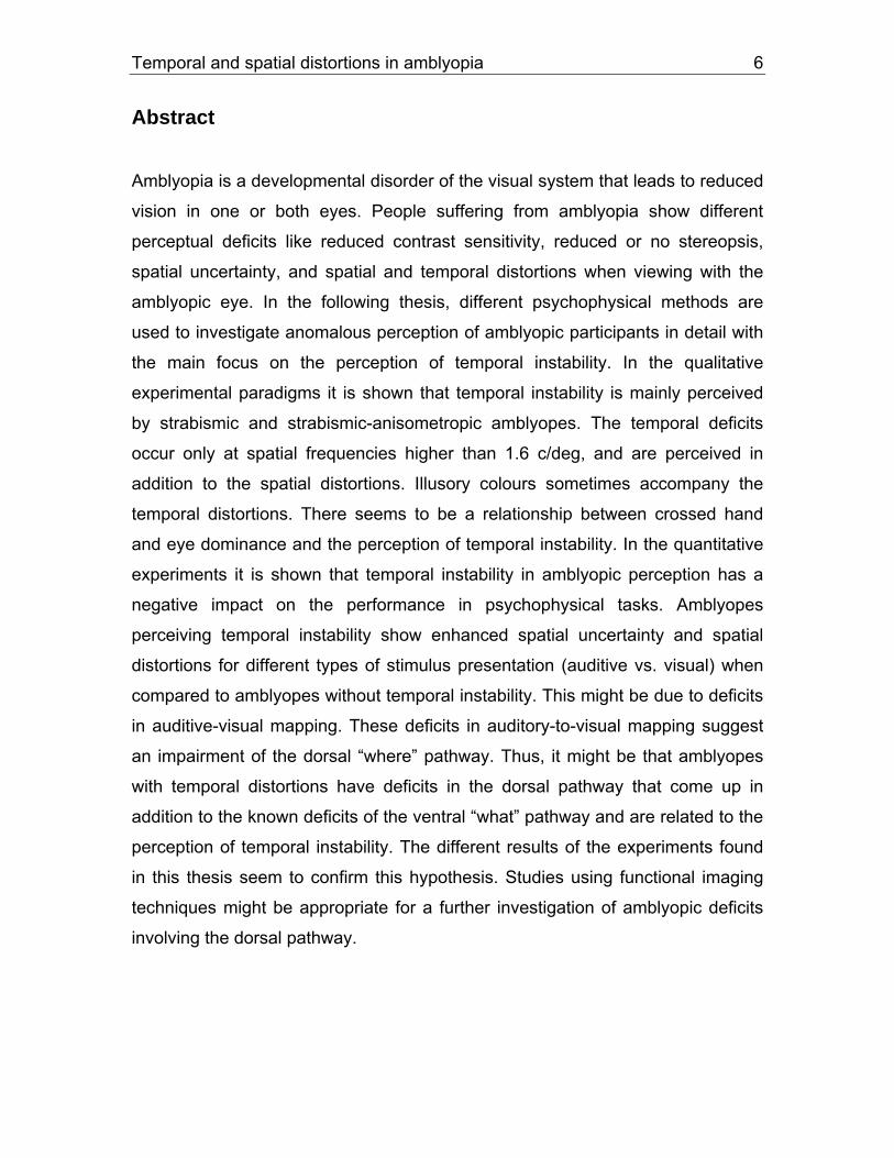
Abstract
Amblyopia is a developmental disorder of the visual system that leads to reduced
vision in one or both eyes. People suffering from amblyopia show different
perceptual deficits like reduced contrast sensitivity, reduced or no stereopsis,
spatial uncertainty, and spatial and temporal distortions when viewing with the
amblyopic eye. In the following thesis, different psychophysical methods are
used to investigate anomalous perception of amblyopic participants in detail with
the main focus on the perception of temporal instability. In the qualitative
experimental paradigms it is shown that temporal instability is mainly perceived
by strabismic and strabismic-anisometropic amblyopes. The temporal deficits
occur only at spatial frequencies higher than 1.6 c/deg, and are perceived in
addition to the spatial distortions. Illusory colours sometimes accompany the
temporal distortions. There seems to be a relationship between crossed hand
and eye dominance and the perception of temporal instability. In the quantitative
experiments it is shown that temporal instability in amblyopic perception has a
negative impact on the performance in psychophysical tasks. Amblyopes
perceiving temporal instability show enhanced spatial uncertainty and spatial
distortions for different types of stimulus presentation (auditive vs. visual) when
compared to amblyopes without temporal instability. This might be due to deficits
in auditive-visual mapping. These deficits in auditory-to-visual mapping suggest
an impairment of the dorsal “where” pathway. Thus, it might be that amblyopes
with temporal distortions have deficits in the dorsal pathway that come up in
addition to the known deficits of the ventral “what” pathway and are related to the
perception of temporal instability. The different results of the experiments found
in this thesis seem to confirm this hypothesis. Studies using functional imaging
techniques might be appropriate for a further investigation of amblyopic deficits
involving the dorsal pathway.

Temporal and spatial distortions in amblyopia 7
1. Introduction
There are five senses humans are endowed with: vision, audition, smell,
taste and touch. Vision is the one most finely tuned sense, which is also capable
of obtaining information from distant elements. The visual system is our major
source of obtaining information about the environment. In order to retrieve
information, we analyze the environmental object. We localize and recognize
objects and use different kinds of information like wavelength, shape and texture.
However, our visual system is not fully developed at birth and needs time to
mature. Especially in the early period of visual development i.e. the first year of
life, the visual system is very sensitive to influences that distract the critical
developmental steps. Everyone knows about the sight of small children wearing
a patch over one of their eyes, for example. These children are in orthoptic
therapy in order to prevent or reverse amblyopia. Amblyopia is one of the most
widespread visual disorders that can occur in early childhood, due to an
undesirable early development of the eyes’ visual perception. Amblyopia carries
many implications for the perception of an amblyopic person and furthermore,
there are several functional aspects of vision which are changed due to
amblyopia.
One way to investigate the visual perception of humans and, more
specifically, the perception of amblyopic people, is to observe the phenomenon
when it occurs naturally. However, as the subjective visual perception is
impossible to study by direct observation, it is examined by indirect observation,
e.g. in an interview (Atkinson, Atkinson, Smith, Bem & Nolen-Hoeksema, 1996),
or by qualitative descriptions of one’s perceptual experiences. Often additional
data to the subjective descriptions are provided by an anamnesis of the medical
history and the actual visual status of the individual. This method of qualitative
investigation has a very old tradition in psychophysical research and, besides the
experimental approach, is one of the two main research methods used in
psychophysical experiments. The experimental approach uses a different type of
Temporal and spatial distortions in amblyopia 8
investigation, gathering quantitative data about the nature of the investigated
object or variable, respectively, and the relations it has with other variables. In
the case of visual perception, this concerns the relation of visual perception to
relevant internal and external stimuli, and, especially, the influence of these
stimuli on perception.
This thesis is about the anomalous visual perception of people suffering
from amblyopia, investigated with both qualitative and quantitative research
methods. First, an introduction on the anatomy and function of the visual system
of normally sighted people is given. Second, the development of the visual
disorder “amblyopia” is explained and visual deficits caused by amblyopia are
presented. After that, four experiments are reported to answer the question of
how a special visual phenomenon, the temporal instability in amblyopic vision, is
actually experienced by amblyopic people and which implications this instable
perception has on other spheres of action. Temporal instabilities are investigated
as well as their relationship with the spatial distortions, which occur in amblyopic
perception. Due to the paucity of qualitative and quantitative data about temporal
instabilities in amblyopic vision, experiments 1 and 2 center upon the issue of
how the perception of temporal instability is subjectively experienced by
amblyopic people. Descriptive data about the amblyopic perception of different
geometrical patterns with low and high spatial frequencies have been collected
and qualitative descriptions of the temporal distortions acquired. In addition,
experiments 3 and 4 investigate the implication of a temporally instable
perception on the performance of psychophysical tasks. Indeed, amblyopes
perceiving temporal instability show deficits in auditory-to-visual mapping. In
sum, it is shown that strabismus, in addition to amblyopia, is needed to elicit
significant spatial and temporal distortions. The results of the qualitative as well
as the quantitative experiments suggest an impairment of the dorsal pathway in
amblyopes with temporal instability in addition to the known deficits in the ventral
pathway.

Temporal and spatial distortions in amblyopia 9
2. The nature of vision
This chapter gives a short overview over the visual system with its
anatomical and functional aspects - starting from the entrance of light into the
eyeball to the processing of an image in higher visual areas. Detailed information
on pathological aspects will be given where it is relevant for the further
understanding of the amblyopic vision described in chapter 3.
2.1 Anatomy and function of the eye
For moving the eyes towards different objects of interest within a wide field
of vision, six muscles are attached to each eye: four rectus muscles and two
oblique ones (Kandel, Schwartz & Jessell, 2000). These extraocular muscles
rotate each eye in its orbit and have a functional complement in the other orbit.
By that, the eyes can rotate in the same plane, but in opposite directions, e.g.
both eyes rotate to the left, one eye away from the nose, and the other nasally.
The proportion of 1) horizontal, 2) vertical and 3) torsional rotation performed by
each muscle depends on the horizontal position of the eye in the orbit. It is critical
for the brain that the muscles function together to interpret the images from each
eye as a single one. An improper functioning of these muscles can cause
strabismus, a misalignment of the two eyes. Result of this misalignment is double
vision, also called diplopia, as the image of the object of interest is no longer
located on the same retinal position in each eye. The six extraocular muscles are
innervated by three groups of motor neurons: the abducens nerve, the trochlear
nerve and the oculomotor nerve. All have specialized functions and if injured,
each shows a characteristic syndrome, e.g. damage to the oculomotor nerve
results in a nearly complete droping of the eyelid, a so-called ptosis.
The cornea is the outermost layer of the eye through which light passes
and - with about 43 diopters - the principal refractive structure of the eye (figure

Temporal and spatial distortions in amblyopia 10
1). Its shape is convex, it has a horizontal diameter of about 11,6 mm and is
surrounded by the sclera, a white, non-transparent tissue. The sclera together
with the cornea forms the outer layer of the eye and is continuous with the dura
mater covering the optic nerve and the brain. The inner space between the
cornea and the lens is called the anterior chamber and is filled with the aqueous
humor, a fairly homogeneous clear liquid. Within this anterior chamber lies the
iris, which surrounds the pupil and is the structure giving "color" to the eye. The
pupil is a hole in the middle of the iris, which bundles the light going into the eye.
The pupil changes its size in response to different light levels: e.g. due to the
muscle sphinctor pupillae the pupil becomes small (minimum about 2 mm), while
its agonist dilatator pupillae causes the pupil to become larger (max. 6-8 mm).
Both muscles are associated with each other forming a systematic alliance.
Figure 1. Anatomy of the eye (from Kandel et al., 2000).
With about 15 diopters, the lens is the second refracting element of the
eye (Dodt, 1975) - the other elements, the humor and vitreous body are only of
marginal influence. To a small amount, the lens is differently curved on the back
in comparison to the front side and is attached via zonular fibers to the ciliary
muscle which can change the shape of the lens. The ciliary muscle is formed like

Temporal and spatial distortions in amblyopia 11
a ring - when it contracts, the diameter of the muscle ring is reduced, thereby
relaxing the tension of the zonular fibers and making the lens more spherical.
The main function of the lens is focusing on objects on different distances. For
focusing on nearby objects the ciliary muscle contracts to unease the curvature
of the lens, for focusing on far objects the ciliary muscle relaxes – rendering the
lens more flat. This process of changing the shape of the lens is called
accommodation.
One’s ability to clearly focus on an object depends on two factors: the
dimension of the eyeball and the shape of the lens and cornea. At the age of 6-8
years, the eye should reach its optimal length of about 24 mm. If the eye is too
long, the image is not focused on the retina, but between the lens and the retina.
This is called myopia, near sighted vision. For hyperopia or farsighted vision
respectively, the opposite is true. The eyeball is too short, and therefore the
image is focused beyond the retina. In some cases the cornea or the lens does
not have an optimal shape; they are not spherical yet rather cylindrically shaped -
the so-called astigmatism. This causes the light to focus on more than one point
on the retina and results in blurred vision. Astigmatism often is accompanied by
myopia or hyperopia.
To conclude, incoming light is focused by the pupil and refracted by the
cornea and the lens, then passing the vitreous humor, a clear liquid between
lens, retina and the vitreous body, and reaching the photoreceptors, which are
located in the retina at the back of the eye.

Temporal and spatial distortions in amblyopia 12
2.2 The visual system
2.2.1 The retina
Talking about the visual system in the brain means starting to talk about
the retina, as the retina is already part of the central nervous system. It is formed
embryologically from the optic vesicle that evaginates from the diencephalon.
The photoreceptors can be considered as the "entrance gate" for the light to the
retinal cells (Birbaumer & Schmidt, 1999), while the ganglion cells are the only
cells with axons that leave the eye, forming together the optic nerve (Held, 1988).
Between the photoreceptors and the ganglion cells three other types of cells are
imbedded: the horizontal cells, the bipolar cells and the amacrine cells, forming
together the bipolar layer. All the different retinal cells are arranged in an inverse
way: the photoreceptors are turned away from light and towards the pigment
epithelium at the outermost surface of the eye. The photoreceptor layer is at the
back of the retina, while the ganglion cells form the inner cell layer. This inverse
system prevents light from being reflected off the back of the eye to the retina
again (Kandel et al., 2000) and provides a very high efficiency of the cells
supporting the photoreceptors with oxygenated blood for the essential
photopigment regeneration. Thus, retinal cells with the highest metabolism are
close to the chorio capillaris. In animals eyes other than mammals, where a lower
efficiency of the photosensitivity is needed, the receptors are arranged in an
everse way (e.g. in the eyes of cephalopods).
The two types of photoreceptors are: cones (about 120 millions) and rods
(about 6 millions). Cones are most prevalent in the central retina, and provide
colour vision and a very good acuity. In the central retina, the fovea centralis –
with the central spot of the foveola - is the area of finest vision. Here, cell bodies
arranged in front of the photoreceptors are shifted away to get highest
photosensitivity (Kandel et al., 2000). The light-sensitive cones are the only
photoreceptors prevalent in the foveloa. They are most sensitive to moderate up

Temporal and spatial distortions in amblyopia 13
to high wavelengths of light (380-760 nm) and mediate information about hue.
Three different types of cones contain each one of the three different visual
pigments whose abs0000orbencies are maximal for blue, green and red lights
(Held, 1988). Rods are more prevalent in the periphery of the retina. In contrast
to cones, rods are sensitive to light of low luminance and process basically
monochromatic information (Carlson, 2001).
For the transduction of the visual information, initiated by a complex chain
reaction, photopigments are involved in the first step. Photopigments decay by
the exposition to light and due to this process the permeability of the sensors and
the membrane potential is changed: with the breakdown of visual pigments
hyperpolarization to -70 mV of the cones and rods follows (Held, 1988). This
hyperpolarization reduces the release of neurotransmitters and causes the
membrane of the bipolar cell to depolarize. In turn, the depolarization causes the
bipolar cells to release more neurotransmitters. This process depolarizes the
membrane of the ganglion cell at the inner cell layer, causing it to increase its
rates of firing (Carlson, 2001). Taken together, light shining on the
photoreceptors triggers a cascade of events leading to the excitation of the
ganglion cell.
Functionally the receptive fields of the ganglion cells consist of two parts:
the receptive field center and the surround. Retinal ganglion cells can be
subdivided into different classes in terms of their size, shape and functional
properties. For excitation of the cell, e.g. on-center ganglion cells are excited
when light falls on the center of the receptive field, while they are inhibited when
light is directed to the surround. Off-center ganglion cells have a reverse
mechanism. On-center and off-center ganglion cells are present in more or less
equal numbers and thus provide two parallel pathways for the processing of
visual information (Kandel et al., 2000). This organization of the receptive fields is
very important as it provides best transduction of the visual information.
Receptive fields "respond best when light intensities in the center and surround

Temporal and spatial distortions in amblyopia 14
are quite different" (Kandel et al., 2000, p 519), e.g. to stimuli with high-contrast
properties like black-and-white patterns. In this context, the firing rate of a
ganglion cell is a fine measure for the difference in intensity of light illuminating
the center and surround. In addition to measuring contrast and illumination,
ganglion cells analyze several other aspects of the visual image, such as color.
Most ganglion cells fall into two functional classes: M (magno) cells and P (parvo)
cells. M cells respond optimally to large objects and rapid changes in the
stimulus, while P cells respond optimally to small, slowly moving objects and to
color. This distinct functionality leads to parallel processing with parallel networks
of ganglion cells.
At the layer of the optic nerve fibers, axons from ganglion cells are
traversing the retina to leave the eyeball at the optic disk and carry visual
information into the brain. The optic disk produces a blind spot since no receptors
are located in that area. On the primary retinal-geniculo-cortical pathway, the
axons of the retinal ganglion cells form the optic nerve pass the chiasma
opticum, reach the lateral geniculate nucleus, and then proceed through the optic
radiations to the primary visual cortex.
2.2.2 Optic nerve and chiasma opticum
The axons of the optic nerves of both eyes intermingle at the basis of the
brain in the chiasma opticum, and then are sorted into the two optic tracts. Here
the optic nerves are divided into temporal and nasal parts. Respectively, axons
from ganglion cells on the nasal sides of the retinae cross the hemispheres
through the chiasma opticum, while axons from cells on the temporal sides
remain at the same side of the brain (figure 2). Thus the left part of the visual
field of each eye is projected to the right hemisphere, while the right hemifield of
vision is projected to the left hemisphere. Therefore, in analogy to the
somatosensory system, each hemisphere receives information from the
contralateral half of the outer world.

Temporal and spatial distortions in amblyopia 15
Figure 2. Course of the optic nerves, chiasma opticum and the tracti optici (from Kandel et al.,
2000).
Beyond the chiasma, the axons of the ganglion cells continue through the
optic tracts to reach several visual relay nuclei in the brain (Held, 1988) and each
of the axonal projections is precisely organized. The distribution of fibers from
one optic nerve into the two optic tracts relates to the amount of binocular vision,
however at this time the two visual impressions of both eyes still remain
separate. The partial crossing of retinal fibers in the chiasma opticum is
necessary for stereoscopic vision, since it is necessary for later fusion of
corresponding retinal images from the two eyes in higher cortical brain areas -
that is, inputs from the two hemiretinae which view the same part of the visual

Temporal and spatial distortions in amblyopia 16
field form two maps that are exactly aligned with each other (Held, 1988). The
optic tract projects onto three subcortical targets: the lateral geniculate nucleus,
the pretectal area of the midbrain and the superior colliculus. Of these three
subcortical regions only the lateral geniculate nucleus processes visual
information that ultimately results in visual perception (Kandel et al., 2000).
2.2.3 The lateral geniculate nucleus
The lateral geniculate nucleus (LGN) of the thalamus is the first station on
the way to further visual processing. The axons of the retinal ganglion cells
ascend through the optic nerves and reach the synapses of the LGN (Carlson,
2001). This is the only relay between retina and cortex. This nucleus contains six
layers of neurons; the inner two ventral layers (laminae 1 and 2) are called
magnocellular layers. They receive input from M-ganglion cells. The outer four
dorsal layers are called parvocellular layers (laminae 3 to 6), receiving input from
P-ganglion cells (see figure 2). The neurons in the two magnocellular layers
contain cell bodies which are much larger than those of the parvocellular layers.
There are striking differences between both cell types: P cells are critical for color
vision and vision that requires high spatial and low temporal resolution vision,
while M cells are most important for low spatial and high temporal resolution
vision (Kandel et al., 2000). A third type of neurons can be seen in the
koniocellular sublayers, which are positioned ventrally to the magno- and
parvocellular layers. All those three types of layers - the magnocellular layer, the
parvocellular layer and the koniocellular layer - belong to three different systems,
each of which analyses different types of visual information.
The LGN is the site of termination of most optic tract fibers (Held, 1988).
Each of its six layers receives input from only one eye, however the areas of the
retina are not represented equally: the fovea - area of finest vision - has a
relatively larger representation than does the periphery of the retina. The neuron
layers are alternatingly allocated to the ipsi- and the contralateral eye. Inputs

Temporal and spatial distortions in amblyopia 17
from the ipsilateral and contralateral eye remain segregated in laminae 2, 3 and 5
and 1, 4 and 6, respectively. Only few interactions between the ipsi- and
contralateral layers occur, therefore no binocular processing of the visual signals
is found in this stage. Going further, the neurons in the LGN send their axons
through the optic radiations to the primary visual cortex (V1).
2.2.4 The striate visual cortex
Visual processing goes through different stages: from low level feature
extraction in primary areas to complex processing of visual information in higher
cortical areas. The visual cortex is organized in a hierarchical way, with the
primary visual cortex at the bottom of such a hierarchy. V1, also called the striate
cortex due to the “stria of Gennari”, which is visible with the naked eye, has six
different layers and gets his main input from the lateral geniculate nucleus via the
optic radiations. From the striate cortex several segregated pathways feed into
extrastriate areas: the most important ones are the dorsal, magnocellular,
“where” pathway and the ventral, parvocellular, “what” pathway (figure 3).
Figure 3. The visual cortex (from Kandel et al., 2000).

Temporal and spatial distortions in amblyopia 18
The primary visual cortex is a horizontal fissure surrounding the calcarine
fissure and is located in the medial and posterior occipital lobe (Carlson, 2001). It
processes the information of the retinal ganglion cells more precisely and then
transmits it to the visual association cortex. The striate cortex has a retinotopic
organization with the fovea having the largest area of representation. Input from
the optic radiations leads mostly to layer IV of the primary visual cortex. Exactly
speaking, from the LGN both magnocellular and parvocellular layers project to
the primary visual cortex to layer 4C - the magnocellular layer to 4Cα and the
parvocellular layer to 4Cβ. Continuing from this point both M and P pathways
remain partially separated from each other through V2 (Kandel et al., 2000).
It is thought that the primary visual cortex is organized into different
functional modules. There are three systems crossing the layers of the primary
visual cortex: a) orientations columns, b) blobs, and c) ocular dominance
columns (Kandel et al., 2000). These vertically oriented systems transfer
information to each other by major horizontally oriented connections. Orientation
columns contain neurons that are selectively sensitive to specific axes of
orientation, while the blob regions (structures about 0.2 mm in diameter, revealed
by a stain for cytochrome oxidase) are sensitive to colour and to low spatial
frequencies. These blob regions are part of the parvocellular system. From the
blob regions of V1, the P pathway continues to the thin stripes of V2 forming the
ventral pathway, which reaches the inferior temporal cortex. In contrast to that,
the M pathway projects from the stripes of V1 to the thick stripes of V2 forming
the dorsal pathway (figure 3).
As pointed out above, neurons in the primary visual cortex are grouped in
functional vertical columns. Each of these columns processes visual information
from a specific region of the visual field (Kandel et al., 2000). They are commonly
referred to as ocular dominance columns, as the left or the right eye dominates
them. Input from the eyes has been found to enter layer IV in alternating patches,
however in between are binocular areas as well that are activated from both, the

Temporal and spatial distortions in amblyopia 19
left and the right eye. This combines the input from the two eyes, a step that is
necessary for the perception of depth (Kandel et al., 2000).
Perception of depth For the perception of depth two types of clues are necessary: monocular
cues for depth and stereoscopic cues for binocular disparity (Kandel et al., 2000).
The six monocular cues are the following: familiarity of size, occlusion of objects,
linear perspective, and size perspective, distribution of shadows and illumination,
and motion parallax. For large distances, perception relies on monocular cues,
as the retinal images seen by the two eyes are almost identical. The slight
difference in an object’s position between the two retinal images is very important
for stereoscopic vision - this difference in position is called binocular disparity.
Stereoscopic vision cannot arise until the disparity information of the two eyes
gets together in the visual cortex. In V1 certain neurons are sensitive to
horizontal disparity. Also cells in the extrastriate areas V2 and V3 respond best to
retinal disparity, and some cells in area MST fire in response to the combination
of disparity and direction of motion (Kandel et al., 2000). Two types of cells can
be found in the striate cortex, simple and complex cells. Simple cells receive only
monocular input and respond best to a bar of light with a specific orientation and
position within the receptive field (Kandel et al., 2000), whereas complex cells
have much larger receptive fields than simple cells and are stimulated by both
eyes – most of them are binocular cells. There seems to be a hierarchy between
the cells: each simple cell surveys the activity of a group of geniculate cells, and
each complex cell surveys the activity of a group of simple cells (Kandel et al.,
2000). Differences can be recognized even in the anatomy of V1: simple cells are
found mostly in layer IV, complex cells more in layers above or beneath layer IV.
To conclude, the only clue necessary for stereoscopic vision is retinal disparity.
Complex cells do respond best to retinal disparity and thus are likely to play a
role in depth perception.

Temporal and spatial distortions in amblyopia 20
2.2.5 The extrastriate visual cortex
The areas of the extrastriate visual cortex are located in two general
regions: in the prestriate cortex and in the inferotemporal cortex (see Pinel,
2003). The prestriate cortex is a band of tissue in the occipital lobe that
surrounds V1. The inferotemporal cortex is the cortex of the inferior temporal
lobe. The secondary extrastriate visual areas (V2, V3, and V4) have still a
retinotopic organization. Signals coming from neurons of area V1 are passed to
V2, V3 or V4 depending on their functional aspect, that is, the neurons in each
functional area respond most vigorously to different aspects of visual stimuli (e.g.
color, movement, or shape). To be more specific, e.g. in area V5, commonly
referred to as area MT (middle temporal area), neurons respond mainly to small,
moving objects, whereas neurons of area MST respond to wide moving visual
stimuli or global motion, respectively.
Although connections between areas of the extrastriate and the
association cortex are reciprocal, the flow of information is up the hierarchy, from
more simple to more complex areas. That means the higher in the visual
hierarchy, the larger receptive fields the neurons represent and the more specific
and complex are the stimuli to which the neurons respond. In addition, higher
cortical areas have also feedback connections lower cortical areas. The
pathways that conduct information from V1 through extrastriate and association
cortex are parts of two major streams: the dorsal stream and the ventral stream.
The dorsal stream flows from the primary visual cortex via the dorsal prestriate
cortex to the posterior parietal cortex - including the middle temporal area -, the
ventral stream flows from the primary visual cortex via the ventral prestriate
cortex to the inferotemporal cortex - including area V4 (figure 4). The dorsal
stream is involved in the perception of where objects are, while the ventral
stream is involved in the perception of what objects are.

Temporal and spatial distortions in amblyopia 21
Figure 4. The visual pathways (from Kandel et al., 2000).
To put it in a nutshell, "the parietal cortex is specialized for spatial
representation, whereas the temporal cortex is specialized for object recognition"
(Kandel et al., 2000, p 498).

Temporal and spatial distortions in amblyopia 22
3. Amblyopia
Amblyopia is a developmental disorder of the visual system that leads to
losses in spatial vision. The medical term "amblyopia" is derived from the Greek
words αµβλυ-ωπός and means dull vision. Amblyopia is medically defined as the
reduction of visual acuity in one or both eyes in absence of ocular disorders - it is
the weak-sightedness of an eye and therefore often referred to as "lazy eye". The
weak-sightedness happens despite good retinal image quality, does not involve
any retinal defects or ocular deficits, the rod and cone receptors are oriented
normally and the foveal pigment density is without pathological findings (Levi &
Carkeet, 1993). Since spectacles cannot rectify the symptoms of the acuity loss,
amblyopia is presumed to have a neurological source. Reason for the
development of amblyopia is most probably abnormal neuronal processing of the
visual information in the visual cortex. A detailed description of the development
of strabismic and anisometropic amblyopia and the impairment of visual functions
due to amblyopia is given in the following subsections.
3.1 Development of amblyopia
Amblyopia is a developmental disorder of spatial vision - it occurs quite
early in life and with up to 2-5 % is "the most common cause of visual loss in
childhood" (Sireteanu, 2000 a, p. 63). Amblyopia results from an abnormal visual
stimulation early in life, and there are different types of amblyopia, which are
defined according to their etiology. The main factors for abnormal postnatal
visual development, which are associated with an enhanced risk for amblyopia,
are an early misalignment of one eye, unequal refraction in both eyes, a cataract
or a ptosis. If these disorders occur late in life, amblyopia does not develop and
therefore a critical period in the development of amblyopia has to be assumed.
There is some evidence that the onset of amblyopia may not begin prior to 3-4
months of age or at least not before the onset of the normal development of

Temporal and spatial distortions in amblyopia 23
binocular interaction in the visual cortex, yet also that the onset of amblyopia is
highly dependent on the type of amblyopia (Levi & Carkeet, 1993). Depending on
these factors for the abnormal visual development, there are two main types of
amblyopia: strabismic amblyopia, and deprivation amblyopia which also includes
anisometropic amblyopia.
3.1.1 Strabismic amblyopia
The onset of eye alignment, and of binocularity, respectively, occurs for
most infants sometime between 2 and 6 months after birth (Tychsen, 1993). In
strabismic amblyopia an early misalignment of the eyes is mostly due to a squint
- the two visual axes differ from each other. Due to miscoordination between both
eyes, the visual axes do not meet at the fixated object. In one eye the fixation
point lies on the fovea, but on another retinal area in the other eye: the signals
from the two eyes are not correlated to each other and diplopia (double images)
occurs. To prevent these double images, the perception of one eye is being
suppressed. By this, the squinting eye is being excluded from vision and the
functionally stronger eye becomes the dominant eye, resulting in a reduction of
visual acuity in the squinting eye after a period of time (Sireteanu & Fronius,
1981). Only 35-50 % of the strabismic children also develop amblyopia (Levi &
Carkeet, 1993). It seems that several factors are important for the development
of strabismic amblyopia, like the age of onset (the earlier the worse) or the type
of strabismus. E.g. strabismic amblyopia is more prevalent among esotropes
(strabismus convergens - inward deviated eye) than among exotropes
(strabismus divergens - outward deviated eye) (Howard, 2002).

Temporal and spatial distortions in amblyopia 24
3.1.2 Deprivation amblyopia
Deprivation amblyopia can occur due to a cataract or a ptosis. These
conditions lead to the deprivation of visual stimuli or to inadequate visual
stimulation respectively and consequently to the loss of form vision in the
affected eye. Anisometropic amblyopia is considered to be a mild form of
deprivation amblyopia - due to unequal refraction in the two eyes, probably
caused by an unequal eye growth. Both eyes have a different focus of image, so
that one eye gets a clear visual image, while the other perceives a blurred one.
Due to the bilateral reflex for accommodation there is only a limited possibility to
independently accommodate both eyes monocularly. In most cases the dominant
eye gets the clear visual image, while the non-dominant eye - like in strabismic
amblyopia - is being suppressed in order to avoid confusion. Although little is
known about the development of anisometropia, it is discussed that
"anisometropic amblyopia may have its onset considerably later than amblyopia
associated with strabismus" (Levi and Carkeet, 1993, p. 402). Continuous blur
has to persist for a longer period to develop anisometropic amblyopia (Daw,
1998). Consequently, anisometropic amblyopes show quite different (more
similar to normal) results in psychophysical and imaging experiments than do
strabismic amblyopes (Sireteanu, Tonhausen, Muckli, Lanfermann, Zanella,
Singer & Goebel, 1998).
In general, any condition which provides no adequate visual stimulation
and therefore causes a constant suppression of one eye early in childhood, is a
potential source of amblyopia. However, due to the fact that only about 1/3 of the
strabismic or anisometropic children are developing amblyopia, strabismus,
anisometropia or other derivational factors are a necessary, still not sufficient
condition for the development of amblyopia. Additionally, in all cases it is valid
that the earlier the visual disorders occur and the greater the degree of the
misalignment, the anisometropia or the stimulus deprivation, the greater the
depth of amblyopia.

Temporal and spatial distortions in amblyopia 25
3.2 Mechanisms of adaptation
3.2.1 Interocular suppression
As pointed out above, interocular suppression is an adaptation
mechanism due to strabismic or anisometropic amblyopia. The unequal
refraction or the squint in one eye would cause double or blurred images,
respectively, and in order to avoid diplopia, the brain of the young child
suppresses the visual input of the most affected eye. There are two ways of
suppression: first, only one eye is suppressed all the time. Due to the
suppression, this eye may develop a reduction of visual acuity – amblyopia -,
which cannot be corrected by spectacles. When the two eyes are suppressed
alternatingly (strabismus alternans) the visual acuity remains normal in both
eyes: due to the alternating fixation each eye contributes to the regions in which
it is best (Sireteanu, 1982). People with alternating suppression do not belong to
the group of amblyopes, as they have no reduction of visual acuity in one eye.
While alternating fixation may only occur in strabismic people, suppression of
always the same eye occurs in strabismics as well as in anisometropes. The
area of the visual field affected by suppression is a different one in strabismic
amblyopia and in deprivation amblyopia. Strabismic amblyopes have an
asymmetric distribution of suppression. The suppressed areas of the deviated
eye involve mainly the central and nasal retina (Sireteanu & Fronius, 1981;
Abdolvahab-Emminger & Sireteanu, 1993; Sireteanu, 2000b). Contrary to this,
anisometropes do not have an asymmetric interocular suppression (Sireteanu &
Fronius, 1981). Sireteanu and Fronius (1981) found that in anisometropic
amblyopes the acuity loss as well as the suppression of the nasal and temporal
fields is symmetrical in the central as well as in the peripheral areas of the
amblyopic eye.
Further, as a consequence of interocular suppression, humans with early
strabismus or anisometropia suffer partial or complete loss of binocular vision

Temporal and spatial distortions in amblyopia 26
and stereopsis (Sireteanu, Fronius & Singer, 1981). Most people who develop an
interocular suppression (or alternating fixation, respectively) have monocular
vision and therefore lost stereopsis. However, probably due to the limitation of
the suppression to the central and nasal retinal areas, in some amblyopes, the
peripheral non-suppressed regions of the amblyopic eye are still involved in
binocular vision (Sireteanu, Fronius & Singer, 1981; Sireteanu, 1982). A reason
for the most evident loss of binocularity in the foveal region may be that "with its
high resolution and small receptive fields, binocular function is more likely to be
disrupted than in the peripheral field, were the resolution is relatively low and the
receptive fields relatively large" (Sireteanu, 2000b, p.40). Consequently, there is
a loss of binocular neurons visible on the neuronal level of V1 (Hubel & Wiesel,
1977). Instead of binocular neurons, neurons that are activated monocularly were
found in the striate cortex of monkeys and cats. However, the total number of
neurons was not reduced (Sireteanu, 1991).
3.2.2 Anomalous retinal correspondence
Dependent on the gathered experiences, the developmental selection
process optimizes and adapts neuronal connections in the visual cortex. One
important function of this process is to make sure that binocular neurons form
precisely aligned receptive fields in both eyes. This exact correspondence of the
receptive fields can not only be defined via genetic information, as it also
depends on individual characteristics, e.g. the distance between the eyes. So, by
definition, those retinal regions are correspondent to each other, which get
identical visual stimulation during binocular fixation (Singer, 1984). In a squinting
eye exact alignment of the receptive fields can no longer be developed, so the
brain evolves an adaptation mechanism. That is, in anomalous retinal
correspondence (arc) the retinal coordinates are shifted in relation to the angle of
squint: in harmonic anomalous retinal correspondence (h-arc) the objective angle
of squint matches the subjective localization. This complete adaptation of
subjective localization to the angle of squint is only possible in small and constant

Temporal and spatial distortions in amblyopia 27
angles, as in so-called microstrabismus. In non-harmonious anomalous retinal
correspondence (nh-arc), objective and subjective localization of the angle of
squint do not match exactly.
The fact that strabismic amblyopes preserve binocularity in the periphery
of the visual field suggests that in this region retinal coordinates are shifted due
to anomalous retinal correspondence. In the central field, with its small receptive
fields, any correspondence is hard to maintain with the deviated eye, and thus
suppression is most evident in that region. However, in the periphery with larger
receptive fields, anomalous retinal correspondence can be prevalent and no
suppression is needed (Sireteanu & Fronius, 1989). There is evidence
(Sireteanu, 1991; Sireteanu & Best, 1992) that mostly extrastriate areas are
involved in this developmental process of anomalous retinal correspondence.
The higher visual areas are more tolerant for deviations due to the squinting eye
due to their bigger receptive fields. In V1, with its small receptive fields, no such
compensation would be possible. So, on the neuronal level a process similar to
that in the fovea and the periphery is assumed.
3.3 Visual deficits
3.3.1 Contrast sensitivity
Visual acuity represents the smallest high contrast stimulus that can be
resolved. Still this is only one limit of visual capacity. In fact, most objects in daily
life are large stimuli with low contrast. That is, every visual object can be
analyzed by a combination of some basic visual patterns, so called sine-wave
gratings. The threshold of contrast is the smallest difference in intensity between
these dark and bright stripes one is able to detect. The contrast sensitivity is
defined in relation to this threshold as the 1/contrast threshold ratio. The

Temporal and spatial distortions in amblyopia 28
measurement for contrast sensitivity with sine-wave gratings is much more exact
(about 4 - 5 times) than for instance with letters, and gives a quite good
impression for the visual sensibility for objects. In addition to visual acuity,
contrast sensitivity function is used for diagnostics in pathological changes of the
visual system.
The reduced ability for strabismic amblyopes to detect low contrast objects
is well known in literature (Asper, Crewther & Crewther, 2000a) - especially the
higher frequencies (6 - 18 c/deg) are affected due to amblyopia. There is no
significant contrast threshold difference between the eyes of normal observers,
so amblyopes can be considered as their own control by comparing their
amblyopic and their dominant and unaffected eye, respectively. Hess and Howell
(1977) argued for two categories of reduced contrast sensitivity in strabismic and
anisometropic amblyopes: either only the higher frequencies are affected (type I
amblyopia), or both, high and low frequencies (type II amblyopia). They
considered the type I amblyopia to be similar to simple dioptric blurring of
moderate degree, however not the type II amblyopia. In the latter "the effect of
defocus on low frequencies is slight ... when the high frequency degradation is
equivalent to the amblyope's high frequency loss" (Hess & Howell, 1977, p.
1053). Note that only the amblyopic eye shows marked losses in contrast
sensitivity, while the dominant eye is mostly unaffected. Secondly, the high
frequency loss tends to be more severe in the high-and-low-frequency-
abnormality class than for the high-frequency-only-abnormality class. Although
the loss of contrast sensitivity at high spatial frequencies increases with the
severity of amblyopia (Levi, 1991), still a small reduction in acuity is reflected by
a large loss in contrast sensitivity. Strabismic amblyopes seem to lose contrast
sensitivity especially in the central part of the retina, anisometropic amblyopes in
the central and the peripheral part (Hess & Pointer, 1985). Most importantly, the
reduced contrast sensitivity of the amblyopic eye is a result of a neural loss in
foveal function (Hess, Bradley & Piotrowski, 1983). It does not result from optical
factors, eccentric fixation or unsteady fixational eye movements (Levi, 1991).

Temporal and spatial distortions in amblyopia 29
3.3.2 Perceptual distortions
Perceptual distortions in adult amblyopia have been reported as early as
in the late 1950ies (Pugh, 1958). In Pugh’s study, participants reported pattern
distortions of letters; the outline of a circle "became "jagged", "dragged",
"smudged", or "rubbed", the blackness faded, and the circle appeared to be
flattened" (p. 453). The distortions did not show any symmetry, however in all
cases a consistency of the pattern distortion was observable.
Later the interest for perceptual distortions in strabismic and non-
strabismic amblyopes increased in connection with several different topics e.g.
concerning the deficit in spatial localization and intrinsic positional uncertainty in
the amblyopic visual system. Quantitative and qualitative approaches have been
used to investigate these amblyopic distortions.
In the qualitative way, investigators asked participants to describe their
perception with the amblyopic eye. This was done mainly with gratings of
different spatial frequencies shown to the amblyopic participants. Hess, Campbell
& Greenhalgh (1978) discussed the effect of spatial frequency on pattern
distortion. They showed participants gratings at a range of different spatial
frequencies and asked them to compare their appearance as seen by the
amblyopic eye with the appearance seen by the fellow normal eye. The vision by
the amblyopic eye gets progressively distorted the higher the spatial frequency,
while the vision of the dominant eye remains normal. Repeatedly, on a number of
different sessions low spatial frequency gratings appeared fairly undistorted,
however at higher spatial frequency gratings spatial distortions increased. The
gratings were perceived as fragmented and jumbled.
Quantitative measurements of spatial errors and uncertainty were
specified in studies from Bedell & Flom (1981; 1983). They described monocular
spatial distortions, defined as the relative overestimations and underestimations

Temporal and spatial distortions in amblyopia 30
in space in the nasal and temporal fields (Bedell & Flom, 1981), and
directionalisation errors, as well as spatial imprecision. Spatial imprecision may
also be labeled as spatial uncertainty and is defined by the variability of a
participant’s judgment regardless of its accuracy (Bedell & Flom, 1983). Bedell
and Flom (1983) showed that considerable distortions and imprecision of spatial
judgements characterize the amblyopic eye of strabismic but not of
anisometropic amblyopes. Especially for the strabismic amblyopic eye, reduced
acuity or unsteady fixation cannot account for the errors of relative
directionalisation and imprecision of the spatial judgments.
On the other hand, the uncertainty and the magnitude of the errors in a
vertical alignment task of Fronius & Sireteanu (1989) were quite closely related to
the participant’s visual acuity. They found a larger area of the visual field of the
deviated eye to be more or less involved in abnormal space perception. The
participant had to align several test stimuli between two vertical reference points.
Instead of building up a straight vertical line with the two reference points as
endpoints of the line, the amblyopic person constructed a curved line. That is, in
the center of the visual field the differences between the amblyopic eye and the
non-amblyopic eye are most pronounced. A map of the two-dimensional space
perception (vertical and horizontal) of amblyopic participants showed spatial
distortions especially in the amblyopic eye of strabismics with a large angle of
squint (Lagrèze & Sireteanu, 1991). Hereby, each participant exhibited his or her
individual distortion pattern for the amblyopic eye. It seems that showing a
reference point during the perception task influences the direction of the
distortions with regard to the position of the reference point. There was no
significant difference between the non-amblyopic eye and the monocular two-
dimensional map of normal observers.
Sireteanu et al. (1993) showed that amblyopic participants perceived
distortions at higher frequency gratings using their amblyopic eye. The gratings
of low spatial frequency seemed to be quite irregular and not exactly straight; the

Temporal and spatial distortions in amblyopia 31
lines seemed to be bended to the outward. The high spatial frequency gratings
were even more blurred with some participants showing a deep scotoma in some
part of the gratings. Sireteanu et al. (1993) compared these individual subjective
distortions by assigning results based on a quantitative psychophysical
experiment (Lagrèze & Sireteanu, 1991; Sireteanu et al., 1993) to the grating
stimuli. Results revealed that these reconstructed patterns only partially reflected
the distortion perceived with the amblyopic eye. The spatial distortion of the
percept through the amblyopic eye was often reported to be less than expected.
The authors argued that this may be due to the different nature of the tasks that
are likely to affect two different mechanisms in the brain.
Recently, Barrett, Pacey, Bradley, Thibos & Morrill (2003) categorized
individual distortion patterns of amblyopic participants. They used the same
procedure as Hess et al. (1978) and Sireteanu et al. (1993) with gratings of
various spatial frequencies and orientations. One third (10 out of 30) of their
participants showed no perceptual distortions, two third reported nonveridical
perception with the amblyopic eye. Barrett et al. (2003) divided the anomalous
spatial perceptions into five distinct classes of distortions: (1) wavy appearance
of straight gratings; (2) the "jagged" type with abrupt positional shifts orthogonal
to the grating orientation; (3) errors in perceived orientation; (4) fragmented
perception in which the gratings appeared broken; (5) and scotomatous
distortions showing large gaps in the gratings. Interestingly, for most of the
participants the type of the perceptual distortion was not constant across different
spatial frequencies or orientations.
In addition to these constant perceptual errors, amblyopic participants
often describe their pattern perception as being temporally unstable on a short
time scale (Barrett et al., 2003). In fact, this phenomenon of temporal instability in
pattern perception has been briefly mentioned in some studies concerning
amblyopia (e.g. Hess et al., 1978; Sireteanu, 2000 a). Amblyopic participants
describe this phenomenon of temporal instability as if images are permanently

Temporal and spatial distortions in amblyopia 32
changing, as if "seen through hot air" (c. f. Sireteanu, 2000 a, p. 71). However,
there are no systematic studies aimed at describing this phenomenon in detail -
no researcher has made the effort to describe this perceptual phenomenon or to
investigate it psychophysically.
On the other hand, several studies have addressed the temporal aspects
of amblyopic perception, especially dealing with motion (c.f. Simmers, Ledgeway,
Hess & McGraw, 2003). Ellemberg, Lewis, Maurer & Brent (2000) found deficits
in amblyopic temporal vision caused by visual deprivation, yet only at low
temporal frequencies and with less severity than deficits in spatial vision.
Ellemberg et al. (2000) concluded that mostly the slower developing aspects of
spatial and temporal vision are affected and therefore the longer the deprivation
of the amblyopic eye lasts, the higher temporal frequencies will be affected.
Further, a deficit in perception of flickering stimuli was found to be highly
dependent on the spatial properties of the stimulus and the individual amblyopic
deficit (Bradley & Freeman, 1985). For large stimuli covering also peripheral
parts of the retina or consisting of low spatial frequencies, no temporal deficits
were found. In a temporal integration task, Altmann & Singer (1986) found a
positive correlation between severity of amblyopia and impaired performance. In
their study, there was a pronounced deficit of the amblyopic system to integrate
temporally separate stimuli, and the interocular differences between the
amblyopic and the dominant eye were getting bigger the longer the interval of the
stimuli. Altmann & Singer (1986) proposed a disturbance of channels dealing
with processing of temporal aspects and high spatial resolution. They suggested
that neuronal responses on the amblyopic pathway might be less sustained in
processing than those of the pathway of the fellow eye.
To summarize, perceptual distortions occur in adult amblyopia in addition
to other perceptual deficits like loss in contrast sensitivity in the amblyopic eye
(c.f. Hess & Howell, 1977), deficits in spatial localization (Bedell & Flom, 1981;
Levi & Klein, 1983; Fronius & Sireteanu, 1989; Lagreze & Sireteanu, 1991;

Temporal and spatial distortions in amblyopia 33
Sireteanu, Lagrèze & Constantinescu, 1993) and spatial uncertainty in the
amblyopic visual system (Bedell & Flom, 1981, 1983; Fronius & Sireteanu, 1989;
Demanins & Hess, 1996; Wang, Levi & Klein, 1998; for a recent review see
Asper et al., 2000a). The perceptual distortions can be separated in spatial and
temporal ones. Strabismic amblyopic participants report their vision to be blurred,
spatially distorted and temporally instable (Sireteanu, 2000 a; Sireteanu, 2000 b).
Spatial misperception is absent or not so obvious at very low spatial frequencies,
but increases for higher spatial frequencies. In amblyopes, vision for high spatial
frequencies and low temporal frequencies is evidently impaired, and probably
higher-level cortical processes are abnormal in amblyopia (Asper et al., 2000a;
Sharma, Levi & Klein, 2000; Simmers et al., 2003; Sireteanu, 1991; Sireteanu et
al., 1998).

Temporal and spatial distortions in amblyopia 34
4. Part I: Qualitative methods
The qualitative part I of this thesis consists of two studies, which attempt
to get step by step as close to a description of the amblyopic perception as
possible. In study 1, participants were shown different geometrical patterns of low
and high spatial frequency and asked for their subjective amblyopic experience:
a descriptive base of spatial and temporal distortions was assessed in this study.
In study 2 an evaluation of the perceptual distortions encountered in study 1 was
performed by the use of a matching paradigm.
4.1 Study 1: Drawing experiment
The purpose of the first study was to investigate the temporal instability
and spatial distortions in amblyopic perception and thus to understand the nature
of the amblyopic vision more fully. Due to the lack of previous research on
temporal distortions, the main interest was in collecting descriptive data on the
amblyopic perception of different geometrical patterns with low and high spatial
frequencies. In addition, it was investigated if there were any systematic relations
between the occurrence of spatial and temporal distortions and other
characteristics of the amblyopic vision.
4.1.1 Method
Participants Fourteen amblyopes participated in the study: seven strabismic
amblyopes, four anisometropic amblyopes without strabismus and three
anisometropic-strabismic amblyopes. Selection criteria for the amblyopic
participants were: corrected visual acuity in the deviated eye of at most 0.5
Snellen acuity, measured with the Landolt C test for single optotypes and an

Temporal and spatial distortions in amblyopia 35
interocular difference between the dominant and the amblyopic eye of at least
0.3 Snellen acuity. To be classified as anisometropic, amblyopes were required
to have a minimum difference in refraction of 1.5 dpt between the eyes. All
participants were 18 - 65 years old, had no red-green deficiencies, and no
neurological or psychiatric ailments. They participated in the experiment for
course credits or for a payment of 10,- € per hour. Informed consent was
obtained from all participants after the nature and purpose of the study has been
fully explained. The experiments were performed in accordance with the
Declaration of Helsinki. The study was approved by the Ethical Committee of the
Johann Wolfgang Goethe-University Frankfurt.
Orthoptic examination All participants underwent full refraction and orthoptic assessment before
testing. The orthoptic measurements included an anamnesis of the patients’
medical history. The subjective refractive error of the eyes was assessed with the
aid of Fronhäuser refraction lenses. Corrected visual acuity for far vision was
measured with the Snellen acuity chart at 5 m distance, for near vision with the
Landolt-C test at 40 cm distance. Angle of squint was assessed with
simultaneous and alternate cover and prism test for far and near fixation. The
visuscope test was used for the determination of fixation. Stereopsis was
assessed with the TNO-, Randot-, Titmus- and Lang-Tests. For the evaluation of
the retinal correspondence, the participants were tested with the Maddox cross in
connection with dark and light red filters and with Bagolini striated glasses for far
and near vision. The orthoptic examinations were performed by a professional
orthoptist. Table 1 provides an overview of the clinical data of all participants.
Contrast sensitivity As loss of contrast sensitivity is a major deficit in spatial amblyopic vision
(see also chapter 3.3), prior to psychophysical testing, the participants’ contrast
sensitivity was tested for far (3.0 m) and near (0.4 m) vision. Monocular and
binocular foveal contrast sensitivity function (CSF) was measured using the VIS -

Temporal and spatial distortions in amblyopia 36 Table 1. Orthoptic data of amblyopic participants.
Partici-pant
Gender, Age
Eye Refraction Visus c.c.(near vision)
Fixation Strabismus(sim. Cover test)
Stereopsis Correspondence History
Strabismic amblyopes R.S.
male, 58 yr
RE LE*
+ 6.00 -1.25/171° + 6.75 -1.50/5°
0.60 0.10
foveolar 1.5°-2° nasal
far +2½°+ VD near +3° + VD
negative, excl. LE
h arc
Occlusion at 1 yr
L.P.
female, 33 yr
RE LE*
+ 0.50 sph + 0.75 sph
1.00 0.25
foveolar temporal
far -12½°+ VD 1°near ~± 0°
negative, excl. LE
nrc
Onset and occlusion at 4 yr, glasses until 15 yr
M.K.
male, 29 yr
RE* LE
+ 5.50 -4.00 / 145° + 5.00 -4.75 / 5°
0.10 1.00
temporal foveolar
far +1½° + VD½°near +1½° + VD¾°
negative, excl. RE
h arc
Strabismus from early childhood, occlusion at 5 yr
S.B.
female, 25 yr
RE* LE
-10.0 sph - 9.0 sph
0.30 0.60
temporal foveolar
far +12° + VD near +12° + VD 7°
negative, excl. RE
nh arc
Family history, strabismus from early childhood, glasses at 5 yr
D.S.
male, 51 yr
RE* LE
+ 5.25 -2.50 / 100° + 4.50 -2.25 / 95°
0.25 1.00
fov. margin foveolar
far ± 0° near +4° - VD
negative, excl. RE
nh arc
Onset and glasses at 6 yr
A.F.
female, 22 yr
RE LE*
- 1.25 -2.00 / 85° - 1.25 -1.75 / 105°
1.00 0.32
foveolar temp. margin
far +3° - VD 1° near +4½°- VD 1°
negative, excl. LE
h arc
Strabismus from early childhood, glasses and occlusion at 5 yr for one year
G.P.
female, 27 yr
RE* LE
+ 1.25 sph + 1.25 sph
0.50 1.00
1° nasal foveolar
far +1° near +1°
negative excl. RE
nrc
Occlusion at 6 yr, glasses at 7 yr
Strabismic and anisometropic amblyopes B.B.
female, 29 yr
RE* LE
- 0.75 sph - 1.50 +2.0 / 175°
0.08 0.90
temp. margin foveolar
far + ½° + VD 3°near - 2½° + VD 2°
negative, excl. RE
nh arc
Strabismus from early childhood, glasses at 3 yr, occlusion 3-6 yr, surgery at 20 months
M.H.
female, 31 yr
RE LE*
+ 5.00 -0.75 / 142° + 1.50 -0.50 / 0°
1.00 0.08
foveolar nasal
far + 1½° ± VD near + 4°
negative, excl. LE
h arc
Family history, strabismus from early childhood, glasses at 3 yr, occlusion 4–5 yr, surgery at 5 yr
C.L.
male, 28 yr
RE LE*
- 3.50 -1.50 / 20° + 1.00 -1.25 / 0°
1.00 0.50
foveolar nasal - fovea
far +15° +VD 1° near +15° +VD 2°
negative, excl. LE
nh arc
Strabismus from early childhood, occlusion in kindergarten for 3 yr, surgery at 4 yr
Anisometropic amblyopes H.L.
male, 27 yr
RE LE*
plano + 6.25 sph
1.40 0.25
foveolar foveolar
± 0°
negative
nrc
Occlusion at 11 yr, glasses at 18 yr
T.S.
male, 30 yr
RE LE*
+ 1.25 sph + 2.75 -3.75/135°
1.00 0.40
foveolar nasal margin
± 0°
negative, excl. LE
nrc
Occlusion and glasses at 6 yr
J.B.
male, 26 yr
RE LE*
- 2.25 sph - 0.75 -2.00 / 15°
1.00 0.50
foveolar temporal
± 0°
Positive
nrc
Occlusion and glasses at 6 yr
M.B.
male, 34 yr
RE LE*
- 0.50 -0.50 / 45° - 3.00 -3.25 / 2°
1.00 0.50
foveolar foveolar
± 0°
Positive
nrc
Glasses at 17 yr
Abbrevations: nrc = normal retinal correspondence, nh arc = non-harmonius anomalous retinal correspondence, h arc = harmonius anomalous retinal correspondence, VD = vertical deviation, + = esotropia, – = exotropia, * = amblyopic eye.

Temporal and spatial distortions in amblyopia 37
TECH CSF test charts (VCTS 6500 charts, VISTECH Consultants, Dayton, Ohio)
consisting of 40 circular targets with gratings of different contrasts and spatial
frequencies, arranged in five rows of eight targets each. The gratings were
vertical or tilted 15 deg clockwise or counter clockwise from vertical and were
arranged randomly in one of the three orientations. The spatial frequency of the
gratings increased from 1.5 c/deg in the top row to 18.0 c/deg in the bottom row
when viewed at 3.0 m distance. The spatial frequencies in the intermediate rows
were 3.0, 6.0 and 12.0 c/deg.
Apparatus and stimuli The stimuli for study 1 were geometrical black-and-white patterns: two
gratings of spatial frequencies of 0.4 c/deg and 1.6 c/deg, one checkerboard with
a spatial frequency of 0.4 c/deg and a rectangular grid with a line spacing
corresponding to 3.2 c/deg (figure 5 a-d). The patterns were computer-
generated, high-contrast and printed on white paper. They were presented to the
participants in a distance of 57 cm.
Figure 5 a-d. Grating stimulus of 0.4 c/deg. b. Grating stimulus of 1.6 c/deg. c. Checkerboard stimulus of 0.4 c/deg. d. Grid stimulus of 3.2 c/deg.
Procedure A three-alternative forced-choice method was used for CSF
measurements. The participants were asked to report the orientation of each of
the eight gratings row by row. If the participants reported that the circular patch
appeared blank, they were required to guess. Then, the number of the correct
choices was recorded.

Temporal and spatial distortions in amblyopia 38
For the assessment of spatial distortions, the participants were asked to
look at the different patterns with their amblyopic eye and to memorize their
perception. The non-amblyopic eye was covered during this time. After
memorizing, the participants were asked to draw the gratings, the checkerboard
and the grid from memory, as they had perceived them with the amblyopic eye.
They used the non-amblyopic eye for drawing the percept of the original patterns.
There was no time limit to fulfill this task. The participants were allowed to look at
the pattern with the amblyopic eye as often as they needed to refresh their
memory. For testing the temporal distortions, the procedure was as described for
the spatial ones. The participants were asked to describe verbally whether the
spatial distortions captured in their drawings were stable or changed over time.
They had to tell in which way they perceived the instability, whether temporal
distortions appeared in addition to spatial ones, if there was a special "type" of
instability occurring in the stimuli and for how long the temporal instability was
perceivable. Based on the descriptions and drawings of the participants, images
of the perception of each pattern and each amblyope were generated.
4.1.2 Results
Results of the orthoptic examination Out of the seven strabismic amblyopes, three (LP, AF and GP) had either
no noteworthy or relatively low refractive errors in either eye and could thus be
classified as primarily strabismic amblyopes. Two other participants (MK and DS)
had a high ametropia in both eyes (hyperopia combined with astigmatism) and
thus can be classified as accommodative strabismic amblyopes. The remaining
two participants (RS and SB) had reduced acuity in both eyes, probably due to
an early, uncorrected high refractive error in both eyes and a consecutive
microtropia. RS had a high bilateral hyperopia with astigmatism, SB a severe
bilateral myopia. In the following, these participants shall be classified as
“refractive strabismic amblyopes”. With the exception of LP and SB, all strabismic
participants were microtropic. None of them underwent surgery, however RS, LP,

Temporal and spatial distortions in amblyopia 39
AF, GP and CL were occluded at various ages. All of them were stereoblind and
showed an exclusion of the most affected eye. Fixation was eccentric in all
cases, correspondence ranged from normal (LP and GP) to differing degrees of
anomality (harmonious in RS, MK and AF, non-harmonious in SB and DS).
The three anisometropic-strabismic amblyopes (BB, MH and CL) showed
differing degrees of refractive errors, usually in combination with astigmatism. All
had been occluded and underwent surgery quite early in life. None of them
showed any residual stereopsis, all showed an exclusion of the amblyopic eye.
Participants BB and MH were microstrabismic, participant CL had a large-angle
consecutive esotropia.
The four anisometropic participants (HL, TS, JB and MB) showed differing
degrees of refractive errors, no squint, normal correspondence and central
fixation in both eyes. Not all of them showed an exclusion of the amblyopic eye.
Participants JB and MB showed some residual stereopsis. All had received their
first treatment after 6 years of age. Participant MB did not undergo occlusion
therapy and even got his first glasses at 17 years of age.
Relationship between spatial and temporal misperception and the orthoptic characteristics of the participants Of the 14 participants included in this study, two did not report any
distortions, six perceived spatial distortions but no temporal instability and
another six perceived temporal instability either in addition or in the absence of
spatial distortions. Out of the six participants presenting spatial distortions only,
one participant was purely strabismic (LP), two were strabismic with a severe
bilateral refraction error (RS and SB) and 3 were purely anisometropic (JB, HL
and MB). Of the temporal-instability group, 3 were strabismic (MK, DS and GP),
2 strabismic-anisometropic (BB and CL), and one purely anisometropic (TS). Of
the two participants experiencing no distortions at all, one was strabismic (AF),

Temporal and spatial distortions in amblyopia 40
and one strabismic-anisometropic (MH). As these data suggest, temporal
instability is perceived mostly in amblyopes with a history of strabismus (table 2).
Type of amblyopia
Strabismic Strabismic & refraction
Strabismic & anisometropic
Anisometropic
No distortion (n = 2)
1
-
1
-
Spatial distortion only (n = 6)
1
2
-
3
Temporal instability (n = 6)
3
-
2
1
Table 2. Type of amblyopia and type of perceptual distortion.
For the purpose of analysis, acuity loss was divided in three categories:
very deep (visus of the amblyopic eye was 0.08-0.19), moderate-to-deep (0.2-
0.39), and moderate (0.4-0.5). The visual acuities of the amblyopic eyes of the
participants not experiencing any perceptual distortions were 0.32 for the
strabismic participant AF and 0.08 for the strabismic-anisometropic participant
MH. Thus, it appeared that even strabismic participants with a very deep
amblyopia do not necessarily experience distorted vision. This finding is quite
surprising, as it seems that neither the aetiology nor the severity of amblyopia are
a requirement for the presence of spatial distortions in amblyopic participants
(see also Barrett et al., 2003). The acuity loss of the participants showing spatial-
distortions-only ranged from moderate to deep (visus of the amblyopic eye was
0.50 for MB and JB, 0.30 for SB, 0.25 for LP and HL, and 0.10 for RS). The same
was true for the group experiencing temporal distortions (visus was 0.50 for GP
and CL, 0.40 for TS, 0.25 for DS, 0.10 for MK and 0.08 for BB, see figure 6).
There was no significant difference in visual acuity between the three distortion
groups (F (2,11) = .36, p = .71). Means for the no-distortion-group, the spatial-
distortion-group and the temporal-distortion-groups were M = .20, M = .32, and M
= .31, respectively.

Temporal and spatial distortions in amblyopia 41
Figure 6. Type of distortions in comparison with visual acuity of the amblyopic eye.
There were no significant correlations between the type of distortion
(spatial, temporal or none) and any of the clinical orthoptic data like angle of
squint, eccentric fixation, residual stereopsis, type of correspondence etc.
However there was an intriguing positive trend between the side of the amblyopic
eye and the type of distortion, meaning that when the right eye is the amblyopic
eye, temporal distortions are more likely to occur: 67% (4 out of 6) of the
participants with a left amblyopic eye perceived temporal instability, while in 83%
(5 out of 6) of the amblyopes with a right amblyopic eye only spatial distortions
occurred. In addition, most of the participants perceiving temporal instability with
the amblyopic right eye (4 out of 6) were right-handed.
Anomalous perception in amblyopic vision 12 out of 14 amblyopes examined in this study (86%) reported anomalous
spatial perception. Two cases of strabismus (participants MH and AF) did not
report visual misperceptions or differences in their perception between the eyes
for any of the four patterns. Both participants without anomalous perception had
a microstrabismus and an exclusion of the left, amblyopic eye. Of the twelve
participants who reported anomalous perception, six perceived spatial distortions
but no temporal instability (the microstrabismic participant LP, the purely
anisometropic participants MB, JB and HL, and the two refractive-strabismic
participants RS and SB). The remaining six participants perceived temporal

Temporal and spatial distortions in amblyopia 42
instability of the original patterns, either in addition (the microstrabismic
participant GP, the anisometropic participant TS and the strabismic–
anisometropic participants CL and BB), or in the absence of spatial distortions
(the microstrabismic participants DS and MK).
In the following, a detailed qualitative description of the subjective
experience of the anomalous perception in the amblyopic participants is given.
a) Spatial distortions Participants RS, LP, SB, JB, HL and MB reported distorted perception for
the amblyopic eye. All experienced constant spatial distortions, yet no temporal
instabilities at any spatial frequency. The results of these participants are shown
in table 3.
We could find in our sample the five different distortions categories
proposed by Barrett et al. (2003). The wavy appearance of vertical lines only
emerged in grating patterns, either in low and high spatial frequency (participant
LP) or only at low spatial frequency (participant SB). This wavy perception or
bending of vertical lines showed up solely in the group of strabismic amblyopes.
Additionally, one participant (participant HL) reported perception of wavy
horizontal lines for the high frequency grid. However the incidence of these
horizontal waves was extremely low, and the wave-like distortions were hardly
observable.
The "jagged type of perceived positional modulation" (Barrett et al., 2003,
p. 1558) was clearly visible in participant MB. In both, high and low frequency
gratings the jags appeared as a quite regular pattern. The jags were all of similar
size and orientation; however, the repetition rate of the jags was much higher for
the high spatial frequency grating. In comparison with the waves perceived by
some participants, the jags seemed to be generally higher in repetition.

Temporal and spatial distortions in amblyopia 43
Table 3. Spatial misperception and contrast sensitivity for amblyopic participants with stable vision in the amblyopic eye.
Participant Grating 0.4 c/deg
Checkerboard 0.4 c/deg
Grating 1.6 c/deg
Grid 3.2 c/deg
Contrast Sensitivity (in c/deg)
RS
Blurred perception
Blurred perception
Blurred perception
LP
SB
JB
No distortions
HL
Blurred perception
MB
No distortions
No distortions
Temporal and spatial distortions in amblyopia 44
Most of the participants reported misperceptions referring to a third
category, the so-called "errors in perceived orientation" (Barrett et al., 2003,
p.1558). These orientation errors were observed by 4 out of 6 amblyopes (67 %:
participants LP, SB, JB and HL). However there seemed to be two subtypes
within this category, at variance from the subtypes proposed by Barrett et al.
(2003). One of them was only appearing in the checkerboard and the grid. Lines
were here bended to the left (participant SB – checkerboard 0.4 c/deg) or bended
inwards or outwards (participant JB – checkerboard 0.4 c/deg and grid 3.2
c/deg). In the second subtype, superimposed lines or contours distorted the high
frequency patterns. Participant LP perceived a shadow-like inward positioned
arrowhead on the left side of the pattern, in addition to the wavy perception of the
lines of the grating. Participants RS and SB reported no perceptual errors of the
high spatial frequency grating or the grid, except that the patterns were broken by
blurred lines of a different orientation, which intersected the patterns. This error in
orientation was observable in all strabismic participants for higher spatial
frequencies.
Finally, three out of six participants (50 %) reported to perceive blurred
images of the patterns. Two of them (participants RS and HL) had a blurred
perception of the whole pattern, while for participant LP either the surround in the
low frequency checkerboard or the left side in the high spatial frequency grating
appeared blurred. Even if in the amblyopic perception of participants RS and HL
the blurring of the pattern was not as unsettling as the spatial distortions, blurred
vision might represent a significant factor in the spatial misperception in
amblyopia.
It is hereby noticeable that in all except two participants (JB and MB),
when distortions were perceived at low spatial frequency grating or
checkerboard, there were also distortions perceived at the high spatial frequency
grating and grid, respectively. There was a strong relationship between the
spatial frequency and the appearance and severity of the spatial misperception:

Temporal and spatial distortions in amblyopia 45
for the higher spatial frequencies (1.6 and 3.2 c/deg), the spatial distortions were
more pronounced than for the lower spatial frequency patterns. In participants
RS and especially SB, lines intersected the grating or grid and distorted the
appearance of the pattern even more extremely.
b) Temporal instability The remaining six amblyopes (43% - MK, DS, GP, TS, CL and BB)
perceived temporal instability, either in addition or in absence of spatial
distortions. The spatial distortions experienced by these participants were
comparable to those of the previous group and shall not be discussed in detail
here. There was a high correlation between the amount of spatial frequency and
the prevalence of pattern instability: Temporal instability was only perceived at
the higher spatial frequency grating and grid, no instability appeared in the 0.4
c/deg spatial frequency patterns (see table 4). Temporal instability was perceived
without any nystagmus evident in the amblyopic or both eyes, respectively.
As described above, the participants perceived the spatial distortions to be
unstable on a short time scale. These temporal instabilities fall into two
categories. In the first one, the whole pattern was perceived as moving or
flickering, while in the second, moving lines pervaded the pattern. Examples for
the first category - the "whole pattern flicker" - were most common (4 out of 6
participants). Participants GP and BB perceived in the higher spatial frequency
grating all stripes as vibrating, participant CL as moving from one side to the
other. In the first two participants the stripes flickered on a short time scale, while
in participant CL movement of the whole grating was experienced. CL also
perceived a movement of the whole pattern in the grid, yet to the opposite
direction than in the grating. Participant DS perceived a movement of the grid as
well, in his case all stripes were moving in the upward direction. All of these
participants were strabismic (participants DS and GP were microstrabismic,
participants CL and BB strabismic-anisometropic amblyopes).

Temporal and spatial distortions in amblyopia 46
Table 4. Spatial misperception and contrast sensitivity for amblyopic participants perceiving temporal instability.
Participant Grating 0.4 c/deg
Checkerboard 0.4 c/deg
Grating 1.6 c/deg
Grid 3.2 c/deg
Contrast Sensitivity (in c/deg)
MK
No distortions
Lines moving
DS
Lines moving Lines moving up GP
Stripes vibrating
TS
Moving inside to out
CL
No distortions
Moving from left - right Moving jerkily to left BB
Lines vibrating

Temporal and spatial distortions in amblyopia 46
Participants in the second category - the "line moving only" – were the two
accommodative strabismic amblyopes MK and DS. They reported lines crossing
the high spatial frequency grating and moving constantly on a small spatial
range. In both participants, the lines distorted the pattern on the horizontal axis,
they moved from the left to the right. Unlike in perception of patterns in the
whole-pattern-flicker category, here the grating itself remained undistorted. There
were no spatial distortions in addition to the temporally instable lines.
The temporal misperception of the anisometropic participant TS was
difficult to allocate to one of the proposed instability categories. TS perceived
squares propagating from the inside to the outside of the grating field. After
focussing on the high frequency pattern, the participant saw an unstable, small
diameter area in the center of the grating. This area of temporal instability was
getting bigger in diameter as it propagated to the periphery of the grating field.
The stripes of the high spatial frequency grating itself remained undistorted.
Generally, it can be said that in the whole-pattern-instability category,
spatial distortions were perceived in addition to the temporal ones, while in the
line-moving-only category no spatial distortions were perceived. Here, temporal
misperception occurred solely.
c) Colour illusions Four participants (MK, DS, CL and BB) reported, in addition to temporal
instability, perception of colour-like contours in the high spatial frequency
patterns. These 4 participants (67%) were strabismic (MK and DS) or strabismic-
anisometropic (CL and BB) amblyopes. Participants MK and DS perceived the
moving lines pervading the grating in rainbow-like or red-and-green colours,
respectively. In participant’s CL vision, the stripes in the high spatial frequency
grating appeared also in prism colours - similar to participant MK -, while the
cubicles in the grid pattern were perceived in greenish colour. Participant BB had
a perception of vibrating lines, appearing in red and yellow.

Temporal and spatial distortions in amblyopia 47
In sum, there seem to be two types of colour misperceptions
accompanying the perception of temporal instability: on the one hand all kinds of
colours in the prism or rainbow spectrum are appearing. On the other hand, only
one or two colours (green, red or yellow) are perceived. There is no relationship
between the appearance of colour misperception and the category or type of
temporal misperception.
Relationship between perceptual errors and contrast sensitivity The findings of different types of contrast sensitivity loss in amblyopia
were replicated in this study (for a short overview to loss in contrast sensitivity in
amblyopic vision see chapter 3.3.1). In the following, the two types of contrast
sensitivity loss in amblyopic participants described by Hess & Howell (1977) shall
be called “high spatial frequency loss” and “high and low spatial frequency loss”,
respectively. Examples for the first type are the participants MH, LP, JB, HL, MK
and TS, for the second participants RS and BB. A third type of contrast sensitivity
loss where participants experience no or only a mild contrast sensitivity loss
(Weiß, Rentschler & Caelli, 1985) or a bilateral contrast sensitivity loss shall be
called “mild contrast sensitivity loss”: This category comprises participants in
which contrast sensitivity was either in the normal range for both eyes
(participants AF, MB, GP and CL), or below the normal range in both eyes
(participants SB and DS).
Of the participants with undistorted perception, participant AF showed a
mild contrast sensitivity loss (both eyes were in the normal range), while MH
showed a much deeper loss for higher spatial frequencies for the amblyopic eye
(the non-amblyopic eye was in the normal range). Thus, contrast sensitivity loss
does not seem to be related to the occurrence of perceptual distortions.
Out of the group of participants showing spatial distortions only, the
participants LP, JB and HL had a deep contrast sensitivity loss in the amblyopic
eye, affecting mainly the higher spatial frequencies (6 c/deg or above; see table

Temporal and spatial distortions in amblyopia 48
3). These participants showed clear perceptual distortions for the higher spatial
frequencies of the patterns. The mildly amblyopic anisometrope MB showed only
moderately reduced contrast sensitivity in the amblyopic eye (both eyes were in
the normal range). He did not experience distortions in the higher, but only in the
lower spatial frequency range, for which his contrast sensitivity was not affected
(0.4 or 1.6 c/deg). For the two refraction-strabismic amblyopes SB and RS,
contrast sensitivity of both eyes was in the lower or below the normal range. Both
participants showed clear distortions, especially for the higher spatial frequency
grating.
Of the participants with temporal instabilities, those showing the deepest
contrast sensitivity losses (the deeply amblyopic microstrabismic participant MK,
the moderately amblyopic anisometropic participant TS and the very deep
amblyopic strabismic-anisometropic participant BB) also showed spectacular
perceptual distortions. The spatial and temporal distortions in the microstrabismic
participant GP and the strabismic-anisometropic participant CL, both moderately
amblyopic, do not seem to be related to their modest pattern of contrast
sensitivity loss. Also, the spectacular pattern of distortions of the microstrabismic
participant DS is difficult to reconcile with his bilateral contrast sensitivity loss.
In sum, participants experiencing constant spatial distortions and
participants with temporal instability nearly fall into the same contrast sensitivity
loss categories (see table 5). Qualitatively, it might seem that participants with
the deepest contrast sensitivity losses (LP, JB, HL, TS and BB) tend to show the
most pronounced perceptual distortions. Nevertheless, as illustrated in table 5,
there was no quantitative relationship between contrast sensitivity losses and the
type or severity of distortions.
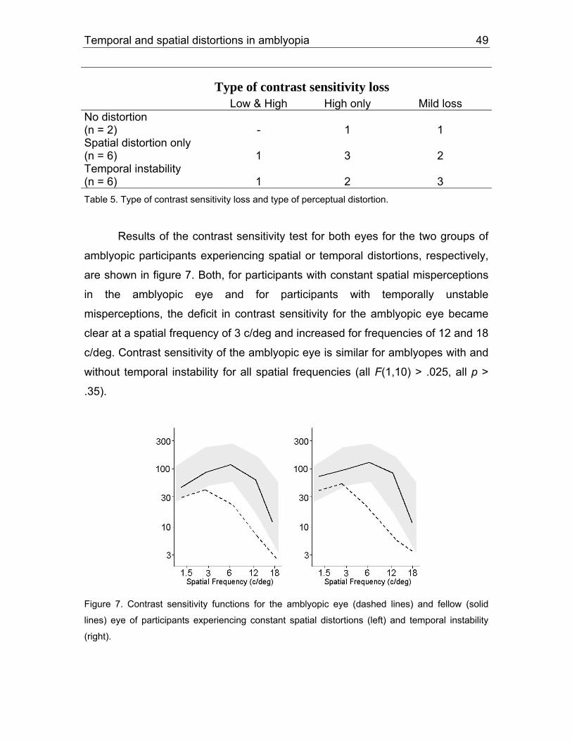
Temporal and spatial distortions in amblyopia 49
Type of contrast sensitivity loss
Low & High High only Mild loss No distortion (n = 2)
-
1
1
Spatial distortion only (n = 6)
1
3
2
Temporal instability (n = 6)
1
2
3
Table 5. Type of contrast sensitivity loss and type of perceptual distortion.
Results of the contrast sensitivity test for both eyes for the two groups of
amblyopic participants experiencing spatial or temporal distortions, respectively,
are shown in figure 7. Both, for participants with constant spatial misperceptions
in the amblyopic eye and for participants with temporally unstable
misperceptions, the deficit in contrast sensitivity for the amblyopic eye became
clear at a spatial frequency of 3 c/deg and increased for frequencies of 12 and 18
c/deg. Contrast sensitivity of the amblyopic eye is similar for amblyopes with and
without temporal instability for all spatial frequencies (all F(1,10) > .025, all p >
.35).
Figure 7. Contrast sensitivity functions for the amblyopic eye (dashed lines) and fellow (solid
lines) eye of participants experiencing constant spatial distortions (left) and temporal instability
(right).

Temporal and spatial distortions in amblyopia 50
4.1.3 Discussion
In this study, different types of distortions became apparent in the
drawings of the amblyopic participants: a) blurring of patterns, b) wavy and
jagged appearance of horizontal and vertical lines, c) distortions due to
superimposed lines or contours, and d) errors in orientation. These different
types of distortions correspond to the five distortion categories proposed by
Barrett et al. (2003).
Concerning the temporal instability, almost half (6 out of 14) of the
investigated amblyopes experienced temporal distortions, either in addition (4
cases) or in absence (2 cases) of spatial misperception. Temporal instability
occured only at high spatial frequency, spatial distortions for low spatial
frequency had a stable character. In total, there were two types of categories
concerning temporal instability: a) the whole pattern is perceived as jittering, and
b) single lines or parts in the pattern are perceived as moving. The temporal
instability patterns were superimposed to the spatial distortions and emerged in
addition to them.
The role of aetiology As this sample of amblyopic participants reflects, there might be a
preponderance of strabismic participants in the temporal-instability group -
temporal instability phenomena seem to occur mainly for strabismic and
strabismic-anisometropic amblyopes: three out of four strabismics and two out of
three stabismic-anisometropic amblyopes, but only one out of three
anisometropic amblyopes sensed temporal instability. One of the unexpected
findings of this study is that the subjective experience of spatial distortions, with
or without temporal instability, can occur in purely anisometropic participants.
This is at variance from previous reports (e.g. Lagrèze & Sireteanu, 1991) and
indicates that an early ocular misalignment is not a necessary condition for
spatial misperceptions. Neither is absence of stereopsis or total exclusion of the

Temporal and spatial distortions in amblyopia 51
amblyopic eye from binocular vision a prerequisite for the occurrence of spatial or
temporal distortions: all four anisometropic amblyopes showed some degree of
spatial or temporal distortion, albeit two of them (JB and MB) had some residual
stereopsis.
There is also no secure relationship between pattern of distortions and
angle of strabismus, presence of eccentric fixation, type of correspondence,
therapy or known early history. However, the positive relationship between the
side of the amblyopic eye (left or right eye is amblyopic) and the type of distortion
might be an important result for understanding the temporal distortions more
deeply. It seems that the eye dominance is a crucial factor for the development of
a temporally instable perception in amblyopic vision in addition to spatial
distortions. 4 out of 6 amblyopes perceiving temporal instability had a right
amblyopic eye and in addition were right-handed. These results show that it is
not only the side of the amblyopic eye by itself, but probably the interaction
between opposite sides of hand and eye dominance.
The role of visual acuity loss Of the six participants with spatial distortions only, one had a very deep
(RS), 3 a moderate-to-deep (LP, SB and HL) and 2 a moderate amblyopia (JB
and MB). In the temporal-distortions group, 2 participants suffered a very deep
(MK and BB), one a moderate-to-deep (DS) and 3 a moderate amblyopia (GP,
TS, and CL). From the two participants without distortions, one had a very deep
(MH) and one a moderate-to-deep amblyopia (AF).
Thus, very deep amblyopia seems not to be a good predictor of either
spatial or temporal distortions. Moderate-to-deep acuity loss is more often
associated with spatial distortions, while temporal distortions occur more often in
moderate amblyopes. It seems indeed that neither the severity of amblyopia nor
any other factors of the orthoptic status of an amblyopic person are good
predictors whether spatial or temporal distortions are going to occur.

Temporal and spatial distortions in amblyopia 52
The role of contrast sensitivity loss Hess & Howell (1977) described two types of amblyopic deficit, which
could be replicated in this study: a type affecting both, higher and lower spatial
frequencies and the other show only a loss of higher spatial frequencies. In
addition, a “mild” loss was defined in which both eyes remain either in the normal
contrast sensitivity range or slightly below it. For most participants, higher spatial
frequencies were progressively more affected in the amblyopic eye. With very
few exceptions (participants RS and BB), all patterns used for examining
distortions had spatial frequencies as low as to be seen equally well through both
eyes of the amblyopic participants. Thus, the experienced distortions could not
have been due to reduced contrast sensitivity.
The „spatial-distortions-only“ group contains one participant with a high
and a low spatial frequency deficit (RS), 3 participants in which only the higher
spatial frequencies were affected (LP, JB and HL), and 2 mild cases (SB and
MB). In the „temporal-distortions” group, one participant presented a deficit for
both higher and lower spatial frequencies (BB), 2 participants had a deficit for the
higher spatial frequencies alone (TS and MK) and 3 participants could be
classified in the „mild loss” type (DS, GP and CL). The two participants not
experiencing any distortions belong to the higher-frequency-only (MH) and the
mild type (AF), respectively.
Thus, the contrast sensitivity losses do not predict accurately the
occurrence and severity of distortions. A deep loss, affecting both high and low
spatial frequencies, is certainly associated with a distorted vision, however it
does not necessarily predict whether a purely spatial or a combination of spatial
and temporal distortions is going to occur. Participants with mild losses often do
not report distortions, still sometimes they do, especially in cases of severe
ametropia, like in participants SB, MB, DS or CL. Spatial distortions without
temporal instability occur more often in participants with a selective loss of higher
Temporal and spatial distortions in amblyopia 53
spatial frequency. Temporal instabilities are most often associated with a mild
contrast sensitivity loss.
4.2 Study 2: Matching experiment
To provide an objective measurement of amblyopic distortions, study 2
operationalises the spatial distortions, reported by the amblyopic participants in
study 1. In a previous study, Sireteanu et al. (1993) transferred experimental data
that mirrored the spatial distortions into computer-generated geometrical
patterns. Their participants were asked to compare the constructed patterns with
their own perception observed through the amblyopic eye. As they found, vision
seems to be more distorted at the higher and more complex patterns, however
the results also indicate that the transformed patterns do not always reflected the
actual perception of the amblyopic eyes (Sireteanu et al., 1993). The aim of the
following study is to go one step further and come as close to the amblyopic
perception as possible. This is done by using transformed patterns that are
based on the amblyopic vision acquired in study 1 and that are computer-
generated stimuli that show the perceived spatial distortions of the original
patterns. The results of this psychophysical study are also used as stimuli for a
following study using functional MRI to investigate the neuronal sites of activation
deficits in strabismic and anisometropic amblyopes (Sireteanu, Bäumer, Sârbu,
Tsujimura & Muckli, in preparation).
4.2.1 Method
Participants Participants for a pretest of the experiment were eight persons with normal
or corrected-to-normal vision (age 26 to 48, three of them male, all right-handed).
Participants for the main experiment were the same as in study 1 (see table 1).

Temporal and spatial distortions in amblyopia 54
All participants participated for course credits or for a payment of 10,- € per hour.
The refractive errors were fully corrected in all participants.
Apparatus and stimuli In study 2, using the drawings from the first part, computer-generated
stimuli were created by the experimenter, which showed the perceived spatial
distortions of the original patterns. Criterion for the computer-generated stimuli
were the individual distortions, and by that individual stimuli were constructed for
all of the four different patterns for each participant. For each distorted pattern,
four experimental stimuli were prepared. These four stimuli differed in amount of
distortion: the first stimulus was the original undistorted pattern, the third stimulus
corresponded to the reported amblyopic perception, mapped from the drawings
of study 1, and the second and fourth were stimuli with smaller and more
exaggerated distortions, respectively. The second and the fourth stimuli were
either more or less distorted than the reported distortions. This was done in order
to appraise the precise spatial distortions in the participants’ amblyopic vision. In
total, there were four test stimuli of differing distortion amount for each pattern:
the first stimulus was not distorted at all, while the fourth stimulus was the most
distorted one.
The stimuli we used were presented on a 19'' Iiyama colour monitor with a
frame rate of 85 Hz. Size of the stimuli was 12.5 degrees. For monocular
stimulation during the experiment, participants wore red-green goggles. Stimuli
were presented in corresponding colours: red stimuli (RGB = 242, 0, 0) were
shown to the dominant eye, green stimuli (RGB = 0, 249, 177) to the amblyopic
eye. Depending on the colour of the stimulus (either red or green), the eye
seeing through the complementary filter of the goggles is thereby excluded from
perception. The perceived colour of the stimulus at the screen was hereby black
and white. The wavelengths of the red and green stimuli and the goggles had
been matched. For exact matching, luminance of the stimuli was measured with
a photometer (LiteMate II, Kollmorgen Co., Burbank, USA) and the colour of the

Temporal and spatial distortions in amblyopia 55
stimuli was adjusted accordingly. All stimuli were presented against a grey
background. The advantage of this presentation procedure was that manipulation
or cover of one eye was not necessary for monocular stimulation.
Procedure The participants were seated in front of the monitor screen in an otherwise
darkened room and wore the red-green goggles. To control for head position, the
participants’ head rested on a chin-rest to maintain an eye-to-monitor distance of
57 cm. It was ensured that the participants understood their task before the
experiment started. The procedure for one trial was the following: an undistorted
reference stimulus (e.g. the low spatial frequency checkerboard) was shown to
the amblyopic eye for five seconds. Then, two test stimuli were presented on the
screen of which the participants had to chose the one which fitted best their
perception of the reference stimulus; that is of the amblyopic perception,
respectively. After that another trial followed. Showing all pairs of the stimuli (4 x
3 pairs) and having five repetitions per pair, the participants had to make 60
comparisons in total. There was no time limit to fulfil the task.
At the beginning, each participant accomplished a test trial with the
dominant eye. For best comparison between the amblyopic and non-amblyopic
perception, the whole experiment was repeated twice. At the end of the second
session, participants were debriefed and the purpose of the experiment was
explained.
4.2.2 Results
Pretest Prior to the main experiment of study 2, a pretest was conducted in order
to investigate if the matching task is a proper tool, capable of identifying the
target reference stimulus from different test stimuli, as well as to test the reliability
of the task. For statistical analyses, the number of hits for each matching were

Temporal and spatial distortions in amblyopia 56
computed. Due to the dichotomous type of each matching variable, data were
analysed first with descriptive statistics, a further analysis of probabilities for each
stimulus was submitted to a paired-samples t-test and an ANOVA with a
repeated measurement design. Here and in all subsequent analyses, the α level
was set to .05.
The pretest showed that all participants (N = 8) had a clear preference for
test stimulus 3, which was the test stimulus that matched 100 % with the
reference stimulus. Means of frequencies for test stimuli 1 through 4 were M1 =
1.5, M2 = 13.25, M3 = 28.63, and M4 = 16.63, respectively. Standard deviation
was lowest for test stimulus 3 (SD = 1.19), and highest for the two test stimuli 2
and 4 (SD2 = 5.06, SD4 = 6.46). The main effect for “test stimulus” was
significant, F(3, 5) = 1670.82, p = .00, and more specifically, a two-tailed t-test for
pair wise comparison revealed a significant difference between the test stimuli 1,
2 and 4 in comparison with test stimulus 3 (ts (7) > 6.28, ps < .00).
A 2-tailed paired-samples t-test was done to calculate the influence on the
stimuli comparisons for showing a test stimulus either on the left or the right side
of the monitor screen. None of the pairs for stimuli 1 through 4 showed a
significant difference between the placed sides - left versus right stimulus side (ts
(7) < 2.05, ps > .08).
For testing the reliability of the task, the experiment was repeated twice.
First and second sessions were submitted to a repeated measurement ANOVA.
Again, as expected, the main effect “test stimulus” was significant, F(3, 2) =
4132.17, p = .00, however the interaction between test stimulus and repetition did
not reach a significant level, F(3, 2) = 1.068, p = .52. Mean and standard
deviation of the two test sessions were exactly the same, Ms = 25, SDs = .00.
The significant effect for test stimulus 3 in difference to the other three test
stimuli, and additionally the low standard deviation for making the decision for

Temporal and spatial distortions in amblyopia 57
this stimulus, indicates that the participants did not have any problem in choosing
the test stimulus that corresponds to the reference stimulus. This also fits the
high standard deviations of test stimuli 2 and 4, which are most similar to
stimulus 3 and therefore most difficult to distinguish from test stimulus 3. The lack
of significance for a side effect as well as for a difference in repetition accounts
for the robustness of this paradigm and shows that it is an appropriate tool for
matching stimuli with each other with regard to a reference stimulus.
Main experiment Amblyopic participants had first to pass a test trial to be sure that they
understood the task. This test trial was the same as in the pretest done by
normally sighted participants, but here it was done by amblyopic participants with
the non-amblyopic eye. All amblyopic participants passed the test trial
(frequencies for test stimulus 3 were fs > 26, minimum differences from test
stimulus 3 to other test stimuli were fs > 4) except participant SB, who chose test
stimulus 4 instead of test stimulus 3. The data of this participant were excluded
from further analysis, as it was not sure if SB was either not willing or able to
follow the instructions properly. SB had a bilateral refractive strabismic amblyopia
and probably the low vision of the non-amblyopic eye influenced the results of
the matching task. Vision of the amblyopic eye could therefore not be tested in
the desired way.
First run. One basic assumption was that the distortion results of the
drawings in study 1 should also emerge in study 2. It was thus expected that the
third test stimulus - which was made exactly like the distortion drawing in study 1
- would be the test stimulus to be chosen by the participants. Results show that
this was only true for participant HL for matching the low frequency grating. For
all other trials or stimuli, respectively, participants chose either the first, or
second, or the fourth test stimulus (see table 6 a-d).

Temporal and spatial distortions in amblyopia 58
For the low spatial frequency grating (table 6 a), participant HL chose test
stimulus 3 with a probability of p = .48 (f = 29), all other test stimuli showed a
probability of p < .30 (f < 18). Participants JB and MB chose test stimulus 2 with
probabilities of pJB = .40 (f = 24) and pMB = .48 (f = 29), probabilities and
frequencies for both participants for all other test stimuli in the low frequency
grating were ps < .35 and fs < 21. Participant LP was the only one to choose the
undistorted test stimulus 1 in the low frequency grating. Success probability of
test stimulus 1 was p = .48 (f = 29), probabilities (with frequencies in
parentheses) of test stimuli 2 through 4 were .35 (21), .16 (10) and .00 (0).
Overall, the difference between the chosen test stimulus which matched best
with the reference stimulus, and all other test stimuli had a range from pmin = .50
(f = 3) to pmax = .18 (f = 11). Participant RS was the only one to report veridical
perception in this grating of .4 c/deg.
Table 6 a. Probabilities (and frequencies) for matching of grating .4 c/deg (matched test stimuli
are put in bold letters).
Participant Test stimulus 1 Test stimulus 2 Test stimulus 3 Test stimulus 4
HL .11 (7) .30 (18) .48 (29) .10 (6)
JB .35 (21) .40(24) .20 (12) .50 (3)
LP .48 (29) .35 (21) .16 (10) .00 (0)
MB .33 (20) .48 (29) .18 (11) .00 (0)
RS No distortions No distortions No distortions No distortions
For the high spatial frequency grating, differences between the test stimuli
were similar to those of grating .4 c/deg (table 6 b). Differences between the test
stimulus matched with the reference stimulus, and all other test stimuli ranged
from pmin = .08 (f = 5) to pmax = .16 (f = 10). With the highest possible score,
participant MB chose test stimulus 2 (p = .50, f = 30) over the other stimuli (ps <
.33, fs < 20). Participants HL and LP chose either the most exaggerated test
stimulus 4 or the undistorted test stimulus 1, respectively, with probabilities of pHL

Temporal and spatial distortions in amblyopia 59
= .38 (f = 23) and pLP = .46 (f = 28), probabilities and frequencies for both
participants for all other test stimuli in the grating were ps < .36 and fs < 22. As
well as participant LP, participant RS matched the undistorted test stimulus 1
with the reference stimulus (p = .50, f = 30), probabilities and frequencies for the
other test stimuli 2 through 4 were ps < .33 and fs < 20. Participant JB did not
report any spatial misperceptions.
Table 6 b. Probabilities (and frequencies) for matching of grating 1.6 c/deg
Participant Test stimulus 1 Test stimulus 2 Test stimulus 3 Test stimulus 4
HL .06 (4) .25 (15) .30 (18) .38 (23) JB No distortions No distortions No distortions No distortions
LP .46 (28) .36 (22) .16 (10) .00 (0)
MB .33 (20) .50 (30) .16 (10) .00 (0)
RS .50 (30) .33 (20) .16 (10) .00 (0)-
Participants JB and LP perceived spatial distortions in the low spatial
frequency checkerboard, while participants HL, MB and RS reported normal
perception of the pattern (table 6 c). Somehow corresponding to the undistorted
perception of participants HL, MB and RS, both, participant JB as well as
participant LP chose the undistorted test stimulus 1 with a very high frequency, f
= 29 and f = 30, respectively. Success probability of test stimulus 1 was p = .48
for participant JB and p = .50 for participant LP.
Table 6 c. Probabilities (and frequencies) for matching of checkerboard .4 c/deg
Participant Test stimulus 1 Test stimulus 2 Test stimulus 3 Test stimulus 4
HL No distortions No distortions No distortions No distortions
JB .48 (29) .35 (21) .16 (10) .00 (0)
LP .50 (30) .33 (20) .16 (10) .00 (0)
MB No distortions No distortions No distortions No distortions
RS No distortions No distortions No distortions No distortions

Temporal and spatial distortions in amblyopia 60
Again, in the high spatial frequency grid (3.2 c/deg), participants chose
either test stimulus 1 or 2 (table 6 d). Success probability was very high for the
chosen test stimulus 1 in participants HL and JB, p = .50 (f = 30) and p = .48 (f =
29), respectively. Differences between test stimulus 1 and all other test stimuli
were quite big, ps > .13, fs > 8. Participant LP preferred test stimulus 2 over all
other test stimuli (p = .46, f = 28), and the differences in success probabilties
were even higher (ps > .20, fs > 12).
Table 6 d. Probabilities (and frequencies) for matching of grid 3.2 c/deg
Participant Test stimulus 1 Test stimulus 2 Test stimulus 3 Test stimulus 4
HL .50 (30) .33 (20) .16 (10) .00 (0)-
JB .48 (29) .35 (21) .13 (8) .03 (2)
LP .23 (14) .46 (28) .26 (16) .03 (2)
MB No distortions No distortions No distortions No distortions
RS No distortions No distortions No distortions No distortions
Overall, participant LP was the one who had spatial misperceptions
throughout all of the four patterns. However, in most of the cases (7 out of 12),
participants chose either the first test stimulus or the last one. In these cases it is
not clear if the distortion perceived is even more exaggerated (when choosing
test stimulus 4) or much less pronounced (when choosing test stimulus 1). For
this reason, the experiment was repeated twice.
Second run. In the second run of the experiment, test stimuli were
provided in a way that the test stimulus which was matched in the first run with
the reference stimulus still was the third test stimulus in the second run.
Depending on the perceived distortion of this matched test stimulus, the
remaining test stimuli were made with much finer more or less exaggerated
distortions to get more precise matching results. Results are now listed not by the
patterns but by each participant for a more individual description of the matched

Temporal and spatial distortions in amblyopia 61
spatial distortions; data are only provided for the final results after two sessions in
the experiment. Participants HL and RS did not take part in the second run of this
experiment. Results of the spatial distortions matched in the first run of the
experiment are provided in table 7 for both participants HL and RS.
Table 7. Results of the matching experiment for participants HL and RS for gratins 0.4 c/deg and
1.6 c/deg, checkerboard 0.4 c/deg, and grid 3.2 c/deg. Participant
Grating
(0.4 c/deg)
Grating (1.6 c/deg)
Checkerboard (0.4 c/deg)
Grid (3.2 c/deg)
HL
No distortion
RS
No distortion
No distortion
No distortion
Participant MB reported spatial misperception only for the grating patterns
of .4 c/deg and 1.6 c/deg (see table 8). Due to the finer gradation, there was no
undistorted test stimulus 1, but one with very small spatial distortions. Participant
MB chose in both, the low spatial frequency grating and the high spatial
frequency grating, test stimulus 1. Probabilities and frequencies for this stimulus
were p = .48, f = 29 and p = .50, f = 30, respectively. Differences between test
stimulus 1 and all other stimuli were ps > .13 for grating .4 c/deg, and ps > .16 for
grating 1.6 c/deg. In sum, the reference stimulus in the second run was matched
with a test stimulus much less pronounced than expected from the results of the
first run in this study.

Temporal and spatial distortions in amblyopia 62
Table 8. Results of the matching experiment for participant MB. Participant
Grating
(0.4 c/deg)
Grating (1.6 c/deg)
Checkerboard (0.4 c/deg)
Grid (3.2 c/deg)
MB
No distortion
No distortion
Having spatial misperceptions in all of the four patterns, participant LP
chose test stimulus 2 to match the reference stimulus throughout the four
patterns. Compared to participant MB, LP had about the same amount of
quantity in spatial distortions. MB chose in the first trials test stimuli 2 and in the
second trials test stimuli 1, while LP chose in the first trials mostly test stimulus 1
(except for checkerboard 3.2 c/deg) and in the second trials test stimulus 2. Both
stimuli treated over the two runs have the same linear gradiations in spatial
distortion. For LP, probabilities (with frequencies in parentheses) of test stimuli 2
for grating .4 c/deg, grating 1.6 c/deg, checkerboard .4 c/deg and checkerboard
3.2 c/deg were .45 (27), .45 (27), .41 (25) and .41 (25). Overall, the difference
between the chosen test stimulus and all other test stimuli had a range from pmin
= .06 (f = 4) to pmax = .15 (f = 9). The test stimuli which matched best with the
reference stimulus and hence with the amblyopic vision can be seen in table 9.
Table 9. Results of the matching experiment for participant LP. Participant
Grating
(0.4 c/deg)
Grating (1.6 c/deg)
Checkerboard (0.4 c/deg)
Grid (3.2 c/deg)
LP

Temporal and spatial distortions in amblyopia 63
The second time, participant JB again always chose the undistorted test
stimulus 1 over the other test stimuli (table 10). Probabilities and frequencies for
the test stimuli 1 were p = .43, f = 26 for the low spatial frequency grating, p =
.43, f = 26 for the low spatial frequency checkerboard, and p = .41, f = 25 for the
high spatial frequency checkerboard. Differences between test stimulus 1 and all
other stimuli were ps > .15 for grating .4 c/deg, ps > .06 for checkerboard .4
c/deg and ps > .02 for checkerboard 3.2 c/deg. As one can see, differences
especially between test stimulus 1 and test stimulus 2 in both of the
checkerboard patterns are very small.
Table 10. Results of the matching experiment for participant JB. Participant
Grating
(0.4 c/deg)
Grating (1.6 c/deg)
Checkerboard (0.4 c/deg)
Grid (3.2 c/deg)
JB
No distortion
4.2.3 Discussion
Distortional effects were very similar to those of the previous experiment.
In analogy to study 1, refractive strabismic amblyope RS did not report any
spatial misperceptions in the low spatial frequency grating and checkerboard and
the high spatial frequency grid. The three anisometropic amblyopes JB, HL and
MB reported undistorted perception in the low spatial frequency checkerboard
(HL), the high spatial frequency grating (JB), and the low spatial frequency
checkerboard and the grid (MB). Strabismic amblyope LP was the only
participant experiencing spatial distortions in all four patterns.

Temporal and spatial distortions in amblyopia 64
Relationships first run of the experiment vs. second run In contrast to study 1, the amblyopic participants perceived much less
pronounced spatial distortions than expected from the results of the previous
study. In half of the cases (6 out of 12), participants chose the undistorted test
stimulus 1, and in 4 out of 12 cases they matched their perception with the least
distorted test stimulus 2. These surprising results gave rise to a second run of the
experiment, which provided a finer gradation of distortions of the amblyopic
vision. Unfortunately, data for a second trial of the experiment were available
only for the two anisometropic participants MB and JB and the strabismic
amblyope LP. Compared to the first run, distortions were once more less
pronounced than expected from the results of the first run. Both anisometropic
participants MB and JB chose test stimulus 1, while LP perceived some spatial
distortions, and chose test stimulus 2. As the probability differences between the
chosen test stimulus and the neighbouring test stimuli were very small, especially
in the anisometropic participants, those participants might still perceive spatial
distortions, however probably be unsure about their real amount.
The results in this study are similar to the results Sireteanu et al. (1993),
and Hess, Pointer, Simmers & Bex (2003) found in their experiments. Hess et al.
had veridical matches in an edge-blur task for amblyopes with a history of
strabismus as well as for strabismic amblyopes with an associated
anisometropia. Hess et al. (2003) concluded that “amblyopes perceive edges as
sharp and not blurred even though their acuity and contrast sensitivity is
dramatically reduced” (p. 2258). In contrast to that, anisometropic amblyopes
without a strabismus did exhibit a mild degree of perceived blur. This is at some
variance from the results of the matching experiment in this thesis, as the
anisometropic amblyopic participants did not report spatial distortions although
the results reflect an increased uncertainty about their matching. A reasonable
explanation for the results in study 2 as well as for Hess et al. (2003)
experiments might be that contour information is sufficient to recognize an object

Temporal and spatial distortions in amblyopia 65
most of the times (Kandel et al., 2000), especially when the uniform interior does
not contain any critical visual information.
Another reason for the small spatial distortions could be that the
participants of this sample chose the more undistorted patterns because of the
special type of the matching paradigm. By the comparison of two test stimuli,
amblyopic participants are more likely to choose the undistorted test stimulus.
This may be either because amblyopes who have a distorted perception could
identify different spatial frequencies as accurately as those with veridical
perception (see also Barrett et al., 2003), and therefore match the distorted
perception with the undistorted pattern in any spatial frequency. Or because of
their amblyopic vision, which might be influenced by external parameters e.g.
such as contour information (Kandel et al., 2000). This would also explain the
results of the pretest with normal sighted adults as well as the results of the
pretest for each amblyope, conducted with the non-amblyopic eye. In each, they
had no problems in matching the distorted stimuli with each other.
In general, the results of the matching task showed distortions that are
much less pronounced than expected from the drawings of study 1. This was
especially conspicuous in the low frequency patterns. However the structure and
complexity of the spatial and temporal distortions remained the same over the
two experiments. That is, in the comparison between study 1 and study 2, one
can say that the type of distortion was the same for the drawings and the
matching task. The spatial distortions did not change in quality for both
paradigms, yet in quantity. This is also consistent with previous results from the
group of Sireteanu (Sireteanu et al., 1993): quantitative data differ from
qualitative descriptions and only partially reflect the amblyopic perception. It
seems that amblyopes experience and describe their distortions subjectively
more vividly than can be captured accurately in quantitative experiments (see
also Hess et al., 2003).

Temporal and spatial distortions in amblyopia 66
5. Part II: Quantitative methods
5.1 Study 3: Circle experiment (auditory instructions)
As pointed out in the introduction chapter, the aim of the following studies
is to quantitatively measure the temporal as well as the spatial distortions in
amblyopic vision, and to relate the temporal distortions to the performance of the
amblyopic participants in psychophysical tasks. The following experiments are a
further development of the work of Lagrèze and Sireteanu (1991), and Sireteanu
et al. (1993). These authors could show that spatial distortions and spatial
uncertainty are increased in strabismic amblyopia, however did not focus on
temporal instability in amblyopic vision. In the following, Lagrèze and Sireteanu’s
(1991) method was improved and further developed by the additional
assessment of temporal instability.
5.1.1 Method
Participants With the exception of strabismic amblyope GP, amblyopic participants in
experiments of part II were the same as in experiment 1. Participants were six
strabismic amblyopes, four anisometropic amblyopes, three strabismic and
anisometropic amblyopes, and additionally three strabismic alternators and
twelve adults with normal vision (see table 11). Selection criteria for the normal
participants and the alternators were: corrected visual acuity of ≥ 1.0 Snellen
acuity in both eyes, no interocular deviation, and for the normal participants good
stereo acuity. The participants with normal vision were matched in gender and
age (± 5 years) each with one of the amblyopes or alternators, respectively.

Temporal and spatial distortions in amblyopia 67 Table 11. Orthoptic data of amblyopic, alternating, and normally sighted participants
Partici-pant
Gender, Age
Eye Refraction Visus c.c.(near vision)
Fixation Strabismus(sim. Cover test)
Stereopsis Correspondence History
Strabismic amblyopes R.S.
male, 58 yr
RE LE*
+ 6.00 -1.25/171° + 6.75 -1.50/5°
0.60 0.10
foveolar 1.5°-2° nasal
far +2½°+ VD near +3° + VD
negative, excl. LE
h arc
Occlusion at 1 yr
L.P.
female, 33 yr
RE LE*
+ 0.50 sph + 0.75 sph
1.00 0.25
foveolar temporal
far -12½°+ VD 1°near ~± 0°
negative, excl. LE
nrc
Onset and occlusion at 4 yr, glasses until 15 yr
M.K.
male, 29 yr
RE* LE
+ 5.50 -4.00 / 145° + 5.00 -4.75 / 5°
0.10 1.00
temporal foveolar
far +1½° + VD½°near +1½° + VD¾°
negative, excl. RE
h arc
Strabismus from early childhood, occlusion at 5 yr
S.B.
female, 25 yr
RE* LE
-10.0 sph - 9.0 sph
0.30 0.60
temporal foveolar
far +12° + VD near +12° + VD 7°
negative, excl. RE
nh arc
Family history, strabismus from early childhood, glasses at 5 yr
D.S.
male, 51 yr
RE* LE
+ 5.25 -2.50 / 100° + 4.50 -2.25 / 95°
0.25 1.00
fov. margin foveolar
far ± 0° near +4° - VD
negative, excl. RE
nh arc
Onset and glasses at 6 yr
A.F.
female, 22 yr
RE LE*
- 1.25 -2.00 / 85° - 1.25 -1.75 / 105°
1.00 0.32
foveolar temp. margin
far +3° - VD 1° near +4½°- VD 1°
negative, excl. LE
h arc
Strabismus from early childhood, glasses and occlusion at 5 yr for one year
Strabismic and anisometropic amblyopes B.B.
female, 29 yr
RE* LE
- 0.75 sph - 1.50 +2.0 / 175°
0.08 0.90
temp. margin foveolar
far + ½° + VD 3°near - 2½° + VD 2°
negative, excl. RE
nh arc
Strabismus from early childhood, glasses at 3 yr, occlusion 3-6 yr, surgery at 20 months
M.H.
female, 31 yr
RE LE*
+ 5.00 -0.75 / 142° + 1.50 -0.50 / 0°
1.00 0.08
foveolar nasal
far + 1½° ± VD near + 4°
negative, excl. LE
h arc
Family history, strabismus from early childhood, glasses at 3 yr, occlusion 4–5 yr, surgery at 5 yr
C.L.
male, 28 yr
RE LE*
- 3.50 -1.50 / 20° + 1.00 -1.25 / 0°
1.00 0.50
foveolar nasal - fovea
far +15° +VD 1° near +15° +VD 2°
negative, excl. LE
nh arc
Strabismus from early childhood, occlusion in kindergarten for 3 yr, surgery at 4 yr
Anisometropic amblyopes H.L.
male, 27 yr
RE LE*
plano + 6.25 sph
1.40 0.25
foveolar foveolar
± 0°
negative
nrc
Occlusion at 11 yr, glasses at 18 yr
T.S.
male, 30 yr
RE LE*
+ 1.25 sph + 2.75 -3.75/135°
1.00 0.40
foveolar nasal margin
± 0°
negative, excl. LE
nrc
Occlusion and glasses at 6 yr
J.B.
male, 26 yr
RE LE*
- 2.25 sph - 0.75 -2.00 / 15°
1.00 0.50
foveolar temporal
± 0°
Positive
nrc
Occlusion and glasses at 6 yr
M.B.
male, 34 yr
RE LE*
- 0.50 -0.50 / 45° - 3.00 -3.25 / 2°
1.00 0.50
foveolar foveolar
± 0°
Positive
nrc
Glasses at 17 yr
Abbrevations: nrc = normal retinal correspondence, nh arc = non-harmonius anomalous retinal correspondence, h arc = harmonius anomalous retinal correspondence, VD = vertical deviation, + = esotropia, – = exotropia, * = amblyopic eye.

Temporal and spatial distortions in amblyopia 68
Partici-pant
Gender, Age
Eye Refraction Visus c.c.(near vision)
Fixation Strabismus(sim. Cover test)
Stereopsis Correspondence History
Alternators M.O.
female, 23 yr
RE LE
- 0.50 -1.0 / 163° - 1.00 -1.0 / 27°
1.00 1.00
foveolar foveolar
far + VD 1.5° near - 1.5° + VD5°
negative
nrc
Family history, glasses at 14 yr
R.F.
female, 25 yr
RE LE
plano - 4.00 -1.0 / 15°
1.00 1.00
foveolar foveolar
far - 8° + VD near - 3°
negative
nrc
Strabismus from early childhood, glasses and pleoptic therapy at 2 yr
J.Z.
male, 27 yr
RE LE
- 7.75 sph - 8.50 sph
1.25 1.25
foveolar foveolar
far + 4° - VD near + 4° - VD
negative
arc
Glasses and occlusion at 5 yr
Normally sighted participants A.Fr.
male, 31 yr
RE LE
- 2.75 -0.5 / 160° - 2.25 -1.0 / 20°
1.00 1.00
foveolar foveolar
far ± 0° near ± 0°
positive
nrc
Glasses at 14 yr
C.E.
female, 25 yr
RE LE
- 0.25 -0.25 / 15° - 0.50
1.25 1.25
foveolar foveolar
far ± 0° near ± 0°
positive
nrc
Glasses at 14 yr
E.B.
female, 21 yr
RE LE
plano - 0.5 / 0°
1.00 1.00
foveolar foveolar
far ± 0° near ± 0°
positive
nrc
M.Y.
female, 22 yr
RE LE
+ 0.25 sph + 0.25 sph
1.00 1.00
foveolar foveolar
far ± 0° near ± 0°
positive
nrc
R.P.
male, 30 yr
RE LE
plano plano
1.00 1.00
foveolar foveolar
far ± 0° near ± 0°
positive
nrc
T.P.
male, 35 yr
RE LE
+ 0.25 sph plano
1.00 1.00
foveolar foveolar
far ± 0° near ± 0°
positive
nrc
T.W.
male, 28 yr
RE LE
- 6.50 -0.75 / 50° - 6.50 -0.5 / 170°
1.00 1.00
foveolar foveolar
far ± 0° near ± 0°
positive
nrc
Glasses at 9 yr
S.H.
male, 31 yr
RE LE
- 0.5 / 95° - 0.75 / 82°
1.00 1.00
foveolar foveolar
far ± 0° near ± 0°
positive
nrc
P.G.
female, 40 yr
RE LE
+ 0.25 sph plano
1.00 1.00
foveolar foveolar
far ± 0° near ± 0°
positive
nrc
M.N.
female, 30 yr
RE LE
- 5.50 -1.25 / 175° - 3.00 -1.0 / 160°
1.00 1.00
foveolar foveolar
far ± 0° near ± 0°
positive
nrc
Glasses at 16 yr
E.G.
female, 21 yr
RE LE
- 2.50 -0.25 / 0° - 3.50 -0.5 / 25°
1.00 1.00
foveolar foveolar
far max. - 1.5° near max. - 3°
positive
nrc
Glasses at 10 yr
D.G.
male, 21 yr
RE LE
- 4.25 sph - 2.50 sph
1.00 1.00
foveolar foveolar
far ± 0° near ± 0°
positive
nrc
Glasses at 4 yr

Apparatus and stimuli The apparatus and the red-green goggles were as described in study 2.
The stimuli in study 3 were circles of six different radii of two, four, six, eight, ten
and twelve degrees. Corresponding to study 2, stimuli shown to the dominant
eye were red, stimuli shown to the amblyopic eye green.
During the experiment participants heard numbers from 1 to 12 by
loudspeakers. In a pretest, the numbers were rated to make sure they were
understood correctly. Participants (N = 15) had to rate the numbers on a scale
from 1 (very hard to understand) to 5 (very easy to understand). As table 12
shows, all numbers were understood correctly and means of the ratings were Ms
> 4.40.
Table 12. Rating of numbers.
Number Percent of correct
understanding
Rating
(mean)
1 100 % 4.73
2 100 % 4.47
3 100 % 4.60
4 100 % 4.40
5 100 % 4.73
6 100 % 4.60
7 100 % 4.60
8 100 % 4.40
9 100 % 4.53
10 100 % 4.87
11 100 % 4.53
12 100 % 4.80

Temporal and spatial distortions in amblyopia 70
Procedure The participants were seated in front of the monitor screen in an otherwise
darkened room and put on the red-green goggles. The participants’ head was
rested on a chin-rest to maintain an eye-to-monitor distance of 57 cm. The
refractive errors were fully corrected in all participants. There had been twelve
test trials to practice the experimental procedure before starting the experiment.
The experimenter made sure that the participant understood his/her task entirely
before the experiment started.
Participants were asked to fixate a continuously presented cross in the
centre of the screen and then memorize the different circles, which were
presented in a random sequence. The fixation cross indicated the midpoint of
each circle. The circles were presented on the screen for five seconds, only one
circle in each trial. One single circle consisted of 12 points, similar to the dial
plate of a watch. The circles were presented to the dominant eye, while the
amblyopic eye did not receive any stimulus. After the circle disappeared
participants heard over loudspeakers a number in the range of 1-12, which was
presented randomly.
The participants’ task was to reproduce a circle presented to their
dominant eye from memory and reconstruct it by bringing a test point on the
imaginary outline of the circle according to the position of the number heard over
the loudspeakers before. On a blank screen the test point always appeared in the
middle of the fixation cross and could be freely moved on the screen with the aid
of the computer mouse. The circle reconstruction had to be done with the non-
amblyopic eye as well as with the amblyopic eye and every position on each
circle was shown up to five times. Depending on the eye (amblyopic or non-
amblyopic) used for the reconstruction, the colour of the test point was either red
or green. Each trial took about 10 seconds. After every 84th trial or after about 15
minutes, respectively, there was a break for five minutes. There was no time limit
for this task and the participants had the opportunity to cancel the last set

Temporal and spatial distortions in amblyopia 71
position if deemed necessary. Informed consent was obtained from all
participants after the nature and the purpose of the study had been explained
fully. At the end of the second half of the experiment all participants were
debriefed and paid 10,- € per participated session.
5.1.2 Results
Data were submitted to a repeated-measures multivariate analysis of
variance (MANOVA) model that included eye, radial position and tangential
position of the points as dependent variables, and amount of spatial distortion
(difference of the adjusted point to the target point) and amount of spatial
uncertainty (standard deviation) as independent variables. The α level was set to
.05 for omnibus tests, Wilk’s Λ was used as a test statistic. Separate error terms
and Bonferroni α adjustments were used for planned comparisons and contrasts.
For appraising the spatial distortions and spatial uncertainty in individual
amblyopes (strabismic, strabismic-anisometropic and anisometropic), strabismic
alternators and normally sighted participants, data of the two eyes were mirrored
to each other, to enable comparison of the nasal versus temporal hemifields,
respectively.
Normally sighted participants Figure 8 shows maps of the central visual fields in all normally sighted
participants for the radius gradations up to 12 deg. Left and right eyes are
displayed separately. The red symbols represent the optimal positions for each
adjusted point; the black symbols represent the mean of the individual mean
settings for each position.

Temporal and spatial distortions in amblyopia 72
Left eyes Right eyes temporal nasal nasal temporal
Figure 8. Monocular localisations for all normally sighted participants. Left: settings in the left eye.
Right: settings in the right eye. Red symbols: optimal position. Black symbols: mean of individual
setting. Radii of the circles are 2, 4, 6, 8, 10 and 12 deg.
Differences in spatial distortions between both eyes were not significant
for radial (F(1,11) = 1.59, p = .23, η² = .13) as well as tangential adjustments (F
(1,11) = .89, p = .37, η² = .07). For spatial uncertainty, radial and tangential
standard deviations of the means were quite small with SDs ≤ .04 for the left eye
and SDs = .03 for the right eye. Although very small, there were systematic
spatial distortions in the perception of each eye even in normal observers.
Positions in the smaller circles with radii of 2 and 4 deg tended to be set larger,
indicating an underestimation of the central visual field, while positions in the
larger circles (10 and 12 deg), especially in the vertical meridian, tended to be set
smaller and were therefore overestimated. In general, spatial distortions (M2 deg =
.26 to M12 deg = .51) as well as positional uncertainty (SD2 deg = .04 to SD12 deg =
0.7) tended to increase significantly with eccentricity of the circle radius (F(5,7) =
4.71, p = .03, η² = .77), however only for radial adjustments. Positions on the
vertical and the horizontal meridian were set more accurately and with a lower
spatial uncertainty (Ms < 1.77, SDs < .36) than on the other positions (Ms > 4.14,

Temporal and spatial distortions in amblyopia 73
SDs > .58). There were no significant differences between the patterns for the left
and the right eye. On an individual level, the results of the normally sighted
participants were very consistent with the group data and showed similar
patterns of the adjusted positions in both visual fields.
To put together, since normal observers had no systematic differences
between the two eyes and showed small yet consistent spatial distortions and
spatial uncertainty, participants with normal binocular vision can be used as
control participants for the group of amblyopes and their results can be adopted
as a baseline for the amblyopic participants in this study.
Strabismic amblyopes Figures 9 to 11 show the individual patterns of spatial distortions and the
relative amount of spatial error in the amblyopic eye for strabismic participants
RS, LP, MK, SB, DS and AF. The bases of the arrows indicate the mean of the
tested positions in the participant’s dominant eye, while the tips of the arrows
indicate the mean of the tested positions in the amblyopic eye. Thus, the longer
the arrow, the more pronounced the spatial distortion in the amblyopic eye in
relation to the non-amblyopic eye.
a) Refractive strabismic amblyopes: SB and RS have a reduced visual
acuity in both eyes - with the amblyopic eye having an acuity of 0.3 and 0.1,
respectively, and the non-amblyopic eye of 0.6. They both showed strong spatial
misperceptions and overestimation of nearly all circles in the non-amblyopic and
the amblyopic eye (figure 9). The overestimation got higher as the radius of the
circle got larger. In the smallest radius with 2 deg, position adjustment was quite
precise, as well as was the position adjustment of the vertical and the horizontal
axis of the non-amblyopic eyes and the amblyopic eye of participant RS.

Temporal and spatial distortions in amblyopia 74
Esotropic and refractive strabismic amblyope RS temporal nasal nasal temporal
Esotropic an blyope SB d refractive strabismic am temporal nasal nasal temporal
Figure 9. Spatial distortions for the amblyopic and the non-am
spatial distortions indicated as vectorial subtraction of the distortio
SB. Positional means of the spatial distortions are connected by
of the arrows indicate the positional mean in the amblyopi
amblyopic eye. Left: settings in the non-amblyopic eye. Middle:
Right: vectorial subtraction of the distortion maps.
Participant SB had not only problems in
blyopic eye, and relative amount of
n maps of participants RS and
lines for better comparison. Tips
c eye, the bases refer to the non-
settings in the amblyopic eye.
adjusting radial distances, but
also in adjusting the tangential position of the circle points, especially in the
amblyopic eye. However, there was no nasal-temporal asymmetry observable in
the data of both amblyopes.

Temporal and spatial distortions in amblyopia 75
b) Accommodative strabismic amblyopes: DS, an amblyope with a
visual acuity of 0.25 in his right eye, has a high ametropia in both eyes. He
showed some overestimation of the circles that was very consistent over the
different radii gradations, however somewhat higher in the right lower visual
emifield. Tangential adjustments on the horizontal and vertical axis were
djusted with a smaller amount of spatial error than on the other positions.
h
a
Participant MK, who is hyperopic with astigmatism, had larger localisation errors.
The subjective circle positions in the amblyopic eye were rotated clockwise by
some degree, and radii from 8-12 deg were underestimated especially on the
horizontal and the vertical axis (figure 10).
Esotropic amblyope DS temporal nasal nasal temporal
Esotropic amblyope MK temporal nasal nasal temporal
Figure 10. Spatial distortions and vectorial subtraction of the distortion maps of participants DS
and MK. Left: non-amblyopic eye. Middle: amblyopic eye. Right: vectorial subtraction.

Temporal and spatial distortions in amblyopia 76
c) Primarily strabismic amblyopes: Participants LP and AF, both
primarily strabismic with a visual acuity of 0.25 and 0.32 of the amblyopic eye,
were characterised by an underestimation of the circles in the amblyopic eye. A
minimally oval, lengthened shape of the adjusted circles was visible in both
participants. Tendency of the adjustments was to the horizontal axis, yet this
tendency was more pronounced in participant LP (figure 11).
strabismic amblyope LP Exotropic
temporal nasal nasal temporal
strabismic AF Exotropic amblyope
temporal nasal nasal temporal
Figure 11. Spatial distortions and vectorial subtraction of the distortion maps of participants LP
and AF. Left: non-amblyopic eye. Middle: amblyopic eye. Right: vectorial subtraction.
All strabismic amblyopes were quite precise in the position adjustment of
the horizontal and the vertical axis. Participants RS, SB, and DS showed an

Temporal and spatial distortions in amblyopia 77
overestimation of the circles, while participants MK, LP, and AF underestimated
the radii of the circles. Except amblyope MK, all other strabismic amblyopes had
howed a pattern in
e non-amblyopic eye which was similar to the normal observers’ (figure 12). In
ed, with a slight
hape of the whole circle
patterns. That is, the horizontal positions were underestimated while distances
on the vertical meridian were overestimated for more than 2 deg.
Strabismic-anisometropic amblyope BB
a tendency to set vertical distances smaller than horizontal distances (also found
by Lagrèze & Sireteanu, 1991). No nasal-temporal asymmetries were observable
in all of the strabismic amblyopes.
Strabismic-anisometropic amblyopes BB, with an acuity of 0.08 in the right amblyopic eye, s
th
contrast to that, the pattern of the amblyopic eye was fairly distort
S-shape of the vertical axis and an overall oval s
temporal nasal nasal temporal
and vectorial subtraction of the distortion maps of strabismic-
BB. Left: non-amblyopic eye. Middle: amblyopic eye. Right: vectorial
Participants MH and CL showed quite similar patterns of their subjective
adjustments (figure 13), even if they differ a lot in their orthoptic status, as MH is
microstrabismic and has a visual acuity of 0.08 and CL shows a large-angle
esotropia and an acuity of 0.5. They both underestimated the horizontal meridian.
Albeit being quite precise with the tangential adjustments on the vertical and the
Figure 12. Spatial distortions
anisometropic amblyope
subtraction.

Temporal and spatial distortions in amblyopia 78
horizontal axis, the adjusted points on the intermediate axis showed a more
distorted pattern.
Esotropic amblyope MH temporal nasal nasal temporal
Esotropic amblyope CL temporal nasal nasal temporal
Figure 13. Spatial distortions and vectorial subtraction of the distortion maps of amblyopes MH
and CL. Left: non-amblyopic eye. Middle: amblyopic eye. Right: vectorial subtraction.
All strabismic-anisometropic amblyopes had a tendency to set vertical
nces. However this tendency was most distances smaller than the horizontal dista
pronounced in amblyope BB as the circles were nearly ovally shaped, amblyopes
CL and MH showed this pattern to a much smaller amount. As for the strabismic
amblyopes, there were no nasal-temporal asymmetries in the circle adjustments
of the strabismic-anisometropic amblyopes.

Temporal and spatial distortions in amblyopia 79
Anisometropic amblyopes TS, who is anisometropic with a visual acuity of the left amblyopic eye of
.4, showed the highest overestimation of the circles of all anisometropic
, however
re shifted to the horizontal
axis (figure 14). This pattern of spatial misperceptions was similar to that of
strabismic participant AF.
Anisometropic amblyope TS
0
amblyopes. This overestimation was quite pronounced in both eyes
circle positions in the amblyopic eye were even mo
temporal nasal nasal temporal
d vectorial subtraction of the distortion maps of anisometropic
Middle: amblyopic eye. Right: vectorial subtraction.
acuity of the left amblyopic eye is 0.5) showed an underestimation of
e 2 deg circle with the amblyopic eye relative to the non-amblyopic eye. The
Figure 14. Spatial distortions an
amblyope TS. Left: non-amblyopic eye.
Both participants HL (visual acuity of the left amblyopic eye is 0.25) and
JB (visual
th
difference between the eyes was very small: in the amblyopic eye spatial
distortions were just slightly more pronounced than in the non-amblyopic eye.
Participant MB, with an acuity of 0.5 in his left eye, exhibited a pattern that was
fairly similar to those of the normally sighted participants. The amblyopic and the
non-amblyopic eye showed both quite regular position adjustments of all circles
with a slight overestimation of the larger 10-12 deg circles (figure 15).

Temporal and spatial distortions in amblyopia 80
Anisome amblyope HL tropic temporal nasal nasal temporal
Anisometropic amblyope JB temporal nasal nasal temporal
Anisometropic amblyope MB temporal nasal nasal temporal
Figure 15. Spatial distortions and vectorial subtraction of the distortion maps of anisometropic
amblyopes HL, JB, and MB. Left: non-amblyopic eye. Middle: amblyopic eye. Right: vectorial
subtraction.

Temporal and spatial distortions in amblyopia 81
The differences between the amblyopic and the non-amblyopic eyes were
quite small in the anisometropic amblyopes, especially for participant MB. All
nisometropic amblyopes had a tendency to overestimate the radial circle
ositions, and also had a tendency to set the vertical distances smaller than the
horizon
val shape of the circles in both eyes,
airly overestimated here. Participant RF showed
tively.
alte
a
p
tal ones, resulting in an oval shaped circle pattern.
Strabismic alternators Strabismic alternators did not show such a clear distortion pattern. All
showed an underestimation of the smallest circle, getting less for the larger radii
(see figure 16). Participant MO showed an o
the vertical meridian was f
similar tangential errors in the left and right eye in the intermediate meridians of
the circles. The positions on these axes had a strong tendency away from
vertical and the horizontal to the middle. JZ exhibited a pattern contrary to that of
RF. He adjusted the positions on the intermediate meridians accordingly to their
main neighbour axis (vertical or horizontal); that was more to the vertical and the
horizontal meridian, respec
Strabismic rnator MO temporal nasal nasal temporal

Temporal and spatial distortions in amblyopia 82
Strabismic alternator RF temporal nasal nasal temporal
Strabismic alternator JZ temporal nasal nasal temporal
Figure 16. Spatial distortions and vectorial subtraction of the distortion maps of strabismic
alternators MO, RF, and JZ. Left: non-amblyopic eye. Middle: amblyopic eye. Right: vectorial
subtraction.
In total, each amblyopic participant and strabismic alternator, respectively,
had his/her individual pattern of spatial distortions. A group analysis revealed that
the difference between the amblyopic and the dominant eye in amblyopic
participants was highly significant for the spatial distortions (F (1,23) = 8.88, p =
07, η² = .279) and for the spatial uncertainty, indicated as standard deviation of
e positional mean (Fs > 7.09, ps < .01, η² > .25). Means of the different groups
table 13.
.0
th
for the spatial distortions and spatial uncertainty are shown in

Temporal and spatial distortions in amblyopia 83
Table 13. Amount of spatial distortions (difference of adjusted point to target point) and spatial
uncertainty (standard deviation) for the amblyopic and non-amblyopic eyes.
Strabismic Anisometropic Strabismic-
anisometropic
Strabismic
alternator
Amblyopic
eye
M = 2.41
SD = .45
M = .72
SD = .31
M = .52
SD = .37
M = .91
SD = .37
Non- M = .79 M = .19
SD = .32
M = .57
SD = .29
M = .19
SD = .34 amblyopic
eye
SD = .38
Group data
absolute value of the difference between
a) Spatial distortion and spatial uncertainty: For group data, the
the amblyopic and the dominant eye
tial uncertainty. The
stortions (linear di om t oint to the target point) and
spatial uncertainty (standard deviation of adjusted points) were most pronounced
in strabismic and strabismic-anisometropic amblyopes, for the radius (figure 15)
as well as for the angle (figure 16) of the adjusted circle points. As reported in
previous research (Lagrèze & Sireteanu, 1991), also in this sample strabismic
and strabismic-anisometropic amblyopes show considerable spatial distortions
and uncertainty in contrast to the anisometropic amblyopes and normally sighted
participants.
As figure 15 shows, mean error in localisation (with spatial uncertainty in
brackets) for radial position was M = .12 deg (SD = .07 deg) for strabismic
amblyopes, M = .07 deg (SD = .08 deg) for strabismic-anisometropic amblyopes,
M = .06 deg (SD = .04 deg) for strabismic alternators, M = .03 deg (SD = .01
deg) for anisometropic amblyopes and M = .02 deg (SD = .01 deg) for normally-
sighted participants. Strabismic alternators which have a history of strabismus
was used for the calculation of spatial distortion and spa
spatial di stance fr he adjusted p
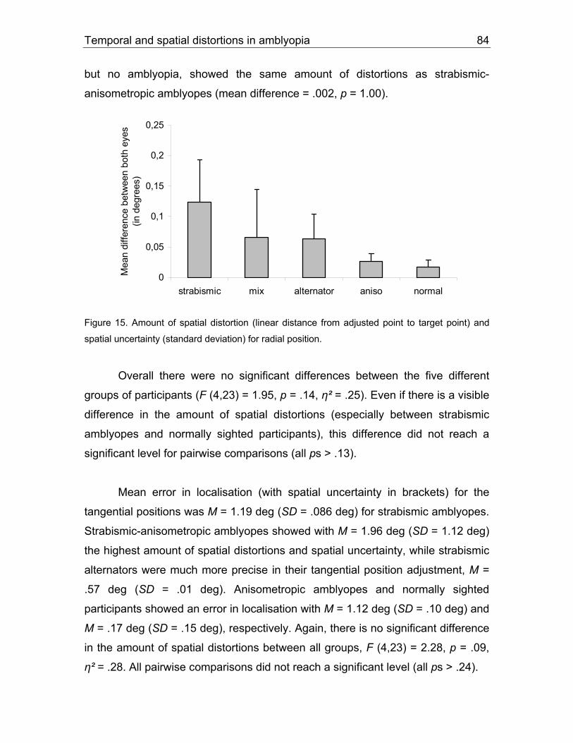
Temporal and spatial distortions in amblyopia 84
but no amblyopia, showed the same amount of distortions as strabismic-
(mean difference = .002, anisometropic amblyopes p = 1.00).
0
0,05
0,1
0,15
0,2
0,25
strabismic mix alternator aniso normal
Mea
n di
ffere
nce
betw
een
both
eye
s(in
deg
rees
)
Figure 15. Amount of spat
spatial uncertainty (standard deviation) f
ial distortion (linear distance from adjusted point to target point) and
or radial position.
ount of spatial distortions (especially between strabismic
and normally sighted participants), this difference did not reach a
opes showed with M = 1.96 deg (SD = 1.12 deg)
distortions and spatial uncertainty, while strabismic
in their tangential position adjustment, M =
Overall there were no significant differences between the five different
groups of participants (F (4,23) = 1.95, p = .14, η² = .25). Even if there is a visible
difference in the am
amblyopes
significant level for pairwise comparisons (all ps > .13).
Mean error in localisation (with spatial uncertainty in brackets) for the
tangential positions was M = 1.19 deg (SD = .086 deg) for strabismic amblyopes.
Strabismic-anisometropic ambly
the highest amount of spatial
alternators were much more precise
.57 deg (SD = .01 deg). Anisometropic amblyopes and normally sighted
participants showed an error in localisation with M = 1.12 deg (SD = .10 deg) and
M = .17 deg (SD = .15 deg), respectively. Again, there is no significant difference
in the amount of spatial distortions between all groups, F (4,23) = 2.28, p = .09,
η² = .28. All pairwise comparisons did not reach a significant level (all ps > .24).

Temporal and spatial distortions in amblyopia 85
0
1
1,5
strabismic mix alternator aniso normal
fere
nce
bet
(in d
egr
Figure 16. Amount of spatial distortion (linear distance from adjusted point to target point) and
spatial uncertainty (standard deviation) for tangential position.
b) Temporal instability: On the basis of these findings, data were
analysed regarding the perception of temporal instability. The amblyopic
participants were grouped in two groups depending on their subjective perception
f temporal instability when viewing with the amblyopic eye. This was done
ased on the results of the qualitative reports in study 1. The group with
mblyopes perceiving no temporal instability consisted of eleven amblyopes:
ope
strabismic alternators MO,
RF, and JZ. Five participants were in the temporal-instabilit
amblyopes MK and DS, strabismic-anisometropic am
anisometropic amblyope TS.
Amblyopes who perceived temporal inst
in adjusting the tangential position of t
amblyopes without temporally instable perception, they
. Mean spatial distortion
tial u certa ) for t ition was M = 2.29 deg (SD
= 1.12 deg) for amblyopes who perceive temporal instability and M = .39 deg (SD
2
2,5
3w
een
both
eye
see
s)
0,5
ean
dif
M
o
b
a
strabismic amblyopes LP, AF, RS, and SB, strabismic-anisometropic ambly
MH, anisometropic amblyopes JB, HL, and MB, and
y group: strabismic
blyopes BB and CL, and
ability had much more difficulties
he test point (figure 17). Compared to
showed a significantly
lowered performance, F (1, 14) = 5.42, p = .03, η² = .28
(with spa n inty in brackets angential pos

Temporal and spatial distortions in amblyopia 86
= .28 deg) for those who have a stable but distorted pe
was no difference in the reconstruction of the r
amblyopes with stable distortions and those with t
(1,14) = .39, p = .54, η² = .03. Mean spatial distortion
brackets) for radial position was M = .1 deg (SD =
instability group and M = .07 deg (SD
distortions.
rception. However, there
adial positions between
emporally instable ones, F
(with spatial uncertainty in
.07 deg) for the temporal
= .02 deg) for amblyopes with stable
0
0,5
1
1,5
2
2,5
Mea
n di
ffere
nce
betw
een
bot
3
h ey
e
3,5
s
temporal instabilityno instability
igure 17. Amount of spatial distortion (linear distance from adjusted point to target point) and
Note that t pr itively,
while the radial position was presented in a visual way. In general, for both
grou g en ively p positio test point
was h harde sti ial, vis nted owever,
amblyopes who perceived temporal instability showed an additional impairment
in a g the a pres gential than those who did not.
re specifically, the values of both groups have the same trends over the
ifferent radius gradations (from 2° to 12°), but differ only in their amount. As
ported above, there is a main effect for the factor “group” for spatial uncertainty
F
spatial uncertainty (standard deviation) for the adjustment of the tangential positions.
the tangential position of the est point was esented aud
ps, adjustin
muc
the tang tial, audit resented n of the
r than adju ng the rad ually prese position. H
djustin
Mo
uditively ented tan positions
d
re

Temporal and spatial distortions in amblyopia 87
in tangential position, but there is also an effect for the interaction between group
and tangential position, which is nearly significant (F(5,8) = 3.06, p = 07, η² =
.66). The impairment of performance for the temporal instability group in the
reconstruction of the angle was only true for circles smaller than 8°. That is, the
performance of the amblyopic eye appeared to be especially impaired in the
central visual field, while in the peripheral field the differences disappeared
(figure 18).
2
2,5
3
in d
egre
es) instable
stable
0
0,5
1
circle 1 circle 2 circle 3 circle 4 circle 5 circle 6
Sta
ndar
d
1,5
dev
iatio
n (
Figure 18. Amount of spatial uncertainty for different radius gradations for amblyopes with (N = 5)
and without (N = 8) perception of temporal instability.
5.1.3 Discussion
In general, the amount of spatial distortions and spatial uncertainty
between the amblyopic and the non-amblyopic eye were significantly different in
all amblyopic participants. However as reported in previous research (Lagrèze &
Sireteanu, 1991), the spatial distortions and spatial uncertainty were most
pronounced in amblyopes with a history of strabismus. Strabismic and
strabismic-anisometropic amblyopes showed higher spatial distortions and

Temporal and spatial distortions in amblyopia 88
uncertainty in contrast to anisometropic amblyopes and normally sighted
participants.
Individual analysis of spatial distortions Normally sighted participants do not show differences in spatial vision
between both eyes, still they show, albeit very small, systematic spatial
distortions in the perception of the circles. The distortions were in a way, that
participants underestimated smaller circles in the central visual field - especially
positions on the horizontal axis - and overestimated positions on the vertical axis
of the bigger circles.
ounced
opes, who overestimated nearly all circle
positions
underestimation of the circle positions
same overestimation visible in the normal participants. Only anisometropic
In contrast to the normally sighted participants, strabismic amblyopes in
this sample showed strong misperceptions. These were especially pron
for the two refractive strabismic ambly
except the ones of the smallest circle of 2°. The patterns of the circles
were distorted in a quite irregular way, a pattern known from previous literature
(Bedell & Flom, 1983; Lagrèze & Sireteanu, 1991). Bedell and Flom (1983)
showed that „spatial errors need not to be in the same direction within a single
eye“ (p. 427). The other strabismic amblyopes also showed a high level of spatial
errors, but this was accompanied by an
and by a systematic clockwise rotation or lengthened ovally shape of the circles,
respectively. The strabismic-anisometropic participants showed patterns which
were much more ovally shaped than those of the purely strabismic amblyopes.
They had a higher accuracy in adjusting the positions on the horizontal and
vertical axis, and therefore had a lower amount of misplacement of individual
circle positions. In all anisometropic participants who experienced some stereo
vision, differences between the two eyes was quite small and spatial distortions
were only slightly more pronounced in the amblyopic eye. The patterns were
fairly similar to those of the normally sighted participants as the circle positions
were regularly adjusted with a slight overestimation of the outermost circles – the

Temporal and spatial distortions in amblyopia 89
amblyope TS, who experienced temporal instability of his amblyopic perception,
had a more pronounced overestimation of the circle positions and showed an
ovally shaped pattern similar to those of the strabismic amblyopes.
Analysis of the perception of temporal instability As the perception of temporal instability seems to have a large influence
on the spatial distortions of the amblyopic participants (see study 1), the data
were analyzed regarding the perception of temporal instability on the basis of the
findings of spatial distortions. Participants who perceived temporal instability
showed a worse performance in the auditive reconstruction of the angular circle
points. They had much more difficulties in adjusting the auditively presented
s which affect especially the
ual fields of the eyes are most affected by the distorted amblyopic
ercep
onocular distortions (see also Lagrèze &
angle of the test point. This was only true for smaller circles with a radius lower
than 8°. This might suggest that the small inner circle
central vis
p tion. These results are in accordance with findings from Bradley and
Freeman (1985), Fronius and Sireteanu (1989), and Fronius and Sireteanu
(1994). Both groups found that the performance of the amblyopic subjects is
worse the smaller the targets and the more targets are located in the central part
of the retina (Bradley & Freeman, 1985). In this central visual areas, amblyopia is
thought to be most severe (Sireteanu & Fronius, 1981) and therefore the
monocular geometry e.g. in a line alignment task (Fronius & Sireteanu, 1989) is
more distorted in the central visual field than in the periphery. In the periphery,
the judgments of the amblyopic and the non-amblyopic eye became very similar.
Fronius and Sireteanu assume that the retinal correspondence is one factor that
can explain those centrally located m
Sireteanu, 1991).
However, there was no difference in the visual reconstruction of the radius
between amblyopes with stable distortions and temporally instable ones. This
suggests that amblyopes with temporal distortions may need visual cues for
handling this construction task as accurately as amblyopes with stable

Temporal and spatial distortions in amblyopia 90
perception. In comparison with the non-instability-group, they had more problems
in adjusting the auditively presented cues, but not the visually presented ones.
Maybe both the group of strabismic-anisometropic amblyopes as well as the
group of strabismic alternators experience similar distortions in perception. On
the one hand it is the amblyopia, on the other the strabismus that may influence
the distortions. Strabismic amblyopes who suffer from both, amblyopia as well as
strabismus, show a higher amount of spatial and temporal distortions than the
other groups in this study, like anisometropic amblyopes or strabismic
alternators, who have to deal only with one of the problems.
5.2 Study 4: Circle experiment (visual instructions)
Circle experiment # 2 was planned to carry on with the psychophysical
task of the preceding experiment but to change the experimental settings in such
a way, that the reconstruction of the circle is based on visual instructions. Thus, a
combination of visual-auditive modalities (radius based on visual input vs. angle
based on auditive input) will be avoided, and participants have the visual
mblyopes
ith and without the perception of temporal instability when the modalities are
Participants for a pretest were 15 adults with normal or corrected to
guidance of radius and angle within the whole experimental task (visual-visual
modalities). The question was whether there is a difference between a
w
visual-visual (i.e. the adjustment of the radial as well as the tangential position
depend on visual stimulation).
5.2.1 Method
Participants
normal vision (age 19 to 37, two of them male, all right-handed). Participants for
the main experiment were in the group of amblyopes with stable distortions

Temporal and spatial distortions in amblyopia 91
participants RS, LP, SB, JB and MB. Participants DS, BB, MK, CL and TS were
in the group of amblyopes with temporal instability (classification of stable or
instable misperception is due to the results of experiment 1). Amblyopes AF, MH,
and HL were not able to participate in this study 4, as were strabismic amblyopes
MO, RF, and JZ.
Apparatus and Stimuli The apparatus and the red-green goggles were as described in
experiment 2. Stimuli for radial reconstruction were circles as described in
experiment 3 with a radius of 2, 4, 6, and 8 degrees, respectively. Stimuli for the
reconstruction of angles were lines, extending from the fixation point in the center
of the screen to the margin of the monitor. Corresponding to the previous
experiment, direction of the lines were 0°, 30°, 60°, 90°, 120°, 150°, 180°, 210°,
240°, 270°, 300°, and 330°, respectively. Lines had the same colour as the
circles; red lines and circles (RGB = 242, 0, 0) were shown to the dominant eye,
green lines and circles (RGB = 0, 249, 177) were shown to the amblyopic eye.
Procedure Procedure was the same as in the previous experiment, except that the
or screen in an otherwise darkened
om. The participants’ head was rested on a chin-rest to maintain an eye-to-
onitor distance of 57 cm. The procedure was adapted from the preceding circle
nts were asked to fixate on a continuously presented cross
the centre of the screen and then memorize the different circles and lines,
ich
auditively presented number was changed into a visually presented line. The
participants were seated in front of the monit
ro
m
experiment: Participa
in
wh were displayed on the monitor (figure 19). The fixation cross indicated the
midpoint of each circle. The circles were presented on the screen for five
seconds. After the circle, a line with a randomized direction was shown for 5
seconds. The circles and lines were presented to the dominant eye, while the
amblyopic eye did not receive any stimulus.
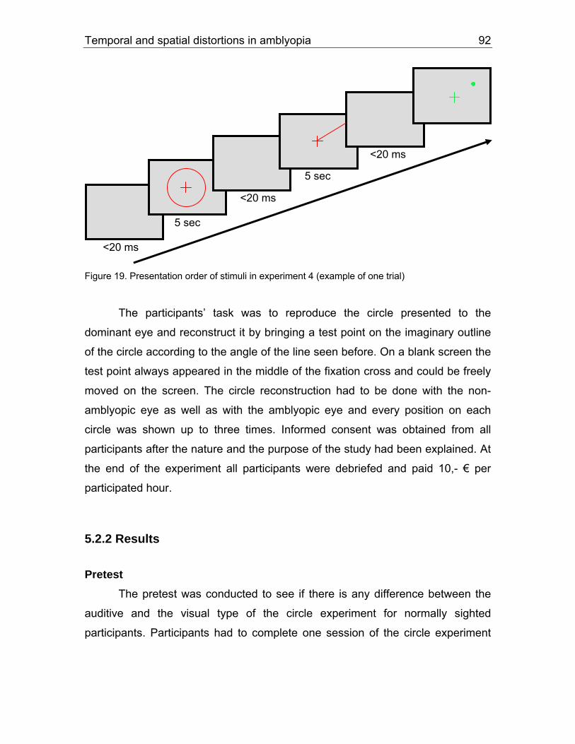
Temporal and spatial distortions in amblyopia 92
igure 19. Presentation order of stimuli in experiment 4 (example of one trial) F
The participants’ task was to reproduce the circle presented to the
dominant eye and reconstruct it by bringing a test point on the imaginary outline
of the circle according to the angle of the line seen before. On a blank screen the
test point always appeared in the middle of the fixation cross and could be freely
moved on the screen. The circle reconstruction had to be done with the non-
amblyopic eye as well as with the amblyopic eye and every position on each
circle was shown up to three times. Informed consent was obtained from all
participants after the nature and the purpose of the study had been explained. At
the end of the experiment all participants were debriefed and paid 10,- € per
participated hour.
5.2.2 Results
Pretest The pretest was conducted to see if there is any difference between the
auditive and the visual type of the circle experiment for normally sighted
participants. Participants had to complete one session of the circle experiment
5 sec
5 sec
<20 ms
<20 ms
<20 ms

Temporal and spatial distortions in amblyopia 93
with the tangential position auditively presented, as done in study 3, and one
session with the tangential position visually presented.
The results of the pretest revealed that for normally sighted participants (N
auditive and the visual type
(RS, LP, SB, JB, and MB) are displayed. The bases of the
rrows indicate the mean of the tested positions in the participant’s dominant
= 16), there is no significant difference between the
of the experiment (F (1, 13) = .46, p = .51, η² = .03). There were no additional
interactions between type of perception and other variables, e.g. eye (left or right
side) or circle radius (all Fs < 2.74, ps > .12, η²s > .14).
The results of the pretest showed that for normally sighted participants,
there was no difference in solving the task based on visual or auditive perception.
Participants had no difficulties in understanding and handling the two different
types of the experiment.
Main experiment Data treatment proceeded as described in study 3 for the amount of
spatial distortions (difference of the adjusted point to the target point) and the
amount of spatial uncertainty (standard deviation). Dependent variables were the
same as in study 3 - eye, radial position, and tangential position of the points -
and, additionally, the type of presenting the tangential position. Amount of spatial
distortion (difference of the adjusted point to the target point) and amount of
spatial uncertainty (standard deviation) were the independent variables. Figures
19 to 24 show the subjective adjustments of the tangential and radial positions,
both visually presented: individual patterns of spatial distortions and the relative
amount of spatial error for amblyopes with (DS, BB, MK, CL, and TS) and without
temporal instability
a
eye, while the tips of the arrows indicate the mean of the tested positions in the
amblyopic eye. Thus, the longer the arrow, the more pronounced the spatial
distortions in the amblyopic eye in relation to the non-amblyopic eye.

Temporal and spatial distortions in amblyopia 94
Amblyopes with stable distortions a) Refractive strabismic amblyopes: RS and SB are both refractive
strabismic amblyopes with a reduced visual acuity in both eyes. RS has a visual
acuity of 0.6 in the non-amblyopic eye and 0.1 in the amblyopic eye; SB has an
acuity of 0.6 in her non-amblyopic eye and 0.3 in her amblyopic eye. Participant
RS showed an overestimation of the inner circles especially for the amblyopic
eye. For the radial adjustments there was a tendency to set the vertical distances
smaller than the horizontal ones, picturing a more ovally shaped circle for the
amblyopic eye (figure 20).
Esotropic and refractive strabismic amblyope RS temporal nasal nasal temporal
Esotropic and refractive strabismic amblyope SB temporal nasal nasal temporal
Figure 20. Visual presentation of the circle positions: spatial distortions and relative amount of
spatial distortions indicated as vectorial subtraction of the distortion maps of participants RS and
SB. Left: settings in the non-amblyopic eye. Middle: settings in the amblyopic eye. Right: vectorial
subtraction of the distortion maps.

Temporal and spatial distortions in amblyopia 95
Amblyope SB underestimated the 2 and 4 deg circles and overestimated
the 6 and 8 deg circles. It seems that she nearly did not distinguish between the
four different radii of the circles. Tangential adjustments were set more precisely,
especially in the non-amblyopic eye. In both subjects, no nasal-temporal
asymmetry occurred in the adjustments of the circle positions.
ismic amblyopes: Primarily strabismic amblyope LP
(visual
b) Primarily strab
acuity of the amblyopic eye 0.25) showed a clear tendency in the
amblyopic eye to set the vertical distances smaller than the horizontal ones. This
pattern was visible especially in the amblyopic eye. The radial settings for the
non-amblyopic eye were all underestimated more than for the amblyopic eye,
tangential setting for the different positions were quite accurate for both eyes
(figure 21).
Exotropic strabismic amblyope LP temporal nasal nasal temporal
Figure 21. Visual presentation of the circle positions: spatial distortions and vectorial subtraction
of the distortion maps of participant LP. Left: non-amblyopic eye. Middle: amblyopic eye. Right:
vectorial subtraction.
c) Anisometropic amblyopes: Both anisometropic amblyopes, JB and
MB, had a visual acuity in the amblyopic eye of 0.5. Participant JB had a slight
tendency to underestimate the circle positions for the adjustments of the
amblyopic eye; however the difference between the amblyopic and the non-
amblyopic eye was quite low. Especially positions for the vertical and horizontal

Temporal and spatial distortions in amblyopia 96
degrees visual angle are set surprisingly correct for the amblyopic eye as well as
for the non-amblyopic eye (figure 22).
Anisometropic amblyope JB temporal nasal nasal temporal
Anisometropic amblyope MB temporal nasal nasal temporal
Figure 22. Visual presentation of the circle positions: spatial distortions and vectorial subtraction
f the distortion maps of anisometropic amblyopes JB and MB. Left: non-amblyopic eye. Middle:
amblyopic eye. Right: vectorial subtraction.
Vertical, horizontal, and all other tangential adjustments were quite correct
etrope MB as well. He showed a small tendency for setting the vertical
distanc
circle-shaped pattern for all different radii.
o
in anisom
es smaller than the horizontal distances, however this pattern was more
pronounced in the non-amblyopic eye. In both anisometropic amblyopes,
patterns of the positions adjustments were very regular, and nearly showed a

Temporal and spatial distortions in amblyopia 97
For the adjustments of radial positions, all amblyopes (except SB)
perceiving stable spatial distortions showed a tendency to set the vertical
ositions of the circles smaller than the horizontal ones (see also Lagrèze &
Siretea
mblyopes with temporal instability a) Accomodative strabismic amblyopes: Comparing amblyopes DS
and M
d a slight overestimation of the circle positions for the
mblyopic eye. The tangential circle positions were set quite precisely for the
s the non-amblyopic eye.
p
nu, 1991). The circles looked more or less ovally shaped, yet not in a very
pronounced way. Additionally, they set the tangential positions adjustments quite
precisely, especially for the vertical and the horizontal circle positions.
A
K, both showed a very different pattern of the circle adjustments. DS set
the position adjustments in a very consistent manner with quite regular circle-
shaped patterns for the non-amblyopic as well as for the amblyopic eye (figure
23). However, he showe
a
amblyopic as well a
The circle adjustments of participant MK imaged a distinctive
underestimation of all four circle radii for both eyes (see figure 23). This
underestimation was even more pronounced in the amblyopic eye. The circle
patterns in the amblyopic eye were also more distorted and irregularly shaped
than in the non-amblyopic eye, and tangential circle positions were adjusted even
more deviating compared to the non-amblyopic eye.
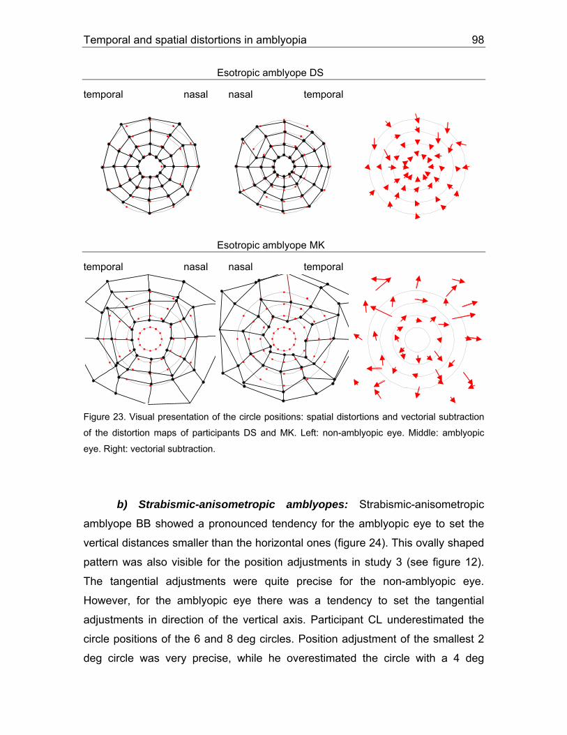
Temporal and spatial distortions in amblyopia 98
Esotropic amblyope DS temporal nasal nasal temporal
Esotropic amblyope MK
nasal nasal temporal temporal
Figure 23. Visual presentation of the circle positions: spatial distortions and vectorial subtraction
s DS and MK. Left: non-amblyopic eye. Middle: amblyopic
b) Strabismic-anisometropic amblyopes: Strabismic-anisometropic
ounced tendency for the amblyopic eye to set the
al ones (figure 24). This ovally shaped
attern was a so vis see figure 12).
genti l adju n quite amblyopic eye.
However, for the amblyopic eye there was a tendency to set the tangential
adjustments in direction of the vertical axis. Participant CL underestimated the
circle positions of the 6 and 8 deg circles. Position adjustment of the smallest 2
deg circle was very precise, while he overestimated the circle with a 4 deg
of the distortion maps of participant
eye. Right: vectorial subtraction.
amblyope BB showed a pron
vertical distances smaller than the horizont
p l ible for the position adjustments in study 3 (
he tan a stme ts were precise for the non-T

Temporal and spatial distortions in amblyopia 99
radius. Tangential position adjustments were very precise for the horizontal axis,
however got less precise for the other tangential adjustments. For the amblyopic
eye, a slight clockwise rotation of the circle positions was observed
Microstrabismic amblyope BB temporal nasal nasal temporal
Esotropic amblyope CL temporal nasal nasal temporal
sual presentation of the circle positions: spatial distortions and vectorial subtraction
f the distortion maps of amblyopes BB and CL. Left: non-amblyopic eye. Middle: amblyopic eye.
25). This ovally shaped pattern of the circles is more pronounced
Figure 24. Vi
o
Right: vectorial subtraction.
c) Anisometropic amblyopes: Amblyope TS is the only anisometropic
amblyope who experienced temporal instability. As most of the other participants
he showed a tendency to set the horizontal distances smaller than the vertical
ones (figure

Temporal and spatial distortions in amblyopia 100
than in the other anisometropic amblyopes JB and MB, who experienced no
inst ility. r ther in the “temporal instability”-
group, TS adjusted the tangential positions quite precisely in the non-amblyopic
eye, however in the amblyopic eye, the tangential adjustments deviated from the
target points. There was a tendency to set the tangential positions in direction to
the vertical axis.
Anisometropic amblyope TS
temporal ab Simila to the o 4 amblyopes
nasal temporal temporal nasal
btraction.
e circles for all of the four different radii.
Figure 25. Visual presentation of the circle positions: spatial distortions and vectorial subtraction
of the distortion maps of anisometropic amblyope TS. Left: non-amblyopic eye. Middle: amblyopic
eye. Right: vectorial su
Overall, in all amblyopes position adjustments for the non-amblyopic eye
were set more precisely than for the amblyopic eye. Four out of five amblyopes
perceiving temporal instability showed a distorted pattern in the adjustment of the
radial as well as the tangential circle positions. There was also a tendency to set
the horizontal distances smaller than the vertical distances. However, amblyope
DS showed a very regular pattern of th

Temporal and spatial distortions in amblyopia 101
Group data a) Spatial distortions and temporal instability: As shown in the
previous experiment, a difference in the amount of spatial distortion between the
amblyopic and the non-amblyopic eye was pronounced for the radial (F (1,8) =
14.08, p = .01, η² = .64) as well as for the tangential position adjustments (F (1,8)
= 12.26, p = .01, η² = .61). The amblyopic eye showed much higher deviations
from the target point than the non-amblyopic eye (Mamblyopic eye = .35 and Mnon-
positions, and Mamblyopic eye = 4.43 and Mnon-amblyopic eye =
). This significant difference in
me for both types of amblyopes, for amblyopes perceiving
opic eye and for amblyopes with a stable
(1,8) < 1.30, p > .25).
s no significant difference in the
F (1,8) = 2.05, p =
9, η² = .20). However, even if there was no significant difference, mean position
le pe less precise than those of
the group with stable spatial distortions (Minstable= .10 and Mstable = .05, see figure
26). For the tangential adjustment of the circle positions, difference in the amount
of spatial distortions between the groups of stable and instable amblyopic
perception approached nearly a significant difference (F (1,8) = 4.23, p = .07, η²
= .35). Mean position adjustments of amblyopes with instable perception were
less precise than those of amblyopes with stable spatial distortions (Minstable=
amblyopic eye = .27 for radial
3.09 for tangential positions, respectively
perception was the sa
temporal instability with the ambly
amblyopic perception (F
For the spatial distortions, there wa
adjustment of the radial positions between the two groups (
.1
adjustments of the group with instab rception were
2.12 and Mstable=.55).

Temporal and spatial distortions in amblyopia 102
a. b.
0
0,12
0,04
0,08
mea
n va
lue
(rad
ial d
e
Stableperception
Temporalinstability
Abso
lute
gree
s)
0
0,5
Stableperception
Temporalinstability
Abso
lute
mea
n v 1
1,5
2
alue
(deg
rees
vis
ual a
ngle
)
2,5
& b. Amount of spatial distortions for the position adjustment of the circles, for
mblyopes with constant distortions and for those with temporal instability. a. Radial adjustments.
t level (all Fs > .02, ps > .14). The
pe of presentation had no influence on the amount of spatial distortions for the
uncertainty was significantly different between the amblyopic and the non-
amblyopic eye for the tangential position adjustments (F (1,7) = 11.68, p = .01, η²
Figure 26 a
a
b. Tangential adjustments.
The main effect „presentation type“ did not show a significant influence on
the radial position adjustments (F (1,8) = .47, p = .51, η² = .06). In the auditive
presentation type, radial position adjustments were minimally less precise than in
the visual presentation type (Mauditive = .09, Mvisual = .06). As in the adjustments of
the radial positions, all other main factors and interactions for the tangential
position adjustments did not reach a significan
ty
tangential adjustments (F (1,8) = .02, p = .89, η² = .00), means were Mauditive =
1.37 for the auditive type of presentation and Mvisual = 1.31 for the visual type.
b) Spatial uncertainty and temporal instability: The amount of spatial

Temporal and spatial distortions in amblyopia 103
= .63). This was not true for the radial position adjustments – here a significant
level was not reached (F (1,7) = 2.36, p = .17, η² = .25). Still in both, the
mblyopic eye showed more pronounced deviations from the target point than
e non-amblyopic eye (Mamblyopic eye = .32 and Mnon-amblyopic eye = .29 for radial
ositions, and Mamblyopic eye = 4.07 and Mnon-amblyopic eye = 3.27 for tangential
ositions, respectively).
As expected for the radial position adjustment, there was no significant
ifference between the two groups in the amount of spatial uncertainty (F (1,7) =
2, p = .74, η² = .02). Nevertheless, mean radial position adjustments for the
roup with instable perception were minimally less precise than those of the
stable = .02, see figure 27).
for the tangential circle
adjustment, the difference in the amount of spatial uncertainty between the two
groups for the tangential adjustment approached nearly a significant level (F (1,7)
= 4.45, p = .07, η² = .38). Mean position adjustments of amblyopes with instable
perception were clearly less precise than those of amblyopes with stable spatial
distortions (Minstable= 1.28 and Mstable = .31).
a
th
p
p
d
.1
g
group with stable spatial distortions (Minstable= .03 and M
However as in the amount of spatial distortions

Temporal and spatial distortions in amblyopia 104
a. b.
0
0,01
0,02
0,03
0,04
Stableperception
Temporalinstability
Stan
dard
dev
iatio
n (ra
dial
deg
rees
)
0
0,5
1
1,5
Stableperception
Temporalinstability
Stan
dard
dev
iatio
n (d
egre
es v
isua
l ang
le)
Figure 27 a & b. Amount of spatial uncertainty for the position adjustment, for amblyopes with
The main effect „presentation type“ as well as the interaction between the
presentation type and the two groups did not reach a significant level for the
ns (Mauditive =
constant distortions and for those with temporal instability. a. Radial adjustments. b. Tangential
adjustments.
radial position adjustments (F (1,7) = .19, p = .68, η² = .38, and F (1,7) = .17, p =
.69, η² = .02, respectively). For the tangential position adjustments, the
interaction between presentation type and group was not significant (F (1,7) =
.57, p = .47, η² = .08) as well as the main effect “presentation type” (F (1,7) =
1.42, p = .27, η² = .17).
However even though a significant level was not reached, there was a
pronounced difference in spatial uncertainty especially for the tangential position
adjustments between amblyopes who perceive temporal instability and those
who do not. Amblyopes with a temporally instable perception showed a higher
amount of spatial uncertainty in adjusting the tangential circle positions (Mauditive =
1.35, Mvisual = 1.22) than amblyopes with constant spatial distortio

Temporal and spatial distortions in amblyopia 105
.60, Mvisual = .02). This was the same for visual or auditive presentation type of
the angle positions (figure 28).
0
0,2
0,4
0,6
0,8
1
1,2
1,4
1,6
Stable perception Temporal instability
Sta
ndar
d de
viat
ion
Figure 28. Amount of spatial u
adjustments, for amblyopes with co
line: visual presentation, dash
ncertainty regarding the presentation type for tangential
nstant distortions and for those with temporal instability. Solid
ed line: auditive presentation.
benefit from switching to
l presentat pe and show results more similar to amblyopes with
stable distortions. Yet, it was found that in the group of amblyopes without the
perception of temporal instability, spatial uncertainty for the adjustment of the
tangential circle positions is extremely reduced due to the visual presentation
type. This was not true for amblyopes perceiving temporal instability: they only
showed a marginal improvement of the tangential adjustments.
5.2.3 Discussion
Study 4 was designed to investigate if and how the performance of
amblyopes perceiving temporal instability with the amblyopic eye depends on the
modalities (visual and visuo-motoric versus auditive) used in a psychophysical
Amblyopes perceiving temporal instability should
the visua ion ty

Temporal and spatial distortions in amblyopia 106
task. This is addressed by the question: Is the change from a visual to auditive
modality for amblyopes with instable perception more difficult to handle than for
amblyopes with a stable amblyopic perception?
First, similar results as in study 3 were shown for the difference between
the amblyopic and the non-amblyopic eye concerning the amount of spatial
distortions and spatial uncertainty. Overall, the amblyopic eye showed much
higher distortions than the non-amblyopic eye. This result applied for amblyopes
perceiving temporal instability as well as for those without instable perception.
In general, there was no difference in spatial distortions and spatial
uncertainty between the two amblyopic groups for the adjustment of the radial
circle positions. For the tangential circle adjustments, a visually based
presentation was much easier to handle for all amblyopes than an auditively
circle position that was presented auditively rather than
isually. They had an increased amount of spatial distortions and spatial
ncertainty for this auditive modality dependent task. However there was also a
based presentation. Amblyopes perceiving temporal instability with the amblyopic
eye as well as amblyopes with stable perceptual distortions showed the same
difficulties in adjusting a
v
u
clear difference between amblyopes with an instable and a stable perception for
the adjustment of the tangential circle positions: amblyopes with an instable
perception were less precise in adjusting the tangential circle positions than
amblyopes with stable spatial distortions. They showed enhanced spatial
distortions and spatial uncertainty for the tangential positions. This was
irrespective of a visual or auditive modality dependent presentation. Here,
amblyopes with an instable perception showed a definitely increased spatial
uncertainty in adjusting the tangential circle position auditively as well as visually.
Nevertheless, the general pattern for larger distortions for auditive adjustments
was also visible.

Temporal and spatial distortions in amblyopia 107
Ho e like for temp instable pwev r, un orally erceiving amblyopes, for
amblyopes with stable spatial distortions, spatial uncertainty in the tangential
position adjustment was extremely reduced by the visual presentation. They
showed nearly no uncertainty in the tangential adjustment of the circle points
anymore. Overall, amblyopes perceiving temporal instability were worse in
accomplishing the tangential circle positions than amblyopes with a stable
amblyopic perception. Amblyopes with a stable amblyopic perception benefited
largely from visual presentation; however amblyopes perceiving temporal
for the visual presentation typ still
much more moderate extent.
in amblyopic perception had a
chophysical tasks. This special type of
added to the spatial amblyopic deficits may contribute
amblyopes with an instable amblyopic
xtent as for amblyopes with stable distortions. Maybe the problem lies in the
ature of the visual stimulus itself: amblyopes with temporal distortions have a
The reason why there was nearly no difference between the two
mblyopic groups for the radial adjustments might be that the radius was shown
twelve times in each trial. This high repetition rate seems to increase the security
the right position for amblyopes perceiving temporal instability: the
percen
instability also showed a small improvement
they seemed to benefit to a
Compendiously, temporal instability
negative impact on the performance in psy
temporal distortion that is
to the inferior results of the group of
perception. It seems that even a visually presented stimulus does not help for the
psychophysical task. The performance does get better, albeit not to the same
e
n
flickering, instable perception of the visual circle position. Due to that instable
vision they are not able to point precisely on the target point but show an
increased spatial uncertainty and deviation from that point. Perhaps the difficulty
for amblyopes with an instable perception lies in that “pointing”-part of the
experiment, not in the “presenting”-part.
a
in pointing at
t of errors was quite low and similar to that of the other amblyopes. It
might be that for amblyopes with an instable perception, it is important to simplify

Temporal and spatial distortions in amblyopia 108
the “pointing”-part of the experiment by a higher repetition rate of the presented
circle lines. This in turn might ease and thus improve the “pointing”-part of the
experiment for those participants.
Another reason for the large errors in the auditively presented tangential
position adjustments of amblyopes with a stable and instable spatial perception
might be deficits especially in the auditive-visual mapping. These deficits in
auditory-to-visual mapping suggest an impairment of the dorsal “where” pathway,
in addition to the known deficits of the ventral “what” pathway (Mishkin &
ngerleider, 1982). This possibility will be discussed in the following chapter in a
genera
U
l discussion of all four experiments.

Temporal and spatial distortions in amblyopia 109
6. General discussion
6.1 What it is all about
Amblyopia is a developmental disorder of the visual system that appears
very early in life and leads to losses in spatial vision. Amblyopia results from an
abnormal visual stimulation in early childhood. Generally, any condition which
provides no adequate visual stimulation is associated with an enhanced risk for
amblyopia. A number of different anomalies could cause amblyopia, but two
seem especially important and are investigated in this thesis. These are an early
misalignment of one eye (strabismic amblyopia), an unequal refraction in both
eyes (anisometropic amblyopia), or a combination of both. In strabismic
amblyopia motor fusion mechanisms are anomalous or inadequate for the
maintenance of ocular alignment, while anisometropic amblyopia is due to an
contrast sensitivity, spatial localisation and spatial
srupted in amblyopia. These deficits often occur along
with perceptual distor
anisometropia have quite different effects on spatial interactions (Ellemberg,
Hess
unequal refraction in the two eyes, probably caused by an unequal eye growth.
Performance in
localisation precision is di
tions in amblyopic vision. Perceptual distortions have been
well investigated in quantitative and qualitative approaches during the last fifty
years. Possibly due to their different aetiologies, anisometropic and strabismic
amblyopes show quite different results in psychophysical experiments. There is
evidence suggesting that spatial capacities of amblyopes are abnormal, with
strabismic and anisometropic amblyopes showing different abnormalities. In
general, anisometropic amblyopes show results more similar to normal than
strabismic amblyopes. Anisometropic amblyopes, but not strabismic amblyopes,
perceive a reduction in contrast typical of normal vision, and strabismus and
& Arsenault, 2002). In particular, Snellen acuity is affected to a greater
degree than grating acuity in strabismic but not in anisometropic amblyopes
(Levi, 1991). Considerable spatial distortions and imprecision of spatial

Temporal and spatial distortions in amblyopia 110
judgements characterise vision through the amblyopic eye of strabismic
amblyopes but not of anisometropic amblyopes (Bedell & Flom, 1983).
Anisometropic and strabismic amblyopes also differ with respect to two-
dimensional spatial distortions (Lagrèze & Sireteanu, 1991). Anisometropic
amblyopes, unlike strabismic amblyopes, do not show high amounts of positional
uncertainty and contour integration is exhibited normally in anisometropic
mblyopes (Hess & Demains, 1998). For anisometropic amblyopes, spatial
integra
The permanent spatial and temporal perceptual deficits may be a result of
e abnormal visual experience during visual development. The main reason for
e development of amblyopia seems to be an abnormal neuronal processing of
e visual information in the cortex. The neurophysiological substrate
orresponding to the visual deficits is still a matter of investigation. The neuronal
rocesses underlying amblyopia and especially the neural site of amblyopia are
ot known yet (Hess, 2001). There is some evidence from imaging studies that
e main site of the abnormal developmental changes is V1, in that the striate
ortex shows reduced activity in human strabismic amblyopes (Barnes, Hess,
umoulin, Achtman & Pike, 2001).
a
tion efficiency is quantitatively similar to that of normally sighted persons,
while for strabismic amblyopes, the spatial integration efficiency is markedly
decreased (Wang, Levi & Klein, 1998). In addition to the pronounced spatial
distortions, temporal distortions are reported frequently among strabismic
amblyopes (Sireteanu, 2000a). Amblyopic patients report their vision to be
temporally unstable and flickering on a short time scale (Barrett et al., 2003), and
more specifically the amblyopic percept of strabismics is perceived as if “seen
through hot air” (Sireteanu, 2000a, p. 71).Compared to the spatial distortions,
there are no studies reporting temporal instability in strabismic and anisometropic
amblyopic vision - not to mention studies investigating the temporal distortions in
detail.
th
th
th
c
p
n
th
c
D

Temporal and spatial distortions in amblyopia 111
However, recent support is coming from brain-imaging studies that the
deficit is also beyond V1 in the extra-striate cortex, and that the relative reduction
of responses to the amblyopic eye increases from striate to extrastriate areas
(e.g. Imamura, Richter, Fischer, Lennerstrand, Franzén, Rydberg, Andersson,
Schneider, Onoe, Watanabe & Långström, 1997). Sireteanu et al. (1998) found
neural deficits in strabismic and anisometropic amblyopia first appearing in V2
and then becoming more pronounced with increasing hierarchical cortical levels
(Muckli, Kiess, Tonhausen, Singer, Goebel & Sireteanu, in press). However, it is
still unclear whether dorsal and ventral processing streams are affected equally.
Several findings suggest that the ventral visual pathway is more affected in
strabismic amblyopia. The ventral “what” pathway extends from the primary
visual cortex to the inferior temporal cortex of the temporal lobe. It functions in
the identification of pattern and shape discrimination, resolution of fine detail and
sychophysical studies, Sireteanu (1991) and Schröder, Fries,
oelfsema, Singer and Engel (2002) found the ventral pathway more affected in
strabis
lyopic eye, and argued that this inability to count features
epends on the malfunction particularly of the dorsal, parietal cortex.
acuity, colour perception, and visual memory. For strabismic amblyopes Altmann
and Singer (1986) proposed a deficit of the P-type cells response. Corresponding
to these p
R
mic amblyopic cats than the dorsal pathway. On the other hand, there are
studies suggesting deficits in the dorsal pathway accompanying strabismic
amblyopia (for a review see Asper, Crewther & Crewther, 2000b). The dorsal
“where” stream is primarily involved with perception of motion, depth, control of
eye movements, localisation of targets in space, and attention shifts. Sharma,
Levi and Klein (2000) found strabismic amblyopes to underestimate the number
of features with the amb
d
Thus, psychophysical, imaging and animal data report impairment of
functions attributed to both ventral and dorsal pathways (Asper et al., 2000b).
Therefore the four studies in this thesis were undertaken to get clearer evidence
for either pathway, or more precisely, to investigate the perception of temporal
instability as well as its relationship to the spatial distortions which occur in

Temporal and spatial distortions in amblyopia 112
amblyopic vision. Both qualitative and quantitative approaches were
accomplished to get a broader view on these different types of perceptual
distortions.
6.2 Spatial distortions and spatial uncertainty in amblyopia
Four different types of distortions became apparent in the experiments.
These were a) blurring of patterns, b) wavy and jagged appearance of horizontal
and vertical lines, c) distortions due to superimposed lines or contours, and d)
errors in orientation. Similar spatial distortions have been found and were
classified (see also Barrett et al., 2003). One surprising result in this thesis was
that in study 2 the amblyopic participants perceived spatial distortions less
ered as likely. When one looks
t the nature of the spatial distortions, it can be said that all spatial distortions
that ca
wavy and jagged
appearance, superimposed lines, and errors in orientation. For the results of the
quantitative part one could also categorise that strabismic participants
circle positions while anisometropic participants slightly
verestimate the outermost circles. Anisometropic amblyopes are more similar to
the no
pronounced than in study 1. But the structure and complexity of the spatial
distortions remained the same throughout both experiments, and the four
categories of distortional errors can still be consid
a
me up in any of the experiments are characterised by quite an amount of
irregularity (see also Bedell & Flom, 1983; Lagrèze & Sireteanu, 1991). In
contrast to the anisometropic amblyopes, this was especially true for the
strabismic amblyopes.
On the one hand, it was possible for the results of the qualitative part to
build categories for the distortions to fit in: blurring of patterns,
underestimate the
o
rmally sighted participants. Additionally, anisometropic and strabismic-
anisometropic amblyopes show patterns that are more ovally shaped, while
those of the purely strabismic amblyopes are - as pointed above - distorted in an

Temporal and spatial distortions in amblyopia 113
irregular way or rotated in a clockwise direction. But on the other hand, in both
qualitative and quantitative approaches results of the spatial distortions were also
very individual (Sireteanu et al., 1993). Each amblyopic subject has his/her
dividual pattern of distortions. These findings are in accordance with previous
results
he
patial distortions and uncertainty were increased or on a more normal level,
respec
) were most pronounced in strabismic amblyopes for all kinds
f experimental approaches. Strabismic and strabismic-anisometropic amblyopes
showed considerably higher spatial distortions and uncertainty than do
in
of Lagrèze and Sireteanu (1991) as well as others (e.g. Barrett et al.,
2003).
There was also an effect of the modality concerning the spatial distortions
and the spatial uncertainty. Depending on a visual or auditive modality, t
s
tively. Presenting the stimulus auditively, an increased amount of
distortions and spatial uncertainty was recorded. This was not the case for a
visual stimulus presentation. This suggests that amblyopes might have difficulties
with this auditive modality dependent task, and, more specifically, might have
problems with auditive-to-visual mapping.
For the spatial uncertainty, strabismic and strabismic-anisometropic
amblyopes show considerable uncertainty, in contrast to the anisometropic
amblyopes and normally sighted participants. This finding was achieved in each
of the four experiments done in this thesis. Recently, Simmers and Bex (2004)
confirmed the widely believed assumption that spatial uncertainty is most
pronounced in amblyopes. They suggested that the spatial deficit accompanying
amblyopia underlies this increased spatial uncertainty. Fronius, Sireteanu and
Zubcov (2004) found this even in the amblyopic perception of children with
strabismic amblyopia. In their study, increased spatial uncertainty outside the
normal range occurred which is similar to that in adult amblyopes.
Overall, spatial localisation deficits (comprising both spatial distortions and
spatial uncertainty
o

Temporal and spatial distortions in amblyopia 114
anisom
f single lines or parts in
e pattern. They occurred only at high spatial frequency patterns, at low spatial
freque
the amblyopic participants experienced
temporal instability. But there is clearly a preponderance of strabismic amblyopes
percei
etropic amblyopes. They were especially pronounced in the central visual
fields (see also Fronius & Sireteanu, 1994). This is in accordance with other
results from the literature (Lagrèze & Sireteanu, 1991; Sireteanu et al., 1993;
Demanins & Hess 1996).
6.3 Temporal instability in the amblyopic percept
For the perception of temporal distortions, two types of instability have
been found: a) jitter of the whole pattern, and b) moving o
th
ncy patterns simply stable spatial distortions were perceived with the
amblyopic eye. That is similar to the findings of Bradley and Freeman (1985).
They concluded that when the experimental stimulus is either large enough for
the amblyopic participant to perceive or consists of low spatial frequencies, no
temporal deficits emerge in the amblyopic perception. These temporal distortions
emerged in addition to the spatial distortions, and moreover, the data in this
thesis suggest that the perception of temporal instability seems to have quite an
influence on the perception of spatial distortions. Especially the central visual
field of the amblyopic eye is most affected by the distorted spatial perception,
and additionally amblyopes perceiving temporal instability show an even
increased amount of spatial distortions in the central visual field.
It can be said that almost half of
ving temporal distortions. It seems that these instability phenomena occur
mainly in strabismic and strabismic-anisometropic amblyopes. In addition, other
than one would expect, a deep visual acuity loss seems not to be a good
predictor of temporal distortions. Temporal distortions rather occur more often in
moderate amblyopes, while spatial distortions are associated with moderate-to-
deep acuity loss. This fits also to the findings reported in study 1 that temporal

Temporal and spatial distortions in amblyopia 115
instability is associated with a mild loss in contrast sensitivity. Thus, moderate
acuity loss and mild loss of contrast sensitivity seem to point to the perception of
temporal instability. A possible explanation for this finding might be that due to
e lower suppression of the amblyopic perception, an instable perception is
coming
might result in an instable percept of
e amblyopic eye that has to deal with other occurring rivalling visual inputs
oming from the “better” non-amblyopic eye. The findings of Ellemberg et al.
000) also fit in this interpretation. In their studies they found that deep visual
eprivation has much smaller effects on neuronal mechanisms that mediate
mporal vision than on those that mediate spatial vision.
One quite different but nevertheless very interesting and important finding
this thesis was that the eye dominance might be a crucial factor for the
evelopment of a temporally instable perception. It might be the interaction
etween opposite sides of hand and eye dominance together that leads to one of
e two types of temporal distortions. Crossed dominance was also found to be
lated to other neuronal disorders that begin quite early in life like dyslexia
tein, Richardson & Fowler, 2000).
An additional visual disturbance that came up in some of the temporal
istortions is the perception of colour illusions. Some participants perceiving
mporal instability also reported the perception of coloured lines or contours.
hese illusions were appearing in all kinds of colours in the rainbow spectrum or
just one or two colours like red, green or yellow. As Eagleman (2000) stated
llusions, often, are those stimuli that exist at the extremes of what our system
th
up. Kilwinger, Spekreijse and Simonsz (2002) found that for the
occurrence of strabismic suppression, the similarity of the images of both eyes
has to be detected by the visual system. They suggested that “two mechanisms
must be at work, a ‘similarity detector’ and a ‘suppressor’” (p.2011), and that the
location of these two mechanisms might be in the lateral occipital cortex. If the
loss in visual acuity in not that extreme and therefore suppression lower, rivalry
between the two eyes might come up. This
th
c
(2
d
te
in
d
b
th
re
(S
d
te
T
in
“i

Temporal and spatial distortions in amblyopia 116
has evolved to handle. Sometimes illusions stem from assumptions made by the
isual system; at other times they represent an active recalibration” (p. 920). But
colour illusions reported by the amblyopes in the
tudies of this thesis? It is true that amblyopes and especially strabismic
amblyo
v
what does this mean for the
s
pes perceive an increased amount of spatial uncertainty. This uncertainty
was even more increased in amblyopes perceiving temporal instability. It might
be that due to this highly increased uncertainty in the perception of the amblyopic
eye additional visual phenomena like those of the visual illusions come up,
spontaneously activated by cortical cells sensitive to colours, even in absence of
any adequate stimuli (Sireteanu, Bäumer & Sârbu, 2005). Probably in the
amblyopic perception a neuronal pathway is affected and spuriously activated
that is sensitive to temporal properties and colour processing. Due to its functions
and important role in vision, especially in the perception of motion, this might be
the magnocellular-processing stream including area MT, but also parts of the
ventral pathway, involving V4.
6.4 Conclusions
There are several models that attempt to explain the visual deficits found
in the perception of strabismic and anisometropic amblyopes. The two most
popular ones for explaining the losses of amblyopic spatial vision are the
topographical disarray model (Hess et al., 1978) and the undersampling model
(Levi & Klein, 1986; Sharma, Levi & Coletta, 1999). It is possible that strabismic
deficits reflect a topographical disorder or jitter of the cortical receptive fields,
which appear to be uncalibrated (Levi & Carkeet, 1993; Hess & Field, 1994). The
failure of the calibration process might come up in early visual development as a
result of the eye misalignment. This means that there is an early developing
scrambling of the retinotopic map, which in turn results in an enhanced positional
uncertainty (Hess, Field & Watt, 1990). Alternatively, the undersampling model
hypothesises that amblyopic vision is due to a reduced cortical spatial sampling

Temporal and spatial distortions in amblyopia 117
density (Levi, Klein & Sharma, 1999). Evidence for this spatial undersampling
comes mainly from experiments showing that amblyopes are not efficient in using
all visual information that defines an image (Levi et al., 1999; Wang et al. 1998).
Sharma et al. (1999) showed that there is a sparse sampling by cortical neurons
in the foveal representation of the amblyopic eye. The authors suggest that this
undersampling may be the reason for the spatial distortions in the perception of
strabismic amblyopes.
However, there are controversies regarding these hypotheses, and neither
of the two models can account for all of the results found in the qualitative and/or
quantitative parts of the experiments in this thesis. It might be possible to explain
some data in terms of the topographical disarray or positional uncertainty,
respectively, or in terms of the spatial undersampling model, but it is not clear
how they deal with most of the higher-order deficits found to appear in the spatial
vision of strabismic and anisometropic amblyopes. That is especially the deficits
in auditory-to-visual mapping and the perception of jitter and coloured illusions in
two-dimensional black-and-white patterns. There is also the unexplained
observation of crossed hand and eye dominance on the occurrence of temporal
istortions, and the finding of a low loss in acuity as well as in contrast sensitivity
in rela
n the psychophysical task that was based on
an auditive rather than on a visual modality. In comparison to amblyopes
perceiving only spatial distortions, they showed an increased amount of spatial
d
tion to the perception of temporal instability.
What do these different findings tell us about the possible cortical site of
the spatial and temporal deficits in strabismic and anisometropic amblyopic
vision? In general, it can be said that the different results of the four experiments
and reported deficits in the amblyopic vision suggest an impairment of the dorsal
“where” pathway, which comes up in addition to the known deficits of the ventral
“what” pathway (Mishkin & Ungerleider, 1982). This is especially true for
amblyopes perceiving temporal instability. First, amblyopes with the perception of
temporal instability had difficulties i

Temporal and spatial distortions in amblyopia 118
distort
te cortex in
igher extra-striate areas including the parietal cortex. It seems that both, the
ions and spatial uncertainty for the auditive modality dependent task. This
was even more pronounced for tangential positions adjustments. It seems that
amblyopes with temporal instabilities might have deficits especially in the
auditory-to-visual mapping. This suggests that cortical cells on the dorsal
pathway might be affected and that higher-order, multisensory association areas
in the posterior parietal cortex might be involved (Sireteanu, Bäumer & Sârbu,
2005).
Second, colour illusions in combination with temporal instability also
suggest an impairment of the magnocellular-processing stream, probably caused
by increased uncertainty in the visual process of the amblyopic perception. It
might be that, due to this increased uncertainty, visual hallucinations of colour-
like contours are coming up. They might be spontaneously activated by cortical
cells in the dorsal pathway which are sensitive to colours, and occur even in
absence of any adequate stimuli. So, there seems to be an impairment in the
dorsal pathway where cortical cells might be spuriously activated, being sensitive
to temporal properties and colour processing together. However, these two
different deficits might also mean that more than one pathway is affected and
that both, the dorsal as well as the ventral pathway contribute to the spurious
perception of colour-like, moving contours.
To conclude, it seems that perception of temporal instability can occur in
addition to the spatial distortions in amblyopia. Regarding the visual deficits
found in strabismic and anisometropic amblyopes, the spatial and temporal
deficits might be located not only in V1 but also beyond the stria
h
ventral and the dorsal pathway contribute to the different spatial and temporal
deficits in amblyopic perception, depending on the type and amount of spatial or
temporal errors the amblyope perceives.

Temporal and spatial distortions in amblyopia 119
6.5 What comes next?
The results of these psychophysical experiments, quantitative as well as
ualitative ones, suggest that one should find an impairment of the ventral
athway for amblyopes perceiving stable spatial distortions and spatial
ility in
ddition to spatial distortions, there should be an additional impairment of the
dorsal
q
p
uncertainty. Also, in amblyopic participants perceiving temporal instab
a
pathway. Regarding the temporal instabilities, this should include deficits
in activation for the areas MT and MST for the amblyopic eye. The best way to
investigate these questions would be a combination of psychophysical,
computational and functional imaging techniques. This is exactly what the group
of Sireteanu is doing right now: ongoing studies in their laboratory suggest that
“mapping of auditory onto visual signals is likely to be disturbed in amblyopic
subjects experiencing anomalous perception, thus suggesting an involvement of
higher-order, multisensory areas in the posterior parietal cortex” (Sireteanu,
Bäumer & Sârbu, 2005).

Temporal and spatial distortions in amblyopia 120
References
Abdolvahab-Emminger, H. & Sireteanu, R. (1993). Residual visual properties in
human amblyopia after monocular deprivation in early life. Clinical Vision
Sciences, 8, 263-279.
Altmann, L. & Singer, W. (1986). Temporal integration in amblyopic vision. Vision
tkinson, R.L., Atkinson, R.C., Smith, E., Bem, D. & Nolen-Hoeksema, S. (1996).
Research, 26, 1959-1968.
Asper, L., Crewther, D. & Crewther, S. (2000a). Strabismic amblyopia. Part 1:
Psychophysics. Clinical and Experimental Optometry, 83, 49-58.
Asper, L., Crewther, D. & Crewther, S. (2000b). Strabismic amblyopia. Part 2:
Neural Processing. Clinical and Experimental Optometry, 83, 200-211.
A
Hilgard’s Introduction to Psychology. Fort Worth: Harcourt Brace College
Publishers.
Barnes, G., Hess, R., Dumoulin, S., Achtman, R. & Pike, G. (2001). The cortical
deficit in humans with strabismic amblyopia. Journal of Physiology, 533,
281-297.
Barrett, B., Pacey, I., Bradley, A., Thibos, L. & Morrill, P. (2003). Nonveridical
visual perception in human amblyopia. Investigative Ophthalmology &
Visual Science, 44, 1555-1567.
Bedell, H. & Flom, M. (1981). Monocular spatial distortion in strabismic
amblyopia. Investigative Ophthalmology & Visual Science, 20, 263-268.
Temporal and spatial distortions in amblyopia 121
Bedell, H. & Flom, M. (1983). Normal and abnormal space perception. American
Journal of Optometry & Physiological Optics, 60, 426-435.
Birbaumer, N. & Schmidt, R. (1999). Biologische Psychologie. Berlin: Springer
Verlag.
in amblyopia: an
explanation of conflicting reports. Vision Research, 25, 39-46.
, 920-926.
Ellemb
Bradley, A. & Freemann, R. (1985). Temporal sensitivity
Carlson, N. (2001). Physiology of Behavior. Boston: Allyn and Bacon.
Daw, N. (1998). Critical periods and amblyopia. Archives of Ophthalmology, 116,
502-505.
Demanins, R. & Hess, R. (1996). Effect of exposure duration on spatial
uncertainty in normal and amblyopic eyes. Vision Research, 36, 1189-
1193.
Dodt, E. (1975). Lichtsinn. In W. Keidel, W. (Ed.), Kurzgefasstes Lehrbuch der
Physiologie (pp. 17-2 – 17-3). Stuttgart: Georg Thieme Verlag.
Eagleman, D. (2001). Visual illusions and neurobiology. Nature, 2
erg, D., Hess, R. & Arsenault, A. (2002). Lateral interactions in amblyopia.
Vision Research, 42, 2471-2478.
Ellemberg, D., Lewis, T., Maurer, D. & Brent, H. (2000). Influence of monocular
deprivation during infancy on the later development of spatial and
temporal vision. Vision Research, 40, 3283-3295.

Temporal and spatial distortions in amblyopia 122
Fronius, M. & Sireteanu, R. (1989). Monocular geometry is selectively distorted in
the central visual field of strabismic amblyopes. Investigative
Ophthalmology & Visual Science, 30, 2034-2044.
Fronius, M. & Sireteanu, R. (1994). Pointing errors in strabismics: complex
patterns of distorted visuomotor coordination. Vision Research, 34, 689-
707.
Fronius, M., Sireteanu, R. & Zubcov, A. (2004). Deficits of spatial localization in
children with strabismic amblyopia. Graefe’s Archiv of Clinical and
Experimental Ophthalmology, 242, 827-839.
Held, R. (1988). Sensory Systems I. Vision and Visual Systems. Boston:
Birkhäuser.
Hess, R. (2001). Amblyopia: site unseen. Clinical and Experimental Optometry,
84, 321-336.
Hess, R. & Demanins, R. (1998). Contour integration in anisometropic
amblyopia. Vision Research, 38, 889-894.
Hess, R. & Field, D. (1994). Is the spatial deficit in strabismic amblyopia due to
loss of cells or and uncalibrated disarray of cells? Vision Research, 34,
3397-3406.
Hess, R. & Howell, E. (1977). The threshold contrast sensitivity function in
strabismic amblyopia: evidence for a two type classification. Vision
Research, 17, 1049-1055.

Temporal and spatial distortions in amblyopia 123
Hess, R. & Pointer, J. (1985). Differences in the neural basis of human
amblyopia: the distribution of the anomaly across the visual field. Vision
Research, 25, 1577-1594.
Hess, R., Bradley, A. & Piotrowski, L. (1983). Contrast coding in amblyopia – I:
Differences in the neural basis of human amblyopia. Proceedings of the
Royal Society London, B 217, 309-330.
Hess, R., Campell, F. & Greenhalgh, T. (1978). On the nature of the neural
abnormality in human amblyopia: neural aberrations and neural sensitivity
loss. Pflügers Archiv, 377, 201-207.
Hess, R., Field, D. & Watt, R. (1990). The puzzle of amblyopia. In C. Blakemore
(Ed.), Vision Coding and Efficiency (pp.267-280). Cambridge: Cambridge
University Press.
Hess, R., Pointer, J., Simmers, A. & Bex, P. (2003). Border distinctness in
ion Research, 43, 2255-2264. amblyopia. Vis
Howard, I. (2002). Seeing in Depth, Vol. I - Basic Mechanisms. Toronto:
Porteons.
Hubel, D. & Wiesel, T. (1977). Functional architecture of macaque visual cortex.
Proceedings of the Royal Society London, B 198, 1-59.
Imamura, K., Richter, H., Fischer, H., Lennerstrand, G., Franzén, O., Rydberg,
A., Andersson, J., Schneider, H., Onoe, H., Watanabe, Y. & Långström
(1997). Reduced activity in the extrastriate visual cortex of individuals with
strabismic amblyopia. Neuroscience Letters, 225, 173-176.

Temporal and spatial distortions in amblyopia 124
Kandel, E., Schwartz, J. & Jessell, T. (2000). Principles of Neural Science. New
York: McGraw-Hill.
Kilwinger, S., Spekreijse, H. & Simosz, H. (2002). Strabismic suppression
depends on the amount of dissimilarity between left- and right-eye images.
Vision Research, 42, 2005-2011.
Lagrèze, W.-D. & Sireteanu, R. (1991). Two-dimensional spatial distortions in
human strabismic amblyopia. Vision Research, 31, 1271-1288.
Levi, D. & Klein, S. (1986). Sampling in spatial vision. Nature, 320, 360-362.
Levi, D. (1991). Spatial vision in amblyopia. In D. Regan (Ed.), Vision and Visual
Dsyfunction, Vol. 10: Spatial Vision (pp. 212-238). Houndmills and
London: MacMillan Press.
Levi, D. & Carkeet, A. (1993). Amblyopia: a consequence of abnormal visual
development. In K. Simons (Ed.), Early visual development, normal and
abnormal (pp. 391-408). New York: Oxford University Press.
Levi, D. & Klein, S. (1983). Spatial localization in normal and amblyopic vision.
Vision Research, 23, 1005-1017.
Levi, D., Klein, S. & Sharma, V. (1999). Position jitter and undersampling in
pattern perception. Vision Research, 39, 445-465.
Mishkin, M. & Ungerleider, L. (1982). Contribution of striate inputs of the
visuospatial functions of parieto-preoccipital cortex in monkeys. Behavioral
Brain Research, 6, 57-77.

Temporal and spatial distortions in amblyopia 125
Muckli, L., Kiess, S., Tonhausen, N., Singer, W., Goebel, R. & Sireteanu, R. (in
press). Cerebral correlates of impaired gratings perception in individual,
psychophysically assessed human amblyopes. Vision Research.
Pinel, J. (2003). Biopsychology. Boston: Ally and Bacon.
Pugh, M. (1958). Visual distortion in amblyopia. British Journal of Ophthalmology,
42, 449-460.
immers, A. & Bex, P. (2004). The representation of global spatial structure in
Schröder, J.-H., Fries, P., Roelfsema, P., Singer, W. & Engel, A. (2002). Ocular
dominance in extrastriate cortex of strabismic amblyopic cats. Vision
Research, 42, 29-39.
Sharma, V., Levi, D. & Coletta, N. (1999). Sparse sampling of gratings in the
visual cortex of strabismic amblyopes. Vision Research, 39, 3526-3536.
Sharma, V., Levi, D. & Klein, S. (2000). Undercounting features and missing
features: evidence for a high-level deficit in strabismic amblyopia. Nature
Neuroscience, 3, 496-501.
S
amblyopia. Vision Research, 44, 523-533.
Simmers, A., Ledgeway, T., Hess, R. & McGraw, P. (2003). Deficits to global
motion processing in human amblyopia. Vision Research, 43, 729-738.
Singer, W. (1984). Neurophysiologische Grundlagen der Amblyopie. In V. Herzau
(Ed.), Pathophysiologie des Sehens (pp.162-165). Stuttgart: Ferdinand
Enke Verlag.

Temporal and spatial distortions in amblyopia 126
Sireteanu, R. (1982a). Human amblyopia: consequence of chronic interocular
suppression. Human Neurobiology, 1, 31-33.
ireteanu, R. & Best, J. (1992). Squint-induced modification of visual receptive
fields in the lateral suprasylvian cortex of the cat: binocular interaction,
vertical effect and anomalous correspondence. European Journal of
Neuroscience, 4, 235-242.
ireteanu, R. & Fronius, M. (1981). Naso-temporal asymmetries in amblyopic
vision: consequence of a long-term interocular suppression. Vision
Research, 21, 1055-1063.
Sireteanu, R. & Fronius, M. (1989). Different patterns of retinal correspondence
in the central and peripheral visual field of strabismics. Investigative
Ophthalmology & Visual Science, 30, 2023-2033.
Sireteanu, R. (1982b). Binocular vision in strabismic humans with alternating
fixation. Vision Research, 22, 889-896.
Sireteanu, R. (1991). Restricted visual fields in both eyes of kittens raised with a
unilateral, surgically induced strabismus: relationship to extrastriate
cortical binocularity. Clinical Vision Sciences, 6, 277-287.
Sireteanu, R. (2000a). The binocular act in strabismus. In G. Lennerstrand & J.
Ygge (Eds.), Advances in strabismus research: basic and clinical aspects
(pp. 63-83). London: Portland Press.
Sireteanu, R. (2000b). The binocular visual system in amblyopia. Strabismus, 8,
39-51.
S
S

Temporal and spatial distortions in amblyopia 127
Sireteanu, R., Bäumer, C. & Sârbu, C. (2005). Temporal instability of amblyopic
vision: evidence for an involvement of the dorsal visual pathway. Journal
Singer, W. (1981). Binocular interaction in the
peripheral visual field of humans with strabismic and anisometropic
ireteanu, R., Lagrèze, W.-D. & Constantinescu, D. (1993). Distortions in two-
bei
Schielamblyopen: Psychophysik und bildgebende Verfahren. Klinische
, N., Muckli, L., Lanfermann, H., Zanella, F., Singer, W.
& Goebel, R. (1998). Cortical site of the amblyopic deficit in strabismic and
tein, J., Richardson, A. & Fowler, M. (2000). Monocular occlusion can improve
nd the origins of infantile strabismus. In K.
Simons (Ed.), Early visual development, normal and abnormal (pp. 364-
ang, H., Levi, D. & Klein, S. (1998). Spatial uncertainty and sampling efficiency
in amblyopic position acuity. Vision Research, 38, 1239-1251.
of Vision, VSS abstracts 294.
Sireteanu, R., Fronius, M. &
amblyopia. Vision Research, 21, 1065-1074.
S
dimensional visual space perception in strabismic observers. Vision
Research, 33, 677-690.
Sireteanu, R., Bäumer, C., Sârbu, C., Tsujimura, S. & Muckli, L. (in preparation).
Verzerrte Raumwahrnehmung und zeitliche Instabilität
Monatsblätter.
Sireteanu, R., Tonhausen
anisometropic subjects, investigated with fMRI. Investigative
Ophthalmology & Visual Science, 39 (Suppl.), 909.
S
binocular control and reading in dyslexics. Brain, 123, 164-170.
Tychsen, L. (1993). Motion sensitivity a
390). New York: Oxford University Press.
W

Temporal and spatial distortions in amblyopia 128
Weiß, C., Rentschler, I. & Caelli, T. (1985). Amblyopic processing of positional
information. Part II: Sensitivity to phase distortion. Experimental Brain
Research, 60, 279-288.

Temporal and spatial distortions in amblyopia 129
Lebenslauf
Persönliche Daten:
Adresse: Claudia Christina Bäumer Kaiserstr. 36 69115 Heidelberg
Tel. 06221 / 4332061
E-Mail: [email protected]
Geburtsdatum: 11.06.1977 (München) Familienstand: ledig, eine Tochter Staatsangehörigkeit: deutsch
Schulausbildung:
1983 - 1987 Besuch der Feldmochinger-Grundschule in München
Abschluss des Abiturs mit Gesamtnote "gut" (2,4)
1987 - 1996 Besuch des Maximiliansgymnasiums in München
Hochschulausbildung:
Okt.´96 - Sept.´98 Psychologie-Grundstudium an der TU Darmstadt Abschluss des Vordiploms mit Gesamtnote „gut“
Schwerpunktfächer
Okt.´98 - Apr.´02 Psychologie-Hauptstudium an der Uni
Heidelberg Abschluss des Diploms mit Gesamtnote "sehr gut"
: Klinische & Pädagogische Psy. Nebenfach: Psychopathologie Diplomarbeit: "Überprüfung zweier kognitiver Zeit-
am Max-Planck-Institut für
Hirnforschung, Frankfurt Doktorarbeit
schätzungstheorien anhand einer klinischen Stichprobe"
seit Juli ´02 Promotionsstelle
: „Temporal and spatial distortions in adult amblyopia“ (Betreuerin: Prof. Sireteanu)

Temporal and spatial distortions in amblyopia 130
Berufliche Tätigkeiten:
seit Juli ´02 Wissenschaftliche Mitarbeiterin an der Universität Frankfurt, Psychologisches Institut (Fachbereich Physiologische Psychologie) im DFG-Projekt "Wahrnehmungsverzerrung bei
itarbeiterin bei der Psycholog.
Beratungsstelle der Psychosozialen Hilfe e.V. Tätigkeit: Durchführung von Erstgesprächen,
Wiss
Schielamblyopie: Psychophysik und bildgebende Verfahren"
seit Feb. ´04 Selbständige M
weiterführende Beratung
enschaftl. Tätigkeiten:
Mai `97 - Juli `98 Beschäftigung als studentische Hilfskraft im
Beschäftigung als stud. Hilfskraft im Bereich "Sozialpsychologie" bei Prof. T. Betsch (DFG-Projekt 504)
raktika:
Bereich "Pädagogische Psychologie" bei Prof. B. Schmitz
Okt.`99 - Juni `01
Tätigkeit: Durchführung einer experimentellen Studie zum Thema Entscheidungsfindung
P
Praktikum bei der Daimler Benz AG im Bereich "Unfallforschung"
Tätigkeit: Durchführung und Auswertung von
nck-Institut für psychologische Forschung im Bereich "Kognition und Handlung"
ische
Tätigkeit: Diagnostik, therapeutische Betreuung von Patienten und deren Angehörigen in der neurologischen Frührehabilitation
Aug. - Sept.´97
Interviews mit verunfallten Autofahrern
Feb. - März ´99 Praktikum am Max-Pla
Tätigkeit: Konzeption und Durchführung einer experimentellen Studie zum „Tapping“
Aug. - Sept.´99 Praktikum am Wilhelm-Griesinger-Krankenhaus Berlin im Fachbereich "KlinNeuropsychologie"
Temporal and spatial distortions in amblyopia 131
Deutsche Zusammenfassung der Dissertation „Temporal and spatial distortions in adult amblyopia“
von Claudia Bäumer
I. Einleitung
Amblyopie ist definiert al
s die Schwachsichtigkeit eines oder beider
ugen, welche ohne organische Fehler im Auge auftritt. Die Entstehung einer
oder S
Verlus nsitivität des
icht-dominanten Auges, und speziell das Auftreten räumlicher Verzerrungen
von Sc Eine zusätzliche zeitliche Instabilität des
eheindrucks wurde zwar vereinzelt berichtet (Barrett, Pacey, Bradley, Thibos &
icht
efergehend untersucht. Ziel der Dissertation ist es, die amblyope Wahrnehmung
Beitrag und zeitlichen
ahrnehmungsverzerrungen bei der Benutzung des amblyopen Auges zu
d Visualisierung von zeitlicher Instabilität im
eheindruck amblyoper Personen wurde bis jetzt noch nicht umgesetzt: diese
beschrieben ihren Seheindruck des amblyopen Auges als zeitlich instabil, das
heißt, die Bilder flimmern, als würden sie durch heiße Luft betrachtet (Sireteanu,
A
Amblyopie kann unter anderem durch Anisometropie (anisometrope Amblyopie)
trabismus (Schielamblyopie) in der frühen Kindheit hervorgerufen werden.
t der Binokularität, verminderte Sehschärfe und Kontrastse
n
sind kennzeichnend für die Wahrnehmung von Amblyopen und im besonderen
hielamblyopen (Sireteanu, 2000).
S
Morill, 2003; Hess, Campbell & Greenhalgh, 1978, Sireteanu, 2000), jedoch n
ti
mit Fokus auf die zeitliche Instabilität näher zu untersuchen, und somit einen
zum Verständnis der räumlichen
W
erbringen.
Die genaue Erfassung un
S

Temporal and spatial distortions in amblyopia 132
2000). Zudem konnte bis zum jetzigen Zeitpunkt noch kein eindeutiges
ales Korrelat des amblyopen Defizits identifiziert werden. Die bisherigen
isse legen allerdings nahe, dass dieses Defizit nicht auf der Eb
neuron
Ergebn ene der
rimären Sehrinde, sondern auf höheren, extrastriären Arealen lokalisiert ist
nu, in press). In der
issertation wurde versucht, diese Fragen über zwei unterschiedliche
durchgeführt, wobei jeweils zwei die amblyope Wahrnehmung quantitativ bzw.
ualitativ erfasst haben.
II. Teil 1: Qualitative Methodik
p
(Muckli, Kiess, Tonhausen, Singer, Goebel & Siretea
D
methodischer Arten anzugehen: es wurden vier psychophysische Experimente
q
d
rthoptische Untersuchung amblyoper Probanden ging den psychophysischen
von d
Schielamblyope, 4 Schielamblyope mit zusätzlicher Anisometropie, 4
nisometrope Amblyope und 3 Schieler mit alternierender Fixation ohne
norma ucht. Zudem wurde das
ontrastsehen unter zu Hilfenahme des „Vision Contrast Test Systems“ für Ferne
überpr
Exper Personen wurden im ersten Experiment Schwarz-Weiß-
uster verschiedener räumlicher Frequenzen gezeigt. Die Probanden wurden
Orthoptische Voruntersuchungen Die Aufstellung einer empirischen Datenbasis durch Rekrutierung un
o
Experimenten voraus. Insgesamt wurden 41 Personen orthoptisch untersucht,
iesen kamen 18 für die Experimente in Frage. Es konnten somit 7
a
Amblyopie rekrutiert werden. Zu Kontrollzwecken wurden zudem 30
lsichtige Personen orthoptisch unters
K
und Nähe sowohl monokular für das rechte und linke Auge als auch binokular
üft.
iment 1 14 amblyopen
M

Temporal and spatial distortions in amblyopia 133
gebeten, ihre individuelle Wahrnehmung der Muster mit dem amblyopen Auge
ilen und aufzuzeichnen. mitzute
Wahrn n: gerade Linien erscheinen als
ellenförmig oder ausgefranst, überlagernde Konturen verzerren das
sind u
Consta
Anisom
den rä ie zeitliche Instabilität trat nur bei
ohen räumlichen Frequenzen der Schwarz-Weiß-Muster auf. In der Gruppe der
Anisometropen eine Instabilität der Wahrnehmung. Es konnten zwei Arten von
stabilität in der amblyopen Wahrnehmung feststellt werden: entweder wurde
Bereic
einige
Die Ergebnisse machen deutlich, dass vor allem Schielamblyope und
Wahrn
Wahrn zu den allgemein bekannten räumlichen
erzerrungen im amblyopen Seheindruck auf. Zudem zeigte sich, dass ein
Freque
Hinsichtlich der räumlichen Fehler konnten verschiedene Arten an
ehmungsverzerrungen feststellt werde
w
Erscheinungsbild, und Teile der Scharz-Weiß-Muster bzw. das Gesamtmuster
nscharf (siehe auch Barrett et al., 2003, Sireteanu, Lagrèze &
ntinescu, 1993).
Drei von vier Schielamblyopen und zwei von drei Schielamblyopen mit
etropie berichteten zudem über zeitliche Instabilitäten, die zusätzlich zu
umlichen Verzerrungen auftraten. D
h
anisometropen Amblyopen dagegen zeigte sich nur bei einem von vier
In
das gesamte Muster als instabil wahrgenommen oder einzelne Linien oder
he schienen sich innerhalb des Musters zu bewegen. Zudem nehmen
Ambylope farbliche Veränderungen in den Mustern wahr.
Schielamblyope mit zusätzlicher Anisometropie zeitliche Instabilitäten in ihrer
ehmung mit dem amblyopen Auge haben. Diese Instabilitäten in der
ehmung treten zusätzlich
V
zeitlich instabiler Seheindruck vor allem in Verbindung mit höheren räumlichen
nzen auftritt.

Temporal and spatial distortions in amblyopia 134
Experiment 2 In einem Matching-Experiment wurden dem nicht-amblyopen Auge der
den unterschiedliche Verzerru
Proban ngsgrade ihres individuellen Seheindrucks
m Computer präsentiert. Die Probanden wurden dann gebeten, diese
abzugl
generi amblyopen Seheindruck
bereinstimmte.
den Schwarz-Weiß-Mustern mit geringen räumlichen Frequenzen nur noch
inimale räumliche Verzerrungen. Dies war besonders deutlich bei „einfacheren“
verzer
diese
die qu ssung der subjektiven amblyopen Wahrnehmung sehr
chwer möglich und spiegelt diese nicht in ausreichendem Maße wider (Hess,
I. Teil 2: Quantitative Methodik
a
verschiedenen Modelle mit dem Seheindruck ihres amblyopen Auges
eichen. Dieser Vorgang wurde solange wiederholt, bis der computer-
erte Seheindruck mit dem tatsächlichen
ü
Überraschenderweise zeigten die amblyopen Probanden hier vor allem in
m
Verzerrungen wie wellenförmigen oder ausgefransten Linien. Allerdings konnten
rende überlagernde Konturen mit dieser Methode gut abgebildet werden;
traten auch eher in höheren räumlichen Frequenzen auf. Anscheinend ist
antitative Erfa
s
Pointer, Simmers & Bex, 2003).
II
ExperIn einem dritten Experiment wurden die Versuchsteilnehmer vor die
amblyo
Experi spatial errors“), die räumliche
ngenauigkeit („spatial uncertainty“) und die zeitliche Flüchtigkeit („temporal
iment 3
Aufgabe gestellt, geometrische Kreise verschiedener Größe sowohl mit dem
pen als auch mit dem dominanten Auge zu rekonstruieren. In diesem
ment wurden die konsistenten Fehler („
U
instability“), die in der amblyopen Wahrnehmung auftreten, erfasst.

Temporal and spatial distortions in amblyopia 135
Aufbauend auf früheren Projekten (Lagrèze & Sireteanu, 1991; Sireteanu
Auf e
dargeboten, welche die Probanden memorisieren mussten. Anschließend sollten
iese Kreise monokular Punkt für Punkt, wie die Zahlen einer Uhr, rekonstruiert
erden (d.h. mit der Maus am Bildschirm eingestellt werden). 6 vorgegebene
adii von 2, 4, 6, 8, 10 und 12 Grad Sehwinkel und 12 Positionen pro Radius
urden getestet. Der Radius wurde visuell am Bildschirm präsentiert, die
weilige Kreisposition auditiv über Kopfhörer vorgespielt. Zur Ermittlung der
umlichen Unsicherheit wurde jede Position sowohl dem amblyopen als auch
em nicht-amblyopen Auge fünfmal präsentiert.
Die Abweichung der gemittelten Einstellung durch das amblyope Auge
on der gemittelten Einstellung durch das nicht-amblyope Auge ergab das Maß
r die konsistenten räumlichen Fehler in der amblyopen Wahrnehmung („spatial
rrors“). Die zwei-dimensionale Streuung dieser Mittelwerte ergab das Maß der
umlichen Unsicherheit („spatial uncertainty“). Zur Erfassung des Einflusses der
eitlichen Instabilität („temporal instability“) wurden die Probanden nach der
tabilität ihrer Wahrnehmung befragt und in zwei Gruppen eingeteilt: amblyope
ahrnehmung mit zeitlicher Instabilität und ohne Instabilität. Räumliche
nsicherheit und lokale Fehler in der Wahrnehmung wurden daraufhin
insichtlich beider Instabilitätsgruppen ausgewertet.
Konkret zeigte sich, dass die räumlichen Fehler („spatial distortions“) und
ie räumliche Unsicherheit („spatial uncertainty“) bei Schielamblyopen wesentlich
usgeprägter waren als bei anisometropen Amblyopen oder alternierenden
chielern. Das selbe traf für die Wahrnehmung zeitlicher Instabilität zu. Es
onnte gezeigt werden, dass Schielamblyope signifikant mehr lokale räumliche
ehler aufweisen und eine höhere räumliche Unsicherheit zeigen als
nisometrope Amblyope, alternierende Schieler oder normalsichtige Probanden.
in Einfluss der zeitlichen Instabilität in der amblyopen Wahrnehmung äußerte
et al., 1993) wurden die Methoden der vorangegangenen Studien übernommen.
inem Computerbildschirm wurden dem nicht-amblyopen Auge Kreise
d
w
R
w
je
rä
d
v
fü
e
rä
z
S
W
U
h
d
a
S
k
F
a
E

Temporal and spatial distortions in amblyopia 136
sich dadurch, dass Amblyope errungen wesentlich größere
Schwierigkeiten hatten, die auditiv pr itionen visuell
mzusetzen. Dieser Einfluss war vor allem in den fovealen
esichtsfeldbereichen zu beobachten und damit mit Ergebnissen früherer
Fronius & Sireteanu, 1989).
en Experiment genauer
esentlich größere Schwierigkeiten, die auditiv präsentierten Kreispositionen
tatt auditiv präsentierter Positionsreize wurden diese nun
visuell
Bereich der visuellen bzw. visuo-motorischen Modalitäten befanden.
Die Ergebnisse des vierte ll
ie visuell basierte Umsetzung der Aufgabe leichter zu lösen war als die auditive.
ope mit als auch ohne instabile Wahrnehmung. Bei
auditiv
n
dementsprechend von einer visu
die Amblyopen ohne zeitliche
wesentlich größeren Ausmaß.
VI. Di
mit zeitlichen Verz
äsentierten Kreispos
u
G
Arbeiten konsistent (siehe
Experiment 4 Hier sollte der Einfluss der zeitlichen Instabilität in der amblyopen
Wahrnehmung auf die Leistung im psychophysisch
abgeklärt werden. Amblyope mit instabilem Seheindruck hatten im Experiment 3
w
visuell umzusetzen. S
als Linien dargeboten, so dass sich sämtliche Versuchsvorgänge im
n Experiments machten deutlich, dass genere
d
Dies galt sowohl für Ambly
er Stiumulspräsentation stieg die Rate der räumlichen Verzerrungen und
wesentlich an. Beide Gruppen konnteder räumlichen Unsicherheit
ellen Stiumulspräsentation profitieren, allerdings
Instabilität in der Wahrnehmung zu einem
skussion
Zusammenfaasend läßt s ngen
und Unsicherheit als auch zeitliche Instabilität bei Ambylopen mit Strabismus
ich sagen, daß sowohl räumliche Verzerru

Temporal and spatial distortions in amblyopia 137
eher auftreten bzw. stärker ausgeprägt sind als bei anisometropen Amblyopen.
Die zeiliche Instabilität in der Wahrnehmung mit dem amblyopen Auge ist auch
ein beeinträchtigender Faktor b
Aufgaben.
Die Ergebnisse der vier E deuten vorallem für Amblyope mit
einer zeitlich instabilen Wahrnehmung auf eine Beeinträchtigung des dorsalen
„Wo“-Pfades hin, welche sich zu
zeigt (Mishkin & Ungerleider, 198 rimente 3 und 4 machen
eutlich, daß diese Amblyopen wahrscheinlich Defizite im Bereich des visuell-
nnte dadurch bedingt sein, daß corticale
Zellen im dorsalen Pfad beeint
posterioren parietalen Cortex invo
e nach Typ und Ausmaß der räumlichen und zeitlichen Verzerrungen scheinen
somit
amblyopen Seheindruck verantwortlich zu sein.
ei der Durchführung anderer psychophysischer
xperimente
sätzlich zu Defiziten im ventralen „Was“-Pfades
2). Besonders die Expe
d
auditiven-Mappings haben. Dies kö
rächtigt sind, und extra-striäre Bereiche des
lviert sind (sireteanu, Bäumer & Sârbu, 2005).
J
sowohl der ventrale als auch der dorsale Pfad mit für den verzerrten
Literaturverzeichnis
arrett, B., Pacey, I., Bradley, A., Thibos, L. & Morrill, P. (2003). Nonveridical
Visual Science, 44, 1555-1
ronius, M. & Sireteanu, R. (1989). Monocular geometry is selectively distorted in
Ophthalmology & Visual S 044.
B
visual perception in human amblyopia. Investigative Ophthalmology &
567.
F
the central visual field of strabismic amblyopes. Investigative
cience, 30, 2034-2

Temporal and spatial distortions in amblyopia 138
Hess, R neural
Hess, R., Pointer, J., Simm 03). Border distinctness in
amblyopia. Vision Research, 43, 2255-2264.
o-dimensional spatial distortions in
Vision Research, 31, 1271-1288.
ishkin, M. & Ungerleider, L. (1982). Contribution of striate inputs of the
ireteanu, R., Lagrèze, W.-D. & Constantinescu, D. (1993). Distortions in two-
., Campell, F. & Greenhalgh, T. (1978). On the nature of the
abnormality in human amblyopia: neural aberrations and neural sensitivity
loss. Pflügers Archiv, 377, 201-207.
ers, A. & Bex, P. (20
Lagrèze, W.-D. & Sireteanu, R. (1991). Tw
human strabismic amblyopia.
M
visuospatial functions of parieto-preoccipital cortex in monkeys. Behavioral
Brain Research, 6, 57-77.
Muckli, L., Kiess, S., Tonhausen, N., Singer, W., Goebel, R. & Sireteanu, R. (in
press). Cerebral correlates of impaired gratings perception in individual,
psychophysically assessed human amblyopes. Vision Research.
Sireteanu, R. (2000). The binocular act in strabismus. In G. Lennerstrand & J.
Ygge (Eds.), Advances in strabismus research: basic and clinical aspects
(pp. 63-83). London: Portland Press.
Sireteanu, R., Bäumer, C. & Sârbu, C. (2005). Temporal instability of amblyopic
vision: evidence for an involvement of the dorsal visual pathway. Journal
of Vision, VSS abstracts 294.
S
dimensional visual space perception in strabismic observers. Vision
Research, 33, 677-690.