Randomized Trial of Treatment of Amblyopia in Children Aged ...
11
CLINICAL TRIALS SECTION EDITOR: ROY W. BECK, MD, PhD Randomized Trial of Treatment of Amblyopia in Children Aged 7 to 17 Years Pediatric Eye Disease Investigator Group* Objective: To evaluate the effectiveness of treatment of amblyopia in children aged 7 to 17 years. Methods: At 49 clinical sites, 507 patients with ambly- opic eye visual acuity ranging from 20/40 to 20/400 were provided with optimal optical correction and then ran- domized to a treatment group (2-6 hours per day of pre- scribed patching combined with near visual activities for all patients plus atropine sulfate for children aged 7 to 12 years) or an optical correction group (optical correc- tion alone). Patients whose amblyopic eye acuity im- proved 10 or more letters (2 lines) by 24 weeks were considered responders. Results: In the 7- to 12-year-olds (n = 404), 53% of the treatment group were responders compared with 25% of the optical correction group (P.001). In the 13- to 17- year-olds (n = 103), the responder rates were 25% and 23%, respectively, overall (adjusted P = .22) but 47% and 20%, respectively, among patients not previously treated with patching and/or atropine for amblyopia (adjusted P = .03). Most patients, including responders, were left with a re- sidual visual acuity deficit. Conclusions: Amblyopia improves with optical correc- tion alone in about one fourth of patients aged 7 to 17 years, although most patients who are initially treated with optical correction alone will require additional treat- ment for amblyopia. For patients aged 7 to 12 years, pre- scribing 2 to 6 hours per day of patching with near vi- sual activities and atropine can improve visual acuity even if the amblyopia has been previously treated. For pa- tients 13 to 17 years, prescribing patching 2 to 6 hours per day with near visual activities may improve visual acu- ity when amblyopia has not been previously treated but appears to be of little benefit if amblyopia was previ- ously treated with patching. We do not yet know whether visual acuity improvement will be sustained once treat- ment is discontinued; therefore, conclusions regarding the long-term benefit of treatment and the development of treatment recommendations for amblyopia in chil- dren 7 years and older await the results of a follow-up study we are conducting on the patients who responded to treatment. Arch Ophthalmol . 2005;123:437-447 A LTHOUGH THERE IS CONSEN- sus that amblyopia can be treated effectively in young children, 1-3 many eye care professionals believe that treatment beyond a certain age is ineffec- tive. Some eye care professionals believe that a treatment response is unlikely after the age of 6 or 7 years, while others con- sider age 9 or 10 years to be the upper age limit for successful treatment. 4-8 The Ameri- can Academy of Ophthalmology Pre- ferred Practice Pattern for amblyopia rec- ommends treatment up to age 10 years. 9 The opinion that amblyopia treatment is ineffective in older children may have arisen because the age of 6 to 7 years is thought to be the end of the “critical pe- riod” for visual development in hu- mans. 10 This belief, however, is not based on adequate prospectively collected data. In fact, there are numerous reports, pri- marily retrospective case series, of older children and adults with amblyopia re- sponding to treatment with patching. 11-24 In preparation for conducting a ran- domized trial, we performed a pilot study of 66 patients with amblyopia 10 to 17 years to estimate the response rate to treat- ment with part-time patching combined with near visual activities. We found im- provement in visual acuity of 2 or more lines in 27% of patients. 13 We now report the results of a randomized clinical trial designed to assess the benefit of treating amblyopia in children aged 7 to 17 years. METHODS The study, supported through a cooperative agreement with the National Eye Institute of For editorial comment see page 557 *Authors: The Writing Committee served as author for the Pediatric Eye Disease Investigator Group (PEDIG). Group Information: A list of the members of PEDIG appears on page 445. Financial Disclosure: None. (REPRINTED) ARCH OPHTHALMOL / VOL 123, APR 2005 WWW.ARCHOPHTHALMOL.COM 437 ©2005 American Medical Association. All rights reserved. on January 3, 2010 www.archophthalmol.com Downloaded from
-
Upload
khangminh22 -
Category
Documents
-
view
0 -
download
0
Transcript of Randomized Trial of Treatment of Amblyopia in Children Aged ...

CLINICAL TRIALS
SECTION EDITOR: ROY W. BECK, MD, PhD
Randomized Trial of Treatmentof Amblyopia in ChildrenAged 7 to 17 YearsPediatric Eye Disease Investigator Group*
Objective: To evaluate the effectiveness of treatment ofamblyopia in children aged 7 to 17 years.
Methods: At 49 clinical sites, 507 patients with ambly-opic eye visual acuity ranging from 20/40 to 20/400 wereprovided with optimal optical correction and then ran-domized to a treatment group (2-6 hours per day of pre-scribed patching combined with near visual activities forall patients plus atropine sulfate for children aged 7 to12 years) or an optical correction group (optical correc-tion alone). Patients whose amblyopic eye acuity im-proved 10 or more letters (�2 lines) by 24 weeks wereconsidered responders.
Results: In the 7- to 12-year-olds (n=404), 53% of thetreatment group were responders compared with 25% ofthe optical correction group (P�.001). In the 13- to 17-year-olds (n=103), the responder rates were 25% and 23%,respectively, overall (adjusted P=.22) but 47% and 20%,respectively, among patients not previously treated withpatching and/or atropine for amblyopia (adjusted P=.03).Most patients, including responders, were left with a re-sidual visual acuity deficit.
Conclusions: Amblyopia improves with optical correc-tion alone in about one fourth of patients aged 7 to 17years, although most patients who are initially treated withoptical correction alone will require additional treat-ment for amblyopia. For patients aged 7 to 12 years, pre-scribing 2 to 6 hours per day of patching with near vi-sual activities and atropine can improve visual acuity evenif the amblyopia has been previously treated. For pa-tients 13 to 17 years, prescribing patching 2 to 6 hoursper day with near visual activities may improve visual acu-ity when amblyopia has not been previously treated butappears to be of little benefit if amblyopia was previ-ously treated with patching. We do not yet know whethervisual acuity improvement will be sustained once treat-ment is discontinued; therefore, conclusions regardingthe long-term benefit of treatment and the developmentof treatment recommendations for amblyopia in chil-dren 7 years and older await the results of a follow-upstudy we are conducting on the patients who respondedto treatment.
Arch Ophthalmol . 2005;123:437-447
A LTHOUGH THERE IS CONSEN-sus that amblyopia can betreated effectively in youngchildren,1-3 many eye careprofessionals believe that
treatment beyond a certain age is ineffec-tive. Some eye care professionals believethat a treatment response is unlikely afterthe age of 6 or 7 years, while others con-sider age 9 or 10 years to be the upper agelimit for successful treatment.4-8 TheAmeri-can Academy of Ophthalmology Pre-ferred Practice Pattern for amblyopia rec-ommends treatment up to age 10 years.9
The opinion that amblyopia treatment isineffective in older children may havearisen because the age of 6 to 7 years isthought to be the end of the “critical pe-riod” for visual development in hu-mans.10 This belief, however, is not basedon adequate prospectively collected data.In fact, there are numerous reports, pri-marily retrospective case series, of older
children and adults with amblyopia re-sponding to treatment with patching.11-24
In preparation for conducting a ran-domized trial, we performed a pilot studyof 66 patients with amblyopia 10 to 17years to estimate the response rate to treat-ment with part-time patching combinedwith near visual activities. We found im-provement in visual acuity of 2 or morelines in 27% of patients.13 We now reportthe results of a randomized clinical trialdesigned to assess the benefit of treatingamblyopia in children aged 7 to 17 years.
METHODS
The study, supported through a cooperativeagreement with the National Eye Institute of
For editorial commentsee page 557
*Authors: The WritingCommittee served as author forthe Pediatric Eye DiseaseInvestigator Group (PEDIG).Group Information: A list ofthe members of PEDIG appearson page 445.Financial Disclosure: None.
(REPRINTED) ARCH OPHTHALMOL / VOL 123, APR 2005 WWW.ARCHOPHTHALMOL.COM437
©2005 American Medical Association. All rights reserved. on January 3, 2010 www.archophthalmol.comDownloaded from
the National Institutes of Health, Bethesda, Md, was con-ducted by the Pediatric Eye Disease Investigator Group at 49clinical sites. The protocol and informed consent forms wereapproved by institutional review boards. The parent or guard-ian (referred to subsequently as “parent”) of each study pa-tient gave written informed consent and each patient gave as-sent to participation. Study oversight was provided by anindependent data and safety monitoring committee. The ma-jor aspects of the protocol are summarized herein.
PATIENT SELECTION
The major eligibility criteria for the trial included age 7 to 17 years,a diagnosis of unilateral amblyopia with a history of strabismusor the presence on examination of an amblyogenic factor meet-ing study-specified criteria for strabismus and/or anisometropia,no amblyopia treatment (other than spectacles) in the past monthand no more than 1 month of amblyopia treatment in the last 6months, best-corrected amblyopic eye visual acuity between 20/40and 20/400 inclusive and best-corrected sound eye acuity of 20/25or better, no ocular cause for reduced acuity, and no more than6 diopters (D) of myopia in the amblyopic eye. For patientsyounger than 13 years, additional eligibility criteria included nomore than 0.50 D of myopia in the sound eye (since this age groupcould be randomized to patching in combination with atropinesulfate penalization and myopia could negate the blurring effectat near of the atropine) and a bifocal not being used. Based on apostrandomization review, 5 patients did not have a study-defined amblyogenic factor (1 in the treatment group and 4 inthe optical correction group); the data of these patients were in-cluded in the analyses.
BASELINE EXAMINATION PROCEDURES
Visual acuity was measured in each eye with the patient wear-ing optimal optical correction by a study-certified vision testerusing the electronic Early Treatment Diabetic Retinopathy Studytesting procedure.25,26 Acuity testing was repeated in the am-blyopic eye. The better of the 2 visual acuity scores in the am-blyopic eye was used to assess eligibility and to serve as the base-line for assessing improvement.
Additional baseline testing included: (1) assessment of bin-ocularity with the Titmus test (fly only) and the Randot Pre-school Stereoacuity Test (Stereo Optical Co, Chicago, Ill), (2) mea-surement of ocular alignment with a simultaneous prism and covertest at distance and near fixation (modified Krimsky test used iffixation poor), (3) cycloplegic refraction (using 1% cyclopen-tolate hydrochloride, performed according to the investigator’susual routine with retinoscopy, subjective refraction, or both),(4) ocular examination including pupillary dilation, and (5) as-sessment of eccentric fixation with a direct ophthalmoscope.
OPTICAL CORRECTION
At a screening visit prior to randomization, a new pair of spec-tacles was provided for all patients regardless of whether a changewas needed. Anisometropia, myopia, and astigmatism were fullycorrected. Hyperopia was either fully corrected or symmetri-cally undercorrected by no more than 1.50 D. Since the studyprovided a pair of glasses to every patient, there were no mini-mums for amount of astigmatism or anisometropia corrected.Patients without refractive error were prescribed safety glasses.The protocol specified that the spectacles were not to be wornprior to the day of the baseline examination. Contact lens wearwas only permitted if in use at the time of study entry.
For classification purposes for analysis, the amblyopic eye inpatients with anisometropic or combined-mechanism ambly-opia who were already wearing optical correction at the time of
enrollment were considered to be optimally corrected when an-isometropia, myopia, and astigmatism were fully corrected andhyperopia was not undercorrected by more than 1.50 D or over-corrected. Patients with strabismic amblyopia currently wearingoptical correction were considered to be optimally corrected whenthe difference between the cycloplegic refraction and the cur-rent optical correction in the amblyopic eye did not exceed 1.50D of hyperopia, 0.25 D of myopia, or 0.50 D of cylinder and thedifference in the cylinder axis did not exceed 5°. In patients withstrabismus who were not wearing spectacles or contact lenses atthe time of enrollment, optical correction was not considered tobe optimal (and spectacles therefore were prescribed) when theamblyopic eye had residual refractive error of more than 1.50 Dof hyperopia, 0.25 D of myopia, or 0.50 D of cylinder.
RANDOMIZATION
Eachpatientwas randomlyassignedwithequalprobability toeitheroptical correction plus amblyopia treatment (treatment group)or to optical correction only (optical correction group). Random-ization was accomplished following data entry by the clinical cen-ter staff on the study’s Web site using a permuted-blocks designof varying block sizes, with a separate sequence of computer-generated random numbers for each clinical site in 4 age strata.
TREATMENT PROTOCOL FORTHE TREATMENT GROUP
In addition to protocol-guided optical correction, patients in thetreatment group were prescribed 2 to 6 hours per day of patch-ing of the sound eye (number of hours at investigator discre-tion). Patients were instructed to perform near visual activitiesfor at least 1 hour a day while patching and were provided witha GameBoy (Nintendo, Redmond, Wash) that could be used forthis purpose. Other suggested near activities included home-work, reading, computer work, and the use of workbooks de-signed for the study with mazes, word finds, and other eye-handactivities. Patients in the younger age group (7-12 years) also wereprescribed 1 drop daily of 1% atropine sulfate for the sound eye.In these patients, reading ability was assessed after cycloplegiaof the sound eye, and glasses for near work were prescribed (tobe used in school) for patients who were unable to read grade-appropriate print. Patients using atropine were advised to wearspectacles or sunglasses with UV protection and a brimmed hatwhen outdoors. As a compliance aid, calendars were provided tothe patients to record the treatment used each day.
The treatment prescribed at baseline was continued for theduration of the randomized trial, with the 1 exception beingthat atropine use could be discontinued if it was not being tol-erated. At each visit, the patient and parent were queried aboutthe adverse effects of treatment.
FOLLOW-UP SCHEDULE
During the randomized trial, follow-up visits occurred every 6weeks until criteria were met to classify the patient as a re-sponder or nonresponder (see “Primary Outcome” subsec-tion). At each visit, visual acuity was measured in each eye us-ing the electronic Early Treatment Diabetic Retinopathy Studyprocedure and then repeated in the amblyopic eye. When theamblyopic eye visual acuity testing (better of the 2 measure-ments) indicated that the patient met the criteria for re-sponder or nonresponder (see “Primary Outcome” subsec-tion), visual acuity in the amblyopic eye was remeasured by amasked examiner (who did not observe the patient prior to oc-cluding the sound eye), either on the same day or within 2 weeks(at the 24-week visit, the masked acuity testing could have beenthe only acuity test performed). Prior to 24 weeks, when the
(REPRINTED) ARCH OPHTHALMOL / VOL 123, APR 2005 WWW.ARCHOPHTHALMOL.COM438
©2005 American Medical Association. All rights reserved. on January 3, 2010 www.archophthalmol.comDownloaded from
masked acuity testing did not confirm the classification of thepatient as a responder or nonresponder, the patient continuedin follow-up (and for patients in the treatment group, contin-ued receiving treatment) and the masked examination was re-peated when indicated at a subsequent visit.
Follow-up of nonresponders in both groups was discontin-ued at the visit at which nonresponder criteria were met (onthe masked testing). Responders in both groups continued infollow-up with visits every 6 weeks until there was no furtherimprovement (visual acuity score no more than 2 letters bet-ter than the score at the prior visit). During this time, respond-ers in the treatment group could continue receiving the same
treatment regimen or could be prescribed additional treat-ment at the discretion of the investigator (additional treat-ment was almost exclusively an increase in patching hours).When there was no further improvement, follow-up of re-sponders in the optical correction group was discontinued. Re-sponders in the treatment group could continue for 1 addi-tional 6-week period, during which time treatment could betapered at investigator discretion, and then enter a 12-monthobservation phase during which time they were seen after 13,26, and 52 weeks to monitor for a worsening of visual acuity.The observation phase of the trial is still in progress, and re-sults will be reported on its completion.
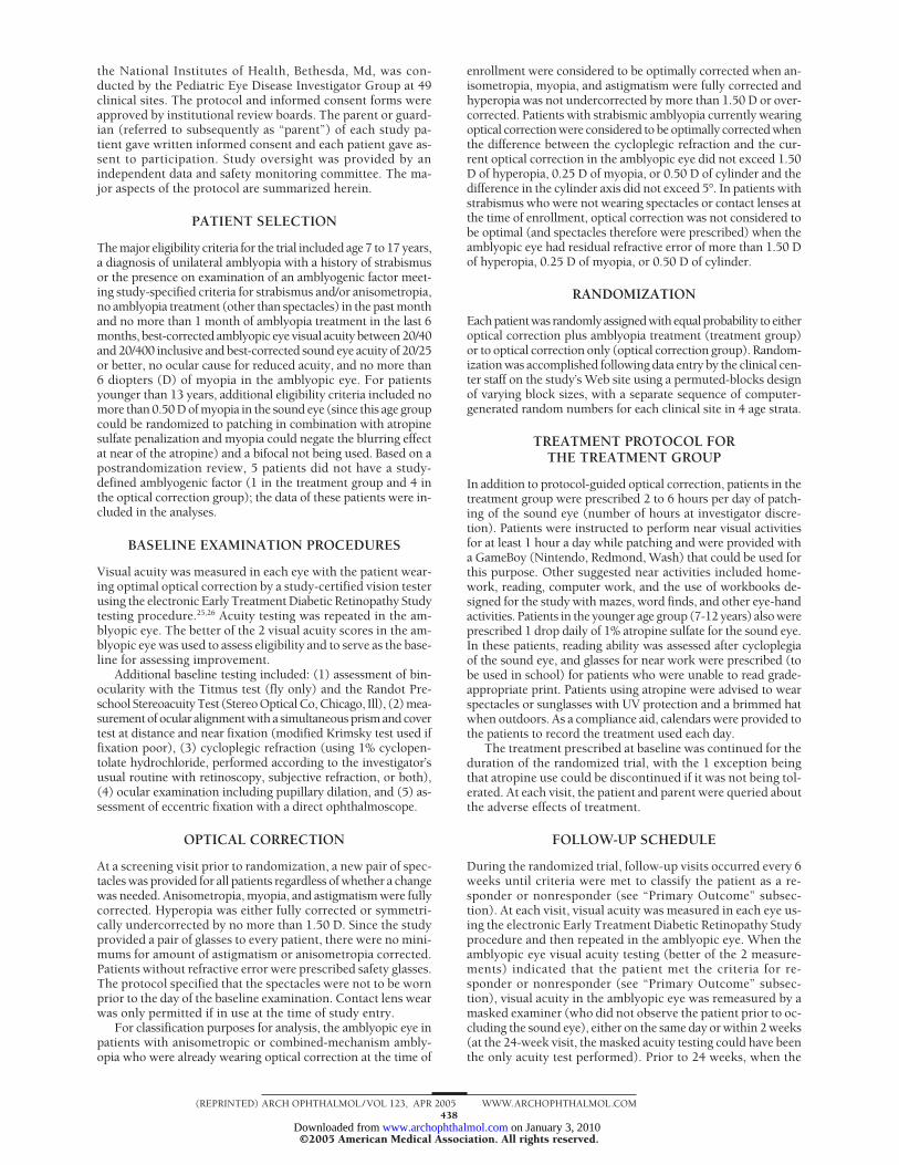
Table 1. Demographic and Baseline Clinical Characteristics*
Patients Aged 7-12 Years Patients Aged 13-17 Years
Treatment Group(n = 201)
Optical Correction Group(n = 203)
Treatment Group(n = 55)
Optical Correction Group(n = 48)
Age at randomization, mean (SD) 9.6 (1.6) 9.5 (1.7) 14.7 (1.4) 14.9 (1.2)Female 89 (44) 87 (43) 31 (56) 25 (52)Race/Ethnicity
White 154 (77) 148 (73) 36 (65) 33 (69)African American 16 (8) 16 (8) 10 (18) 6 (13)Hispanic or Latino 29 (14) 34 (17) 6 (11) 6 (13)Other 2 (1) 5 (2) 3 (5) 3 (6)
Prior treatment for amblyopiaNone 96 (48) 99 (49) 17 (31) 20 (42)Patching 77 (38) 74 (36) 32 (58) 27 (56)Atropine sulfate 6 (3) 2 (1) 0 0Patching and atropine sulfate 22 (11) 28 (14) 6 (11) 1 (2)
Cause of amblyopia†Strabismus 52 (26) 52 (26) 11 (20) 14 (29)Anisometropia 75 (38) 81 (41) 20 (36) 17 (35)Strabismus and anisometropia 73 (37) 66 (33) 24 (44) 17 (35)
Distance visual acuity in amblyopic eye20/200-20/400 (�37 letters) 16 (8) 18 (9) 2 (4) 3 (6)20/100-20/160 (38-52 letters) 44 (22) 48 (24) 20 (36) 16 (33)20/40-20/80 (�53 letters) 141 (70) 137 (67) 33 (60) 29 (60)Median (quartiles) logMAR 0.52 (0.40, 0.70) 0.54 (0.40, 0.72) 0.60 (0.40, 0.72) 0.59 (0.44, 0.71)
Distance visual acuity in sound eye‡Median (quartiles) logMAR 0.00 (−0.06, 0.06) −0.02 (−0.06, 0.04) −0.04 (−0.10, 0.02) −0.05 (−0.11, 0.00)
Intereye acuity differenceMedian (quartiles) letters 28 (19, 36) 28 (21, 37) 30 (20, 38) 32 (24.5, 39)
Refractive error in amblyopic eye�0 9 (4) 9 (4) 4 (7) 8 (17)0 to �1.00 D 15 (7) 18 (9) 7 (13) 6 (13)1.00 to �2.00 D 18 (9) 16 (8) 4 (7) 6 (13)2.00 to �3.00 D 20 (10) 16 (8) 4 (7) 3 (6)3.00 to �4.00 D 28 (14) 24 (12) 11 (20) 6 (13)�4.00 D 111 (55) 120 (59) 25 (45) 19 (40)Median (quartiles) spherical equivalent 4.25 (2.38, 5.75) 4.50 (2.50, 5.75) 3.75 (1.25, 5.13) 3.13 (0.50, 4.63)
Refractive error in sound eyeMedian (quartiles) spherical equivalent 1.50 (0.50, 3.25) 1.50 (0.63, 3.00) 0.75 (0.00, 2.25) 0.50 (0.00, 1.00)
Optical correction§No correction worn/no correction needed 7 (3) 10 (5) 1 (2) 6 (13)Correction worn optimal 32 (16) 30 (15) 5 (9) 7 (15)Correction worn requires change 82 (41) 75 (37) 24 (44) 11 (23)No correction worn/correction needed 77 (38) 83 (41) 25 (45) 24 (50)
Abbreviation: D, diopter.*Values are expressed as number (percentage) of patients unless otherwise indicated.†One patient younger than 13 years in the treatment group and 4 patients younger than 13 years in the optical correction group had an indeterminate cause of
amblyopia. These were not included in the denominators for cause of amblyopia.‡Two patients younger than 13 years in the treatment group and 1 patient younger than 13 years in the optical correction group had distance visual acuity in the
sound eye worse than 20/25 (�80 letters).§See “Methods” section for definitions of the classification. Current optical correction was with contact lenses for 6 patients: in the younger age group, 1 in the
treatment group and 3 in the optical correction group, and in the older age group, 2 in the optical correction group. Eight patients were missing optical correctionand were not included in the denominators: 5 were missing because cause of amblyopia was indeterminate and 3 because spectacle correction was unknown.
(REPRINTED) ARCH OPHTHALMOL / VOL 123, APR 2005 WWW.ARCHOPHTHALMOL.COM439
©2005 American Medical Association. All rights reserved. on January 3, 2010 www.archophthalmol.comDownloaded from
PRIMARY OUTCOME
The primary outcome was the proportion of patients in eachgroup classified as a responder. A patient was classified as aresponder if the amblyopic eye acuity was 10 or more letters(2 lines) better than the baseline acuity on the testing con-ducted by the masked examiner at the 6-week, 12-week,18-week, or 24-week visit. By the 24-week visit, if the ambly-opic eye acuity had not improved 10 or more letters, then thepatient was classified as a nonresponder. A patient could alsobe classified as a nonresponder at an earlier visit if there wasno improvement (0 letters) from the prior follow-up visit orminimal improvement from baseline (defined at the 6-weekvisit as 0-letter improvement from baseline, at the 12-weekvisit as �3-letter improvement from baseline, and at the18-week visit as �5-letter improvement from baseline).Patients who did not complete the randomized trial andpatients in the optical correction group who received ambly-opia treatment prior to being classified as a responder or non-responder were considered to be nonresponders in the pri-mary analysis.
DIPLOPIA ASSESSMENT
At each visit, the patient and parent were queried about the oc-currence and frequency of symptoms of diplopia. The patient wasasked “Do you ever see 2 (double) of the same thing when youare looking directly at it?” and the parent was asked if the childever complained of double vision. Frequency was recorded as 1of the following: less than once a week, once a week, once a day,up to 10 times a day, more than 10 times a day, and all the time.
STATISTICAL METHODS
The minimum sample size was computed to be 90 patients ineach of 4 age strata (7-8 years, 9-10 years, 11-12 years, and 13-17years) based on having a minimum of 80% power for each agestratum with a 5% 1-sided type 1 error rate to detect a differ-ence in responder rates between an optical correction grouprate of 5% and a treatment group rate of 25%. With these as-sumptions, statistical power for the primary analysis of the 404patients in the younger age group (7-12 years) was 99% andfor the 103 patients in the older age group (13-17 years), 86%.
Separate analyses were preplanned for the younger age group(7-12 years) and older age group (13-17 years) because of theexpectation of statistical interaction between age group and ran-domization group (P value for the observed interaction in thetrial between randomization group and age group=.03) and be-cause the treatment regimens were not the same in the 2 agegroups. The primary analysis compared the proportion of pa-tients in each randomization group who were classified as a re-sponder using a Fisher exact test. Unadjusted and adjusted oddsratios were computed in logistic regression models. Addi-tional analyses compared the maximum improvement achieved(at any visit) between randomization groups and the interocu-lar difference at the time of maximum improvement in analy-sis of covariance models adjusted for baseline amblyopic eyevisual acuity and baseline interocular difference, respectively.Confounding and interaction between baseline factors and ran-domization group on the primary outcome were assessed byincluding covariates of interest and interaction terms in the mod-els. Linearity of the relationship of continuous covariates withthe outcome was verified. Visual acuity of 20/25 or better wasconsidered a secondary outcome for moderate amblyopia (20/40-20/80) and visual acuity of 20/40 or better was considereda secondary outcome for severe amblyopia (20/100-20/400);proportions meeting these criteria were compared between ran-domization groups using the Fisher exact test. Similar resultsfor all analyses were obtained in secondary analyses that ex-cluded patients in both randomization groups who dropped outand patients in the optical correction group who received am-blyopia treatment (other than spectacles) prior to being clas-sified as a responder or nonresponder (data not shown).
All analyses followed the intention-to-treat principle. Re-ported P values are 1-tailed for between–randomization groupcomparisons and 2-tailed for within–randomization group com-parisons. Analyses were conducted using SAS version 8.2 (SASInstitute, Cary, NC).
RESULTS
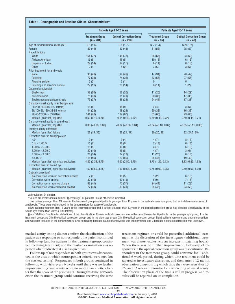
Between October 2002 and March 2004, 507 patients en-tered the trial. There were 170 patients aged 7 to 8 years,150 aged 9 to 10 years, 84 aged 11 to 12 years, and 103aged 13 to 17 years. The number of patients random-ized per site at the 49 sites ranged from 1 to 44 (median,7). Table 1 provides the baseline characteristics by ran-domization group for the younger (7-12 years) and older(13-17 years) age groups.
57 Responders12 Nonresponders
151 Completed15 Missed
17 Responders57 Nonresponders
39 Responders31 Nonresponders
85 Completed9 Missed
9 Responders27 Nonresponders
15 Responders41 Nonresponders
7 Responders36 Nonresponders
14 Responders30 Nonresponders
80 Completed8 Missed
150 Completed15 Missed
256Treatment
Group
251Optical Correction
Group
13 Dropped
6 wk
12 wk
18 wk
24 wk
9 Dropped
8 Dropped
2 Dropped 3 Dropped
2 Dropped 1 Dropped
56 Completed 43 Completed
10 Dropped
235 Completed7 Missed
238 Completed5 Missed
23 Responders44 Nonresponders
507Randomized
Figure 1. Flowchart of the 507 randomized patients through the 24 weeks ofthe randomized trial phase of the study.
(REPRINTED) ARCH OPHTHALMOL / VOL 123, APR 2005 WWW.ARCHOPHTHALMOL.COM440
©2005 American Medical Association. All rights reserved. on January 3, 2010 www.archophthalmol.comDownloaded from

FOLLOW-UP
Figure 1 provides a summary of patient follow-up dur-ing the randomized trial phase by randomization group.The completion rates of the randomized trial phase in theyounger group were 91% in the treatment group and 90%in the optical correction group and in the older group, 87%and 94%, respectively. Among the patients classified as aresponder in the randomized trial phase, follow-up con-tinued until maximal improvement was reached in theyounger group for 87 (82%) of the 106 responders in thetreatment group and for 43 (86%) of the 50 responders inthe optical correction group and in the older group for 13(93%) of 14 and 9 (82%) of 11, respectively.
The visual acuity tester was masked to randomiza-tion group for 97% of the visual acuity measurements usedto classify each patient as a responder or nonresponder.
TREATMENT
Most patients met our criteria for not having optimal op-tical correction, with most having moderate to high de-grees of hyperopia in the amblyopic eye. In the youngergroup, 79 patients (20%) were classified as having opti-mal optical correction at baseline, 157 (39%) were wear-ing optical correction meeting criteria for a change, and160 (40%) needed optical correction that was newly pre-scribed at the time of enrollment (see “Methods” sec-tion for definitions of the classification). In the older
group, the number of patients and percentages were 19(18%), 35 (34%), and 49 (48%), respectively.
In the treatment group, among the 201 patients in theyounger group, 2 hours of patching per day were pre-scribed for 101 patients (50%), 4 hours for 82 patients(41%), and 6 hours for 18 patients (9%) and atropine wasprescribed for all but 1 patient. Glasses for near work wereprescribed for 46 (23%) of these patients. Among the 55patients in the older group assigned to the treatmentgroup, 2 hours of patching per day were prescribed for34 patients (62%), 4 hours for 19 patients (35%), and 6hours for 2 patients (4%).
In the optical correction group, 3 patients (3 in theyounger group and 0 in the older group) began patchingand/or atropine treatment prior to being classified as a re-sponder or nonresponder (violations of the protocol).
PRIMARY OUTCOME:AMBLYOPIC EYE VISUAL ACUITY
Younger Group (7-12 Years)
The responder criterion was met by 106 (53%) of the 201patients in the treatment group and by 50 (25%) of the 203patients in the optical correction group (Fisher exact testP value �.001; unadjusted odds ratio, 3.41 [95% confi-dence interval, 2.24-5.21]; P�.001; adjusted odds ratio [forage, baseline visual acuity, history of prior amblyopia treat-ment, current optical correction, cause], 4.19 [95% con-
Table 2. Randomization Group Comparisons for Younger Age Group (7-12 Years)*
Responder Rate† Maximum Improvement Interocular Difference‡
TreatmentGroup,No. (%)
of Patients
OpticalCorrection
Group, No. (%)of Patients
Unadjusted/AdjustedP Value§
TreatmentGroup, Mean
Letters
OpticalCorrection
Group,Mean Letters
Unadjusted/AdjustedP Value§
TreatmentGroup, Mean
Letters
OpticalCorrection
Group,Mean Letters
Unadjusted/AdjustedP Value§
All patientsn = 201/203 106 (53) 50 (25) �.001/�.001 13.3 7.3 �.001/�.001 12.6 23.2 �.001/�.001
Severity of amblyopiaModerate (20/40-20/80)
n = 141/137 70 (50) 30 (22) �.001/�.001 11.8 6.2 �.001/�.001 7.3 18.2 �.001/�.001Severe (20/100-20/400)
n = 60/66 36 (60) 20 (30) �.001/�.001 17.0 9.6 �.001/�.001 25.4 34.0 �.001/�.001Cause of amblyopia
Strabismusn = 52/52 21 (40) 10 (19) .01/.001 11.7 5.9 �.001/�.001 10.0 20.4 �.001/�.001
Anisometropian = 75/81 47 (63) 24 (30) �.001/�.001 15.6 8.8 �.001/�.001 9.8 24.0 �.001/�.001
Combinedn = 73/66 37 (51) 15 (23) �.001/�.001 12.3 7.1 �.001/�.001 17.3 24.4 �.001/�.001
Prior amblyopia treatmentYes
n = 105/104 42 (40) 14 (13) �.001/�.001 10.8 5.3 �.001/�.001 14.5 23.6 �.001/�.001No
n = 96/99 64 (67) 36 (36) �.001/�.001 16.2 9.6 �.001/�.001 10.4 22.8 �.001/�.001
*Maximum improvement and interocular difference columns exclude 16 patients who were randomized but dropped out with no follow-up visits.†Responder has improved at least 10 letters. Among the 106 responders in the treatment group, the responder criterion was met at the 6-week visit by 51 (48%), at
the 12-week visit by 35 (33%), at the 18-week visit by 6 (6%), and at the 24-week visit by 14 (13%). Among the 50 responders in the optical correction group, thenumber of patients meeting the criterion at each visit was 16 (32%), 16 (32%), 11 (22%), and 7 (14%), respectively.
‡At visit of maximum improvement.§Adjusted P values adjusted for age (continuous), baseline amblyopic eye acuity (continuous), cause (strabismus/anisometropia/combined), prior treatment (yes/no),
and current optical correction at enrollment (yes/no). P values under responder rate are from logistic regression models. P values under maximum improvement andinterocular difference are from analysis of covariance models, adjusting for baseline acuity and baseline interocular difference, respectively.
(REPRINTED) ARCH OPHTHALMOL / VOL 123, APR 2005 WWW.ARCHOPHTHALMOL.COM441
©2005 American Medical Association. All rights reserved. on January 3, 2010 www.archophthalmol.comDownloaded from
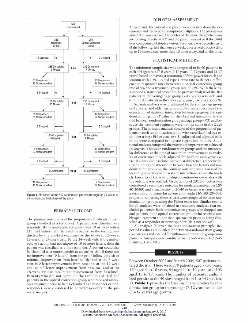
fidence interval, 2.63-6.67]; P�.001). A benefit of treat-ment was seen for both moderate amblyopia (20/40-20/80) and severe amblyopia (20/100-20/400) in responderrates, maximal improvement, and interocular difference atthe time of maximal improvement (Table 2). For mod-erate amblyopia, 36% of the treatment group compared with14% of the optical correction group achieved 20/25 or bet-ter acuity (P�.001) (Figure 2A), and for severe ambly-opia, 23% of the treatment group compared with 5% of theoptical correction group achieved 20/40 or better acuity(P=.004) (Figure 2B).
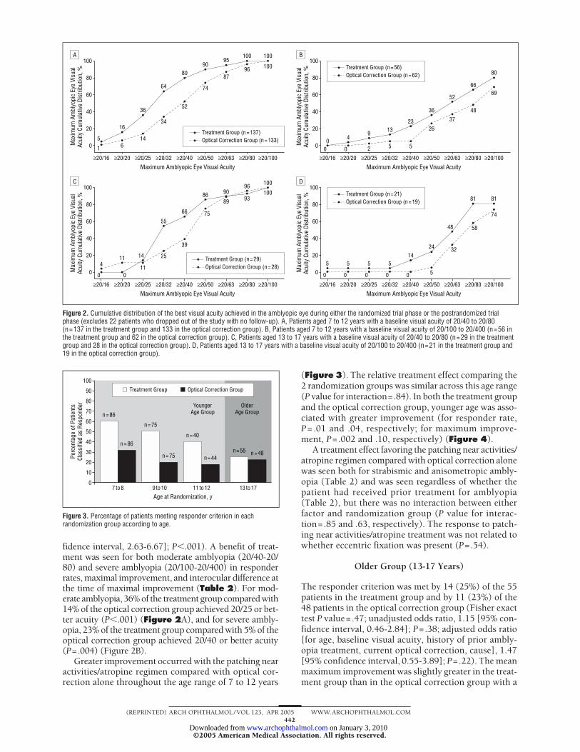
Greater improvement occurred with the patching nearactivities/atropine regimen compared with optical cor-rection alone throughout the age range of 7 to 12 years
(Figure 3). The relative treatment effect comparing the2 randomization groups was similar across this age range(P value for interaction=.84). In both the treatment groupand the optical correction group, younger age was asso-ciated with greater improvement (for responder rate,P= .01 and .04, respectively; for maximum improve-ment, P=.002 and .10, respectively) (Figure 4).
A treatment effect favoring the patching near activities/atropine regimen compared with optical correction alonewas seen both for strabismic and anisometropic ambly-opia (Table 2) and was seen regardless of whether thepatient had received prior treatment for amblyopia(Table 2), but there was no interaction between eitherfactor and randomization group (P value for interac-tion=.85 and .63, respectively). The response to patch-ing near activities/atropine treatment was not related towhether eccentric fixation was present (P=.54).
Older Group (13-17 Years)
The responder criterion was met by 14 (25%) of the 55patients in the treatment group and by 11 (23%) of the48 patients in the optical correction group (Fisher exacttest P value=.47; unadjusted odds ratio, 1.15 [95% con-fidence interval, 0.46-2.84]; P=.38; adjusted odds ratio[for age, baseline visual acuity, history of prior ambly-opia treatment, current optical correction, cause], 1.47[95% confidence interval, 0.55-3.89]; P=.22). The meanmaximum improvement was slightly greater in the treat-ment group than in the optical correction group with a
100
60
80
40
20
0
≥20/16 ≥20/20 ≥20/25 ≥20/32 ≥20/40 ≥20/50 ≥20/63 ≥20/80 ≥20/100
Maximum Amblyopic Eye Visual Acuity
5
16
36
64
8090
95100 100
96 100
87
74
52
34
1461
Max
imum
Am
blyo
pic
Eye
Visu
alAc
uity
Cum
ulat
ive
Dist
ribut
ion,
%
A
Treatment Group (n = 137)Optical Correction Group (n = 133)
Treatment Group (n = 56)Optical Correction Group (n = 62)
Treatment Group (n = 29)Optical Correction Group (n = 28)
Treatment Group (n = 21)Optical Correction Group (n = 19)
100
60
80
40
20
0
≥20/16 ≥20/20 ≥20/25 ≥20/32 ≥20/40 ≥20/50 ≥20/63 ≥20/80 ≥20/100
Maximum Amblyopic Eye Visual Acuity
Max
imum
Am
blyo
pic
Eye
Visu
alAc
uity
Cum
ulat
ive
Dist
ribut
ion,
%
B
04
0
9
2
13
5
23
5
36
26
52
37
66
48
80
69
0
100
60
80
40
20
0
≥20/16 ≥20/20 ≥20/25 ≥20/32 ≥20/40 ≥20/50 ≥20/63 ≥20/80 ≥20/100
Maximum Amblyopic Eye Visual Acuity
411
0 011
25
39
66
86 9096 100
1009389
7555
14
Max
imum
Am
blyo
pic
Eye
Visu
alAc
uity
Cum
ulat
ive
Dist
ribut
ion,
%
C100
60
80
40
20
0
≥20/16 ≥20/20 ≥20/25 ≥20/32 ≥20/40 ≥20/50 ≥20/63 ≥20/80 ≥20/100
Maximum Amblyopic Eye Visual AcuityM
axim
um A
mbl
yopi
c Ey
e Vi
sual
Acui
ty C
umul
ativ
e Di
strib
utio
n, %
D
5 5 5 514
24
48
81 81
74
58
32
50 0 0 0 0
Figure 2. Cumulative distribution of the best visual acuity achieved in the amblyopic eye during either the randomized trial phase or the postrandomized trialphase (excludes 22 patients who dropped out of the study with no follow-up). A, Patients aged 7 to 12 years with a baseline visual acuity of 20/40 to 20/80(n=137 in the treatment group and 133 in the optical correction group). B, Patients aged 7 to 12 years with a baseline visual acuity of 20/100 to 20/400 (n=56 inthe treatment group and 62 in the optical correction group). C, Patients aged 13 to 17 years with a baseline visual acuity of 20/40 to 20/80 (n=29 in the treatmentgroup and 28 in the optical correction group). D, Patients aged 13 to 17 years with a baseline visual acuity of 20/100 to 20/400 (n=21 in the treatment group and19 in the optical correction group).
100
70
60
80
90
50
40
20
30
10
0
n = 86
n = 86
n = 75
n = 75
n = 40
n = 44n = 55 n = 48
YoungerAge Group
OlderAge Group
Age at Randomization, y
Perc
enta
ge o
f Pat
ient
sCl
assi
fied
as R
espo
nder
7 to 8 9 to 10 11 to 12 13 to 17
Treatment Group Optical Correction Group
Figure 3. Percentage of patients meeting responder criterion in eachrandomization group according to age.
(REPRINTED) ARCH OPHTHALMOL / VOL 123, APR 2005 WWW.ARCHOPHTHALMOL.COM442
©2005 American Medical Association. All rights reserved. on January 3, 2010 www.archophthalmol.comDownloaded from
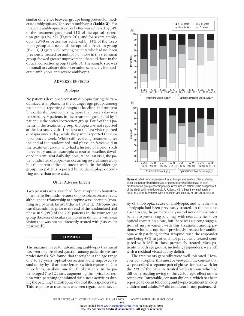
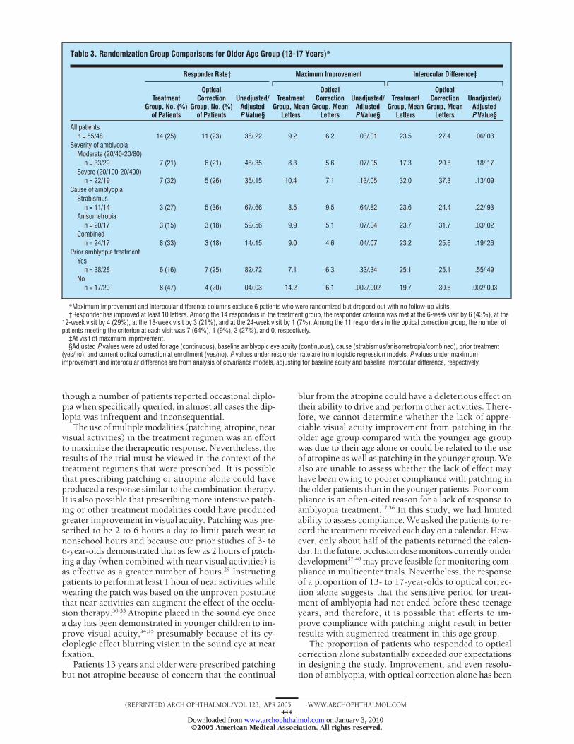
similar difference between groups being present for mod-erate amblyopia and for severe amblyopia (Table 3). Formoderate amblyopia, 20/25 or better was achieved by 14%of the treatment group and 11% of the optical correc-tion group (P=.52) (Figure 2C), and for severe ambly-opia, 20/40 or better was achieved by 14% of the treat-ment group and none of the optical correction group(P=.13) (Figure 2D). Among patients who had not beenpreviously treated for amblyopia, those in the treatmentgroup showed greater improvement than did those in theoptical correction group (Table 3). The sample size wastoo small to evaluate this observation separately for mod-erate amblyopia and severe amblyopia.
ADVERSE EFFECTS
Diplopia
No patients developed constant diplopia during the ran-domized trial phase. In the younger age group, amongpatients not reporting diplopia at baseline, intermittentbinocular diplopia occurring more than once a day wasreported by 4 patients in the treatment group and by 1patient in the optical correction group. For 3 of the 4 pa-tients in the treatment group, diplopia was not reportedat the last study visit; 1 patient at the last visit reporteddiplopia once a day, while the parent reported the dip-lopia once a week. While still receiving treatment afterthe end of the randomized trial phase, an 8-year-old inthe treatment group, who had a history of a prior sixthnerve palsy and an esotropia at near at baseline, devel-oped intermittent daily diplopia; at the last visit, the pa-tient indicated diplopia was occurring several times a daybut the parent indicated once a week. In the older agegroup, no patients reported binocular diplopia occur-ring more than once a day.
Other Adverse Effects
Two patients were switched from atropine to homatro-pine methylbromide because of possible adverse effects,although the relationship to atropine was uncertain (vom-iting in 1 patient, tachycardia in 1 patient). Atropine usewas discontinued prior to the end of the randomized trialphase in 9 (4%) of the 201 patients in the younger agegroup (because of ocular symptoms or difficulty with nearvision that was not satisfactorily treated with glasses fornear work).
COMMENT
The maximum age for attempting amblyopia treatmenthas been an unresolved question among pediatric eye careprofessionals. We found that throughout the age rangeof 7 to 17 years, optical correction alone improved vi-sual acuity by 10 or more letters (which equates to 2 ormore lines) in about one fourth of patients. In the pa-tients aged 7 to 12 years, augmenting the optical correc-tion with patching (combined with near activities dur-ing the patching) and atropine doubled the responder rate.This response to treatment was seen regardless of sever-
ity of amblyopia, cause of amblyopia, and whether theamblyopia had been previously treated. In the patients13-17 years, the primary analysis did not demonstrate abenefit to prescribing patching (with near activities) overoptical correction alone, but there was a strong sugges-tion of improvement with this treatment among pa-tients who had not been previously treated for ambly-opia with patching and/or atropine, with the responderrate being 47% in patients not previously treated com-pared with 16% in those previously treated. Most pa-tients in both age groups, including responders, were leftwith a residual visual acuity deficit.
The treatments generally were well tolerated. How-ever, for atropine, this must be viewed in the context thatwe prescribed a separate pair of glasses for near work forthe 23% of the patients treated with atropine who haddifficulty reading owing to the cycloplegic effect on thesound eye. Intractable, constant diplopia, which has beenreported to occur following amblyopia treatment in olderchildren and adults,27,28 did not occur in any patients. Al-
≥15 Letters10-14 Letters
5-9 Letters<5 Letters
100
70
60
80
90
50
40
20
30
10
0
Perc
enta
ge
n = 537 to <9
n = 569 to <11
n = 2811 to <13
n = 2913 to <18
n = 587 to <9
n = 499 to <11
n = 2611 to <13
n = 2813 to <18
A
Treatment Group, Age, y Optical Correction Group, Age, y
100
70
60
80
90
50
40
20
30
10
0
Perc
enta
ge
n = 287 to <9
n = 179 to <11
n = 1111 to <13
n = 2113 to <18
n = 217 to <9
n = 269 to <11
n = 1511 to <13
n = 1913 to <18
B
Treatment Group, Age, y Optical Correction Group, Age, y
Figure 4. Maximum improvement in amblyopic eye acuity achieved duringeither the randomized trial phase or postrandomized trial phase in eachrandomization group according to age (excludes 22 patients who dropped outof the study with no follow-up). A, Patients with a baseline visual acuity of20/40 to 20/80. B, Patients with a baseline visual acuity of 20/100 to 20/400.
(REPRINTED) ARCH OPHTHALMOL / VOL 123, APR 2005 WWW.ARCHOPHTHALMOL.COM443
©2005 American Medical Association. All rights reserved. on January 3, 2010 www.archophthalmol.comDownloaded from
though a number of patients reported occasional diplo-pia when specifically queried, in almost all cases the dip-lopia was infrequent and inconsequential.
The use of multiple modalities (patching, atropine, nearvisual activities) in the treatment regimen was an effortto maximize the therapeutic response. Nevertheless, theresults of the trial must be viewed in the context of thetreatment regimens that were prescribed. It is possiblethat prescribing patching or atropine alone could haveproduced a response similar to the combination therapy.It is also possible that prescribing more intensive patch-ing or other treatment modalities could have producedgreater improvement in visual acuity. Patching was pre-scribed to be 2 to 6 hours a day to limit patch wear tononschool hours and because our prior studies of 3- to6-year-olds demonstrated that as few as 2 hours of patch-ing a day (when combined with near visual activities) isas effective as a greater number of hours.29 Instructingpatients to perform at least 1 hour of near activities whilewearing the patch was based on the unproven postulatethat near activities can augment the effect of the occlu-sion therapy.30-33 Atropine placed in the sound eye oncea day has been demonstrated in younger children to im-prove visual acuity,34,35 presumably because of its cy-cloplegic effect blurring vision in the sound eye at nearfixation.
Patients 13 years and older were prescribed patchingbut not atropine because of concern that the continual
blur from the atropine could have a deleterious effect ontheir ability to drive and perform other activities. There-fore, we cannot determine whether the lack of appre-ciable visual acuity improvement from patching in theolder age group compared with the younger age groupwas due to their age alone or could be related to the useof atropine as well as patching in the younger group. Wealso are unable to assess whether the lack of effect mayhave been owing to poorer compliance with patching inthe older patients than in the younger patients. Poor com-pliance is an often-cited reason for a lack of response toamblyopia treatment.17,36 In this study, we had limitedability to assess compliance. We asked the patients to re-cord the treatment received each day on a calendar. How-ever, only about half of the patients returned the calen-dar. In the future, occlusion dose monitors currently underdevelopment37-40 may prove feasible for monitoring com-pliance in multicenter trials. Nevertheless, the responseof a proportion of 13- to 17-year-olds to optical correc-tion alone suggests that the sensitive period for treat-ment of amblyopia had not ended before these teenageyears, and therefore, it is possible that efforts to im-prove compliance with patching might result in betterresults with augmented treatment in this age group.
The proportion of patients who responded to opticalcorrection alone substantially exceeded our expectationsin designing the study. Improvement, and even resolu-tion of amblyopia, with optical correction alone has been
Table 3. Randomization Group Comparisons for Older Age Group (13-17 Years)*
Responder Rate† Maximum Improvement Interocular Difference‡
TreatmentGroup, No. (%)
of Patients
OpticalCorrection
Group, No. (%)of Patients
Unadjusted/AdjustedP Value§
TreatmentGroup, Mean
Letters
OpticalCorrection
Group, MeanLetters
Unadjusted/AdjustedP Value§
TreatmentGroup, Mean
Letters
OpticalCorrection
Group, MeanLetters
Unadjusted/AdjustedP Value§
All patientsn = 55/48 14 (25) 11 (23) .38/.22 9.2 6.2 .03/.01 23.5 27.4 .06/.03
Severity of amblyopiaModerate (20/40-20/80)
n = 33/29 7 (21) 6 (21) .48/.35 8.3 5.6 .07/.05 17.3 20.8 .18/.17Severe (20/100-20/400)
n = 22/19 7 (32) 5 (26) .35/.15 10.4 7.1 .13/.05 32.0 37.3 .13/.09Cause of amblyopia
Strabismusn = 11/14 3 (27) 5 (36) .67/.66 8.5 9.5 .64/.82 23.6 24.4 .22/.93
Anisometropian = 20/17 3 (15) 3 (18) .59/.56 9.9 5.1 .07/.04 23.7 31.7 .03/.02
Combinedn = 24/17 8 (33) 3 (18) .14/.15 9.0 4.6 .04/.07 23.2 25.6 .19/.26
Prior amblyopia treatmentYes
n = 38/28 6 (16) 7 (25) .82/.72 7.1 6.3 .33/.34 25.1 25.1 .55/.49No
n = 17/20 8 (47) 4 (20) .04/.03 14.2 6.1 .002/.002 19.7 30.6 .002/.003
*Maximum improvement and interocular difference columns exclude 6 patients who were randomized but dropped out with no follow-up visits.†Responder has improved at least 10 letters. Among the 14 responders in the treatment group, the responder criterion was met at the 6-week visit by 6 (43%), at the
12-week visit by 4 (29%), at the 18-week visit by 3 (21%), and at the 24-week visit by 1 (7%). Among the 11 responders in the optical correction group, the number ofpatients meeting the criterion at each visit was 7 (64%), 1 (9%), 3 (27%), and 0, respectively.
‡At visit of maximum improvement.§Adjusted P values were adjusted for age (continuous), baseline amblyopic eye acuity (continuous), cause (strabismus/anisometropia/combined), prior treatment
(yes/no), and current optical correction at enrollment (yes/no). P values under responder rate are from logistic regression models. P values under maximumimprovement and interocular difference are from analysis of covariance models, adjusting for baseline acuity and baseline interocular difference, respectively.
(REPRINTED) ARCH OPHTHALMOL / VOL 123, APR 2005 WWW.ARCHOPHTHALMOL.COM444
©2005 American Medical Association. All rights reserved. on January 3, 2010 www.archophthalmol.comDownloaded from
The Pediatric Eye Disease Investigator Group
Writing CommitteeLead authors: Mitchell M. Scheiman, OD; Richard W. Hertle, MD; Roy W. Beck, MD, PhD; Allison R. Edwards, MS. Additional writing committeemembers (alphabetical): Eileen Birch, PhD; Susan A. Cotter, OD; Earl R. Crouch, Jr, MD; Oscar A. Cruz, MD; Bradley V. Davitt, MD; Sean Dona-hue, MD; Jonathan M. Holmes, BM, BCh; Don W. Lyon, OD; Michael X. Repka, MD; Nicholas A. Sala, DO; David I. Silbert, MD; Donny W. Suh,MD; Susanna M. Tamkins, OD.
Clinical SitesListed in order of number of patients randomized into the study. The number of patients randomized is noted in parentheses preceded by the sitename and location. Personnel are listed as (I) for investigator, (C) for coordinator, and (V) for visual acuity tester.
Pediatric Ophthalmology of Erie, Erie, Pa (44): Nicholas A. Sala (I); Rhonda M. Hodde (C); Veda L. Zeto (C); Cindy E. Tanner (V). Wolfe Clinic,West Des Moines, Iowa (41): Donny W. Suh (I); Kim S. Walters (C); Heather K. Sipes (C); Shannon L. Craig (V); Rhonda J. Swisher (V); Lisa M.Fergus (V). Indiana University School of Optometry, Indianapolis (29): Don W. Lyon (I); John P. Downey (V); Christy C. Hohenbary (V); DeborahJ. Plass (V); Brad M. Sutton (V); Danielle F. Warren (V). Pediatric Ophthalmology Associates Inc, Columbus, Ohio (28): Richard W. Hertle (I); DonL. Bremer (I); Mary Lou McGregor (I); Gary L. Rogers (I); Rae R. Fellows (C); Vanessa M. Hill (C); Ninon M. Greene (V); Rebecca A. Murray (V);Teresa M. Rhinehart (V); Angela M. Serna (V); Laura J. Shenberger (V). Southern California College of Optometry, Fullerton (25): Susan A. Cotter (I);Carmen N. Barnhardt (I); Raymond H. Chu (I); Monique M. Nguyen (I); Susan M. Shin (I); Erin Song (I); Tracy R. Leonhardt (C); Lourdes Asiain(C). Family Eye Group, Lancaster, Pa (25): David I. Silbert (I); Eric L. Singman (I); Noelle S. Matta (C); Suanne E. Carner (V); Cristina M. Cor-radino (V); Troy J. Hosey (V); Diane M. Jostes (V); Alyson B. Keene (V); Melissa A. Kelly (V); Stephanie R. Kilgore (V); Michelle M. Lindsey (V);Danae L. Nelson (V); Tonji L. Nelson (V); Tiana M. Ober (V); Wendy L. Piper (V); Sara L. Weit (V); Sylvia R. Wright (V); Shannon M. Butler (V);Scarlett T. Musser (V). Bascom Palmer Eye Institute, Miami, Fla (23): Susanna M. Tamkins (I); Joseph H. Sugg (I); Eva M. Olivares (C); Nihusa P.Oviedo (C); Bruce D. Bailey (V); Mirna Garcia (V); Maria Georgatos (V). Eastern Virginia Medical School, Norfolk (23): Earl R. Crouch, Jr (I); EricR. Crouch III (I); Kristen D. Ruark (C); Gaylord G. Ventura (V). Pennsylvania College of Optometry, Philadelphia (20): Mitchell M. Scheiman (I);Karen E. Pollack (C); Michael F. Gallaway (V); Nicole I. Lynch (V); Tomo Yamada (V); Brandy J. Scombordi (V). Cardinal Glennon Children’sHospital, St Louis, Mo (14): Oscar A. Cruz (I); Bradley V. Davitt (I); Emily A. Miyazaki (C). Eye Physicians & Surgeons, PC, Milford, Conn (13):Darron A. Bacal (I); Donna Martin (C); Jodi A. Bregman (C). University of Texas Southwestern Medical Center, Dallas (12): David R. Weakley, Jr (I);Clare L. Dias (C); Eileen E. Birch (V); Christina S. Cheng (V); Joost Felius (V); Sarah E. Morale (V). Baylor College of Medicine, Texas Children’sHospital, Houston (11): Evelyn A. Paysse (I); David K. Coats (I); Jane Covington Edmond (I); Kimberly G. Yen (I); Michele L. Fulton (C); La-Shunna D. Jamerson (V); Alma D. Sanchez (V); M. Louise Thomas (V). State University of New York, College of Optometry, New York (11): RobertH. Duckman (I); David E. FitzGerald (I); Marilyn Vricella (V). Children’s Eye Care & Adult Strabismus Surgery, South Charleston, WVa (11): Debo-rah L. Klimek (I); Dani L. Clay (V); Lisa L. Winter (V). Katherine Ann Lee, MD, PA, Boise, ID (10): Katherine A. Lee (I); Bonita R. Schweinler (C);Jenny N. Marshall (V); Melissa K. Schweigert (V). Pediatric Ophthalmology, PC, Grand Rapids, Mich (10): Patrick J. Droste (I); Robert J. Peters (I);Jan Hilbrands (C); Sandra K. Rogers (V); Maggie A. Santure (V). Pediatric Ophthalmology, PA, Dallas (9): David R. Stager, Sr (I); David R. Stager, Jr(I); Priscilla M. Berry (I); Joost Felius (C); Elaine R. Hammett (V); Sarah E. Morale (V). Casey Eye Institute, Portland, Ore (9): David T. Wheeler (I);Kimberley A. Beaudet (C); Paula K. Rauch (C); Annika S. Joshi (V). Goldblum Family Eye Care Center, PC, Albuquerque, NM (8): Todd A. Goldblum(I); Rachel Baca (C); Angela Alfaro (V); Regan R. Foster (V); Antoinette Ramirez (V). Ophthalmic Associates, Anchorage, Alaska (8): Robert W.Arnold (I); Mary Diane Armitage (C); Nancy H. Brusseau (V); Maru V. Gindling (V); Karen M. Lowe (V). Wilmer Institute, Baltimore, Md (8): Mi-chael X. Repka (I); Carole R. Goodman (C); Xiaonong Liu (C). The Cleveland Clinic Foundation, Ophthalmology at Beachwood, Beachwood, Ohio (8):Diane L. Tucker (I); Andreas Marcotty (I); Laurie A. Slaby (C); Tamara M. Roman (V). Southern College of Optometry, Memphis, Tenn (8): Erin R.Nosel (I); Kristin K. Anderson (I); Ingryd J. Lorenzana (I); Lindsay C. Moran (C); Christopher W. Lievens (V); Bernard I. Sparks (V); Tom R.Karkkainen (V). Eye Centers of Ohio, Canton (7): Elbert H. Magoon (I); Paula A. Kannam (C); Kathy Ann Earl (C); Lynn A. McAtee (C); MargieAndrews (V); Caroline M. Hoge (V); Debby Ann Null (V); Denise Richards (V); Judy A. Swartz (V). Rhode Island Eye Institute, Providence (7): JohnP. Donahue (I); Melissa A. Corrente (V); Robin L. Darpino (V); Patricia Reale (V); Nicole L. Waterman (V). Grene Vision Group, Wichita, Kan (7):David A. Johnson (I); Ruth D. Dannar (C); Amy Melissa Wheeler (V). University of Alabama at Birmingham School of Optometry, Birmingham (6):Robert P. Rutstein (I); Marcela Frazier (I); Kristine T. Hopkins (I); Wendy L. Marsh-Tootle (I); Katherine K. Weise (I); Cathy H. Baldwin (C);Sophocles Sophocleous (V); Ann M. Wonderling (V). The Ohio State University, Columbus (6): Marjean T. Kulp (I); Tracy L. Kitts (C); Michael J.Earley (V); Anna J. Schlesselman (V); Andrew J. Toole (V); Ann Hickson (V). Vanderbilt Eye Center, Nashville, Tenn (6): Sean Donahue (I); SandyA. Owings (C); Ronald J. Biernacki (V); Genise G. Mofiled (V); Neva J. Palmer (V). New England College of Optometry, Boston, Mass (5): Erik M.Weissberg (I); Barry S. Kran (I); Nicole M. Quinn (I); Melissa A. Suckow (V). Children’s Hospital Medical Center, Cincinnati, Ohio (5): Constance E.West (I); Stephanie C. Fort (C); Brandy L. Bocklett (V); Nicole M. Burton (V); Kathryn M. Carter (V); Laura E. Dickman (V); Sarah L. Lopper (V);Laurie A. Hahn-Parrott (V); Melanie M. Hounchell (N); Kelli N. Kinder (V); Debbie A. Meister (V); Walker W. Motley (V); Shannen L. Nelson (V);Daniele P. Saltarelli (V); Shannon R. Walsh (V). Medical College of Wisconsin, Milwaukee (5): Jane D. Kivlin (I); Mark S. Ruttum (I); Veronica R.Picard (C); Margaret L. Willett (V). Eye Care Group, PC, Waterbury, Conn (5): Andrew J. Levada (I); Tabitha L. Matchett (C); David N. Comstock(C); Lisa A. Marcil (V); Nicole G. Rannazzisi (V); Cheryl Schleif (V); Shelley K. Weiss (V). Pediatric Eye Associates, Wilmette, Ill (5): Deborah R.Fishman (I); Lisa C. Verderber (I); JoAnn Spieker (C). Alberta Children’s Hospital, Calgary (4): William F. Astle (I); Maria del Pilar Echeverri (I);Anna L. Ells (I); Heather J. Peddie (C); Trena L. Beer (C); Cheryl R. Hayduk (C); April D. Ingram (C); Catriona I. Kerr (C); Heather M. Vibert (C).Cinnaminson, NJ (4): Michael F. Gallaway (I); Alan Bacho (V). Mayo Clinic, Rochester, Minn (4): Jonathan M. Holmes (I); Brian G. Mohney (I);Melissa L. Rice (I); Rebecca A. Nielsen (C); Julie A. Holmquist (V); Pamela Kirsh (V); Rose M. Kroening (V); David A. Leske (V); Marna L. Levisen(V); Deborah K. Miller (V); Debbie M. Priebe (V); Julie A. Spitzer (V). Pediatric Ophthalmology Service, Boston (3): Linda R. Dagi (I); MarietteTyedmers (C); Derek P. Guanaga (V). Midwest Eye Institute, Indianapolis, Ind (3): Derek T. Sprunger (I); Jay G. Galli (C). University of Minnesota,Minneapolis (3): C. Gail Summers (I); Stephen P. Christansen (I); Erick D. Bothun (I); Ann M. Holleschau (C); Kim S. Merrill (V); Jane D. Lavoie.Vision Center of Delaware, Newark (3): Don D. Blackburn (I); Patti E. MacDonald (V). The Emory Eye Center, Atlanta, Ga (2): Scott R. Lambert (I);Amy K. Hutchinson (I); Rachel A. Reeves (C). Palm Beach Eye Foundation/Visual Health and Surgical Center, Lake Worth, Fla (2): Lee S. Friedman(I). Dean A. McGee Eye Institute, University of Oklahoma, Oklahoma City (2): Lucas Trigler (I); Dana M. Jones (I); R. Michael Siatkowski (I); StefanieKraeer (C); Lisa M. Ogilbee (C); Sara L. Ceresa (V); Connie J. Dwiggins (V); Jason D. Jobson (V). Associated Eye Care, Saint Paul, Minn (2): SusanSchloff (I); Anthony R. Brown (C); Valori E. Olson (C). Ophthalmology Consultants, Inc, Willoughby, Ohio (2): Bernard D. Perla (I); Christine A.Hochrein (C); Pam R. Meyer (V); Danielle M. Zahler (V). The State University of New York Downstate Medical Center, Brooklyn (1): Janine N. Smith (I).
Coordinating Center, Tampa, FlaRoy W. Beck, Gladys N. Bernett, Nicole M. Boyle, Christina M. Cagnina-Morales, Esmeralda L. Cardosa, Danielle L. Chandler, Laura E. Clark,Sharon R. Constantine, Quayleen Donahue, Mitchell Dupre, Allison R. Edwards, Heidi A. Gillespie, Julie A. Gillett, Raymond T. Kraker, Shelly T.Mares, Amanda R. McCarthy, Holly J. McCombs, B. Michele Melia, Pamela S. Moke.
National Eye Institute, Bethesda, MdDonald F. Everett.
PEDIG Executive CommitteeMichael X. Repka (chair), Jonathan M. Holmes (vice chair), Roy W. Beck, Eileen E. Birch, Susan A. Cotter, Donald F. Everett, Pamela S. Moke.
Amblyopia Treatment Study Steering CommitteeRoy W. Beck, Eileen E. Birch, Stephen P. Christiansen, Susan A. Cotter, Donald F. Everett, Richard W. Hertle, Jonathan M. Holmes, Don W. Lyon,Noelle S. Matta, Brian G. Mohney, Pamela S. Moke, Graham E. Quinn, Michael X. Repka, Mitchell M. Scheiman, David K. Wallace, David R. Weakley.
(REPRINTED) ARCH OPHTHALMOL / VOL 123, APR 2005 WWW.ARCHOPHTHALMOL.COM445
©2005 American Medical Association. All rights reserved. on January 3, 2010 www.archophthalmol.comDownloaded from
reported by other investigators.41-45 This “refractive adap-tation” is more than just the immediate effect of wearingspectacles but represents actual treatment of amblyopiasince the visual acuity improves in a gradual and sus-tained manner.37 Another consideration for the improve-ment seen in the optical correction group is a learning effect;however, this is likely not the explanation for most of theresponder cases based on the fact that 2 visual acuity testswere performed to establish the baseline and based on priortest-retest studies that did not demonstrate a meaningfullearning effect.25,26 In designing the trial, had we knownthat there would be such a large proportion of patients whohad never been treated for amblyopia and were not wear-ing optimal optical correction, we might have included ano treatment arm as part of the randomization of patientswith uncorrected refractive error. Alternately, we mighthave followed up all patients with spectacle correction aloneuntil improvement stopped prior to randomizing those pa-tients who still had amblyopia.
We could identify no sources of bias or confounding toexplain our findings. Accounting for differences in the dis-tribution of baseline factors between groups in the analy-sesdidnotalter the interpretationof theresults.The follow-up visit rate was similar in the 2 groups, and analyses thatincludedandexcludedthedroppedpatientsprovidedsimi-lar results. Although the patients and investigators were bythe nature of this study unmasked to the treatment groupassignments, responder-nonresponder statuswasbasedona visual acuity test administered by an individual maskedto the treatmentassignment. Inaddition, thecomputerizedmethod of visual acuity testing used in the trial minimizesthe possibility that knowledge of treatment group will biasthe results. The responder definition of a 10 or more letter(�2 lines) improvement frombaselinewasselected for thisprotocol to provide a measure of acuity improvement thatexceeded testing variability. This was based on prior stud-ies that determined that a change of 7 or more letters is un-likely to be due to measurement variability.25,26
In translating the results into clinical practice, it is im-portant to recognize that patients participating in a clini-cal trial may differ from patients in usual practice, and ourpatients’ level of compliance may have been better than whatmay be achieved in clinical practice. Although our resultsindicate that visual acuity can be improved by treating am-blyopia in older children, it is not known whether the im-provement will be sustained after treatment is discontin-ued. Therefore, a conclusion regarding the long-term benefitof treatment and the development of treatment recommen-dations for amblyopia in children 7 years and older will needto await the results of a follow-up study we are conduct-ing on the patients who responded to treatment.
Submitted for Publication: December 6, 2004; final revi-sion received February 1, 2005; accepted February 1, 2005.Correspondence: Mitchell M. Scheiman, OD, c/o JaebCenter for Health Research, 15310 Amberly Dr, Suite 350,Tampa, FL 33647 ([email protected]).
REFERENCES
1. Pediatric Eye Disease Investigator Group. A randomized trial of atropine vs patch-ing for treatment of moderate amblyopia in children. Arch Ophthalmol. 2002;120:268-278.
2. Neumann E, Friedman Z, Abel-Peleg B. Prevention of strabismic amblyopia ofearly onset with special reference to the optimal age for screening. J Pediatr Oph-thalmol Strabismus. 1987;24:106-110.
3. Scott WE, Dickey CF. Stability of visual acuity in amblyopic patients after visualmaturity. Graefes Arch Clin Exp Ophthalmol. 1988;226:154-157.
4. Quah BL, Tay MT, Chew SJ, Lee LK. A study of amblyopia in 18-19 year old males.Singapore Med J. 1991;32:126-129.
5. Epelbaum M, Milleret C, Buisseret P, Dufier JL. The sensitive period for strabis-mic amblyopia in humans. Ophthalmology. 1993;100:323-327.
6. Flynn JT, Schiffman J, Feuer W, Corona A. The therapy of amblyopia: an analy-sis of the results of amblyopia therapy utilizing the pooled data of published studies.Trans Am Ophthalmol Soc. 1998;96:431-453.
7. Simons K, Preslan M. Natural history of amblyopia untreated owing to lack ofcompliance. Br J Ophthalmol. 1999;83:582-587.
8. Assaf AA. The sensitive period: transfer of fixation after occlusion for strabismicamblyopia. Br J Ophthalmol. 1982;66:64-70.
9. American Academy of Ophthalmology. Preferred Practice Pattern: Amblyopia.San Francisco, Calif: American Academy of Ophthalmology; 2002.
10. von Noorden GK, Crawford ML. The sensitive period. Trans Ophthalmol Soc U K.1979;99:442-446.
11. Woodruff G, Hiscox F, Thompson JR, Smith LK. Factors affecting the outcomeof children treated for amblyopia. Eye. 1994;8:627-631.
12. Simmers AJ, Gray LS. Improvement of visual function in an adult amblyope. Op-tom Vis Sci. 1999;76:82-87.
13. Pediatric Eye Disease Investigator Group. A prospective, pilot study of treat-ment of amblyopia in children 10 to � 18 years old. Am J Ophthalmol. 2004;137:581-583.
14. Mohan K, Saroha V, Sharma A. Successful occlusion therapy for amblyopiain 11- to 15-year-old children. J Pediatr Ophthalmol Strabismus. 2004;41:89-95.
15. Brown MH, Edelman PM. Conventional occlusion in the older amblyope. Am Or-thopt J. 1976;26:34-36.
16. Sen DK. Results of treatment in amblyopia associated with unilateral high myo-pia without strabismus. Br J Ophthalmol. 1984;68:681-685.
17. Oliver M, Neumann E, Chaimovitch Y, Gotesman N, Shimshoni M. Complianceand results of treatment for amblyopia in children more than 8 years old. Am JOphthalmol. 1986;102:340-345.
18. Noda S, Hayasaka S, Setogawa T. Occlusion therapy of Japanese children withanisometropic amblyopia without strabismus. Ann Ophthalmol. 1993;25:145-147.
19. Tsubota K, Yamada M. Treatment of amblyopia by extended-wear occlusion softcontact lenses. Ophthalmologica. 1994;208:214-215.
20. Mintz-Hittner HA, Fernandez KM. Successful amblyopia therapy initiated after age7 years: compliance cures. Arch Ophthalmol. 2000;118:1535-1541.
21. Kupfer C. Treatment of amblyopia exanopsia in adults; a preliminary report ofseven cases. Am J Ophthalmol. 1957;43:918-922.
22. Wick B, Wingard M, Cotter S, Scheiman M. Anisometropic amblyopia: is the pa-tient ever too old to treat? Optom Vis Sci. 1992;69:866-878.
23. Park KH, Hwang JM, Ahn JK. Efficacy of amblyopia therapy initiated after 9 yearsof age. Eye. 2004;18:571-574.
24. Magoon EH, Garuda S. Visual acuity plasticity in amblyopes between age 10 and14. In: Scott A, ed. Proceedings of the Jampolsky Festschrift. San Francisco, Calif:Smith-Kettlewell Eye Research Institute; 2000.
25. Beck R, Moke P, Turpin A, et al. A computerized method of visual acuity testing:adaptation of the early treatment of diabetic retinopathy study testing protocol.Am J Ophthalmol. 2003;135:194-205.
26. Cotter SA, Chu RH, Chandler DL, et al. Reliability of the Electronic Early Treat-ment Diabetic Retinopathy Study testing protocol in children 7 to �13 years old.Am J Ophthalmol. 2003;136:655-661.
27. Swan KC. Esotropia precipitated by occlusion. Am Orthopt J. 1980;30:49-59.28. von Noorden GK. Binocular Vision and Ocular Motility: Theory and Management
of Strabismus. St Louis, Mo: Mosby; 1996:206-296.29. Pediatric Eye Disease Investigator Group. A randomized trial of patching regi-
mens for treatment of moderate amblyopia in children. Arch Ophthalmol. 2003;121:603-611.
30. Francois J, James M. Comparative study of amblyopic treatment. Am Orthopt J.1955;5:61-64.
31. Callahan WP, Berry D. The value of visual stimulation during contact and directocclusion. Am Orthopt J. 1968;18:73-74.
32. von Noorden GK, Springer F, Romano P, Parks M. Home therapy for amblyopia.Am Orthopt J. 1970;20:46-50.
33. Watson PG, Sanac AS, Pickering MS. A comparison of various methods of treat-ment of amblyopia: a block study. Trans Ophthalmol Soc U K. 1985;104:319-328.
34. Pediatric Eye Disease Investigator Group. A comparison of atropine and patch-ing treatments for moderate amblyopia by patient age, cause of amblyopia, depthof amblyopia, and other factors. Ophthalmology. 2003;110:1632-1638.
35. Pediatric Eye Disease Investigator Group. A randomized trial of atropine regi-mens for treatment of moderate amblyopia in children. Ophthalmology. 2004;111:2076-2085.
36. Pritchard C. Why won’t you treat my 10 year old’s lazy eye? Am Orthopt J. 1990;40:15-18.
37. Stewart CE, Moseley MJ, Stephens DA, Fielder AR. Treatment dose-response inamblyopia therapy: the Monitored Occlusion Treatment of Amblyopia Study. In-vest Ophthalmol Vis Sci. 2004;45:3048-3054.
(REPRINTED) ARCH OPHTHALMOL / VOL 123, APR 2005 WWW.ARCHOPHTHALMOL.COM446
©2005 American Medical Association. All rights reserved. on January 3, 2010 www.archophthalmol.comDownloaded from
38. Fielder AR, Irwin M, Auld R, Cocker KD, Jones HS, Moseley MJ. Compliance inamblyopia therapy: objective monitoring of occlusion. Br J Ophthalmol. 1995;79:585-589.
39. Loudon SE, Polling JR, Simonsz HJ. Electronically measured compliance withocclusion therapy for amblyopia is related to visual acuity increase. Graefes ArchClin Exp Ophthalmol. 2003;241:176-180.
40. Simonsz HJ, Polling JR, Voorn R, et al. Electronic monitoring of treatment com-pliance in patching for amblyopia. Strabismus. 1999;7:113-123.
41. Clarke WN, Noel LP. Prognostic indicators for avoiding occlusion therapy in an-isometropic amblyopia. Am Orthopt J. 1990;40:51-63.
42. Kivlin JD, Flynn JT. Therapy of anisometropic amblyopia. J Pediatr OphthalmolStrabismus. 1981;18:47-56.
43. Moseley MJ, Neufeld M, McCarry B, et al. Remediation of refractive amblyopiaby optical correction alone. Ophthalmic Physiol Opt. 2002;22:296-299.
44. Nordlow W. Anisometropia, amblyopia, induced aniseikonia and estimated cor-rection with iseikonic lenses in 4 year-olds. Acta Ophthalmol (Copenh). 1970;48:959-970.
45. Clarke MP, Wright CM, Hrisos S, Anderson JD, Henderson J, Richardson SR.Randomised controlled trial of treatment of unilateral visual impairment de-tected at preschool vision screening. BMJ. 2003;327:1251.
ARCHIVES Web Quiz Winner
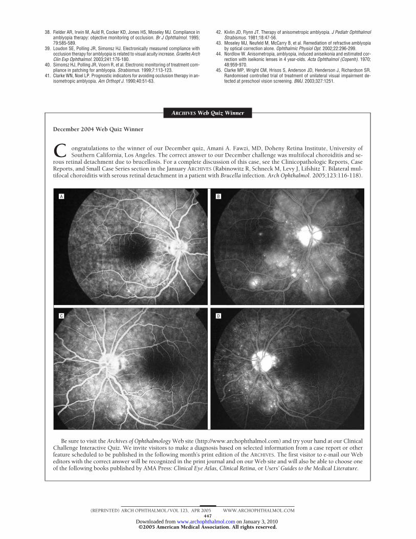
December 2004 Web Quiz Winner
C ongratulations to the winner of our December quiz, Amani A. Fawzi, MD, Doheny Retina Institute, University ofSouthern California, Los Angeles. The correct answer to our December challenge was multifocal choroiditis and se-
rous retinal detachment due to brucellosis. For a complete discussion of this case, see the Clinicopathologic Reports, CaseReports, and Small Case Series section in the January ARCHIVES (Rabinowitz R, Schneck M, Levy J, Lifshitz T. Bilateral mul-tifocal choroiditis with serous retinal detachment in a patient with Brucella infection. Arch Ophthalmol. 2005;123:116-118).
Be sure to visit the Archives of Ophthalmology Web site (http://www.archophthalmol.com) and try your hand at our ClinicalChallenge Interactive Quiz. We invite visitors to make a diagnosis based on selected information from a case report or otherfeature scheduled to be published in the following month’s print edition of the ARCHIVES. The first visitor to e-mail our Webeditors with the correct answer will be recognized in the print journal and on our Web site and will also be able to choose oneof the following books published by AMA Press: Clinical Eye Atlas, Clinical Retina, or Users’ Guides to the Medical Literature.
BA
DC
(REPRINTED) ARCH OPHTHALMOL / VOL 123, APR 2005 WWW.ARCHOPHTHALMOL.COM447
©2005 American Medical Association. All rights reserved. on January 3, 2010 www.archophthalmol.comDownloaded from