Synthesis of silver nanoparticles using the fish scales of Labeo rohita and their application for...
11
Biomimetic synthesis of silver nanoparticles using the fish scales of Labeo rohita and their application as catalysts for the reduction of aromatic nitro compounds Tanur Sinha, M. Ahmaruzzaman ⇑ , A.K. Sil, Archita Bhattacharjee Department of Chemistry, National Institute of Technology, Silchar-788010, Assam, India highlights Silver nanoparticles are synthesized using fish scales of Labeo rohita. Size tailoring of nanoparticles is achieved by varying the reaction parameters. The method is simple, economic, and environmental friendly. The catalytic property of silver nanoparticles is investigated in aqueous and micellar medium. Silver nanoparticles have excellent potential for catalytic reduction of aromatic nitro compounds. graphical abstract AgNO3 Fish scale extract 70degree 10 mins, cooled 3 days 4-NP BH4- Fast reaction LDS BH4- 4-NP Intermediate reaction TX-100 BH4- Slowest reaction CPC AgNP e- NO2 O- Slow O- NH2 4-NP BH4- article info Article history: Received 31 December 2013 Received in revised form 7 April 2014 Accepted 13 April 2014 Available online 30 April 2014 Keywords: Silver nanoparticles Fish scales Gelatin Nitro-compounds Reduction Micelle abstract In this article, a cleaner, greener, cheaper and environment friendly method for the generation of self assembled silver nanoparticles (Ag NPs) applying a simple irradiation technique using the aqueous extract of the fish scales (which is considered as a waste material) of Labeo rohita is described. Gelatin is considered as the major ingredient responsible for the reduction as well as stabilisation of the self assembled Ag NPs. The size and morphology of the individual Ag NPs can be tuned by controlling the various reaction parameters, such as temperature, concentration, and pH. Studies showed that on increasing concentration and pH Ag NPs size decreases, while on increasing temperature, Ag NPs size increases. The present process does not need any external reducing agent, like sodium borohydride or hydrazine or others and gelatin itself can play a dual role: a ‘reducing agent’ and ‘stabilisation agent’ for the formation of gelatin–Ag NPs colloidal dispersion. The synthesized Ag NPs were characterised by Ultraviolet–Visible spectroscopy (UV–Vis), Transmission electron microscopy (TEM) and Selected area electron diffraction (SAED) analyses. The synthesized Ag NPs was used to study the catalytic reduction of various aromatic nitro compounds in aqueous and three different micellar media. The hydrophobic and electrostatic interaction between the micelle and the substrate is responsible for the catalytic activity of the nanoparticles in micelle. Ó 2014 Elsevier B.V. All rights reserved. Introduction Noble metal nanoparticles (NPs) especially silver nanoparticles (Ag NPs) have shown enormous interest to many research groups for their fascinating applications in the field of electronics, catalysis, http://dx.doi.org/10.1016/j.saa.2014.04.065 1386-1425/Ó 2014 Elsevier B.V. All rights reserved. ⇑ Corresponding author. Tel./fax: +91 3842 224797. E-mail address: [email protected] (M. Ahmaruzzaman). Spectrochimica Acta Part A: Molecular and Biomolecular Spectroscopy 131 (2014) 413–423 Contents lists available at ScienceDirect Spectrochimica Acta Part A: Molecular and Biomolecular Spectroscopy journal homepage: www.elsevier.com/locate/saa
-
Upload
independent -
Category
Documents
-
view
0 -
download
0
Transcript of Synthesis of silver nanoparticles using the fish scales of Labeo rohita and their application for...

Spectrochimica Acta Part A: Molecular and Biomolecular Spectroscopy 131 (2014) 413–423
Contents lists available at ScienceDirect
Spectrochimica Acta Part A: Molecular andBiomolecular Spectroscopy
journal homepage: www.elsevier .com/locate /saa
Biomimetic synthesis of silver nanoparticles using the fish scalesof Labeo rohita and their application as catalysts for the reductionof aromatic nitro compounds
http://dx.doi.org/10.1016/j.saa.2014.04.0651386-1425/� 2014 Elsevier B.V. All rights reserved.
⇑ Corresponding author. Tel./fax: +91 3842 224797.E-mail address: [email protected] (M. Ahmaruzzaman).
Tanur Sinha, M. Ahmaruzzaman ⇑, A.K. Sil, Archita BhattacharjeeDepartment of Chemistry, National Institute of Technology, Silchar-788010, Assam, India
h i g h l i g h t s
� Silver nanoparticles are synthesizedusing fish scales of Labeo rohita.� Size tailoring of nanoparticles is
achieved by varying the reactionparameters.� The method is simple, economic, and
environmental friendly.� The catalytic property of silver
nanoparticles is investigated inaqueous and micellar medium.� Silver nanoparticles have excellent
potential for catalytic reduction ofaromatic nitro compounds.
g r a p h i c a l a b s t r a c t
AgNO3
Fish scale extract
70degree
10 mins,cooled 3 days
4-NPBH4
-
Fast reactionLDS
BH4-4-NP
Intermediate reactionTX-100
BH4-
Slowest reactionCPC
AgNP
e-
NO2
O-
Slow
O-
NH2
4-NP
BH4-
a r t i c l e i n f o
Article history:Received 31 December 2013Received in revised form 7 April 2014Accepted 13 April 2014Available online 30 April 2014
Keywords:Silver nanoparticlesFish scalesGelatinNitro-compoundsReductionMicelle
a b s t r a c t
In this article, a cleaner, greener, cheaper and environment friendly method for the generation of selfassembled silver nanoparticles (Ag NPs) applying a simple irradiation technique using the aqueousextract of the fish scales (which is considered as a waste material) of Labeo rohita is described. Gelatinis considered as the major ingredient responsible for the reduction as well as stabilisation of the selfassembled Ag NPs. The size and morphology of the individual Ag NPs can be tuned by controlling thevarious reaction parameters, such as temperature, concentration, and pH. Studies showed that onincreasing concentration and pH Ag NPs size decreases, while on increasing temperature, Ag NPs sizeincreases. The present process does not need any external reducing agent, like sodium borohydride orhydrazine or others and gelatin itself can play a dual role: a ‘reducing agent’ and ‘stabilisation agent’for the formation of gelatin–Ag NPs colloidal dispersion. The synthesized Ag NPs were characterised byUltraviolet–Visible spectroscopy (UV–Vis), Transmission electron microscopy (TEM) and Selected areaelectron diffraction (SAED) analyses. The synthesized Ag NPs was used to study the catalytic reductionof various aromatic nitro compounds in aqueous and three different micellar media. The hydrophobicand electrostatic interaction between the micelle and the substrate is responsible for the catalytic activityof the nanoparticles in micelle.
� 2014 Elsevier B.V. All rights reserved.
Introduction
Noble metal nanoparticles (NPs) especially silver nanoparticles(Ag NPs) have shown enormous interest to many research groupsfor their fascinating applications in the field of electronics, catalysis,

350 400 450 500 550 600 6500.0
0.5
1.0
1.5
2.0
2.5
At40perc
At30perc
At20perc
At10perc
Abs
orba
nce
(a.u
.)
Wavelength (nm)
Fig. 1. UV–Vis absorption spectra recorded of synthesized Ag NPs with different concentrations of fish scale extract (10%, 20%, 30%, and 40%) solution treated to 0.1 M AgNO3
solution. The colours of the solution shown in the inset after 3 days in increasing order of concentration from left to right. (For interpretation of the references to colour in thisfigure legend, the reader is referred to the web version of this article.)
400 500 600 7000
1
2
At60C
At70C
At80C
At90C
At100C
Abs
orba
nce
(a.u
.)
Wavelength (nm)
Fig. 2. UV–Vis absorption spectra recorded for Ag NPs at different temperatures (60, 70, 80, 90 and 100 �C) when 10 ml of 0.1 M silver nitrate solution treated to 10 ml of 10%extract. The colours of the solution after 3 days in increasing order of temperature from left to right. (For interpretation of the references to colour in this figure legend, thereader is referred to the web version of this article.)
414 T. Sinha et al. / Spectrochimica Acta Part A: Molecular and Biomolecular Spectroscopy 131 (2014) 413–423
and pharmaceuticals etc. [1]. These applications are based on differ-ent chemical and physical properties attributed with nanomaterialsdue to their unique electronic structure and extremely large surfacearea which were not observed from those of the bulk and atomicspecies. However, various methods have been developed [2] forthe synthesis of Ag NPs in organic solvents, using toxic reducingagents like sodium borohydride and N, N-dimethyl formamide.
Hence, for cleaner preparation of these noble metal nanoparti-cles, various biomimetic approaches [3,4] are being explored.Green technology, in the niche of preparative protocols of nanoma-terial stands for switching to environmentally benign startingmaterial and reducing agent, water as the reaction medium andselection of non-toxic substances of Ag NPs stability [5] withminimal wastage in terms of energy and raw materials [6–27].
Hence, in this paper, we develop an ecofriendly, clean,non-toxic, facile chemically preparative method, for the generationof Ag NPs using the extract of fish scales of Labeo rohita, acting as
reducing as well as stabilising agent. The effects of various reactionparameters, such as temperature, pH and concentration of theextract in the synthesis of Ag NPs were also investigated. We havealso studied the catalytic activity of these synthesized Ag NPs inthe reduction of several aromatic nitro compounds in aqueousand micellar medium. The present synthesis process and the catal-ysis reaction are simple, environment friendly, and robust.
Experimental details
Materials
Silver nitrate (AgNO3), p-nitro phenol (4-NP), o-nitro phenol(2-NP), p-nitro aniline (4-NP), sodium borohydride (NaBH4),lithium dodecyl sulphate (LDS), cetyl pyridinium chloride (CPC),and triton x-100 (TX-100) of A.R Grade was purchased from

T. Sinha et al. / Spectrochimica Acta Part A: Molecular and Biomolecular Spectroscopy 131 (2014) 413–423 415
Sigma–Aldrich and used as received. Double distilled water wasused in all experiments.
Preparation of fish scale extract
2 g of Labeo rohita fish scales were thoroughly washed andboiled for 20 min at 60 �C in 100 ml double distilled water. Theresulting solution was filtered and used for further experiments.
Synthesis of silver nanoparticles
10 ml of 0.1 M aqueous solution of silver nitrate was taken in acontainer and 10 ml of different concentrations of fish scale extract(10%, 20%, 30% and 40%) were added separately and heated at 70 �Cfor 10 min followed by slow cooling of the solutions to room tem-perature. The solution was then allowed to stabilize for 3 days.After 3 days the solutions with brown sediments were formed atthe bottom of the container which indicated the presence of AgNPs. These were then centrifuged, filtered and the residues werewashed several times with double distilled water to removeunbound polymers to yield the Ag NPs. Also samples of Ag NPswere prepared by adding 10 ml of 0.1 M aqueous solution of silvernitrate to 10 ml of 10% of fish scale extract and heating at differenttemperatures (60, 70, 80, 90 and 100 �C) separately. Another set ofsamples of Ag NPs were prepared by heating 10 ml of 0.1 M aque-ous solution of silver nitrate and 10 ml of 10% of fish scale extractat 70 �C and keeping the pH of the stock solutions at (0.5, 1, 1.5, 2, 3and 4) separately.
Characterisation of silver nanoparticles
The UV–Vis absorption spectra of the synthesized Ag NPs wererecorded on Cary 100 Bio spectrophotometer (kmax in nm)equipped with 1-cm quartz cell. The TEM, HR-TEM images, andSAED pattern were recorded using JEOL-JEM 2100 transmissionelectron microscope operated at an accelerating voltage of200 kV. The TEM samples of Ag NPs were prepared by placingthe solution drops over the carbon coated copper grids and allow-ing the solvent to evaporate at room temperature.
400 500 6000.0
0.2
0.4
0.6
0.8
1.0
1.2
1.4
1.6 Atp
Atp
Abs
orba
nce
(a.u
.)
Wavelength (nm)
Fig. 3. UV–Vis absorption spectra recorded for Ag NPs at different pH(3 and 4) when 10 mof the solution after 3 days in increasing order of pH from left to right. (For interpretatioversion of this article.)
Catalytic activity of the synthesized silver nanoparticles
In a standard quartz cuvette, having 1 cm path length, 2 ml ofwater and 60 ll of (6.07 � 10�3 M) nitro compounds (4-nitro phe-nol, 2-nitro phenol, and 4-nitro aniline) were taken separately andthe cuvette was then placed in a UV–Vis spectrometer and theabsorbance was recorded. To each of these nitro compound solu-tions, 350 ll of aqueous NaBH4 (0.1 M) was added and the absor-bance was noted. Thereafter, 150 ll of 10% Ag NPs solution wasadded to that mixture and the absorbance was recorded up to20 m when the peak due to the nitro compounds was no longerobserved and the appearance of a new peak at �430 nm wasnoticed.
Results and discussion
UV–Vis analysis
The Ag NPs exhibits a unique UV–Visible adsorption bandderived from collective oscillation of conduction electrons uponinteraction with electromagnetic radiation, which is known aslocalised surface plasmon resonance (LSPR). The shifting of thesebands provides information of the particle size, chemical surround-ing, adsorbed species on the surface and dielectric constant. Fig. 1shows the UV–Visible spectra of the synthesized Ag NPs withdifferent concentration of fish scale extract (10%, 20%, 30%, and40%).
The spectra revealed the presence of Ag NPs in all the mixturesand the optical properties exhibited by the Ag NPs (shown in theinset of Fig. 1) clearly indicates that the particles may have differ-ent size or shape (Morphology). At low concentration, the absorp-tion band (�420 nm) is originating mostly from surface Plasmonvibrations of spherical Ag NPs. However with increase in concen-tration, the absorption bands become broader with increasedintensity and shifted to longer wavelength region (�450 nm). Thebroad absorption indicates the aggregation of spherical NPs or achange in the dimension of spherical anisotropic nanostructures,while an increased intensity reflects that the percentage of popula-tion of the particles are increasing with increase in concentrationof the extract in the solution.
700
H4
H3
l of 0.1 M silver nitrate solution treated to 10 ml of 10% extract at 70 �C. The coloursn of the references to colour in this figure legend, the reader is referred to the web

Fig. 4. TEM images of Ag NPs synthesized with different concentrations of fish scale extract (a) 10% (b) magnified image of (a), (c) 40% (d) magnified image of (c, e and f)selected area electron diffraction (SAED) pattern of the TEM image shown in (a) and (c) respectively.
416 T. Sinha et al. / Spectrochimica Acta Part A: Molecular and Biomolecular Spectroscopy 131 (2014) 413–423
Fig. 2 represents the UV–visible spectra of Ag NPs formed atdifferent temperatures (60, 70, 80, 90, and 100 �C). The spectrarevealed the formation of Ag NPs possessing different size andmorphology. At low temperature (60 �C), a band due to the surfacePlasmon resonance of Ag NPs appeared at about �420 nm andshifted to longer wavelength (red shift) with an increase in broad-ness of the band at high temperature (100 �C). Usually, a red shift is
associated with an increase in particle size or with the withdrawalof electron density from the surface [28–33]. Further, the opticalproperties exhibited by the solutions of Ag NPs (shown in insetof Fig. 2) indicated that the particles may have different size orshape (morphology). This increase in broadness reflects a changein dimension of spherical anisotropic nanostructures or aggrega-tion of Ag NPs.

T. Sinha et al. / Spectrochimica Acta Part A: Molecular and Biomolecular Spectroscopy 131 (2014) 413–423 417
Fig. 3 shows the UV–visible spectra of Ag NPs formed at differ-ent pH (3 and 4). At low pH (pH = 3), a band due to the surfacePlasmon resonance of Ag NPs appeared at about �440 nm whichshifted to shorter wavelength (blue shift) at high pH (pH = 4).Usually, a blue shift is associated with a decrease in particle sizeor donation of electron density from the surface [34]. These resultsshow that concentration, temperature and pH are importantfactors for the formation and size distribution of nanoparticles.
TEM and SAED studies
The morphology and size of the Ag NPs have been analysed usingTEM and SAED pattern. Fig. 4a and c corresponds to the TEM imagesof the synthesized Ag NPs using 10% and 40% fish scale extractrespectively. The SAED pattern of Fig. 4a and c is shown in Fig. 4b,d, e, and f, are the magnified images of Fig. 4a and c, respectively.Fig. 4a and c display the formation of spherical Ag NPs. It is apparentfrom the TEM images that spherical Ag NPs are polydispersed innature with the average size of spherical NPs �17.47, and�16.5 nm. Thus with increase in concentration of the extract, parti-cle size of Ag NPs decreases. The polycrystalline nature of nanopar-ticles can be evidenced from the concentric diffraction rings in aSAED pattern as shown in Fig. 4e and f, respectively. The spots inFig. 4e could be indexed corresponding to the reflections from the
Fig. 5. TEM images of the Ag NPs synthesized when 10% extract solution treated to 0.1 Mpattern of TEM image shown in (a) and (b) respectively.
(111) and (200) lattice planes, while the spots in Fig. 4(f) couldbe indexed corresponding to the reflections from the (111), (200)(220), and (311) lattice planes. The lattice spacing calculated fromFig. 4b and d is almost 0.26 and 0.23 nm and corresponds to (111)lattice plane of face centered cubic (FCC) structure of silver [35].
TEM images of the synthesized Ag NPs at 60 �C and 100 �C arepresented in Fig. 5a and b, respectively. The SAED pattern ofFig. 5a and b is shown in Fig. 5c and d. The magnified images ofFig. 5a and b are presented in Supplementary material as shownin Fig. A (1) and A (2), respectively. Fig. 5a and b displayed the for-mation of spherical Ag NPs. It is apparent from the TEM image thatspherical Ag NPs are polydispersed with an average size of spher-ical NPs �9.36, and �22.96 nm. Hence, average particle size of AgNPs increases with increase in temperature. The concentric diffrac-tion rings in SAED pattern shown in Fig. 5c and d reveals thepolycrystalline nature of particles. The spots could be indexed cor-responding to the reflections from the (111), (200), and (111),(200), (220), and (311) lattice planes as shown in Fig. 5c and d,respectively. The fringe spacing calculated from Fig. A (1) and A(2) are 0.21 nm and 0.23 nm, respectively and corresponds to(111) lattice plane of face centered cubic (FCC) structure of silver[35].
TEM images of the synthesized Ag NPs at pH = 3 and pH = 4 arepresented in Fig. 6a and b, respectively. Fig. 6c and d are the SAED
silver nitrate solution at different temperatures (a) 60 �C, (b) 100 �C (c) and (d) SAED

Fig. 6. TEM images of the Ag NPs synthesized when 10% extract solution treated to 0.1 M silver nitrate solution heated at 70 �C at (a) pH = 3, (b) at pH = 4, (c) and (d) SAEDpattern of TEM image shown in (a) and (c) respectively.
250 300 350 400 450 500 550 600
0.0
0.5
1.0
1.5
2.0
2.5
Abs
orba
nce
(a.u
.)
Wavelength (nm)
Fig. 7. UV–Vis absorption spectra recorded of Ag NPs.250 300 350 400 450 500
0.0
0.5
1.0
1.5
2.0
2.5
Abs
orba
nce
(a.u
.)
Wavelength (nm)
Fig. 8. UV–Vis absorption spectra recorded of 60 ll (6.07 � 10�3 M) 4-NP inaqueous solution.
418 T. Sinha et al. / Spectrochimica Acta Part A: Molecular and Biomolecular Spectroscopy 131 (2014) 413–423
pattern of Fig. 6a and b, respectively. Fig. B (1) and B (2) (shown inSupplementary material) are the magnified images of Fig. 6a and b,respectively. Fig. 6a and b indicated the formation of spherical Ag
NPs. It is apparent from the TEM image that spherical Ag NPs arepolydispersed in nature with the average size of spherical NPs�46.14 nm, and �10.33 nm. Therefore, on increase in pH of thesolution, average particle size decreases. Polycrystalline nature ofparticles can be evidenced from the concentric diffraction ringsin a SAED pattern shown in Fig. 6c and d. The spots could beindexed corresponding to the reflections from the (111), (200),

200 250 300 350 400 450 500 550 600 6500.0
0.5
1.0
1.5
2.0
2.5
3.0
Initial0sec At300secs At600secs At900secs At1200secs
Abs
orba
nce
(a.u
.)
Wavelength (nm)
Fig. 9. UV–Vis absorption spectra recorded of 60 ll (6.07 � 10�3 M) 4-NP + 350 ll(0.1 M) NaBH4 + 150 ll 10% Ag NPs in aqueous solution.
300 400 500 600 700 800 900
-0.6
-0.5
-0.4
-0.3
-0.2
ln (
At/A
0)
Time (secs)
Fig. 10. Plot of ln (At/A0) against time for pseudo-first-order reduction kinetics of4-NP in the presence of excess NaBH4 (0.1 M) in aqueous solutions.
T. Sinha et al. / Spectrochimica Acta Part A: Molecular and Biomolecular Spectroscopy 131 (2014) 413–423 419
(220) and (311) lattice planes in Fig. 6c and (111), (200), (220)lattice planes in Fig. 6d respectively. The lattice spacing calculatedfrom Fig. B(1) and B(2) are found to be 0.25 nm and 0.23 nm,respectively, both of which corresponds to (111) lattice plane offace centered cubic (FCC) structure of silver [35]. From all theTEM images it is found that the Ag NPs are predominantly spheri-cal in shape. These spherical NPs also consist of dark regions sur-rounded by a faint thin shell with a capping agent possibly oforganic material (gelatin) present in the extract. Hence, from theabove it is apparent that particle size can be tuned by controllingthe reaction parameters.
Probable mechanism for the formation of Ag NPs
It is well known that aqueous AgNO3 is not a stable chemicaland can be decomposed easily either by heating or by UV light illu-mination [36]. Under such conditions, Ag (0) is the sole productafter realising oxygen and nitrogen dioxide and in such case theAg (0) formed non-uniform micron-size Ag particles in the absenceof any stabilising agent. So, the presence of stabilising agent is verymuch essential for the synthesis of Ag NPs. Here, fish scale extractserves as both reducing as well as stabilising agent.
Fish scales are biocomposites of high ordered, type-I collagenfibres (41–84%) and hydroxyapatite Ca10(OH)2 (PO4)6 [37–39].Type-I collagen is a heterotrimeric copolymer composed of twoa1 (I) and one a2 (I) polypeptide chains, containing approximately1050 amino acids each. Each polypeptide chain has a conformationof a left handed, polyproline-II –type helix, which are twistedtogether into a right handed coil, a triple helix which representsa quaternary structure, being stabilised by numerous hydrogenbonds and intramolecular vander waals interactions [40] as wellas by some covalent bonds [41], and further associated into right–handed microfibrils and fibrils, being further assembled into col-lagen fibres [42] with unusual strength and stability.
When the aqueous solution of these fish scales is heated at60 �C for 20 min, the collagen present in these scales get denaturedto a mixture of random-coil single, double and triple strands [43]which is accompanied by change in physical and chemical proper-ties due to collapse of the triple helical structure. Such modifiedcollagen is called denatured collagen (gelatin) and can be evi-denced from Fig. C (Supplementary material), where a peak at�270 nm corresponds to the absorption of denatured collagen orgelatin [44]. These collagens are made up of polypeptide chains,containing amino acids, having variable compositions, particularlyfor the minor constituents, depending on raw materials andprocesses used, proximate value by weight are [45] glycine 21%,proline 12%, hydroxyproline 12%, glutamic acid 10%, alanine 9%,arginine 8%, aspartic acid 6%, lysine 4%, serine 4%, leucine 3%, valine2%, phenyl alanine 2%, threonine 2%, isoleucine 1%, hydroxylysine1%, methionine and histidine <1% with tyrosine <0.5%, and containfunctional groups such as suspended double bonds, –NH2, –SH and–COOH endowing it as a reducing as well as stabilising agent.
It is believed that the mechanism for the formation of Ag NPsproceeds via 2 steps as shown in Scheme 1. In the first step, whenAgNO3 solution is added to the fish scale extract, they formed aninstantaneous gelatin-Ag+ complex where the positively chargedAg (I) ions were initially bound with the negatively charged groupof gelatin via electrostatic interaction which can be evidenced bythe shift of the absorption of the UV–visible spectra (Fig. D) (Sup-plementary material) by 5–6 nm as well as dramatic increase inthe absorption intensity as compared to the absorption band ofpure gelatin (Fig. C) (Supplementary material). In addition, a weakhump appeared at around 350–400 nm due to the formation of Ag(I)-gelatin complex. The next step involves the heating of thesolution mixture for 10 min, so that the formed complex startsreducing and ultimately self-assembled Ag NPs were formed after
cooling for 3 days at room temperature (Fig. 7). we believe that thereduction mechanism of all the Ag (I) ions proceed via the autocat-alytic growth route, where once a metal atom is evolved as anucleation centre it can act as a catalyst for the reduction of theremaining metal ions present in the solution via autocatalysis [46].
Thus, it was observed that gelatin itself acted as a reducing aswell as stabilising agent for the formation of self-assembled AgNPs without the addition of reducing agent like NaBH4, sugar,hydrazine etc.
Catalytic reduction of 4-NP, 2-NP and 4-NA using self-assembled AgNPs on gelatin as a catalyst in aqueous medium
To investigate the catalytic efficiency of self-assembled Ag NPson gelatin in depth, the reduction of 4-NP was monitored in thepresence of NaBH4 as a model reaction [47,48]. The reduction of4-NP [E0 (4-NP/4-AP) = �0.76 V] by NaBH4 [E0 (H3BO3/BH4
�) =�1.33 V] is thermodynamically feasible but kinetically restrictedin the absence of a catalyst.
It is observed that 4-NP in aqueous medium has a maximumabsorption at 317 nm as shown in Fig. 8. However, when freshlyprepared NaBH4 solution is added, there was a red shift from317 nm to 403 nm, and the light yellow colour of the solutionchanges to intense yellow (Fig. E) (Supplementary material), due

0 300s 600s 900s 1200s
0.60.81.01.21.41.61.82.02.22.42.62.83.03.23.43.6
Time (secs)
Abs
orba
nce
at 4
03 n
m
Water LDS CPC TX100
Fig. 12. Absorbance versus time (secs) plot for the reduction of 4-NP prompted byAg NPs as catalyst concentration of LDS = CPC = TX-100 = 60 � 10�3 M, 4-
420 T. Sinha et al. / Spectrochimica Acta Part A: Molecular and Biomolecular Spectroscopy 131 (2014) 413–423
to the formation of 4-nitrophenolate ions in alkaline condition. Thepeak at 403 nm remains unchanged even for a couple of days in theabsence of any catalyst.
Addition of 150 ll of 10% Ag NPs solution into the reaction mix-ture caused a gradual fading of the characteristic yellow colour dueto 4-NP and finally complete bleaching of the yellow colour of the4-NP solution was observed. This decolourisation was quantita-tively monitored spectrophotometrically with time and it has beennoted as a successive decrease of peak height. This is due to thereduction of 4-NP to 4-amino phenol (4-AP). A new peak centredat 420 nm due to the surface Plasmon resonance of Ag NPs wasobserved after completion of the reduction (at 1200 s). In the meantime, a new peak at 298 nm appeared with progressive increase inintensity (Fig. 9), which is attributed to the typical absorption of4-AP. This results suggests that the catalytic reduction of 4-NPexclusively yielded 4-AP without any side products [49].
In the reduction process, the overall concentration of NaBH4
was 0.1 M and 4-NP was 6.07 � 10�3 M. Considering much higherconcentration of NaBH4 compared to 4-NP, the pseudo-first-order
Silver nanoparticle Ag NP
BH4-
H
H
HH
H H
H H
Boride ions adsorbed on thesurface of AgNP & transfer asurface-hydrogen species tothe surface of AgNP
4-NP
H
H
HH
H
H H
H 4-NP
At the same time 4-NP adsorbs on to the surface of AgNP & adsorption/desorption of both the reagents on thesurface is fast following LangmuirIsotherm
4-AP
Final product 4-AP desorbs leaving AgNP surface& thecatalytic cycle can begin again
O-
NO2
4-Nitrophenol(4-NP)
NaBH4-
Ag NP
NH2
OH
4-Aminophenol(4-AP)
Fig. 11. Representation of Langmuir-Hinshelwood (LH) model for the mechanism of the reduction of 4-NP to 4-AP by NaBH4 in presence of Ag NPs.
NP = 6.07 � 10�3 M, NaBH4 = 0.1 M & Ag NP = 10%.
T. Sinha et al. / Spectrochimica Acta Part A: Molecular and Biomolecular Spectroscopy 131 (2014) 413–423 421
kinetics has been applied with respect to 4-NP to determine thecatalytic activity of Ag NPs. The absorbance of 4-NP is proportionalto its concentration in solution; the absorbance at time t (At) andtime t = 0 (A0) are equivalent to the concentration at time t (Ct)and time t = 0 (C0). The rate constant (k) has been determined fromthe linear plot of ln (At/A0) versus reduction time in seconds, andthe constant was estimated to be 6.0 � 10�4 s�1 (Fig. 10).
Reduction of 2-nitro phenol (2-NP) and 4-nitro aniline (4-NA)(each having concentration 6.07 � 10�3 M), was also examinedusing 0.1 M NaBH4 and 10% Ag NPs as catalyst (spectra not shown).The yellow colour due to 2-NP and 4-NA was discharged during thecourse of the reduction and their absorbance values at their respec-tive kmax positions decreased gradually in presence of NaBH4 andAg NPs as catalyst. It was found that two new peaks graduallyappeared for 4-nitro aniline (4-NA) at 300 and 209 nm, both ofwhich are due to p-phenylenediamine in alkaline aqueous solu-tions [50]. Similarly, in aqueous alkaline solution, a new peakwas observed at 406 nm due to 2-nitrophenolate ion and the peak
4-NPBH4
-
Fast reactionLDS
Slowest reactionCPC
AgNP
e-
O-
N
4-NP
BH4-
Fig. 13. Schematic representation of electron t
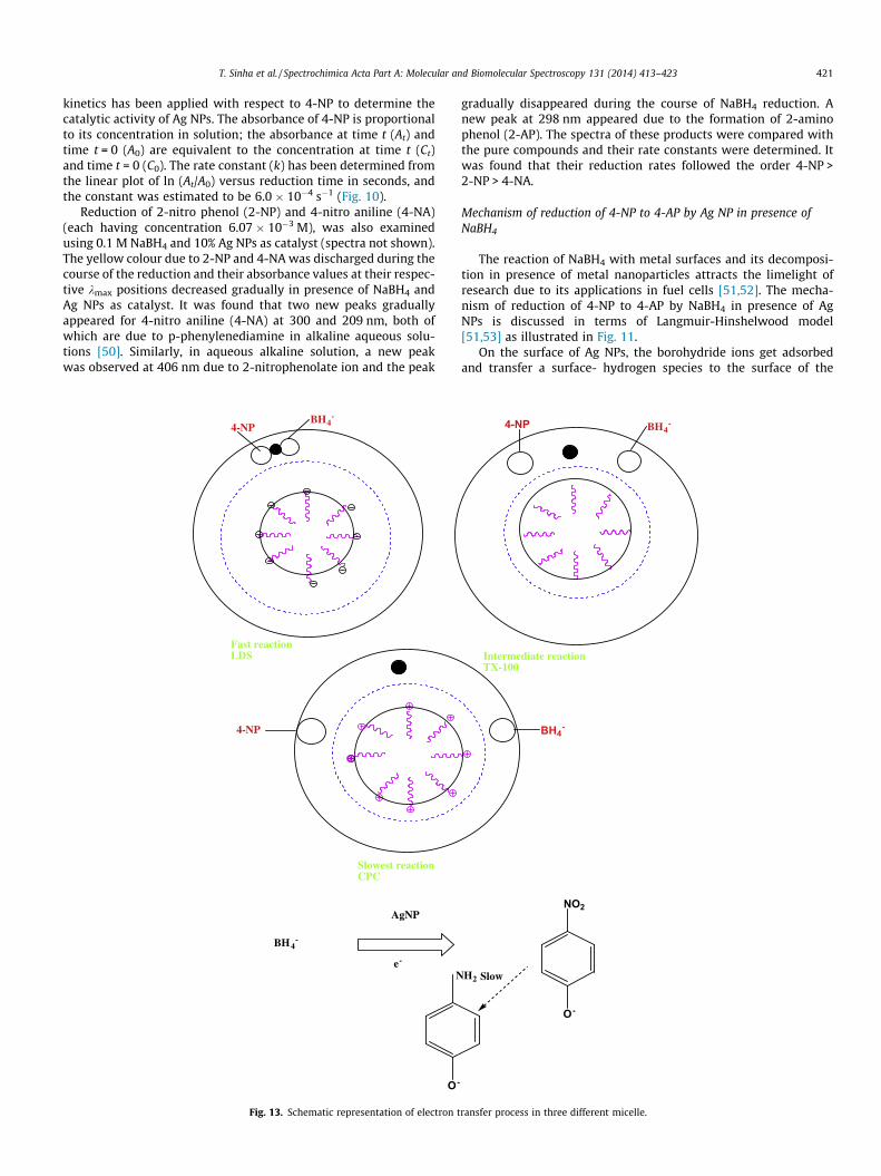
gradually disappeared during the course of NaBH4 reduction. Anew peak at 298 nm appeared due to the formation of 2-aminophenol (2-AP). The spectra of these products were compared withthe pure compounds and their rate constants were determined. Itwas found that their reduction rates followed the order 4-NP >2-NP > 4-NA.
Mechanism of reduction of 4-NP to 4-AP by Ag NP in presence ofNaBH4
The reaction of NaBH4 with metal surfaces and its decomposi-tion in presence of metal nanoparticles attracts the limelight ofresearch due to its applications in fuel cells [51,52]. The mecha-nism of reduction of 4-NP to 4-AP by NaBH4 in presence of AgNPs is discussed in terms of Langmuir-Hinshelwood model[51,53] as illustrated in Fig. 11.
On the surface of Ag NPs, the borohydride ions get adsorbedand transfer a surface- hydrogen species to the surface of the
BH4-4-NP
Intermediate reactionTX-100
BH4-
NO2
O-
SlowH2
ransfer process in three different micelle.

Gelatin + Ag+ Gelatin-Ag+ complex
Heat,10min,Cooled for 3 days
Self-assembledSilver nanoparticle
Scheme 1. Mechanism for the formation of self-assembled Ag NPs.
422 T. Sinha et al. / Spectrochimica Acta Part A: Molecular and Biomolecular Spectroscopy 131 (2014) 413–423
nanoparticles [51,54]. It is a reversible step and can be explained interms of a Langmuir isotherm [51]. The 4-NP molecules areadsorbed on the surface of the nanoparticles at the same time. Itis also a reversible step and can also be explained in terms of Lang-muir isotherm [51]. The adsorption/desorption equilibriums anddiffusion of reactants to the nanoparticles are considered to be fast[51]. The reduction of 4-NP is the rate determining step whichoccurs due to the reaction of adsorbed 4-NP with the nanoparticlessurface-bound hydrogen atoms (Fig. 11). When the final product,4-AP undergoes desorption, the metal surface is set free and thecatalytic cycle begins again [51].
Catalytic reduction of 4-NP using self-assembled Ag NPs on gelatin as acatalyst in micellar medium
Micelles not only increase the suspendibility of metal nanopar-ticles (stabilisation) but also refresh the surface of catalyst particles[55–57] and thereby facilitate in studying the reaction kinetics [58]for heterogeneous catalysis. It is also used in various ratios to con-trol the particle shape and size [59]. However, the exact mecha-nism of interaction between metal particles and the surfactantstabilizer has not yet been resolved [60,61]. To study the interac-tions between the substrates and the catalyst, the reduction of4-NP was meticulously examined in three different micelles.
In micelles, a weak hydrophobic force operates between thesubstrate (4-nitrophenolate ion) and the micelle. This hydrophobicinteraction restricts the easy approach of the substrate towards themetal nanoparticle surfaces. In addition, micelles build wrapper-like barriers surrounding the metal particles, whereas BH4
� tendsto remain outside the dynamic barrier due to lack of hydrophobicpart. Accordingly, lowering the reduction rate in micelles in com-parison to the reduction rate in aqueous medium for a particularmetal is perceivable.
Adsorption of the phenolate ion onto the nanoparticle surfacedoes not get disturbed, when the aqueous system is replaced byan anionic micellar solution, lithium dodecyl sulphate (LDS). Thisis due to the negatively charged micellar surface that preventedincorporation of the substrate (4-NP) into the LDS micelle. As aresult, the reduction rate in LDS rendered itself comparable to thatin aqueous medium. Due to the presence of both hydrophobic andhydrophilic interactions between the phenolate ion and thepositively charged micellar surface in cationic micelle cetyl pyrid-inium chloride (CPC), adsorption of the substrate onto the catalystwas retarded which decreased the rate to a considerable extent.Whereas hydrophobic interaction becomes operative in non-ionicmicelle, Triton-X-100 (TX-100), between the substrate and theuncharged micellar surface, which give rise to an intermediate rateof reaction. Therefore, the rate of reaction in three different micellefollowed the sequence of H2O � LDS > TritonX-100 > CPC. Theabsorbance of 4-NP as a function of time in three different micelle(just above CMC) has been depicted in Fig. 12. From the shift of
acid–base equilibrium [62], the location of the substrate, phenolateions in micelle can be quantified. Finally, Fig. 13 schematically rep-resents the location of the substrate (4-NP), BH4
�, and Ag NPs inthree different micellar surfaces and electron transfer reaction.
Conclusion
In this paper, for the first time we have successively developedan environment friendly, green, cheap and simple method for thesynthesis of self-assembled Ag nanoparticles using a waste mate-rial extract (fish scale extracts) of Labeo rohita which itself playeda dual role of stabilising and reducing agent. Thus, the presentmethod does not require the addition of any external reducingagents, such as sodium borohydride or hydrazine or others. Thesizes of the Ag NPs were tuned just by controlling the variousreaction parameters, such as temperature, concentration and pH.It is observed from the TEM images that spherical Ag NPs arepolydispersed in nature with the average size of spherical NPs of�16.5, and �17.47 nm using 10% and 40% fish scale extract, respec-tively. It was also found that average size of spherical Ag NPsincreased with increase in temperature and found to be �9.36,and �22.96 nm, at 60 �C and 100 �C, respectively. The TEM studiesindicated that the average size of Ag NPs are �46.14 nm, and�10.33 nm, at pH = 3 and 4, respectively. The results showed thatconcentration, temperature and pH are important factors for theformation and size distribution of nanoparticles. These Ag NPswere utilised as a catalyst for the reduction of several aromaticnitro-compounds into their corresponding amino derivatives. Thisprocedure for the synthesis of Ag NPs is useful for large-scale syn-thesis. This study gives a clear insight of heterogeneous catalysiseven in micellar media.
Acknowledgements
Tanur Sinha wants to acknowledge TEQIP-II of NIT Silchar forproviding the financial assistance and SAIF–NEHU Shillong forproviding the TEM facilities.
Appendix A. Supplementary material
Supplementary data associated with this article can be found, inthe online version, at http://dx.doi.org/10.1016/j.saa.2014.04.065.
References
[1] N.L. Rosi, C.A. Mirikin, Chem. Rev. 105 (2005) 1547–1562.[2] X. Chen, H.J. Schluesener, Toxicol. Let. 176 (2008) 1–12.[3] V.K. Sharma, R.A. Yngard, Y. Lin, Adv. Colloid Interface Sci. 145 (2009) 83–96.[4] H. Korbekandi, S. Iravani, S. Abbasi, Crit. Rev. Biotechnol. 29 (2009) 279–306.[5] P. Raveendran, J. Fu, S.L. Wallen, J. Am. Chem. Soc. 125 (2003) 13940–13941.[6] K.F. Schmidt, ‘‘Green Nanotechnology: It’s Easier than you think’’ Presentation
given at Green Nanotechnology Event Hosted by the Projects on EmergingNanotechnologies at the Woodrow Wilson International Center for Scholar,2007.
[7] A. Mittal, J. Mittal, A. Malviya, V.K. Gupta, J. Colloid Interface Sci. 344 (2010)497–507.
[8] V.K. Gupta, S. Agarwal, T.A. Saleh, J. Hazard. Mater. 185 (2011) 17–23.[9] A. Mittal, L. Kurup (Krishnam), V.K. Gupta, J. Hazard. Mater. 117 (2005) 171–
178.[10] V.K. Gupta, A. Mittal, L. Kurup, J. Mittal, J. Colloid Interface Sci. 304 (2006) 52–
57.[11] V.K. Gupta, A. Mittal, L. Krishnam, J. Mittal, J. Colloid Interface Sci. 293 (2006)
16–26.[12] V.K. Gupta, R. Jain, S. Varshney, J. Hazard. Mater. 142 (2007) 443–448.[13] V.K. Gupta, R. Jain, S. Varshney, J. Colloid Interface Sci. 312 (2007) 292–296.[14] V.K. Gupta, I. Ali, V.K. Saini, Water Res. 41 (2007) 3307–3316.[15] V.K. Gupta, A. Rastogi, J. Hazard. Mater. 163 (2009) 396–402.[16] V.K. Gupta, A. Mittal, A. Malviya, J. Mittal, J. Colloid Interface Sci. 335 (2009)
24–33.[17] S. Karthikeyan, V.K. Gupta, R. Boopathy, A. Titus, G. Sekaram, J. Mol. Liq. 173
(2012) 153–163.

T. Sinha et al. / Spectrochimica Acta Part A: Molecular and Biomolecular Spectroscopy 131 (2014) 413–423 423
[18] V.K. Gupta, I. Ali, T.A. Saleh, A. Nayak, S. Agarwal, RSC Adv. 2 (2012) 6380–6388.
[19] T.A. Saleh, V.K. Gupta, Environ. Sci. Pollut. Res. 19 (2012) 1224–1228.[20] A.K. Jain, V.K. Gupta, S. Jain, Suhas, Environ. Sci. Technol. 38 (2004) 1195–
1200.[21] V.K. Gupta, S. Sharma, Ind. Eng. Chem. Res. 42 (2003) 6619–6624.[22] V.K. Gupta, B. Gupta, A. Rastogi, S. Agarwal, A. Nayak, J. Hazard Mater. 186
(2011) 891–901.[23] A.K. Jain, V.K. Gupta, A. Bhatnagar, Suhas, Sep. Sci. Technol. 38 (2003) 463–
481.[24] A. Mittal, V.K. Gupta, A. Malviya, J. Mittal, J. Hazard. Mater. 151 (2008) 821–
832.[25] V.K. Gupta, R. Jain, A. Mittal, M. Mathur, S. Sikarwar, J. Colloid Interface Sci. 309
(2007) 464–469.[26] A. Mittal, J. Mittal, D. Kaur, V.K. Gupta, J. Colloid Interface Sci. 343 (2010) 463–
473.[27] V.K. Gupta, A. Rastogi, A. Nayak, J. Colloid Interface Sci. 342 (2010) 533–539.[28] T. Linnert, P. Mulvaney, A. Henglein, J. Am. Chem. Soc. 112 (1990) 4657–
4664.[29] P. Mulvaney, A. Henglein, J. Phys. Chem. 94 (1990) 4182–4188.[30] T. Linnert, P. Mulvaney, A. Henglein, J. Phys. Chem. 97 (1993) 679–682.[31] A. Henglein, J. Phys. Chem. 97 (1993) 5457–5471.[32] F. Strelow, A. Henglein, J. Phys. Chem. 99 (1995) (1838) 11834–11838.[33] A. Henglein, Chem. Mater. 10 (1998) 444–450.[34] S. Raza, N. Stenger, S. Kadkhodazadeh, S.V. Fischer, N. Kostesha, A.P. Jauho, A.
Burrows, M. Wubs, N.A. Mortensen, Nanopho-tonics 2 (2013) 131–138.[35] M.G. Guzman, J. Dille, S. Godet, World Acad. Sci. Eng Technol. 67 (2008) 357–
364.[36] S. Kundu, Phys. Chem. Chem. Phys. 15 (2013) 14107–14119.[37] H. Onozato, N. Watabe, Cell Tissue Res. 201 (1980) 303–316.[38] L. Zulberberg, J.B. Hahn, J.Y. Sire, Cell Tissue Res. 253 (1988) 597–607.[39] T. Ikoma, H. Kobayashi, J. Tanaka, D. Walsh, S. Mann, J. Struct. Bio. 142 (2003)
327–333.
[40] J. Brinckmann, H. Notbohm, P.K. Mueller (Eds.), Collagen Primer in Structure,Processing And Assembly, Springer, 2005, pp. 1–252.
[41] R.D. Harkness, ‘‘Collagen’’, Science Progress. 54 (1966) 257–274.[42] L. He, C. Mu, J. Ski, Q. Zhang, B. Shi, Q. Lin, Int. J. Biol. Macromol. 48 (2011) 354–
359.[43] B.K. Simpson, S. Benjakul, P. Kittiphattanabawon, J.M. Regenstein, ‘‘Food
Biochemistry and Food processing’’, second ed., 2012 (21st Chapter (Fishgelatin)).
[44] L. O’Donovan, P.A. De Bank, J. Mater. Chem. 22 (2012) (1884) 21878–21884.[45] A. Sharma, P. Dour, P. Gupta, World J. Microbial Biotechnol. 22 (2006) 1049–
1054.[46] A. Henglein, Chem. Rev. 89 (1989) 1861–1873.[47] J. Liu, G. Qin, P. Raveendran, Y. Ikushima, Chem. Eur. J. 12 (2006) 2131–2138.[48] A. Murugadoss, A. Chattopadhyay, J. Phys. Chem. C. 112 (2008) 11265–11271.[49] Q. An, M. Yu, Y. Zhang, W. Ma, J. Guo, C. Wang, J. Phys. Chem. C. 116 (2012)
22432–22440.[50] J.P. Phillips, H. Feuer, B.S. Thyagasrajan, Org. Electron. Spect. Data 10 (1968)
50–58.[51] S. Wunder, F. Polzer, Y. Lu, Y. Mei, M. Ballauff, J. Phys. Chem. C 114 (2010)
8814–8820.[52] B.H. Liu, P.Z. Li, J. Power Sources 187 (2009) 527–534.[53] M.A. Vannice, Reactions, Springer Science + Business Media, Philadelphia, PA,
2005.[54] G. Guella, B. Patton, A. Miotello, J. Phys. Chem. C. 111 (2007) 18744–18750.[55] T. Pal, T.K. Sau, N.R. Jana, Langmuir 13 (1997) 1481–1485.[56] T. Pal, N.R. Jana, T.K. Sau, Corros. Sci. 39 (1997) 981–986.[57] R. Breslow, Acc. Chem. Res. 24 (1991) 159–164.[58] N.R. Jana, T.K. Sau, T. Pal, J. Phys. Chem. B. 103 (1998) 115–121.[59] M.A. Markowitz, D.N. Dunn, G.M. Chow, J. Zhang, J. Colloid Interface Sci. 210
(1999) 73–85.[60] H. Hirai, H. Alzawa, J. Colloid Interface Sci. 161 (1993) 471–474.[61] D. Yogev, S. Efrima, Langmuir 7 (1991) 267–271.[62] T. Pal, N.R. Jana, Langmuir 12 (1996) 3114–3121.