![Synthesis and Biological Evaluation of Novel Pyrazoles and Pyrazolo[3,4- d ]pyrimidines Incorporating a Benzenesulfonamide Moiety](https://static.fdokumen.com/doc/165x107/6334f3f76c27eedec605f93f/synthesis-and-biological-evaluation-of-novel-pyrazoles-and-pyrazolo34-d-pyrimidines.jpg)
Synthesis, DNA/RNA affinity and antitumour activity of new aromatic diamidines linked by...
14
This article appeared in a journal published by Elsevier. The attached copy is furnished to the author for internal non-commercial research and education use, including for instruction at the authors institution and sharing with colleagues. Other uses, including reproduction and distribution, or selling or licensing copies, or posting to personal, institutional or third party websites are prohibited. In most cases authors are permitted to post their version of the article (e.g. in Word or Tex form) to their personal website or institutional repository. Authors requiring further information regarding Elsevier’s archiving and manuscript policies are encouraged to visit: http://www.elsevier.com/copyright
Transcript of Synthesis, DNA/RNA affinity and antitumour activity of new aromatic diamidines linked by...
This article appeared in a journal published by Elsevier. The attachedcopy is furnished to the author for internal non-commercial researchand education use, including for instruction at the authors institution
and sharing with colleagues.
Other uses, including reproduction and distribution, or selling orlicensing copies, or posting to personal, institutional or third party
websites are prohibited.
In most cases authors are permitted to post their version of thearticle (e.g. in Word or Tex form) to their personal website orinstitutional repository. Authors requiring further information
regarding Elsevier’s archiving and manuscript policies areencouraged to visit:
http://www.elsevier.com/copyright
Author's personal copy
Original article
Synthesis, DNA/RNA affinity and antitumour activity of new aromatic diamidineslinked by 3,4-ethylenedioxythiophene
Ivana Stoli�c a,1, Katarina Mi�skovi�c b,1, Ivo Piantanida c, Mirela Baus Lon�car b,d, Ljubica Glava�s-Obrovac b,e,*,Miroslav Baji�c a,**
aDepartment of Chemistry and Biochemistry, Faculty of Veterinary Medicine, University of Zagreb, Heinzelova 55, 10000 Zagreb, Croatiab Laboratory of Functional Genomics and Tissue Culture, School of Medicine, J. J. Strossmayer University of Osijek, 31000 Osijek, CroatiacDivision of Organic Chemistry and Biochemistry, RuCer Bo�skovi�c Institute, Zagreb, CroatiadDivision of Molecular Medicine, RuCer Bo�skovi�c Institute, Zagreb, CroatiaeClinical Institute of Nuclear Medicine and Rad. Protection, Clinical Hospital Centre Osijek, Osijek, Croatia
a r t i c l e i n f o
Article history:Received 23 September 2010Received in revised form4 December 2010Accepted 14 December 2010Available online 21 December 2010
Keywords:Diphenylamidine3,4-EthylenedioxythiopheneDNA/RNA bindingAntitumour activity
a b s t r a c t
A series of novel 2,5-bis(amidinophenyl)-3,4-ethylenedioxythiophenes (5e10 and 15) has beensynthesized. Compounds 5e10 bind to the DNA minor groove as the dominant binding site and stronglystabilize the double helix of ct-DNA. Surprisingly, the same compounds also thermally stabilize ds-RNA,whereby most of them form stacked dimers along the RNA double helix. The only exception is compound15 which, due to its structural features, showed no interaction with DNA or RNA. Compounds 5e10 haveshown a moderate to strong cytotoxic effect (GI50 ¼ 1.5e9.0 mM) on a panel of seven tumour cell lines.The diimidazoline derivative 9, due to its highest inhibitory potential on the growth of all tested tumourcell lines, was investigated in more detail by testing its ability to enter into cells and influence the cellcycle. Compound 9 (5 mM) was internalized successfully in cell cytoplasm during a 30-min incubationperiod, followed by nuclear localization upon 90-min incubation. Significant arrest in HeLa cells in theG2/M phase, shown by cell cycle analysis at an equitoxic (50 mM) concentration, suggests interaction ofa studied compound with cellular DNA as the main mode of biological action.
� 2010 Elsevier Masson SAS. All rights reserved.
1. Introduction
With the aging of the world’s population, changes in dietaryhabits and the increasing environmental pollution, cancer hasemerged as the top threat to human life worldwide [1]. A majorgoal in the current development of novel chemotherapeutics is tomaximize therapeutic responses by increasing their selectivity [2].One of the approaches is synthesis of small organic molecules withthe ability to recognize and bind to specific DNA sequences [3].
The most studied DNA minor groove binders are aromatic dia-midines [4e9]. The ability of this class of compounds to interferewith processes vital to cell survival, e.g. replication and transcrip-tion, by competing with proteins such as transcription factors and
topoisomerases, has been confirmed by a number of studies per-formed over the last few decades [10e18].
The amidinium moiety is known to contribute to the DNAbinding of small molecules as well as to the recognition of targetedDNA sequences by electrostatic, van der Waals and hydrogenbonding interactions [5,8]. Although DNA binding is clearlyinvolved, not all diamidines are biologically active. For this reason,it is important to understand how small changes in the structure ofa novel compound affect its biological activity.
In our search for analogues with improved DNA binding andantitumour activity, we have focused on bisarylamidines, with anextended thiophene core, namely 3,4-ethylenedioxythiophene orbenzo[c]thiophenes, as the central linker [10,19]. We presumed thatsuch a structural change could improve affinity and selectivity ofnovel molecules for specific DNA sequences by increasing theirelectron-donating capabilities and potential for hydrogen bondingand van der Waals interactions, as it has been shown by studies ofBoykin and co-workers [9,20e23].
In our previous paper, the effect of 3,4-ethylenedioxy-extensionof thiophene core on the DNA/RNA binding properties and bio-logical activity of bisbenzimidazole amidines was described [10].
* Corresponding author. Laboratory of Functional Genomics and Tissue Culture,School of Medicine, J. J. Strossmayer University of Osijek, 31000 Osijek, Croatia. Fax:þ385 31 512 227.** Corresponding author. Tel.: þ385 1 2390300; fax: þ385 1 2441390.
E-mail addresses: [email protected] (L. Glava�s-Obrovac), [email protected] (M. Baji�c).
1 These authors are contributed equally to this work.
Contents lists available at ScienceDirect
European Journal of Medicinal Chemistry
journal homepage: http: / /www.elsevier .com/locate/ejmech
0223-5234/$ e see front matter � 2010 Elsevier Masson SAS. All rights reserved.doi:10.1016/j.ejmech.2010.12.010
European Journal of Medicinal Chemistry 46 (2011) 743e755
Author's personal copy
In this series of compounds, the most promising biological activitywas shown by the alkyl substituted amidine derivative. Followingthese results, we report here the synthesis, DNA/RNA bindingproperties and antitumour activities of 2,5-bis(4-amidinophenyl)-3,4-ethylenedioxythiophene derivatives with alkyl substituted orcyclic amidine at terminal positions. In addition, we have alsosynthesized compound 15with amidino moieties attached directlyto 2- and 5-positions of 3,4-ethylenedioxythiophene linker andphenyl at terminal positions to compare the impact of the amidinemoiety position on DNA affinity and biological properties.
2. Results and discussion
2.1. Chemistry
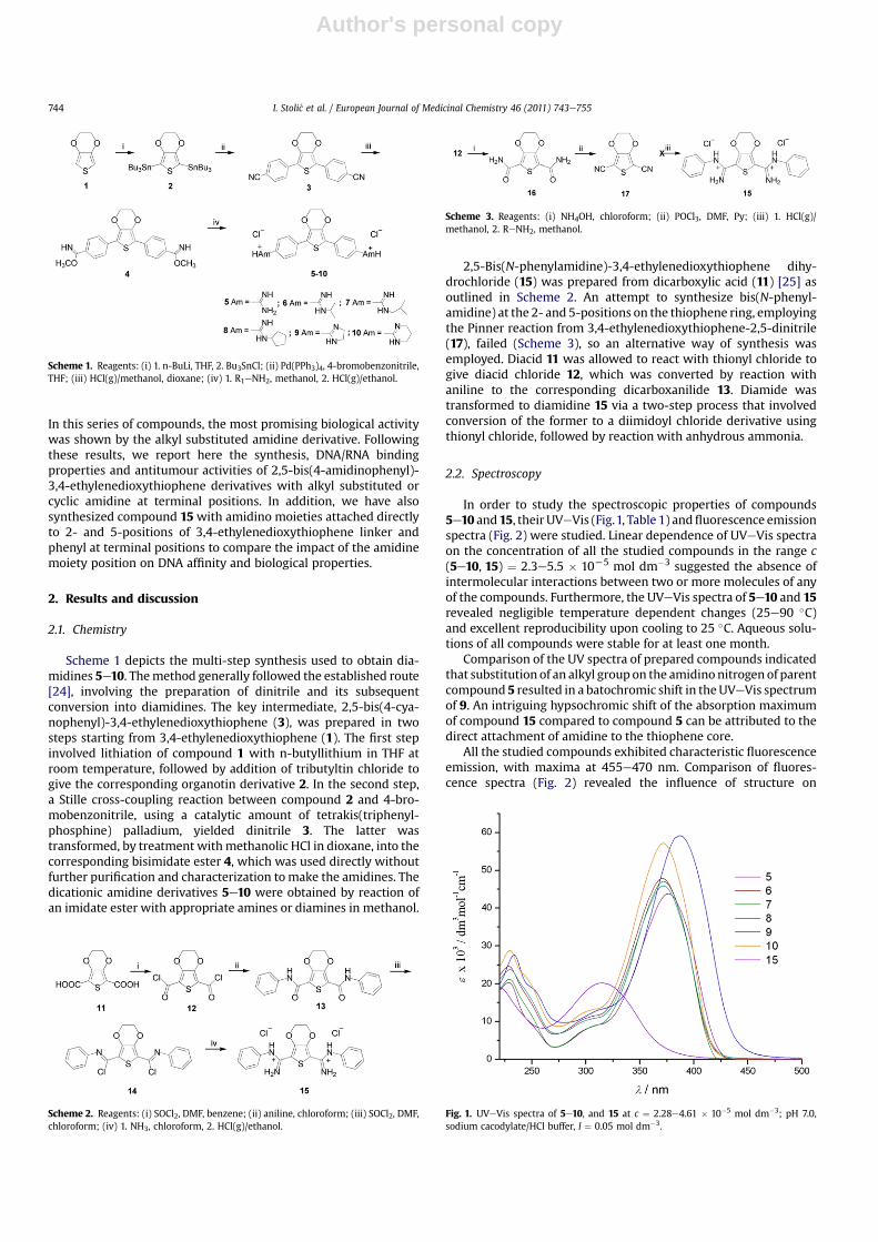
Scheme 1 depicts the multi-step synthesis used to obtain dia-midines 5e10. Themethod generally followed the established route[24], involving the preparation of dinitrile and its subsequentconversion into diamidines. The key intermediate, 2,5-bis(4-cya-nophenyl)-3,4-ethylenedioxythiophene (3), was prepared in twosteps starting from 3,4-ethylenedioxythiophene (1). The first stepinvolved lithiation of compound 1 with n-butyllithium in THF atroom temperature, followed by addition of tributyltin chloride togive the corresponding organotin derivative 2. In the second step,a Stille cross-coupling reaction between compound 2 and 4-bro-mobenzonitrile, using a catalytic amount of tetrakis(triphenyl-phosphine) palladium, yielded dinitrile 3. The latter wastransformed, by treatment withmethanolic HCl in dioxane, into thecorresponding bisimidate ester 4, which was used directly withoutfurther purification and characterization tomake the amidines. Thedicationic amidine derivatives 5e10 were obtained by reaction ofan imidate ester with appropriate amines or diamines in methanol.
2,5-Bis(N-phenylamidine)-3,4-ethylenedioxythiophene dihy-drochloride (15) was prepared from dicarboxylic acid (11) [25] asoutlined in Scheme 2. An attempt to synthesize bis(N-phenyl-amidine) at the 2- and 5-positions on the thiophene ring, employingthe Pinner reaction from 3,4-ethylenedioxythiophene-2,5-dinitrile(17), failed (Scheme 3), so an alternative way of synthesis wasemployed. Diacid 11 was allowed to react with thionyl chloride togive diacid chloride 12, which was converted by reaction withaniline to the corresponding dicarboxanilide 13. Diamide wastransformed to diamidine 15 via a two-step process that involvedconversion of the former to a diimidoyl chloride derivative usingthionyl chloride, followed by reaction with anhydrous ammonia.
2.2. Spectroscopy
In order to study the spectroscopic properties of compounds5e10 and15, their UVeVis (Fig.1, Table 1) andfluorescence emissionspectra (Fig. 2) were studied. Linear dependence of UVeVis spectraon the concentration of all the studied compounds in the range c(5e10, 15) ¼ 2.3e5.5 � 10L5 mol dm�3 suggested the absence ofintermolecular interactions between two or more molecules of anyof the compounds. Furthermore, the UVeVis spectra of 5e10 and 15revealed negligible temperature dependent changes (25e90 �C)and excellent reproducibility upon cooling to 25 �C. Aqueous solu-tions of all compounds were stable for at least one month.
Comparison of the UV spectra of prepared compounds indicatedthat substitution of an alkyl group on the amidino nitrogen of parentcompound 5 resulted in a batochromic shift in theUVeVis spectrumof 9. An intriguing hypsochromic shift of the absorption maximumof compound 15 compared to compound 5 can be attributed to thedirect attachment of amidine to the thiophene core.
All the studied compounds exhibited characteristic fluorescenceemission, with maxima at 455e470 nm. Comparison of fluores-cence spectra (Fig. 2) revealed the influence of structure on
Scheme 1. Reagents: (i) 1. n-BuLi, THF, 2. Bu3SnCl; (ii) Pd(PPh3)4, 4-bromobenzonitrile,THF; (iii) HCl(g)/methanol, dioxane; (iv) 1. R1eNH2, methanol, 2. HCl(g)/ethanol.
Scheme 2. Reagents: (i) SOCl2, DMF, benzene; (ii) aniline, chloroform; (iii) SOCl2, DMF,chloroform; (iv) 1. NH3, chloroform, 2. HCl(g)/ethanol.
Scheme 3. Reagents: (i) NH4OH, chloroform; (ii) POCl3, DMF, Py; (iii) 1. HCl(g)/methanol, 2. ReNH2, methanol.
Fig. 1. UVeVis spectra of 5e10, and 15 at c ¼ 2.28e4.61 � 10�5 mol dm�3; pH 7.0,sodium cacodylate/HCl buffer, I ¼ 0.05 mol dm�3.
I. Stoli�c et al. / European Journal of Medicinal Chemistry 46 (2011) 743e755744

Author's personal copy
emission of the compounds. It is interesting to note that alkylderivatives 6 and 7 exhibited stronger fluorescence than cyclo-pentyl derivate 8 and cyclic analogues 9 and 10 compared to parentcompound 5. These data show a pronounced impact of electronicproperties of the studied molecules on fluorescence emission.
2.3. Interactions with double stranded (ds-) DNA and RNA
2.3.1. UVeVis titrations with double stranded DNAThe UVeVis spectra of the studied compounds exhibited strong
hypochromic and batochromic changes upon addition of calfthymus (ct)-DNA under the conditions of an excess of compoundover DNA base pairs (ratio r[compd]/[ct-DNA] > 0.5 for 5 and 9; r ¼ 0.4for 6; r ¼ 0.2 for 7, 8 and 10), while further additions of ct-DNA(excess of DNA base pairs over compound; r << 0.2) resulted ina hyperchromic effect and additional batochromic shift of maximain the UVeVis spectra (Fig. 3). Thus, the final (at high excess ofct-DNA; r[compd]/[ct-DNA] ¼ 0.02) batochromic shifts of the absorp-tion maxima of 5e10 at lmax > 300 nm were between þ16 andþ25 nm. The observed changes in 5e10/ct-DNA titrations, alongwith a clear deviation from the isosbestic points, supportedformation of at least two different types of complexes with ct-DNA.Surprisingly, addition of ct-DNA did not yield any measurablechanges in the UVeVis spectrum of 15.
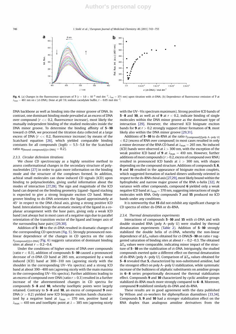
2.3.2. Fluorimetric titrations with ct-DNAAddition of ct-DNA led to a high increase of fluorescence
emission of compounds 5e10 (Fig. 4). Similarly as observed inUVeVis titration experiments, titration with ct-DNA resulted in
opposite changes of fluorescence of compounds 5e10, where thebreakpoint for all titrations was at about the ratio r [compound]/
[ct-DNA] ¼ 0.2. Several different binding modes were present underthe conditions of an excess of compound over DNA (r > 0.2, fluo-rescence quenching), including non-specific agglomeration ofpositively charged small molecules along the negatively charged
Table 1Electronic absorption data of 5e10 and 15.a
lmax/nm e � 103/dm3 mol�1 cm�1
5 376 45.916 371 49.357 371 47.478 372 46.619 387 59.6610 372 57.1115 315 22.02
a Sodium cacodylate buffer, pH 7.0, I ¼ 0.05 mol dm�3.
Fig. 2. Fluorescence emission spectra of 5e10 at c ¼ 3.98e4.09 � 10�6 mol dm�3; pH7.0, sodium cacodylate/HCl buffer, I ¼ 0.05 mol dm�3; at lexc. ¼ 376 (5), 371 (6, 7), 372(8, 10), 387 (9) nm.
Fig. 3. (a) Changes in UVeVis spectrum of 7 (c ¼ 1.97 � 10�5 mol dm�3) upon titrationwith ct-DNA; Dependence of the absorbance of 7 at lmax ¼ 391 nm (b) and 371 nm(c) on c(ct-DNA), at pH 7.0, sodium cacodylate buffer, I ¼ 0.05 mol dm�3.
I. Stoli�c et al. / European Journal of Medicinal Chemistry 46 (2011) 743e755 745
Author's personal copy
DNA backbone as well as binding into the minor groove of DNA. Incontrast, one dominant bindingmode prevailed at an excess of DNAover compound (r << 0.2, fluorescence increase), most likely themutually independent binding of the studied molecules inside theDNA minor groove. To determine the binding affinity of 5e10toward ct-DNA, we processed the titration data collected at a largeexcess of DNA (r << 0.2, fluorescence increase) by means of theScatchard equation [26], which yielded comparable bindingconstants for all compounds (logKs ¼ 5.5e5.8 for the Scatchardratio n[bound compound]/[ct-DNA] ¼ 0.2).
2.3.3. Circular dichroism titrationsWe chose CD spectroscopy as a highly sensitive method to
assess conformational changes in the secondary structure of poly-nucleotides [27] in order to gain more information on the bindingmode and the structure of the complexes formed. In addition,achiral small molecules can show induced CD signals (ICD) uponbinding to polynucleotides, giving useful information about themodes of interaction [27,28]. The sign and magnitude of the ICDband can depend on the binding geometry. Ligandeligand stackingis expected to give a strong bisignate exciton CD band. Minorgroove binding to ds-DNA orientates the ligand approximately at45� in respect to the DNA chiral axis, giving a strong positive ICDband. Intercalation brings the aromatic moiety of the ligand in a co-planar arrangement with the base pairs, giving only a weak ICDband (not always but in most cases of a negative sign due to parallelorientation of the transition vector of the ligand and longer axis ofthe surrounding base pairs) [29,30].
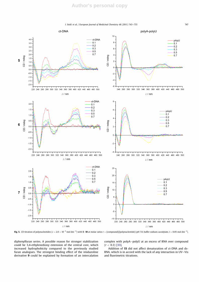
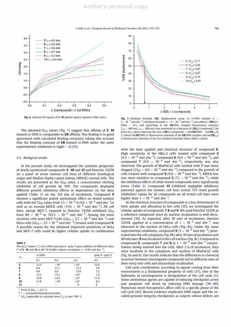
Addition of 5e10 to the ct-DNA resulted in dramatic changes ofthe corresponding CD spectrum (Fig. 5). Strongly pronounced non-linear dependence of the changes in CD spectra on the ratior[compound]/[ct-DNA] (Fig. 6) suggests saturation of dominant bindingsites at about r ¼ 0.2e0.4.
Under the conditions of higher excess of DNA over compounds(ratio r < 0.3), addition of compounds 5e10 caused a pronounceddecrease of ct-DNA CD band at 285 nm, accompanied by a weakinduced (ICD) band at 300e310 nm (agreeing nicely with theshoulder in the corresponding UVeVis spectra) and a strong ICDband at about 390e400 nm (agreeing nicely with themainmaximain the corresponding UVeVis spectra). Further additions leading toan excess of compound over DNA (ratio r> 0.3) resulted in a furtherincrease of the aforementioned changes in CD spectra forcompounds 5e8 and 10, whereby isoelliptic points were largelyretained. Contrary to 5e8 and 10, an excess of compound 9 overDNA (r > 0.2) yielded new ICD bisignate exciton bands character-ized by a negative band at lmax ¼ 370 nm, positive band atlmax ¼ 410 nm and isoelliptic point at l ¼ 385 nm (agreeing nicely
with the UVeVis spectrummaximum). Strong positive ICD bands of5e8 and 10, as well as of 9 at r < 0.2, indicate binding of singlemolecules within the DNA minor groove as the dominant type ofinteraction [29]. However, the observed ICD bisignate excitonbands for 9 at r > 0.2 strongly support dimer formation of 9, mostlikely also within the DNA minor groove [29,31].
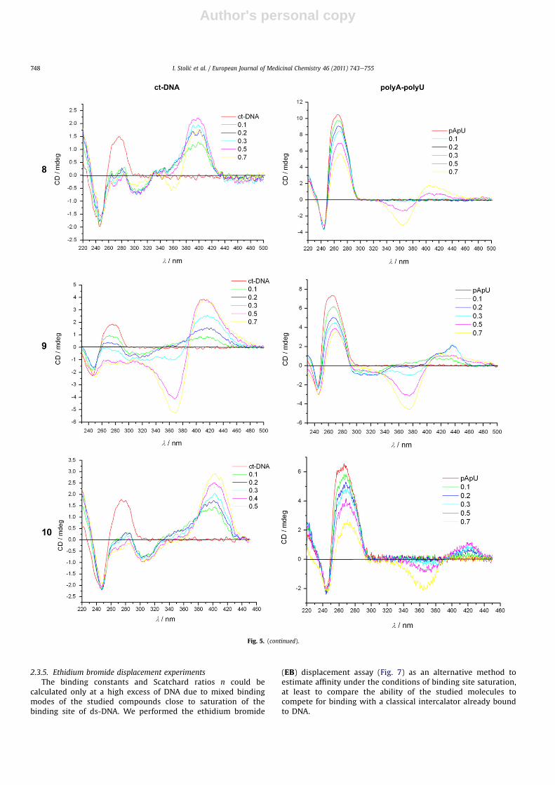
Additions of 5e10 to ds-RNA at the ratio r[compound]/[poly Aepoly U]
< 0.2 (excess of RNA over compound) in most cases resulted in onlya minor decrease of the RNA CD band at lmax ¼ 265 nm. No induced(ICD) bands were observed at l > 300 nm, with the exception of theweak positive ICD band of 9 at lmax ¼ 410 nm. However, furtheradditions ofmost compounds (r> 0.2, excess of compoundoverRNA)resulted in pronounced ICD bands at l > 300 nm, with shapesdepending on the compound structure. Additions of compounds 5, 8,9 and 10 resulted in the appearance of bisignate exciton couplets,which suggested formation of stacked dimers uniformly oriented inrespect to theds-RNAchiral axis [27,29],most likely boundwithin thehydrophobic and narrow major groove of the RNA a-helix [32]. Atvariance with other compounds, compound 6 yielded only a weaknegative ICD band at lmax¼ 370 nm, suggesting interactions of singlemolecules with RNA. Only compounds 7 and 15 produced no ICDbands under any conditions.
It is noteworthy that 15 did not exhibit any significant change inCD spectra of either ds-DNA or ds-RNA.
2.3.4. Thermal denaturation experimentsInteractions of compounds 5e10 and 15 with ct-DNA and with
double stranded RNA (poly Aepoly U) were studied by thermaldenaturation experiments (Table 2). Addition of 5e10 stronglystabilized the double helix of ct-DNA, whereby the non-lineardependence of DTm values obtained for ct-DNA/5e10 on ratio r sug-gested saturation of binding sites at about r ¼ 0.2e0.3. The obtainedDTm values were comparable, indicating minor impact of the struc-ture of 5e10 on the stabilization of ct-DNA. Intriguingly, the studiedcompounds exerted quite a different effect on thermal denaturationof ds-RNA (poly Aepoly U). Comparison of DTm values obtained for5e8 revealed that 5, characterized by non-substituted amidine, hadthe strongest effect on poly Aepoly U stabilization, while systematicincrease of the bulkiness of aliphatic substituents on amidine groupsin 6e8 series proportionally decreased the thermal stabilizationeffect. Compounds 9 and 10 characterized by cyclic amidine groupsstabilized ds-RNA much more strongly compared to 6e8. Moreover,compound 9 stabilized similarly ds-DNA and ds-RNA.
These results are in good agreement with the data publishedby Wilson and co-workers for diphenylfuran diamidines [33,34].Compounds 5, 9 and 10 had a stronger stabilization effect on theRNA duplex than analogous amidine derivatives from the
Fig. 4. (a) Changes in the fluorescence spectrum of 7 (c ¼ 1.0 � 10�6 mol dm�3, lexc ¼ 371 nm) upon titration with ct-DNA; (b) Dependence of fluorescence emission of 7 atlmax ¼ 461 nm on c (ct-DNA). Done at pH 7.0, sodium cacodylate buffer, I ¼ 0.05 mol dm�3.
I. Stoli�c et al. / European Journal of Medicinal Chemistry 46 (2011) 743e755746
Author's personal copy
diphenylfuran series. A possible reason for stronger stabilizationcould be 3,4-ethylenedioxy extension of the central core, whichincreased hydrophobicity compared to the previously studiedfuran analogues. The strongest binding effect of the imidazolinederivative 9 could be explained by formation of an intercalation
complex with polyAepolyU at an excess of RNA over compound(r < 0.3) [33].
Addition of 15 did not affect denaturation of ct-DNA and ds-RNA, which is in accord with the lack of any interaction in UVeVisand fluorimetric titrations.
Fig. 5. CD titration of polynucleotides (c ¼ 2.0 � 10�5 mol dm�3) with 5e10 at molar ratios r ¼ [compound]/[polynucleotide] (pH 7.0, buffer sodium cacodylate, I ¼ 0.05 mol dm�3).
I. Stoli�c et al. / European Journal of Medicinal Chemistry 46 (2011) 743e755 747
Author's personal copy
2.3.5. Ethidium bromide displacement experimentsThe binding constants and Scatchard ratios n could be
calculated only at a high excess of DNA due to mixed bindingmodes of the studied compounds close to saturation of thebinding site of ds-DNA. We performed the ethidium bromide
(EB) displacement assay (Fig. 7) as an alternative method toestimate affinity under the conditions of binding site saturation,at least to compare the ability of the studied molecules tocompete for binding with a classical intercalator already boundto DNA.
Fig. 5. (continued).
I. Stoli�c et al. / European Journal of Medicinal Chemistry 46 (2011) 743e755748
Author's personal copy
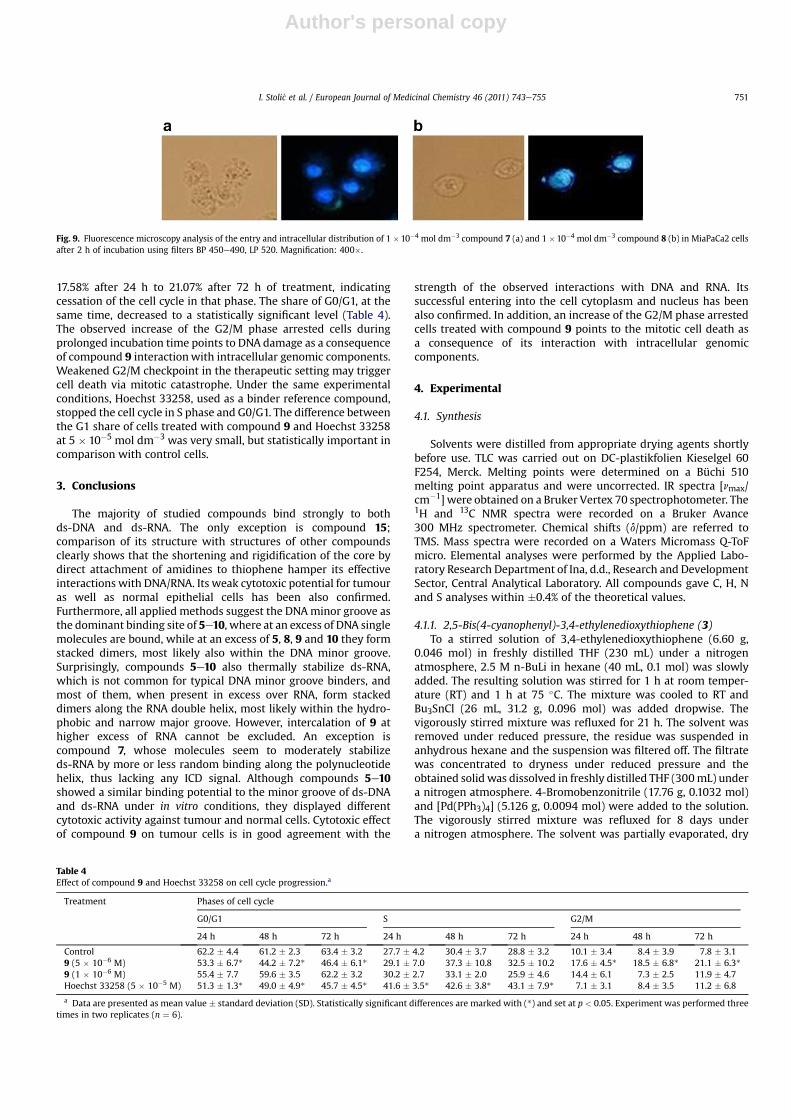
The obtained IC50 values (Fig. 7) suggest that affinity of 5e10toward ct-DNA is comparable to EB affinity. This finding is in goodagreement with calculated binding constants, taking into accountthat the binding constant of EB toward ct-DNA under the sameexperimental conditions is logKs ¼ 6 [35].
2.4. Biological results
In the present study, we investigated the cytotoxic propertiesof newly synthesized compounds 5e10 and 15 and Hoechst 33258on a panel of seven tumour cell lines of different histologicalorigin and Madine-Darby canine kidney (MDCK) normal cells. Theresults are presented as the GI50 value, a concentration elicitinginhibition of cell growth by 50%. The compounds displayeddifferent growth inhibitory effects in dependence on the doseapplied (Table 3) on the 3rd day of incubation. Compound 9showed a significant potent antitumour effect on tested tumourcells with the GI50 value from 1.5 � 10�6 to 9.0 � 10�6 mol dm�3aswell as on normal MDCK cells (17.0 � 10�6 mol dm�3). All celllines, except MCF-7, exposed to Hoechst 33258 exhibited GI50from 84 � 10�6 to 191.5 � 10�6 mol dm�3. Among the mostsensitive cells were MCF-7cells (GI50 ¼ 2.7 � 10�6 mol dm�3) andHeLa cells (GI50 ¼ 1.5 � 10�6 mol dm�3) treated with compound 9.A possible reason for the obtained improved sensitivity of HeLaand MCF-7 cells could be higher cellular uptake in combination
with the dose applied and chemical structure of compound 9.High sensitivity of the HEp-2 cells treated with compound 5(9.3 � 10�6 mol dm�3), compound 6 (9.0 � 10�6 mol dm�3), andcompound 7 (9.9 � 10�6 mol dm�3), respectively, was alsoobserved. The growth of MiaPaCa2 cells treated with 7 was moreslugged (GI50 ¼ 4.9 � 10�6 mol dm�3) compared to the growth ofcells treated with compound 9 (9.0 � 10�6 mol dm�3). MDCK linewas most sensitive to compound 5 (7.1 � 10�6 mol dm�3), whilethe inhibitory effects of other tested compounds were significantlylower (Table 3). Compound 15 exhibited negligible inhibitorypotential against the tumour cell lines tested. TGI (total growthinhibition) values for all compounds on all tested cell lines werehigher than 1 � 10�4 mol dm�3.
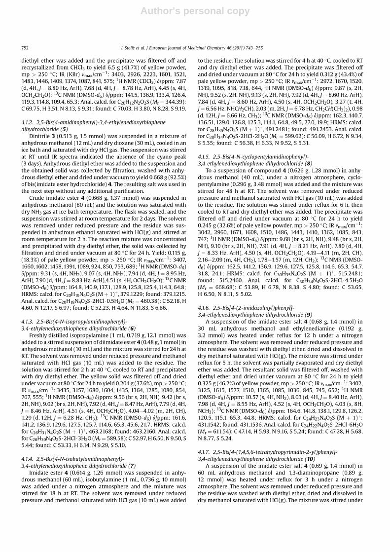
As the chemical structure of compounds is a key determinant oftheir uptake and allocation in live cells [10], we investigated thecellular uptake of compounds 7, 8 and 9. We used Hoechst 33258 asa reference compound since its nucleus localization is well docu-mented [36]. As expected, after 30 min of incubation, Hoechst33258, applied at a concentration of 1 � 10�4 mol dm�3, wasobserved in the nucleus of HeLa cells (Fig. 8a). Under the sameexperimental conditions, compound 9 (5 � 10�6 mol dm�3) pene-trated into the cell cytoplasm (Fig. 8b) after 30min of incubation and60min later,9was localized in the cell nucleus (Fig. 8c). Compared tocompound 9, compounds 7 and 8 at 1 � 10�4 mol dm�3 concen-tration slowly entered into the cells. After 2 h of incubation, theywere localized in the cytoplasm and nucleus of MiaPaCa2 cells(Fig. 9a and b). Our results indicate that the differences in chemicalstructure between investigated compounds led to different rates oftheir entry into cells and intracellular localization.
Cell cycle coordination, according to signals coming from theirenvironment is a fundamental property of cells [37]. One of thehallmarks of carcinogenesis is deregulation of the cell cycle [1].Some antitumour agents are capable of inducing checkpoint arrestand apoptotic cell death by inducing DNA damage [38e40].Numerous novel therapeutics affect cells in a specific phase of thecell cycle [41]. Recent evidence implicates DNA repair and the so-called genome integrity checkpoints as culprits whose defects are
Fig. 6. Induced CD signals of 5e10 plotted against ligand/ct-DNA ratios.
Table 2The DTm
a values (�C) of ct-DNA and poly Aepoly U upon addition of different ratiosrb of 5e10 and 15 at pH 7.0 (buffer sodium cacodylate, I ¼ 0.05 mol dm�3).
rb¼ct-DNA poly Aepoly U
0.1 0.2 0.3 0.3
5 12.2 >16c >16c 12.66 8.4 11.1 12.3 5.87 9.0 9.0 15.0 5.28 12.3 15.2 >16c 1.49 10.8 14.3 >16c 17.010 11.8 >16c >16c 8.215 e e 0 0
a Error in DTm: � 0.5 �C.b r ¼ [compound]/[polynucleotide].c DTm impossible to calculate since Tm is over 100 �C.
Fig. 7. Ethidium bromide (EB) displacement assay: to ct-DNA solution (c ¼5 � 10�5 mol dm�3), ethidium bromide (c ¼ 5 � 10�6 mol dm�3) was added (r [EB]/[ct-DNA] ¼ 0.1), and quenching of the EB/DNA complex fluorescence emission(lex ¼ 480 nm, lem ¼ 600 nm) was monitored as a function of c(EB)/c(compound). Thegiven IC50 values represent the ratio c(EB)/c(compound) ¼ [Int(EB/DNA) � Int(EBfree)]/2, where Int(EB/DNA) is fluorescence intensity of the EB/DNA complex and Int(EBfree)is fluorescence intensity of the free ethidium bromide before DNA is added.
I. Stoli�c et al. / European Journal of Medicinal Chemistry 46 (2011) 743e755 749

Author's personal copy
largely responsible for the enhanced genetic instability of tumourcells [42]. The G2/M checkpoint prevents a DNA damaged cell fromentering mitosis and gives more time to various repair mechanismsto repair the damage, if possible, or directs the cell into pro-grammed cell death. For this reason, the G2/M checkpoint is ofparticular interest for novel drug design in the development ofantitumour therapy [43].
To elucidate the mechanism of cytotoxic action of compound 9,its effect on the cell cycle of HeLa cells wasmonitored. Compound 9,applied at a concentration of 5 � 10�5 mol dm�3, significantlyaffected the cell cycle of HeLa cells during monitored incubationperiods (24e72 h). Accumulation of cells in the G2/M phase started24 h after application and increased up to 3 fold over incubationtime. Percentage of cells in the G2/M phase slowly increased from
Table 3Cytotoxic activity of tested compounds.
GI50 (1 � 10�6 mol dm�3)a
Compound 5 6 7 8 9 10 15 Hoechst
Tumour cellsMCF-7 25.0 � 15.6 67.5 � 27.6 7.3 � 0.8 7.8 � 1.2 2.7 � 1.7 8.4 � 1.2 129.0 � 18.4 5.7 � 4.5NCI-H358 33.0 � 12.7 112.7 � 9.5 73.3 � 2.9 62.2 � 6.3 4.1 � 1.0 98.7 � 10.0 112.0 � 8.5 91.7 � 18CaCo-2 13.0 � 4.2 76.5 � 14.8 42.8 � 29.9 41.0 � 26.9 3.1 � 1.3 101.3 � 15.1 134.0 � 11.3 84.0 � 9.8HEp-2 9.3 � 1.2 9.0 � 0 9.9 � 0.6 61.3 � 19.9 3.0 � 0.8 97.7 � 18.1 100.3 � 17 132.5 � 7.8HeLa 32.3 � 12.4 72.3 � 13.3 18.8 � 9.1 26.7 � 11.2 1.5 � 0.8 97.5 � 10.6 138.5 � 3.5 115.7 � 25.5AGS 116.5 � 34.6 72.3 � 18.4 59.3 � 3.8 45.0 � 2.6 4.3 � 3.8 81.7 � 12.6 113.5 � 4.9 112.0 � 12.7MiaPaCa2 9.3 � 1.5 13.0 � 3.5 4.9 � 7.0 10.9 � 5.7 9.0 � 0.9 64.7 � 25.8 128.5 � 21.9 191.5 � 17.7Normal cellsMDCK 7.1 � 1.6 65.3 � 4.0 59.7 � 4.2 50.0 � 5.9 17.0 � 0.9 82.0 � 17.7 79.5 � 17.7 157.5 � 20.5
Abbreviation (used cell lines): breast adenocarcinoma (MCF-7), bronchioalveolar carcinoma (NCI-H358), colon adenocarcinoma (CaCo-2), larynx carcinoma (HEp-2), cervixadenocarcinoma (HeLa), gastric adenocarcinoma (AGS), pancreatic carcinoma (MiaPaCa) and Madine-Darby canine kidney (MDCK).Bold values emphasized significant difference in cytotoxic efficiency of investigated compounds on tumour cells compared to normal cells.
a GI50 valueeconcentration eliciting inhibition of cell growth by 50%. Data represent the mean GI50 value of three independent experiments � SD. Cytotoxic activity wasanalyzed by the MTT assay.
Fig. 8. Fluorescence microscopy analysis of the entry and intracellular distribution of 1 � 10�4 mol dm�3 Hoechst 33258 (a), 5 � 10�6 mol dm�3 compound 9 (b) in HeLa cells after30 min of incubation, and 5 � 10�6 mol dm�3 compound 9 (c) after 90 min of incubation using BP 450e490 filters, LP 520. Magnification: 400�.
I. Stoli�c et al. / European Journal of Medicinal Chemistry 46 (2011) 743e755750
Author's personal copy
17.58% after 24 h to 21.07% after 72 h of treatment, indicatingcessation of the cell cycle in that phase. The share of G0/G1, at thesame time, decreased to a statistically significant level (Table 4).The observed increase of the G2/M phase arrested cells duringprolonged incubation time points to DNA damage as a consequenceof compound 9 interactionwith intracellular genomic components.Weakened G2/M checkpoint in the therapeutic setting may triggercell death via mitotic catastrophe. Under the same experimentalconditions, Hoechst 33258, used as a binder reference compound,stopped the cell cycle in S phase and G0/G1. The difference betweenthe G1 share of cells treated with compound 9 and Hoechst 33258at 5 � 10�5 mol dm�3 was very small, but statistically important incomparison with control cells.
3. Conclusions
The majority of studied compounds bind strongly to bothds-DNA and ds-RNA. The only exception is compound 15;comparison of its structure with structures of other compoundsclearly shows that the shortening and rigidification of the core bydirect attachment of amidines to thiophene hamper its effectiveinteractions with DNA/RNA. Its weak cytotoxic potential for tumouras well as normal epithelial cells has been also confirmed.Furthermore, all applied methods suggest the DNAminor groove asthe dominant binding site of 5e10, where at an excess of DNA singlemolecules are bound, while at an excess of 5, 8, 9 and 10 they formstacked dimers, most likely also within the DNA minor groove.Surprisingly, compounds 5e10 also thermally stabilize ds-RNA,which is not common for typical DNA minor groove binders, andmost of them, when present in excess over RNA, form stackeddimers along the RNA double helix, most likely within the hydro-phobic and narrow major groove. However, intercalation of 9 athigher excess of RNA cannot be excluded. An exception iscompound 7, whose molecules seem to moderately stabilizeds-RNA by more or less random binding along the polynucleotidehelix, thus lacking any ICD signal. Although compounds 5e10showed a similar binding potential to the minor groove of ds-DNAand ds-RNA under in vitro conditions, they displayed differentcytotoxic activity against tumour and normal cells. Cytotoxic effectof compound 9 on tumour cells is in good agreement with the
strength of the observed interactions with DNA and RNA. Itssuccessful entering into the cell cytoplasm and nucleus has beenalso confirmed. In addition, an increase of the G2/M phase arrestedcells treated with compound 9 points to the mitotic cell death asa consequence of its interaction with intracellular genomiccomponents.
4. Experimental
4.1. Synthesis
Solvents were distilled from appropriate drying agents shortlybefore use. TLC was carried out on DC-plastikfolien Kieselgel 60F254, Merck. Melting points were determined on a Büchi 510melting point apparatus and were uncorrected. IR spectra [nmax/cm�1] were obtained on a Bruker Vertex 70 spectrophotometer. The1H and 13C NMR spectra were recorded on a Bruker Avance300 MHz spectrometer. Chemical shifts (d/ppm) are referred toTMS. Mass spectra were recorded on a Waters Micromass Q-ToFmicro. Elemental analyses were performed by the Applied Labo-ratory Research Department of Ina, d.d., Research and DevelopmentSector, Central Analytical Laboratory. All compounds gave C, H, Nand S analyses within �0.4% of the theoretical values.
4.1.1. 2,5-Bis(4-cyanophenyl)-3,4-ethylenedioxythiophene (3)To a stirred solution of 3,4-ethylenedioxythiophene (6.60 g,
0.046 mol) in freshly distilled THF (230 mL) under a nitrogenatmosphere, 2.5 M n-BuLi in hexane (40 mL, 0.1 mol) was slowlyadded. The resulting solution was stirred for 1 h at room temper-ature (RT) and 1 h at 75 �C. The mixture was cooled to RT andBu3SnCl (26 mL, 31.2 g, 0.096 mol) was added dropwise. Thevigorously stirred mixture was refluxed for 21 h. The solvent wasremoved under reduced pressure, the residue was suspended inanhydrous hexane and the suspension was filtered off. The filtratewas concentrated to dryness under reduced pressure and theobtained solid was dissolved in freshly distilled THF (300mL) undera nitrogen atmosphere. 4-Bromobenzonitrile (17.76 g, 0.1032 mol)and [Pd(PPh3)4] (5.126 g, 0.0094 mol) were added to the solution.The vigorously stirred mixture was refluxed for 8 days undera nitrogen atmosphere. The solvent was partially evaporated, dry
Table 4Effect of compound 9 and Hoechst 33258 on cell cycle progression.a
Treatment Phases of cell cycle
G0/G1 S G2/M
24 h 48 h 72 h 24 h 48 h 72 h 24 h 48 h 72 h
Control 62.2 � 4.4 61.2 � 2.3 63.4 � 3.2 27.7 � 4.2 30.4 � 3.7 28.8 � 3.2 10.1 � 3.4 8.4 � 3.9 7.8 � 3.19 (5 � 10�6 M) 53.3 � 6.7* 44.2 � 7.2* 46.4 � 6.1* 29.1 � 7.0 37.3 � 10.8 32.5 � 10.2 17.6 � 4.5* 18.5 � 6.8* 21.1 � 6.3*9 (1 � 10�6 M) 55.4 � 7.7 59.6 � 3.5 62.2 � 3.2 30.2 � 2.7 33.1 � 2.0 25.9 � 4.6 14.4 � 6.1 7.3 � 2.5 11.9 � 4.7Hoechst 33258 (5 � 10�5 M) 51.3 � 1.3* 49.0 � 4.9* 45.7 � 4.5* 41.6 � 3.5* 42.6 � 3.8* 43.1 � 7.9* 7.1 � 3.1 8.4 � 3.5 11.2 � 6.8
a Data are presented as mean value � standard deviation (SD). Statistically significant differences are marked with (*) and set at p < 0.05. Experiment was performed threetimes in two replicates (n ¼ 6).
Fig. 9. Fluorescence microscopy analysis of the entry and intracellular distribution of 1 �10�4 mol dm�3 compound 7 (a) and 1 �10�4 mol dm�3 compound 8 (b) in MiaPaCa2 cellsafter 2 h of incubation using filters BP 450e490, LP 520. Magnification: 400�.
I. Stoli�c et al. / European Journal of Medicinal Chemistry 46 (2011) 743e755 751
Author's personal copy
diethyl ether was added and the precipitate was filtered off andrecrystallized from CHCl3 to yield 6.5 g (41.7%) of yellow powder,mp > 250 �C; IR (KBr) nmax/cm�1: 3403, 2926, 2223, 1601, 1521,1483, 1446, 1409, 1374, 1087, 841, 575; 1H NMR (CDCl3) d/ppm: 7.87(d, 4H, J ¼ 8.80 Hz, ArH), 7.68 (d, 4H, J ¼ 8.78 Hz, ArH), 4.45 (s, 4H,OCH2CH2O); 13C NMR (DMSO-d6) d/ppm: 141.5, 136.9, 133.4, 126.4,119.3, 114.8, 109.4, 65.3; Anal. calcd. for C20H12N2O2S (Mr ¼ 344.39):C 69.75, H 3.51, N 8.13, S 9.31; found: C 70.03, H 3.80, N 8.28, S 9.19.
4.1.2. 2,5-Bis(4-amidinophenyl)-3,4-ethylenedioxythiophenedihydrochloride (5)
Dinitrile 3 (0.513 g, 1.5 mmol) was suspended in a mixture ofanhydrous methanol (12 mL) and dry dioxane (30 mL), cooled in anice bath and saturated with dry HCl gas. The suspensionwas stirredat RT until IR spectra indicated the absence of the cyano peak(3 days). Anhydrous diethyl ether was added to the suspension andthe obtained solid was collected by filtration, washed with anhy-drous diethyl ether and dried under vacuum to yield 0.668 g (92.5%)of bis(imidate ester hydrochloride) 4. The resulting salt was used inthe next step without any additional purification.
Crude imidate ester 4 (0.668 g, 1.37 mmol) was suspended inanhydrous methanol (80 mL) and the solution was saturated withdry NH3 gas at ice bath temperature. The flask was sealed, and thesuspensionwas stirred at room temperature for 2 days. The solventwas removed under reduced pressure and the residue was sus-pended in anhydrous ethanol saturated with HCl(g) and stirred atroom temperature for 2 h. The reaction mixture was concentratedand precipitated with dry diethyl ether, the solid was collected byfiltration and dried under vacuum at 80 �C for 24 h. Yield: 0.115 g,(18.3%) of pale yellow powder, mp > 250 �C; IR nmax/cm�1: 3407,1660,1602,1458,1391,1089, 924, 850, 753, 689; 1HNMR (DMSO-d6)d/ppm: 9.31 (s, 4H, NH2), 9.07 (s, 4H, NH2), 7.94 (d, 4H, J ¼ 8.95 Hz,ArH), 7.90 (d, 4H, J¼ 8.83 Hz, ArH),4.51 (s, 4H, OCH2CH2O); 13C NMR(DMSO-d6) d/ppm: 164.8,140.9,137.1,128.9,125.8,125.4,114.3, 64.8;HRMS: calcd. for C20H19N4O2S (Mþ 1)þ, 379.1229; found: 379.1215.Anal. calcd. for C20H18N4O2S$2HCl$0.5H2O (Mr¼ 460.38): C 52.18, H4.60, N 12.17, S 6.97; found: C 52.23, H 4.64, N 11.83, S 6.86.
4.1.3. 2,5-Bis(4-N-isopropylamidinophenyl)-3,4-ethylenedioxythiophene dihydrochloride (6)
Freshly distilled isopropylamine (1 mL, 0.719 g, 12.1 mmol) wasadded to a stirred suspension of diimidate ester 4 (0.48 g,1mmol) inanhydrousmethanol (10mL) and themixturewas stirred for 24 h atRT. The solvent was removed under reduced pressure andmethanolsaturated with HCl gas (10 mL) was added to the residue. Thesolution was stirred for 2 h at 40 �C, cooled to RT and precipitatedwith dry diethyl ether. The yellow solid was filtered off and driedunder vacuumat 80 �C for 24 h to yield 0.204 g (37.6%),mp> 250 �C;IR nmax/cm�1: 3435, 3157, 1680, 1604, 1435, 1364, 1285, 1080, 854,767, 555; 1H NMR (DMSO-d6) d/ppm: 9.56 (br s, 2H, NH), 9.42 (br s,2H, NH), 9.02 (br s, 2H, NH), 7.92 (d, 4H, J¼ 8.47Hz, ArH), 7.79 (d, 4H,J ¼ 8.46 Hz, ArH), 4.51 (s, 4H, OCH2CH2O), 4.04e4.02 (m, 2H, CH),1.29 (d, 12H, J ¼ 6.28 Hz, CH3); 13C NMR (DMSO-d6) d/ppm: 161.6,141.2, 136.9, 129.6, 127.5, 125.7, 114.6, 65.3, 45.6, 21.7; HRMS: calcd.for C26H31N4O2S (M þ 1)þ, 463.2168; found: 463.2160. Anal. calcd.for C26H30N4O2S$2HCl$3H2O (Mr¼589.58): C 52.97, H 6.50, N 9.50, S5.44; found: C 53.33, H 6.14, N 9.29, S 5.10.
4.1.4. 2,5-Bis(4-N-isobutylamidinophenyl)-3,4-ethylenedioxythiophene dihydrochloride (7)
Imidate ester 4 (0.614 g, 1.26 mmol) was suspended in anhy-drous methanol (60 mL), isobutylamine (1 mL, 0.736 g, 10 mmol)was added under a nitrogen atmosphere and the mixture wasstirred for 18 h at RT. The solvent was removed under reducedpressure and methanol saturated with HCl gas (10 mL) was added
to the residue. The solutionwas stirred for 4 h at 40 �C, cooled to RTand dry diethyl ether was added. The precipitate was filtered offand dried under vacuum at 80 �C for 24 h to yield 0.312 g (43.4%) ofpale yellow powder, mp > 250 �C; IR nmax/cm�1: 2972, 1670, 1520,1319, 1095, 818, 738, 644, 1H NMR (DMSO-d6) d/ppm: 9.87 (s, 2H,NH), 9.52 (s, 2H, NH), 9.13 (s, 2H, NH), 7.92 (d, 4H, J ¼ 8.60 Hz, ArH),7.84 (d, 4H, J ¼ 8.60 Hz, ArH), 4.50 (s, 4H, OCH2CH2O), 3.27 (t, 4H,J¼ 6.56 Hz, NHCH2CH), 2.03 (m, 2H, J¼ 6.78 Hz, CH2CH(CH3)2), 0.98(d, 12H, J ¼ 6.66 Hz, CH3); 13C NMR (DMSO-d6) d/ppm: 162.3, 140.7,136.51, 129.0, 126.8, 125.3, 114.1, 64.8, 49.5, 27.0, 19.9; HRMS: calcd.for C28H35N4O2S (M þ 1)þ, 491.2481; found: 491.2453. Anal. calcd.for C28H34N4O2S$2HCl$2H2O (Mr ¼ 599.62): C 56.09, H 6.72, N 9.34,S 5.35; found: C 56.38, H 6.33, N 9.52, S 5.31.
4.1.5. 2,5-Bis(4-N-cyclopentylamidinophenyl)-3,4-ethylenedioxythiophene dihydrochloride (8)
To a suspension of compound 4 (0.626 g, 1.28 mmol) in anhy-drous methanol (40 mL), under a nitrogen atmosphere, cyclo-pentylamine (0.296 g, 3.48 mmol) was added and the mixture wasstirred for 48 h at RT. The solvent was removed under reducedpressure and methanol saturated with HCl gas (10 mL) was addedto the residue. The solution was stirred under reflux for 6 h, thencooled to RT and dry diethyl ether was added. The precipitate wasfiltered off and dried under vacuum at 80 �C for 24 h to yield0.245 g (32.6%) of pale yellow powder, mp > 250 �C; IR nmax/cm�1:3042, 2960, 1671, 1608, 1510, 1486, 1443, 1410, 1362, 1085, 843,747; 1H NMR (DMSO-d6) d/ppm: 9.68 (br s, 2H, NH), 9.48 (br s, 2H,NH), 9.10 (br s, 2H, NH), 7.91 (d, 4H, J ¼ 8.21 Hz, ArH), 7.80 (d, 4H,J ¼ 8.33 Hz, ArH), 4.50 (s, 4H, OCH2CH2O), 4.19e4.11 (m, 2H, CH),2.16e2.09 (m, 4H, CH2), 1.78e1.57 (m, 12H, CH2); 13C NMR (DMSO-d6) d/ppm: 162.5, 141.2, 136.9, 129.6, 127.5, 125.8, 114.6, 65.3, 54.7,31.8, 24.1; HRMS: calcd. for C30H35N4O2S (M þ 1)þ, 515.2481;found: 515.2460. Anal. calcd. for C30H34N4O2S$2HCl$4.5H2O(Mr ¼ 668.68): C 53.89, H 6.78, N 8.38, S 4.80; found: C 53.65,H 6.50, N 8.11, S 5.02.
4.1.6. 2,5-Bis[4-(2-imidazolinyl)phenyl]-3,4-ethylenedioxythiophene dihydrochloride (9)
A suspension of the imidate ester salt 4 (0.68 g, 1.4 mmol) in30 mL anhydrous methanol and ethylenediamine (0.192 g,3.2 mmol) was heated under reflux for 12 h under a nitrogenatmosphere. The solvent was removed under reduced pressure andthe residue was washed with diethyl ether, dried and dissolved indry methanol saturated with HCl(g). The mixture was stirred underreflux for 5 h, the solvent was partially evaporated and dry diethylether was added. The resultant solid was filtered off, washed withdiethyl ether and dried under vacuum at 80 �C for 24 h to yield0.325 g (46.2%) of yellow powder, mp> 250 �C; IR nmax/cm�1: 3402,3125, 1615, 1577, 1510, 1365, 1085, 1036, 845, 745, 652; 1H NMR(DMSO-d6) d/ppm: 10.57 (s, 4H, NH2), 8.03 (d, 4H, J¼ 8.40 Hz, ArH),7.98 (d, 4H, J ¼ 8.55 Hz, ArH), 4.52 (s, 4H, OCH2CH2O), 4.03 (s, 8H,NCH2); 13C NMR (DMSO-d6) d/ppm: 164.6, 141.8, 138.1, 129.8, 126.2,120.5, 115.1, 65.3, 44.8; HRMS: calcd. for C24H23N4O2S (M þ 1)þ:431.1542; found: 431.1536. Anal. calcd. for C24H22N4O2S$2HCl$6H2O(Mr ¼ 611.54): C 47.14, H 5.93, N 9.16, S 5.24; found: C 47.28, H 5.68,N 8.77, S 5.24.
4.1.7. 2,5-Bis[4-(1,4,5,6-tetrahydropyrimidin-2-yl)phenyl]-3,4-ethylenedioxythiophene dihydrochloride (10)
A suspension of the imidate ester salt 4 (0.69 g, 1.4 mmol) in60 mL anhydrous methanol and 1,3-diaminopropane (0.89 g,12 mmol) was heated under reflux for 3 h under a nitrogenatmosphere. The solvent was removed under reduced pressure andthe residue was washed with diethyl ether, dried and dissolved indry methanol saturated with HCl(g). The mixture was stirred under
I. Stoli�c et al. / European Journal of Medicinal Chemistry 46 (2011) 743e755752
Author's personal copy
reflux for 2 h, the solvent was partially evaporated and dry diethylether was added. The resultant solid was filtered off, washed withdiethyl ether and dried under vacuum at 80 �C for 24 h to yield0.56 g (75.1%) of yellow powder, mp > 250 �C; IR nmax/cm�1: 2972,2038, 1610, 1578, 1452, 1365, 1308, 1086, 840, 744, 665; 1H NMR(DMSO-d6) d/ppm: 10.20 (s, 4H, NH), 7.92 (d, 4H, J ¼ 8.43 Hz, ArH),7.87 (d, 4H, J ¼ 8.43 Hz, ArH), 4.50 (s, 4H, OCH2CH2O), 3.50 (t, 8H,J ¼ 5.52 Hz, N-CH2CH2), 1.98 (q, 4H, J ¼ 5.34 Hz, N-CH2CH2); 13CNMR (D2O) d/ppm: 159.3, 140.4, 137.0, 127.1, 126.0, 125.7, 114.9, 64.7,38.9, 17.6; HRMS: calcd. for C26H27N4O2S (M þ 1)þ: 459.1855;found: 459.1837. Anal. calcd. for C26H26N4O2S$2HCl$3H2O(Mr¼ 585.55): C 53.33, H 5.85, N 9.57, S 5.48; found: C 53.52, H 5.79,N 9.54, S 5.37.
4.1.8. 3,4-Ethylenedioxythiophene-2,5-dicarboxanilide (13)3,4-Ethylenedioxythiophene-2,5-dicarboxylic acid (11) (2.465 g,
0.0107 mol) was suspended in dry benzene (120 mL), thionylchloride (12 mL, 19.56 g, 0.164 mol) and 12 drops of DMF wereadded, and the resulting mixture was heated under reflux for 12 h.The solvent was removed under reduced pressure and the residuewas washed with dry benzene. The formed diacid chloride wasused in the next step without further purification.
To a suspension of diacid chloride in dry chloroform (100 mL),aniline (4.09 g, 0.044 mol) was added under a nitrogen atmosphere,and the mixture was stirred at RT for 24 h. The solvent was evapo-rated under reduced pressure and the residue was suspended inwater. The formed solid was filtered off and washed with 10% HCl,10% NaHCO3 and water, and recrystallized from chloroform to yield2.98 g (73%) of white powder, mp 240e241 �C; IR nmax/cm�1: 3380,3035, 2955,1652,1601,1544,1484,1459,1434,1374,1307,1290,1240,1147, 1089, 878, 752, 685, 617, 576, 491; 1H NMR (CDCl3) d/ppm: 8.53(s, 2H, NH), 7.62 (d, 4H, J¼ 7.67 Hz, ArH), 7.35 (t, 4H, J¼ 8.08Hz, ArH),7.13 (t, 2H, J ¼ 7.41 Hz, ArH), 4.59 (s, 4H, OCH2CH2O); 13C NMR(CHCl3): 153.02, 133.46, 132.32, 123.84, 119.33, 114.86, 60.13; MS(m/z): 381.2 (M þ 1)þ; Anal. calcd. for C20H16N2O4S$0.25H2O(Mr ¼ 384.93): C 62.41, H 4.32, N 7.28, S 8.33; found: C 62.35, H 4.25,N 7.27, S 7.95.
4.1.9. 3,4-Ethylenedioxythiophene-2,5-bis(N-phenylamidine)dihydrochloride (15)
3,4-Ethylenedioxythiophene-2,5-dicarboxanilide (13) (0.997 g,2.6 mmol) was suspended in dry chloroform (50 mL) undera nitrogen atmosphere, SOCl2 (2 mL) and 6 drops of dry DMF wereadded, and the resulting mixture was heated under reflux for 13 h.The solvent was removed under reduced pressure and the residuewas washed with dry benzene. Diimidoyl chloride was then sus-pended in dry chloroform (50 mL), chilled in an ice bath and dryNH3 gas was passed through for 4 h. The reaction mixture wasstirred at RT for 7 days. The solvent was evaporated under reducedpressure and the residuewas suspended inwater, made basic to pH10 with 1M NaOH and the precipitate was filtered off, washed withwater and dried. The solid was suspended in anhydrous methanolsaturated with HCl gas and stirred at RT for 15 h. The volume of thesolvent was reduced under reduced pressure and dry diethyl etherwas added. The solid was filtered off, washed with dry diethyl etherand dried under vacuum at 80 �C for 24 h to yield 0.9 g (76.8%) ofpale yellow powder, mp > 250 �C; IR nmax/cm�1: 3066, 1600, 1481,1357, 1083, 846, 657; 1H NMR (DMSO-d6) d/ppm: 12.07 (br s, 2H,NH), 9.89 (br s, 2H, NH), 9.36 (br s, 2H, NH), 7.55 (t, 4H, J ¼ 7.64 Hz,ArH), 7.45e7.41 (m, 6H, ArH), 4.46 (s, 4H, OCH2CH2O); 13C NMR(DMSO-d6) d/ppm: 154.59, 144.84, 135.39, 130.37, 128.54, 125.40,108.06, 65.61; HRMS: calcd. for C20H19N4O2S (M þ 1)þ: 379.1229;found: 379.1226. Anal. calcd. for C20H18N4O2S$2HCl$3H2O(Mr¼ 505.42): C 47.53, H 5.19, N 11.09, S 6.34; found: C 47.57, H 5.14,N 11.04, S 6.27.
4.1.10. 3,4-Ethylenedioxythiophene-2,5-dicarboxamide (16)Ammonium hydroxide solution (140 mL) was added dropwise
to a stirred solution of diacid chloride 12 (3.52 g, 13.2 mmol) indichloromethane. The formed solid was filtered off, washed withwater and recrystallized from methanol to yield 2.41 g (80.2%) ofpale brown powder, mp > 250 �C; IR nmax/cm�1: 3426, 3185, 1680,1615, 1484, 1456, 1412, 1354, 1129, 1092, 857, 777, 678, 568; 1H NMR(CDCl3) d/ppm: 7.72 (s, 2H, NH), 6.81 (s, 2H, NH), 4.35 (s, 4H,OCH2CH2O); 13C NMR (DMSO-d6) d/ppm: 161.70, 141.04, 116.69,65.44; MS (m/z): 229.1 (M þ 1)þ.
4.1.11. 3,4-Ethylenedioxythiophene-2,5-dicarbonitrile (17)Dry acetonitrile (110 mL) was cooled in an ice bath and then
POCl3 (1.71mL) and DMF (1.7mL)were added. The resulting solutionwas stirred for 1.5 h in an ice bath and 3,4-ethylenedioxythiophene-2,5-dicarboxamide (0.94 g, 4.1 mmol) was added. Pyridine (5.6 mL)was added after stirring for 2 h at RT and the formed suspensionwasstirred for an additional 1 h at RT. The reactionmixturewas extractedwith chloroform and the collected organic layers were washed with10% HCl, saturated NaCl solution and then with water until neutralreaction. The organic phase was dried at Na2SO4, the solvent wasremoved and the residue was recrystallized from ethanol to yield0.49 g (62.9%) of white powder, mp 133e134 �C; IR nmax/cm�1: 3172,2964, 2214, 1665, 1607, 1506, 1471, 1454, 1390, 1359, 1262, 1083, 851,801, 670, 583; 1H NMR (CDCl3) d/ppm: 4.43 (s, 4H); 13C NMR (CDCl3)d/ppm: 147.69, 110.45, 91.23, 65.12; MS (m/z): 193.1 (M þ 1)þ. Anal.calcd. for C8H4N2O2S(Mr ¼ 192.20): C 49.99, H 2.10, N 14.58, S 16.68;found: C 49.85, H 2.33, N 14.29, S 16.37.
4.2. Spectroscopic studies
Polynucleotides were purchased as noted: polyAepolyU(Sigma), calf thymus (ct)-DNA (Aldrich). Polynucleotides weredissolved in Na-cacodylate buffer, I ¼ 0.05 mol dm�3, pH 7.0. Calfthymus ct-DNA was additionally sonicated and filtered througha 0.45 mm filter. Polynucleotide concentration was determinedspectroscopically as concentration of phosphates [35].
Electronic absorption spectrawereobtained onaVarianCary 100Bio spectrometer, CD spectra were collected on a Jasco J-810 spec-trometer and fluorescence spectra were recorded on a Varian CaryEclipse fluorimeter; all in quartz cuvettes (1 cm). Measurementswere performed in an aqueous buffer solution (pH 7.0 e Na-caco-dylate buffer, I ¼ 0.05 mol dm�3). Absorbance and fluorescenceintensities of the studied compounds were proportional to theirconcentration under the experimental conditions used. Excitationwavelength of lexc > 360 nm was used in fluorimetric titrations toavoid inner filter effects caused by absorption of excitation light byadded polynucleotide. The binding constant (Ks) and [boundcompound]/[polynucleotide phosphate] ratio (n) were calculatedaccording to the Scatchard equation by non-linear least-squarefitting, giving excellent correlation coefficients (>0.999) for theobtained Ks and n values. Thermal melting curves for ds-poly-nucleotides and their complexes with the compounds were deter-mined, as previously described, by following the absorption changeat 260 nm as a function of temperature [35]. The absorbance ofa studied compound was subtracted from every curve, and theabsorbance scale was normalized. The obtained Tm values are themidpoints of the transition curves, determined from the maximumof the first derivative or graphically by a tangentmethod. GivenDTmvalues were calculated subtracting Tm of the free nucleic acid fromTmof the complex. EveryDTmvalue here reportedwas the average ofat least two measurements, the error in DTm is �0.5 �C.
For the ethidium bromide (EB) displacement assay, ethidiumbromide (c ¼ 5 � 10�6 mol dm�3) was added (r [EB]/[poly-nucleotide] ¼ 0.1) to a polynucleotide solution (c ¼ 5 � 10�5
I. Stoli�c et al. / European Journal of Medicinal Chemistry 46 (2011) 743e755 753

Author's personal copy
mol dm�3) and quenching of the EB/polynucleotide complexfluorescence emission (lex ¼ 480 nm, lem ¼ 600 nm) was moni-tored as a function of c(EB)/c(compound). The given IC50 valuesrepresent the ratio c(EB)/c(compound) ¼ [Int(EB/polynucleo-tide) � Int(EBfree)]/2, where Int(EB/polynucleotide) is the fluores-cence intensity of EB/polynucleotide complex and Int(EBfree) is thefluorescence intensity of free ethidium bromide before poly-nucleotide addition.
4.3. Biological experiments
4.3.1. Cell culturingHuman tumour cell lines, breast adenocarcinoma (MCF-7),
bronchioalveolar carcinoma (NCI-H358), colon adenocarcinoma(CaCo-2), larynx carcinoma (HEp-2), cervix adenocarcinoma(HeLa), gastric adenocarcinoma (AGS), pancreatic carcinoma(MiaPaCa2), and Madine-Darby canine kidney (MDCK) cells, werecultured in tissue culture flasks and grown as monolayers. MCF-7,NCI-H358 and AGS cells were grown in RPMI 1640 medium (Gibco,EU) supplemented with 10% heat-inactivated fetal bovine ser-umeFBS (Gibco, EU), 2 mM glutamine, 1 mM sodium pyruvate,HEPES and 100 U/0.1 mg penicillin/streptomycin. CaCo-2, HEp-2,HeLa, MiaPaCa and MDCK cells were cultured in Dulbecco’s Modi-fied Eagle Medium e DMEM (Gibco, EU) supplemented with 10%FBS 2 mM glutamine, and 100 U/0.1 mg penicillin/streptomycin. Todetach them from the flask surface, cells were trypsinized usinga 0.25% trypsin/EDTA solution. Cells were cultured in a humidifiedatmosphere under the conditions of 37 �C/5% of CO2 gas in a CO2incubator (Shell Lab, Sheldon Manufacturing, USA).
4.3.2. Cytotoxicity evaluation by the MTT assay [44]Synthesized compounds 5e10 and 15 and Hoechst 33258 were
prepared as stock solutions in highly pure water. Working solu-tions in a concentration range of 10�3e10�6 mol dm�3 wereprepared prior to testing. Cytotoxic effects of the compounds ontested cell lines were determined by the MTT assay. Cells wereseeded in 96 micro well flat bottom plates (Greiner, Austria) ata concentration of 2 � 104 cells/mL and left overnight in the CO2incubator allowing them to attach to the plate surface. Growingmedium was replaced with compound supplemented or controlmedium and incubated for 72 h. Fresh medium with 5 mg/mL ofMTT was added onto cells and incubated for 4 h at 37 �C. Uponmedia removal, water insoluble MTT-formazan crystals formedinside the living cells were dissolved in DMSO and the absorbanceat 570 nm proportional to the number of living cells was measuredon an Elisa Microplate Reader (Stat fax 2100, Pharmacia Biotech,Uppsala, Sweden). All experiments were performed at least threetimes in triplicates.
The GI50 value, defined as the compound concentration (mM)leading to cellular growth inhibitionby50%,was calculated andusedas a parameter to compare cytotoxicity among the compounds.
4.3.3. Intracellular distributionHeLa and MiaPaCa cells (1 �105 cells per slide) were seeded on
microscopic slides 24 h prior to the experiment. Cells were incu-bated with compound 7 (10�4 mol dm�3), 8 (10�4 mol dm�3),9 (5 � 10�6 mol dm�3) and Hoechst 33258 (10�4 mol dm�3) for 30,60, 90, and 120 min, respectively. In order to assess intracellulardistribution of tested chemicals over a specific time range, cellswere washed with PBS at a specific time, and analyzed undera fluorescence microscope (Axioskop 2 MOT, Carl Zeiss Jena GmbH,Jena, Germany) with Zeiss filter combinations: BP 450e490 and LP520. Untreated cells washed with PBS were used as negativecontrols for the presence of intrinsic cell fluorescence. To confirmcell viability, control cells were treated in a propidium iodide
solution (10�8 mol dm�3) for 5 min, washed with PBS, and entryand intracellular distribution of tested chemicals were analyzedunder the microscope.
4.3.4. Cell cycle analysisFor cell cycle analysis, HeLa cells (3 � 105 cells per well) were
seeded into 6-well plates (Greiner, Austria). Twenty-four hourslater, the tested compound 5 and Hoechst 33285 were added ata concentration of 1 � 10�6 mol dm�3 and 1 � 10�5 mol dm�3,respectively. Cells were harvested at different intervals (24, 48 and72 h) following drug treatment. Floating and adherent cells werecollected separately, then combined, washedwith phosphate buffersaline (PBS), fixed with 70% ethanol, and stored at �20 �C. Imme-diately before the analysis, the cell pellets were washed with PBSand resuspended in 1 mg/mL of propidium iodide and 0.2 mg/mL ofRNase A. Stained cells were then analyzed with a Becton DickinsonFACScalibur (Becton Dickenson) flow cytometer (20 000 cells wereanalyzed). The percentage of cells in each cell cycle phase wasdetermined using the ModFit LT� software (Verity SoftwareHouse) based on DNA histograms. Tests were performed in tripli-cates and repeated at least twice.
Acknowledgments
The authors thank Carl F. Verkoelen, PhD, Erasmus MedicalCenter Rotterdam, Rotterdam, The Netherlands, for providingMDCK cells, RuCer Bo�skovi�c Institute, Department of MolecularMedicine, Laboratory for Systematic Biomedicine, for their helpwith FACS analysis and Igor Brato�s, PLIVA Croatia Ltd., Research &Development, for providing high-resolutionmass spectral analyses.The Ministry of Science, Education and Sports of the Republic ofCroatia financially supported this work through Grants No: 219-0982914-2176, 219-0982914-2179, 053-0982914-2965 and 098-0982914-2918.
References
[1] Y. Wang, P. Ji, J. Liu, R.R. Broaddus, F. Xue, W. Zhang, Mol. Cancer 8 (8) (2009)1e13.
[2] R. Yoshikawa, M. Kusunoki, H. Yanagi, M. Noda, J.-I. Furuyama, T. Yamamura,T. Hashimoto-Tamaoki, Cancer Res. 61 (2001) 1029e1037.
[3] C. Bailly, G. Chessari, C. Carrasco, A. Joubert, J. Mann, W.D. Wilson, S. Neidle,Nucleic Acids Res. 31 (2003) 1514e1524.
[4] R.R. Tidwell, D.W. Boykin, Dicationic DNA minor groove binders as antimi-crobial agents. in: M. Demeunynck, C. Bailly, W.D. Wilson (Eds.), DNA and RNABinders: From Small Molecules to Drugs, vol. 2. Wiley-VCH, Weinheim,Germany, 2003, pp. 414e460.
[5] C.J. Suckling, Expert Opin. Ther. Patents 14 (2004) 1693e1724.[6] P.G. Baraldi, A. Bovero, F. Fruttarolo, D. Preti, M.A. Tabrizi, M.G. Pavani,
R. Romagnoli, Med. Res. Rev. 24 (2004) 475e528.[7] B.S. Praveen Reddy, S. Murari Sondhi, J.W. Lown, Pharmacol. Ther. 84 (1999)
1e111.[8] S. Neidle, Nat. Prod. Rep. 18 (2001) 291e309.[9] W.D. Wilson, B. Nguyen, F.A. Tanious, A. Mathis, J.E. Hall, C.E. Stephens,
D.W. Boykin, Curr. Med. Chem.eAnti-Cancer Agents 5 (2005) 389e408.[10] I. Stoli�c, K. Mi�skovi�c, A. Magdaleno, A.M. Silber, I. Piantanida, M. Baji�c,
Lj Glava�s-Obrovac, Bioorg. Med. Chem. 17 (2009) 2544e2554.[11] S. Neidle, L.R. Kelland, O.J. Trent, I.J. Simpson, D.W. Boykin, A. Kumar,
W.D. Wilson, Biorg. Med. Chem. 7 (1997) 1403e1408.[12] A. Lansiaux, L. Dassonneville, M. Facompre, A. Kumar, C.E. Stephens, M. Baji�c,
F. Tanious, W.D. Wilson, D.W. Boykin, C. Bailly, J. Med. Chem. 45 (2002)1994e2002.
[13] A. Lansiaux, F. Tanious, Z. Mishal, L. Dassonneville, A. Kumar, C.E. Stephens,Q. Hu, W.D. Wilson, D.W. Boykin, C. Bailly, Cancer Res. 62 (2002) 7219e7229.
[14] J.J. Vanden Eynde, A. Mayence, M.T. Johnson, T.L. Huang, M.S. Collins,S. Rebholz, P.D. Walzer, M.T. Cushion, I.O. Donkor, Med. Chem. Res. 14 (2005)143e157.
[15] A. Mayence, J.J. Vanden Eynde, F.M. Krogstad, D.J. Krogstad, M.T. Cushion,T.L. Huang, J. Med. Chem. 47 (2004) 2700e2705.
[16] K. Bielawski, S. Wolczynski, A. Bielawska, Pol. J. Pharmacol. 53 (2001)143e147.
[17] J. Spychala, Bioorg. Chem. 36 (2008) 183e189.
I. Stoli�c et al. / European Journal of Medicinal Chemistry 46 (2011) 743e755754
Author's personal copy
[18] D. Maciejewska, P. Kazmierczak, J. Zabinski, I. Wolska, S. Popis, Monatsh.Chem. 137 (2006) 1225e1240.
[19] M. Ko�zul, I. Stoli�c, B. �Zini�c, M. Baji�c, Croat. Chem. Acta 78 (2005) 551e555.[20] S. Mallena, M.P.H. Lee, C. Bailly, S. Neidle, A. Kumar, D.W. Boykin, W.D. Wilson,
J. Am. Chem. Soc. 126 (2004) 13659e13669.[21] Y. Liu, C.J. Collar, A. Kumar, C.E. Stephens, D.W. Boykin, W.D. Wilson, J. Phys.
Chem. B. 112 (2008) 11809e11818.[22] M. Del Poeta, W.A. Schell, C.C. Dykstra, S. Jones, R.R. Tidwell, A. Czarny,
M. Bajic, Ma. Bajic, A. Kumar, D.W. Boykin, J.R. Perfect, Antimicrob. AgentsChemother. 42 (1998) 2495e2502.
[23] J.J. Brendle, A. Outlaw, A. Kumar, D.W. Boykin, D.A. Patrick, R.R. Tidwell,K.A. Werbovetz, Antimicrob. Agents Chemother. 46 (2002) 797e807.
[24] a) R.G. Hicks, M.B. Nodwell, J. Am. Chem. Soc. 122 (2000) 6746e6753;b) P. Bilik, F. Tanious, A. Kumar, W.D. Wilson, D.W. Boykin, P. Colson,C. Houssier, M. Facompré, C. Tardy, C. Bailly, ChemBioChem 2 (2001) 559e569.
[25] M. Coffey, B.R. McKellar, B.A. Reinhardt, T. Nijakowski, W.A. Feld, Synth.Commun. 26 (1996) 2205e2212.
[26] J.D. McGhee, P.H. von Hippel, J. Mol. Biol. 103 (1976) 679e684.[27] A. Rodger, B. Norden, Circular Dichroism and Linear Dichroism. Oxford
University Press, New York, 1997.[28] N. Berova, K. Nakanishi, R.W. Woody, Circular Dichroism Principles and
Applications, second ed. Wiley-VCH, New York, 2000.[29] M. Eriksson, B. Norden, Methods Enzymol. 340 (2001) 68e98 (Table I).[30] a) B. Norden, F. Tjerneld, Biopolymers 21 (1982) 1713e1734;
b) R. Lyng, A. Rodger, B. Norden, Biopolymers 31 (1991) 1709e1720;c) P.E. Schipper, B. Norden, F. Tjerneld, Chem. Phys. Lett. 70 (1980) 17e21.
[31] M. Munde, M.A. Ismail, R. Arafa, P. Peixoto, C.J. Collar, Y. Liu, L. Hu, H. M-David-Cordonnier, A. Lansiaux, C. Bailly, D.W. Boykin, W.D. Wilson, J. Am. Chem. Soc.129 (2007) 13732e13743.
[32] C.R. Cantor, P.R. Schimmel, Biophysical Chemistry, vol. 3, WH Freeman andCo., San Francisco, 1980.
[33] M. Zhao, L. Ratmeyer, R.G. Peloquin, S. Yao, A. Kumar, J. Spychala, D.W. Boykin,W.D. Wilson, Bioorg. Med. Chem. 3 (1995) 785e794.
[34] N. Gelus, C. Bailly, F. Hamy, T. Klimkait, W.D. Wilson, D.W. Boykin, Bioorg.Med. Chem. 7 (1999) 1086e1096.
[35] B.S. Palm, I. Piantanida, M. �Zini�c, H.-J. Schneider, J. Chem. Soc. Perkin Trans. 2(2000) 385e392.
[36] D.A. Hewitt, P.F.Watson, G.C.W. England, Theriogenology 49 (1998) 1083e1101.[37] B. Novak, J.T. Tyson, B. Gyorffy, A. Csikasz-Nagy, Nat. Cell Biol. 9 (2007)
724e728.[38] R.S. DiPaola, Clin. Cancer Res. 8 (2002) 3512e3519.[39] T. Sato, T. Koseki, K. Yamato, K. Saiki, K. Konishi, M. Yoshikawa, I. Ishikawa,
T. Nishihara, Infect. Immun. 70 (2002) 528e534.[40] S.-C. Lin, S.-C. Chuehy, C.-J. Hsiaoz, T.-K. Li, T.-H. Chen, C.-H. Liao, P.-C. Lyuz,
J.-H. Guh, Neoplasia 9 (2007) 830e839.[41] R. Venkatasubramanian, M.A. Henson, N.S. Forbes, Theor. Biol. 253 (2008)
98e117.[42] J. Bartek, J. Lukas, Cancer Cell 3 (2003) 421e429.[43] F.S. Hogan, N.K. Krishnegoweda, M. Mikhalova, M.S. Kahlenberg, J. Surg. Res.
143 (2007) 58e65.[44] G. Mickisch, S. Fajta, H. Bier, R. Tschada, P. Alken, Urol. Res. 19 (1991)
99e103.
I. Stoli�c et al. / European Journal of Medicinal Chemistry 46 (2011) 743e755 755