
N -(3,4-Dimethylphenyl)benzenesulfonamide
10
N-(3,4-Dimethylphenyl)benzene- sulfonamide Peter John, a Shahzad Sharif, a Islam Ullah Khan, a ‡ Saima Khizar a and Edward R. T. Tiekink b * a Materials Chemistry Laboratory, Department of Chemistry, Government College, University, Lahore 54000, Pakistan, and b Department of Chemistry, University of Malaya, 50603 Kuala Lumpur, Malaysia Correspondence e-mail: [email protected] Received 28 June 2010; accepted 6 July 2010 Key indicators: single-crystal X-ray study; T = 293 K; mean (C–C) = 0.007 A ˚ ; R factor = 0.055; wR factor = 0.196; data-to-parameter ratio = 17.0. The structure of the title compound, C 14 H 15 NO 2 S, shows the sulfonamide N atom to be approximately perpendicular to the plane through the S-bound benzene ring [the N—S—C—C torsion angle is 87.4 (3) ] and to lie to the opposide side of this ring to the two sulfonamide O atoms. The N-bound benzene ring is splayed out with respect to the rest of the molecule so that overall, the molecule adopts a twisted conformation. The dihedral angle between the two benzene rings is 64.5 (3) . In the crystal, supramolecular chains aligned along the b axis are formed via N—HO hydrogen bonds. Related literature For background to the pharmacological uses of sulfonamides, see: Korolkovas (1988); Mandell & Sande (1992). For the structure of the S-tosyl derivative, see: Gowda et al. (2009). For related structures, see: Khan et al. (2010); Sharif et al. (2010). Experimental Crystal data C 14 H 15 NO 2 S M r = 261.33 Monoclinic, C2=c a = 24.2129 (12) A ˚ b = 9.2616 (5) A ˚ c = 16.5584 (16) A ˚ = 132.014 (2) V = 2758.9 (3) A ˚ 3 Z =8 Mo K radiation = 0.23 mm 1 T = 293 K 0.31 0.08 0.07 mm Data collection Bruker APEXII CCD diffractometer Absorption correction: multi-scan (SADABS; Sheldrick, 1996) T min = 0.795, T max = 0.947 11486 measured reflections 2864 independent reflections 2112 reflections with I >2(I) R int = 0.036 Refinement R[F 2 >2(F 2 )] = 0.055 wR(F 2 ) = 0.196 S = 1.09 2864 reflections 168 parameters 37 restraints H atoms treated by a mixture of independent and constrained refinement max = 0.56 e A ˚ 3 min = 0.65 e A ˚ 3 Table 1 Hydrogen-bond geometry (A ˚ , ). D—HA D—H HA DA D—HA N1—H1nO2 i 0.85 (3) 2.07 (2) 2.921 (3) 177 (5) Symmetry code: (i) x þ 1 2 ; y 1 2 ; z þ 1 2 . Data collection: APEX2 (Bruker, 2007); cell refinement: SAINT (Bruker, 2007); data reduction: SAINT; program(s) used to solve structure: SHELXS97 (Sheldrick, 2008); program(s) used to refine structure: SHELXL97 (Sheldrick, 2008); molecular graphics: ORTEP-3 (Farrugia, 1997) and DIAMOND (Brandenburg, 2006); software used to prepare material for publication: publCIF (Westrip, 2010). We are grateful to Mr Munawar Hussain, Engineering Cell GC University, Lahore, for providing support services to the Materials Chemistry Laboratory. Supplementary data and figures for this paper are available from the IUCr electronic archives (Reference: HB5533). References Brandenburg, K. (2006). DIAMOND. Crystal Impact GbR, Bonn, Germany. Bruker (2007). APEX2 and SAINT. Bruker AXS Inc., Madison Wisconsin, USA. Farrugia, L. J. (1997). J. Appl. Cryst. 30, 565. Gowda, B. T., Foro, S., Nirmala,P. G., Terao, H.& Fuess, H. (2009). Acta Cryst. E65, o877. Khan, I. U., Mariam, I., Zia-ur-Rehman, M., Arif Sajjad, M. & Sharif, S. (2010). Acta Cryst. E66, o1088. Korolkovas, A. (1988). Essentials of Medicinal Chemistry, 2nd ed., pp. 699–716. New York: Wiley. Mandell, G. L. & Sande, M. A. (1992). In Goodman and Gilman, The Pharmacological Basis of Therapeutics 2, edited by A. Gilman, T. W. Rall, A. S. Nies & P. Taylor, 8th ed., pp. 1047–1057. Singapore: McGraw–Hill. Sharif, S., Iqbal, H., Khan, I. U., John, P. & Tiekink, E. R. T. (2010). Acta Cryst. E66, o1288. Sheldrick, G. M. (1996). SADABS. University of Go ¨ttingen, Germany. Sheldrick, G. M. (2008). Acta Cryst. A64, 112–122. Westrip, S. P. (2010). J. Appl. Cryst. 43, 920–925. organic compounds Acta Cryst. (2010). E66, o1983 doi:10.1107/S1600536810026656 John et al. o1983 Acta Crystallographica Section E Structure Reports Online ISSN 1600-5368 ‡ Additional correspondence author, e-mail: [email protected].
-
Upload
independent -
Category
Documents
-
view
0 -
download
0
Transcript of N -(3,4-Dimethylphenyl)benzenesulfonamide
N-(3,4-Dimethylphenyl)benzene-sulfonamide
Peter John,a Shahzad Sharif,a Islam Ullah Khan,a‡ Saima
Khizara and Edward R. T. Tiekinkb*
aMaterials Chemistry Laboratory, Department of Chemistry, Government College,
University, Lahore 54000, Pakistan, and bDepartment of Chemistry, University of
Malaya, 50603 Kuala Lumpur, Malaysia
Correspondence e-mail: [email protected]
Received 28 June 2010; accepted 6 July 2010
Key indicators: single-crystal X-ray study; T = 293 K; mean �(C–C) = 0.007 A;
R factor = 0.055; wR factor = 0.196; data-to-parameter ratio = 17.0.
The structure of the title compound, C14H15NO2S, shows the
sulfonamide N atom to be approximately perpendicular to the
plane through the S-bound benzene ring [the N—S—C—C
torsion angle is �87.4 (3)�] and to lie to the opposide side of
this ring to the two sulfonamide O atoms. The N-bound
benzene ring is splayed out with respect to the rest of the
molecule so that overall, the molecule adopts a twisted
conformation. The dihedral angle between the two benzene
rings is 64.5 (3)�. In the crystal, supramolecular chains aligned
along the b axis are formed via N—H� � �O hydrogen bonds.
Related literature
For background to the pharmacological uses of sulfonamides,
see: Korolkovas (1988); Mandell & Sande (1992). For the
structure of the S-tosyl derivative, see: Gowda et al. (2009).
For related structures, see: Khan et al. (2010); Sharif et al.
(2010).
Experimental
Crystal data
C14H15NO2SMr = 261.33
Monoclinic, C2=ca = 24.2129 (12) A
b = 9.2616 (5) Ac = 16.5584 (16) A� = 132.014 (2)�
V = 2758.9 (3) A3
Z = 8
Mo K� radiation� = 0.23 mm�1
T = 293 K0.31 � 0.08 � 0.07 mm
Data collection
Bruker APEXII CCDdiffractometer
Absorption correction: multi-scan(SADABS; Sheldrick, 1996)Tmin = 0.795, Tmax = 0.947
11486 measured reflections2864 independent reflections2112 reflections with I > 2�(I)Rint = 0.036
Refinement
R[F 2 > 2�(F 2)] = 0.055wR(F 2) = 0.196S = 1.092864 reflections168 parameters37 restraints
H atoms treated by a mixture ofindependent and constrainedrefinement
��max = 0.56 e A�3
��min = �0.65 e A�3
Table 1Hydrogen-bond geometry (A, �).
D—H� � �A D—H H� � �A D� � �A D—H� � �A
N1—H1n� � �O2i 0.85 (3) 2.07 (2) 2.921 (3) 177 (5)
Symmetry code: (i) �x þ 12; y� 1
2;�zþ 12.
Data collection: APEX2 (Bruker, 2007); cell refinement: SAINT
(Bruker, 2007); data reduction: SAINT; program(s) used to solve
structure: SHELXS97 (Sheldrick, 2008); program(s) used to refine
structure: SHELXL97 (Sheldrick, 2008); molecular graphics:
ORTEP-3 (Farrugia, 1997) and DIAMOND (Brandenburg, 2006);
software used to prepare material for publication: publCIF (Westrip,
2010).
We are grateful to Mr Munawar Hussain, Engineering Cell
GC University, Lahore, for providing support services to the
Materials Chemistry Laboratory.
Supplementary data and figures for this paper are available from theIUCr electronic archives (Reference: HB5533).
References
Brandenburg, K. (2006). DIAMOND. Crystal Impact GbR, Bonn, Germany.Bruker (2007). APEX2 and SAINT. Bruker AXS Inc., Madison Wisconsin,
USA.Farrugia, L. J. (1997). J. Appl. Cryst. 30, 565.Gowda, B. T., Foro, S., Nirmala, P. G., Terao, H. & Fuess, H. (2009). Acta Cryst.
E65, o877.Khan, I. U., Mariam, I., Zia-ur-Rehman, M., Arif Sajjad, M. & Sharif, S. (2010).
Acta Cryst. E66, o1088.Korolkovas, A. (1988). Essentials of Medicinal Chemistry, 2nd ed., pp. 699–716.
New York: Wiley.Mandell, G. L. & Sande, M. A. (1992). In Goodman and Gilman, The
Pharmacological Basis of Therapeutics 2, edited by A. Gilman, T. W. Rall,A. S. Nies & P. Taylor, 8th ed., pp. 1047–1057. Singapore: McGraw–Hill.
Sharif, S., Iqbal, H., Khan, I. U., John, P. & Tiekink, E. R. T. (2010). Acta Cryst.E66, o1288.
Sheldrick, G. M. (1996). SADABS. University of Gottingen, Germany.Sheldrick, G. M. (2008). Acta Cryst. A64, 112–122.Westrip, S. P. (2010). J. Appl. Cryst. 43, 920–925.
organic compounds
Acta Cryst. (2010). E66, o1983 doi:10.1107/S1600536810026656 John et al. o1983
Acta Crystallographica Section E
Structure ReportsOnline
ISSN 1600-5368
‡ Additional correspondence author, e-mail: [email protected].

supplementary materials

supplementary materials
sup-1
Acta Cryst. (2010). E66, o1983 [ doi:10.1107/S1600536810026656 ]
N-(3,4-Dimethylphenyl)benzenesulfonamide
P. John, S. Sharif, I. U. Khan, S. Khizar and E. R. T. Tiekink
Comment
In continuation of on-going structural studies of sulfonamides (Khan et al., 2010; Sharif et al., 2010), of interest owing totheir putative anti-microbial activity (Korolkovas, 1988; Mandell & Sande, 1992), the crystal and molecular structure ofthe title compound, (I), was investigated.
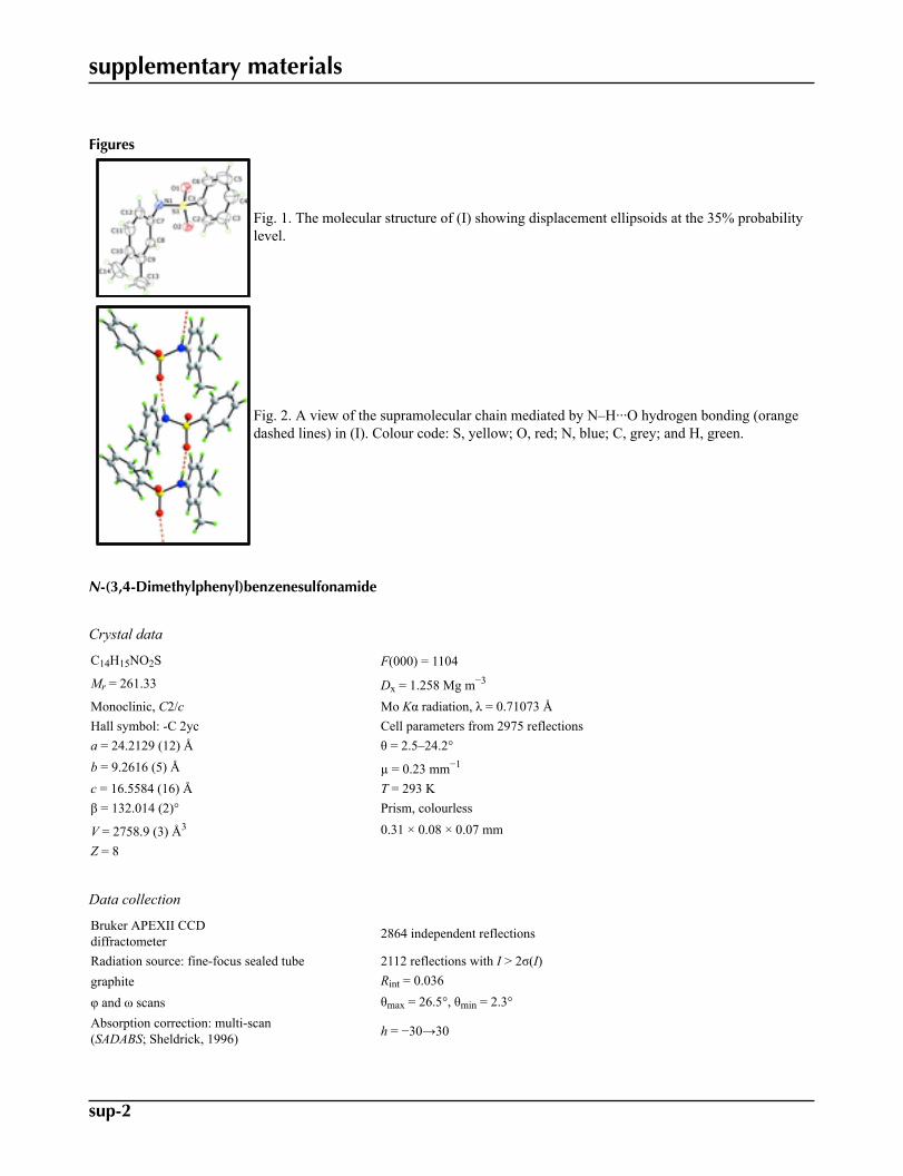
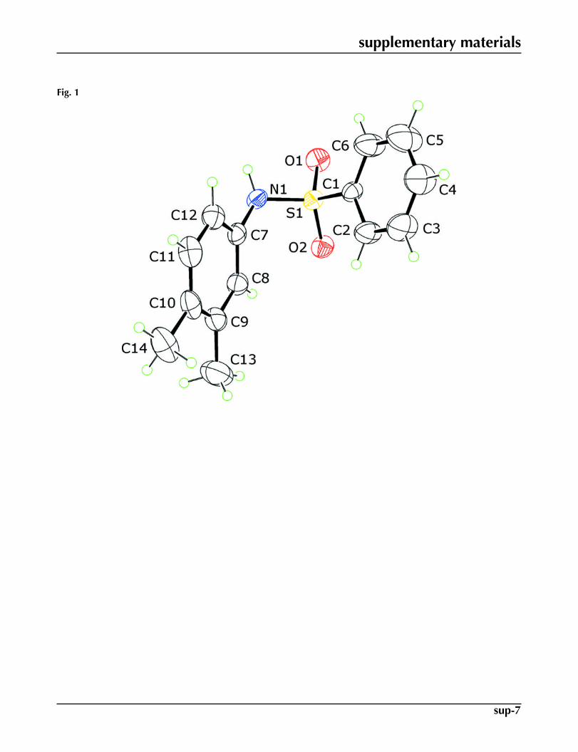
In (I), both sulfonamido-O atoms lie to one side of the S-bound benzene ring [the O1–S1–C1–C6 and O2–S1–C1–C2torsion angles are 19.3 (4) and -28.0 (3) °, respectively] and are on the opposite side of this ring to the sulfonamido-N1atom, Fig. 1. The latter is approximately perpendicular to the benzene ring [N1–S1–C1–C2 is -87.4 (3) °] with the N-boundbenzene ring clearly displaced to one side of the molecule; the dihedral angle formed between the two benzene rings is64.5 (3) °. Although not isomorphous, the overall molecular conformation found in the closely related derivative, with anS-bound tolsyl group, is almost identical (Gowda et al. 2009).
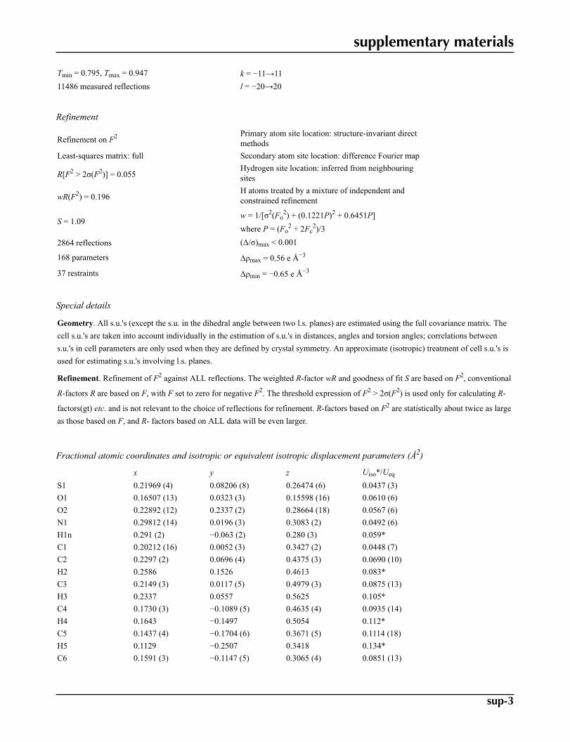
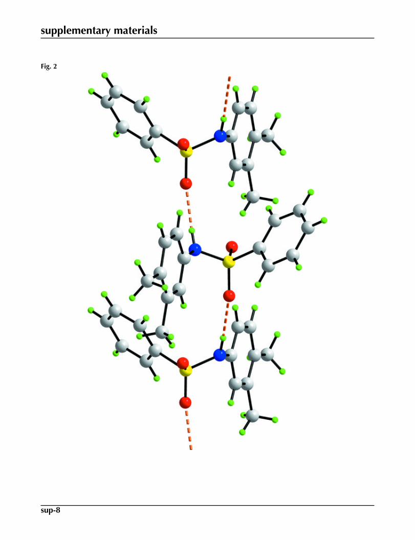
The presence of N1–H···O2 hydrogen bonding, Table 1, leads to the formation of supramolecular chains along the baxis, Fig. 2.
Experimental
To 3,4-dimethyl aniline (484 mg, 4 mmol) in distilled water (10 ml) was added benzene sulfonyl chloride (510 ml, 4 mmol)with stirring at room temperature while maintaining the pH of the reaction mixture at 8 using 3% sodium carbonate. Theprogress of the reaction was monitored by TLC. The precipitate formed was washed with water, dried and crystallized froma methanol/ethyl acetate mixture (50:50 V/V) to yield colourless prisms of (I); m. pt 414 K.
Refinement
The C-bound H atoms were geometrically placed (C–H = 0.93–0.96 Å) and refined as riding with Uiso(H) = 1.2Ueq(C). The
N-bound H atom was refined with the distance restraint N–H = 0.86±0.01 Å, and with Uiso(H) = 1.2Ueq(N). High thermal
motion was noted for several atoms in the S-bound benzene ring but multiple positions were not resolved. The anisotropicdisplacement parameters for this ring were refined with the ISOR command to constrain these to be approximately isotropic.
supplementary materials
sup-2
Figures
Fig. 1. The molecular structure of (I) showing displacement ellipsoids at the 35% probabilitylevel.
Fig. 2. A view of the supramolecular chain mediated by N–H···O hydrogen bonding (orangedashed lines) in (I). Colour code: S, yellow; O, red; N, blue; C, grey; and H, green.
N-(3,4-Dimethylphenyl)benzenesulfonamide
Crystal data
C14H15NO2S F(000) = 1104
Mr = 261.33 Dx = 1.258 Mg m−3
Monoclinic, C2/c Mo Kα radiation, λ = 0.71073 ÅHall symbol: -C 2yc Cell parameters from 2975 reflectionsa = 24.2129 (12) Å θ = 2.5–24.2°b = 9.2616 (5) Å µ = 0.23 mm−1
c = 16.5584 (16) Å T = 293 Kβ = 132.014 (2)° Prism, colourless
V = 2758.9 (3) Å3 0.31 × 0.08 × 0.07 mmZ = 8
Data collection
Bruker APEXII CCDdiffractometer 2864 independent reflections
Radiation source: fine-focus sealed tube 2112 reflections with I > 2σ(I)graphite Rint = 0.036
φ and ω scans θmax = 26.5°, θmin = 2.3°Absorption correction: multi-scan(SADABS; Sheldrick, 1996) h = −30→30
supplementary materials
sup-3
Tmin = 0.795, Tmax = 0.947 k = −11→1111486 measured reflections l = −20→20
Refinement
Refinement on F2 Primary atom site location: structure-invariant directmethods
Least-squares matrix: full Secondary atom site location: difference Fourier map
R[F2 > 2σ(F2)] = 0.055Hydrogen site location: inferred from neighbouringsites
wR(F2) = 0.196H atoms treated by a mixture of independent andconstrained refinement
S = 1.09w = 1/[σ2(Fo
2) + (0.1221P)2 + 0.6451P]where P = (Fo
2 + 2Fc2)/3
2864 reflections (Δ/σ)max < 0.001
168 parameters Δρmax = 0.56 e Å−3
37 restraints Δρmin = −0.65 e Å−3
Special details
Geometry. All s.u.'s (except the s.u. in the dihedral angle between two l.s. planes) are estimated using the full covariance matrix. Thecell s.u.'s are taken into account individually in the estimation of s.u.'s in distances, angles and torsion angles; correlations betweens.u.'s in cell parameters are only used when they are defined by crystal symmetry. An approximate (isotropic) treatment of cell s.u.'s isused for estimating s.u.'s involving l.s. planes.
Refinement. Refinement of F2 against ALL reflections. The weighted R-factor wR and goodness of fit S are based on F2, conventional
R-factors R are based on F, with F set to zero for negative F2. The threshold expression of F2 > 2σ(F2) is used only for calculating R-
factors(gt) etc. and is not relevant to the choice of reflections for refinement. R-factors based on F2 are statistically about twice as largeas those based on F, and R- factors based on ALL data will be even larger.
Fractional atomic coordinates and isotropic or equivalent isotropic displacement parameters (Å2)
x y z Uiso*/Ueq
S1 0.21969 (4) 0.08206 (8) 0.26474 (6) 0.0437 (3)O1 0.16507 (13) 0.0323 (3) 0.15598 (16) 0.0610 (6)O2 0.22892 (12) 0.2337 (2) 0.28664 (18) 0.0567 (6)N1 0.29812 (14) 0.0196 (3) 0.3083 (2) 0.0492 (6)H1n 0.291 (2) −0.063 (2) 0.280 (3) 0.059*C1 0.20212 (16) 0.0052 (3) 0.3427 (2) 0.0448 (7)C2 0.2297 (2) 0.0696 (4) 0.4375 (3) 0.0690 (10)H2 0.2586 0.1526 0.4613 0.083*C3 0.2149 (3) 0.0117 (5) 0.4979 (3) 0.0875 (13)H3 0.2337 0.0557 0.5625 0.105*C4 0.1730 (3) −0.1089 (5) 0.4635 (4) 0.0935 (14)H4 0.1643 −0.1497 0.5054 0.112*C5 0.1437 (4) −0.1704 (6) 0.3671 (5) 0.1114 (18)H5 0.1129 −0.2507 0.3418 0.134*C6 0.1591 (3) −0.1147 (5) 0.3065 (4) 0.0851 (13)
supplementary materials
sup-4
H6 0.1403 −0.1588 0.2420 0.102*C7 0.36691 (17) 0.0393 (3) 0.4183 (2) 0.0470 (7)C8 0.39442 (19) 0.1756 (3) 0.4603 (3) 0.0537 (8)H8 0.3673 0.2562 0.4178 0.064*C9 0.46209 (19) 0.1938 (4) 0.5652 (3) 0.0607 (9)C10 0.50388 (19) 0.0737 (4) 0.6287 (3) 0.0638 (10)C11 0.4750 (2) −0.0623 (4) 0.5834 (3) 0.0683 (10)H11 0.5022 −0.1436 0.6246 0.082*C12 0.4080 (2) −0.0805 (4) 0.4804 (3) 0.0581 (8)H12 0.3902 −0.1728 0.4523 0.070*C13 0.4905 (3) 0.3462 (5) 0.6065 (4) 0.1030 (16)H13A 0.5075 0.3559 0.6778 0.154*H13B 0.4511 0.4139 0.5581 0.154*H13C 0.5309 0.3653 0.6096 0.154*C14 0.5778 (2) 0.0900 (6) 0.7430 (3) 0.0921 (15)H14A 0.6053 0.1672 0.7454 0.138*H14B 0.6054 0.0017 0.7654 0.138*H14C 0.5697 0.1115 0.7912 0.138*
Atomic displacement parameters (Å2)
U11 U22 U33 U12 U13 U23
S1 0.0524 (5) 0.0421 (5) 0.0424 (4) 0.0023 (3) 0.0340 (4) 0.0028 (3)O1 0.0623 (14) 0.0769 (16) 0.0388 (12) −0.0017 (12) 0.0317 (11) −0.0011 (10)O2 0.0678 (14) 0.0410 (12) 0.0680 (14) 0.0076 (10) 0.0483 (13) 0.0091 (10)N1 0.0572 (15) 0.0462 (14) 0.0533 (15) −0.0004 (12) 0.0407 (13) −0.0075 (12)C1 0.0560 (16) 0.0416 (15) 0.0466 (15) −0.0004 (12) 0.0384 (14) −0.0012 (12)C2 0.089 (2) 0.072 (2) 0.0622 (19) −0.0237 (18) 0.0574 (19) −0.0184 (16)C3 0.118 (3) 0.102 (3) 0.071 (2) −0.024 (2) 0.075 (2) −0.019 (2)C4 0.132 (3) 0.098 (3) 0.091 (3) −0.025 (3) 0.092 (3) −0.005 (2)C5 0.155 (4) 0.101 (3) 0.118 (3) −0.054 (3) 0.108 (3) −0.021 (2)C6 0.121 (3) 0.079 (2) 0.084 (2) −0.038 (2) 0.080 (2) −0.026 (2)C7 0.0513 (16) 0.0521 (17) 0.0538 (17) 0.0004 (13) 0.0418 (15) 0.0004 (14)C8 0.0551 (18) 0.0536 (19) 0.0588 (19) 0.0010 (14) 0.0408 (17) −0.0001 (14)C9 0.0553 (19) 0.071 (2) 0.064 (2) −0.0095 (17) 0.0436 (18) −0.0090 (18)C10 0.0481 (19) 0.094 (3) 0.058 (2) −0.0018 (18) 0.0389 (17) 0.0061 (19)C11 0.062 (2) 0.075 (3) 0.074 (2) 0.0142 (18) 0.048 (2) 0.0238 (19)C12 0.061 (2) 0.0554 (19) 0.067 (2) 0.0035 (15) 0.0462 (18) 0.0068 (16)C13 0.078 (3) 0.098 (4) 0.091 (3) −0.020 (3) 0.039 (3) −0.022 (3)C14 0.058 (2) 0.142 (4) 0.068 (3) −0.008 (2) 0.038 (2) 0.011 (3)
Geometric parameters (Å, °)
S1—O1 1.420 (2) C7—C8 1.379 (4)S1—O2 1.430 (2) C7—C12 1.381 (4)S1—N1 1.621 (3) C8—C9 1.386 (5)S1—C1 1.758 (3) C8—H8 0.9300N1—C7 1.436 (4) C9—C10 1.394 (5)N1—H1n 0.85 (3) C9—C13 1.518 (6)

supplementary materials
sup-5
C1—C6 1.356 (5) C10—C11 1.392 (5)C1—C2 1.364 (4) C10—C14 1.508 (5)C2—C3 1.379 (5) C11—C12 1.365 (5)C2—H2 0.9300 C11—H11 0.9300C3—C4 1.351 (6) C12—H12 0.9300C3—H3 0.9300 C13—H13A 0.9600C4—C5 1.363 (6) C13—H13B 0.9600C4—H4 0.9300 C13—H13C 0.9600C5—C6 1.384 (6) C14—H14A 0.9600C5—H5 0.9300 C14—H14B 0.9600C6—H6 0.9300 C14—H14C 0.9600
O1—S1—O2 119.67 (15) C12—C7—N1 119.2 (3)O1—S1—N1 105.54 (14) C7—C8—C9 120.7 (3)O2—S1—N1 107.46 (13) C7—C8—H8 119.7O1—S1—C1 108.80 (14) C9—C8—H8 119.7O2—S1—C1 106.57 (13) C8—C9—C10 120.1 (3)N1—S1—C1 108.39 (14) C8—C9—C13 118.5 (4)C7—N1—S1 122.54 (19) C10—C9—C13 121.4 (4)C7—N1—H1N 117 (2) C11—C10—C9 117.8 (3)S1—N1—H1N 109 (2) C11—C10—C14 120.9 (4)C6—C1—C2 120.5 (3) C9—C10—C14 121.3 (4)C6—C1—S1 119.9 (2) C12—C11—C10 122.2 (3)C2—C1—S1 119.6 (2) C12—C11—H11 118.9C1—C2—C3 120.0 (4) C10—C11—H11 118.9C1—C2—H2 120.0 C11—C12—C7 119.5 (3)C3—C2—H2 120.0 C11—C12—H12 120.2C4—C3—C2 120.1 (4) C7—C12—H12 120.2C4—C3—H3 120.0 C9—C13—H13A 109.5C2—C3—H3 120.0 C9—C13—H13B 109.5C3—C4—C5 119.7 (4) H13A—C13—H13B 109.5C3—C4—H4 120.1 C9—C13—H13C 109.5C5—C4—H4 120.1 H13A—C13—H13C 109.5C4—C5—C6 120.7 (4) H13B—C13—H13C 109.5C4—C5—H5 119.7 C10—C14—H14A 109.5C6—C5—H5 119.7 C10—C14—H14B 109.5C1—C6—C5 119.0 (4) H14A—C14—H14B 109.5C1—C6—H6 120.5 C10—C14—H14C 109.5C5—C6—H6 120.5 H14A—C14—H14C 109.5C8—C7—C12 119.7 (3) H14B—C14—H14C 109.5C8—C7—N1 121.0 (3)
O1—S1—N1—C7 −176.1 (2) C4—C5—C6—C1 −2.1 (9)O2—S1—N1—C7 55.1 (3) S1—N1—C7—C8 −60.8 (4)C1—S1—N1—C7 −59.7 (3) S1—N1—C7—C12 123.0 (3)O1—S1—C1—C6 19.3 (4) C12—C7—C8—C9 −1.6 (4)O2—S1—C1—C6 149.6 (3) N1—C7—C8—C9 −177.7 (3)N1—S1—C1—C6 −95.0 (4) C7—C8—C9—C10 1.4 (5)O1—S1—C1—C2 −158.3 (3) C7—C8—C9—C13 178.7 (3)O2—S1—C1—C2 −28.0 (3) C8—C9—C10—C11 −0.5 (5)

supplementary materials
sup-6
N1—S1—C1—C2 87.4 (3) C13—C9—C10—C11 −177.8 (4)C6—C1—C2—C3 0.9 (6) C8—C9—C10—C14 179.9 (3)S1—C1—C2—C3 178.6 (3) C13—C9—C10—C14 2.6 (5)C1—C2—C3—C4 0.1 (7) C9—C10—C11—C12 −0.2 (5)C2—C3—C4—C5 −2.1 (9) C14—C10—C11—C12 179.4 (3)C3—C4—C5—C6 3.1 (10) C10—C11—C12—C7 −0.1 (5)C2—C1—C6—C5 0.1 (7) C8—C7—C12—C11 0.9 (5)S1—C1—C6—C5 −177.6 (4) N1—C7—C12—C11 177.1 (3)
Hydrogen-bond geometry (Å, °)
D—H···A D—H H···A D···A D—H···A
N1—H1n···O2i 0.85 (3) 2.07 (2) 2.921 (3) 177 (5)Symmetry codes: (i) −x+1/2, y−1/2, −z+1/2.
supplementary materials
sup-7
Fig. 1
supplementary materials
sup-8
Fig. 2


![2-(1 H -Benzimidazol-2-yl)- N -[( E )-(dimethylamino)methylidene]benzenesulfonamide](https://static.fdokumen.com/doc/165x107/63369e96242ed15b940dcdfc/2-1-h-benzimidazol-2-yl-n-e-dimethylaminomethylidenebenzenesulfonamide.jpg)

![1-[2-(3,4-Dichlorobenzyloxy)-2-phenylethyl]-1 H -benzimidazole](https://static.fdokumen.com/doc/165x107/63152ee185333559270d05af/1-2-34-dichlorobenzyloxy-2-phenylethyl-1-h-benzimidazole.jpg)


![Synthesis and evaluation of conformationally restricted N-[2-(3,4-dichlorophenyl)ethyl]-N-methyl-2-(1-pyrrolidinyl)ethylamines at .sigma. receptors. 2. Piperazines, bicyclic amines,](https://static.fdokumen.com/doc/165x107/631289503ed465f0570a465c/synthesis-and-evaluation-of-conformationally-restricted-n-2-34-dichlorophenylethyl-n-methyl-2-1-pyrrolidinylethylamines.jpg)


![A-740003 [N-(1-{[(Cyanoimino)(5-quinolinylamino) methyl]amino}-2,2-dimethylpropyl)-2-(3,4-dimethoxyphenyl)acetamide], a Novel and Selective P2X7 Receptor Antagonist, Dose-Dependently](https://static.fdokumen.com/doc/165x107/63441f69596bdb97a9085093/a-740003-n-1-cyanoimino5-quinolinylamino-methylamino-22-dimethylpropyl-2-34-dimethoxyphenylacetamide.jpg)


![Pyrazolo[3,4- c ]pyridazines as Novel and Selective Inhibitors of Cyclin-Dependent Kinases](https://static.fdokumen.com/doc/165x107/632451b94d8439cb620d53b7/pyrazolo34-c-pyridazines-as-novel-and-selective-inhibitors-of-cyclin-dependent.jpg)

