Study of microdroplet generation from vacuum arcs on graphite cathodes
Upload
independentCategory
view
0download
0
POLYMERS FOR ADVANCED TECHNOLOGIES
Polym. Adv. Technol. 2008; 19: 371–376
science.wiley.com) DOI: 10.1002/pat.1018
Published online 5 December 2007 in Wiley InterScience (www.interSynthesis, characterization and evaluation of semi-IPN
hydrogels consisted of poly(methacrylic acid) and guar
gum for colon-specific drug delivery
Shengfang Li* and Xianli LiuSchool of Chemical and Material Engineering, Huangshi Institute of Technology, Huangshi 435003, Hubei, PR China
Received 29 July 2007; Revised 24 September 2007; Accepted 26 September 2007
*Correspoeering, HHubei, PE-mail: li
The semi-IPN hydrogels consisting of poly(methacrylic acid) and guar gum (GG) are prepared at
room temperature using water as solvent. 5-aminosalicylic acid (5-ASA) is entrapped in the hydrogel
in the synthesis of hydrogel and all entrapment efficiencies are found above 85%. The hydrogel
shows excellent pH-sensitivity. It exhibited minimum swelling in an acidic pHmedium through the
formation of a complex hydrogen-bonded structure and maximal swelling due to the electrostatic
repulsion due to the ionization of the carboxylic groups in pH 7.4 medium. The degradation in vitro
shows that the degree of degradation (R%) depended on the concentration of cross-linking agent and
content of GG. The hydrogel shows a minimum release of 5-ASA due to the complex hydrogen
bonded structure of the hydrogels in the medium of pH 2.2. The enzymatic degradation of hydrogels
by cecal bacteria can accelerate the release of 5-ASA entrapped in the hydrogel in pH 7.4 medium.
Copyright # 2007 John Wiley & Sons, Ltd.
KEYWORDS: guar gum; Semi-IPN hydrogels; enzymatic degradation in vitro; release
INTRODUCTION
Colon-specific drug delivery has gained increased import-
ance not only for its potential for the delivery of proteins and
peptides but also for the delivery of the drugs for the
treatment special diseases such as ulcerative colitis, Crohn’s
diseases, inflammatory bowel diseases (IBD), infectious
diseases, and colon cancer.1,2 To achieve successful colonic
delivery, a drug needs to be prevented from absorption of the
environment of upper gastrointestinal tract (GIT) and then be
released into the colon, which is considered the optimum site
for colon-specific drug delivery. The various approaches for
colon-specific drug delivery mainly include time-dependent
release systems, pH-dependent systems and enzymatically
controlled delivery systems. pH-sensitive hydrogel could be
potentially used for the delivery of drugs to the colon.
However, site-specific drug delivery to the colon cannot be
achieved by the only pH-sensitive hydrogels, because the pH
of the small intestine and the large intestine are almost same.3
Guar gum (GG), a naturally occurring glactomannan
polysaccharide, has been well studied as a carrier for
colon-specific drug delivery due to its drug release retarding
property and susceptibility to microbial degradation in the
colon.4,5 From the literatures, GG used for colon-specific
ndence to: S. Li, School of Chemical and Material Engin-uangshi Institute of Technology, Huangshi 435003,R [email protected]
drug delivery includes mainly coating and hydrogels.6
However, the studies on hydrogels of GG mostly emphasize
on enzymatic degradation of GG and its release of drugs in
the environment of colon. Few researchers focus on the
release behavior in the pH environment of stomach.7–10
5-Aminosalicylic acid (5-ASA) is widely accepted in the
treatment of IBD, including ulcerative colitis and Crohn’s
disease. When orally administered, 5-ASA is unstable in the
gastric conditions and prone to be absorbed or degraded in
the stomach and small intestine before reaching the colon
sites, which causes low drug bioavailability and low
efficiency for inflammatory colon disease. In addition,
5-ASA is easily oxidated at high temperature. In the present
study, we synthesized a new pH sensitive and enzymatic
degradable semi-IPN hydrogel containing both pH-sensitive
acidic monomers and enzymatically degradable GG for
colon-specific drug delivery. The hydrogels are consisted of
poly(methacrylic acid) and GG. Swelling of such hydrogels
in the stomach (lower pH value) is minimal due to the
presence of carboxylic groups. The extent of swelling
increases as the hydrogel passes down the intestinal tract
because of increase in pH leading to ionization of the
carboxylic groups. Once inside the colon, highly swollen
hydrogels become accessible to the enzymes produced by
microflora in the colon. The enzymatic degradation of GG
will occur. 5-ASA, as model drug, was entrapped in the
hydrogel in the synthesis of hydrogel at room temperature
using water as a solvent. The semi-IPN hydrogels were
characterized by FTIR, SEM, etc. The swelling and degra-
Copyright # 2007 John Wiley & Sons, Ltd.
372 S. Li and X. Liu
dation properties of semi-IPN hydrogels were also studied.
The release behavior of 5-ASA in vitro from semi-IPN
hydrogel was evaluated.
EXPERIMENTAL
MaterialsMethacrylic acid (MAA) were purchased from Tianjin
Chemical Group, China and were distilled before use. GG,
N,N0-methylenebisacrylamide (MBA), K2S2O8, and NaHSO3
(purchased from Shanghai Chemical Group, China) were of
analytical reagent grade and used without any further
purification. 5-ASA was a gift from Yuancheng Co. (Wuhan,
China), and recrystallized with distilled water.
Preparation of semi-IPN hydrogelsThe hydrogels were prepared through the free-radical
copolymerization of MAA, MBA in the present of GG in
an aqueous medium. Briefly, the mixture solutions were
made up of varied amounts of MAA,MBA in distilled water.
Then various amounts of GG, K2S2O8, and NaHSO3 were
added into the mixture solutions. This homogeneous
solution was bubbled with nitrogen to discharge oxygen
for about 30min. The free radical copolymerization was
carried out at room temperature for 4 days. After
copolymerization, the solid copolymer slab was cut into
circular disks using punches. The samples were immersed in
deionized water for 6 days to remove the unreacted
monomers. Finally, the samples were dried in vacuum at
258C to a constant weight and stored for further use. The feed
composition for the preparation of hydrogels is listed in
Table 1 and the samples are designated as PMGx.
Fourier transform infrared (FIIR)measurementsFIIR spectroscopy was used to confirm the structure of
semi-IPN. The hydrogel samples and GGwere analyzed on a
Bruker EQUNINOX FIIR spectrophotometer in the region of
400–4000 cm�1. Before themeasurements, the dried hydrogel
samples were crushed down (KBr, pellet).
Morphology of semi-IPN xerogelsThe equilibrium-swollen gels were frozen at �808C for 12 hr
in refrigerator, then freeze-dried and fractured. The fractured
specimens were covered with gold vapor, and then the
morphology of the fractured surface of the xerogels were
observed by field emission scanning electron microscopy
Table 1. The feed composition of semi-IPN hydrogels
Sample GG (g) MAA (g) MBA (g)
PMG0 0 1 0.02PMG1 0.4 1 0.02PMG2 0.4 1 0.04PMG3 0.4 1 0.06PMG4 0.4 1 0.08PMG5 0.4 1 0.1PMG6 0.2 1 0.02PMG7 0.1 1 0.02
Copyright # 2007 John Wiley & Sons, Ltd.
(FE-SEM; FEI Sirion 200) with an acceleration voltage of
10 kV.
Swelling studiesThe dry hydrogel was immersed in the swelling medium at
378C. At regular time intervals, the gels were removed from
the medium, the weight of the swollen hydrogels was
determined after the removal of the surface water through
blotting with filter paper. The equilibrium-swelling ratio was
calculated by the following equation:
SR ¼ Ws �Wd
Wdð1Þ
when the swollen hydrogels reached a constant weight, the
SR was considered to be the equilibrium. Wd and Ws was
the weight of xerogels and equilibrium-swollen gels,
respectively.
In vitro degradation of hydrogelsThe enzymatic degradation of the hydrogels was performed
in a flask filled with 150ml of phosphate buffer (pH 7.4,
0.1mol/L, 378C) which contained a certain quantity of freeze
dried rat cecum content (from male Sprague–Dawley rats,
about 250 g). The degradation experiments were conducted
by incubating the hydrogel mass in buffer placed in a
thermostatic rotary shaker (HQ45Z, Chinese Academy of
Sciences instrument Corp. Ltd., Wuhan, China) and by the
determination of the weight loss after recovery of the
samples at predetermined time intervals. The buffer solution
was changed every 2 days to maintain enzymatic activity.
After a predetermined time, the samples were removed from
the solution, washed thoroughly with distilled water, and
then dried below 608C. The degradation was assessed by
measuring the weight loss ratio (R%), which was defined as
the following equation:
R ð%Þ ¼ W1 �W2
W1ð2Þ
where, W1 and W2 were the weights of the gel before and
after degradation, respectively.
In vitro loading and release of 5-ASAof the hydrogelsIn order to determine the actual amount of drug entrapped
into the hydrogel, various sample synthesized were washed
in 1L of distilled water taken in five installments of 200ml
each and the absorbance of each solution was measured at
275 nm. In this way, the actual drug entrapped in the
K2S2O8 (g) NaHSO3 (g) H2O (g)
0.02 0.01 100.02 0.01 100.02 0.01 100.02 0.01 100.02 0.01 100.02 0.01 100.02 0.01 100.02 0.01 10
Polym. Adv. Technol. 2008; 19: 371–376
DOI: 10.1002/pat
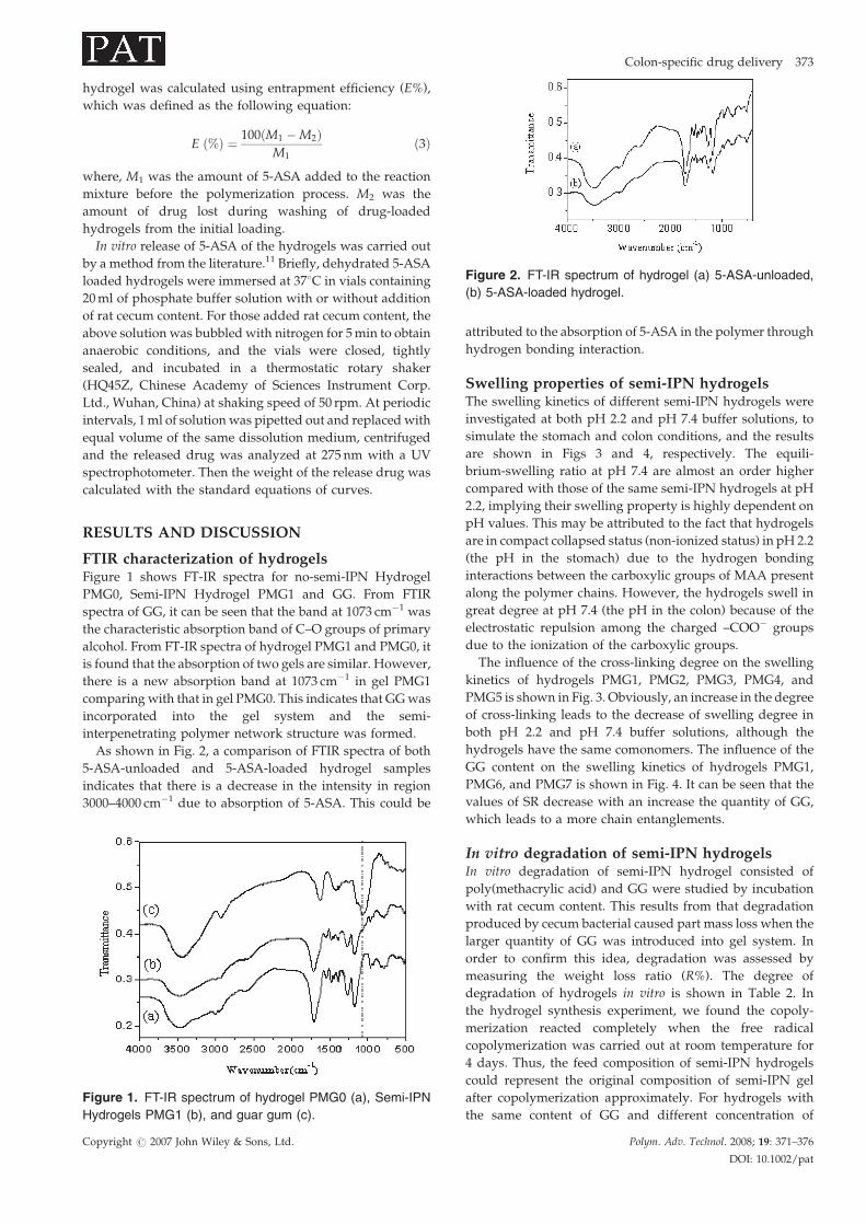
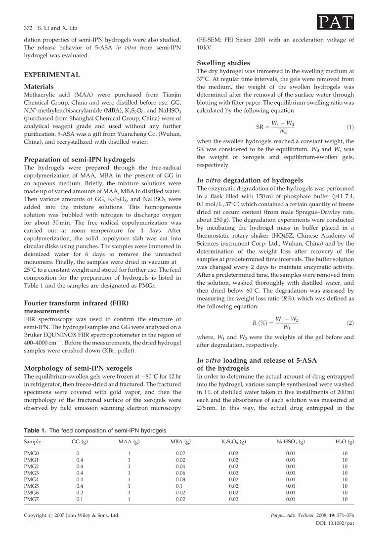
Figure 2. FT-IR spectrum of hydrogel (a) 5-ASA-unloaded,
(b) 5-ASA-loaded hydrogel.
Colon-specific drug delivery 373
hydrogel was calculated using entrapment efficiency (E%),
which was defined as the following equation:
E ð%Þ ¼ 100ðM1 �M2ÞM1
ð3Þ
where, M1 was the amount of 5-ASA added to the reaction
mixture before the polymerization process. M2 was the
amount of drug lost during washing of drug-loaded
hydrogels from the initial loading.
In vitro release of 5-ASA of the hydrogels was carried out
by a method from the literature.11 Briefly, dehydrated 5-ASA
loaded hydrogels were immersed at 378C in vials containing
20ml of phosphate buffer solution with or without addition
of rat cecum content. For those added rat cecum content, the
above solution was bubbled with nitrogen for 5min to obtain
anaerobic conditions, and the vials were closed, tightly
sealed, and incubated in a thermostatic rotary shaker
(HQ45Z, Chinese Academy of Sciences Instrument Corp.
Ltd., Wuhan, China) at shaking speed of 50 rpm. At periodic
intervals, 1ml of solutionwas pipetted out and replacedwith
equal volume of the same dissolution medium, centrifuged
and the released drug was analyzed at 275 nm with a UV
spectrophotometer. Then the weight of the release drug was
calculated with the standard equations of curves.
RESULTS AND DISCUSSION
FTIR characterization of hydrogelsFigure 1 shows FT-IR spectra for no-semi-IPN Hydrogel
PMG0, Semi-IPN Hydrogel PMG1 and GG. From FTIR
spectra of GG, it can be seen that the band at 1073 cm�1 was
the characteristic absorption band of C–O groups of primary
alcohol. From FT-IR spectra of hydrogel PMG1 and PMG0, it
is found that the absorption of two gels are similar. However,
there is a new absorption band at 1073 cm�1 in gel PMG1
comparingwith that in gel PMG0. This indicates that GGwas
incorporated into the gel system and the semi-
interpenetrating polymer network structure was formed.
As shown in Fig. 2, a comparison of FTIR spectra of both
5-ASA-unloaded and 5-ASA-loaded hydrogel samples
indicates that there is a decrease in the intensity in region
3000–4000 cm�1 due to absorption of 5-ASA. This could be
Figure 1. FT-IR spectrum of hydrogel PMG0 (a), Semi-IPN
Hydrogels PMG1 (b), and guar gum (c).
Copyright # 2007 John Wiley & Sons, Ltd.
attributed to the absorption of 5-ASA in the polymer through
hydrogen bonding interaction.
Swelling properties of semi-IPN hydrogelsThe swelling kinetics of different semi-IPN hydrogels were
investigated at both pH 2.2 and pH 7.4 buffer solutions, to
simulate the stomach and colon conditions, and the results
are shown in Figs 3 and 4, respectively. The equili-
brium-swelling ratio at pH 7.4 are almost an order higher
compared with those of the same semi-IPN hydrogels at pH
2.2, implying their swelling property is highly dependent on
pH values. This may be attributed to the fact that hydrogels
are in compact collapsed status (non-ionized status) in pH 2.2
(the pH in the stomach) due to the hydrogen bonding
interactions between the carboxylic groups of MAA present
along the polymer chains. However, the hydrogels swell in
great degree at pH 7.4 (the pH in the colon) because of the
electrostatic repulsion among the charged –COO� groups
due to the ionization of the carboxylic groups.
The influence of the cross-linking degree on the swelling
kinetics of hydrogels PMG1, PMG2, PMG3, PMG4, and
PMG5 is shown in Fig. 3. Obviously, an increase in the degree
of cross-linking leads to the decrease of swelling degree in
both pH 2.2 and pH 7.4 buffer solutions, although the
hydrogels have the same comonomers. The influence of the
GG content on the swelling kinetics of hydrogels PMG1,
PMG6, and PMG7 is shown in Fig. 4. It can be seen that the
values of SR decrease with an increase the quantity of GG,
which leads to a more chain entanglements.
In vitro degradation of semi-IPN hydrogelsIn vitro degradation of semi-IPN hydrogel consisted of
poly(methacrylic acid) and GG were studied by incubation
with rat cecum content. This results from that degradation
produced by cecum bacterial caused part mass loss when the
larger quantity of GG was introduced into gel system. In
order to confirm this idea, degradation was assessed by
measuring the weight loss ratio (R%). The degree of
degradation of hydrogels in vitro is shown in Table 2. In
the hydrogel synthesis experiment, we found the copoly-
merization reacted completely when the free radical
copolymerization was carried out at room temperature for
4 days. Thus, the feed composition of semi-IPN hydrogels
could represent the original composition of semi-IPN gel
after copolymerization approximately. For hydrogels with
the same content of GG and different concentration of
Polym. Adv. Technol. 2008; 19: 371–376
DOI: 10.1002/pat
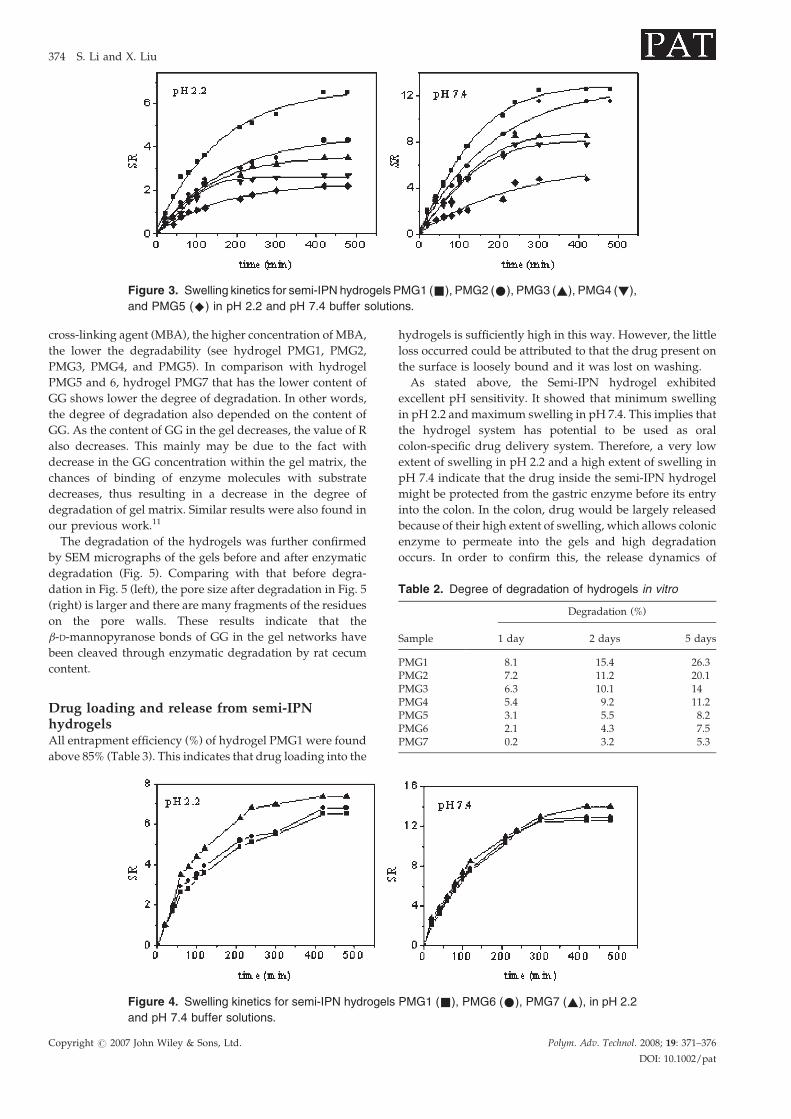
Figure 3. Swelling kinetics for semi-IPN hydrogels PMG1 (&), PMG2 (*), PMG3 (~), PMG4 (!),
and PMG5 (^) in pH 2.2 and pH 7.4 buffer solutions.
Table 2. Degree of degradation of hydrogels in vitro
Sample
Degradation (%)
1 day 2 days 5 days
PMG1 8.1 15.4 26.3PMG2 7.2 11.2 20.1PMG3 6.3 10.1 14PMG4 5.4 9.2 11.2PMG5 3.1 5.5 8.2PMG6 2.1 4.3 7.5PMG7 0.2 3.2 5.3
374 S. Li and X. Liu
cross-linking agent (MBA), the higher concentration of MBA,
the lower the degradability (see hydrogel PMG1, PMG2,
PMG3, PMG4, and PMG5). In comparison with hydrogel
PMG5 and 6, hydrogel PMG7 that has the lower content of
GG shows lower the degree of degradation. In other words,
the degree of degradation also depended on the content of
GG. As the content of GG in the gel decreases, the value of R
also decreases. This mainly may be due to the fact with
decrease in the GG concentration within the gel matrix, the
chances of binding of enzyme molecules with substrate
decreases, thus resulting in a decrease in the degree of
degradation of gel matrix. Similar results were also found in
our previous work.11
The degradation of the hydrogels was further confirmed
by SEM micrographs of the gels before and after enzymatic
degradation (Fig. 5). Comparing with that before degra-
dation in Fig. 5 (left), the pore size after degradation in Fig. 5
(right) is larger and there are many fragments of the residues
on the pore walls. These results indicate that the
b-D-mannopyranose bonds of GG in the gel networks have
been cleaved through enzymatic degradation by rat cecum
content.
Drug loading and release from semi-IPNhydrogelsAll entrapment efficiency (%) of hydrogel PMG1 were found
above 85% (Table 3). This indicates that drug loading into the
Figure 4. Swelling kinetics for semi-IPN hydrogels
and pH 7.4 buffer solutions.
Copyright # 2007 John Wiley & Sons, Ltd.
hydrogels is sufficiently high in this way. However, the little
loss occurred could be attributed to that the drug present on
the surface is loosely bound and it was lost on washing.
As stated above, the Semi-IPN hydrogel exhibited
excellent pH sensitivity. It showed that minimum swelling
in pH 2.2 andmaximum swelling in pH 7.4. This implies that
the hydrogel system has potential to be used as oral
colon-specific drug delivery system. Therefore, a very low
extent of swelling in pH 2.2 and a high extent of swelling in
pH 7.4 indicate that the drug inside the semi-IPN hydrogel
might be protected from the gastric enzyme before its entry
into the colon. In the colon, drug would be largely released
because of their high extent of swelling, which allows colonic
enzyme to permeate into the gels and high degradation
occurs. In order to confirm this, the release dynamics of
PMG1 (&), PMG6 (*), PMG7 (~), in pH 2.2
Polym. Adv. Technol. 2008; 19: 371–376
DOI: 10.1002/pat
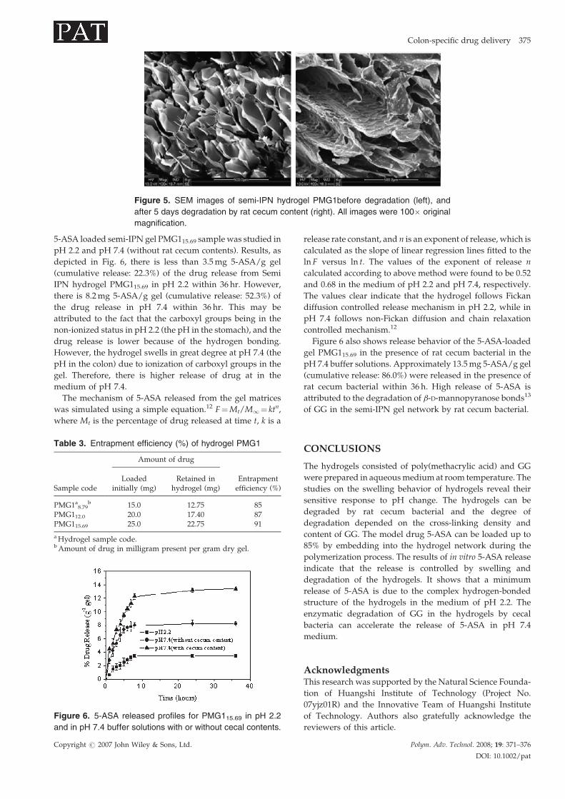
Figure 5. SEM images of semi-IPN hydrogel PMG1before degradation (left), and
after 5 days degradation by rat cecum content (right). All images were 100� original
magnification.
Colon-specific drug delivery 375
5-ASA loaded semi-IPN gel PMG115.69 samplewas studied in
pH 2.2 and pH 7.4 (without rat cecum contents). Results, as
depicted in Fig. 6, there is less than 3.5mg 5-ASA/g gel
(cumulative release: 22.3%) of the drug release from Semi
IPN hydrogel PMG115.69 in pH 2.2 within 36 hr. However,
there is 8.2mg 5-ASA/g gel (cumulative release: 52.3%) of
the drug release in pH 7.4 within 36 hr. This may be
attributed to the fact that the carboxyl groups being in the
non-ionized status in pH 2.2 (the pH in the stomach), and the
drug release is lower because of the hydrogen bonding.
However, the hydrogel swells in great degree at pH 7.4 (the
pH in the colon) due to ionization of carboxyl groups in the
gel. Therefore, there is higher release of drug at in the
medium of pH 7.4.
The mechanism of 5-ASA released from the gel matrices
was simulated using a simple equation.12 F¼Mt/M1¼ ktn,
where Mt is the percentage of drug released at time t, k is a
Table 3. Entrapment efficiency (%) of hydrogel PMG1
Sample code
Amount of drug
Entrapmentefficiency (%)
Loadedinitially (mg)
Retained inhydrogel (mg)
PMG1a8.79b 15.0 12.75 85
PMG112.0 20.0 17.40 87PMG115.69 25.0 22.75 91
aHydrogel sample code.bAmount of drug in milligram present per gram dry gel.
Figure 6. 5-ASA released profiles for PMG115.69 in pH 2.2
and in pH 7.4 buffer solutions with or without cecal contents.
Copyright # 2007 John Wiley & Sons, Ltd.
release rate constant, and n is an exponent of release, which is
calculated as the slope of linear regression lines fitted to the
ln F versus ln t. The values of the exponent of release n
calculated according to above method were found to be 0.52
and 0.68 in the medium of pH 2.2 and pH 7.4, respectively.
The values clear indicate that the hydrogel follows Fickan
diffusion controlled release mechanism in pH 2.2, while in
pH 7.4 follows non-Fickan diffusion and chain relaxation
controlled mechanism.12
Figure 6 also shows release behavior of the 5-ASA-loaded
gel PMG115.69 in the presence of rat cecum bacterial in the
pH 7.4 buffer solutions. Approximately 13.5mg 5-ASA/g gel
(cumulative release: 86.0%) were released in the presence of
rat cecum bacterial within 36 h. High release of 5-ASA is
attributed to the degradation of b-D-mannopyranose bonds13
of GG in the semi-IPN gel network by rat cecum bacterial.
CONCLUSIONS
The hydrogels consisted of poly(methacrylic acid) and GG
were prepared in aqueousmedium at room temperature. The
studies on the swelling behavior of hydrogels reveal their
sensitive response to pH change. The hydrogels can be
degraded by rat cecum bacterial and the degree of
degradation depended on the cross-linking density and
content of GG. The model drug 5-ASA can be loaded up to
85% by embedding into the hydrogel network during the
polymerization process. The results of in vitro 5-ASA release
indicate that the release is controlled by swelling and
degradation of the hydrogels. It shows that a minimum
release of 5-ASA is due to the complex hydrogen-bonded
structure of the hydrogels in the medium of pH 2.2. The
enzymatic degradation of GG in the hydrogels by cecal
bacteria can accelerate the release of 5-ASA in pH 7.4
medium.
AcknowledgmentsThis research was supported by the Natural Science Founda-
tion of Huangshi Institute of Technology (Project No.
07yjz01R) and the Innovative Team of Huangshi Institute
of Technology. Authors also gratefully acknowledge the
reviewers of this article.
Polym. Adv. Technol. 2008; 19: 371–376
DOI: 10.1002/pat
376 S. Li and X. Liu
REFERENCES
1. Gombotz WR, Pettite DK. Biodegradable polymers forprotein and peptide drug delivery. Bioconjug. Chem. 1995;6: 332–351.
2. Kim B, Peppas NA. Synthesis and characterization of pHsensitive lycopolymers for oral drug delivery systems.J. Biomater. Sci. Polymer Edn. 2002; 13: 1271–1281.
3. Friend D. Colonic-specific drug delivery. Adv. Drug. DelievRev. 1991; 7: 149–201.
4. Macfarlane GT, Hay S, Macfarlane S, Gibson GR. Effect ofdifferent carbohydrates on growth, polysaccharidase andglycosidase production by Bacteroides ovatus, in batchand continuous. J. Appl. Bacteriol. 1990; 68: 179–187.
5. Bayliss CE, Houston AP. Degradation of guar gum by faecalbacteria. Appl. Environ. Microbiol. 1986; 48: 626–632.
6. Sinha VR, Kumria R. Microbially triggered drug delivery tothe colon. Eur. J. Pharma. Sci. 2003; 18: 3–18.
7. Rubinstein A, Gliko-Kabir I. Synthesis and swelling depen-dent enzymatic degradation of borax modified guar gum forcolonic delivery purpose. S.T.P. Pharm. Sci. 1995; 5: 41–46.
Copyright # 2007 John Wiley & Sons, Ltd.
8. Gliko-Kabir I, Yagen B, Baluom M, Rubinstein A.Phosphated cross-linked guar for colon-specific drug deliv-ery II. In vitro and in vivo evaluation in the rat. J. Control. Rel.2000; 63: 129–134.
9. Robinsin A. Natural polysaccharides as targeting tools ofdrugs to the human colon. Drug Dev. Res. 2000; 50: 435–439.
10. Gliko-Kabir I, Yagen B, Penhasi A, Rubinstein A. Lowswelling, crosslinked guar and its potential use ascolon-specific drug carrier. Pharm. Res. 1998; 7(15):1019–1025.
11. Li S, Yang Y, Yang X, Xu H. In vitro degradation and ProteinRelease of Semi-IPN hydrogels consisted of poly(acrylicacid-acrylamide-methacrylate) and amylose. J. Appl. Polym.Sci. 2007; 105: 3432–3438.
12. Ritger PL, Peppas NA. A simple equation for description ofsolute release. II. Fickan and anomalous release from swel-lable devices. J. Control. Rel. 1987; 5: 37–42.
13. Sinha VR, Kumria R. Polysaccharides in colon-specific drugdelivery. Int. J. Pharm. 2001; 224: 19–38.
Polym. Adv. Technol. 2008; 19: 371–376
DOI: 10.1002/pat
Copyright © 2022 FDOKUMEN