Synthesis and Characterization of Ag@TiO2 Core-Shell Nanoparticles and Study of its Antibacterial...
10
SYNTHESIS AND CHARACTERIZATION OF Ag@TiO 2 CORE-SHELL NANOPARTICLES AND STUDY OF ITS ANTIBACTERIAL ACTIVITY K. I. DHANALEKSHMI, K. S. MEENA & I. RAMESH PG & Research Department of Chemistry, Queen Mary’s College, Chennai, Tamil Nadu, India ABSTRACT Core-shell type Ag@TiO 2 nano particles were prepared by one pot simultaneous reduction of AgNO 3 and hydrolysis of Ti(IV) isopropoxide. They were characterized by absorption, XRD, FTIR, TGA, DSC and HR-TEM techniques. XRD patterns show the presence of anatase form of TiO 2 and the noble metal (Ag). High resolution transmission electron microscopic measurements revealed that their size is below 50 nm. The antibacterial properties of Ag@TiO 2 core-shell nanoparticles against Escherichia coli (E.coli) and Staphylococcus aureus (S.aureus) were examined by the agar diffusion method .As a result E.coli and S.aureus were shown to be substantially inhibited by Ag@TiO 2 core-shell nanoparticles. These result demonstrated that TiO 2 supported on the surface of Ag NPs without aggregation was proved to be a good novel antibacterial activity. KEYWORDS: Core- Shell Nanoparticles, Escherichia coli, Staphylococcus aureus, Antibacterial Activity INTRODUCTION Antibacterial activity agents are very important in the textile industry , water disinfection, medicine and food packaging. Organic compounds used for disinfection have some disadvantages, including toxicity to the human body, therefore the interest in inorganic disinfectants such as metal oxide nanoparticles is increasing [1]. Inorganic antibacterial agents are superior to organic antibacterial materials in terms of their durability, heat resistance, toxicity, selectivity and various other characteristics [2]. Among the inorganic nanoparticles, silver has been recognized as an effective antimicrobial agents that exhibit low toxicity in human and has diverse in vitro and in vivo application [3]. Ag nanoparticles have attracted considerable attention and have been used as an antibacterial activity and the unlikeness to develop resistant microorganisms [4,5]. The principal characteristic of Ag nanoparticles is their high surface area to volume ratio, resulting in appearance of new mechanical, chemical, electrical, optical, magnetic, electro-optical, and magneto-optical properties of the nanoparticles that are different from their bulk properties [6]. The antimicrobial of activity of Ag nanoparticles is result of the interaction of silver ions with the three main components of the bacterial cell: the peptidoglycan cell wall and plasma membrane, bacterial DNA and bacterial protein, particularly enzymes [7]. It is widely believed that silver nanoparticles are incorporated in the cell membrane, which cause leakage of intracellular substances and eventually causes cell death [8,9]. Some of the silver nanoparticles incorporated, also penetrate into the cells. The antimicrobial activity increases with decreasing size of the Ag nanoparticles [10].But Ag nanoparticles with diameter less than 200 nm tend to aggregate spontaneously, and their stability in air, water, or sunlight is not good enough for long term applications [11], which will decrease their antibacterial performance. To solve this problem, a wide range of materials TiO 2 [12], SiO 2 [13], Al 2 O 3 [14] , zeolites [15]and activated carbon fibres [16] have been employed to support Ag nanoparticles, so that the ultra fine Ag nanoparticles can be homogeneously formed without aggregation [17]. International Journal of Nanotechnology and Application (IJNA) ISSN(P): 2277-4777; ISSN(E): 2278-9391 Vol. 3, Issue 5, Dec 2013, 5-14 ©TJPRC Pvt. Ltd.
-
Upload
independent -
Category
Documents
-
view
0 -
download
0
Transcript of Synthesis and Characterization of Ag@TiO2 Core-Shell Nanoparticles and Study of its Antibacterial...
SYNTHESIS AND CHARACTERIZATION OF Ag@TiO2 CORE-SHELL NANOPARTICLES
AND STUDY OF ITS ANTIBACTERIAL ACTIVITY
K. I. DHANALEKSHMI, K. S. MEENA & I. RAMESH
PG & Research Department of Chemistry, Queen Mary’s College, Chennai, Tamil Nadu, India
ABSTRACT
Core-shell type Ag@TiO2 nano particles were prepared by one pot simultaneous reduction of AgNO3 and
hydrolysis of Ti(IV) isopropoxide. They were characterized by absorption, XRD, FTIR, TGA, DSC and HR-TEM
techniques. XRD patterns show the presence of anatase form of TiO2 and the noble metal (Ag). High resolution
transmission electron microscopic measurements revealed that their size is below 50 nm. The antibacterial properties of
Ag@TiO2 core-shell nanoparticles against Escherichia coli (E.coli) and Staphylococcus aureus (S.aureus) were examined
by the agar diffusion method .As a result E.coli and S.aureus were shown to be substantially inhibited by Ag@TiO2
core-shell nanoparticles. These result demonstrated that TiO2 supported on the surface of Ag NPs without aggregation was
proved to be a good novel antibacterial activity.
KEYWORDS: Core- Shell Nanoparticles, Escherichia coli, Staphylococcus aureus, Antibacterial Activity
INTRODUCTION
Antibacterial activity agents are very important in the textile industry , water disinfection, medicine and food
packaging. Organic compounds used for disinfection have some disadvantages, including toxicity to the human body,
therefore the interest in inorganic disinfectants such as metal oxide nanoparticles is increasing [1]. Inorganic antibacterial
agents are superior to organic antibacterial materials in terms of their durability, heat resistance, toxicity, selectivity and
various other characteristics [2].
Among the inorganic nanoparticles, silver has been recognized as an effective antimicrobial agents that exhibit
low toxicity in human and has diverse in vitro and in vivo application [3]. Ag nanoparticles have attracted considerable
attention and have been used as an antibacterial activity and the unlikeness to develop resistant microorganisms [4,5]. The
principal characteristic of Ag nanoparticles is their high surface area to volume ratio, resulting in appearance of new
mechanical, chemical, electrical, optical, magnetic, electro-optical, and magneto-optical properties of the nanoparticles that
are different from their bulk properties [6]. The antimicrobial of activity of Ag nanoparticles is result of the interaction of
silver ions with the three main components of the bacterial cell: the peptidoglycan cell wall and plasma membrane,
bacterial DNA and bacterial protein, particularly enzymes [7].
It is widely believed that silver nanoparticles are incorporated in the cell membrane, which cause leakage of
intracellular substances and eventually causes cell death [8,9]. Some of the silver nanoparticles incorporated, also penetrate
into the cells. The antimicrobial activity increases with decreasing size of the Ag nanoparticles [10].But Ag nanoparticles
with diameter less than 200 nm tend to aggregate spontaneously, and their stability in air, water, or sunlight is not good
enough for long term applications [11], which will decrease their antibacterial performance. To solve this problem, a wide
range of materials TiO2 [12], SiO2 [13], Al2O3 [14], zeolites [15]and activated carbon fibres [16] have been employed to
support Ag nanoparticles, so that the ultra fine Ag nanoparticles can be homogeneously formed without aggregation [17].
International Journal of Nanotechnology
and Application (IJNA)
ISSN(P): 2277-4777; ISSN(E): 2278-9391
Vol. 3, Issue 5, Dec 2013, 5-14
©TJPRC Pvt. Ltd.
6 K.I.Dhanalekshmi, K.S.Meena & I.Ramesh
Among the various oxide semiconductors, titania has proven to be the most suitable for widespread environment
application for its high chemical stability, non-toxicity, low cost and excellent degradation for organic pollutants [18-21].
TiO2 nanoparticles are easy to attach to the cell membranes and accumulate [22].
The core-shell composite structures, as a kind of new nanostructures, have received intense attention due to their
improved physical and chemical properties over their single components, and thus many efforts have been made to
synthesize such special core-shell nanostructures [23,24]. The core-shell composite has many advantages as antibacterial
agent, such as high antibacterial activity, low toxicity, chemical stability, long lasting action period and thermal resistance
versus organic antibacterial agents [25]. In an earlier report it has shown that Ag nanoparticles deposited on TiO2
nanostructures enhance their bacterial property [26]. Such a structure, through effective, results in exposing both metal
Ag to reactants and the surrounding medium. The Ag core-shell nanoparticles would provide a new possibility to be good
candidates for antibacterial materials due to their unique structures and at the same time the shell to protect Ag
nanoparticles and stabilize them against chemical corrosion. It could be reasonably envisioned that Ag cores would release
Ag ion slowly through the outer porous coating layer, thereby producing and maintaining the excellent antibacterial effects
[27].
In this study, Ag@TiO2 core-shell NPs was synthesized and characterized by absorption, XRD, FTIR, TGA, DSC
and HR-TEM techniques. The antibacterial activity of the as prepared Ag@TiO2 core-shell NPs was also examined against
E.coli and S.aureus.
MATERIALS AND METHODS
Reagents
Titanium (iv) isopropoxide was purchased from Sigma Aldrich. AgNO3 was obtained from Merck and all the
other chemicals used were of Analar grade. The water used was of Milli–Q type.
Synthesis of Ag@TiO2 Core-Shell Nanoparticles
The core-shell type Ag@ TiO2 was prepared by slight modification of the method described in literature [28]. In
brief 20 mM each of Ti (IV) isopropoxide and acetylacetone in 30ml of isopropanol was prepared by sonicating the
mixture for 15 minutes. 10 mM solutions of AgNO3 in 5ml of milli-Q water was prepared and 20ml of DMF was added to
it and stirred well. To this solution 30ml of the above sonicated solution was added and stirring continued for 10 more
minutes. The final mixture was refluxed between 60 - 70°C for 1 hour. The solution became greenish black and the
refluxing was continued for 1 more hour. The precipitate obtained was sonicated for 2 hours to disperse. On adding toluene
the colloidal material was precipitated and washed several times with toluene and redissolved in isopropanol. The solvent
was evaporated at room temperature to get a greenish black powder of Ag@TiO2 core –shell nanoparticles.
Characterization
UV–visible spectra were recorded in a spectrophotometer (Perkin Elmer Lambda 35). FTIR spectroscopic
measurements were carried out with a Perkin Elmer FTIR Spectrum RXI spectrometer. Thermogravimetric analysis (TGA)
was performed using WATERS SDT Q600 thermal analysis instrument. About 10-20 mg of sample was used at a heating
rate of 10° C /min and the data were collected between 35 and 1000oC. The measurements were made in N2 atmosphere.
DSC measurements were carried out in a WATERS DSC Q10 Differential Scanning Calorimeter in oxygen. High
resolution transmission electron microscopy (HRTEM) photographs were taken using a JEOL JEM -3010 Electron
microscope operating at 300 keV. The magnifying power used was 600 and 800k times.
Synthesis and Characterization of Ag@TiO2 Core-Shell Nanoparticles and Study of its Antibacterial Activity 7
Antibacterial Activity Test
The in vitro antibacterial activity of Ag@TiO2 core-shell NPs were tested against the bacterial species S. aureus,
and E. coli by agar diffusion method [29]. Initially, the stock cultures of bacteria were revived by inoculating in broth
media and grown at 37 ºC for 18 hrs. The agar plates of media (peptone-10 g, NaCl-10 g and yeast extract 5 g, agar 20 g in
1000 ml of distilled water) were prepared and wells were made in the plate. Each plate was inoculated with 18 h old
cultures (100 μl) and spread evenly on the plate. After 20 minutes, the wells were filled with compound at 100,
200,300,400,500 and 600 µg/mL concentrations, to determine the minimum inhibitory concentration (MIC) value. All the
plates were incubated at 37 ºC for 24 h and the diameter of inhibition zone were noted.
RESULTS AND DISCUSSIONS
UV-Visible Spectral Analysis
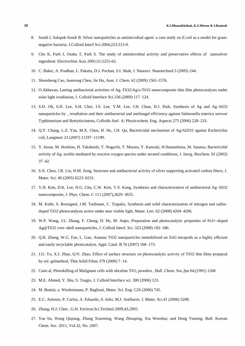
The absorption spectrum of Ag@TiO2 is shown in Figure 1. It has a broad band centered at 425nm. However it is
reported that the plasmon absorption band of the small Ag particles prepared using borohydride reduction is around 380
nm. The red shift in the Ag@TiO2 spectrum may be due to increase in the particle dimension and /or change in the
dielectric constant of the surrounding matrix upon encapsulation.[28].
XRD Analysis
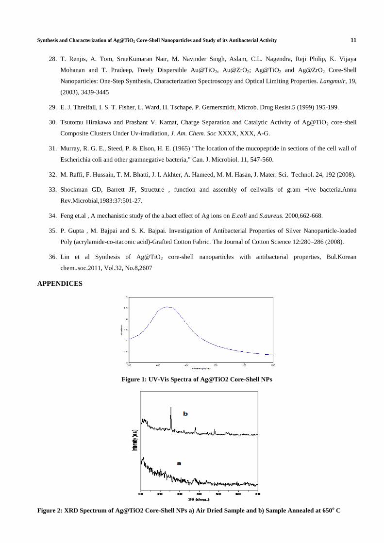
Standard X-ray powder diffraction was used to determine the identity, relative proportion and average grain size
of the core-shell nanoparticles. Figure (2a & 2b) shows the X-ray diffraction pattern of pre and post annealed Ag@TiO2
(at 650°C in air for 5hrs) respectively. The Figure 2(a) shows that the air dried Ag@TiO2 core-shell nanoparticle is fully
amorphous since there is no peak corresponding to TiO2 and Ag. The Figure 2(b) shows 4 characteristic peaks of
nanocrystalline pure Ag@TiO2 of monoclinic structure (JCPDS # 52-1202). The major diffraction intensity peaks at 2θ,
about 25.42, 32.182, 37.995, 47.839° were identified to originate from (220), (420) (112) and (040) planes of Ag@TiO2
respectively. The XRD patterns could be indexed to the C2/c (15) space group, end centered monoclinic structure with cell
parameters a = 16.77, b = 7.594, c = 5.044 and β = 102.01. Absence of peaks in Figure 2(a) and appearance of these peaks
in Figure 2(b) clearly establishes the sintering of both TiO2 particles to produce bulk dimension. Generally formation of
rutile phase is a common thing during calcination. The patters are very well comparable to those reported in literature [28].
FTIR Spectrum
The FTIR spectrum of Ag@ TiO2 is shown in Figure 3. The air dried sample of Ag@TiO2 shows no characteristic
peaks due to DMF. The defective OH vibrations below 3500cm-1
, OH stretch of water at 3362cm-1
, the OH2 bend at
1625.1cm-1
and 531 cm-1
for the Ti-O stretching, vibration are observed. In addition there are peaks at about 1114cm-1
and
1382cm-1
and these peaks are assigned to alkoxide C-O and CH2 bending vibrations respectively. Hence alcohol might be
adsorbed on the surface. The CH2 stretching vibrations which generally occur just below 3000cm-1
are not well resolved in
this figure. The FTIR spectrum is very well comparable to that reported in literature [28].
Thermo Gravimetric Analysis (TGA)
The thermo gravimetric analysis of nano Ag@TiO2 was carried out between 50 and 900°C at a heating rate of
10°C/min in nitrogen atmosphere. The traces of thermogram are illustrated in Figure 4. The mass loss on ignition was
related to the content of volatile components, especially physi or chemisorbed water. There is a weight loss between 50 and
125°C due to desorption of adsorbed water. There is a weight loss above 200°C. It involves condensation of defective–OH
groupings and oxidation of adsorbed organics on the surface. There is a weight loss above 550°C due to condensation of
8 K.I.Dhanalekshmi, K.S.Meena & I.Ramesh
difficultly condensable groupings and / or oxidation of organic residues. Thus the thermo gravimetric study of Ag@TiO2
core-shell nanoparticles shows a monotonous mass decrease with temperature.
Differential Scanning Colorimetry (DSC)
The DSC analysis of the air-dried Ag@TiO2 core-shell nanoparticles was carried out between 50 and 400°C at a
heating rate of 10°C/min in nitrogen atmosphere. The DSC trace of Ag@TiO2 is shown in Figure 5.There is an
endothermic peak between 50 and 250°C. This endothermic peak coincides with the second major weight loss in the TGA
traces shown in Figure 4. The energy changes in this process are also comparable with the percentage weight loss in TGA.
There is an exothermic peak between 300 and 400 °C. This may be due to the crystallization of Ag@TiO2 at this
temperature which is in accordance with XRD results shown in Figure 4b.This sharp peak at this higher temperature may
presumably involve some form of structural modifications within the Ag@TiO2 lattice.
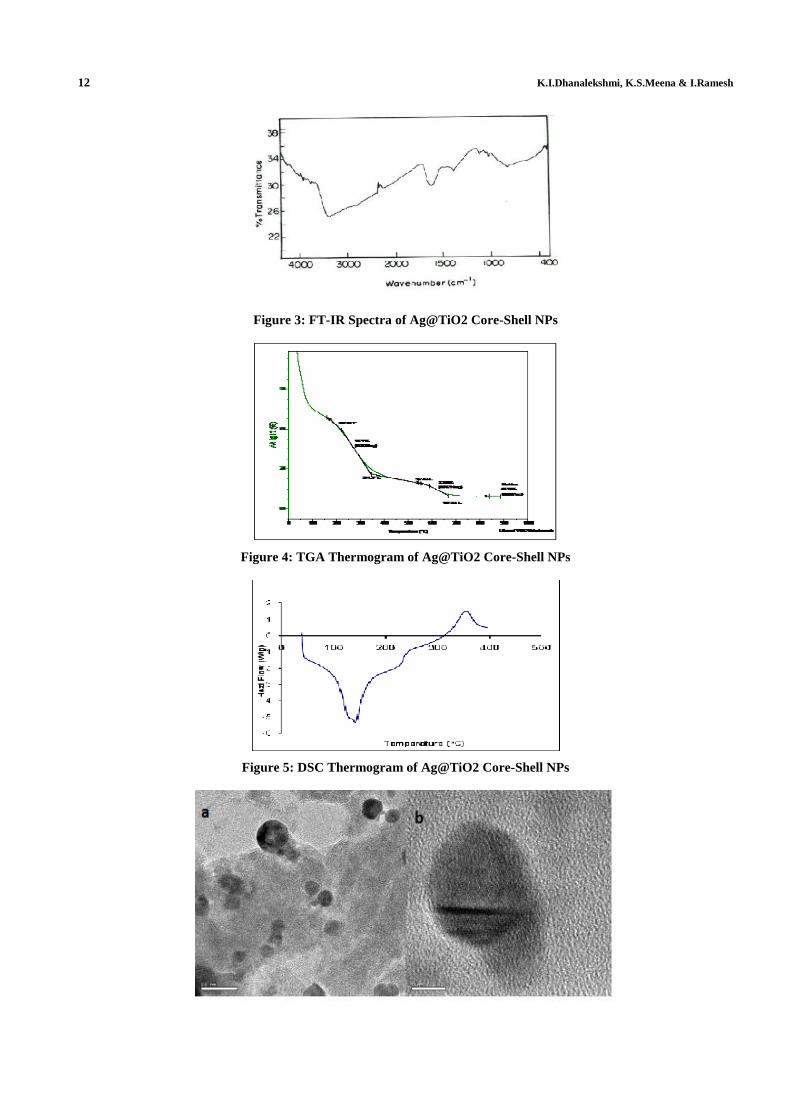
High Resolution Scanning Electron Microscopy (HR-TEM)
The HR-TEM images of Ag@TiO2 are shown in Figure 6(a-c).The images show a majority of dark images of Ag
core in the size regime. Figure 6(a). Illustrates the formation of nearly spherical particles of silver core with particle
diameter of 15nm to 20nm and a range of particle morphologies are seen. Although most of the particles seen in this image
are spherical or oval, faceted structures were also observed. All of them appear to be associated with TiO2 shell. The
boundary between core (Ag) and shell (TiO2) is very much distinct Figure 6 (b-c). HR-TEM image of single Ag@TiO2
particle is illustrated in Figure 6(b & c). This image illustrates that each particle has a thin capping of TiO2 shell of
thickness in the range 2 to3nm. Figure 6(a) illustrates aggregation of fine particles of Ag represented by dense regions and
associated TiO2.
Microscopy reveals no free particles of either silver or oxide. The presence of completely covering shells is
confirmed with reactivity studies as well. Capping of TiO2 shell on the Ag core was confirmed by checking the stability in
an acidic solution (HNO3). The Ag cluster, stabilized by citric acid, is readily dissolved in an acidic solution (pH=2).
Ag@TiO2 on the other hand is quite stable in HNO3 solution even when the TiO2 shell is thin. If the formation of TiO2
clusters in DMF was independent such that both clusters are formed separately or in the form of a TiO2/Ag sandwich
structure we would have observed dissolution of Ag clusters The stability test in acidic solution asserts the argument that
the TiO2 shell on the Ag core is uniform and provides the protection against acid induced corrosion [30].
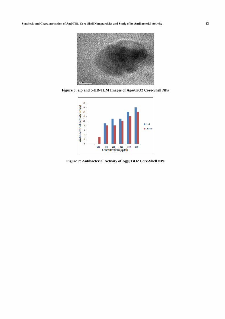
Antibacterial Activity of Ag@TiO2 Core-Shell Nanoparticles
The inhibition zone values were determined for Ag@TiO2 core-shell NPs were tested against two types of bacteria
E.coli and S.aureus results are shown in the Figure 7. The MIC observed in this study for Ag@TiO2 core-shell NPs are 100
µg/ml for S.aureus and 200 µg/ml for E.coli.
The obtained data demonstrates that all the investigated Ag@TiO2 core-shell NP had relatively high antibacterial
activity against Gram negative (E.coli) bacteria as compared to that of Gram positive (S.aureus) bacteria. The antibacterial
activity increases as the concentration of core-shell increases.
The inhibition zone of S.aureus is smaller than for E.coli. It has been known, that the structure of gram positive
and gram negative cell was has different compositions. The layer of peptidoglycan in gram positive bacteria is thicker than
for gram negative bacteria which consequently decrease the penetrations of NPs to the cell wall of gram positive bacteria.
Ag NPs shows three different way of mechanism of action.
Synthesis and Characterization of Ag@TiO2 Core-Shell Nanoparticles and Study of its Antibacterial Activity 9
Firstly, Ag NPs attach to the surface of the cell membrane and disturb its power functions. Such as permeability
and respiration [31]. It is reasonable to state that the binding of the particles to the bacteria depends on the interaction of
the surface area available.
Secondly Ag NPs are able to penetrate the bacteria and came further damage, possibly by interacting with sulfur
and phosphorus containing compounds such as DNA [32]. In addition , it is believed that Ag NPs after penetration into the
bacteria have inactivated their enzymes, generating hydrogen peroxide and causing bacterial cell death [32,33].
Third, Ag NPs release silver ions, which made an additional contribution to the bactericidal effect [34]. The Ag
NPs also contain micro molar concentration of Ag+ and they have shown that Ag
+ and Ag
0 both contribute to the
antibacterial activity. The mechanism of inhibition by silver ions on micro organism in partially known. It is believed that
DNA loses its replication ability and cellular proteins become inactivated on silver ion treatment [35]. Higher
concentrations of Ag+ ions have been shown to interact with cytoplasmic components and nucleic acids [34].
It could be concluded that Ag ions from Ag cores could be released through porous TiO2 shells. It may be
reasonable to presume that such core/shell structures will benefit preventing corrosion and prolonging the release time of
Ag ions and preserving the sustained antibacterial behavior [36].
Therefore such Ag@TiO2 core-shell nanoparticles could be useful and effective in bactericidal applications, and
would present a reasonable alternative for the development of new bactericides.
CONCLUSIONS
Ag@TiO2 core-shell nanoparticles were successfully prepared and their structure were analyzed by techniques
such as UV-vis spectroscopy, XRD, FTIR, TGA, DSC and HR-TEM. Positively charged silver reacted easily with gram
negative bacteria rather than gram positive bacteria could be selectively killed by the Ag@TiO2 core-shell NPs more
effectively. The core-shell NPs showed good antibacterial activities because TiO2 deposited on the surface of Ag NPs
prevent aggregation. Such core-shell NPs could have promising application as antibacterial materials for microbiocides and
water treatment.
REFERENCES
1. Hajipour et al, Antibacterial properties of nanoparticles.
2. Q.X.Li , H.A.Tang, Y.Z.Li, M.Wang, L.F.Wang, C.G.Xia, J.Inorg.Biochem.78 (2000) 167-174.
3. Farooqui, M.D.A, Chauhan, P.S., Krishanmoorthy, P and Shaik ,J .Extraction of Ag Nanoparticles from the leaf
extracts of Clerodendrum inerme, Digest Journal of nanomaterials and biostructures, 2010; 5:43-49.
4. V.K.Sharma, R.A.Yngard, Y.Lin, Silver nanoparticles: green synthesis and their antimicrobial activites, Advances
in colloid and Interface Science 145 (2009) 83-96.
5. Q.Li, S. Mahaendra, D.Y. Lyon, l. Brunet, M.V. Liga, D. Li, P.J.J. Alvarez, Antimicrobial nanomaterials for
water disinfection and microbial control: potential applications and implications, Water research 42 (2008) 4591-
4602.
6. Whitesides, G.M. (2005) Nanoscience, nanotechnology, and chemistry. Small 1,172-179.
7. K.Chaloupka , Y. Malam, A.M. Seifalian, Trends Biotechnol.28 (2010) 580.
10 K.I.Dhanalekshmi, K.S.Meena & I.Ramesh
8. Sondi I, Salopek-Sondi B. Silver nanoparticles as antimicrobial agent: a case study on E.coli as a model for gram-
negative bacteria. J.Colloid Interf Sci-2004;223:313-9.
9. Cho K, Park J, Osaka T, Park S. The study of antimicrobial activity and preservative effects of nanosilver
ingredient. Electrochim Acta 2005;51:5255-62.
10. C. Baker, A. Pradhan, L. Pakstis, D.J. Pochan, S.I. Shah, J. Nanosci. Nanotechnol.5 (2005) 244.
11. Shunsheng Cao, Juanrong Chen, Jie Hu, Aust. J. Chem. 62 (2009) 1561-1576.
12. O.Akhavan, Lasting antibacterial activities of Ag–TiO2/Ag/a-TiO2 nanocomposite thin film photocatalysts under
solar light irradiation, J. Colloid Interface Sci.336 (2009) 117–124.
13. S.D. Oh, S.H. Lee, S.H. Choi, I.S. Lee, Y.M. Lee, J.H. Chun, H.J. Park, Synthesis of Ag and Ag–SiO2
nanoparticles by _-irradiation and their antibacterial and antifungal efficiency against Salmonella enterica serovar
Typhimurium and Botrytiscinerea, Colloids Surf. A: Physicochem. Eng. Aspects 275 (2006) 228–233.
14. Q.Y. Chang, L.Z. Yan, M.X. Chen, H. He, J.H. Qu, Bactericidal mechanism of Ag/Al2O3 against Escherichia
coli, Langmuir 23 (2007) 11197–11199.
15. Y. Inoue, M. Hoshino, H. Takahashi, T. Noguchi, T. Murata, Y. Kanzaki, H.Hamashima, M. Sasatsu, Bactericidal
activity of Ag–zeolite mediated by reactive oxygen species under aerated conditions, J. Inorg. Biochem. 92 (2002)
37–42.
16. S.X. Chen, J.R. Liu, H.M. Zeng, Structure and antibacterial activity of silver supporting activated carbon fibers, J.
Mater. Sci. 40 (2005) 6223–6231.
17. Y.H. Kim, D.K. Lee, H.G. Cha, C.W. Kim, Y.S. Kang, Synthesis and characterization of antibacterial Ag–SiO2
nanocomposite, J. Phys. Chem. C 111 (2007),3629–3635.
18. M. Ksibi, S. Rossignol, J.M. Tatibouet, C. Trapalis, Synthesis and solid characterization of nitrogen and sulfur-
doped TiO2 photocatalysts active under near visible light, Mater. Lett. 62 (2008) 4204–4206.
19. W.P. Wang, J.L. Zhang, F. Cheng, D. He, M. Anpo, Preparation and photocatalytic properties of Fe3+-doped
Ag@TiO2 core–shell nanoparticles, J. Colloid Interf. Sci. 323 (2008) 182–186.
20. Q.H. Zhang, W.G. Fan, L. Gao, Anatase TiO2 nanoparticles immobilized on ZnO tetrapods as a highly efficient
and easily recyclable photocatalyst, Appl. Catal. B 76 (2007) 168–173.
21. J.G. Yu, X.J. Zhao, Q.N. Zhao, Effect of surface structure on photocatalytic activity of TiO2 thin films prepared
by sol–gelmethod, Thin Solid Films 379 (2000) 7–14.
22. Caiet.al, Photokilling of Malignant cells with ultrafine TiO2 powders , Bull .Chem. Soc,Jpn 64,(1991) 1268
23. M.E. Ahmed, Y. Shu, S. Tsugio, J. Colloid Interface sci. 300 (2006) 123.
24. M. Bonini, a. Wiedenmann, P. Baglioni, Mater. Sci. Eng. C26 (2006) 745.
25. E.C. Antonio, P. Carlos, A. Eduardo, S. Julio, M.J. Antibacte. J. Mater. Sci.41 (2006) 5208.
26. Zhang, H.J; Chen , G.H. Environ.Sci.Technol.2009,43,2905.
27. Yue lin, Wang Qiqiang, Zhang Xiaoming, Wang Zhouping, Xia Wenshui, and Dong Yuming. Bull. Korean
Chem. Soc. 2011, Vol.32, No. 2607.
Synthesis and Characterization of Ag@TiO2 Core-Shell Nanoparticles and Study of its Antibacterial Activity 11
28. T. Renjis, A. Tom, SreeKumaran Nair, M. Navinder Singh, Aslam, C.L. Nagendra, Reji Philip, K. Vijaya
Mohanan and T. Pradeep, Freely Dispersible Au@TiO2, Au@ZrO2; Ag@TiO2 and Ag@ZrO2 Core-Shell
Nanoparticles: One-Step Synthesis, Characterization Spectroscopy and Optical Limiting Properties. Langmuir, 19,
(2003), 3439-3445
29. E. J. Threlfall, I. S. T. Fisher, L. Ward, H. Tschape, P. Gernersmidt, Microb. Drug Resist.5 (1999) 195-199.
30. Tsutomu Hirakawa and Prashant V. Kamat, Charge Separation and Catalytic Activity of Ag@TiO2 core-shell
Composite Clusters Under Uv-irradiation, J. Am. Chem. Soc XXXX, XXX, A-G.
31. Murray, R. G. E., Steed, P. & Elson, H. E. (1965) "The location of the mucopeptide in sections of the cell wall of
Escherichia coli and other gramnegative bacteria," Can. J. Microbiol. 11, 547-560.
32. M. Raffi, F. Hussain, T. M. Bhatti, J. I. Akhter, A. Hameed, M. M. Hasan, J. Mater. Sci. Technol. 24, 192 (2008).
33. Shockman GD, Barrett JF, Structure , function and assembly of cellwalls of gram +ive bacteria.Annu
Rev.Microbial,1983:37:501-27.
34. Feng et.al , A mechanistic study of the a.bact effect of Ag ions on E.coli and S.aureus. 2000,662-668.
35. P. Gupta , M. Bajpai and S. K. Bajpai. Investigation of Antibacterial Properties of Silver Nanoparticle-loaded
Poly (acrylamide-co-itaconic acid)-Grafted Cotton Fabric. The Journal of Cotton Science 12:280–286 (2008).
36. Lin et al Synthesis of Ag@TiO2 core-shell nanoparticles with antibacterial properties, Bul.Korean
chem..soc.2011, Vol.32, No.8,2607
APPENDICES
Figure 1: UV-Vis Spectra of Ag@TiO2 Core-Shell NPs
Figure 2: XRD Spectrum of Ag@TiO2 Core-Shell NPs a) Air Dried Sample and b) Sample Annealed at 650o C
12 K.I.Dhanalekshmi, K.S.Meena & I.Ramesh
Figure 3: FT-IR Spectra of Ag@TiO2 Core-Shell NPs
Figure 4: TGA Thermogram of Ag@TiO2 Core-Shell NPs
Figure 5: DSC Thermogram of Ag@TiO2 Core-Shell NPs
Synthesis and Characterization of Ag@TiO2 Core-Shell Nanoparticles and Study of its Antibacterial Activity 13
Figure 6: a,b and c-HR-TEM Images of Ag@TiO2 Core-Shell NPs
Figure 7: Antibacterial Activity of Ag@TiO2 Core-Shell NPs