Large Deletion Mutations Involving the First Pyrimidine-Rich ...
Upload
independentCategory
view
0download
0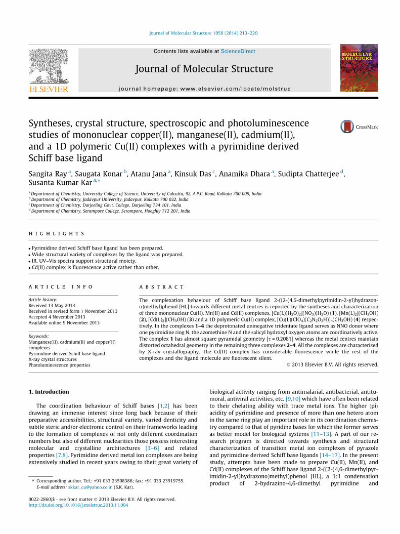
Journal of Molecular Structure 1058 (2014) 213–220
Contents lists available at ScienceDirect
Journal of Molecular Structure
journal homepage: www.elsevier .com/ locate /molst ruc
Syntheses, crystal structure, spectroscopic and photoluminescencestudies of mononuclear copper(II), manganese(II), cadmium(II),and a 1D polymeric Cu(II) complexes with a pyrimidine derivedSchiff base ligand
0022-2860/$ - see front matter � 2013 Elsevier B.V. All rights reserved.http://dx.doi.org/10.1016/j.molstruc.2013.11.004
⇑ Corresponding author. Tel.: +91 033 23508386; fax: +91 033 23519755.E-mail address: [email protected] (S.K. Kar).
Sangita Ray a, Saugata Konar b, Atanu Jana a, Kinsuk Das c, Anamika Dhara a, Sudipta Chatterjee d,Susanta Kumar Kar a,⇑a Department of Chemistry, University College of Science, University of Calcutta, 92, A.P.C. Road, Kolkata 700 009, Indiab Department of Chemistry, Jadavpur University, Jadavpur, Kolkata 700 032, Indiac Department of Chemistry, Darjeeling Govt. College, Darjeeling 734 101, Indiad Department of Chemistry, Serampore College, Serampore, Hooghly 712 201, India
h i g h l i g h t s
� Pyrimidine derived Schiff base ligand has been prepared.� Wide structural variety of complexes by the ligand was prepared.� IR, UV–Vis spectra support structural moiety.� Cd(II) complex is fluorescence active rather than other.
a r t i c l e i n f o
Article history:Received 13 May 2013Received in revised form 1 November 2013Accepted 4 November 2013Available online 9 November 2013
Keywords:Manganese(II), cadmium(II) and copper(II)complexesPyrimidine derived Schiff base ligandX-ray crystal structuresPhotoluminescence properties
a b s t r a c t
The complexation behaviour of Schiff base ligand 2-((2-(4,6-dimethylpyrimidin-2-yl)hydrazon-o)methyl)phenol [HL] towards different metal centres is reported by the syntheses and characterizationof three mononuclear Cu(II), Mn(II) and Cd(II) complexes, [Cu(L)(H2O)2](NO3)(H2O) (1), [Mn(L)2](CH3OH)(2), [Cd(L)2](CH3OH) (3) and a 1D polymeric Cu(II) complex, [Cu(L)(ClO4)(C2N2O2H)]n(CH3OH) (4) respec-tively. In the complexes 1–4 the deprotonated uninegative tridentate ligand serves as NNO donor whereone pyrimidine ring N, the azomethine N and the salicyl hydroxyl oxygen atoms are coordinatively active.The complex 1 has almost square pyramidal geometry [s = 0.2081] whereas the metal centres maintaindistorted octahedral geometry in the remaining three complexes 2–4. All the complexes are characterizedby X-ray crystallography. The Cd(II) complex has considerable fluorescence while the rest of thecomplexes and the ligand molecule are fluorescent silent.
� 2013 Elsevier B.V. All rights reserved.
1. Introduction
The coordination behaviour of Schiff bases [1,2] has beendrawing an immense interest since long back because of theirpreparative accessibilities, structural variety, varied denticity andsubtle steric and/or electronic control on their frameworks leadingto the formation of complexes of not only different coordinationnumbers but also of different nuclearities those possess interestingmolecular and crystalline architectures [3–6] and relatedproperties [7,8]. Pyrimidine derived metal ion complexes are beingextensively studied in recent years owing to their great variety of
biological activity ranging from antimalarial, antibacterial, antitu-moral, antiviral activities, etc. [9,10] which have often been relatedto their chelating ability with trace metal ions. The higher hpiiacidity of pyrimidine and presence of more than one hetero atomin the same ring play an important role in its coordination chemis-try compared to that of pyridine bases for which the former servesas better model for biological systems [11–13]. A part of our re-search program is directed towards synthesis and structuralcharacterization of transition metal ion complexes of pyrazoleand pyrimidine derived Schiff base ligands [14–17]. In the presentstudy, attempts have been made to prepare Cu(II), Mn(II), andCd(II) complexes of the Schiff base ligand 2-((2-(4,6-dimethylpyr-imidin-2-yl)hydrazono)methyl)phenol [HL], a 1:1 condensationproduct of 2-hydrazino-4,6-dimethyl pyrimidine and
Scheme 1. Schematic representation of ligand (HL) and their complexes.
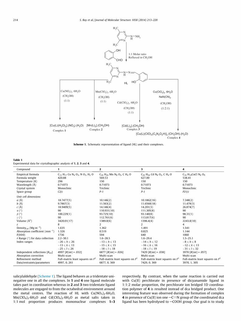
Table 1Experimental data for crystallographic analysis of 1, 2, 3 and 4.
Compound 1 2 3 4
Empirical formula C13 H17 Cu N4 O3, N O3, H2 O C26 H26 Mn N8 O2, C H4 O C26 H26 Cd N8 O2, C H4 O C16 H19CuCl N6 O9
Formula weight 420.88 569.53 627.00 538.41Temperature (K) 296 150 150 150Wavelength (Å) 0.71073 0.71073 0.71073 0.71073Crystal system Monoclinic Triclinic Triclinic MonoclinicSpace group C2/c P-1 P-1 P21/c
Unit cell dimensionsa (Å) 18.7477(5) 10.146(2) 10.1862(16) 7.348(2)b (Å) 9.7867(3) 11.563(2) 11.6560(18) 11.479(3)c (Å) 18.9409(5) 14.146(4) 14.201(3) 26.874(7)a (�) 90 110.931(10) 111.305(8) 90b (�) 100.229(1) 93.725(10) 93.140(8) 98.31(1)c (�) 90 112.761(6) 113.817(6) 90Volume (Å3) 3420.01(17) 1389.0(6) 1396.4(4) 2243.0(10)z 8 2 2 4Densitycal (Mg m�3) 1.635 1.362 1.491 1.541Absorption coefficient (mm�1) 1.326 0.519 0.825 1.144F(000) 1736 594 640 1060h Range (�) for data collection 2.2–30.1 1.6–28.5 1.6–29.4 1.5–25.1Index ranges �26 6 h 6 26 �13 6 h 6 13 �14 6 h 6 12 �8 6 h 6 8
�13 6 k 6 13 �15 6 k 6 15 �16 6 k 6 16 �12 6 k 6 13�25 6 l 6 26 �18 6 l 6 18 �18 6 l 6 19 �31 6 l 6 32
Independent reflections [Rint] 4997 [R(int) = .032] 6877 [R(int) = .026] 7429 [R(int) = .038] 3970 [R(int) = .057]Absorption correction Multi-scan Multi-scan Multi-scan Multi-scanRefinement method Full-matrix least squares on F2 Full-matrix least squares on F2 Full-matrix least squares on F2 Full-matrix least squares on F2
Data/restraints/parameters 4997, 0, 261 6877, 0, 369 7429, 0, 369 3970, 1, 305
214 S. Ray et al. / Journal of Molecular Structure 1058 (2014) 213–220
salicylaldehyde (Scheme 1). The ligand behaves as a tridentate uni-negative one in all the complexes. In 1 and 4 one ligand moleculetakes part in coordination whereas in 2 and 3 two tridentate ligandmolecules are engaged to from the octahedral environment aroundthe metal centres. The reaction of HL with Cu(NO3)2�6H2O,Mn(ClO4)2�6H2O and Cd(ClO4)2�6H2O as metal salts taken in1:1 mol proportion produces mononuclear complexes 1–3
respectively. By contrast, when the same reaction is carried outwith Cu(II) perchlorate in presence of dicyanamide ligand in1:1:2 molar proportion, the perchlorate ion bridged 1D coordina-tion polymer of 4 is resulted instead of dca bridged product. Oneinteresting feature was observed during the formation of complex4 in presence of Cu(II) ion one AC„N group of the coordinated dcaligand has been hydrolysed to ACOOH group. Our goal is to study

Table 2Selected bond distances (Å) and angles (�) in 1, 2, 3 and 4.
Selected bonds Value (Å) Selected angles (�)
Complex 1O1ACu1AO2 91.82(6)
Cu1AO1 1.9009(13) O1ACu1AO3 85.95(6)Cu1AO2 2.2662(15) O1ACu1AN1 91.74(5)Cu1AO3 2.0159(16) O1ACu1AN3 172.62(6)Cu1AN1 1.9500(13) O2ACu1AO3 93.81(6)Cu1AN3 2.0280(15) O2ACu1AN1 113.19(6)
O2ACu1AN3 90.93(6)O3ACu1AN1 152.97(6)O3ACu1AN3 100.70(6)N1ACu1AN3 80.88(6)Cu1AN1AN2 113.53(10)
Complex 2Mn1AO1 2.0897(15) O1AMn1AO2 103.38(6)Mn1AO2 2.0858(16) O1AMn1AN1 150.54(6)Mn1AN1 2.3029(17) O1AMn1AN4 80.66(5)Mn1AN4 2.2748(16) O1AMn1AN5 90.01(5)Mn1AN5 2.3376(17) O1AMn1AN8 96.97(5)Mn1AN8 2.2604(16) O2AMn1AN1 89.61(6)
O2AMn1AN4 100.10(5)O2AMn1AN5 150.72(6)O2AMn1AN8 81.50(5)N1AMn1AN4 70.99(6)N1AMn1AN5 91.12(6)N1AMn1AN8 111.20(6)N4AMn1AN5 107.79(6)N4AMn1AN8 177.38(5)N5AMn1AN8 70.99(6)O1ACd1AO2 106.26(10)
Complex 3Cd1AO1 2.218(3) O1ACd1AN1 146.34(10)Cd1AO2 2.222(2) O1ACd1AN4 79.01(9)Cd1AN1 2.385(3) O1ACd1AN5 88.02(10)Cd1AN4 2.356(3) Cd1AN8AN7 114.5(2)Cd1AN5 2.362(3) O1ACd1AN8 102.77(10)Cd1AN8 2.369(3) O2ACd1AN1 90.34(9)
O2ACd1AN4 98.77(9)O2ACd1AN5 147.05(10)O2ACd1AN8 78.30(9)N1ACd1AN4 69.48(11)N1ACd1AN5 93.58(10)N1ACd1AN8 109.27(11)N4ACd1AN5 113.24(11)N4ACd1AN8 176.88(10)N5ACd1AN8 69.52(11)
Complex 4Cu1AN1 1.933(5) O1ACu1AO4_a 96.21(18)Cu1AN4 2.044(5) O1ACu1AN1 90.24(19)Cu1AN5 1.939(6) O1ACu1AN4 170.61(19)Cu1AO1 1.919(4) O1ACu1AN5 86.9(2)Cu1AO4_a 2.615(5) O4_aACu1AN1 84.1(2)Cu1AO6 2.725(6) O4_aACu1AN4 86.44(19)
O4_aACu1AN5 90.7(2)O4_aACu1AO6 165.7(2)N1ACu1AN4 81.05(19)N1ACu1AN5 173.7(2)N4ACu1AN5 102.2(2)O6ACu1AN1 101.9(2)O6ACu1AN4 81.72(19)O6ACu1AN5 84.1(2)O1ACu1AO6 96.77(19)
Symmetry transformations used to generate equivalent atoms:For complex 4, a = �1 + x, y, z.
Fig. 1. Structural representation and atom numbering scheme of 1 (H-atoms areomitted for clarity).
S. Ray et al. / Journal of Molecular Structure 1058 (2014) 213–220 215
the coordination modes of the ligand towards different metal ionsand also to draw a comparative account of the special characterizesin the structures of the metal complexes. Two molecules of thesame ligand form the distorted octahedral geometry of MN4O2
(M = Mn and Cd) chromophore around the metal centres in 2 and3. The 4 is a 1D polymeric Cu(II) complex where the distorted octa-hedral geometry is maintained around each Cu(II) centre by one
ligand molecule, unhydrolysed cyanamide part of one dicyanamidemoiety and two l-bridged propagating perchlorate group. In thecomplex 1 the ligand utilizes one pyrimidine N, the azomethineN and the salicyl hydroxyl oxygen atoms along with two oxygenatoms of two coordinated water molecules to form square pyrami-dal geometry. We have also studied the photoluminescence prop-erties of the ligand HL and its metal complexes. Interestingly wehave observed that the ligand HL, complexes 1, 2 and 4 are fluores-cence silent where as complex 3 shows chelation enhancedfluorescence.
2. Experimental section
2.1. Materials and physical methods
All chemicals were of reagent grade, purchased from commer-cial sources and used without further purification.
Caution! Although we have not encountered any problem, itshould be kept in mind that perchlorate compounds of metal ionsare potentially explosive in the presence of organic ligands. Only asmall amount of the material should be prepared and it should behandled with care.
Elemental analyses (carbon, hydrogen, oxygen and nitrogen) ofthe ligand and the metal complexes were determined with aPerkin–Elmer CHN analyzer 2400 at the Indian Association forthe Cultivation of Science, Kolkata. The electronic spectra of thecomplexes in methanolic solution were recorded on a Hitachimodel U-3501 spectrophotometer. IR spectra (KBr pellet, 400–4000 cm�1) were recorded on a Perkin–Elmer model 883 infraredspectrophotometer. The fluorescence spectra were recorded on aPerkin Elmer LS 50B fluorimeter.
The ligand HL was synthesized using literature method [18].
2.2. Syntheses of complexes
2.2.1. Preparation of complex 1A methanolic solution (10 ml) of Cu(NO3)2�6H2O (1 mmol,
0.295 g) was added dropwise to a solution of HL (1 mmol,0.241 g) in the same solvent taken in 1:1 molar ratio with constantstirring. The solution turned yellow and stirring was continued forca. 2 h. It was left for slow evaporation at room temperature. After2 weeks X-ray quality crystals of 1 were formed and were collectedby usual technique. (Yield: 0.548 g, 65.5%). Anal. Calc. for C13H19
CuN5O7: C, 37.10; H, 4.55; N, 16.64; O, 26.61%. Found: C, 35.00;H, 3.44; N, 16.54; O, 23.23%. IR (KBr, cm�1): 1385 (m of free NO�3 ),1609 (s) (mc@Npym), 3346 (m of coordinated H2O). UV–Vis (MeOH):kmax/nm = 385, 605; e (DMF, M�1 cm�1): 8500.
2.2.2. Preparation of complexes 2 and 3Complexes 2 and 3 were prepared following the same synthetic
procedure as that for 1 using Mn(ClO4)2�6H2O and Cd(ClO4)2�6H2Oas metal salts respectively. The ligand and metal was in 1:2 molar

Fig. 2. Hydrogen bonding interactions in complex 1.
Table 3Details of hydrogen bond distances (Å) and angles (�) for 1, 2, 3 and 4.
DAH� � �A d(DAH) d(H� � �A) d(D� � �A) <(DHA)
Complex 1N2AH4� � �O7 0.8600 1.9300 2.771(2) 167.00O2AH14� � �O1 0.82(3) 2.05(3) 2.864(2) 173(3)O3AH16� � �O5 0.78(3) 2.07(3) 2.846(2) 175(2)O3AH17� � �O4 0.79(3) 2.02(3) 2.801(2) 171(3)O7AH18� � �O6 0.88(3) 2.06(3) 2.904(3) 162(3)O7AH19� � �O7 0.67(5) 2.22(4) 2.859(3) 159(6)
Complex 2N3AH3N� � �O3 0.86(2) 2.01(2) 2.863(3) 174(2)O3AH3O� � �O2 0.81(3) 1.96(3) 2.752(3) 166(3)N7AH7N� � �O1 0.88(2) 1.91(2) 2.794(2) 175.0(18)
Complex 3N6AH6� � �O1 0.8800 1.9400 2.775(9) 158.00N14AH14� � �O3 0.8800 1.9000 2.751(11) 162.00
Complex 4N2AH2N� � �O7 1.12(9) 2.07(9) 3.027(8) 142(6)O2AH2O� � �O1 1.05(6) 1.82(6) 2.875(7) 177(7)O8AH8O� � �O6 1.0500 1.9500 2.998(9) 176.00
Fig. 3. Structural representation and atom numbering scheme of 2 (H-atoms areomitted for clarity).
216 S. Ray et al. / Journal of Molecular Structure 1058 (2014) 213–220
ratio in each case to afford orange and yellow compoundsrespectively.
For 2: (Yield: 0.773 g, 73%). Anal. Calc. for C27H30MnN8O3: C,56.94; H, 5.31; N, 19.68; O, 8.43%. Found: C, 55.90; H, 4.56; N,18.41; O, 6.40%. IR (KBr, cm�1): 1606 (s) (mc@Npym). UV–Vis (MeOH):kmax/nm = 580; e (DMF, M�1 cm�1): 15,000.
For 3: (Yield: 0.602 g, 67%). Anal. Calc. for C27H30CdN8O3: C,51.72; H, 4.82; N, 17.87; O, 7.66%. Found: C, 51.49; H, 3.63; N,15.99; O, 6.66%. IR (KBr, cm�1): 1608 (s) (mc@Npym). UV–Vis (MeOH):kmax/nm = 360 and 470; e (DMF, M�1 cm�1): 89,000.
2.2.3. Preparation of complex 4To a methanolic solution (30 ml) of Cu(ClO4)2, 6H2O (2 mmol,
0.744 g), a solution of the Schiff base HL in the same solvent(2 mmol, 0.482 g) was slowly added, followed by a solution of so-dium dicyanamide (4 mmol, 0.356 g) in a minimum volume ofaqueous methanol with constant stirring. The stirring was contin-ued for another 2 h and filtered. The light yellow solution was keptin a refrigerator at 10 �C which after 10 days afforded green hexag-onal crystals suitable for X-ray diffraction. The crystals were iso-lated by filtration and made air dried. (Yield: 0.657 g, 61.5%).Anal. Calc. for C16H15ClN8O6Cu: C, 37.36; H, 2.94; N, 21.79; O,18.66%. Found: C, 35.31; H, 2.21; N, 21.51; O, 16.94%. IR (KBr,cm�1): 1100 (m of coordinated perchlorate), 1610 (s) (mc@Npym),UV–Vis (MeOH): kmax/nm = 386, 607; e (DMF, M�1 cm�1): 10,000.
3. X-ray crystallography
Selected crystal data for 1–4 are given in Table 1 and selectedmetrical parameters of these complexes are given in Table 2.Diffraction data were collected on a Bruker-APEX II SMART CCDdiffractometer at 296 K (for 1, 2, 3 and 4) using graphite-mono-chromated Mo Ka radiation (k = 0.71073 (Å). Cell parameter refine-ment and data reduction were carried out using Bruker SMART [19]and Bruker SAINT softwares for 1, 2, 3 and 4 [20]. The structureswere solved by direct and Fourier methods and refined by full-ma-trix least-squares based on F2 using the SHELXS-97 and SHELXL-97programs [21]. .An empirical absorption correction for 1, 2, 3 and 4were carried out using the SADABS [22]. One methanol moleculewas present as the solvent molecule which was removed usingSQUEEZE. Thus four hydrogen atoms, one carbon and one oxygenatom of methanol molecule were added to the chemical formulato adjust the density, molecular mass and F000 value in 3. A water
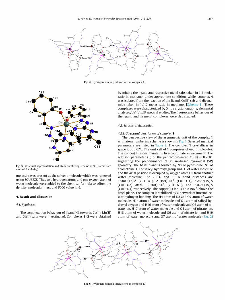
Fig. 5. Structural representation and atom numbering scheme of 3 (H-atoms areomitted for clarity).
Fig. 4. Hydrogen bonding interactions in complex 2.
S. Ray et al. / Journal of Molecular Structure 1058 (2014) 213–220 217
molecule was present as the solvent molecule which was removedusing SQUEEZE. Thus two hydrogen atoms and one oxygen atom ofwater molecule were added to the chemical formula to adjust thedensity, molecular mass and F000 value in 4.
4. Result and discussion
4.1. Syntheses
The complexation behaviour of ligand HL towards Cu(II), Mn(II)and Cd(II) salts were investigated. Complexes 1–3 were obtained
Fig. 6. Hydrogen bonding in
by mixing the ligand and respective metal salts taken in 1:1 molarratio in methanol under appropriate condition, while, complex 4was isolated from the reaction of the ligand, Cu(II) salt and dicyna-mide taken in 1:1:2 molar ratio in methanol [Scheme 1]. Thesecomplexes were characterized by X-ray crystallography, elementalanalyses, UV–Vis, IR spectral studies. The fluorescence behaviour ofthe ligand and its metal complexes were also studied.
4.2. Structural description
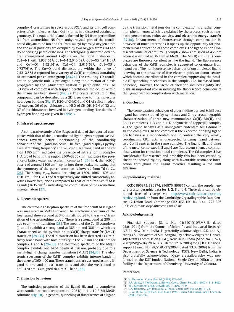
4.2.1. Structural description of complex 1The perspective view of the asymmetric unit of the complex 1
with atom numbering scheme is shown in Fig. 1. Selected metricalparameters are listed in Table 2. The complex 1 crystallizes inspace group C2/c. The unit cell of 1 comprises of eight molecules.The copper(II) atom maintains five-coordinate environment. TheAddison parameter (s) of the pentacoordinated Cu(II) is 0.2081suggesting the predominance of square-based pyramidal (SP)geometry. The basal plane is formed by N3 of pyrimidine, N1 ofazomethine, O1 of salicyl hydroxyl group and O3 of water moleculeand the axial position is occupied by oxygen atom O2 from anotherwater molecule. The CuAO and CuAN bond distances are1.9009(13) Å (Cu1AO1), 2.0159(16) Å (Cu1AO3), 2.2662(15) Å(Cu1AO2) axial, 1.9500(13) Å (Cu1AN1), and 2.0280(15) Å(Cu1AN3) respectively. The copper(II) ion is at 0.196 Å above thebasal plane. The complex is stabilized by a network of intermolec-ular hydrogen bonding. The H4 atom of N2 and O7 atom of watermolecule, H14 atom of water molecule and O1 atom of salicyl hy-droxyl oxygen and H16 atom of water molecule and O5 atom of ni-trate ion, H17 atom of water molecule and O4 atom of nitrate ion,H18 atom of water molecule and O6 atom of nitrate ion and H19atom of water molecule and O7 atom of water molecule (Fig. 2)
teractions in complex 3.

Fig. 7. Structural representation and atom numbering scheme of 4 (H-atoms are omitted for clarity).
Fig. 8. 3D view of complex 4 with trapped per chlorate molecules within the chains.
Fig. 9. Hydrogen bonding interactions in complex 4.
Fig. 10. Fluorescence emission property of free ligand HL and complexes 1, 2, 3 and4.
218 S. Ray et al. / Journal of Molecular Structure 1058 (2014) 213–220
are involved in the hydrogen bonding. The details of hydrogenbonding are given in Table 3.
4.2.2. Structural description of complexes 2 and 3The perspective view of the asymmetric units of the complexes
2 and 3 with the atom numbering schemes are shown in Figs. 3 and5 respectively. Selected metrical parameters are listed in Table 2.The complexes 2 and 3 crystallize in same space group P-1. Theunit cells of 2 and 3 comprise of two molecules. In all the threecomplexes, two tridentate ligand molecules are involved in
coordination. The central metal ion M [where, M = Mn(II) andCd(II)] in these complexes attains distorted octahedral geometrywith N4O2 chromophore. Here the equatorial positions are occu-pied by the salicyl hydroxyl oxygen atom O1, one pyridmidyl nitro-gen atom N1 from one ligand and another salicyl hydroxyl oxygenatom O2, pyridmidyl nitrogen atom N5 from another ligand whilethe axial positions are occupied by the azomethine nitrogen atomN4 from one ligand and N8 from another ligand. The averageMAN1(pym) distance falls in the range 2.3029(17)–2.385(3) Å,MAN5(pym) 2.3376(17)–2.362(3) Å, MAO1(alkoxy) 2.0897(15)–2.218(3) Å, MAO2(alkoxy) 2.0858(16)–2.222(2) Å, MAN4(azomethine) 2.2748(16)–2.356(16) Å, MAN8(azomethine) 2.2604(16)–2.369(3) Å. The average O1AMAO2 bond angle falls in therange 103.38(6)–106.26(10), O2AMAN1 89.61(6)–90.34(9),N1AMAN5 91.12(6)–93.58(10), N5AMAO1 88.02(10)–90.01(5),N8AMAN4 176.88(610)–177.38(5) respectively. The structures 2and 3 are stabilized by a network of intermolecular hydrogenbonding.(O3 of methanol molecule and H3N of N3, N7H of N7and O1 of salicyl hydroxyl oxygen, H30 of methanol moleculeand O2 of salicyl hydroxyl oxygen) (Fig. 4), O1 of salicyl hydroxyoxygen and H4A of water molecule) and O2 of salicyl hydroxyloxygen and H3N of N3, O1 of salicyl hydroxyl oxygen and H3O ofmethanol molecule, O3 of methanol molecule and H7N of N7(Fig. 6) are involved in hydrogen bonding. The details of H-bondingare presented in Table 3.
4.2.3. Structural description of complex 4The perspective view with atom numbering scheme is shown in
Fig. 7. Selected metrical parameters are given in Table 2. The
S. Ray et al. / Journal of Molecular Structure 1058 (2014) 213–220 219
complex 4 crystallizes in space group P21/c and its unit cell com-prises of six molecules. Each Cu(II) ion is in a distorted octahedralgeometry. The equatorial plane is formed by N4 from pyrimidine,N1 from azomethine, N5 from unhydrolysed part of the coordi-nated dicyanamide ion and O1 from salicyl hydroxyl oxygen atomand the axial positions are occupied by two oxygen atoms O4 andO5 of bridging perchlorate ions. The tetragonally distorted octahe-dral arrangement around Cu(II) gives the bond distances ofCu1AN1 with 1.937(5) Å, Cu1AN4 2.045(5) Å, Cu1AN5 1.941(6) Åand Cu1AO1 1.921(4) Å, Cu1AO4 2.615(5) Å, Cu1AO5_b2.727(6) Å. The CuAO bond distances are within the range of2.52–2.883 Å reported for a variety of Cu(II) complexes containingco-ordinated per chlorate group [23,24]. The resulting 1D coordi-nation polymeric unit is prolonged along the direction of b-axispropagated by the l-bidentate ligation of perchlorate ions. The3D view of complex 4 with trapped perchlorate molecules withinthe chains has been shown (Fig. 8). The crystal structure of thiscompound can be described as a 2D layer due to intermolecularhydrogen bonding (Fig. 9). H2O of CH3OH and O1 of salicyl hydro-xyl oxygen, O6 of per chlorate and H8O of CH3OH, H2N of N2 andO7 of perchlorate are involved in hydrogen bonding. The details ofhydrogen bonding are given in Table 3.
5. Infrared spectroscopy
A comparative study of the IR spectral data of the reported com-plexes with that of the uncoordinated ligand gives supportive evi-dences towards better understanding of the coordinatingbehaviour of the ligand molecule. The free ligand displays pyridylC@N stretching frequency at 1528 cm�1. A strong band in the re-gion 1385 cm�1 indicates the presence of nitrate ion in complex1. A broad band in the region 3500–3200 cm�1 indicates the pres-ence of lattice water molecules in complex 1 [25]. In 4, the mðClO�4 Þobserved around 1100 cm�1 splits into three peaks, indicating thatthe symmetry of the per chlorate ion is lowered from Td to C2V
[26]. The strong mC@N bands occurring at 1609, 1606, 1608 and1610 cm�1 for 1, 2, 3 and 4 respectively are shifted considerably to-wards lower frequencies compared to that of the free Schiff baseligands (1635 cm�1), indicating the coordination of the azomethinenitrogen atom [27].
6. Electronic spectra
The electronic absorption spectrum of the free Schiff base ligandwas measured in MeOH solvent. The electronic spectrum of thefree ligand shows a band at 345 nm attributed to the n ? p� tran-sition of the azomethine group. There is a strong band at 280 nmdue to a p ? p� transition [28]. The spectra of the Cu(II) complexes(1 and 4) exhibit a strong band at 385 nm and 386 nm which arecharacterized as the pyrimidine to Cu(II) charge transfer (LMCT)transition [29–33]. The d–d transition has been detected as a rela-tively broad band with low intensity in the 605 nm and 607 nm forcomplex 1 and 4 [29–33]. The electronic spectrum of the Mn(II)complex exhibits one band nearly at 580 nm, probably due to ametal–ligand charge transfer transition (MLCT) [34,35]. The elec-tronic spectrum of the Cd(II) complex exhibits intense bands inthe range of 360–400 nm. These transitions are assigned as intra li-gand p ? p� and n ? p� transitions and also the weak band at450–470 nm is assigned to a MLCT band [36].
7. Emission behaviour
The emission properties of the ligand HL and its complexeswere studied at room temperature (298 K) in 1 � 10�5(M) MeOHsolutions (Fig. 10). In general, quenching of fluorescence of a ligand
by the transition metal ions during complexation is a rather com-mon phenomenon which is explained by the process, such as mag-netic perturbation, redox activity, and electronic energy transfer[37,38]. Enhancement of fluorescence through complexation is,however, of much interest as it opens up the opportunity for pho-tochemical application of these complexes. The ligand is non fluo-rescent while its cadmium(II) complex shows emission at 455 nmwhen it is excited at 360 nm in MeOH. The Mn(II) and Cu(II) com-plexes are fluorescence silent as like the ligand. The fluorescencebehaviour of the Cd(II) complex is suggested to originate fromligand part. The nonfluorescence behaviour of uncomplexed ligandis owing to the presence of free electron pairs on donor centreswhich become coordinated in the complex suppressing the possi-ble ET quenching mechanism in the complex (i.e. increased lumi-nescence) However, the factor of chelation induced rigidity alsoplays an important role in inducing the fluorescence behaviour ofthe ligand part on complexation with metal ion.
8. Conclusion
The complexation behaviour of a pyrimidine derived Schiff baseligand has been studied by syntheses and X-ray crystallographiccharacterization of three new mononuclear Cu(II), Mn(II), andCd(II) complexes 1–3 and a 1-D polymeric of copper(II) complex4. The ligand behaves as a uninegative tridentate NNO donor inall the complexes. In the complex 4 the expected bridging liganddca behaves as a monodentate one. In contrast, the very weaklycoordinating ClO�4 acts as unexpected bridging ligand betweentwo Cu(II) centres in the same complex. The ligand HL and threeof the metal complexes 1, 2 and 4 are fluorescent silent, a commonobservation for transition metal complexes. The Cd complex 3 hasconsiderable fluorescence and probably this has been caused bychelation induced rigidity along with favourable resonance inter-action throughout the ligand moieties resulting a red shiftemission.
Supplementary material
CCDC 896873, 896874, 896876, 896877 contain the supplemen-tary crystallographic data for 1, 2, 3 and 4. These data can be ob-tained free of charge via http://www.ccdc.cam.ac.uk/conts/retrieving.html, or from the Cambridge Crystallographic Data Cen-tre, 12 Union Road, Cambridge CB2 1EZ, UK; fax: +44 1223 336033; or e-mail: [email protected].
Acknowledgements
Financial support [Sanc. No. 01(2401)/10/EMR-II, dated05.01.2011] from the Council of Scientific and Industrial Research(CSIR), New Delhi, India, is gratefully acknowledged. S.K. and A.J.thank CSIR for award of SRF. Sangita Ray acknowledges the Univer-sity Grants Commission (UGC), New Delhi, India [Sanc. No. F. 5-1/2007(BSR)/5-16/ 2007(BSR), dated 12.02.2006] for a J.R.F. Financialsupport [Sanc. No. SR/S1/IC-27/2008, dated 13.03.2009] from theDepartment of Science & Technology (DST), New Delhi, India, isalso gratefully acknowledged. X-ray crystallography was per-formed at the DST funded National Single Crystal Diffractometerfacility at the Department of Chemistry, University of Calcutta.
References
[1] V. Alexander, Chem. Rev. 95 (1995) 273–345.[2] P.A. Vigato, S. Tamburini, L. Bertolo, Coord. Chem. Rev. 251 (2007) 1311–1492.[3] M.J. Zaworotko, Cryst. Growth Des. 7 (2007) 4–9.[4] G.A. Hembury, V.V. Borovkov, Y. Inoue, Chem. Rev. 108 (2008) 1–73.[5] Q. Ye, D.-W. Fu, H. Tian, R.-G. Xiong, P.W.H. Chen, S.D. Huang, Inorg. Chem. 47
(2008) 772–774.

220 S. Ray et al. / Journal of Molecular Structure 1058 (2014) 213–220
[6] X.-H. Chen, Q.-J. Wu, Z.-Y. Liang, C.-R. Zhan, J.-B. Liu, Acta Crystallogr. Sect. C 65(2009) m190–m194.
[7] V. Balzani, A. Credi, M. Venturi, Molecular Devices and Machines, vol. 4, Wiley-VCH, Weinheim, 2003.
[8] J.S. Miller, in: M. Drillon (Ed.), Magnetism: Molecules to Materials V, vol. 5,Wiley-VCH, Weinheim, 2005, pp. 283–346.
[9] J.S. Casas, E.E. Castellans, M.D. Louce, J. Ellena, A. Sanchez, J. Sordo, C. Taboada,J. Inorg. Biochem. 11 (2006) 1858–1860.
[10] U. Koch, B. Attenni, S. Malancona, S. Colarusso, I. Conte, M.D. Filippo, S. Harper,B. Pacini, C. Giomini, S. Thomas, I. Incitti, L. Tomei, R.D. Francesco, S. Altamura,V.G. Matassa, F. Narjes, J. Med. Chem. 49 (2006) 1693–1705.
[11] F. Zamora, M. Kunsman, M. Sabat, B. Lippart, Inorg. Chem. 36 (1997) 1583–1587.
[12] F. Jolibois, J. Cadet, A. Grand, R. Subra, N. Raga, V. Barone, J. Am. Chem. Soc. 120(1998) 1864–1871.
[13] A.R. Katritzky, C.W. Pees, A.J. Boulton, C. Mckillop, J. Heterocycl. Chem. 3(1984) 57–68.
[14] S. Roy, T.N. Mandal, A.K. Barik, S. Gupta, R.J. Butcher, M.S. El Fallah, J. Tercero,S.K. Kar, Polyhedron 27 (2008) 105–112.
[15] A. Hazra, A.K. Barik, S. Pal, S. Gupta, S. Roy, R.J. Butcher, S.M. Peng, G.H. Lee, S.K.Kar, Polyhedron 26 (2007) 773–781.
[16] T.N. Mandal, S. Roy, A.K. Barik, S. Gupta, R.J. Butcher, S.K. Kar, Polyhedron 27(2008) 3267–3274.
[17] S. Roy, T.N. Mandal, A.K. Barik, S. Gupta, M.S. El Fallah, J. Tercero, R.J. Butcher,S.K. Kar, J. Chem. Soc., Dalton Trans. 1 (2009) 8215–8226.
[18] S. Roy, T.N. Mandal, A.K. Barik, S. Gupta, R.J. Butcher, M. Nethaji, S.K. Kar,Polyhedron 27 (2008) 593–601.
[19] Bruker, SMART v5.631, Bruker AXS Inc., Madison, WI, USA, 2001.[20] CRYSALIS, Oxford Diffraction Ltd., Abingdon, UK, 2006.
[21] G.M. Sheldrick, SHELXS-97 and SHELXL-97, University of Göttingen, Göttingen,Germany, 1997.
[22] SAINT, Version 6.02, SADABS, Version 2.03, Bruker AXS Inc., Madison, WI,2002.
[23] T.G.M. Fawcett, S.M. Rudich, B.H. Toby, R.A. Lanlancette, J.A. Potenza, H.J.Schugar, Inorg. Chem. 19 (1980) 940–945.
[24] P.A. Tasker, L.J. Sklar, J. Cryst. Mol. Struct. 5 (1975) 329–344.[25] P.A. Kumar, Indian J. Chem. A 37 (1998) 460–465.[26] K. Nakamoto (Ed.), Infrared and Raman spectra of Inorganic and Coordination
Compounds, fourth ed., Wiley, New York, 1986. 147–150, 227–233, 251–253.[27] L. Xue, Q. Liu, H. Jiang, Org. Lett. 11 (2009) 3454–3457.[28] V.L. Siji, M.R. Sudarsanakumar, S. Suma, Polyhedron 29 (2010) 2035–2040.[29] D. Volkmer, B. Hommerich, K. Griesar, W. Haase, B. Krebs, Inorg. Chem. 35
(1996) 3792–3803.[30] M. Ciampolini, Inorg. Chem. 5 (1966) 35–40.[31] L. Sacconi, M. Ciampolini, G.P. Speroni, J. Am. Chem. Soc. 87 (1965) 3102–3106.[32] M. Ciampolini, N. Nardi, Inorg. Chem. 5 (1966) 41–44.[33] E.V.R. Akimova, A.Y. Nazarenko, L. Chen, P.W. Krieger, A.M. Herrera, V.V.
Tarasov, P.D. Robinson, Inorg. Chim. Acta 324 (2001) 1–15.[34] S. Belaid, A. Landreau, S. Djebbar, O.B. Baitich, G. Bouet, J.P. Bouchara, J. Inorg.
Biochem. 102 (2008) 63–69.[35] S. Biswas, K. Mitra, C.H. Schwalbe, C.R. Lucas, S.K. Chattopadhyay, B. Adhikary,
Inorg. Chim. Acta 358 (2005) 2473–2481.[36] M.K. Paria, J. Dinda, T.H. Lu, A.R. Paital, C. Sinha, Polyhedron 26 (2007) 4131–
4140.[37] N. Chattopadhyay, A. Mallick, S. Sengupta, J. Photochem. Photobiol., A 177
(2005) 55–60.[38] P. Purkayastha, G.K. Patra, D. Datta, N. Chattopadhyay, Indian J. Chem. A 39
(2000) 375–381.
Copyright © 2022 FDOKUMEN