Can you tickle yourself if you swap bodies with someone else?
Upload
independentCategory
view
3download
0
SWAP-70 regulates mast cell FceRI-mediated signalingand anaphylaxis
Raja R. Sivalenka1, Manoj Sinha1 and Rolf Jessberger1,2
1 Department of Gene and Cell Medicine, Mount Sinai School of Medicine, New York,USA
2 Institute of Physiological Chemistry, Dresden University of Technology, Dresden,Germany
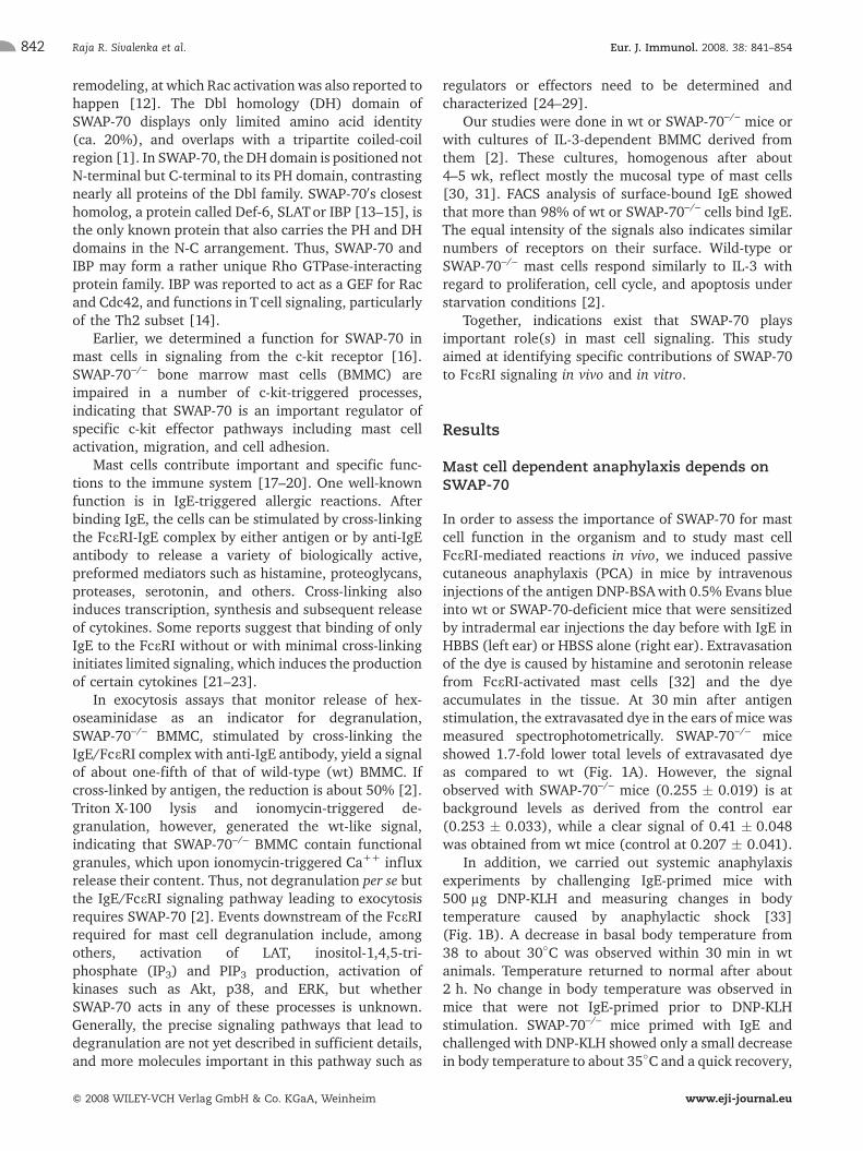
Mast cells, perhaps best known by their ability to trigger allergic reactions afterstimulation through the FceRI, express the unusual phosphatidylinositol 3-kinase(PI3K)-dependent, Rac-binding protein SWAP-70. Here, we show that the IgE-mediatedpassive cutaneous and the systemic anaphylactic responses are strongly reduced inSWAP-70–/– mice. Cultured SWAP-70–/– immature bone marrowmast cells (BMMC) arealso impaired in FceRI-mediated degranulation, which can be restored by expression ofexogenous wild-type SWAP-70, but less so if a phosphatidylinositol trisphosphate (PIP3)binding mutant is expressed. SWAP-70 itself supports inositol-3-phosphate and PIP3production, the latter indicating a potential feedback from SWAP-70 towards PI3K.FceRI-stimulated transcription and release of cytokines is controlled by SWAP-70. KeyFceRI signal transduction events like activation of LAT by phosphorylation, activation ofAkt/PKB and of p38 MAP kinase are reduced in SWAP-70–/– BMMC, but ERK is stronglyhyperactivated. Some requirements for SWAP-70 were apparent only under limited-strength signaling conditions. We suggest that SWAP-70 defines a new element ofefficient mast cell activation upon FceRI signaling, important for the control of mastcell-dependent anaphylaxis.
Supporting Information for this article is available athttp://www.wiley-vch.de/contents/jc_2040/2008/37597_s.pdf
Introduction
Murine SWAP-70, originally isolated from activated,mature B lymphocytes [1], is strongly expressed in mastcells [2]. Molecularly, SWAP-70 loosely resemblesproteins of the Dbl-family of guanine nucleotideexchange factors (GEF) for small Rho GTPases [3, 4].
SWAP-70 interacts specifically with the Rho GTPase Rac,which is a central molecular switch in a number of signaltransduction pathways including FceRI and c-kitsignaling [5, 6], and supports accumulation of itsactivated form, to which it preferentially binds [7]. Inmast cells, SWAP-70 is found in the cytoplasm and at thecytoplasmic membrane, but unlike in activated B cells isnot observed in the nucleus [2, 8, 9]. A pleckstrinhomology (PH) domain [10] exists in the center regionof SWAP-70, binds phosphatidylinositol trisphosphate(PIP3), the second messenger product generated byphosphatidylinositol 3-kinase (PI3K), and mediatesmembrane localization of the protein. As shown witha mutant (R230C) impaired in PIP3 binding, PIP3binding is necessary for SWAP-70 to localize tomembrane F-actin structures called membrane ruffles[7, 11], which represent sites of membrane F-actin
Correspondence: Rolf Jessberger, Institute of PhysiologicalChemistry, Dresden University of Technology, Fiedlerstr. 42,D-01307 Dresden, GermanyFax: +49-351-4586305e-mail: [email protected]
Received 26/6/07Revised 7/11/07
Accepted 12/12/07
[DOI 10.1002/eji.200737597]
Key words:Anaphylaxis
� Degranulation� FceRI � Mast cells
� SWAP-70
Abbreviations: BMMC: bone marrow mast cell � DH: Dblhomology � GEF: guanine nucleotide exchange factors �IP3: inositol-1,4,5-triphosphate � PCA: passive cutaneousanaphylaxis � PH: pleckstrin homology � PIP3: phosphatidyl-inositol trisphosphate
Eur. J. Immunol. 2008. 38: 841–854 Molecular immunology 841
f 2008 WILEY-VCH Verlag GmbH & Co. KGaA, Weinheim www.eji-journal.eu
remodeling, at which Rac activationwas also reported tohappen [12]. The Dbl homology (DH) domain ofSWAP-70 displays only limited amino acid identity(ca. 20%), and overlaps with a tripartite coiled-coilregion [1]. In SWAP-70, the DH domain is positioned notN-terminal but C-terminal to its PH domain, contrastingnearly all proteins of the Dbl family. SWAP-700s closesthomolog, a protein called Def-6, SLATor IBP [13–15], isthe only known protein that also carries the PH and DHdomains in the N-C arrangement. Thus, SWAP-70 andIBP may form a rather unique Rho GTPase-interactingprotein family. IBP was reported to act as a GEF for Racand Cdc42, and functions in Tcell signaling, particularlyof the Th2 subset [14].
Earlier, we determined a function for SWAP-70 inmast cells in signaling from the c-kit receptor [16].SWAP-70–/– bone marrow mast cells (BMMC) areimpaired in a number of c-kit-triggered processes,indicating that SWAP-70 is an important regulator ofspecific c-kit effector pathways including mast cellactivation, migration, and cell adhesion.
Mast cells contribute important and specific func-tions to the immune system [17–20]. One well-knownfunction is in IgE-triggered allergic reactions. Afterbinding IgE, the cells can be stimulated by cross-linkingthe FceRI-IgE complex by either antigen or by anti-IgEantibody to release a variety of biologically active,preformed mediators such as histamine, proteoglycans,proteases, serotonin, and others. Cross-linking alsoinduces transcription, synthesis and subsequent releaseof cytokines. Some reports suggest that binding of onlyIgE to the FceRI without or with minimal cross-linkinginitiates limited signaling, which induces the productionof certain cytokines [21–23].
In exocytosis assays that monitor release of hex-oseaminidase as an indicator for degranulation,SWAP-70–/– BMMC, stimulated by cross-linking theIgE/FceRI complex with anti-IgE antibody, yield a signalof about one-fifth of that of wild-type (wt) BMMC. Ifcross-linked by antigen, the reduction is about 50% [2].Triton X-100 lysis and ionomycin-triggered de-granulation, however, generated the wt-like signal,indicating that SWAP-70–/– BMMC contain functionalgranules, which upon ionomycin-triggered Ca++ influxrelease their content. Thus, not degranulation per se butthe IgE/FceRI signaling pathway leading to exocytosisrequires SWAP-70 [2]. Events downstream of the FceRIrequired for mast cell degranulation include, amongothers, activation of LAT, inositol-1,4,5-tri-phosphate (IP3) and PIP3 production, activation ofkinases such as Akt, p38, and ERK, but whetherSWAP-70 acts in any of these processes is unknown.Generally, the precise signaling pathways that lead todegranulation are not yet described in sufficient details,and more molecules important in this pathway such as
regulators or effectors need to be determined andcharacterized [24–29].
Our studies were done in wt or SWAP-70–/– mice orwith cultures of IL-3-dependent BMMC derived fromthem [2]. These cultures, homogenous after about4–5 wk, reflect mostly the mucosal type of mast cells[30, 31]. FACS analysis of surface-bound IgE showedthat more than 98% of wt or SWAP-70–/– cells bind IgE.The equal intensity of the signals also indicates similarnumbers of receptors on their surface. Wild-type orSWAP-70–/– mast cells respond similarly to IL-3 withregard to proliferation, cell cycle, and apoptosis understarvation conditions [2].
Together, indications exist that SWAP-70 playsimportant role(s) in mast cell signaling. This studyaimed at identifying specific contributions of SWAP-70to FceRI signaling in vivo and in vitro.
Results
Mast cell dependent anaphylaxis depends onSWAP-70
In order to assess the importance of SWAP-70 for mastcell function in the organism and to study mast cellFceRI-mediated reactions in vivo, we induced passivecutaneous anaphylaxis (PCA) in mice by intravenousinjections of the antigen DNP-BSAwith 0.5% Evans blueinto wt or SWAP-70-deficient mice that were sensitizedby intradermal ear injections the day before with IgE inHBBS (left ear) or HBSS alone (right ear). Extravasationof the dye is caused by histamine and serotonin releasefrom FceRI-activated mast cells [32] and the dyeaccumulates in the tissue. At 30 min after antigenstimulation, the extravasated dye in the ears of mice wasmeasured spectrophotometrically. SWAP-70–/– miceshowed 1.7-fold lower total levels of extravasated dyeas compared to wt (Fig. 1A). However, the signalobserved with SWAP-70–/– mice (0.255 � 0.019) is atbackground levels as derived from the control ear(0.253 � 0.033), while a clear signal of 0.41 � 0.048was obtained from wt mice (control at 0.207 � 0.041).
In addition, we carried out systemic anaphylaxisexperiments by challenging IgE-primed mice with500 lg DNP-KLH and measuring changes in bodytemperature caused by anaphylactic shock [33](Fig. 1B). A decrease in basal body temperature from38 to about 30�C was observed within 30 min in wtanimals. Temperature returned to normal after about2 h. No change in body temperature was observed inmice that were not IgE-primed prior to DNP-KLHstimulation. SWAP-70–/– mice primed with IgE andchallenged with DNP-KLH showed only a small decreasein body temperature to about 35�C and a quick recovery,
Raja R. Sivalenka et al. Eur. J. Immunol. 2008. 38: 841–854842
f 2008 WILEY-VCH Verlag GmbH & Co. KGaA, Weinheim www.eji-journal.eu

indicating a strong reduction in the systemic anaphy-lactic response induced by FceRI.
Since mature peritoneal mast cells do not expressSWAP-70 [2], we asked which types of mature tissuemast cells express SWAP-70. Analyses of fixed, paraffin-embedded sections from back skin and sections fromfrozen stomach mucosa by immunohistochemistry,using FITC-conjugated avidin to stain mast cells [34]and polyclonal rabbit anti-SWAP-70 followed by sec-ondary anti-rabbit-Cy3 antibody, showed that mast cellsfrom back skin as well as mucosal mast cells stronglyexpress SWAP-70 (not shown). Back skin sections fromSWAP-70–/– mice showed fewer numbers of mast cells,in agreement with an earlier report on twofold reducedskin mast cells in these mice [2], while mucosal stomachmast cell numbers are unaltered compared to wt.Gastrointestinal tract mast cells were stained withtoluidine blue and no difference in numbers betweenwt and SWAP-70–/– mice were found.
SWAP-70 reconstitution restores thedegranulation efficiency of SWAP-70–/– BMMC
Earlier, we reported that SWAP-70–/– BMMC aredefective in vitro in FceRI-mediated degranulation ofpreformed mediators such as hexoseaminidase [2]. Toconfirm a direct role of SWAP-70 and to get insights intothe molecular mechanisms of its function, we trans-fected mutant BMMC with pEGFP constructs totransiently express SWAP-70, or with the site-specificmutant R230C which is reduced in PIP3 binding [7], orwith vector alone for control. Expression of SWAP-70 in
transfected SWAP-70–/– and control wt BMMC wasassessed by performing Western blot analysis (Fig. 2A).The transfected cells were starved for 2 h, primed withanti-DNP-KLH IgE for 1 h, followed by cross-linkingusing 100 ng of the antigen DNP-KLH. The results are asshown in Fig. 2B as the average of three experiments.Compared to wt cells, SWAP-70–/– BMMC show *1.4-fold reduced degranulation after cross-linking withantigen, similar to the twofold reduction reported earlier[2]. Infection of wt or SWAP-70–/– cells with controlvector had only marginal effects. Wild-type BMMC thatwere transfected with SWAP-70 showed an increase ofabout 1.8-fold in degranulation upon stimulation withantigen. If background levels are deducted, thestimulation is 2.5-fold. Reconstitution of SWAP-70–/–
BMMC with SWAP-70 restored degranulation to levelssimilar to that seen in non-transfected wt BMMC.Expression of the SWAP-70 R230C mutant, which iscompromised in PIP3 binding [35], in wt or SWAP-70–/–
cells stimulated degranulation to a lesser degree thanexpression of the unaltered SWAP-70. The failure ofR230C to efficiently restore degranulation despite itsquite strong expression indicates a requirement for PIP3binding of SWAP-70 in signaling to degranulation.
SWAP-70 controls cytokine gene expression
Activation of mast cells induces synthesis and secretionof cytokines, rendering mast cells important contribu-tors to the cytokine repertoire of an organism. Earlier,we obtained indications for reduced secretion of IL-6 orTNF-a by FceRI-stimulated SWAP-70–/– BMMC [2]. To
Figure 1. Quantification of IgE-induced passive cutaneous and active systemic anaphylaxis. SWAP-70+/+ or SWAP-70–/– mice wereprimed intradermally in their left earswith 20 lg IgE (anti-DNP antibody clone SPE-7) and injectedwith HBSS in their right ears forinternal controls, followed after 24 h by intravenous injections of 100 lg antigen DNP-BSA together with 0.5% Evans blue in HBSSto induce PCA reactions. Control mice received HBSS with Evans blue alone without DNP-BSA. After 30 min, extracted dye fromminced ear tissues was measured spectrophotometrically at OD 610 nm and normalized against the appropriate controls. Eachgroup under study comprised five mice. The average difference in OD of sensitized left ears versus the HBBS-injected right earcontrols was 0.41 � 0.048 versus 0.207 � 0.041 for SWAP-70+/+ and 0.255 � 0.019 versus 0.253 � 0.033 for SWAP-70–/– mice (A).SWAP-70+/+ or SWAP-70–/– mice priorly sensitized intravenously or not with 50 lg IgE were challenged 48 h later with 500 lg DNP-KLH. Changes in basal body temperatureweremonitored at 20-min intervals for up to 2.5 h. Tenmicewere analyzed in each group.Values are means � SEM (B).
Eur. J. Immunol. 2008. 38: 841–854 Molecular immunology 843
f 2008 WILEY-VCH Verlag GmbH & Co. KGaA, Weinheim www.eji-journal.eu

assay not only for degranulation of preformedmediatorsas above, but also for induction of new synthesis ofmediators, we asked whether SWAP-70 is involved inregulating transcription of cytokine genes. Using RT-PCR with RNA prepared at various time points aftereither cross-linking of the IgE-loaded FceRI or after IgEloading only without cross-linking, we show here that
transcription of several interleukins is altered inSWAP-70–/– BMMC (Fig. 3A, B). Upon cross-linking,induction of transcription of IL-1, IL-2 or IL-10 largelyfails in SWAP-70–/– cells, and transcription of IL-6 andTNF-a is mildly reduced if compared to wt cells. IL-5transcription is up-regulated as in wt cells, IL-4expression is mildly increased, and IL-3 transcriptionmay be slightly reduced. Transcription of b-micro-globulin is unchanged as compared to the HPRTcontrol.
To test whether additional requirements forSWAP-70 would become apparent under limitingsignaling conditions, i.e. by sensitization with IgE alone,we tested cytokine gene expression at different timepoints after IgE binding (Fig. 3B). Expression of IL-6,IL-10 or TNF-a under these conditions is readilyobserved in wt but very weak in SWAP-70–/– cells,where IFN-c is also clearly reduced. IL-5 expression issimilarly induced in both genotypes, and IL-3 (notshown) is not detectable in either. Remarkably, expres-sion of IL-4 is strongly up-regulated in mutant BMMC,while it is not induced in wt cells.
Since IL-4 released by mast cells upon activationthrough the IgE receptor has been previously shown toprovide an initial pulse to trigger T cells for sustainedrelease of IL-4 [36] and to contribute to B cell activation[37], and thus is biologically relevant, we investigatedbasal levels of IL-4 in the serum of SWAP-70–/– mice,compared to wt (Fig. 4A). Analysis by ELISA revealedtwofold higher levels of IL-4 in SWAP-70-deficient miceindependent of the strain background, i.e. in both, the129SvEMS and C57BL/6 wt or SWAP-70–/– strains. Tofurther evaluate the effect of IgE on the IL-4 levels inSWAP-70–/– mice compared to wt, we injected differentconcentrations of IgE and 24 h later analyzed the serumby ELISA for IL-4 (Fig. 4B). Both in control mice and inmice injected with 50 or 100 lg IgE, SWAP-70–/– miceshowed higher serum IL-4 levels compared to wt, eventhough at 100 lg IgE an inhibitory effect was observedin all strains.
Activation of Akt, ERK, and p38
Degranulation as well as cytokine gene expressiontriggered by FceRI stimulation is mediated by activationof kinases such as Akt, ERK and p38. Therefore, weanalyzed activation of these kinases in SWAP-70-deficient BMMC. Western blotting using antibodiesspecific for phosphorylated Akt (P-Ser 473) revealeddifferences between wt and SWAP-70–/– BMMC, bothupon IgE sensitization only and after FceRI cross-linking. In total cell lysates of wt cells, Akt phosphoryl-ation is higher than in lysates from SWAP-70–/– cells(Fig. 5A, B). Phosphorylation was further mildlyincreased for a few minutes after cross-linking.SWAP-70–/– cells showed only weak Ser 473 phosphor-
Figure 2. In vitro reconstitution of SWAP-70 expression anddegranulation in SWAP-70–/– BMMC. Immunoblots probedwithanti-SWAP-70 antibody showing expression of SWAP-70 orSWAP-70-EGFP in transfected BMMC. SWAP-70–/– (top) or wt(lower panel) BMMC transiently transfected with pEGFP vectoralone, pEGFP-SWAP-70, or pEGFP-R230C were sorted 48 h posttransfection for GFP expression using a MoFlo cell sorter. RIPAextracts from GFP– (nos. 1–4) or GFP+ cells (nos. 5–8) of1 � 105 wt and 5 � 105 SWAP-70–/– cells were subjected toSDS-PAGE followed by immunoblot with anti-SWAP-70 anti-body. The upper panel shows extracts from mock transfectedwt (no. 1) as a control followed bymock transfected SWAP-70–/–
BMMC extract (no. 5). Extracts from wt or SWAP-70–/– BMMCare shown in the upper and lower panels, respectively, fromcells that were transfected with pEGFP vector alone (nos. 2, 6),pEGFP-SWAP-70 (nos. 3, 7), and pEGFP-R230C (nos. 4, 8) (A).Hexoseaminidase assay for degranulation in reconstitutedSWAP-70–/– BMMC (B). Cells (105) from each batch of wt orSWAP-70–/– BMMCafter transfection and sorting for expressionof GFP as described above for (A) were starved for 4 h,sensitized with 0.5 lg anti-DNP IgE for 1 h, and stimulatedwith the antigen DNP-KLH (100 ng) for 15 min. Supernatantswere subjected to spectrophotometric quantification of thereleased hexoseaminidase using fluorescent N-acetyl-b-D-glucosaminidine as a substrate, as a measure for degranula-tion. Degranulation was calculated and plotted as % of totalhexoseaminidase from the respective Triton X-100-lysed cellpellets. Values shown are the averages from three experiments� SEM (A).
Raja R. Sivalenka et al. Eur. J. Immunol. 2008. 38: 841–854844
f 2008 WILEY-VCH Verlag GmbH & Co. KGaA, Weinheim www.eji-journal.eu

ylation of Akt, visible often only upon prolongedexposure of the film. Total amounts of Akt protein werethe same in both cell types.
Since phosphorylation of Akt is affected inSWAP-70–/– BMMC, we performed Akt kinase assaysusing GSK3-a fusion protein as substrate in the presenceof ATP with BMMC lysates from SWAP-70–/– or wtBMMC (Fig. 5C). About tenfold less phosphorylatedGSK-3a was noted when SWAP-70–/– BMMC lysateswere used as compared to wt lysates. This confirms therequirement of SWAP-70 for proper activation of Akt inFceRI-stimulated BMMC.
The extracellular signal-regulated kinases (ERK)constitute a subfamily of mitogen-activated protein
kinases (MAP kinases). ERK are involved in the controlof cell proliferation, cell shape, cell mobility, and cell-cell interactions [38–40]. Since signaling through theFceRI stimulates ERK [21, 41, 42], we asked whetherERK activation may be affected by SWAP-70 deficiencyas well. We tested phosphorylation of ERK1 and ERK2 bya phospho-specific antibody. After triggering signaltransduction by IgE sensitization alone (Fig. 6A) or bycross-linking with anti-IgE antibody (Fig. 6B), we foundphosphorylation of both ERK1 and ERK2 – though underthese conditions primarily ERK2 – up-regulated in wtcells. However, phosphorylation in SWAP-70–/– cells ismuch stronger than in wt cells. This increasedphosphorylation is independent of the mode of
Figure 3. Cytokine gene expression of SWAP-70+/+ vs. SWAP-70–/– BMMC. RNA from SWAP-70+/+ or SWAP-70–/– BMMC uponstimulation by IgE binding for 4 h followedby anti-IgE antibody cross-linkingwas subjected to RT-PCR analysis tomonitor cytokinegene expression after IgE binding followed by anti-IgE antibody cross-linking (A) or IgE binding alone (B), for the indicated times(min). Results are representative of at least four, in many cases six experiments for each of the cytokines shown.
Figure 4. Effect of SWAP-70 on serum IL-4 levels: Serum IL-4 levels from SWAP-70+/+ or SWAP-70–/– mice on the C57BL/6 and129SvEMSbackgroundwere quantified using ELISA. Each symbol represents onemouse (A). Effect of IgE stimulation on serum IL-4levels (B). Groups of four each of SWAP-70–/– or wtmicewere sensitized intravenously with 50 or 100 lg IgE (anti-DNP antibody) inHBSS or with HBBS alone. After 24 h, IL-4 levels from sera of these mice were quantified. Values are plotted as means � SEM.
Eur. J. Immunol. 2008. 38: 841–854 Molecular immunology 845
f 2008 WILEY-VCH Verlag GmbH & Co. KGaA, Weinheim www.eji-journal.eu

stimulation, although the difference between wt andSWAP-70–/– cells was highest if the FceRI was cross-linked. The total amount of ERK2 is similar in cells ofboth genotypes. In SWAP-70–/– BMMC, an additionalband appears that reacts with the anti-ERK1/2 antibodyand migrates at a position between ERK1 and ERK2. Acomparable polypeptide has been observed earlier [16]and seems to replace the ERK1 band in non-stimulatedor briefly stimulated cells. After 1 h of IgE stimulation,the typical ERK1 signal appears. The identity of theSWAP-70–/–-specific band is subject to future studies.Differentially regulated isoforms of ERK have beenreported in various systems [43].
Kinase assays using an ELK-1 fusion protein as asubstrate for ERK immunoprecipitated from BMMClysates, which were prepared after stimulation with onlyIgE, revealed much higher levels of activated ERK inSWAP-70–/– BMMC lysates as compared to wt lysates(Fig.6C)(*12-foldhigherat the10-min timepoint).Thissupports our notion of hyperactivation of ERK in theabsenceofSWAP-70.Similarly increasedERKactivitywasfound upon cross-linking of the FceRI (data not shown).
As another key component, kinase p38 has beenimplicated in mast cell signaling to control migration,adhesion, cytokine production, and survival [21, 41, 44,45]. We analyzed p38 phosphorylation and activity.Upon FceRI stimulation through IgE binding only, weobserved that SWAP-70–/– BMMC show similar or onlymildly lower phosphorylation of p38 compared to wtBMMC (Fig. 7A). The difference is much morepronounced when the FceRI/IgE complex is cross-linked(Fig. 7B; there is no detectable signal after 1 h ofpretreatment with IgE only). There also appears to bemore p38 expressed in wt BMMC before and afterstimulation, although slightly more lysate may havebeen loaded in Fig. 7B. To assess p38 kinase activity, wesubjected lysates from wt or SWAP-70–/– cells with orwithout FceRI cross-linking to immunokinase assayswith the ATF2-GST fusion protein as substrate (Fig. 7C).Very little p38 activity was observed in SWAP-70-deficient BMMC, indicating a reduction of ca. 10–25-fold(5-min time point) compared to wt.
Figure 5.Akt activation upon FceRI stimulation. Phosphorylation of Akt at Ser 473 was analyzed by immunoblotting extracts fromSWAP-70+/+ or SWAP-70–/– BMMC stimulated with IgE only (A) or with IgE followed by anti-IgE antibody (B). Extracts were probedwith antibodies for either phosphorylated or total Akt. Representative of five experiments. (C) Akt activity was measured byimmunokinase assays using GSK3-a-GST fusion protein as substrate for Akt immunoprecipitated from extracts from SWAP-70+/+
or SWAP-70–/– BMMC. The p-GSK3-a generatedwas analyzed by immunoblotting (C) and quantitation is shown in the lower panel.The experiment was repeated three times.
Raja R. Sivalenka et al. Eur. J. Immunol. 2008. 38: 841–854846
f 2008 WILEY-VCH Verlag GmbH & Co. KGaA, Weinheim www.eji-journal.eu

PI3K activity and IP3 production are impairedin SWAP-70–/– BMMC
Phospholipids are produced during FceRI and serve askey second messengers. PI3K is activated and catalyzesthe synthesis of PIP3 [46]. This is considered a keyreaction, for several signaling proteins like serine/threonine kinases, protein tyrosine kinases, GEF andGTPases depend on PIP3, which they bind for theirtranslocation from the cytosol to the membrane [47].SWAP-70, which also binds PIP3, is considered to act
downstream of PI3K, but we wondered whether itsabsence nevertheless affects PIP3 production, since IL-4production is increased and known to down-regulatePI3K by recruiting SHP-1 [48]. We assayed PIP3production by PI3K, which was immunoprecipitatedfromwt or SWAP-70–/– cells (Fig. 8A). Upon IgE binding,PIP3 production by PI3K was found to be *1.5-foldlower in SWAP-70–/– cells than in wt. By further cross-linking of the IgE-loaded FceRI, PIP3 productionremained consistently lower (average wt 48 � 5 pmol;pmol; SWAP-70–/– 40 � 4.5 pmol). Immunoprecipita-
Figure 6. ERK phosphorylation and activity in SWAP-70+/+ or SWAP-70–/– BMMC. Extracts fromBMMC stimulatedwith IgE alone (A)or with IgE followed by anti-IgE antibody (B) were probed by immunoblotting with antibodies specific for phosphorylated or totalERK. Antibody specific against actinwas used as the loading control. The experimentwas repeated four or five times, respectively.(C) ERK activity was measured in immunokinase assays of SWAP-70+/+ or SWAP-70–/– BMMC extracts, using immunoprecipitatedphosphorylated ERK and ELK-1–GST fusion protein as substrate; p stands for the control protein (GST-ELK-1). Immunoblots probedwith phospho-specific ELK-1 (left) or GST control antibodies (right) are shown in the upper panel. The GST-ELK-1 band and theantibody L-chain of the anti-phospho-ERK antibody are indicated. The lower panel shows quantification of the kinase activity byImagequant analysis of immunoblots.
Eur. J. Immunol. 2008. 38: 841–854 Molecular immunology 847
f 2008 WILEY-VCH Verlag GmbH & Co. KGaA, Weinheim www.eji-journal.eu

tion experiments from RIPA lysates of IgE/anti-IgEantibody-activated BMMC with anti-phosphotyrosineantibody (4G10) and subsequent probing of theprecipitate by immunoblotting with anti-p85 antibodyindicated reduced phosphorylation of the p85 regula-tory subunit of PI3K (not shown).
Phospholipase Cc (PLCc) converts phosphatidylinos-itol-4,5-bisphosphate (PIP2) to IP3, which throughbinding to IP3 receptors on the endoplasmic reticulumallows Ca++ channels to be opened [49]. This processmay be at least partially dependent on PIP3 productionand is required for FceRI-triggered mast cell degranula-tion [49–52]. Ca++ flux is mildly impaired upon FceRI
Figure 7. Phosphorylation and activity of p38 in SWAP-70+/+ or SWAP-70–/– BMMC. Phosphorylation of p38 was analyzed byimmunoblotting of cells stimulated with IgE alone (A) or IgE followed by anti-IgE antibody (B). Antibodies specific forphosphorylated or for total p38 were used. Antibody specific against actin was used as the loading control. These experimentswere repeated three times. (C) p38 immunoprecipitated from extracts of IgE/anti-IgE antibody-treated SWAP-70+/+ or SWAP-70–/–
BMMC was assayed in kinase assays with ATF2-GST fusion protein as substrate. Products were analyzed by immunoblottingwith antibodies specific for phosphorylated ATF2 and GST (detects all ATF-GST, indicated by ATF-2, right hand blot) for control asshown in the left two panels. The right two panels show quantitation of p38 activity for either BMMC treatedwith IgE only or withIgE/anti-IgE antibody. Representative of three experiments.
Figure 8. PI3K activity and IP3 production in SWAP-70+/+ or SWAP-70–/– BMMC. ELISAmeasuring PI3K activity (A). PI3K activity wasmeasured in kinase reactions using SWAP-70+/+ or SWAP-70–/– BMMC extracts with the amount of PIP3 generated, plotted againstthe indicated time intervals of activationwith IgE, as the read-out. One representative experiment of three is shown. Generation ofthe second messenger IP3 upon stimulation by IgE binding followed by anti-IgE antibody cross-linking (B). IP3 generated uponstimulation of SWAP-70+/+ or SWAP-70–/– BMMCwasmeasured using the BIOTRAKTRK-1000 kit. The concentration of IP3 (pmol) isplotted against the time intervals of stimulation.
Raja R. Sivalenka et al. Eur. J. Immunol. 2008. 38: 841–854848
f 2008 WILEY-VCH Verlag GmbH & Co. KGaA, Weinheim www.eji-journal.eu

in SWAP-70–/– BMMC (data not shown), similar to ourobservation of reduced Ca++ flux upon c-kit activationof these cells [16]. PLCc1 and PLCc2 phosphorylation inFceRI-triggered SWAP-70–/– BMMC are similar to wt,but their distribution between NP40-soluble and-insoluble fractions differs, since we find much loweramounts of PLCc in the soluble fraction, but more in thecytoskeletal fraction, prepared from SWAP-70–/– cells ascompared to wt fractions (not shown). We analyzedlevels of IP3 in FceRI-stimulated SWAP-70–/– or wtBMMC. Kinetic analysis in 30 s intervals of IP3 levels indefined numbers of cells revealed that the spike of IP3production in wt cells, seen within 1–3 min after FceRIcross-linking, is missing in mutant cells where only amild increase was observed (Fig. 8B). In wt cells, levelsof IP3 increase from an average of 8 to 24 pmol per107 cells. Analyzing IP3 levels at 15 s intervals con-firmed the absence of the spike in IP3 production inSWAP-70-deficient cells (not shown).
LAT phosphorylation is reduced in SWAP-70–/–
BMMC
The transmembrane adaptor molecule LAT is anotherkey element in early steps of FceRI signaling [53]. Uponits phosphorylation and activation by tyrosine kinases, it
binds to adaptors and other signaling proteins to driveseveral transduction pathways, including PLCc, Grb2,Gads, and SLP-76, and forms membrane-bound signal-ing complexes including many of these proteins [26]. Todetermine whether SWAP-70 is involved in a step asearly as LAT phosphorylation, we analyzed SDS-solubilized total extracts (RIPA extracts) fromSWAP-70–/– and wt BMMC for LAT phosphorylationusing phospho-specific antibodies to LAT (pTyr 171).SWAP-70–/– BMMC showed reduced phosphorylation ofLATas compared with wt BMMC, both upon and prior tostimulation with IgE/anti-IgE antibody, while total LATexpression remained unaltered in both (Fig. 9A). Wefurther performed immunoprecipitation experimentsusing anti-SWAP-70 antibody with BMMC lysatesprepared in less stringent RIPA buffer that onlysolubilized a fraction of LAT. Still, we observed someIgE-dependent coprecipitation of SWAP-70 and phos-phorylated LAT, which disappeared after anti-IgE anti-body cross-linking (Fig. 9B). However, the reverseprecipitation, i.e. using anti-LAT antibody, did notprecipitate LATwith any antibody tried. Together, theseexperiments indicate that SWAP-70 also affects LAT andthus an early step in FceRI signaling.
Figure 9. Activation of LAT upon FceRI stimulation. SDS extracts from BMMC sensitized with IgE followed by anti-IgE antibodycross-linking (A) were subjected to SDS-PAGE followed by immunoblotting with antibodies specific for P-LAT (Tyr 171), total LATand actin for control. The results shown are representative of three experiments. (B) Co-immunoprecipitation of LAT withSWAP-70. RIPA extracts from BMMC upon stimulation by anti-IgE antibody cross-linking following IgE presensitization weresubjected to immunoprecipitation with anti-SWAP-70 antibody and immunoblotted with anti-P-LAT, anti-LAT followed by anti-SWAP-70 antibody.
Eur. J. Immunol. 2008. 38: 841–854 Molecular immunology 849
f 2008 WILEY-VCH Verlag GmbH & Co. KGaA, Weinheim www.eji-journal.eu

Discussion
Initial data from a previous study suggested a role forSWAP-70 in mast cell FceRI signaling [2], and we set outto identify specific requirements for SWAP-70 in theFceRI signaling pathway. Our data reveal roles forSWAP-70 in the activation of LAT, Akt, ERK, and p38, inthe production of PIP3 and IP3, and in the regulation ofcytokine gene expression as illustrated in a workingmodel (Fig. 10). In addition, IgE-dependent local andsystemic anaphylaxis are impaired in SWAP-70–/– mice,correlating with impaired FceRI signaling and degran-ulation seen in vitro.
The key contribution of mast cells to anaphylacticreactions has been widely demonstrated. For example,anti-IgE antibody-induced mast cell degranulation isinvolved in anaphylactic reactions that result incardiopulmonary changes and mortality in mice [54].Mast cell-deficient W/Wv as well as Sl/Sld mice wereshown to exhibit negligible anaphylactic responses uponanti-IgE antibody stimulation as compared to wt mice.Upon mast cell reconstitution, cardiopulmonary altera-tions and mortality were restored to wt levels, indicatingthe essential role for mast cell FceRI signaling inanaphylaxis. Further, functional elimination of certaineffectors of FceRI signaling, like LAT [55], SLP-76 [56],Btk [57], or vav [42], causes diminished responses inmast cell-mediated anaphylaxis. In contrast, mice
lacking the signal transducer PIPKIa, which negativelyregulates mast cell degranulation, exhibit enhancedanaphylactic responses upon FceRI stimulation [58].However, only a limited number of FceRI signalingcomponents are known, and only few have been shownto be involved in the anaphylactic reaction. Here, wedemonstrate SWAP-70 as an important factor in passivecutaneous and active systemic anaphylaxis. The reduc-tion in anaphylactic responses is likely a directconsequence of impaired FceRI signaling as observedin SWAP-70–/– BMMC; however, not all types of maturemast cells express SWAP-70, which is absent in matureperitoneal mast cells [2]. We have now determined thatskin mast cells and stomach mucosal mast cells expressSWAP-70. As reported earlier [2], there are somewhatfewer skin mast cells present in SWAP-70–/– animals, butthe number of mucosal mast cells, at least in thestomach, is like in wt mice. The strong reduction ofsystemic anaphylaxis indicates that SWAP-70 is partic-ularly important in the mucosal cells, since they expressSWAP-70, show an unaltered frequency in SWAP-70–/–
mice, and are similar to BMMC, which are impaired indegranulation in the absence of SWAP-70. The cuta-neous anaphylactic response cannot be explained by thetwofold reduced number of skin mast cells inSWAP-70–/– mice since, if background signal isdeducted, SWAP-70–/– mice show essentially no cuta-neous anaphylaxis signal at all. Thus, SWAP-70 is of key
Figure 10. Hypothetical model for SWAP-70 function in mast cell FceRI signaling (see Discussion section). Several of the processesindicated here occur at the cytoplasmicmembrane, and there are certainly levels of crosstalk between these processes that are notillustrated. The LAT-Ras-ERK pathway is adopted fromGu et al. [71]. The Akt-mediated inhibitory effect of SWAP-70 on ERK cannotbe less potent than the stimulatory effect of SWAP-70 through LAT on ERK, for ERK becomes hyperactivated in the absence ofSWAP-70. It cannot be excluded that SWAP-70 also acts directly on PLCc, ERK, Akt, and/or PI3K. SWAP-70 inhibition of the negativeeffect of IL-4 on PI3K supports PI3K activity. Based on our earlier reports, we hypothesize thatmany or all of the effects of SWAP-70shown here are exercised through control of Rho GTPases and F-actin rearrangements of membrane-proximal cytoskeletalstructures and their associated signaling molecules.
Raja R. Sivalenka et al. Eur. J. Immunol. 2008. 38: 841–854850
f 2008 WILEY-VCH Verlag GmbH & Co. KGaA, Weinheim www.eji-journal.eu
importance to the anaphylactic reaction. In any case, thetwo explanations for the reduced anaphylactic responsein the skin – impaired FceRI signaling in mature mastcells or developmentally decreased numbers of maturemast cells – are not mutually exclusive.
To unravel the specific contribution of SWAP-70 toFceRI signaling, we tested the SWAP-70 dependence of anumber of events associated with degranulation andFceRI signaling and demonstrate a series of hithertounknown functions of SWAP-70. Some functions, suchas regulation of cytokine expression, are apparentlydownstream of PI3K. The supportive role of SWAP-70 inPI3K activation and PIP3 production, however, isseemingly upstream of PI3K. Rather than classifyingSWAP-70 as an upstream or downstream factor, wehypothesize that SWAP-70 plays a complex role inregulating and perhaps coordinating different signalingevents within a signaling pathway such as the FceRIpathway. Once activated by binding PIP3, and likely byother events such as phosphorylation (Pearce andJessberger, manuscript in preparation), SWAP-70 may– as our data indicate – in a feedback loop directly orindirectly (see Results section on PI3K activity andFig. 8A) further increase or maintain the generation ofPIP3 itself. Thus, SWAP-70 may have a signal multiplierrole. Although SWAP-70 may serve as a feedbackmultiplier for PI3K activation, e.g. by down-regulatingIL-4 production, which has been shown to inhibit PI3Kactivity in an autocrine fashion [48], the loss of thisfeedback loop still allows production of some PIP3.
The impairment in PIP3 production seen inSWAP-70–/– BMMC may be caused by increased IL-4expression in these mutants, and therefore be anindirect effect. Interestingly, in c-fos-deficient mastcells, expression of many cytokines is reduced but IL-4expression is increased [59], similar to SWAP-70–/–
BMMC. Since Lee et al. [59] also showed that c-fos–/–
mast cells express much less SWAP-70, our resultssuggest that the effect on cytokine expression seen inc-fos–/– mast cells may be a consequence of reducedSWAP-70 expression. Mast cells contribute IL-4 espe-cially in the early phase of an immune response, wheretheir IL-4 supports expression of IL-4 in T cells [19, 60].
The cytokines that are only weakly expressed inSWAP-70–/– BMMC belong to various classes, amongthem pro-inflammatory cytokines such as IL-1, IL-2, orTNF-a and the anti-inflammatory IL-10, rendering thebiological consequences complex. While IL-4 may down-regulate expression of the FceRI [61], IL-6 is thought toup-regulate it [62]. There is induction of IL-6 expressionupon FceRI cross-linking to almost wt levels inSWAP-70–/– cells, but when only IgE is bound,SWAP-70–/– BMMC only weakly express IL-6. Theparallel up-regulation of IL-4 may suggest that expres-sion of the FceRI is no longer as much supported in the
SWAP-70–/– cells as it is in wt cells if treated with IgEonly. However, RT-PCR and FACS indicated equal levelsof FceRI expression in wt and SWAP-70–/– BMMC beforeand after stimulation [2].
Cytokine gene expression depends on upstreamsignaling events that include molecules like ERK(IL-4) [63–65] orAkt (IL-2) [66]. ImpairedAkt activationand ERK hyperactivation in SWAP-70–/– BMMC maycontribute to the observed changes in cytokine expres-sion, which, however, may also be generated throughother pathways, e.g. altered activation of transcriptionfactorsor translocationof factors intothenucleus throughaltered Rac activation and F-actin rearrangements.Nevertheless, the increase in IL-4 production correlateswith increased ERK activation, and positive stimulatorycrosstalk between IL-4 and ERK was reported [63].
Akt kinase acts in multiple processes includingapoptosis and cell survival, proliferation, cell motility,gene expression and angiogenesis [67]. Akt is activatedin signaling from several receptors including the FceRIand c-kit and its activation depends among others onPIP3 [68, 69]. Akt binds PIP3 in the cytoplasmicmembrane via its PH domain, becomes phosphorylated,and then dissociates from the membrane to act onvarious substrates. Phenotypic consequences of im-paired Akt activation in BMMC may include reducedexocytosis, gene expression, or compromised cellsurvival [66]. Indeed, SWAP-70–/– BMMC were foundto be more sensitive to c-irradiation [2], impaired indegranulation and to display altered cytokine geneexpression patterns. Since activated Akt may inhibit ERK[70], hyperactivation of ERK observed in stimulatedSWAP-70–/– BMMC may be a result of impaired Aktactivation. It remains to be shown, however, throughwhich mechanism ERK becomes hyperactivated. ERK isknown to be activated via the Ras/Raf/MEK pathwayindependently of PI3K/Akt [71, 72]. The up-regulationof ERK activity may constitute a compensatory mechan-ism for the loss of Akt activation to support cell survival.However, we cannot exclude that SWAP-70 acts throughan Akt-independent pathway as a negative regulator ofERK. This pathway is also likely not dependent on theLAT/PLCc/IP3 pathway, for LAT and IP3 are reduced inthe absence of SWAP-70. Experiments to pharmacolo-gically inhibit PI3K activity by the broad-spectrum PI3Kinhibitor LY294002 abolished Akt and ERK activation inwt BMMC with and without FceRI cross-linking, but didnot abolish most of the activated ERK in SWAP-70–/–
BMMC, pointing to a PI3K-independent ERK activationpathway (not shown). IL-4 gene expression was notaffected by the LY294002 treatment, indicating thatregulation of cytokine gene expression occurs through aPI3K/Akt-independent pathway.
There are several lines of evidence indicating that theimportance of SWAP-70 depends on the strength of the
Eur. J. Immunol. 2008. 38: 841–854 Molecular immunology 851
f 2008 WILEY-VCH Verlag GmbH & Co. KGaA, Weinheim www.eji-journal.eu

signal originating at the FceRI. Cross-linking IgE boundto FceRI sometimes triggers similar responses in wt ormutant cells, e.g. in expression of IL-4, IL-5, IFN-c, IL-6,or TNF-a. Binding of IgE, which weakly cross-links dueto its partially oligomeric form to the FceRI withoutfurther cross-linking – thus triggering a weaker signal –reveals a requirement for SWAP-70 for proper tran-scription of IL-4, IL-6 or TNF-a. Similarly, somephenotypes seen after FceRI cross-linking are barelyobservable upon binding of IgE only, e.g. low p38phosphorylation. Thus, for full amplification of a signalof limiting strength, SWAP-70 is often required. Uponcross-linking of the receptor, SWAP-70 deficiency cansometimes be overcome, probably by complementingfactors or alternate converging pathways. The impor-tance of the degree of activation of the FceRI for theplasticity of mast cell responses has been described [21,73]. SWAP-70, with its roles in low-strength signalingand as a regulator and perhaps signal multiplier, likelyconstitutes an important factor in determining FceRI-dependent mast cell plasticity. Given the known role ofSWAP-70 in rearrangement of cytoskeletal, cytoplasmicmembrane-proximal F-actin structures, we hypothesizethat SWAP-70 affects the FceRI pathway and the specificcomponents identified here through intracellular re-cruitment, assembly into proper signaling complexes atthe right time and the right location. This hypothesis cannow be tested.
Materials and methods
Bone marrow-derived murine mast cell (BMMC) cultures werederived fromwt or SWAP-70–/– mice on the 129SvEMS geneticbackground. Cell culture procedures, reagents and assays forphosphorylation and activity of Akt and ERK are described inSivalenka and Jessberger [16]. pEGFP vectors expressingSWAP-70 or the PIP3 binding-defective SWAP-70 mutantR230C have been described earlier [7], as were degranulationassays [2]. The IL-4 ELISA kit was obtained from Peprotech,NJ, USA. p38 phosphorylation and activity assays and RT-PCRfor cytokine expression are described in the SupportingInformation.
Passive cutaneous anaphylaxis
To induce PCA, SWAP-70–/– or wt control mice were injectedintradermally with 20 lg IgE (anti-DNP antibody, clone SPE-7)in HBSS into their left ears and HBSS as control into the rightears. After 24 h, the mice were challenged intravenously with100 lg DNP-BSA in HBSS containing 1% Evans blue. Controlmice received HBSS with Evans blue alone without DNP-BSA.At 30 min after antigen challenge, quantification of extra-vasated Evans blue dye was performed. Briefly, ear tissueswere minced in formamide and incubated at 80�C for 2 h. Theoptical density of the dye in the extract was measured at610 nm using a spectrophotometer. The optical density of
samples extracted from the right ears (IgE-primed) wasnormalized to the internal control samples from the respectiveleft ears (non-primed).
Active systemic anaphylaxis
Wild-type or SWAP-70–/– mice were sensitized by intravenousinjection of 50 lg anti-DNP IgE, and were challenged with500 lg DNP-KLH per mouse intravenously after 48 h.Respective controls were performed without IgE sensitization.Changes in body temperature associated with anaphylaxis [33]were monitored by measuring the rectal temperature beforeand up to 2.5 h after antigen challenge using a rectal probe(Yellow Springs Instrument Co., Yellow Springs, OH).
Nucleofection of BMMC
Cultured wt or SWAP-70–/– BMMC (1 � 106/sample) werenucleofected with 2.5 lg pEGFP-C1 vector alone, pEGFPcontaining SWAP-70, pEGFP containing the R230C PIP3binding-defective mutant of SWAP-70, or were mock trans-fected without DNA for control. An AMAXA nucleofection kit(macrophage kit) from AMAXA Biosystems was used with theY01 program as per the manufacturer's instructions. After48 h, BMMC were harvested and transfection efficiency wasmeasured by FACS analysis. An average of about 20–30%BMMC were positive for GFP. In control nucleotransfections,viability and proliferation of transfected BMMC and theirresponsiveness to IL-3 or SCF were found to be normal.
Statistics
Where indicated, statistics were calculated using Student'st-test.
PI3K activity assay
PI3K activity was measured using an activity ELISA kitobtained from Echelon Biosciences Inc., USA, as per themanufacturer's instructions. Briefly, PI3K was immunoprecip-itated (rabbit anti-p85 antibody; Cell Signaling TechnologyInc.) from 106 BMMC stimulated with 0.5 lg/mL IgE and0.5 lg/mL anti-IgE antibody for various time intervals.Subsequently, immunoprecipitated PI3K was used in kinaseassays, with 100 pmol (2.4 lg) diC8 PI(4,5)P2 as substrate.Reactions were terminated by addition of EDTA to 5 mM, andthe reaction mixtures were transferred to an incubation plate.Alternatively, standards were prepared in separate wells of thesame incubation plate containing PIP3 at concentrationsranging from 0 to 100 pmol per 50 lL standard solution.Standards and samples were run in triplicate. An equal volumeof PIP3 detector (1 : 200) was added to each of the wells andincubated at room temperature for 60 min in the dark. Of thesecondary detection reagent (1 : 40 dilution), 100 lL wasadded and incubated for another 60 min. Solutions were thendiscarded and wells washed with 300 lL Tris-buffered saline(TBS) or TBS containing 0.05% Tween-20. 100 lL TMB wasadded to each well, and reactions were stopped after colordevelopment (within 20–30 min) by addition of 100 lL 0.5 MH2SO4. Absorbance was measured at 450 nm.
Raja R. Sivalenka et al. Eur. J. Immunol. 2008. 38: 841–854852
f 2008 WILEY-VCH Verlag GmbH & Co. KGaA, Weinheim www.eji-journal.eu

Measurement of IP3 production
IP3 production was measured using the commercially availableassay kit BIOTRAK TRK1000 (Amersham Pharmacia Biotech,NJ). BMMC (107) were sensitized with anti-DNP IgE andstimulated in PBS containing protease and phosphataseinhibitors in the absence or presence of 0.5 lg/mL anti-IgEantibody. Stimulation was terminated at the desired timeintervalsbyadding1 : 10v/vice-cold100%trichloroaceticacid.Samples were then neutralized and IP3 partitioned into theaqueous phase usingwater-saturated diethyl ether. The amountof IP3 in each sample was quantified by employing competitivebindingassaysof IP3 toanIP3-bindingprotein, competedforbyatracer of D-myo-H3 inositol 1,4,5-trisphosphate. Standard IP3provided with the kit was assayed simultaneously inconcentrations ranging between 0.19 and 25 pmol; pmol IP3in a sample was derived from the standard curve.
Acknowledgements: We thank Dr. Svetlana Kupersh-tokh, Ms. Astrid Berg, and Mr. Michael Chopin fortechnical help, and Dr. Glen Pearce for discussions. Thisworkwas supported by a grant from theNIH (AI 049282).
Conflict of interest: The authors declare no financial orcommercial conflict of interest.
References
1 Borggrefe, T., Wabl, M., Akhmedov, A. T. and Jessberger, R., A B-cell-specific DNA recombination complex. J. Biol. Chem. 1998. 273:17025–17035.
2 Gross, B., Borggrefe, T., Wabl, M., Sivalenka, R. R., Bennett, M., Rossi, A.B. and Jessberger, R., SWAP-70-deficient mast cells are impaired indevelopment and IgE-mediated degranulation. Eur. J. Immunol. 2002. 32:1121–1128.
3 Zheng, Y., Dbl family guanine nucleotide exchange factors. Trends Biochem.Sci. 2001. 26: 724–732.
4 Rossman, K. L., Der, C. J. and Sondek, J., GEF means go: Turning on RhoGTPases with guanine nucleotide-exchange factors. Nat. Rev. Mol. Cell Biol.2005. 6: 167–180.
5 Takai, Y., Sasaki, T. and Matozaki, T., Small GTP-binding proteins. Physiol.Rev. 2001. 81: 153–208.
6 Etienne-Manneville, S. and Hall, A., Rho GTPases in cell biology. Nature2002. 420: 629–635.
7 Shinohara, M., Terada, Y., Iwamatsu, A., Shinohara, A., Mochizuki, N.,Higuchi, M., Gotoh, Y. et al., SWAP-70 is a guanine-nucleotide-exchangefactor that mediates signalling of membrane ruffling. Nature 2002. 416:759–763.
8 Borggrefe, T., Keshavarzi, S., Gross, B., Wabl, M. and Jessberger, R.,Impaired IgE response in SWAP-70-deficient mice. Eur. J. Immunol. 2001.31: 2467–2475.
9 Masat, L., Caldwell, J., Armstrong, R., Khoshnevisan, H., Jessberger, R.,Herndier, B., Wabl, M. and Ferrick, D., Association of SWAP-70 with theB cell antigen receptor complex. Proc. Natl. Acad. Sci. USA 2000. 97:2180–2184.
10 Lemmon, M. A., Ferguson, K. M. and Abrams, C. S., Pleckstrin homologydomains and the cytoskeleton. FEBS Lett. 2002. 513: 71–76.
11 Fukui, Y., Wakamatsu, I., Tachikawa, H., Okamura, Y., Tanaka, T. andIhara, S., Activity of beta3-beta4 loop of the PH domain is required for themembrane targeting of SWAP-70. IUBMB Life 2007. 59: 99–103.
12 Kraynov, V. S., Chamberlain, C., Bokoch, G. M., Schwartz, M. A.,Slabaugh, S. andHahn, K.M., Localized Rac activation dynamics visualizedin living cells. Science 2000. 290: 333–337.
13 Hotfilder, M., Baxendale, S., Cross, M. A. and Sablitzky, F., Def-2, -3, -6and -8, novel mouse genes differentially expressed in the haemopoieticsystem. Br. J. Haematol. 1999. 106: 335–344.
14 Tanaka, Y., Bi, K., Kitamura, R., Hong, S., Altman, Y., Matsumoto, A.,Tabata, H., Lebedeva, S. et al., SWAP-70-like adapter of T cells, an adapterprotein that regulates early TCR-initiated signaling in Th2 lineage cells.Immunity 2003. 18: 403–414.
15 Gupta, S., Lee, A., Hu, C., Fanzo, J., Goldberg, I., Cattoretti, G. andPernis, A. B.,Molecular cloning of IBP, a SWAP-70 homologous GEF, whichis highly expressed in the immune system. Hum. Immunol. 2003. 64:389–401.
16 Sivalenka, R. R. and Jessberger, R., SWAP-70 regulates c-kit-induced mastcell activation, cell-cell adhesion, and migration. Mol. Cell. Biol. 2004. 24:10277–10288.
17 Metcalfe, D. D., Baram, D. and Mekori, Y. A.,Mast cells. Physiol. Rev. 1997.77: 1033–1079.
18 Li, L. and Krilis, S. A., Mast-cell growth and differentiation. Allergy 1999.54: 306–312.
19 Galli, S. J., Nakae, S. and Tsai, M., Mast cells in the development ofadaptive immune responses. Nat. Immunol. 2005. 6: 135–142.
20 Metz, M. and Maurer, M., Mast cells – key effector cells in immuneresponses. Trends Immunol. 2007. 28: 234–241.
21 Kalesnikoff, J., Huber, M., Lam, V., Damen, J. E., Zhang, J., Siraganian,R. P. and Krystal, G.,Monomeric IgE stimulates signaling pathways in mastcells that lead to cytokine production and cell survival. Immunity 2001. 14:801–811.
22 Kitaura, J., Song, J., Tsai, M., Asai, K., Maeda-Yamamoto, M., Mocsai, A.,Kawakami, Y. et al., Evidence that IgE molecules mediate a spectrum ofeffects on mast cell survival and activation via aggregation of theFcepsilonRI. Proc. Natl. Acad. Sci. USA 2003. 100: 12911–12916.
23 Kitaura, J., Kinoshita, T., Matsumoto, M., Chung, S., Kawakami, Y.,Leitges, M., Wu, D. et al., IgE- and IgE+Ag-mediated mast cell migration inan autocrine/paracrine fashion. Blood 2005. 105: 3222–3229.
24 Daeron, M., Fc receptor biology. Annu. Rev. Immunol. 1997. 15: 203–234.
25 Tkaczyk, C. and Gilfillan, A. M., Fc(epsilon)RI-dependent signalingpathways in human mast cells. Clin. Immunol. 2001. 99: 198–210.
26 Rivera, J., Arudchandran, R., Gonzalez-Espinosa, C., Manetz, T. S. andXirasagar, S., A perspective: Regulation of IgE receptor-mediated mast cellresponses by a LAT-organized plasma membrane-localized signalingcomplex. Int. Arch. Allergy Immunol. 2001. 124: 137–141.
27 Iwaki, S., Tkaczyk, C., Metcalfe, D. D. and Gilfillan, A. M., Roles ofadaptor molecules in mast cell activation. Chem. Immunol. Allergy 2005. 87:43–58.
28 Gilfillan, A. M. and Tkaczyk, C., Integrated signalling pathways for mast-cell activation. Nat. Rev. Immunol. 2006. 6: 218–230.
29 Kopec, A., Panaszek, B. and Fal, A. M., Intracellular signaling pathways inIgE-dependentmast cell activation. Arch. Immunol. Ther. Exp. (Warsz.) 2006.54: 393–401.
30 Matsuda, H., Kannan, Y., Ushio, H., Kiso, Y., Kanemoto, T., Suzuki, H.and Kitamura, Y., Nerve growth factor induces development of connectivetissue-type mast cells in vitro from murine bone marrow cells. J. Exp. Med.1991. 174: 7–14.
31 Smith, T. J., Ducharme, L. A. and Weis, J. H., Preferential expression ofinterleukin-12 or interleukin-4 by murine bone marrowmast cells derived inmast cell growth factor or interleukin-3. Eur. J. Immunol.1994. 24: 822–826.
32 Inagaki, N., Goto, S., Yamasaki, M., Nagai, H. and Koda, A., Studies onvascular permeability increasing factors involved in 48-hour homologousPCA in the mouse ear. Int. Arch. Allergy Appl. Immunol. 1986. 80: 285–290.
33 Dombrowicz, D., Flamand, V., Brigman, K. K., Koller, B. H. and Kinet, J.P., Abolition of anaphylaxis by targeted disruption of the high affinityimmunoglobulin E receptor alpha chain gene. Cell 1993. 75: 969–976.
34 Tharp, M. D., Seelig, L. L., Jr., Tigelaar, R. E. and Bergstresser, P. R.,Conjugated avidin binds to mast cell granules. J. Histochem. Cytochem.1985.33: 27–32.
Eur. J. Immunol. 2008. 38: 841–854 Molecular immunology 853
f 2008 WILEY-VCH Verlag GmbH & Co. KGaA, Weinheim www.eji-journal.eu

35 Wakamatsu, I., Ihara, S. and Fukui, Y.,Mutational analysis on the functionof the SWAP-70 PH domain. Mol. Cell. Biochem. 2006. 293: 137–145.
36 Bradding, P., Feather, I. H., Howarth, P. H., Mueller, R., Roberts, J. A.,Britten, K., Bews, J. P. et al., Interleukin 4 is localized to and released byhuman mast cells. J. Exp. Med. 1992. 176: 1381–1386.
37 Tkaczyk, C., Villa, I., Peronet, R., David, B., Chouaib, S. and Mecheri, S.,In vitro and in vivo immunostimulatory potential of bone marrow-derivedmast cells on B- and T-lymphocyte activation. J. Allergy Clin. Immunol. 2000.105: 134–142.
38 Howe, A. K., Aplin, A. E. and Juliano, R. L., Anchorage-dependent ERKsignaling – mechanisms and consequences. Curr. Opin. Genet. Dev. 2002. 12:30–35.
39 Johnson, G. L. and Lapadat, R.,Mitogen-activated protein kinase pathwaysmediated by ERK, JNK, and p38 protein kinases. Science 2002. 298:1911–1912.
40 Yamasaki, S., Ishikawa, E., Kohno, M. and Saito, T., The quantity andduration of FcRgamma signals determine mast cell degranulation andsurvival. Blood 2004. 103: 3093–3101.
41 Ishizuka, T., Chayama, K., Takeda, K., Hamelmann, E., Terada, N., Keller,G. M., Johnson, G. L. and Gelfand, E. W.,Mitogen-activated protein kinaseactivation through Fc epsilon receptor I and stem cell factor receptor isdifferentially regulated by phosphatidylinositol 3-kinase and calcineurin inmouse bone marrow-derived mast cells. J. Immunol. 1999. 162: 2087–2094.
42 Manetz, T. S., Gonzalez-Espinosa, C., Arudchandran, R., Xirasagar, S.,Tybulewicz, V. and Rivera, J., Vav1 regulates phospholipase cgammaactivation and calcium responses in mast cells. Mol. Cell. Biol. 2001. 21:3763–3774.
43 Yung, Y., Yao, Z., Hanoch, T. and Seger, R., ERK1b, a 46-kDa ERK isoformthat is differentially regulated by MEK. J. Biol. Chem. 2000. 275:15799–15808.
44 Boudreau, R. T., Hoskin, D. W. and Lin, T. J., Phosphatase inhibitionpotentiates IL-6 production by mast cells in response to FcepsilonRI-mediated activation: Involvement of p38 MAPK. J. Leukoc. Biol. 2004. 76:1075–1081.
45 Lam, V., Kalesnikoff, J., Lee, C. W., Hernandez-Hansen, V., Wilson, B. S.,Oliver, J. M. and Krystal, G., IgE alone stimulates mast cell adhesion tofibronectin via pathways similar to those used by IgE + antigen but distinctfrom those used by Steel factor. Blood 2003. 102: 1405–1413.
46 Fukao, T., Terauchi, Y., Kadowaki, T. and Koyasu, S., Role ofphosphoinositide 3-kinase signaling in mast cells: New insights fromknockout mouse studies. J. Mol. Med. 2003. 81: 524–535.
47 Cantley, L. C., The phosphoinositide 3-kinase pathway. Science 2002. 296:1655–1657.
48 Imani, F., Rager, K. J., Catipovic, B. and Marsh, D. G., Interleukin-4 (IL-4)induces phosphatidylinositol 3-kinase (p85) dephosphorylation. Implica-tions for the role of SHP-1 in the IL-4-induced signals in human B cells.J. Biol. Chem. 1997. 272: 7927–7931.
49 Kurosaki, T., Maeda, A., Ishiai, M., Hashimoto, A., Inabe, K. and Takata,M., Regulation of the phospholipase C-gamma2 pathway in B cells.Immunol. Rev. 2000. 176: 19–29.
50 Wilde, J. I. and Watson, S. P., Regulation of phospholipase C gammaisoforms in haematopoietic cells: Why one, not the other? Cell Signal. 2001.13: 691–701.
51 Tkaczyk, C., Beaven, M. A., Brachman, S. M., Metcalfe, D. D. andGilfillan, A. M., The phospholipase C gamma 1-dependent pathway of Fcepsilon RI-mediated mast cell activation is regulated independently ofphosphatidylinositol 3-kinase. J. Biol. Chem. 2003. 278: 48474–48484.
52 Ozawa, K., Szallasi, Z., Kazanietz, M. G., Blumberg, P. M., Mischak, H.,Mushinski, J. F. and Beaven, M. A., Ca(2+)-dependent and Ca(2+)-independent isozymes of protein kinase C mediate exocytosis in antigen-stimulated rat basophilic RBL-2H3 cells. Reconstitution of secretoryresponses with Ca2+ and purified isozymes in washed permeabilized cells.J. Biol. Chem. 1993. 268: 1749–1756.
53 Saitoh, S., Arudchandran, R., Manetz, T. S., Zhang, W., Sommers, C. L.,Love, P. E., Rivera, J. and Samelson, L. E., LAT is essential forFc(epsilon)RI-mediated mast cell activation. Immunity 2000. 12: 525–535.
54 Martin, T. R., Galli, S. J., Katona, I. M. and Drazen, J. M.,Role of mast cellsin anaphylaxis. Evidence for the importance of mast cells in thecardiopulmonary alterations and death induced by anti-IgE in mice.J. Clin. Invest. 1989. 83: 1375–1383.
55 Saitoh, S., Odom, S., Gomez, G., Sommers, C. L., Young, H. A., Rivera, J.and Samelson, L. E., The four distal tyrosines are required for LAT-dependent signaling in FcepsilonRI-mediated mast cell activation. J. Exp.Med. 2003. 198: 831–843.
56 Pivniouk, V. I., Martin, T. R., Lu-Kuo, J. M., Katz, H. R., Oettgen, H. C. andGeha, R. S., SLP-76 deficiency impairs signaling via the high-affinity IgEreceptor in mast cells. J. Clin. Invest. 1999. 103: 1737–1743.
57 Hata, D., Kawakami, Y., Inagaki, N., Lantz, C. S., Kitamura, T., Khan, W.N., Maeda-Yamamoto, M. et al., Involvement of Bruton's tyrosine kinase inFcepsilonRI-dependent mast cell degranulation and cytokine production.J. Exp. Med. 1998. 187: 1235–1247.
58 Sasaki, J., Sasaki, T., Yamazaki, M., Matsuoka, K., Taya, C., Shitara, H.,Takasuga, S. et al., Regulation of anaphylactic responses by phosphatidy-linositol phosphate kinase type I alpha. J. Exp. Med. 2005. 201: 859–870.
59 Lee, Y. N., Tuckerman, J., Nechushtan, H., Schutz, G., Razin, E. andAngel, P., c-Fos as a regulator of degranulation and cytokine production inFcepsilonRI-activated mast cells. J. Immunol. 2004. 173: 2571–2577.
60 Gessner, A., Mohrs, K. and Mohrs, M., Mast cells, basophils, andeosinophils acquire constitutive IL-4 and IL-13 transcripts during lineagedifferentiation that are sufficient for rapid cytokine production. J. Immunol.2005. 174: 1063–1072.
61 Ryan, J. J., DeSimone, S., Klisch, G., Shelburne, C., McReynolds, L. J.,Han, K., Kovacs, R. et al., IL-4 inhibits mouse mast cell Fc epsilonRIexpression through a STAT6-dependent mechanism. J. Immunol. 1998. 161:6915–6923.
62 Conti, P., Kempuraj, D., Di Gioacchino, M., Boucher, W., Letourneau, R.,Kandere, K., Barbacane, R. C. et al., Interleukin-6 and mast cells. AllergyAsthma Proc. 2002. 23: 331–335.
63 So, E. Y., Oh, J., Jang, J. Y., Kim, J. H. and Lee, C. E., Ras/Erk pathwaypositively regulates Jak1/STAT6 activity and IL-4 gene expression in JurkatT cells. Mol. Immunol. 2007. 44: 3416–3426.
64 Lin, D. A. and Boyce, J. A., IL-4 regulates MEK expression required forlysophosphatidic acid-mediated chemokine generation by humanmast cells.J. Immunol. 2005. 175: 5430–5438.
65 Turner, H. and Cantrell, D. A., Distinct Ras effector pathways are involvedin Fc epsilon R1 regulation of the transcriptional activity of Elk-1 and NFATin mast cells. J. Exp. Med. 1997. 185: 43–53.
66 Kitaura, J., Asai, K., Maeda-Yamamoto, M., Kawakami, Y., Kikkawa, U.and Kawakami, T., Akt-dependent cytokine production in mast cells. J. Exp.Med. 2000. 192: 729–740.
67 Brazil, D. P., Park, J. and Hemmings, B. A., PKB binding proteins. Gettingin on the Akt. Cell 2002. 111: 293–303.
68 Yang, F. C., Kapur, R., King, A. J., Tao, W., Kim, C., Borneo, J., Breese, R.et al., Rac2 stimulates Akt activation affecting BAD/Bcl-xl expression whilemediating survival and actin function in primary mast cells. Immunity 2000.12: 557–568.
69 Djouder, N., Schmidt, G., Frings, M., Cavalie, A., Thelen, M. andAktories, K., Rac and phosphatidylinositol 3-kinase regulate the proteinkinase B in Fc epsilon RI signaling in RBL 2H3 mast cells. J. Immunol. 2001.166: 1627–1634.
70 Friedman, A. and Perrimon, N., A functional RNAi screen for regulators ofreceptor tyrosine kinase and ERK signalling. Nature 2006. 444: 230–234.
71 Gu, H., Saito, K., Klaman, L. D., Shen, J., Fleming, T.,Wang, Y., Pratt, J. C.et al., Essential role for Gab2 in the allergic response. Nature 2001. 412:186–190.
72 Ali, K., Bilancio, A., Thomas, M., Pearce, W., Gilfillan, A. M., Tkaczyk, C.,Kuehn, N. et al., Essential role for the p110delta phosphoinositide 3-kinasein the allergic response. Nature 2004. 431: 1007–1011.
73 Gonzalez-Espinosa, C., Odom, S., Olivera, A., Hobson, J. P., Martinez, M.E., Oliveira-Dos-Santos, A., Barra, L. et al., Preferential signaling andinduction of allergy-promoting lymphokines upon weak stimulation of thehigh affinity IgE receptor on mast cells. J. Exp. Med. 2003. 197: 1453–1465.
Raja R. Sivalenka et al. Eur. J. Immunol. 2008. 38: 841–854854
f 2008 WILEY-VCH Verlag GmbH & Co. KGaA, Weinheim www.eji-journal.eu
Copyright © 2022 FDOKUMEN