Elementary mechanisms of calmodulin regulation of NaV1.5 ...
Upload
independentCategory
view
0download
0
doi:10.1182/blood-2007-05-091173Prepublished online October 1, 2007;
Mario Vitale, Vincenzo Cimini, Lucio Pastore, Anthony R Means, Guido Rossi and Luigi RacioppiAnna Maria Masci, Francesca R Bertani, Elena Ciaglia, Dalila Astone, Giuseppe Maulucci, Anna Cavallo, Maddalena Illario, Maria L Giardino-Torchia, Uma Sankar, Thomas J Ribar, Mario Galgani, Laura Vitiello, survival pathway of activated denditric cellsCalmodulin-dependent kinase IV links toll-like receptor 4 signaling with
(1930 articles)Signal Transduction � (5022 articles)Immunobiology �
Articles on similar topics can be found in the following Blood collections
http://bloodjournal.hematologylibrary.org/site/misc/rights.xhtml#repub_requestsInformation about reproducing this article in parts or in its entirety may be found online at:
http://bloodjournal.hematologylibrary.org/site/misc/rights.xhtml#reprintsInformation about ordering reprints may be found online at:
http://bloodjournal.hematologylibrary.org/site/subscriptions/index.xhtmlInformation about subscriptions and ASH membership may be found online at:
digital object identifier (DOIs) and date of initial publication. theindexed by PubMed from initial publication. Citations to Advance online articles must include
final publication). Advance online articles are citable and establish publication priority; they areappeared in the paper journal (edited, typeset versions may be posted when available prior to Advance online articles have been peer reviewed and accepted for publication but have not yet
Copyright 2011 by The American Society of Hematology; all rights reserved.20036.the American Society of Hematology, 2021 L St, NW, Suite 900, Washington DC Blood (print ISSN 0006-4971, online ISSN 1528-0020), is published weekly by
For personal use only. by guest on June 8, 2013. bloodjournal.hematologylibrary.orgFrom

1
Calmodulin-Dependent Kinase IV links Toll-like receptor 4 signaling with
survival pathway of activated denditric cells
Authors
Maddalena Illario1, Maria L. Giardino-Torchia1, Uma Sankar2, Thomas J. Ribar2, Mario Galgani1, Laura Vitiello1,3, Anna Maria Masci1,3, Francesca R. Bertani4, Elena Ciaglia1,, Dalila Astone5-6, Giuseppe Maulucci7, Anna Cavallo1, Mario Vitale8, Vincenzo Cimini9, Lucio Pastore5-6, Anthony R. Means2, Guido Rossi1,10 and Luigi Racioppi1,10.
Affiliations
1Department of Molecular and Cellular Biology and Pathology, “Federico II” University of Naples, Naples, Italy. 2Department of Pharmacology and Cancer Biology, Duke University Medical Center, Durham. 3Laboratory of Immunobiology of Cardiovascular Diseases, Department of Medical Science and Rehabilitation, IRCCS San Raffaele Pisana, Roma. 4Laboratotory of Cellular and Molecular Pathology, IRCCS San Raffaele Pisana, Roma. 5CEINGE-Advanced Biothechology, Naples, Italy. 6Department of Biochemistry and Medical Biotechnology, “Federico II” University of Naples: 7Institute of Physic, University “Sacro Cuore”, Rome. 8Department of Endocrinology and Molecular and Clinical Oncology, “Federico II” University of Naples. 9Department of Biomorphological and Functional Science, “Federico II” University of Naples. 10Interdipartimental Center for Immunological Sciente (CISI), “Federico II” University of Naples.
Reprints Prof. Luigi Racioppi, Departmnent of Cellular and Molecular Biology and Pathology, Via S. Pansini 5, 80131 Napoli-Italy, E-mail: [email protected]: +39-081-7463311, Fax: +39-081-7701016.
Blood First Edition Paper, prepublished online October 1, 2007; DOI 10.1182/blood-2007-05-091173
Copyright © 2007 American Society of Hematology
For personal use only. by guest on June 8, 2013. bloodjournal.hematologylibrary.orgFrom

2
Abstract
Microbial products, including lipopolysacharide, an agonist of Toll-like receptor 4
(TLR4), regulate the lifespan of dendritic cells (DC) by largely undefined mechanisms.
Here, we identify a role for calcium-calmodulin-dependent kinase IV (CaMKIV) in this
survival program. The pharmacological inhibition of CaMKs as well as ectopic
expression of kinase-inactive CaMKIV decreases the viability of monocyte-derived
dendritic cells (DC) exposed to bacterial lipopolysacharide (LPS). The defect in TLR-4
signaling includes a failure to accumulate the phosphorylated form of the cAMP response
element-binding protein (pCREB), Bcl-2 and Bcl-xl. CaMKIV null mice have a
decreased number of DC in lymphoid tissues and fail to accumulate mature DC in spleen
upon in vivo exposure to LPS. Although isolated Camk4-/- DC are able to acquire the
phenotype typical of mature cells and release normal amounts of cytokines in response to
LPS, they fail to accumulate pCREB, Bcl-2 and Bcl-xl and therefore do not survive. The
transgenic expression of Bcl-2 in CaMKIV null mice results in full recovery of DC
survival in response to LPS. These results reveal a novel link between TLR-4 and a
calcium-dependent signaling cascade comprised of CaMKIV-CREB-Bcl-2 that is
essential for DC survival.
(Key words: Dendritic Cells, CaMKIV, Survival, TLR-4, LPS, CREB, Bcl-2)
For personal use only. by guest on June 8, 2013. bloodjournal.hematologylibrary.orgFrom

3
Introduction
Dendritic cells (DC) are Antigen Presenting Cells (APC) that circulate in the
blood, and are also present in peripheral tissues and lymphoid organs. They are able to
sustain and polarize the primary adaptive immune response, and are involved in the
mechanisms of tolerance toward self-antigens (1-3). These cells recognize microbial
products by using a variety of molecules expressed on their surface that enable them to
detect infections in the periphery. Among these molecules, the Toll-like receptors (TLR)
bind pathogen-derived molecules to trigger the activation programs of DC, thus inducing
the release of cytokines and driving DC migration to the T cell zone (4-6). Visualization
of cellular interactions in intact lymphoid tissues reveals that DC-T cell conjugates must
remain stable for up to 2 days in order for lymphocytes to become fully activated (7) .
Therefore, the lifespan of DC is an essential factor in controlling the number of viable
antigen-bearing DC in the T cell zone, and in turn, to regulate the quality and magnitude
of the adaptive immune response.
Agonists of TLR, including the Gram-negative bacterial lipopolisacharide (LPS),
control survival of DC by mechanisms only partially defined (8) . LPS signals DC via-
TLR4, an interaction that requires the lipopolysacharide-binding protein (LBP) and MD2,
a TLR4-associated molecule (4, 6). Two distinct biochemical pathways are activated by
this interaction. The “MyD88-dependent” cascade, involving Toll-interleukin-1 receptor
domain adaptors MyD88 and Mal, regulates activation of the NF-kB transcription factor
and drives the synthesis of cytokines and the terminal differentiation program. The
triggering of the “MyD88-independent” pathway requires TRIF and TRAM (a second set
of Toll-interleukin-1 receptor domain adaptors) and stimulates phosphorylation and
For personal use only. by guest on June 8, 2013. bloodjournal.hematologylibrary.orgFrom
4
dimerization of IRF-3, a key event regulating the synthesis of interferon-γ. A number of
reports have suggested that TLR-4 agonists activate anti-apoptotic as well as pro-
apoptotic pathways (8-12). Recently, it has been proposed that LPS controls
accumulation of both pro-apoptotic and anti-apoptotic members of the Bcl-2 family of
proteins and in so doing regulates the lifespan of DC (8).
Calcium (Ca++) is a pervasive intracellular second messenger that initiates
signaling cascades leading to essential biological processes such as secretion, cell
proliferation, differentiation and movement (13). In DC, many critical functions involve
Ca++ signaling. For example, apoptotic body engulfment and processing are accompanied
by a rise in intracellular Ca++ and are dependent on external Ca++ (14). In addition,
chemotactic molecules produce Ca++ increases in DC (15-18) suggesting the involvement
of a Ca++-dependent pathway in the regulation of DC migration. The role of a Ca++-
dependent pathway in the mechanism regulating DC maturation is suggested by the
opposite effects induced by Ca++ ionophores or chelation of extracellular Ca++ on this
process (19-21).
Many of the effects of Ca++ are mediated via Ca++-induced activation of the
ubiquitous Ca++ receptor calmodulin (CaM) (22). In turn, Ca++/CaM stimulates a plethora
of enzymes including those that comprise the family of multifunctional, serine-threonine
kinases (CaMKs), two of which are CaMKII and CaMKIV (23). These protein kinases
have different tissue distributions, as CaMKII is ubiquitous (24) while CaMKIV is tissue-
selective, and expressed primarily in brain, thymus, testis, ovary, bone marrow and
adrenal glands (25). Whereas CaMKIV is expressed in immature thymocytes and mature
T cells, it is absent in from B cells. Previous studies have revealed roles for CaMKIV in
For personal use only. by guest on June 8, 2013. bloodjournal.hematologylibrary.orgFrom

5
regulating thymic selection as well as activation of naïve and memory T cells. Moreover,
CaMKIV plays a role in regulating the survival of hematopoietic progenitor cells (26).
Importantly, in addition to a rise in intracellular Ca++ activation of CaMKIV requires
phosphorylation by an upstream CaMKK leading to the suggestion that these two
Ca++/CaM-dependent enzymes constitute a “CaM kinase cascade”.
In this study, we demonstrate that CaMKIV is expressed in DC, and plays a key
role in the pathway linking the TLR-4 with the control of DC lifespan by regulating the
temporal expression of Bcl-2. These findings, which have been confirmed in human
monocyte-derived DC as well as in DC derived from mice null for CaMKIV, reveal the
importance of a CaMK cascade in mediating DC survival.
Materials and Methods
Mice and dendritic cells
Mice were housed and maintained in the Levine Science Research Center Animal
Facility located at Duke University under a 12-h light, 12-h dark cycle. Food and water
were provided ad libitum, and all care was given in compliance within National Institutes
of Health and institutional guidelines on the use of laboratory and experimental animals
under an approved Duke IACUC protocol.
Camk4–/– mice were generated as previously described (27). The BCL-2
transgenic mice (a kind gift from Dr. Tannishtha Reya, Duke University) have been
previously described (28).
BCL-2tg/tg/Camk4-/- mice were generated by crossing BCL-2tg/tg with Camk4+/-
mice to generate BCL-2tg/tg/Camk4+/- hybrids. These hybrids were crossed to generate the
For personal use only. by guest on June 8, 2013. bloodjournal.hematologylibrary.orgFrom

6
BCL-2tg/tg/Camk4-/- mice used in our experiments. All mice were screened by PCR to
confirm the presence of the BCL-2 transgene and the absence of the Camk4 gene.
Mouse DCs were isolated from spleen, thymus and lymph nodes of 4-8 week old
mice. CD11c+ cells were positively selected using an anti-CD11 antibody (Miltenyi
Biotech). The purity of DC determined by flow cytometry was 80-92%.
Human DCs were generated from CD14+ monocytes isolated from peripheral
blood of healthy donors (Miltenyi Biotech) cultured for 5 days in RPMI 1640
(Invitrogen), 10% FCS (Fetal Calf Serum), 50 ng/ml GM-CSF (Schering-Plough,
Kenilworth, NJ) and 250 ng/ml IL-4 (PeproTech, Rocky Hill, NJ). Phenotype was
evaluated by cytometry. LPS was from Sigma.
Measurement of viability
The percentage of apoptotic cells was quantified using Annexin V FITC kits (Bender
MedSystem GmbH, Vienna, Austria) according to the manufacturer’s instructions. Viable
cells were evaluated by the exclusion of Trypan blue using a kit from Invitrogen.
Protein and RNA analyses
Immunoblots were performed as described (29). Calpain inhibitors ALLM and
ALLN were obtained from Calbiochem, San Diego, CA. Primary antibodies were: anti-
CaMKII (Santa Cruz Biotech, CA), anti-CaMKIV (BD Biosciences and Acris), anti-
Actin (Sigma), anti-pCREB, anti-pAkt, anti-Bcl-2 family proteins (Cell Signaling), anti-
human Bcl-2 (BD Pharmigen). Binding was detected by HRP-conjugated secondary
For personal use only. by guest on June 8, 2013. bloodjournal.hematologylibrary.orgFrom

7
antibody and chemiluminescence (Amersham Pharmacia Biotech, Chalfont, UK). NIH
Image 1.61 was used to quantify bands.
RNA was isolated by using Trizol kits (Invitrogen), and first strand cDNA
prepared by using SuperScript III (Invitrogen), according to the manufacturer's
directions. PCR-based gene expression analysis was performed as reported elsewhere
(27). The sequences of all the primers used in this study are available upon request.
Immunocytochemistry
CD14+ monocytes were resuspended at 1x10⁶ cells/ml in regular medium
supplemented with IL-4 (1000 IU/ml, Immunotools, Friesoythe, Germany) and GM-CSF
(50 ng/ml, Schering-Plough, Kenilworth, NJ) and adhered to microscope slides coated
with 0.05 mg/ml of poly-L-lysine in 24-well plates. DC were fixed and permeabilized
with the Cytofix/Cytoperm reagent (Becton Dickinson, Milano, Italy) according to the
manufacturer’s instruction and left in 3% BSA solution in PBS for 30 min. at room
temperature. Than were incubated with a rabbit polyclonal antibody to CaMKIV (0.5
µg/ml Acris Antibodies, Germany), stained with Alexa Fluor 594 goat anti-rabbit IgG
(0.5 µg/ml Molecular Probes Eugene USA) and counterstained with Hoechst 33342
(Vector). Images were acquired by using an inverted confocal microscope (DMIRE2,
Leica Microsystems, Germany), 40X oil immersion objective (NA 1.25) and LCS 2.61
Software (Leica Microsystems). Internal photon multiplier tubes collected images in an
eight bit, unsigned images at a 400 Hz scan speed. Hoechst 33342 fluorescence was
excited with a mode-locked Titanium-sapphire Laser (Chameleon, Coherent, Santa Clara,
CA; excitation wavelength: 740 nm, emission range: 410-470 nm). Two photons intensity
For personal use only. by guest on June 8, 2013. bloodjournal.hematologylibrary.orgFrom

8
input was regulated with an amplitude modulator linked to the Leica Software System.
Alexa Fluor 594 was excited by a helium-neon laser line (excitation wavelength: 543 nm,
emission range: 600-700 nm). Line profiles of acquired images were performed with LCS
2.61 image analysis software (Leica Microsystems).
Flow Cytometry
Antibodies used for human DC analysis: FITC-anti-CD14, PE-anti-CD86, PE-CD1a,
FITC-anti-CD83. Mouse DC staining were performed with: FITC-anti-I-A, PE-anti-CD8,
APC-anti-CD11c, FITC-anti-CD86, PE-anti-TNF, FITC-anti-IL-6. All of these
antibodies were purchased from Becton Dickinson, Pharmingen, St. Josè, CA.
Lentiviral infection
The lentiviral constructs were generated and characterized by Kitsos et al (26).
Briefly, CaMKIV-WT and CaMKIV-K71M cDNA were cloned into Lenti-IRES-GFP
vectors, and high titer control and recombinant viruses were prepared by pseudo-typing
with VSV.G using a quadruple transfection protocol in 293T cells according to Follenzi
et al. (30). Approximately 5x10⁶ of immature monocyte-derived DC were infected with
the appropriate Lentivirus at a MOI of 5. Two days after infections GFP+ cells were
sorted by flow cytometry and cultured for an additional 18 h in the presence of LPS
(1g/ml), or left in regular medium.
Results
CaMKIV accumulates during differentiation of human monocytes-derived dendritic
For personal use only. by guest on June 8, 2013. bloodjournal.hematologylibrary.orgFrom

9
cells.
CD14+ cells were cultured in the presence of optimal amounts of GM-CSF and
IL-4 and at different time points aliquots of cells were lysate to measure CaMKIV
accumulation. Immunoblots showed a barely detectable amount of CaMKIV in freshly
isolated monocytes (fig. 1A). However, within 2 h. after cytokine exposure CaMKIV was
robustly upregulated and remained so after 48 h. After 120 h. of stimulus, cells have
acquired the phenotype typical of immature dendritic cells (CD14-
/CD1a+/CD86+/CD83-; data not shown) and still expressed CaMKIV. No significant
modulation in the amount of CaMKI occurred during the monocyte differentiation
process (data not shown). Finally, parallel analysis showed that CaMKIV mRNA
remained stable during the differentiation process. Based on these findings we reasoned
CaMKIV expression likely to be largely regulated by a post-transcriptional mechanism.
Pharmacological inhibition of calpain activity leads to the rapid accumulation of
CaMKIV.
Previous studies have suggested that accumulation of CaMKIV in neuronal cells
is regulated by a Ca++-sensitive protease, calpain (31). Thus, we evaluated CaMKIV
expression in fresh isolated monocytes and in monocytes cultured for 2 h. in regular
medium, with or without ALLM, a cell permeable calpain inhibitor, or the cytokine
cocktail composed of GM-CSF and IL-4. The immunoblot in fig. 1B reveals that the
exposure to ALLM or GM-CSF/IL-4 resulted in a statistically significant and comparable
accumulation of CaMKIV. This contention was confirmed using an additional calpain
inhibitor ALLN (data not shown). Since the anti-CaMKIV antibody used recognizes the
For personal use only. by guest on June 8, 2013. bloodjournal.hematologylibrary.orgFrom

10
entire p55 molecule (31), the barely detectable amount of p55 observed in untreated
monocytes as well as the ability of calpain to increase its expression led us to hypothesize
that a protease-dependent mechanism was likely to play a role in the control of CaMKIV
accumulation in myeloid cells.
Confocal analysis of CaMKIV expression
To analyze the intracellular distribution of CaMKIV in differentiating monocytes
we used two-photon confocal microscopy (fig. 1C-F). The image analysis confirmed a
low level of CaMKIV in untreated monocytes and revealed that this kinase was primarily
localized in close proximity to the nuclear membrane (fig. 1C, panels a-b). Exposure to
the cytokine cocktail or to ALLM induced a rapid increase in CaMKIV (fig 1C, panels c-
f). However, whereas inhibition of calpain activity did not stimulate nuclear
accumulation of this kinase, a large amount of CaMKIV is detected in nuclei of
monocytes exposed to GM-CSF/IL-4 for 2 h. (fig. 1C, c-d). Finally, in monocytes treated
with cytokines for 120 h, conditions that generate a phenotype typical of DC, CaMKIV is
detected predominantly in the perinuclear region as well as in spotted zones in proximity
to plasma membranes (fig. 1C, panels g, h).
A quantitative 20-µm line profile analysis of the acquired images confirmed a
very low nuclear/perinuclear ratio of CaMKIV in unstimulated monocytes (fig. 1D, b’).
This ratio is similar to that observed in freshly isolated cells and did not increase upon
culture in the absence of differentiating stimuli or in the presence of calpain inhibitors
(fig.1D, c’-d’). Kinetic analysis showed that at a later time point (6 h.) the nuclear
accumulation of CaMKIV in cytokine-treated cells decreases and CaMKIV returns to be
For personal use only. by guest on June 8, 2013. bloodjournal.hematologylibrary.orgFrom

11
localized mainly in the perinuclear region (fig. 1D, bottom panel). The quantitative
analysis of DC images confirmed that, at this stage of differentiation, CaMKIV is located
predominantly outside the nucleus (fig. 1F).
CaMKs regulate differentiation and survival of monocyte-derived dendritic
cells.
To examine potential roles of the multifunctional CaMKs in the activation process
of DC, we tested the ability of KN93, a selective inhibitor of the multifunctional CaMKs
(CaMKI, CaMKII, CaMKIV), to alter terminal differentiation and/or survival of DC
exposed to LPS. As shown in fig. 2A, KN-93 interfered with up-regulation of CD83 and
CD86 induced by LPS (fig. 2A). To analyze the effect of KN93 on survival, we exposed
DC to increasing concentrations of the kinase inhibitor and double-stained cells at
different time points with Annexin-V and propidium iodide. Finally, we quantified the
number of double-negative viable cells by flow cytometry or by using a trypan blue-
exclusion assay (fig. 2B top and bottom panels, respectively). The exposure of DC to
LPS normally increases their lifespan: 50% of DC treated with LPS were still viable after
2 days of culture compared to 25% of cells left in regular medium alone (fig. 2B).
Probably due to its inhibitory effect on all three multifunctional CaMKs (I, II and IV),
high doses of KN93 (>5 µM) also led to a decrease in the survival of unstimulated DC
(fig. 2B, upper panels and lower left panel). However, at lower doses this drug exerted its
effect preferentially on the LPS-stimulated DC by preventing the pro-survival ability of
the bacterial endotoxin with barely detectable effects on the viability of unstimulated DC
(fig. 2B, lower right panel). Of note KN92, a KN93 derivative that is 10-fold less potent
For personal use only. by guest on June 8, 2013. bloodjournal.hematologylibrary.orgFrom

12
that KN93 as a CaMK inhibitor, had no effect on differentiation and survival of DC at a
concentration equivalent to the effective dose of KN93 (data not shown). These results
suggest the importance of multifunctional CaM kinases in LPS-mediated DC survival.
The ectopic expression of kinase-inactive CaMKIV decreases the viability of LPS-
stimulated DC.
Our experiments using KN93 indicated that CaMKs play an important role in the
activation programs triggered by TLR-4 stimulation. However, due to the ability of KN93
to equivalently inhibit CaMKI, CaMKII and CaMKIV, it is impossible to identify the
relevant multifunctional CaMK family members. To directly investigate a role for
CaMKIV in DC activation, we infected human immature monocyte-derived DC with
lentiviral vectors encoding wild type or kinase-inactive Camk4 (Lenti-IRES-GFP
CaMKIV-WT and CaMKIV-K71M, respectively). Aliquots of DC were also infected
with the control virus (Lenti-IRES-GFP). After 2 days, GFP+ cells were sorted, washed
and cultured for additional 24 h. in the presence or absence of LPS (1 g/ml), before being
analyzed by flow cytometry (fig. 3). Although DC infected with CaMKIV-K71M or
control viruses (DN and Mock, respectively) left in regular medium expressed
comparable amounts of CD86, infection of the cells with the CaMKIV-WT virus (WT)
induced a significant up-regulation of this co-stimulatory molecule. However, neither
CaMKIV-WT, CaMKIV-K71M nor control virus interfered with LPS-induced increases
in the surface level of CD86 or CD83 (fig. 3).
The effect of ectopic CaMKIV expression on DC survival was evaluated at 24 h.
by both Annexin-V staining and the trypan blue-exclusion assay. As shown in fig. 3, in
For personal use only. by guest on June 8, 2013. bloodjournal.hematologylibrary.orgFrom

13
mock-infected DC, LPS treatment led to a significant decrease in the percentage of
Annexin-V positive cells and induced a parallel increase in the percentage of viable cells
(trypan blue unstained cells), compared to DC cultured in regular medium. Over-
expression of CaMKIV-WT in DC induced a detectable anti-apoptotic effect (fig. 3).
Contrariwise, CaMKIV-K71M did not affect viability of untreated DC, but abrogated the
anti-apoptotic effect induced by LPS, suggesting that the kinase-inactive protein might
play a dominant/negative role in this instance. These results clearly implicate CaMKIV in
survival of human monocyte-derived DC and suggest that the absence of CaMKIV in
mice should negatively impact the number of DC cells.
Camk4-/- mice contain a decreased number of DC.
To evaluate the above-stated hypothesis, we analyzed spleen-derived DC in WT
and Camk4-/- mice. Splenocytes from Camk4-/- and WT mice were isolated, counted and
stained with anti-CD11c and CD8 antibodies. Immunoblots were performed to measure
CaMKIV expression (fig.1A, left panel). Camk4-/- and WT mice contained comparable
numbers of splenocytes (fig. 4A, middle panel), but the former genotype showed a
significant decrease in the percentage of both CD11c+ CD8+ and CD8- subsets (fig 4A,
right panel). Similar results were found upon analysis of CD11c+ cells present in the
lymph nodes of WT vs. CaMKIV null mice (data not shown).
The injection of LPS in WT resulted in a significant increase in the percentage of
cells with a phenotype typical of mature myeloid DC: CD11chigh/CD11bhigh/I-Ahigh
(0.28+0.05 vs 0.65+0.07, untreated vs LPS-treated; fig. 4B, right panels). Otherwise, this
treatment did not induced similar changes in Camk4-/-: the CD11chigh/CD11bhigh/I-Ahigh
For personal use only. by guest on June 8, 2013. bloodjournal.hematologylibrary.orgFrom

14
population failed to accumulate in response to LPS and only 37% of the
CD11chigh/CD11bhigh subset, compared to the 87% detected in WT mice, expressed high
levels of I-A molecules. Therefore, genetic ablation of CaMKIV led to a marked defect in
the accumulation of cells showing typical markers of mature myeloid DC in response to
LPS.
The CD11chigh/CD11blow population contains a mixture of DC at different stage of
differentiation including DC precursors (DCp), immature DC (iDC), and plasmacytoid
DC (pDC) which display different abilities to replicate and differentiate in basal
condition as well as in response to LPS (Diao J et al. J. Immunol 2006, 176:7196). Our
data show a significant decrease in the percentage of CD11chigh/CD11blow cells in
untreated Camk4-/- mice (0.1% vs 0.04%, WT and Camk4-/, respectively). However,
although LPS did not induce significant changes in the percentage of CD11chigh/CD11blow
cells, this did occur in Camk4-/- mice (fig. 4B, lower panels). Thus, in WT, the
CD11chigh/CD11blow population seems to be composed predominantly of LPS-
unresponsive DC subsets (i.e. pDC). However, in Camk4-/- mice, the
CD11chigh/CD11blow cells appear to be mainly derived from LPS-responsive DC subsets
(i.e. DCp, iDC). These data suggest either that CaMKIV may be involved in the
developmental program of other DC subsets (i.e. pDC) or in the control of the
proliferative capacity of DC precursors.
Genetic ablation of CaMKIV does not prevent the ability of DC to differentiate and
secrete cytokines in response to LPS.
For personal use only. by guest on June 8, 2013. bloodjournal.hematologylibrary.orgFrom

15
The involvement of CaMKIV in LPS-signaling was evaluated in vitro using
purified DC. To this end, CD11c+ cells were positively selected from splenocytes of
Camk4-/- and WT mice before being cultured in the presence or absence of LPS (10
g/ml). After 24 h., we measured the cell surface expression of I-A and CD86 by flow
cytometry as well as the intracellular levels of TNF- and IL-6 by immunocytochemistry
(fig. 5A and B, respectively). LPS treatment induced a comparable increase of I-A and
CD86 expression in WT and Camk4-/- DC (fig. 5A). Moreover, cells from both genotypes
accumulated comparable levels of IL-6 and TNFα in response to LPS (fig. 5B).
Therefore, we conclude that CaMKIV is largely dispensable for this branch of the LPS
signaling pathway.
DC from CaMKIV null mice fail to increase CREB phosphorylation in response to
LPS.
To investigate the role of CaMKIV in the early events induced by LPS signaling
we compared the levels of pCREB and p-AKT in DC isolated from WT and Camk4-/-
mice cultured for 1 h. in the presence or absence of the bacterial endotoxin. Although a
comparable up-regulation in the levels of p-AKT was observed in DC isolated from both
genotypes, the ablation of CaMKIV prevented the increase in CREB phosphorylation
(pCREB) in response to LPS (fig. 6). This finding suggests a role for CaMKIV in the
CREB-dependent pathway by which TLR-4 regulates DC survival.
CaMKIV regulates survival of DC.
For personal use only. by guest on June 8, 2013. bloodjournal.hematologylibrary.orgFrom

16
To evaluate whether CaMKIV plays a direct role in regulating the survival of DC,
we isolated CD11c+ from Camk4-/- and WT mice and measured their ability to survive in
vitro in the absence or presence of LPS. At different time points, cell viability was tested
by trypan blue exclusion (fig. 7A). The number of viable DC remaining in the culture in
the absence of treatment decreased progressively as a function of days in culture and the
time course was similar in WT and Camk4-/-cells (fig. 6A). On the other hand, whereas
LPS clearly increased viability of WT cells, it failed to alter the lifespan of Camk4-/- DC
(fig. 7A).
To begin to evaluate the mechanism by which CaMKIV might participate in LPS-
initiated signaling, we quantified the expression of Bcl-2 family proteins. CD11c+ cells
were isolated by positive selection from spleens of WT and Camk4-/- mice. Freshly
isolated WT and Camk4-/- DC expressed comparable amounts of Bcl-2 but undetectable
levels of Bcl-xl (fig. 7B). Although the amount of Bcl-2 decreased similarly in cells of
both genotypes cultured for 24 h, LPS prevented the decrease in WT but not Camk4-/-
DC. In addition, LPS induced accumulation of Bcl-xl in WT cells, and this effect was
markedly decreased in Camk4-/- DC (fig. 7B).
Trangenic expression of Bcl-2 reverses the ability of CaMKIV-null DC to survive.
As reported above, the genetic ablation of CaMKIV prevents the ability of LPS to
maintain the survival of activated DC cells and to support Bcl-2 and Bcl-xl accumulation.
To analyze the contribution of the decreased amount of Bcl-2 to survival, we generated
BCL-2tg/tg/Camk4-/- mice by crossing BCL-2tg/tg mice with Camk4-/- mice to generate
BCL-2tg/tg/Camk4-/- hybrid mice that over-express Bcl-2 in a CaMKIV-null background.
For personal use only. by guest on June 8, 2013. bloodjournal.hematologylibrary.orgFrom

17
The immunoblot in fig 7C shows a typical result obtained in mice carrying the four
different genotypes. CD11c+ cells were recovered by positive selection from spleens of
BCL-2tg/tg and BCL-2tg/tg/Camk4-/- mice, cultured in the presence or absence of LPS and
analyzed for viability as described previously (fig. 7D). DC from BCL-2tg/tg and BCL-
2tg/tg/Camk4-/- mice cultured in regular medium show a comparable and prolonged
lifespan. Furthermore, regardless to genotypes, the presence of LPS in the culture
medium did not result in a significant increase in the number of viable cells (fig. 7D).
The immunoblot in fig. 7E shows the typical expression of total Bcl-2 and Bcl-XL
in these trangenic mouse strains. Regardless of CaMKIV expression but correlated with
the presence of the human Bcl-2 transgene, mice carrying the hybrid genotypes show a
high level of total Bcl-2 protein that was barely altered by LPS treatment (fig. 7E). Since
most of the effect exerted by the LPS-CaMKIV pathway on Bcl-2 was at the
transcriptional level (data not shown), we reasoned that the ectopic promoter of the hu-
bcl-2 transgene would require a different set of transcription factors compared to the
endogenous mouse gene and, in turn, be less dependent on the presence of CaMKIV. On
theother hand, CaMKIV was still required in BCL-2tg/tg hybrid mice to link the LPS-
mediated pathway with Bcl-xl expression (fig. 7D). This finding provides an additional
evidence for a role for CaMKIV in the pathway responsible for Bcl-xl expression, and
also documents the dominant role played by Bcl-2 in modulating the lifespan of LPS-
activated DC in a manner that involves CaMKIV signaling.
Discussion
Stimulation of TLR-4 has been associated with the initiation of both apoptotic and
For personal use only. by guest on June 8, 2013. bloodjournal.hematologylibrary.orgFrom

18
anti-apoptotic pathways, the balance of which determines the outcome of innate and
adaptive immune responses (4-6). Here we describe a novel CaMK cascade-dependent
anti-apoptotic pathway responsible for the survival of LPS-activated DC. The results
obtained, using pharmacological inhibition of CaMKs, ectopic expression of CaMKIV
and a CaMKIV kinase–inactive mutant as well as mice null for CaMKIV, demonstrate
that a CaMKIV signaling cascade controls the phosphorylaion of CREB and
accumulation of Bcl-2 necessary to support the anti-apoptotic branch of the TLR-4
pathway.
The multifunctional CaMK family proteins are involved in the control of
differentiation and survival of several cell types including neurons and hematopoietic
stem cells (26, 27). Analysis of mouse embryos revealed expression of CaMKIV mRNA
in the developing nervous system as well as in the hematopoietic-related tissues (25, 26).
These developmental patterns coincide temporally with periods of significant cellular
differentiation in the nervous system, axonal migration and neuron survival. Correlation
of CaMKIV expression with differentiation is also evident in adult animals as Camk4-/-
mice show major defects in maintenance of hematopoeitic stem cells, postnatal
maturation of Purkinje cells, thymopoiesis, ovulation and terminal differentiation of
spermatozoa (26, 32-38). Here we show that CaMKIV expression is tightly regulated
during the developmental program of human monocyte-derived dendritic cells, a well-
characterized model of myeloid cell differentiation, and is also expressed in murine
mature dendritic cells isolated from secondary lymphoid tissues.
Extensive gene expression analyses performed using microarray or SAGE
technologies failed to identify Camk4 among the mRNAs that were altered during the
For personal use only. by guest on June 8, 2013. bloodjournal.hematologylibrary.orgFrom

19
monocyte-derived DC differentiation process (39-41). In agreement with these findings,
we show comparable Camk4 mRNA levels in monocyte and monocyte-derived DC.
However, we provide evidence for a cytokine-dependent, rapid accumulation of CaMKIV
in differentiating DCs that is overcome by a calpain-dependent mechanism that keeps
CaMKIV levels low in the absence of stimulation. Calpain is a cysteine protease
activated by an increase in intracellular Ca2+ (42, 43) that influences normal signal
transduction pathways by cleaving cytoskeletal proteins, membrane proteins, and
enzymes normally involved in cell survival (44). The susceptibility of CaMKIV to
calpain has been documented in cerebellar granule cell neurons (31). More recently, a
role for calpain has been proposed in the mechanism regulating podosome turnover and
composition in murine dendritic cells (45). Here, we show that inhibition of calpain
activity leads to accumulation of CaMKIV in the perinuclear region of monocytes
cultured in regular medium. However, our data also reveal that stabilization of CaMKIV
by calpain inhibition is not sufficient to promote the nuclear translocation of CaMKIV
that occurs in response to GM-CSF and IL-4. Our findings provide novel evidence to
suggest that differentiating cytokines may inhibit the degradation of CaMKIV and
stimulate the entry of this enzyme into the nucleus where it participates in the regulation
of genes, such as Bcl-2, that are necessary to support the survival of DCs.
Recently, it has been reported that the selective inhibition of another
multifunctional CaMK, CaMKII, interferes with terminal differentiation of monocyte-
derived DC by preventing up-regulation of co-stimulatory and MHC II molecules as well
as secretion of cytokines induced by TLR-4 agonists (46). The findings described in the
present study indicate that CaMKIV selectively regulates survival of stimulated DCs
For personal use only. by guest on June 8, 2013. bloodjournal.hematologylibrary.orgFrom

20
without interfering with their differentiation. Thus in DC, like in neuronal cells, the
coordinated activation of CaMKII and CaMKIV seems to be required to orchestrate the
differentiation and survival programs (32).
Isolated DC are prone to apoptosis that can be modulated by a variety of bioactive
molecules including cytokines, CD40 agonists and TLR ligands, that share the ability to
regulate the levels of Bcl-2 family proteins (8). TLR agonists seem to promote DC
survival mainly by controlling the timing of the accumulation of Bcl-2 family proteins (8)
leading to the idea that Bcl-2 acts as a “molecular timer” to set the lifespan of DC and the
magnitude of the adaptive immune response (8). The crucial role of Bcl-2 in the
regulation of DC lifespan has been confirmed in vivo using transgenic mice expressing
the human bcl-2 gene under the control of the murine CD11c promoter as well as by
testing the ability of Bcl-2 null DC to survive (8, 45). In agreement with these findings,
we show here that the number of viable CD11c+ cells progressively decreases during
culture, a phenomenon that is associated with a parallel decline in the level of Bcl-2. As
mentioned above, stimulation of TLR-4 triggers accumulation of pro-apoptotic Bcl-2
family proteins and induces the progressive temporal decline in the level of Bcl-2. The
timing of these events sets the lifespan of DC and our results support this contention.
That is, freshly isolated DC (time 0) contain higher amounts of Bcl-2 relative to DC
cultured for 24 h in the presence of LPS. However, a different conclusion can be drawn
when taking into account the spontaneous loss of Bcl-2 expression observed in DC
cultured in absence of any stimuli coupled with the net accumulation of Bcl-2 that occurs
in cells cultured with LPS for 24 h. Therefore, while isolated DC activate the “Bcl-2
molecular timer” and undergo spontaneous apoptosis, signals transduced by TLR-4
For personal use only. by guest on June 8, 2013. bloodjournal.hematologylibrary.orgFrom

21
modify the loss of Bcl-2 and, thus, prolong the lifespan of activated DC.
Our results reveal that genetic ablation of CaMKIV results in a decrease in the
number of mature DC present in lymphoid tissues of adult mice. Moreover, isolated DC
derived from Camk4-/- genotype show a marked defect in their capability to prolong
lifespan in response to LPS, a phenomenon that is associated with the failure of TLR-4
signaling to prevent the temporal decline in Bcl-2 and accumulation of Bcl-xl. However,
the analysis of CaMKIV-null DC over-expressing transgenic Bcl-2 provide support for a
dominant role Bcl-2 in regulating the lifespan of LPS-activated DC, and demonstrate that
one of the crucial roles of the CaMKIV cascade in activated DC might be the regulation
of the temporal accumulation of Bcl-2.
CaMKIV regulates survival and differentiation of several cell types including
hematopoietic progenitors, neurons, thymocytes and osteoblasts and one common
mechanism is by activating transcription by stimulating CREB phsosphorylation (26, 27,
33, 47, 48). We show that pharmacological inhibition of CaMKs as well as the genetic
ablation of the CaMKIV gene affects the early events triggered by TLR-4 stimulation by
preventing accumulation of pCREB. Recently, it has been shown that a CREB-dependent
pathway inhibits the pathogen-induced apoptosis of bone marrow- derived macrophages
(12). Our data confirm the relevance of pCREB in the survival program and slos
document its involvement in the molecular mechanisms regulating the lifespan of LPS-
activated DC Furthermore, they identify the CaMKIV/CREB signaling cascade as a novel
pathway that is essential in the anti-apoptotic branch of the TLR-4 signaling pathway.
Genetic or micro-environmental factors may control the lifespan of activated DC
and in turn regulate the adaptive immune response by promoting the eradication of
For personal use only. by guest on June 8, 2013. bloodjournal.hematologylibrary.orgFrom

22
pathogens or the development of immune-mediated diseases. In this context, our results
may contribute to a better understanding of the mechanisms used by pathogens to control
the lifespan of antigen-presenting cells, and may inform novel perspectives to manipulate
the immune response by targeting components of the CaMKIV cascade in DC.
Acknowledgements
The authors thank Prof. Salvatore Formisano for providing human buffy-coats;
Dr. Jiro Kasahara for IC anti-CaMKIVantibody, Dr. Alessio Cardinale for helpful
discussion and technical assistance on calpain experiments, Dr. Antonio Staiano and
Salvatore Sequino for animal care and procedures in Italy.
Grant support
Italian National Program for AIDS research grant #40F.66 and 40G.49 (LR); PRIN 2004
# 2004055579 and 2006051402(LR); NIH grant DK074701 (ARM). U.S. was supported
by ACS fellowship # PF-05-171-01-LIB. L.P. was also supported also by PRIN 2004 #
2004069479 and Telethon #GGP04039.
Statement of authorship
M.I.: Designed and performed research, drafted the manuscript. MG, LV, AMM, FB, EC,
DA, AC, VC, GM: performed research on human monocyte-derived DC. L.M.G.-T, US,
TJR: performed research on murine models. LP: Designed research using lentivaral
vectors. ARM: interpreted data and drafted the manuscript. GR, MV: interpreted data and
For personal use only. by guest on June 8, 2013. bloodjournal.hematologylibrary.orgFrom

23
drafted the manuscript. LR: designed and performed research, analyzed and interpreted,
data, drafted the manuscript.
The authors declare no competing financial intersts.
LEGENDS TO FIGURES
FIG. 1. CaMKIV accumulates during differentiation of monocyte-derived dendritic
cells. (A) CD14+ mononuclear cells were cultured in the presence of GM-CSF and IL-4.
(A) Whole-cell lysates were prepared at the indicated times and analyzed by immunoblot
with specific antibodies (left margins). Aliquots of cells were used to measure CaMKIV
and actin mRNA levels by quantitative RT-PCR. Lower panel shows mean +/- SD of
optical density measurements expressed as the ratio between the CaMKs and actin bands
(n=4). * denotes statistical significance (p < 0.01). (B) Calpain regulates CaMKIV
accumulation in differentiating monocytes. CD14+ mononuclear cells were cultured in
the presence of ALLM, a selective calpain inhibitor (ALLM), or GM-CSF and IL-4 and
analyzed for CaMKIV expression by immunoblot (top panels). The lower panel shows
mean +/- SD of the optical density measurements expressed as the ratio between the
CaMKIV and actin bands (n=4). * denotes statistical significance (p < 0.01). (C-E)
Intracellular distribution of CaMKIV in differentiating monocytes. (C) Trasmission and
Confocal fluorescent immunocytochemistry images of CaMKIV expression in monocytes
cultured for: 2 h in regular medium (a-b); 2 h in the presence of GM-CSF/IL-4 or
ALLLM (c-d and e-f, respectively); 120 h in the presence of GM-CSF/IL-4 (g-h). (D)
Line profiles of cells indicated by white arrows in the corresponding panel C. The line
segment is 20 µm long and F reports the fluorescence intensity in arbitrary units (a.u.).
For personal use only. by guest on June 8, 2013. bloodjournal.hematologylibrary.orgFrom

24
Lower right graph reports the Ratio (R) between the mean fluorescence intensity of Alexa
Fluor 594 (CaMKIV) and Hoechst 33342 (nuclear staining) in the nuclear region along
different line profiles. Means +/- SDs represent 20 independent line profiles. * denotes
statistical significance (p < 0.01). (E) Expression and line profile analysis of CaMKIV in
monocytes treated for 120 h with GM-CSF/IL-4. Inset in the right panel shows the ratio
(R) between the mean fluorescence intensity of Alexa Fluor 594 and Hoechst 33342 in
the nuclear region (n=20).
FIG. 2. CaMKs regulate terminal differentiation and survival of monocyte-derived
dendritic cells. Immature monocyte-derived DC were cultured untreated or stimulated
with LPS in the presence or absence of KN93 (10 µM), a selective inhibitor of the
multifunctional CaMKs. After 24 h cells were recovered and double-stained with anti-
CD86/anti-CD83 antibodies or with Annexin V/propidium iodide (A, B upper panels). A,
lower panels: effects of KN93 on CD83 and CD86 expression as a function of the LPS
dose. Mean +/- SD represent 6 independent experiments. B, lower panels: effects of
KN93 on survival of LPS-stimulated DC (LPS, 10 µg/ml) as a function of time or KN93
dose (left or right panels, respectively). Viability was calculated by trypan blue exclusion.
Mean +/- SD represent 6 independent experiments. * denotes statistical significance (p <
0.01).
FIG. 3. CaMKIV regulates survival of monocyte-derived DC. Monocyte-derived DCs
were infected with Lenti-IRES-GFP lentivirus expressing Camk4, Camk4-WT, or
Camk4-K71M (Mock, WT and DN, respectively). After 48 h, cells were cultured for an
For personal use only. by guest on June 8, 2013. bloodjournal.hematologylibrary.orgFrom

25
additional 18 h in the presence of LPS (1g/ml) or left untreated. Upper panels show
FACS profiles of DC stained with CD86, CD83 and Annexin-V. Table reports Mean +/-
SD.
FIG. 4. The number of splenic mature DCs is reduced in Camk4-/- mice. Splenocytes
from normal or Camk4-/- mice were counted and stained for CD11c and CD8. A, left
panel: immunoblots show CaMKIV and actin expression in splenocytes isolated from
two mice from each genotype. A, middle panel: bar graph reports Mean +/- SD of the
total number of splenocytes (n=15 mice per genotype). A, right panel: percentage of WT
and Camk4-/- CD11c+ subsets. Bars graphs show Mean +/- SD representing 15 mice per
genotype. * denotes statistical significance (p < 0.01). (B) LPS-induced mature DC
accumulation is impaired in Camk4-/- mice in vivo. LPS or PBS was injected into Camk4-
/- and control WT mice. Eighteen hours later, splenocytes were isolated and triple-stained
with anti-CD11b, -CD11c and -I-A antibodies. (B) Typical dot plot profiles. The inset
values show the percentage of cells in the R6/R7 gates (CD11bhigh/CD11chigh and
CD11blow/CD11chigh, respectively). FACS profile histograms show I-A expression. Inset
values refer to the percentage of I-Ahigh cells in the R6/R7 gates. Bracket values display
the percentage of CD11bhigh/CD11chigh/I-Ahigh and CD11blow/CD11chigh/I-Ahigh in whole
splenocytes. * denotes statistical significance (p < 0.01).
FIG. 5. CaMKIV is not required for terminal differentiation and cytokine synthesis
induced by LPS. Isolated CD11c+ splenic DC from WT and Camk4-/- were exposed to
LPS (10 g/ml) or left untreated. After 16 h, cells were recovered and double-stained for I-
For personal use only. by guest on June 8, 2013. bloodjournal.hematologylibrary.orgFrom

26
A and CD8 (A). Aliquots of cells were stained for the presence of intracellular TNFα and
IL-6 (B). A and B upper panels are typical FACS profiles of stained DC. Mean +/- SD
are reported in the lower table (n=6).
Fig. 6. CaMKIV is required to link TLR-4 signaling with pCREB accumulation.
CD11c+ DC were isolated from spleens of WT or Camk4-/- mice and cultured in the
presence or absence of LPS (10 g/ml) for 1 h. Whole lysates were separated by SDS-
PAGE and immunoblotted with the reported antibodies (left margin). Left panels show a
typical immunoblot analysis. Table reports Mean +/- SD of the optical density
measurements expressed as the ratio between pCREB or pAKT and
FIG. 7. CaMKIV regulates lifespan and Bcl-2 family protein accumulation. CD11c+
DC were positively selected from WT (Camk4+/+), Camk4-/-, Bcl-2tg/tg transgenic and
Camk4-/-/Bcl-2tg/tg hybrid mice cultured in the presence or absence of LPS (10 µg/ml). (A,
D) Viability was assayed by trypan blue exclusion at daily intervals. The results are
representative of 6 independent experiments. * denotes statistical significance (p < 0.01).
(B, E) Typical results obtained by immunoblot analysis. Bar graphs show Mean +/- SD of
the optical density measurements expressed as the ratio between Bcl-2 or Bcl-xl and actin
bands (n=6). (C) Immunoblot shows the typical expression of CaMKIV and hu-Bcl-2
detected in WT (Camk4+/+), Camk4-/-, Bcl-2tg/tg transgenic and Camk4-/-/Bcl-2tg/tg hybrid
mice (lane 1, 2, 3, 4, respectively).
For personal use only. by guest on June 8, 2013. bloodjournal.hematologylibrary.orgFrom

27
References
1. Banchereau, J., and Steinman, R. M. (1998). Dendritic cells and the control of
immunity. Nature 392, 245-252.
2. Steinman, R. M. (1991). The dendritic cell system and its role in immunogenicity.
Annu Rev Immunol 9, 271-296.
3. Steinman, R. M., Bonifaz, L., Fujii, S., Liu, K., Bonnyay, D., Yamazaki, S., Pack,
M., Hawiger, D., Iyoda, T., Inaba, K., and Nussenzweig, M. C. (2005). The innate
functions of dendritic cells in peripheral lymphoid tissues. Adv Exp Med Biol
560, 83-97.
4. Akira, S., and Takeda, K. (2004). Toll-like receptor signalling. Nat Rev Immunol
4, 499-511.
5. De Smedt, T., Pajak, B., Muraille, E., Lespagnard, L., Heinen, E., De Baetselier,
P., Urbain, J., Leo, O., and Moser, M. (1996). Regulation of dendritic cell
numbers and maturation by lipopolysaccharide in vivo. J Exp Med 184, 1413-
1424.
6. Takeda, K., and Akira, S. (2004). TLR signaling pathways. Semin Immunol 16,
3-9.
7. Stoll, S., Delon, J., Brotz, T. M., and Germain, R. N. (2002). Dynamic imaging of
T cell-dendritic cell interactions in lymph nodes. Science 296, 1873-1876.
8. Hou, W. S., and Van Parijs, L. (2004). A Bcl-2-dependent molecular timer
regulates the lifespan and immunogenicity of dendritic cells. Nat Immunol 5, 583-
589.
For personal use only. by guest on June 8, 2013. bloodjournal.hematologylibrary.orgFrom
28
9. Bae, J. S., Jang, M. K., Hong, S., An, W. G., Choi, Y. H., Kim, H. D., and
Cheong, J. (2003). Phosphorylation of NF-kappa B by calmodulin-dependent
kinase IV activates anti-apoptotic gene expression. Biochem Biophys Res
Commun 305, 1094-1098.
10. Franchi, L., Condo, I., Tomassini, B., Nicolo, C., and Testi, R. (2003). A
caspaselike activity is triggered by LPS and is required for survival of human
dendritic cells. Blood 102, 2910-2915.
11. Rescigno, M., Martino, M., Sutherland, C. L., Gold, M. R., and Ricciardi-
Castagnoli, P. (1998). Dendritic cell survival and maturation are regulated by
different signaling pathways. J Exp Med 188, 2175-2180.
12. Park J.M., Greten F.R., Wong A., Westrick R.J., Arthur J.S., Otsu K., Hoffmann
A., Montminy M., Karin M. (2005). Signaling pathways and genes that inhibit
pathogen-induced macrophage apoptosis: CREB and NF-kappaB as key
regulators. Immunity. 23, 319-329.
13. Berridge, M. J., Bootman, M. D., and Roderick, H. L. (2003). Calcium signalling:
dynamics, homeostasis and remodelling. Nat Rev Mol Cell Biol 4, 517-529.
14. Rubartelli, A., Poggi, A., and Zocchi, M. R. (1997). The selective engulfment of
apoptotic bodies by dendritic cells is mediated by the alpha(v)beta3 integrin and
requires intracellular and extracellular calcium. Eur J Immunol 27, 1893-1900.
15. Chan, V. W., Kothakota, S., Rohan, M. C., Panganiban-Lustan, L., Gardner, J. P.,
Wachowicz, M. S., Winter, J. A., and Williams, L. T. (1999). Secondary
lymphoid-tissue chemokine (SLC) is chemotactic for mature dendritic cells.
Blood 93, 3610-3616.
For personal use only. by guest on June 8, 2013. bloodjournal.hematologylibrary.orgFrom

29
16. Delgado, E., Finkel, V., Baggiolini, M., Mackay, C. R., Steinman, R. M., and
Granelli-Piperno, A. (1998). Mature dendritic cells respond to SDF-1, but not to
several beta-chemokines. Immunobiology 198, 490-500.
17. Dieu, M. C., Vanbervliet, B., Vicari, A., Bridon, J. M., Oldham, E., Ait-Yahia, S.,
Briere, F., Zlotnik, A., Lebecque, S., and Caux, C. (1998). Selective recruitment
of immature and mature dendritic cells by distinct chemokines expressed in
different anatomic sites. J Exp Med 188, 373-386.
18. Yanagihara, S., Komura, E., Nagafune, J., Watarai, H., and Yamaguchi, Y.
(1998). EBI1/CCR7 is a new member of dendritic cell chemokine receptor that is
up-regulated upon maturation. J Immunol 161, 3096-3102.
19. Czerniecki, B. J., Carter, C., Rivoltini, L., Koski, G. K., Kim, H. I., Weng, D. E.,
Roros, J. G., Hijazi, Y. M., Xu, S., Rosenberg, S. A., and Cohen, P. A. (1997).
Calcium ionophore-treated peripheral blood monocytes and dendritic cells rapidly
display characteristics of activated dendritic cells. J Immunol 159, 3823-3837.
20. Koski, G. K., Schwartz, G. N., Weng, D. E., Czerniecki, B. J., Carter, C., Gress,
R. E., and Cohen, P. A. (1999). Calcium mobilization in human myeloid cells
results in acquisition of individual dendritic cell-like characteristics through
discrete signaling pathways. J Immunol 163, 82-92.
21. Koski, G. K., Schwartz, G. N., Weng, D. E., Gress, R. E., Engels, F. H. C.,
Tsokos, M., Czerniecki, B. J., and Cohen, P. A. (1999b). Calcium Ionophore-
Treated Myeloid Cells Acquire Many Dendritic Cell Characteristics Independent
of Prior Differentiation State, Transformation Status, or Sensitivity to Biologic
Agents. Blood 94, 1359-1371.
For personal use only. by guest on June 8, 2013. bloodjournal.hematologylibrary.orgFrom

30
22. Chin D, M. A., Means A.R. (2000). Calmodulin: a prototypical calcium sensor.
Trends Cell Biol 10, 322-328.
23. Hook, S. S., and Means, A. R. (2001). Ca2+/CaM-dependent kinase: from
activation to function. Annual Review of Pharmacology and Toxicology 41, 471-
505.
24. Yamauchi, T. (2005). Neuronal Ca2+/calmodulin-dependent protein kinase II--
discovery, progress in a quarter of a century, and perspective: implication for
learning and memory. Biol Pharm Bull 28, 1342-1354.
25. Wang, S. L., Ribar, T. J., and Means, A. R. (2001). Expression of
Ca(2+)/calmodulin-dependent protein kinase IV (CaMKIV) messenger RNA
during murine embryogenesis. Cell Growth Differ 12, 351-361.
26. Kitsos, C. M., Sankar, U., Illario, M., Colomer-Font, J. M., Duncan, A. W., Ribar,
T. J., Reya, T., and Means, A. R. (2005). Calmodulin-dependent protein kinase IV
regulates hematopoietic stem cell maintenance. J Biol Chem 280, 33101-33108.
27. Ribar, T. J., Rodriguiz, R. M., Khiroug, L., Wetsel, W. C., Augustine, G. J., and
Means, A. R. (2000). Cerebellar Defects in Ca2+/Calmodulin Kinase IV-
Deficient Mice. J Neurosci 20, 107RC-.
28. Nopora A. and Brocker T. (2002). Bcl-2 controls dendritic cell longevity in vivo.
J Immunol. 169; 3006-3014.
29. Galgani M., De Rosa V., De Simone S., Leonardi A., D'Oro U., Napolitani G.,
Masci A.M., Zappacosta S., Racioppi L. (2004). Cyclic AMP modulates the
functional plasticity of immature dendritic cells by inhibiting Src-like kinases
through protein kinase A-mediated signaling. J Biol Chem. 279, 32507-32514.
For personal use only. by guest on June 8, 2013. bloodjournal.hematologylibrary.orgFrom
31
30. Follenzi, A., Ailles, L. E., Bakovic, S., Geuna, M., and Naldini, L. (2000). Gene
transfer by lentiviral vectors is limited by nuclear translocation and rescued by
HIV-1 pol sequences. Nat Genet 25, 217-222.
31. Tremper-Wells, B., and Vallano, M. L. (2005). Nuclear calpain regulates Ca2+-
dependent signaling via proteolysis of nuclear Ca2+/calmodulin-dependent
protein kinase type IV in cultured neurons. J Biol Chem 280, 2165-2175.
32. Hansen, M. R., Bok, J., Devaiah, A. K., Zha, X. M., and Green, S. H. (2003).
Ca2+/calmodulin-dependent protein kinases II and IV both promote survival but
differ in their effects on axon growth in spiral ganglion neurons. J Neurosci Res
72, 169-184.
33. Raman, V., Blaeser, F., Ho, N., Engle, D. L., Williams, C. B., and Chatila, T. A.
(2001). Requirement for Ca2+/calmodulin-dependent kinase type IV/Gr in setting
the thymocyte selection threshold. J Immunol 167, 6270-6278.
34. Blaeser, F., Toppari, J., Heikinheimo, M., Yan, W., Wallace, M., Ho, N., and
Chatila, T. A. (2001). CaMKIV/Gr is dispensable for spermatogenesis and
CREM-regulated transcription in male germ cells. Am J Physiol Endocrinol
Metab 281, E931-937.
35. Wu, J. Y., Ribar, T. J., Cummings, D.E., Kimberly, A., Burton, G. , McKnight, S.,
Means A. R. (2000). Spermiogenesis and exchange of basic nuclear proteins are
impaired in male germ cells lacking Camk4. Nat Genet 25, 448.
36. Wu, J. Y., Gonzalez-Robayna, I. J., Richards, J. S., and Means, A. R. (2000).
Female fertility is reduced in mice lacking Ca2+/calmodulin-dependent protein
kinase IV. Endocrinology 141, 4777-4783.
For personal use only. by guest on June 8, 2013. bloodjournal.hematologylibrary.orgFrom

32
37. Wu, J. Y., and Means, A. R. (2000). Ca2+/Calmodulin-dependent Protein Kinase
IV Is Expressed in Spermatids and Targeted to Chromatin and the Nuclear
Matrix. J Biol Chem 275, 7994-7999.
38. Wu, J. Y., Ribar, T. J., and Means, A. R. (2001). Spermatogenesis and the
Regulation of Ca2+-Calmodulin-Dependent Protein Kinase IV Localization Are
Not Dependent on Calspermin. Mol Cell Biol 21, 6066-6070.
39. Le Naour, F., Hohenkirk, L., Grolleau, A., Misek, D. E., Lescure, P., Geiger, J.
D., Hanash, S., and Beretta, L. (2001). Profiling changes in gene expression
during differentiation and maturation of monocyte-derived dendritic cells using
both oligonucleotide microarrays and proteomics. J Biol Chem 276, 17920-17931.
40. Messmer, D., Messmer, B., and Chiorazzi, N. (2003). The global transcriptional
maturation program and stimuli-specific gene expression profiles of human
myeloid dendritic cells. Int Immunol 15, 491-503.
41. Wilson, H. L., and O'Neill, H. C. (2003). Identification of differentially expressed
genes representing dendritic cell precursors and their progeny. Blood 102, 1661-
1669.
42. Croall, D. E., and DeMartino, G. N. (1991). Calcium-activated neutral protease
(calpain) system: structure, function, and regulation. Physiol Rev 71, 813-847.
43. Saido, T. C., Sorimachi, H., and Suzuki, K. (1994). Calpain: new perspectives in
molecular diversity and physiological-pathological involvement. Faseb J 8; 814-
822.
44. Huang, Y., and Wang, K. K. (2001). The calpain family and human disease.
Trends Mol Med 7, 355-362.Calle, Y., Carragher, N.O., Thrasher, A.J., Jones,
For personal use only. by guest on June 8, 2013. bloodjournal.hematologylibrary.orgFrom

33
G.E. ###2006). Inhibition of calpain stabilises podosomes and impairs dendritic
cell motility. J Cell Sci. 119, 2375-85.
45. Herrmann T.L., Morita C.T., Lee K., Kusner D.J. (2005). Calmodulin kinase II
regulates the maturation and antigen presentation of human dendritic cells. J
Leukoc Biol. 78, 1397-1407.
46. Anderson, K. A., Ribar, T. J., Illario, M., and Means, A. R. (1997). Defective
Survival and Activation of Thymocytes in Transgenic Mice Expressing a
Catalytically Inactive Form of Ca2+/Calmodulin-Dependent Protein Kinase IV.
Mol Endocrinol 11, 725-737.
47. Sato K., Suematsu A., Nakashima T., Takemoto-Kimura S., Aoki K., Morishita
Y., Asahara H., Ohya K., Yamaguchi A., Takai T., Kodama T., Chatila T.A., Bito
H., Takayanagi H. (2007). Regulation of osteoclast differentiation and function by
the CaMK-CREB pathway. Nat Med 12; 1410:1416.
For personal use only. by guest on June 8, 2013. bloodjournal.hematologylibrary.orgFrom

Figure 1
For personal use only. by guest on June 8, 2013. bloodjournal.hematologylibrary.orgFrom

Figure 1 (cont)
For personal use only. by guest on June 8, 2013. bloodjournal.hematologylibrary.orgFrom

Figure 2
For personal use only. by guest on June 8, 2013. bloodjournal.hematologylibrary.orgFrom

Figure 3
For personal use only. by guest on June 8, 2013. bloodjournal.hematologylibrary.orgFrom

Figure 4
For personal use only. by guest on June 8, 2013. bloodjournal.hematologylibrary.orgFrom
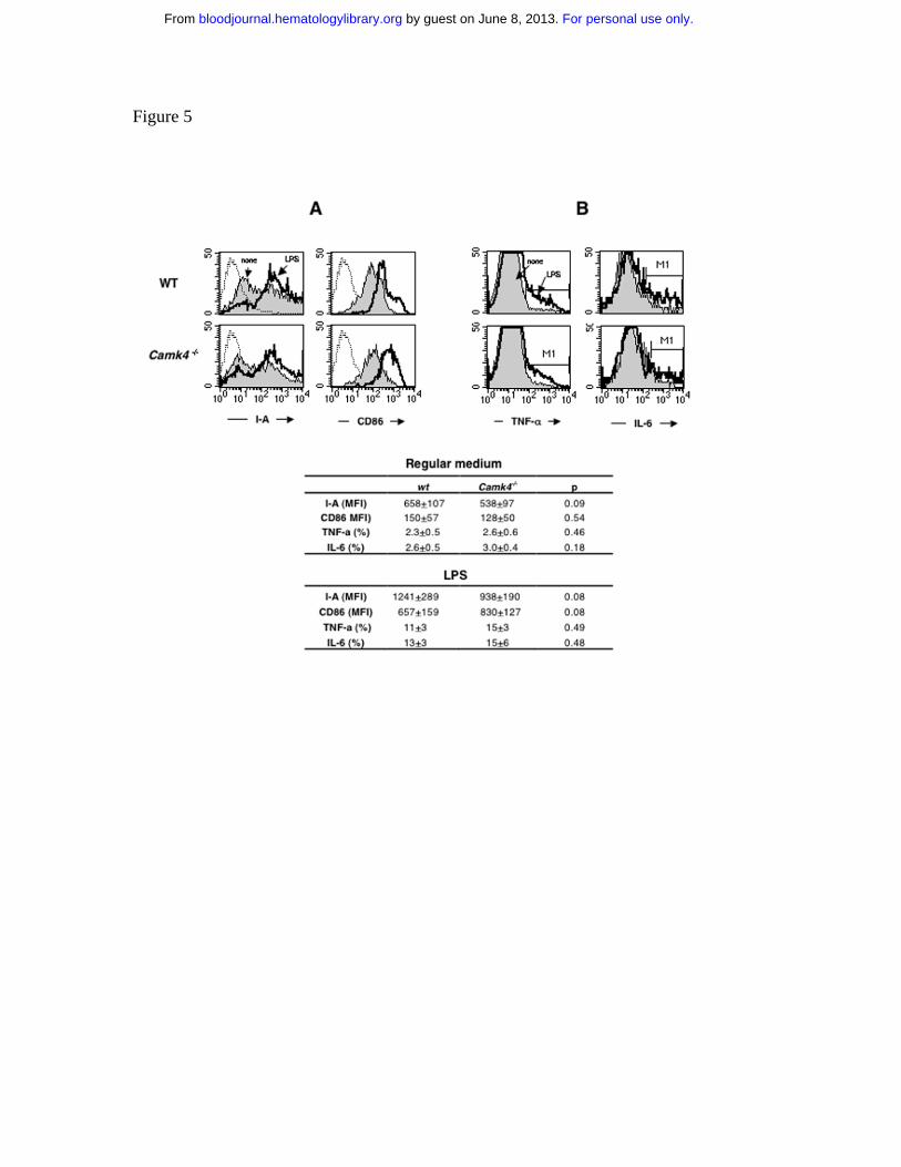
Figure 5
For personal use only. by guest on June 8, 2013. bloodjournal.hematologylibrary.orgFrom

Figure 6
For personal use only. by guest on June 8, 2013. bloodjournal.hematologylibrary.orgFrom

Figure 7
For personal use only. by guest on June 8, 2013. bloodjournal.hematologylibrary.orgFrom
Copyright © 2022 FDOKUMEN