Surface properties and cell adhesion onto allylamine-plasma and amine-plasma coated glass coverslips
12
Surface properties and cell adhesion onto allylamine-plasma and amine-plasma coated glass coverslips Marianne Crespin • Nicolas Moreau • Bernard Masereel • Olivier Feron • Bernard Gallez • Thierry Vander Borght • Carine Michiels • Stephane Lucas Received: 21 September 2010 / Accepted: 18 January 2011 / Published online: 3 February 2011 Ó Springer Science+Business Media, LLC 2011 Abstract Surface properties of nanoparticles to be used for radioimmunotherapy need to be optimized to allow antibody conjugation while ensuring biocompatibility. We aimed to investigate cell adhesion and proliferation onto different coatings to be used for nanoparticles. C, CH x or SiO x coatings deposited onto glass coverslips by magnetron deposition as well as nitrogen functionalized materials synthetized using different reactive sputtering conditions and PPAA (plasma polymerized allylamine) coating, were compared. Amine functionalization did increase hydro- philicity in all the materials tested. Biocompatibility was assessed by measuring cell viability, morphology, attach- ment, spreading, and pro-inflammatory cytokine secretion. The results show that C and CN x were the most biocom- patible substrates while SiO x and SiO x N y were the most toxic materials. PPAA coatings displayed unexpectedly an intermediate biocompatibility. A correlation could be observed between wettability and cell proliferation except for C coated surface, indicating that more complex pro- cesses than hydrophilicity alone are taking place that affect cell functions. 1 Introduction Lung cancer is the leading cause of cancer-related mor- tality worldwide in both men and women [1, 2]. Non-small cell lung cancer (NSCLC) accounts for approximately 85% of all lung cancers and its 5-year survival is only 5% [3, 4]. The conventional treatment of extended NSCLC disease combining systemic chemotherapy with sometimes radio- therapy is unfortunately only palliative: it aims to reduce symptoms, to improve life quality, and to prolong patient survival (6–10 weeks) [1]. Therefore, in order to counter this disease, there is a growing demand for more effective new therapeutic strategies. One of them consists of tar- geting the vasculature that feeds the tumor by radioim- munotherapy (RIT). RIT uses target-specific monoclonal antibodies combined with a radionuclide to deliver radia- tion into the tumor [5–7]. Up to now, available RIT treat- ments are based on conjugates made of one radioactive atom per vector. In order to improve its efficacy, the antibody could be linked to a nanoparticle, containing a larger number of radioactive atoms rather than a single one [8]. Its much higher specific activity is counterbalanced by a more complex radiochemistry. The coating of the M. Crespin C. Michiels (&) Unite ´ de Recherche en Biologie Cellulaire, NARILIS, University of Namur-FUNDP, URBC, 61 rue de Bruxelles, 5000 Namur, Belgium e-mail: [email protected] N. Moreau S. Lucas Research Center in Physics of Matter and Radiation (PMR), NARILIS, University of Namur-FUNDP, Namur, Belgium B. Masereel Drug Design and Discovery Center, NARILIS, University of Namur-FUNDP, Namur, Belgium O. Feron Unit of Pharmacology and Therapeutics, Universite ´ catholique de Louvain, UCL-FATH, Brussels, Belgium B. Gallez Biomedical Magnetic Resonance Unit, Universite ´ catholique de Louvain, UCL-CMFA, Brussels, Belgium T. Vander Borght Division of Nuclear Medicine, Mont-Godinne University Hospital, Universite ´ catholique de Louvain, Yvoir, Belgium 123 J Mater Sci: Mater Med (2011) 22:671–682 DOI 10.1007/s10856-011-4245-3
Transcript of Surface properties and cell adhesion onto allylamine-plasma and amine-plasma coated glass coverslips

Surface properties and cell adhesion onto allylamine-plasmaand amine-plasma coated glass coverslips
Marianne Crespin • Nicolas Moreau • Bernard Masereel •
Olivier Feron • Bernard Gallez • Thierry Vander Borght •
Carine Michiels • Stephane Lucas
Received: 21 September 2010 / Accepted: 18 January 2011 / Published online: 3 February 2011
� Springer Science+Business Media, LLC 2011
Abstract Surface properties of nanoparticles to be used
for radioimmunotherapy need to be optimized to allow
antibody conjugation while ensuring biocompatibility. We
aimed to investigate cell adhesion and proliferation onto
different coatings to be used for nanoparticles. C, CHx or
SiOx coatings deposited onto glass coverslips by magnetron
deposition as well as nitrogen functionalized materials
synthetized using different reactive sputtering conditions
and PPAA (plasma polymerized allylamine) coating, were
compared. Amine functionalization did increase hydro-
philicity in all the materials tested. Biocompatibility was
assessed by measuring cell viability, morphology, attach-
ment, spreading, and pro-inflammatory cytokine secretion.
The results show that C and CNx were the most biocom-
patible substrates while SiOx and SiOxNy were the most
toxic materials. PPAA coatings displayed unexpectedly an
intermediate biocompatibility. A correlation could be
observed between wettability and cell proliferation except
for C coated surface, indicating that more complex pro-
cesses than hydrophilicity alone are taking place that affect
cell functions.
1 Introduction
Lung cancer is the leading cause of cancer-related mor-
tality worldwide in both men and women [1, 2]. Non-small
cell lung cancer (NSCLC) accounts for approximately 85%
of all lung cancers and its 5-year survival is only 5% [3, 4].
The conventional treatment of extended NSCLC disease
combining systemic chemotherapy with sometimes radio-
therapy is unfortunately only palliative: it aims to reduce
symptoms, to improve life quality, and to prolong patient
survival (6–10 weeks) [1]. Therefore, in order to counter
this disease, there is a growing demand for more effective
new therapeutic strategies. One of them consists of tar-
geting the vasculature that feeds the tumor by radioim-
munotherapy (RIT). RIT uses target-specific monoclonal
antibodies combined with a radionuclide to deliver radia-
tion into the tumor [5–7]. Up to now, available RIT treat-
ments are based on conjugates made of one radioactive
atom per vector. In order to improve its efficacy, the
antibody could be linked to a nanoparticle, containing a
larger number of radioactive atoms rather than a single one
[8]. Its much higher specific activity is counterbalanced by
a more complex radiochemistry. The coating of the
M. Crespin � C. Michiels (&)
Unite de Recherche en Biologie Cellulaire, NARILIS, University
of Namur-FUNDP, URBC, 61 rue de Bruxelles, 5000 Namur,
Belgium
e-mail: [email protected]
N. Moreau � S. Lucas
Research Center in Physics of Matter and Radiation (PMR),
NARILIS, University of Namur-FUNDP, Namur, Belgium
B. Masereel
Drug Design and Discovery Center, NARILIS,
University of Namur-FUNDP, Namur, Belgium
O. Feron
Unit of Pharmacology and Therapeutics, Universite catholique
de Louvain, UCL-FATH, Brussels, Belgium
B. Gallez
Biomedical Magnetic Resonance Unit, Universite catholique de
Louvain, UCL-CMFA, Brussels, Belgium
T. Vander Borght
Division of Nuclear Medicine, Mont-Godinne University
Hospital, Universite catholique de Louvain, Yvoir, Belgium
123
J Mater Sci: Mater Med (2011) 22:671–682
DOI 10.1007/s10856-011-4245-3
nanoparticle with a surface-functionalized material may be
used to build this conjugate. The latter should however not
induce an inflammatory response once injected to avoid
compromising the potential therapeutic effect.
This work is aimed to assess such a risk by testing, on
endothelial cells, the biocompatibility of different materials
that could be used as the interface between the nanoparticle
and the monoclonal antibody. Among the different coating
processes available, the magnetron sputtering deposition
process [9–11] was selected because the radioactive
nanoparticles will also be produced by vacuum based
technique [12]. The inclusion of all steps in the same
reactor spares time [13], which may become critical when
using short half-life radioactive materials. Cell adhesion is
dependent on the surface properties of materials [14] such
as high wettability [15] and basic property [16] which are
able to increase their biocompatibility. Furthermore, coat-
ing may require chemical functionalization for antibody
immobilization [17] usually using amine functions. Two
different nitrogen-functionalized coatings can be obtained
either by plasma treatment carried out under nitrogen or by
plasma polymerization of allylamine [18]. The goal of this
study is to assess the biocompatibility of the plasma-syn-
thesized materials themselves, deposited on flat substrates.
It is well known that the PVD deposition mechanisms at
the early stages of the film growth are highly dependant on
the substrate [19]. In some cases, nanometer-ranged films
can grow as islands instead of continuous thin layers.
Therefore, the deposited layer does not entirely cover the
substrate, and its intrinsic roughness is strongly modified
by the plasma deposition. A way to avoid those issues is to
make a rather thick deposit ([50 nm) onto the glass cov-
erslips. This is the option chosen in this work.
In this work, we studied the biocompatibility of these
plasma-deposited materials with and without amine-
functionalization using endothelial cells. Endothelial cells
were chosen because they will be targeted by RIT using
antibodies coupled to nanoparticles.
2 Materials and methods
2.1 Coating synthesis
All materials were deposited on glass coverslips thanks to a
Physical Vapor Deposition System (PVD). It consists of a
vacuum chamber equipped with 200 unbalanced-magnetron
sputtering guns, powered by supply configured in DC or
pulsed bipolar mode. The plasma excitation mode was
chosen in order to obtain the best deposition rate and
plasma stability as well as the least porous material. The
different deposition conditions are listed in Table 1. Two
cathodes were used: carbon for the carbon or silicon for the
silicon based coatings respectively (99.9% purity for each).
Coating thickness was about 100 nm for all samples.
Carbon, CHx and SiOx are the nitrogen-free materials
studied in this work. They were synthesized in either DC
(direct current) for C and PPAA coatings, or pulsed bipolar
(Ton? = Ton-: 10 ls, Toff? = Toff-: 50 ls for CHx, Ton?:
20 ls, Ton-: 90 ls, Toff?: 5 ls, Toff-: 5 ls for SiOx).
PPAA coatings were produced as described in [20]. The
properties of carbon and CHx films have been previously
characterized [21]. This coating is amorphous. The
hydrogen content of the CHx film is about 55%, and its
density is closed to 1.2 g/cm3. They are described as a
highly branched and highly cross-linked hydrocarbon net-
work with a fairly large number of free radicals. When air-
exposed, they become slightly oxidized as revealed by the
presence of C=O stretching band measured by FTIR. The
oxygen bulk concentration of SiOx films has been mea-
sured by Rutherford Backscattering Spectrometry (RBS)
and is the one of SiOx compound. In order to functionalize
these coatings with nitrogen, N2 gas was injected into the
chamber during the plasma deposition process, either pure
(CNx) or mixed with argon (CHxNy) following the
parameters listed in Table 1.
Plasma Polymerized AllylAmine (PPAA) coatings were
obtained by generating a discharge in pure allylamine
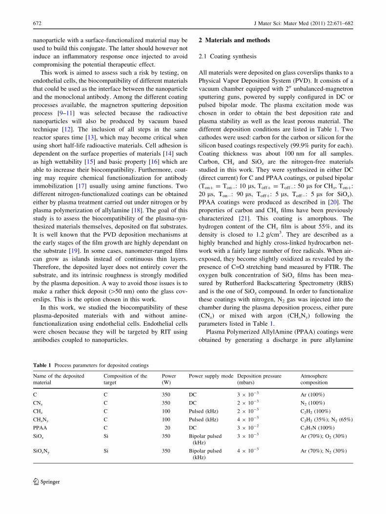
Table 1 Process parameters for deposited coatings
Name of the deposited
material
Composition of the
target
Power
(W)
Power supply mode Deposition pressure
(mbars)
Atmosphere
composition
C C 350 DC 3 9 10-3 Ar (100%)
CNx C 350 DC 2 9 10-3 N2 (100%)
CHx C 100 Pulsed (kHz) 2 9 10-3 C2H2 (100%)
CHxNy C 100 Pulsed (kHz) 4 9 10-3 C2H2 (35%); N2 (65%)
PPAA C 20 DC 3 9 10-2 C3H7N (100%)
SiOx Si 350 Bipolar pulsed
(kHz)
3 9 10-3 Ar (70%); O2 (30%)
SiOxNy Si 350 Bipolar pulsed
(kHz)
4 9 10-3 Ar (70%); N2 (30%)
672 J Mater Sci: Mater Med (2011) 22:671–682
123
vapor, according to the procedure described by Lucas et al.
[20]. Their properties depend on the power fed into the
plasma: here, we generated coatings that are C0.45H0.32
N0.13O0.1. To achieve sterile conditions, the coverslips
were autoclaved prior to their utilization for cell seeding
(during 20 min at 121�C and 16 Psi). The stability of
allylamine deposit is high after autoclaving with only
minor changes in nitrogen composition and wettability
[16].
2.2 Plasma-coated substrate characterization
After the deposition and the sterilization processes, all
coated substrates were characterized by XPS in order to
probe for the chemical composition of their extreme sur-
face. Measurements were performed with a SSX-100
spectrometer (Surface Science Instruments), with incident
X-Ray beam of 1486.6 eV (Al Ka), and without any ionic
etching prior the analysis. To determine the stoichiometry,
all areas of peaks were normalized with appropriate Scof-
field factors given by the spectrometer manufacturer. Evi-
dence of the chemical functions (especially the presence of
amine groups) of the PPAA layer was obtained by chem-
ical derivatization of amine groups into imine with penta-
fluorobenzaldehyde (PFBA) [22]. The reaction was carried
out in ethanol for 15 h (50�C, nitrogen-flushed reactor).
Then, the substrate with the PPAA layer was thoroughly
washed by different organic solvents (ethanol, dichloro-
methane, acetone and hexane) under ultrasonication, in
order to release the PFBA molecules non-covalently bound
to the surface. Finally, the amine group concentration was
obtained by measuring the fluorine atomic concentration on
derivatized samples by XPS, normalized by the carbon
peak area. The surface amine concentration (in atomic
percents) was determined by the formula: ½NH2� ¼½F�=5
½C��7½F�=5� 100; adapted from Choukourov et al. [23]. The
layer of PPAA was also characterized by Nuclear Reac-
tions Analysis (NRA), using a 2.385 MeV 3He beam [20].
This technique provides the measurement of atomic con-
centrations of all elements present in the entire layer,
whereas the XPS measurements only give information
coming from the extreme surface of the layer. Wettability
of the substrates was characterized by contact angle mea-
surements, carried out with VCA 2500 XE system. The
measurements were performed, after being washed in
ethanol, at room temperature with droplets of ca. 0.5 ll of
ultrapure water.
2.3 Cell culture
The human endothelial cell line EAhy926 was kindly
provided by Prof. C. Edgell (Pathology Department,
University of North Carolina) [24]. Cells were maintained
in culture at 37�C under 5% CO2 and 95% air atmosphere,
in 75 cm2 polystyrene flasks (Costar) with Dulbecco’s
Modified Eagle Medium (DMEM, Gibco) (4.5 g/l D-glu-
cose) containing NaHCO3 (1.5 g/l) but no pyruvate and
10% foetal calf serum. For the experiments, cells were
harvested by trypsinization and seeded in 24-well plates
onto the different coverslips at a density of 10,000 cells per
well. They were then incubated for different times in
complete medium.
2.4 Coomassie blue staining
The morphology of cells seeded on the different substrates
was assessed after cell protein staining with Coomassie
Blue. Briefly, EAhy926 endothelial cells were first washed
twice with PBS, and fixed with 3.7% PFA in distilled water
for 10 min at RT. Then, they were permeabilized with 70%
ethanol in distilled water for 10 min at RT and dried at
37�C. The cells were finally stained with Coomassie Blue
for 30 s, washed with distilled water and dried at 37�C,
before being photographed with an inverted microscope
(Labovert FS, Leitz).
2.5 Cell viability assay
To assess cell proliferation/viability on the different sub-
strates, an MTT assay was performed using EAhy926
endothelial cells. Six days after plating, 500 ll of MTT
solution (2.5 mg/ml of [3-(4,5-diMethylThiazol-2-yl)-2,5-
diphenyTetrazolium bromide], Sigma in PBS) were added
to the culture medium for 2 h at 37�C. The medium was
then removed and 1 ml of lysis solution (2 volumes of SDS
30% and 1 volume of N,N-dimethyl-formamide) was added
to the cells for 1 h at 37�C on a rotating plate (80 rpm)
before the optical absorbance measurement using a plate
reader (BIO-RAD) at a wavelength of 570 nm.
2.6 Immunofluorescence staining
The EAhy926 endothelial cells were fixed with 4% para-
formaldehyde in PBS for 10 min at 20�C, and washed three
times with PBS. They were then permeabilized with 1%
Triton-X-100 (Sigma) in PBS for 5 min. They were then
washed three times with PBS-BSA (Sigma). For actin
staining, cells were incubated with Alexa Fluor 546 phal-
loıdin (Molecular Probes) at 1:50 dilution, overnight at
4�C. For focal adhesion plaques staining, cells were incu-
bated with the primary antibody diluted in PBS-BSA,
overnight at 4�C. The next day, cells were washed three
times with PBS-BSA and incubated for 1 h in the presence
J Mater Sci: Mater Med (2011) 22:671–682 673
123
of the secondary antibody in PBS-BSA at 20�C. For both
stainings, after three washes in PBS, nuclei were stained
with To-Pro-3 (Molecular Probes) diluted (1:80) in PBS-
RNase (2 mg/ml). The samples were again washed three
times before being embedded in Mowiol (Aldrich) and
photographed with a confocal microscope TCS (Leica SP1)
using a 409 objective and a constant photomultiplier.
Mouse anti-Vinculin monoclonal antibody (Sigma) was
used at 1:800 dilution. Alexa Fluor 488 goat anti-mouse
IgG conjugate (Molecular Probes) was used at 1:1000
dilution.
2.7 Scanning electron microscopy
The samples were fixed with 2% glutaraldehyde in 0.1 M
cacodylate buffer pH 7.4 overnight at 4�C. Samples were
then washed three times for 15 min in 0.1 M cacodylate
buffer pH 7.4 containing 7.5% sucrose. The specimens
were then dehydrated in a series of increasing concentra-
tions of acetone, critical-point dried by use of liquid CO2,
and sputter coated with gold. Samples were examined with
a scanning electron microscope (SEM) Philips XL-20
operating at an accelerating voltage of 15 kV and photo-
graphs were taken.
2.8 Signal chip human cytokine array
SignalChip Human Cytokine microarrays (Eppendorf
Array Technologies) were used to analyze the protein level
of 20 human cytokines, namely eotaxin, GM-CSF, IFN,
IL-1a, IL-1b, IL-2, IL-4, IL-6, IL-8, IL-10, IL-12p40,
IL-12p70, IL-17, IP-10, MIP-1a, MIP-1b, RANTES,
TNFa, TNF-RI and TNF-RII. The assay is based on a
sandwich ELISA, where capture antibodies are spotted on
glass slides in an array format. Samples are contacted with
the array, and detection is performed using labeled detec-
tion antibodies. Positive and negative controls are included
to normalize the data, and calibration curves constructed
from known amounts of purified cytokines are used to
generate quantitative data. Arrays were contacted with
8.7 ll of sample diluted 10 times and detection was per-
formed in fluorescence, according to the manufacturer’s
protocol. Scanning was performed using a ScanArray
scanner, and signals were quantified using the Imagene
software (Perkin Elmer). Data analysis was performed
using the data analysis software provided with the arrays.
2.9 Statistical analysis
One way analysis of variance (ANOVA) with post hoc
pairwise comparison using Holm-Sidak method was per-
formed using SigmaStat.3 software.
3 Results
3.1 Coating characteristics
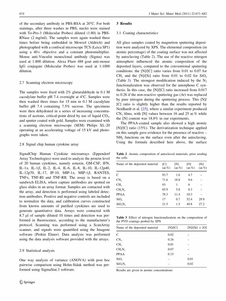
All glass samples coated by magnetron sputtering deposi-
tion were analyzed by XPS. The elemental composition (in
atomic percentage) of the coating surface was not affected
by autoclaving (Table 2). The use of the reactive nitrogen
atmosphere influenced the atomic composition of the
deposited layers, compared to the conventional sputtering
conditions: the [N]/[C] ratio varies from 0.01 to 0.07 for
CHx and the [N]/[Si] ratio from 0.01 to 0.02 for SiOx
(Table 3). The strongest modification induced by the N2
functionalization was observed for the amorphous C syn-
thesis. In this case, the [N]/[C] ratio increased from 0.017
to 0.26 if the non-reactive sputtering gas (Ar) was replaced
by pure nitrogen during the sputtering process. This [N]/
[C] ratio is slightly higher than the results reported by
Neidhardt et al. [25], where a similar experiment produced
CNx films, with [N] values between 16 and 25 at.% while
the [N] content was 18.8% in our experiments.
The PPAA-coated sample also showed a high atomic
[N]/[C] ratio (15%). The derivatization technique applied
on this sample gave evidence for the presence of reactive –
NH2 functions on the surface even after the autoclaving.
Using the formula described here above, the surface
Table 2 Atomic composition of autoclaved materials, prior seeding
the cells
Name of the deposited material [C] [N] [O] [Si]
(at.%) (at.%) (at.%) (at.%)
C 93.7 1.6 4.7 –
CNx 71.6 18.8 9.6 –
CHx 93 1 6 –
CHxNy 85.9 5.8 8.3 –
PPAA 78.3 11.4 10.3 –
SiOx 17 0.7 52.4 29.9
SiOxNy 21.5 1.5 49.8 27.2
Table 3 Effect of nitrogen functionalization on the composition of
the PVD coatings probed by XPS
Name of the deposited material [N]/[C] [N]/[Si] ? [O]
C 0.02 –
CNx 0.26 –
CHx 0.01 –
CHxNy 0.07 –
PPAA 0.15 –
SiOx – 0.01
SiOxNy – 0.02
Results are given in atomic concentrations
674 J Mater Sci: Mater Med (2011) 22:671–682
123
concentration of amine was determined as 1.3% under the
deposition conditions given in Table 1. The stoechiometry
of this material (bulk measurements) probed by NRA
technique is CH0.7N0.29O0.2 [20]. The value of [N]/[C] ratio
obtained by NRA (28%) averaged for the entire layer was
slightly higher than the one measured by XPS.
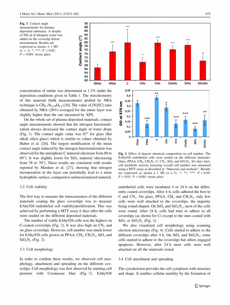
On the whole set of plasma-deposited materials, contact
angle measurements showed that the nitrogen functionali-
zation always decreased the contact angle of water drops
(Fig. 1). The contact angle value was 67� for glass (flat
alkali silica glass) which is similar to values obtained by
Huber et al. [26]. The largest modification of the mean
contact angle induced by the nitrogen functionalization was
observed for the amorphous C material (decrease from 80 to
69�). It was slightly lower for SiOx material (decreasing
from 78 to 70�). These results are consistent with results
reported by Mardare et al. [27], showing that nitrogen
incorporation in the layer can potentially lead to a more
hydrophilic surface, compared to unfunctionalized material.
3.2 Cell viability
The first way to measure the innocuousness of the different
materials coating the glass coverslips was to measure
EAhy926 endothelial cell viability/proliferation. This was
achieved by performing a MTT assay 6 days after the cells
were seeded on the different deposited materials.
The number of viable EAhy926 cells was the highest on
C-coated coverslips (Fig. 2). It was also high on CNx and
on glass coverslips. However, cell number was much lower
for EAhy926 cells grown on PPAA, CHx, CHxNy, SiOx and
SiOxNy (Fig. 2).
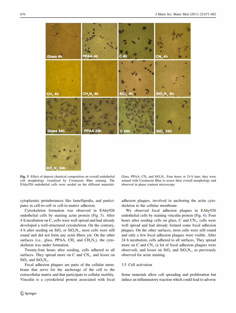
3.3 Cell morphology
In order to confirm these results, we observed cell mor-
phology, attachment and spreading on the different cov-
erslips. Cell morphology was first observed by staining cell
proteins with Coomassie blue (Fig. 3). EAhy926
endothelial cells were incubated 4 or 24 h on the differ-
ently coated coverslips. After 4 h, cells adhered the best to
C and CNx. On glass, PPAA, CHx and CHxNy, only few
cells were well attached to the coverslips, the majority
being round-shaped. On SiOx and SiOxNy, most of the cells
were round. After 24 h, cells had time to adhere to all
coverslips (as shown for C) except to the ones coated with
SiOx or SiOxNy (Fig. 3).
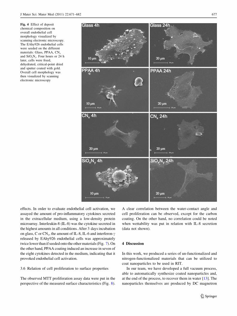
We also visualized cell morphology using scanning
electron microscopy (Fig. 4). Cells started to adhere to the
different coverslips after 4 h. On SiOx and SiOxNy, some
cells started to adhere to the coverslips but others engaged
apoptosis. However, after 24 h most cells were well
attached on all the materials tested.
3.4 Cell attachment and spreading
The cytoskeleton provides the cell cytoplasm with structure
and shape. It enables cellular motility by the formation of
Fig. 1 Contact angle
measurements for plasma-
deposited substrates. A droplet
of 500 ll of ultrapure water was
added on the coverslip before
measurement. Results are
expressed as means ± 1 SD
(n = 3). *; ***: P \ 0.05;
P \ 0.001 versus glass
Fig. 2 Effect of deposit chemical composition on cell number. The
EAhy926 endothelial cells were seeded on the different materials:
Glass, PPAA, CHx, CHxNy, C, CNx, SiOx and SiOxNy. Six days later,
cell metabolic activity assessing overall cell number was measured
using a MTT assay as described in ‘‘Materials and methods’’. Results
are expressed as means ± 1 SD (n = 3). *; **; ***: P \ 0.05;
P \ 0.01; P \ 0.001 versus glass
J Mater Sci: Mater Med (2011) 22:671–682 675
123
cytoplasmic protuberances like lamellipodia, and partici-
pates in cell-to-cell or cell-to-matrix adhesion.
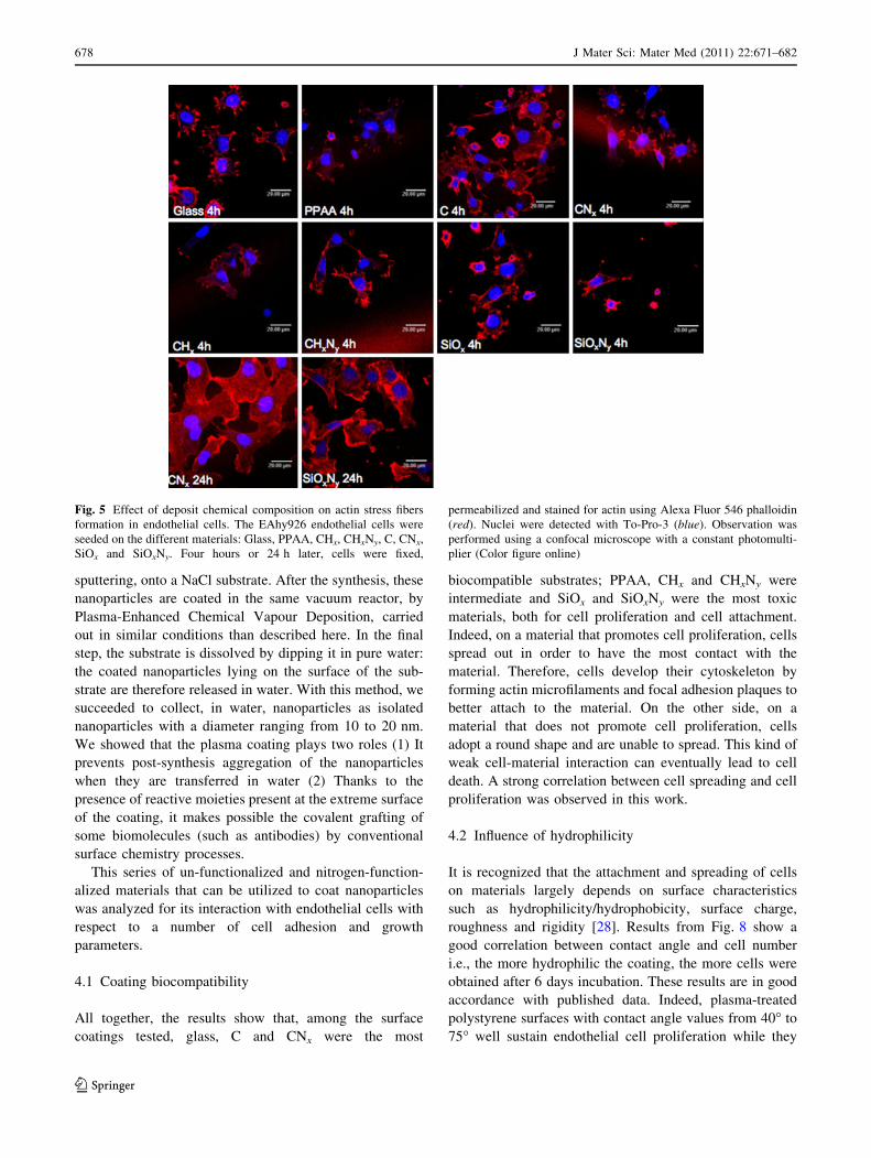
Cytoskeleton formation was observed in EAhy926
endothelial cells by staining actin protein (Fig. 5). After
4 h incubation on C, cells were well spread and had already
developed a well-structured cytoskeleton. On the contrary,
4 h after seeding on SiOx or SiOxNy, most cells were still
round and did not form any actin fibers yet. On the other
surfaces (i.e., glass, PPAA, CHx and CHxNy), the cyto-
skeleton was under formation.
Twenty-four hours after seeding, cells adhered to all
surfaces. They spread more on C and CNx, and lesser on
SiOx and SiOxNy.
Focal adhesion plaques are parts of the cellular mem-
brane that serve for the anchorage of the cell to the
extracellular matrix and that participate to cellular motility.
Vinculin is a cytoskeletal protein associated with focal
adhesion plaques, involved in anchoring the actin cyto-
skeleton to the cellular membrane.
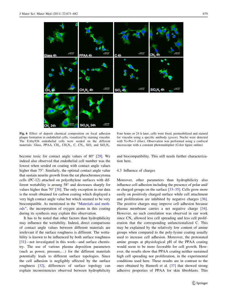
We observed focal adhesion plaques in EAhy926
endothelial cells by staining vinculin protein (Fig. 6). Four
hours after seeding cells on glass, C and CNx, cells were
well spread and had already formed some focal adhesion
plaques. On the other surfaces, most cells were still round
and only a few focal adhesion plaques were visible. After
24 h incubation, cells adhered to all surfaces. They spread
more on C and CNx (a lot of focal adhesion plaques were
observed), and lesser on SiOx and SiOxNy, as previously
observed for actin staining.
3.5 Cell activation
Some materials allow cell spreading and proliferation but
induce an inflammatory reaction which could lead to adverse
Fig. 3 Effect of deposit chemical composition on overall endothelial
cell morphology visualized by Coomassie Blue staining. The
EAhy926 endothelial cells were seeded on the different materials:
Glass, PPAA, CNx and SiOxNy. Four hours or 24 h later, they were
stained with Coomassie Blue to assess their overall morphology and
observed in phase contrast microscopy
676 J Mater Sci: Mater Med (2011) 22:671–682
123
effects. In order to evaluate endothelial cell activation, we
assayed the amount of pro-inflammatory cytokines secreted
in the extracellular medium, using a low-density protein
microarray. Interleukin-8 (IL-8) was the cytokine secreted in
the highest amounts in all conditions. After 3 days incubation
on glass, C or CNx, the amount of IL-8, IL-6 and interferon-creleased by EAhy926 endothelial cells was approximately
twice lower than if seeded onto the other materials (Fig. 7). On
the other hand, PPAA coating induced an increase in seven of
the eight cytokines detected in the medium, indicating that it
provoked endothelial cell activation.
3.6 Relation of cell proliferation to surface properties
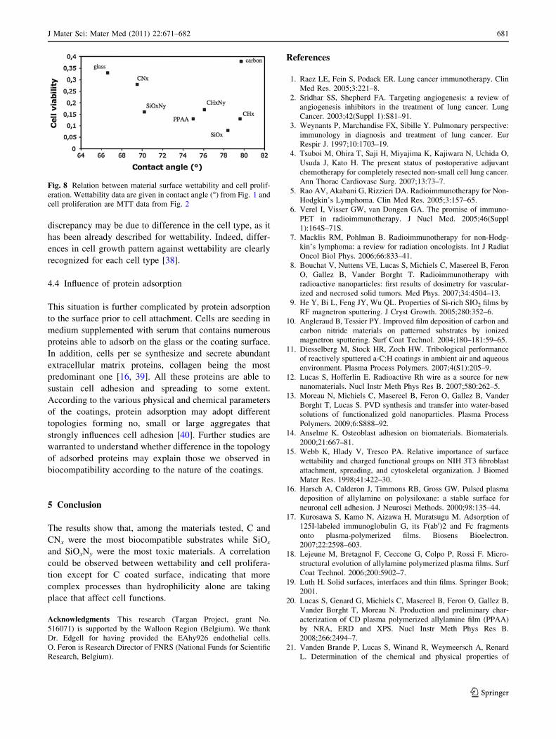
The observed MTT proliferation assay data were put in the
perspective of the measured surface characteristics (Fig. 8).
A clear correlation between the water-contact angle and
cell proliferation can be observed, except for the carbon
coating. On the other hand, no correlation could be noted
when wettability was put in relation with IL-8 secretion
(data not shown).
4 Discussion
In this work, we produced a series of un-functionalized and
nitrogen-functionalized materials that can be utilized to
coat nanoparticles to be used in RIT.
In our team, we have developed a full vacuum process,
able to automatically synthesize coated nanoparticles and,
at the end of the process, to recover them in water [13]. The
nanoparticles themselves are produced by DC magnetron
Fig. 4 Effect of deposit
chemical composition on
overall endothelial cell
morphology visualized by
scanning electronic microscopy.
The EAhy926 endothelial cells
were seeded on the different
materials: Glass, PPAA, CNx
and SiOxNy. Four hours or 24 h
later, cells were fixed,
dehydrated, critical-point dried
and sputter coated with gold.
Overall cell morphology was
then visualized by scanning
electronic microscopy
J Mater Sci: Mater Med (2011) 22:671–682 677
123
sputtering, onto a NaCl substrate. After the synthesis, these
nanoparticles are coated in the same vacuum reactor, by
Plasma-Enhanced Chemical Vapour Deposition, carried
out in similar conditions than described here. In the final
step, the substrate is dissolved by dipping it in pure water:
the coated nanoparticles lying on the surface of the sub-
strate are therefore released in water. With this method, we
succeeded to collect, in water, nanoparticles as isolated
nanoparticles with a diameter ranging from 10 to 20 nm.
We showed that the plasma coating plays two roles (1) It
prevents post-synthesis aggregation of the nanoparticles
when they are transferred in water (2) Thanks to the
presence of reactive moieties present at the extreme surface
of the coating, it makes possible the covalent grafting of
some biomolecules (such as antibodies) by conventional
surface chemistry processes.
This series of un-functionalized and nitrogen-function-
alized materials that can be utilized to coat nanoparticles
was analyzed for its interaction with endothelial cells with
respect to a number of cell adhesion and growth
parameters.
4.1 Coating biocompatibility
All together, the results show that, among the surface
coatings tested, glass, C and CNx were the most
biocompatible substrates; PPAA, CHx and CHxNy were
intermediate and SiOx and SiOxNy were the most toxic
materials, both for cell proliferation and cell attachment.
Indeed, on a material that promotes cell proliferation, cells
spread out in order to have the most contact with the
material. Therefore, cells develop their cytoskeleton by
forming actin microfilaments and focal adhesion plaques to
better attach to the material. On the other side, on a
material that does not promote cell proliferation, cells
adopt a round shape and are unable to spread. This kind of
weak cell-material interaction can eventually lead to cell
death. A strong correlation between cell spreading and cell
proliferation was observed in this work.
4.2 Influence of hydrophilicity
It is recognized that the attachment and spreading of cells
on materials largely depends on surface characteristics
such as hydrophilicity/hydrophobicity, surface charge,
roughness and rigidity [28]. Results from Fig. 8 show a
good correlation between contact angle and cell number
i.e., the more hydrophilic the coating, the more cells were
obtained after 6 days incubation. These results are in good
accordance with published data. Indeed, plasma-treated
polystyrene surfaces with contact angle values from 40� to
75� well sustain endothelial cell proliferation while they
Fig. 5 Effect of deposit chemical composition on actin stress fibers
formation in endothelial cells. The EAhy926 endothelial cells were
seeded on the different materials: Glass, PPAA, CHx, CHxNy, C, CNx,
SiOx and SiOxNy. Four hours or 24 h later, cells were fixed,
permeabilized and stained for actin using Alexa Fluor 546 phalloidin
(red). Nuclei were detected with To-Pro-3 (blue). Observation was
performed using a confocal microscope with a constant photomulti-
plier (Color figure online)
678 J Mater Sci: Mater Med (2011) 22:671–682
123
become toxic for contact angle values of 80� [29]. We
indeed also observed that endothelial cell number was the
lowest when seeded on coating with contact angle values
higher than 75�. Similarly, the optimal contact angle value
that sustain neurite growth from the rat pheochromocytoma
cells (PC-12) attached on polyethylene surfaces with dif-
ferent wettability is aroung 58� and decreases sharply for
values higher than 70� [30]. The only exception in our data
is the result obtained for carbon coating which displayed a
very high contact angle value but which seemed to be very
biocompatible. As mentioned in the ‘‘Materials and meth-
ods’’, the incorporation of oxygen atoms in this coating
during its synthesis may explain this observation.
It has to be noted that other factors than hydrophilicity
may influence the wettability. Indeed, direct comparisons
of contact angle values between different materials are
irrelevant if the surface roughness is different. The wetta-
bility is known to be influenced by both surface roughness
[31]—not investigated in this work—and surface chemis-
try. The use of various plasma deposition parameters
(such as power, pressure,…) for the different materials
potentially leads to different surface topologies. Since
the cell adhesion is negligibly affected by the surface
roughness [32], differences of surface topology can
explain inconsistencies observed between hydrophilicity
and biocompatibility. This still needs further characteriza-
tion here.
4.3 Influence of charges
Moreover, other parameters than hydrophilicity also
influence cell adhesion including the presence of polar and/
or charged groups on the surface [33–35]. Cells grow more
easily on positively charged surface while cell attachment
and proliferation are inhibited by negative charges [36].
The positive charges may improve cell adhesion because
plasma membrane carries a net negative charge [34].
However, no such correlation was observed in our work
since CNx allowed less cell spreading and less cell prolif-
eration that the corresponding unfunctionalized C. This
may be explained by the relatively low content of amine
groups when compared to the poly-lysine coating usually
used to increase cell adhesion. Moreover, the protonated
amine groups at physiological pH of the PPAA coating
would seem to be more favorable for cell growth. How-
ever, the results show that PPAA coating neither sustained
high cell spreading nor proliferation, in the experimental
conditions used here. These results are in contrast to the
ones obtained by Hamerli et al. [37] that showed strong
adhesive properties of PPAA for skin fibroblasts. This
Fig. 6 Effect of deposit chemical composition on focal adhesion
plaque formation in endothelial cells, visualized by staining vinculin.
The EAhy926 endothelial cells were seeded on the different
materials: Glass, PPAA, CHx, CHxNy, C, CNx, SiOx and SiOxNy.
Four hours or 24 h later, cells were fixed, permeabilized and stained
for vinculin using a specific antibody (green). Nuclei were detected
with To-Pro-3 (blue). Observation was performed using a confocal
microscope with a constant photomultiplier (Color figure online)
J Mater Sci: Mater Med (2011) 22:671–682 679
123
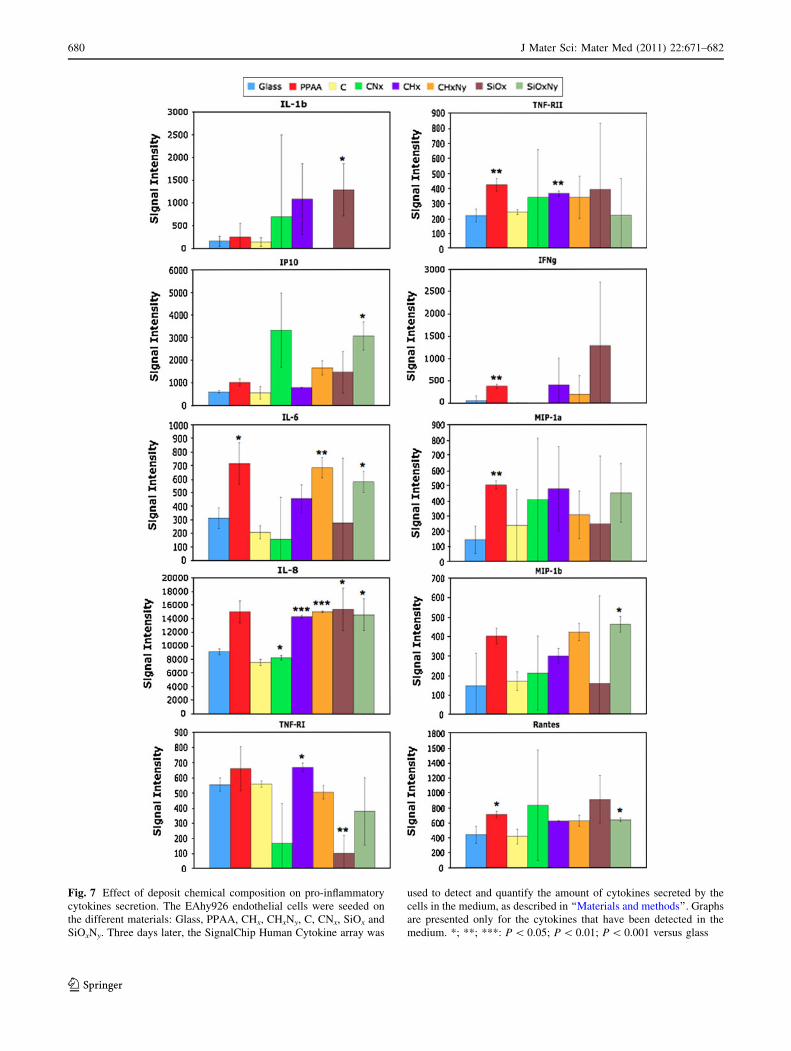
Fig. 7 Effect of deposit chemical composition on pro-inflammatory
cytokines secretion. The EAhy926 endothelial cells were seeded on
the different materials: Glass, PPAA, CHx, CHxNy, C, CNx, SiOx and
SiOxNy. Three days later, the SignalChip Human Cytokine array was
used to detect and quantify the amount of cytokines secreted by the
cells in the medium, as described in ‘‘Materials and methods’’. Graphs
are presented only for the cytokines that have been detected in the
medium. *; **; ***: P \ 0.05; P \ 0.01; P \ 0.001 versus glass
680 J Mater Sci: Mater Med (2011) 22:671–682
123
discrepancy may be due to difference in the cell type, as it
has been already described for wettability. Indeed, differ-
ences in cell growth pattern against wettability are clearly
recognized for each cell type [38].
4.4 Influence of protein adsorption
This situation is further complicated by protein adsorption
to the surface prior to cell attachment. Cells are seeding in
medium supplemented with serum that contains numerous
proteins able to adsorb on the glass or the coating surface.
In addition, cells per se synthesize and secrete abundant
extracellular matrix proteins, collagen being the most
predominant one [16, 39]. All these proteins are able to
sustain cell adhesion and spreading to some extent.
According to the various physical and chemical parameters
of the coatings, protein adsorption may adopt different
topologies forming no, small or large aggregates that
strongly influences cell adhesion [40]. Further studies are
warranted to understand whether difference in the topology
of adsorbed proteins may explain those we observed in
biocompatibility according to the nature of the coatings.
5 Conclusion
The results show that, among the materials tested, C and
CNx were the most biocompatible substrates while SiOx
and SiOxNy were the most toxic materials. A correlation
could be observed between wettability and cell prolifera-
tion except for C coated surface, indicating that more
complex processes than hydrophilicity alone are taking
place that affect cell functions.
Acknowledgments This research (Targan Project, grant No.
516071) is supported by the Walloon Region (Belgium). We thank
Dr. Edgell for having provided the EAhy926 endothelial cells.
O. Feron is Research Director of FNRS (National Funds for Scientific
Research, Belgium).
References
1. Raez LE, Fein S, Podack ER. Lung cancer immunotherapy. Clin
Med Res. 2005;3:221–8.
2. Sridhar SS, Shepherd FA. Targeting angiogenesis: a review of
angiogenesis inhibitors in the treatment of lung cancer. Lung
Cancer. 2003;42(Suppl 1):S81–91.
3. Weynants P, Marchandise FX, Sibille Y. Pulmonary perspective:
immunology in diagnosis and treatment of lung cancer. Eur
Respir J. 1997;10:1703–19.
4. Tsuboi M, Ohira T, Saji H, Miyajima K, Kajiwara N, Uchida O,
Usuda J, Kato H. The present status of postoperative adjuvant
chemotherapy for completely resected non-small cell lung cancer.
Ann Thorac Cardiovasc Surg. 2007;13:73–7.
5. Rao AV, Akabani G, Rizzieri DA. Radioimmunotherapy for Non-
Hodgkin’s Lymphoma. Clin Med Res. 2005;3:157–65.
6. Verel I, Visser GW, van Dongen GA. The promise of immuno-
PET in radioimmunotherapy. J Nucl Med. 2005;46(Suppl
1):164S–71S.
7. Macklis RM, Pohlman B. Radioimmunotherapy for non-Hodg-
kin’s lymphoma: a review for radiation oncologists. Int J Radiat
Oncol Biol Phys. 2006;66:833–41.
8. Bouchat V, Nuttens VE, Lucas S, Michiels C, Masereel B, Feron
O, Gallez B, Vander Borght T. Radioimmunotherapy with
radioactive nanoparticles: first results of dosimetry for vascular-
ized and necrosed solid tumors. Med Phys. 2007;34:4504–13.
9. He Y, Bi L, Feng JY, Wu QL. Properties of Si-rich SIO2 films by
RF magnetron sputtering. J Cryst Growth. 2005;280:352–6.
10. Angleraud B, Tessier PY. Improved film deposition of carbon and
carbon nitride materials on patterned substrates by ionized
magnetron sputtering. Surf Coat Technol. 2004;180–181:59–65.
11. Diesselberg M, Stock HR, Zoch HW. Tribological performance
of reactively sputtered a-C:H coatings in ambient air and aqueous
environment. Plasma Process Polymers. 2007;4(S1):205–9.
12. Lucas S, Hofferlin E. Radioactive Rh wire as a source for new
nanomaterials. Nucl Instr Meth Phys Res B. 2007;580:262–5.
13. Moreau N, Michiels C, Masereel B, Feron O, Gallez B, Vander
Borght T, Lucas S. PVD synthesis and transfer into water-based
solutions of functionalized gold nanoparticles. Plasma Process
Polymers. 2009;6:S888–92.
14. Anselme K. Osteoblast adhesion on biomaterials. Biomaterials.
2000;21:667–81.
15. Webb K, Hlady V, Tresco PA. Relative importance of surface
wettability and charged functional groups on NIH 3T3 fibroblast
attachment, spreading, and cytoskeletal organization. J Biomed
Mater Res. 1998;41:422–30.
16. Harsch A, Calderon J, Timmons RB, Gross GW. Pulsed plasma
deposition of allylamine on polysiloxane: a stable surface for
neuronal cell adhesion. J Neurosci Methods. 2000;98:135–44.
17. Kurosawa S, Kamo N, Aizawa H, Muratsugu M. Adsorption of
125I-labeled immunoglobulin G, its F(ab0)2 and Fc fragments
onto plasma-polymerized films. Biosens Bioelectron.
2007;22:2598–603.
18. Lejeune M, Bretagnol F, Ceccone G, Colpo P, Rossi F. Micro-
structural evolution of allylamine polymerized plasma films. Surf
Coat Technol. 2006;200:5902–7.
19. Luth H. Solid surfaces, interfaces and thin films. Springer Book;
2001.
20. Lucas S, Genard G, Michiels C, Masereel B, Feron O, Gallez B,
Vander Borght T, Moreau N. Production and preliminary char-
acterization of CD plasma polymerized allylamine film (PPAA)
by NRA, ERD and XPS. Nucl Instr Meth Phys Res B.
2008;266:2494–7.
21. Vanden Brande P, Lucas S, Winand R, Weymeersch A, Renard
L. Determination of the chemical and physical properties of
Fig. 8 Relation between material surface wettability and cell prolif-
eration. Wettability data are given in contact angle (�) from Fig. 1 and
cell proliferation are MTT data from Fig. 2
J Mater Sci: Mater Med (2011) 22:671–682 681
123
hydrogenated carbon deposits produced by DC magnetron reac-
tive sputtering. Surf Coat Technol. 1994;68–69:656–61.
22. Moon JH, Shin JW, Kim SY, Park JW. Formation of uniform
aminosilane thin layers: an imine formation to measure relative
surface density of the amine group. Langmuir. 1996;12:4621–4.
23. Choukourov A, Biederman H, Slavinska D, Trchova M, Hol-
lander A. The influence of pulse parameters on film composition
during plasma polymerization of diaminocyclohexane. Surf Coat
Technol. 2003;174:863–6.
24. Edgell CJ, McDonald CC, Graham JB. Permanent cell line
expressing human factor VIII-related antigen established by
hybridization. Proc Natl Acad Sci USA. 1983;80:3734–7.
25. Neidhardt J, Hultman L, Broitman E, Scharf TW, Singer IL.
Structural, mechanical and tribological behavior of fullerene-like
and amorphous carbon nitride coatings. Diam Relat Mater.
2004;13:1882–8.
26. Huber G, Mantz H, Spolenak R, Mecke K, Jacobs K, Gorb SN,
Arzt E. Evidence for capillarity contributions to gecko adhesion
from single spatula nanomechanical measurements. Proc Natl
Acad Sci USA. 2005;102:16293–6.
27. Mardare D, Luca D, Teodorescu CM, Macovei D. On the
hydrophilicity of nitrogen-doped TiO2 thin films. Surf Sci.
2007;601:4515–20.
28. Ramires PA, Mirenghi L, Romano AR, Palumbo F, Nicolardi G.
Plasma-treated PET surfaces improve the biocompatibility of
human endothelial cells. J Biomed Mater Res. 2000;51:535–9.
29. van Kooten TG, Spijker HT, Busscher HJ. Plasma-treated poly-
styrene surfaces: model surfaces for studying cell-biomaterial
interactions. Biomaterials. 2004;25:1735–47.
30. Lee SJ, Khang G, Lee YM, Lee HB. The effect of surface wet-
tability on induction and growth of neurites from the PC-12 cell
on a polymer surface. J Colloid Interface Sci. 2003;259:228–35.
31. Wenzel RN. Surface roughness and contact angle. J Phys Colloid
Chem. 1949;5:1466–7.
32. Richards RG. The effect of surface roughness on fibroblast
adhesion in vitro. Injury. 1996;27(Suppl 3):SC38–43.
33. Maroudas NG. Adhesion and spreading of cells on charged sur-
faces. J Theor Biol. 1975;49:417–24.
34. van Wachem PB, Hogt AH, Beugeling T, Feijen J, Bantjes A,
Detmers JP, van Aken WG. Adhesion of cultured human endo-
thelial cells onto methacrylate polymers with varying surface
wettability and charge. Biomaterials. 1987;8:323–8.
35. Davies JE. The importance and measurement of surface charge
species in cell behavior at the biomaterial interface. In: Ratner
BD, editor. Surface characterization of biomaterials. Amsterdam:
Elsevier; 1998. p. 219–34.
36. Lee JH, Lee JW, Khang G, Lee HB. Interaction of cells on
chargeable functional group gradient surfaces. Biomaterials.
1997;18:351–8.
37. Hamerli P, Weigel T, Groth T, Paul D. Surface properties of and
cell adhesion onto allylamine-plasma-coated polyethyl-
enterephtalat membranes. Biomaterials. 2003;24:3989–99.
38. Fuse Y, Hirata I, Kurihara H, Okazaki M. Cell adhesion and
proliferation patterns on mixed self-assembled monolayers car-
rying various ratios of hydroxyl and methyl groups. Dent Mater J.
2007;26:814–9.
39. Dewez JL, Doren A, Schneider YJ, Rouxhet PG. Competitive
adsorption of proteins: key of the relationship between substra-
tum surface properties and adhesion of epithelial cells. Bioma-
terials. 1999;20:547–59.
40. Satriano C, Marletta G, Carnazza S, Guglielmino S. Protein
adsorption and fibroblast adhesion on irradiated polysiloxane
surfaces. J Mater Sci Mater Med. 2003;14:663–70.
682 J Mater Sci: Mater Med (2011) 22:671–682
123