Supporting Information Mar 24 09
14
1 Supporting Information This supplement contains several figures related to the manuscript listed below. The figures provide important information supporting the conclusions of this report. Figure 1 and 2 provide information on the purification of the title enzyme. Figure 3 shows the time-resolved conversion of the proenzyme to the mature enzyme form. Figure 4 defines the N-terminal sequence of the mature enzyme form obtained in these studies. Figure 5 shows close-up views of the interaction of compound 1 with plasmepsin 2 (PfPM2). Recombinant plasmepsin 1 from the human malaria parasite Plasmodium falciparum: Enzymatic characterization, active site inhibitor design, and structural analysis Peng Liu, Melissa R. Marzahn, Arthur H. Robbins, Hugo Gutiérrez-de-Terán, David Rodríguez, Scott McClung, Stanley M. Stevens, Jr., Charles A. Yowell, John B. Dame, Robert McKenna, and Ben M. Dunn Department of Biochemistry and Molecular Biology, University of Florida, College of Medicine, Gainesville, Florida, USA; Department of Infectious Diseases and Pathology, University of Florida, College of Veterinary Medicine, Gainesville, Florida, USA; Protein Chemistry Core Facility, ICBR, University of Florida, College of Medicine, Gainesville, Florida, USA [email protected] Figure Legends Figure S1. Gel filtration chromatogram of the proPfPM1 K110pN mutant. The protein concentration of each fraction was represented by A 280 . A 50 μL sample from each fraction was pre-incubated in 0.1 M sodium acetate pH 4.5, at 37 °C for 20 min, the enzymatic activity of each fraction was determined by measuring the initial cleavage rates on the chromogenic peptide substrate, Lys-Pro-Ile-Leu-Phe*Nph-Arg-Leu (100
Transcript of Supporting Information Mar 24 09

1
Supporting Information
This supplement contains several figures related to the manuscript listed below. The figures provide important information supporting the conclusions of this report. Figure 1 and 2 provide information on the purification of the title enzyme. Figure 3 shows the time-resolved conversion of the proenzyme to the mature enzyme form. Figure 4 defines the N-terminal sequence of the mature enzyme form obtained in these studies. Figure 5 shows close-up views of the interaction of compound 1 with plasmepsin 2 (PfPM2). Recombinant plasmepsin 1 from the human malaria parasite Plasmodium falciparum: Enzymatic characterization, active site inhibitor design, and structural analysis Peng Liu, Melissa R. Marzahn, Arthur H. Robbins, Hugo Gutiérrez-de-Terán, David Rodríguez, Scott McClung, Stanley M. Stevens, Jr., Charles A. Yowell, John B. Dame, Robert McKenna, and Ben M. Dunn Department of Biochemistry and Molecular Biology, University of Florida, College of
Medicine, Gainesville, Florida, USA; Department of Infectious Diseases and Pathology,
University of Florida, College of Veterinary Medicine, Gainesville, Florida, USA;
Protein Chemistry Core Facility, ICBR, University of Florida, College of Medicine,
Gainesville, Florida, USA
Figure Legends
Figure S1. Gel filtration chromatogram of the proPfPM1 K110pN mutant. The protein
concentration of each fraction was represented by A280. A 50 µL sample from each
fraction was pre-incubated in 0.1 M sodium acetate pH 4.5, at 37 °C for 20 min, the
enzymatic activity of each fraction was determined by measuring the initial cleavage
rates on the chromogenic peptide substrate, Lys-Pro-Ile-Leu-Phe*Nph-Arg-Leu (100

2
µM). The recombinant zymogen material was collected as the 43 kDa monomer in a
single peak.
Figure S2. SDS-PAGE analysis of the over-expression and purification of the proPfPM1
K110pN mutant. 1. High molecular weight marker; 2. Total cell lysate before IPTG
induction; 3. Total cell lysate after 3 h IPTG induction; 4. Inclusion body extracts; 5.
Soluble dialysate; 6. Anion exchange chromatography purification; 7. Gel filtration
chromatography purification.
Figure S3. SDS-PAGE analysis of time-resolved auto-maturation of the proPfPM1
K110pN mutant. All the assays were performed at 37 °C by incubating 3 µg of the
proPfPM1 mutant in 0.1 M sodium formate pH 3.5 (I); 0.1 M sodium formate pH 4.0 (II);
0.1 M sodium acetate pH 4.5 (III); and 0.1 M sodium citrate pH 5.0 (IV). The conversion
from zymogen to mature enzyme was monitored at the incubation times indicated (unit:
min). The self-processing of proPfPM1 was effectively carried out at pH 4.0 and 4.5, as
shown in (II) and (III).
Figure S4. N-terminal protein sequencing analyses on the auto-converted PfPM1. The
naturally-occurring mature enzyme portion is underlined. The mature enzyme sequence
starts with Asn (Gluzman et al. 1994), which is indicated by the arrowhead. Two major
cleavage events, occurring between Phe111p-Phe112p and between L116p-Thr117p,
contributed evenly to the final mature species of this study. The N-terminal residues of
the two resulting products are indicated by the arrows.

3
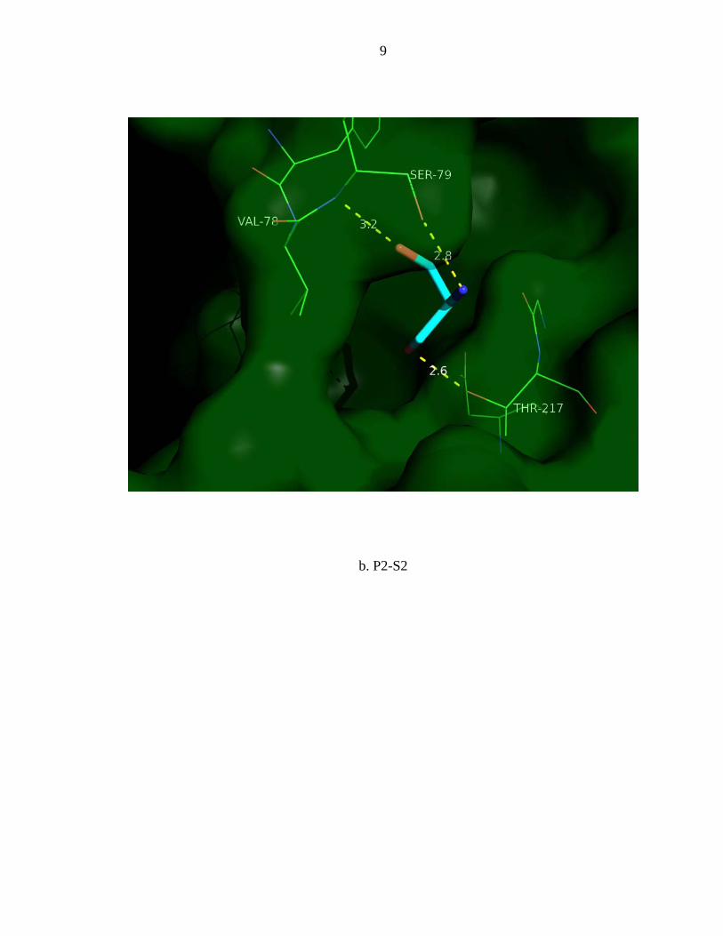
Figure S5. Detailed views of interactions between side chains of compound 1 binding to
PfPM2 based on X-ray determined structure.
a. P1-S1 interaction
b. P2-S2 interaction
c. P3-S3 interaction
d. P4-S4 interaction
e. P1’-S1’ interaction
f. P2’-S2’ interaction
g. P3’-S3’ interaction

4
FIGURES
Figure S1.

5
Figure S2.
1 2 3 4 5 6 7kDa
45
30
66
97
1 2 3 4 5 6 7kDa
45
30
66
97

6
Figure S3.

7
Figure S4.

8
Figure S5.
a. P1-S1
9
b. P2-S2

10
c. P3-S3

11
d. P4-S4

12
e. P1’-S1’

13
f. P2’-S2’

14
g. P3’-S3’