Suicide gene therapy with HSV-TK in pancreatic cancer has no effect in vivo in a mouse model
10
Suicide gene therapy with HSV-TK in pancreatic cancer has no effect in vivo in a mouse model P. Fogar*, E. Greco†, D. Basso†, W. Habeler‡, F. Navaglia†, C. -F. Zambon*, D. Tormen§, N. Gallo†, A. Cecchetto§, M. Plebani† and S. Pedrazzoli * Departments of *Medical and Surgical Sciences, University of Padova, Clinica Chirurgica IV, Via Giustiniani 2, †Laboratory Medicine, University Hospital, Via Giustiniani 2, ‡Oncological and Surgical Sciences—c/o Ospedale Busonera, Via Gattamelata 64, and §Pathology, Via Gabelli 61, University of Padova, Padova, Italy Aim: To study in vivo whether pancreatic cancer tumour growth and metastasis can be modified by a gene construct with HSV-TK suicide gene and IL2 co-expression. Methods: Seventy-eight female SCID mice were i.p. inoculated with retrovirally transduced or control MIA PaCa 2, CAPAN-1 and PANC-1 cell lines. The animals were then randomly selected for saline or ganciclovir (GCV) treatment from the second week, for a total of two weeks. Results: Most inoculated mice developed tumour nodules and spleen metastases. The liver was colonized by control CAPAN-1 and MIA PaCa 2, but not by PANC-1. Tumours in transduced MIA PaCa 2 cell injected mice were smaller, and in transduced CAPAN-1 injected mice larger, than in control-inoculated mice. There were increased pancreatic and decreased spleen metastases from transduced CAPAN-1, and diminished liver involvement from transduced MIA PaCa 2. No differences were found between mice inoculated with transduced and control PANC-1 cell lines. GCV treatment had no effect on tumour’s size or metastases. Conclusions: The HSV-TK suicide gene does not confer GCV sensitivity to pancreatic cancer in this in vivo model. Different pancreatic cancer cell lines cause different growth and metastasis patterns after inoculation in SCID mice, possibly because of variations in their inherent characteristics. The different effects of our vector on cell growth and metastasis may be attributable to the effects of the immunostimulatory cytokine IL2. Q 2003 Elsevier Ltd. All rights reserved. Key words: suicide gene therapy; pancreatic cancer; HSV-TK; animal model. INTRODUCTION Most pancreatic adeno-carcinomas are refractory to conventional treatment. 1,2 Cancer gene therapy involves suicide genes. A gene encoding for an enzyme which converts a pro-drug into a cytotoxic agent is transferred into the cancer cell. 3–7 The most commonly used suicide gene approach uses the herpes simplex virus-thymidine kinase (HSV-TK). This converts the pro-drug ganciclovir (GCV) into its toxic phosphate derivate. The phos- phorylated compound, incorporated into newly syn- thesized DNA, induces chain termination, and selectively kills dividing cells. 8–12 The HSV-TK system is efficient, and encouraging results have been obtained in several tumours, including pancreatic cancer. 13–18 However, complete tumour eradication is not always achieved because the current vector cannot deliver the functional gene to a sufficient number of cells. The bystander effect, which allows an amplification of the suicide effect, is limited for some tumour cell types. 19–26 Tumour cells can be genetically engineered to produce various cytokines which enhance the host immune antitumour response. Pancreatic cancer cell lines trans- duced with interleukin (IL)2, 4, 6, 12, 15, the tumour necrosis factor alpha, or the granulocyte-macrophage colony-stimulating factor, shows an inhibited growth. Some authors have found that they lead to complete tumour regression in subcutaneous nude mice models. 27–32 IL2 and IL4 also provide protection against subsequent re-inoculation of parental pancreatic cell lines. 27 IL2, in particular, is a T-cell growth factor that 0748–7983/$30.00 Q 2003 Elsevier Ltd. All rights reserved. EJSO 2003; 29: 721–730 doi:10.1016/j.ejso.2003.08.001 Correspondence to: Professor Sergio Pedrazzoli, Department of Medical and Surgical Sciences, University of Padova, Clinica Chirurgica IV, Via Giustiniani 2, 35128 Padova, Italy. Tel.: þ 39-0498070603; Fax: þ 39-0498076196; E-mail: [email protected]
-
Upload
independent -
Category
Documents
-
view
3 -
download
0
Transcript of Suicide gene therapy with HSV-TK in pancreatic cancer has no effect in vivo in a mouse model
Suicide gene therapy with HSV-TK in pancreaticcancer has no effect in vivo in a mouse modelP. Fogar*, E. Greco†, D. Basso†, W. Habeler‡, F. Navaglia†,C. -F. Zambon*, D. Tormen§, N. Gallo†, A. Cecchetto§, M. Plebani† andS. Pedrazzoli*
Departments of *Medical and Surgical Sciences, University of Padova, Clinica Chirurgica IV, Via Giustiniani 2, †Laboratory Medicine,
University Hospital, Via Giustiniani 2, ‡Oncological and Surgical Sciences—c/o Ospedale Busonera, Via Gattamelata 64, and §Pathology,
Via Gabelli 61, University of Padova, Padova, Italy
Aim: To study in vivo whether pancreatic cancer tumour growth and metastasis can be modified by a gene constructwith HSV-TK suicide gene and IL2 co-expression.Methods: Seventy-eight female SCID mice were i.p. inoculated with retrovirally transduced or control MIA PaCa 2,CAPAN-1 and PANC-1 cell lines. The animals were then randomly selected for saline or ganciclovir (GCV) treatmentfrom the second week, for a total of two weeks.Results: Most inoculated mice developed tumour nodules and spleen metastases. The liver was colonized by controlCAPAN-1 and MIA PaCa 2, but not by PANC-1. Tumours in transduced MIA PaCa 2 cell injected mice were smaller, andin transduced CAPAN-1 injected mice larger, than in control-inoculated mice. There were increased pancreatic anddecreased spleen metastases from transduced CAPAN-1, and diminished liver involvement from transduced MIA PaCa2. No differences were found between mice inoculated with transduced and control PANC-1 cell lines. GCV treatmenthad no effect on tumour’s size or metastases.Conclusions: The HSV-TK suicide gene does not confer GCV sensitivity to pancreatic cancer in this in vivo model.Different pancreatic cancer cell lines cause different growth and metastasis patterns after inoculation in SCID mice,possibly because of variations in their inherent characteristics. The different effects of our vector on cell growth andmetastasis may be attributable to the effects of the immunostimulatory cytokine IL2.
Q 2003 Elsevier Ltd. All rights reserved.
Key words: suicide gene therapy; pancreatic cancer; HSV-TK; animal model.
INTRODUCTION
Most pancreatic adeno-carcinomas are refractory toconventional treatment.1,2 Cancer gene therapy involvessuicide genes. A gene encoding for an enzyme whichconverts a pro-drug into a cytotoxic agent is transferredinto the cancer cell.3–7 The most commonly used suicidegene approach uses the herpes simplex virus-thymidinekinase (HSV-TK). This converts the pro-drug ganciclovir(GCV) into its toxic phosphate derivate. The phos-phorylated compound, incorporated into newly syn-thesized DNA, induces chain termination, and selectivelykills dividing cells.8–12 The HSV-TK system is efficient,
and encouraging results have been obtained in severaltumours, including pancreatic cancer.13–18 However,complete tumour eradication is not always achievedbecause the current vector cannot deliver the functionalgene to a sufficient number of cells. The bystander effect,which allows an amplification of the suicide effect, islimited for some tumour cell types.19–26
Tumour cells can be genetically engineered to producevarious cytokines which enhance the host immuneantitumour response. Pancreatic cancer cell lines trans-duced with interleukin (IL)2, 4, 6, 12, 15, the tumournecrosis factor alpha, or the granulocyte-macrophagecolony-stimulating factor, shows an inhibited growth.Some authors have found that they lead to completetumour regression in subcutaneous nude micemodels.27–32 IL2 and IL4 also provide protection againstsubsequent re-inoculation of parental pancreatic celllines.27 IL2, in particular, is a T-cell growth factor that
0748–7983/$30.00 Q 2003 Elsevier Ltd. All rights reserved.
EJSO 2003; 29: 721–730
doi:10.1016/j.ejso.2003.08.001
Correspondence to: Professor Sergio Pedrazzoli, Department of
Medical and Surgical Sciences, University of Padova, Clinica Chirurgica
IV, Via Giustiniani 2, 35128 Padova, Italy. Tel.: þ39-0498070603; Fax:
þ39-0498076196; E-mail: [email protected]

enhances: (1) non-specific immune responses, such asactivation of macrophages, natural killer cells, andlymphokine-activated killer cells; (2) major histocompat-ibility complex-restricted cytotoxic T-cell responses.
The aim of the present study was, therefore, toevaluate whether a gene construct co-expressing humanIL-2 and HSV-TK can counteract pancreatic cancergrowth and spread in an in vivo experimental model.
MATERIALS AND METHODS
Reagents
Dulbecco’s modified Eagle’s medium (DMEM), RPMI1640 medium, fetal calf serum (FCS), trypsin, genetycin(G418), superscript IIe reverse transcriptase andgentamycin were purchased from Invitrogen (Italy). TaqDNA polymerase was purchased from Promega (Italy).GCV, high pure RNA isolation kit and XTT assay werepurchased from Roche Molecular Biochemicals (Italy).The EIA assay for IL2 measurement was supplied byBioSource Europe S.A. (Italy).
Cell cultures
Three human pancreatic cancer cell lines were used. MIAPaCa 2 and CAPAN-1 were supplied by the AmericanType Culture Collection (ATCC) and PANC-1, was a giftfrom Professor Aldo Scarpa, University of Verona-Italy.MIA PaCa 2 and PANC-1 cells were from human primarypancreatic adenocarcinomas, CAPAN-1 cells were froma liver metastasis from pancreatic ductal adenocarci-noma. The cells were kept in continuous culture (75 cm2
flasks) at 37 8C in a humid atmosphere with 5% CO2 and95% air. MIA PaCa 2 cells were grown in DMEMsupplemented with 0.1% gentamycin, 2% glutamine and10% FCS; PANC-1 cells were grown in RPMI sup-plemented with 0.1% gentamycin and 10% FCS, whereas,CAPAN-1 cells were grown in RPMI supplemented with0.1% gentamycin and 20% FCS. Cell growth was assessedusing the XTT assay. Briefly, for each cell line, 1000 cellswere seeded per well in a 96 well culture plate on day 0.XTT assay was made by adding 50 mL reagent to eachwell, followed by 4 h incubation at 37 8C and by finalspectrophotometric reading at 450 nm.
Retroviral vector
The retroviral vectors pLIL-2TKSN and LXSN, donatedby Professor Giorgio Palu, Institute of Microbiology,University of Padova-Italy, were Moloney-based retro-viral vectors expressing human IL2 and HSV-TK (pLIL-2TKSN) or the only IL2 (LXSN), through a cap-dependent translation and an internal ribosome entrysite (IRES)-regulated translation.33
Transduction of pancreatic cancer celllines with the retroviral vector
All pancreatic cancer cell lines were transduced with thevector pLIL-2TKSN. The pancreatic cancer cell line MiaPaCa 2 was also transduced with the vector LXSN.Tumour cells (5 £ 105) were plated on 75 cm2 tissueculture flasks and cultured for 7 days. The culturemedium was replaced with 5 mL of retroviral vectorssupernatants and polybrene (8 mg/mL) for 4 h. Forty-eight hours after transduction, the cells were selectedwith a neomycin analogue, genetycin (G418, 0.5–1.5 mg/mL) for 15 days. The surviving colonies weresubcultured and established as permanent transducedcell lines.
Detection of HSV-TK gene expressionby RT-PCR analysis and IL2measurement
Total RNA was isolated from pLIL-2TKSN transducedand control cell lines using affinity columns. Reversetranscription was performed using at least 3 mg of totalcellular RNA, random hexamer primers (250 ng) and theenzyme, Superscript IIe reverse transcriptase (200 U).Two microlitre of cDNA were added to 23 mL of mixturecontaining 2.5 mL of 10 £ PCR buffer, 1.5 mL ofmagnesium chloride (25 mM), 1 mL of 5 mM dNTPsolution, 1.25 mL each of the forward(50CTGCGGGTTTATATAGACGG30) and reverse(50CATTGTTATCTGGGCGCT30) TK primers(12.5 pmol), 0.25 mL of Taq DNA polymerase (1.25 U)and 16 mL of H2O. The PCR cycles were: 94 8C for 3 min;94 8C for 30 sec; 53 8C for 1 min; and 72 8C for 1 min for40 cycles; a final extension step was run at 72 8C for7 min. The PCR product (a 237 bp region of the TK gene)was visualized by electrophoresis on 3% agarose gelstained with ethidium bromide. IL2 protein levels weremeasured in transduced and non-transduced MIA PaCa 2cell culture supernatants.
Mouse experiments
The three pLIL-2TKSN and the three control cell lineswere tested in mice. Seventy-eight female SCID mice(mean weight 60 g, age six weeks, Charles RiverLaboratories S.p.A., Milano, Italy) were used for theexperiments. All animals received i.p. 5 £ 106 transducedor control cells suspended in 500 mL phosphate bufferedsaline (PBS) at the beginning of the experiments. For eachcell line, six groups were identified (Table 1). Starting onday eight of cell inoculation, 500 mL PBS or GCV(100 mg/kg/day) were i.p. injected daily until day 21.The mice were then followed up until day 31 (end of theexperiment). At autopsy, any peritoneal tumours weremacroscopically and microscopically evaluated and thelarger (A) and smaller (B) diameters measured and
P. FOGAR ET AL.722

recorded. Tumour volume (V; a rotational ellipsoid)was calculated according to the formula: V(mm3) ¼A(mm) £ B 2(mm)2/2, in agreement with previous litera-ture reports.30,34–37 To reduce data dispersion andsimplify the analysis, tumour masses were grouped intothree main categories: (1) absent; (2) volume less than60 mm3; (3) volume greater than 60 mm3. None of theanimals had macroscopically evident metastases. Thepresence or absence of liver, pancreas, lung, kidney andspleen metastases was verified by microscopy. Eight micedied during the experimental protocol.
Statistical analysis
The statistical analysis of data was made using the analysisof variance, the chi-square test and Fisher’s exact test.
RESULTS
Growth and IL2 secretion of transducedand non-transduced cell lines
Figure 1 panel A shows the growth pattern of controlMIA PaCa 2 and of the same cell line after transductionwith the retroviral vectors, pLIL-2TKSN and LXSN.Although, the two permanently transduced cells linestended to have a more accelerated growth pattern thancontrol cells, the difference was not statisticallysignificant.
No detectable levels of IL2 protein were found in the
control MIA PaCa 2 culture medium, whereas pro-duction paralleling cell growth and cell number was foundin the culture media of permanently transduced MIAPaCa 2, irrespective of the vector used. Figure 1 panel Bshows the pattern of IL2 levels in pLIL-2TKSN MIAPaCa 2.
Figure 1 panel C shows the growth pattern of pLIL-2TKSN transduced and non-transduced CAPAN-1 andPANC-1 cell lines.
Detection of HSV-TK gene expressionin cell lines
The expression of HSV-TK mRNA of permanentlytransduced MIA PaCa 2, CAPAN-1 and PANC-1, beforethey were inoculated in mice, was evidenced by RT-PCR.All three transduced cell lines expressed HSV-TK mRNA,which appeared as a 237 bp product after agarose gelelecrophoresis.
Results on day 7
Table 2 shows the results obtained after one week frompLIL-2TKSN transduced or control cell lines inoculation.The number of mice developing or not developingtumour masses, liver or spleen metastases are reportedtogether with a statistical analysis of data comparing, foreach cell line, pLIL-2TKSN transduced and controlinjected mice. Sixty-six percent of MIA PaCa 2 injectedmice did not develop primary tumour masses. AllCAPAN-1 and 86% of PANC-1 injected mice developed
Table 1 Identification of SCID mice experimental groups
Cell lines and number of mice per group (in brackets) Day of sacrifice pLIL-2TKSN vector Treatment
CAPAN-1 ðn ¼ 3ÞMIA PaCa2 ðn ¼ 3Þ 7 Yes NoPANC-1 ðn ¼ 3ÞCAPAN-1 ðn ¼ 5ÞMIA PaCa2 ðn ¼ 5Þ 30 Yes PBSPANC-1 ðn ¼ 4ÞCAPAN-1 ðn ¼ 4ÞMIA PaCa2 ðn ¼ 5Þ 30 Yes GCVPANC-1 ðn ¼ 4ÞCAPAN-1 ðn ¼ 3ÞMIA PaCa2 ðn ¼ 3Þ 7 No NoPANC-1 ðn ¼ 4ÞCAPAN-1 ðn ¼ 3ÞMIA PaCa2 ðn ¼ 6Þ 30 No PBSPANC-1 ðn ¼ 4ÞCAPAN-1 ðn ¼ 3ÞMIA PaCa2 ðn ¼ 3Þ 30 No GCVPANC-1 ðn ¼ 5Þ
For each cell line (CAPAN-1, MIA PaCa 2, and PANC-1), the groups were identified on the basis of pLIL-2TKSN transduced orcontrol injected cells, treatment type and sacrifice time. At the beginning of the experiments all mice received i.p. 5 £ 106 pLIL-2TKSN transduced or control cells. PBS or GCV treatments were started on day eight.
HSV-TK GENE THERAPY IN VIVO 723

Figure 1 Panel A: Growth curves of non-transduced (control), LXSN and pLIL-TKSN-transduced MIA PaCa 2 cell lines. Barsrepresent standard errors. Panel B: IL2 levels measured in tissue culture media of pLIL-2TKSN transduced MIA PaCa 2 cell lines. Adifferent number of cells were seeded in a 24 well culture plate. Bars represent standard errors. Panel C: Growth curves of non-transduced (control) and pLIL-2TKSN transduced PANC-1 and CAPAN-1 cell lines.
P. FOGAR ET AL.724
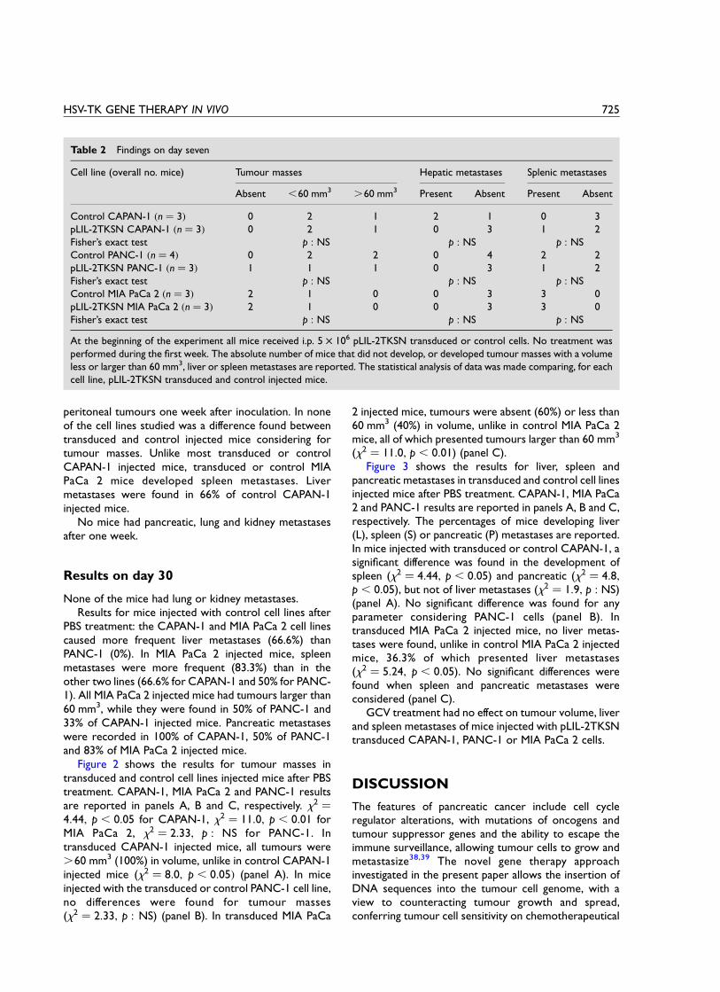
peritoneal tumours one week after inoculation. In noneof the cell lines studied was a difference found betweentransduced and control injected mice considering fortumour masses. Unlike most transduced or controlCAPAN-1 injected mice, transduced or control MIAPaCa 2 mice developed spleen metastases. Livermetastases were found in 66% of control CAPAN-1injected mice.
No mice had pancreatic, lung and kidney metastasesafter one week.
Results on day 30
None of the mice had lung or kidney metastases.Results for mice injected with control cell lines after
PBS treatment: the CAPAN-1 and MIA PaCa 2 cell linescaused more frequent liver metastases (66.6%) thanPANC-1 (0%). In MIA PaCa 2 injected mice, spleenmetastases were more frequent (83.3%) than in theother two lines (66.6% for CAPAN-1 and 50% for PANC-1). All MIA PaCa 2 injected mice had tumours larger than60 mm3, while they were found in 50% of PANC-1 and33% of CAPAN-1 injected mice. Pancreatic metastaseswere recorded in 100% of CAPAN-1, 50% of PANC-1and 83% of MIA PaCa 2 injected mice.
Figure 2 shows the results for tumour masses intransduced and control cell lines injected mice after PBStreatment. CAPAN-1, MIA PaCa 2 and PANC-1 resultsare reported in panels A, B and C, respectively. x2 ¼
4:44; p , 0:05 for CAPAN-1, x2 ¼ 11:0; p , 0:01 forMIA PaCa 2, x2 ¼ 2:33; p : NS for PANC-1. Intransduced CAPAN-1 injected mice, all tumours were.60 mm3 (100%) in volume, unlike in control CAPAN-1injected mice (x2 ¼ 8:0; p , 0:05Þ (panel A). In miceinjected with the transduced or control PANC-1 cell line,no differences were found for tumour masses(x2 ¼ 2:33; p : NS) (panel B). In transduced MIA PaCa
2 injected mice, tumours were absent (60%) or less than60 mm3 (40%) in volume, unlike in control MIA PaCa 2mice, all of which presented tumours larger than 60 mm3
(x2 ¼ 11:0; p , 0:01) (panel C).Figure 3 shows the results for liver, spleen and
pancreatic metastases in transduced and control cell linesinjected mice after PBS treatment. CAPAN-1, MIA PaCa2 and PANC-1 results are reported in panels A, B and C,respectively. The percentages of mice developing liver(L), spleen (S) or pancreatic (P) metastases are reported.In mice injected with transduced or control CAPAN-1, asignificant difference was found in the development ofspleen (x2 ¼ 4:44; p , 0:05) and pancreatic (x2 ¼ 4:8;p , 0:05), but not of liver metastases (x2 ¼ 1:9; p : NS)(panel A). No significant difference was found for anyparameter considering PANC-1 cells (panel B). Intransduced MIA PaCa 2 injected mice, no liver metas-tases were found, unlike in control MIA PaCa 2 injectedmice, 36.3% of which presented liver metastases(x2 ¼ 5:24; p , 0:05). No significant differences werefound when spleen and pancreatic metastases wereconsidered (panel C).
GCV treatment had no effect on tumour volume, liverand spleen metastases of mice injected with pLIL-2TKSNtransduced CAPAN-1, PANC-1 or MIA PaCa 2 cells.
DISCUSSION
The features of pancreatic cancer include cell cycleregulator alterations, with mutations of oncogens andtumour suppressor genes and the ability to escape theimmune surveillance, allowing tumour cells to grow andmetastasize38,39 The novel gene therapy approachinvestigated in the present paper allows the insertion ofDNA sequences into the tumour cell genome, with aview to counteracting tumour growth and spread,conferring tumour cell sensitivity on chemotherapeutical
Table 2 Findings on day seven
Cell line (overall no. mice) Tumour masses Hepatic metastases Splenic metastases
Absent ,60 mm3 .60 mm3 Present Absent Present Absent
Control CAPAN-1 ðn ¼ 3Þ 0 2 1 2 1 0 3pLIL-2TKSN CAPAN-1 ðn ¼ 3Þ 0 2 1 0 3 1 2Fisher’s exact test p : NS p : NS p : NSControl PANC-1 ðn ¼ 4Þ 0 2 2 0 4 2 2pLIL-2TKSN PANC-1 ðn ¼ 3Þ 1 1 1 0 3 1 2Fisher’s exact test p : NS p : NS p : NSControl MIA PaCa 2 ðn ¼ 3Þ 2 1 0 0 3 3 0pLIL-2TKSN MIA PaCa 2 ðn ¼ 3Þ 2 1 0 0 3 3 0Fisher’s exact test p : NS p : NS p : NS
At the beginning of the experiment all mice received i.p. 5 £ 106 pLIL-2TKSN transduced or control cells. No treatment wasperformed during the first week. The absolute number of mice that did not develop, or developed tumour masses with a volumeless or larger than 60 mm3, liver or spleen metastases are reported. The statistical analysis of data was made comparing, for eachcell line, pLIL-2TKSN transduced and control injected mice.
HSV-TK GENE THERAPY IN VIVO 725

Figure 2 Results at one month for tumour masses in pLIL-2TKSN transduced and control injected mice after PBS treatment.
P. FOGAR ET AL.726

agents and/or enhancing the host immune response. Intheory, a gene construct consisting of more than onegene, and acting on different targets, should have anenhanced therapueutic potential. We verified in vivo theeffects of a gene construct consisting of the suicide geneHSV-TK and the immunostimulatory cytokine, IL2, which
was retrovirally transduced into three pancreatic cancerderived cell lines with different metastatic potentials inorder to cover, at least in part, the broad spectrum ofbiological variability of this tumour type. A vectorexpressing only IL2 was also tested in vitro in one ofthese lines, MIA PaCa 2. The in vitro growth patterns of
Figure 3 Results at one month for hepatic (L), splenic (S) and pancreatic (P) metastases in pLIL-2TKSN-transduced and controlinjected mice after PBS treatment. Fisher’s exact test: * ¼ p , 0:05.
HSV-TK GENE THERAPY IN VIVO 727

MIA PaCa 2 non-transduced and transduced with thetwo vectors were quite similar, although the twotransduced lines exerted growth that was slightly fasterthan that in control line (Fig. 1). Furthermore, the twotransduced lines released similar amounts of IL2 inculture media. This suggests that the presence of theHSV-TK gene in the vector does not interfere with IL2transcription and translation.
HSV-TK, a suicide gene, causes transfected cells to behighly sensitive to the pro-drug, GCV.5,8,28 In severaltumour types, including pancreatic cancer, the HSV-TKgene inhibits cellular growth after GCV treatment invitro,12,17,25,40 although this effect cannot be reproducedin all pancreatic cancer derived cell lines.11,19–21,40 Acontroversial issue is the transferral of in vitro data into invivo applications.34,41 SCID mice were used in order toobviate any immunological response to foreign cells.Before inoculation in mice, used RT-PCR to ascertainwhether the new transduced TK gene was correctlytranslated into the corresponding mRNA. The resultsobtained indicate that all transduced lines expressed thesuicide gene and should, therefore, be sensitive to GCV.
All mice were i.p. inoculated with pLIL-2TKSNtransduced and control cell lines. Nineteen mice weresacrificed after one week, before any treatment, toevaluate: (1) the starting tumour mass and its spread; and(2) any differences between transduced and controlinjected mice. No differences were observed, and thisindicates that early in vivo growth and spread are notinfluenced by HSV-TK or by IL2, co-expressed by ourvector. On considering the different pancreatic cancercell lines, differences in tumour growth pattern clearlyemerged just after one week: in all mice, the metastasisderived CAPAN-1 line produced evident tumournodules, while the primary line MIA PaCa 2 causedtumour onset in only a few mice (2/6). An intermediatepattern was observed when PANC-1 cells were inocu-lated. The spleen was rapidly colonized by MIA PaCa 2(all mice) and PANC-1 (3/7 mice), but not by CAPAN-1(1/6), which, however, rapidly colonized the liver (2/6mice). In all injected mice, the pancreas was metastasesfree. The above findings may have depended on differentcell lines causing different tumour types due to theirinherent characteristics: the high hepatic tropism inCAPAN-1 cells may have depended on their human pre-selection, since they were isolated from liver metastasesfrom pancreatic cancer. The high splenic tropism of MIAPaCa 2, and the high hepatic tropism of CAPAN-1 cellswere confirmed at the end of experiments consideringcontrol cells. Furthermore, the liver was targeted by MIAPaCa 2 cells while in no cases did PANC-1 metastasize tothe liver. The similarity between the CAPAN-1 and MIAPaCa 2 metastatic patterns was further observed onconsidering pancreatic involvement at the end of theexperimental protocol, which was recorded in none ofthe CAPAN-1 injected mice, and in only 1/6 MIA PaCa 2
injected mice, while it was the target in 50% of PANC-1injected mice.
Interestingly, our pLIL-2TKSN vector modified ‘perse’ the behavior of MIA PaCa 2 and CAPAN-1 transducedpancreatic cancer cell lines 30 days after i.p. inoculation:on comparing tumour growth and spread in miceinoculated with transduced pancreatic cancer cell linesand saline treated with patterns in mice inoculated withcontrol cell lines, we found: (1) smaller tumour volumesin mice injected with transduced MIA PaCa 2 cells, unlikein transduced CAPAN-1 injected mice; (2) increasedpancreatic and decreased spleen metastases fromtransduced CAPAN-1, and diminished liver involvementfrom transduced MIA PaCa 2. No differences were foundbetween mice inoculated with transduced and thoseinoculated with control PANC-1 cell lines. Since theseeffects were correlated with the vector itself, rather thanwith the sensitivity conferred by it on GCV, the aboveeffects probably depended on IL2. This cytokine is knownto stimulate the specific immune response in immuno-competent animals, but it also causes non-specificactivation of the macrophages as well as the naturalkiller cells, both of which are borne by SCID mice.29–32
Our results can be explained only in part by IL2 activationof these cells. If IL2 activation of macrophages and naturalkiller cells had an anti-neoplastic effect in SCID mice, allpLIL-2TKSN injected mice are expected to have smallertumours and fewer metastases than control injectedmice. As this was not the case, we believe that IL2probably acts directly in an autocrine manner on tumourcells, possibly evoking a different response depending onthe tumour cell status, as suggested by other authors onanalysing the effects of TGF-b1.42,43 This responsepattern, however, does not seem to comprise cellgrowth, which was unaffected in vitro (Fig. 3).
HSV-TK did not lead to cellular death after GCVtreatment. None of the analysed parameters (tumourvolume, liver, spleen and pancreatic metastases) weremodified after GCV treatment in pLIL-2TKSN cellinjected mice. The HSV-TK gene cannot, therefore, beconsidered a suicide gene for pancreatic cancer, probablybecause pancreatic cancer cells can escape the lethalconsequences of GCV, possibly by rapidly metabolizingthis compound. This pro-drug is, in fact, first phosphory-lated by the HSV-TK enzyme. The resultant GCV-monophosphate is subsequently converted by cellularkinases into the toxic GCV-triphosphate nucleotide, asubstrate for DNA polymerase. Any alteration in cellularkinases, often found in tumour cells, might compromiseGCV activation, and therefore, disable any HSV-TKsuicide effect.19–21
In conclusion, in vivo suicide gene therapy with HSV-TK for pancreatic cancer has failed to meet expectations.A search should, therefore, be made for alternativetherapeutic genes in the attempt to cure this disease.
P. FOGAR ET AL.728

ACKNOWLEDGEMENTS
This study was supported by the Ministero Universita eRicerca (Cofin 2001068593), Rome, Italy.
REFERENCES
1. Greenlee RT, Murray T, Bolden S, Wingo PA. Cancer statistics.
Cancer J Clin 2000; 50: 7–33.
2. Sperti C, Pasquali C, Piccoli A, Pedrazzoli S. Recurrence after
resection for ductal adenocarcinoma of the pancreas. World J Surg
1997; 21: 195–200.
3. Miller AD. Human gene therapy comes of age. Nature 1992; 357:
455–60.
4. Zhang J, Russell SJ. Vectors for cancer gene therapy. Cancer
Metastasis Rev 1996; 15: 385–401.
5. Rigg AS, Scarpa A, Pandha HS, Lemoine NR. Gene therapy for
pancreatic cancer. Ital J Gastroenterol Hepatol 1998; 30: 462–6.
6. Rosenberg L. Pancreatic cancer. a review of emerging therapies.
Drugs 2000; 59: 1071–89.
7. Danthinne X, Imperiale MJ. Production of first generation
adenovirus vectors: a review. Gene Ther 2000; 7: 1707–14.
8. Moolten FL. Drug sensitivity (‘suicide’) genes for selective cancer
chemotherapy. Cancer Gene Ther 1994; 1: 279–87.
9. Black ME, Newcomb TG, Wilson HM, Loeb LA. Creation of drug-
specific herpes simplex virus type 1 thymidine kinase mutants for
gene therapy. Proc Natl Acad Sci U S A 1996; 93: 3525–9.
10. Yang L, Hwang R, Pandit L, Gordon EM, Anderson WF, Parekh D.
Gene therapy of metastatic pancreas cancer with intraperitoneal
injections of concentrated retroviral herpes simplex thymidine
kinase vector supernatant and ganciclovir. Ann Surg 1996; 224:
405–17.
11. Kimura M, Tagawa M, Takenaga K et al. Drug resistance to
ganciclovir observed in suicide gene therapy is due to the loss of
integrated herpes simplex virus-thymidine kinase gene. Int J Oncol
1997; 10: 775–8.
12. Faulds D, Rennie CH. Ganciclovir. A review of its antiviral activity,
pharmacokinetic properties and therapeutic efficacy in cytomega-
lovirus infection. Drugs 1990; 39: 567–638.
13. Aoki K, Yoshida T, Matsumoto N et al. Gene therapy for peritoneal
dissemination of pancreatic cancer by liposome-mediated transfer
of herpes simplex virus thymidine kinase gene. Hum Gene Ther
1997; 8: 1105–13.
14. Grignet-Debrus C, Calberg-Bacq C-M. Potential of varicella zoster
virus thymidine kinase as a suicide gene in breast cancer cells. Gene
Ther 1997; 4: 560–9.
15. Block A, Chen SH, Kosai K, Finegold M, Woo SL. Adenoviral-
mediated herpes simplex virus thymidine kinase gene transfer:
regression of hepatic metastasis of pancreatic tumors. Pancreas
1997; 15: 25–34.
16. Mizuno M, Yoshida J, Colosi P, Kurtzman G. Adeno-associated virus
vector containing the herpes simplex virus thymidine kinase gene
causes complete regression of intracerebrally implanted human
gliomas in mice, in conjunction with ganciclovir administration. Jpn J
Cancer Res 1998; 89: 76–80.
17. Hall SJ, Mutchnik S, Yang G et al. Cooperative therapeutic effects of
androgen ablation and adenovirus-mediated herpes simplex virus
thymidine kinase gene and ganciclovir therapy in experimental
prostate cancer. Cancer Gene Ther 1999; 6: 54–63.
18. Shalev M, Kadmon D, Teh B et al. Suicide gene therapy toxicity after
multiple and repeat injections in patients with localized prostate
cancer. J Urol 2000; 163: 1747–50.
19. Beck C, Cayeux S, Lupton SD, Dorken B, Blonkenstein T. The
thymidine kinase/ganciclovir-mediated ‘suicide’ effect is variable in
different tumor cells. Hum Gene Ther 1995; 6: 1525–30.
20. Yang L, Hwang R, Chiang Y, Gordon EM, Anderson WF, Parekh D.
Mechanisms for ganciclovir resistance in gastrointestinal tumor
cells transduced with a retroviral vector containing the herpes
simplex virus thymidine kinase gene. Clin Cancer Res 1998; 4:
731–41.
21. Boucher PD, Ruch RJ, Shewach DS. Differential ganciclovir-
mediated cytotoxicity and bystander killing in human colon
carcinoma cell lines expressing herpes simplex virus thymidine
kinase. Hum Gene Ther 1998; 9: 801–14.
22. Elshami AA, Saavedra A, Zhang H et al. Gap junction plays a role in
the ‘bystander effect’ of the herpes simplex virus thymidine kinase/
ganciclovir system in vitro. Gene Ther 1996; 3: 85–92.
23. Kumar NM, Gilula NB. The gap junction communication channel.
Cell 1996; 84: 381–8.
24. Mesnil M, Yamasaki H. Bystander effects in herpes simplex virus-
thymidine kinase/ganciclovir cancer gene therapy: role of gap-
junctional intercellular communication. Cancer Res 2000; 60:
3989–99.
25. Qiao J, Black ME, Caruso M. Enhanced ganciclovir killing and
bystander effect of human tumor cells transduced with a retroviral
vector carrying a herpes simplex virus thymidine kinase gene
mutant. Hum Gene Ther 2000; 11: 1569–76.
26. Carrio M, Mazo A, Lopez-Iglesias C, Estivill X, Fillat C. Retrovirus-
mediated transfer of the herpes simplex virus thymidine kinase and
connexin26 genes in pancreatic cells. Results in variable efficiency
on the bystander killing: implications for gene therapy. Int J Cancer
2001; 94: 81–8.
27. Humphreys MJ, Greenhalf W, Neoptolemos JP, Ghaneh P. The
potential for gene therapy in pancreatic cancer. Int J Pancreatol 1999;
26: 5–21.
28. Gilliam AD, Watson SA. Emerging biological therapies for
pancreatic carcinoma. EJSO 2002; 28: 370–8.
29. Clary BM, Coveney EC, Philip R et al. Inhibition of established
pancreatic cancers following specific active immunotherapy with
interleukin-2 gene transduced tumour cells. Cancer Gene Ther 1997;
4: 97–104.
30. Kimura M, Tagawa M, Takenaga K et al. Loss of tumorigenicity of
human pancreatic carcinoma cells engineered to produce inter-
leukin-2 or interleukin-4 in nude mice: a potentiality for cancer
gene therapy. Cancer Lett 1998; 128: 47–53.
31. Kimura M, Yoshida Y, Narita M et al. Acquired immunity in
nude mice induced by expression of the IL-2 or IL-4 gene in
human pancreatic carcinoma cells and anti-tumor effect generated
by in vivo gene transfer using retrovirus. Int J Cancer 1999; 82:
549–55.
32. D’Angelica M, Karpoff H, Halterman M et al. In vivo
interleukin-2 gene therapy of established tumors with herpes
simplex amplicon vectors. Cancer Immunol Immunother 1999; 47:
265–71.
33. Pizzato M, Franchin E, Calvi P et al. Production and characterization
of a bicistronic Moloney-based retroviral expressing human
interleukin 2 and herpes simplex virus thymidine kinase for gene
therapy of cancer. Gene Ther 1998; 5: 1003–7.
34. Carrio M, Romagosa A, Mercade E et al. Enhanced pancreatic tumor
regression by a combination of adenovirus and retrovirus-mediated
delivery of herpes simplex virus thymidine kinase gene. Gene Ther
1999; 6: 547–53.
35. Evoy D, Hirschowitz EA, Naama Ha et al. In vivo adenoviral-
mediated gene transfer in the treatment of pancreatic cancer. J Surg
Res 1997; 69: 226–31.
36. Shirakawa T, Gardner TA, Ko S-C et al. Cytotoxicity of adenoviral-
mediated cytosine deaminase plus 5-fluorocytosine gene therapy is
superior to thymidine kinase plus acyclovir in a human renal cell
carcinoma model. J Urol 1999; 162: 949–54.
37. Erbs P, Regulier E, Kintz J et al. In vivo cancer gene therapy by
adenovirus-mediated transfer of a bifunctional yeast cytosine
HSV-TK GENE THERAPY IN VIVO 729

deaminase/uracil phosphoribosyltransferase fusion gene. Cancer Res
2000; 60: 3813–22.
38. Bardeesy N, DePinho RA. Pancreatic cancer biology and genetics.
Nat Rev Cancer 2002; 2: 897–909.
39. Westphal S, Kalthoff H. Apoptosis: targets in pancreatic cancer. Mol
Cancer 2003; 2: 6.
40. Greco E, Fogar P, Basso D et al. Retrovirus-mediated herpes
simplex virus thymidine kinase gene transfer in pancreatic cancer
cell lines: an incomplete antitumor effect. Pancreas 2002; 25(2):
21–9.
41. Schwarz RE, McCarty TM, Peralta EA, Diamond DJ, Ellenhorn JDI.
An orthotopic in vivo model of human pancreatic cancer. Surgery
1999; 126: 562–7.
42. Derynck R, Akhurst RJ, Balmain A. TGF-b signaling in tumor
suppression and cancer progression. Nat Genet 2001; 29: 117–29.
43. Wakefield LM, Roberts AB. TGF-b signaling: positive and
negative effects on tumorigenesis. Curr Opin Genet Dev 2002; 12:
22–9.
Accepted for publication 18 August 2003
P. FOGAR ET AL.730