Imaging of lipids in rat heart by MALDI-MS with silver nanoparticles
Upload
independentCategory
view
1download
0
© 2001 Oxford University Press Nucleic Acids Research, 2001, Vol. 29, No. 19 e91
Sugar additives for MALDI matrices improve signalallowing the smallest nucleotide change (A:T) in a DNAsequence to be resolvedMona Shahgholi1, Benjamin A. Garcia1, Norman H. L. Chiu1,2, Paul J. Heaney1 andKai Tang1,*
1Sequenom Inc., 3595 John Hopkins Court, San Diego, CA 92121, USA and 2Center for Advanced Biotechnology,Boston University, Boston, MA 02215, USA
Received May 1, 2001; Revised July 15, 2001; Accepted August 1, 2001
ABSTRACT
Sample preparation for matrix-assisted laser desorp-tion/ionization (MALDI) mass spectrometry (MS) ofDNA is critical for obtaining high quality massspectra. Sample impurity, solvent content, substratesurface and environmental conditions (temperatureand humidity) all affect the rate of matrix–analyte co-crystallization. As a result, laser fluence thresholdfor desorption/ionization varies from spot to spot.When using 3-hydroxypicolinic acid (3-HPA) as thematrix, laser fluence higher than the threshold valuereduces mass resolution in time-of-flight (TOF) MSas the excess energy transferred to DNA causesmetastable decay. This can be overcome by eithersearching for ‘hot’ spots or adjusting the laserfluence. However, both solutions may require asignificant amount of operator manipulation and arenot ideal for automatic measurements. We haveadded various sugars for crystallization with thematrix to minimize the transfer of excess laserenergy to DNA molecules. Fructose and fucose werefound to be the most effective matrix additives. Usingthese additives, mass resolution for DNA moleculesdoes not show noticeable deterioration as laserenergy increases. Improved sample preparation isimportant for the detection of single nucleotide poly-morphisms (SNPs) using primer extension with asingle nucleotide. During automatic data acquisition itis difficult to routinely detect heterozygous A/T muta-tions, which requires resolving a mass difference of9 Da, unless a sugar is added during crystallization.
INTRODUCTION
The success of matrix-assisted laser desorption/ionization(MALDI) in analysis of oligonucleotides has introduced adirect procedure for examination of products of molecularbiology assays. The specificity and ‘softness’ of MALDIcombined with the simplicity and speed of time-of-flight (TOF)
mass spectrometers has been particularly attractive togenomics endeavors where both reliability and throughput arecritical. For example, MALDI has been used to detect PCRproducts for deletion/insertion (1,2) and for microsatelliteanalyses (3–5), to detect sequencing ladders (6–9) and to detectprimer extension products for polymorphism analyses (10–13).Despite its success, continued efforts are being made toenhance sensitivity and resolution of oligonucleotides either bynew instrument design (14–17) or by innovative MALDIsample preparation (18–26).
Improved sensitivity is important in achieving lower limitsof detection, e.g. for multiplexed assays where product yieldsmay not be high for all variable sites on the DNA template.Sample miniaturization was previously shown to directlyinfluence detection sensitivity (20,25). The miniaturizedmatrix spot size concentrates analyte molecules in a muchsmaller region, thus increasing the amount of analyte in thelaser interrogation area. As a result, MALDI-mass spectro-metry (MS) detection sensitivity can be dramatically improved.
Improved resolution is highly desirable in assays wheremasses of diagnostic DNA products are very close or partiallyoverlap (single base extension and multiplex assays) and inlinear TOF mass spectrometers, where achievable resolution ismodest. A variety of factors can lead to degradation of resolu-tion in MALDI-MS analysis of nucleic acids. Most notable areformation of salt adducts with sodium or potassium ions andfragmentation of the molecular ion during MALDI ionization.Formation of cation adducts depends on oligonucleotidepreparation, namely the effectiveness of the cleaning step,while DNA fragmentation during MALDI is largely dependenton the nature of the matrix used. The use of cation exchangebeads (27) or ammonium salts (28–31) has proven to be veryadvantageous in decreasing the amount of salt adducts withDNA. Similarly, addition of basic organic species (32–34) toDNA samples has been somewhat effective in decreasing saltadducts and hence in increasing mass resolution. DNA frag-mentation during MALDI results from excess energy depositedduring laser irradiation. Some matrices are referred to as ‘hot’if they result in excessive fragmentation, e.g. 2,4,6-trihydroxy-acetophenone (29,35,36), while others are ‘cool’, e.g. 3-hydroxy-picolinic acid (3-HPA) (37) and 6-aza-2-thiothymine (38), andresult in very little fragmentation. The internal energy of the
*To whom correspondence should be addressed: Tel: +1 858 202 9089; Fax: +1 858 202 9001; Email: [email protected] address:Mona Shahgholi, Department of Chemistry, California Institute of Technology, 1200 East California Boulevard, Pasadena, CA 91125, USA
e91 Nucleic Acids Research, 2001, Vol. 29, No. 19 PAGE 2 OF 10
analyte ions may be reduced by collision with an inert gas as inan ion trap (39) or Fourier transform ion cyclotron resonance(FT-ICR) cell (16,40). Addition of a sugar to the matrix mayalso have a ‘cooling’ effect. Sugars have been reported toenhance ionization and/or to reduce metastable fragmentation ofions produced by MALDI. For example, Beavis et al. (41) usedsucrose and glucose as a matrix for the multi-photon ionizationof dipeptides in a reflectron TOF mass spectrometer. The useof sugars suppressed fragment ions resulting from samplepyrolysis due to IR laser irradiation.
Wilkins’ (42,43) research group used sugars as additives inMALDI ionization of peptides and small proteins. In theirexperiment, a mixture of analyte and 2,5-dihydroxybenzoicacid matrix in methanol/water/TFA was saturated with aselected sugar and air sprayed onto the probe tip of a 7 T FT-ICRmass spectrometer. The sugar additives in conjunction withoptimization of electrostatic deceleration times and laserenergies allowed successful coupling of the energetic MALDIions to a FT-ICR mass spectrometer. Further experimentsdemonstrated that sugar additives enhanced the observedresolution, presumably by slowing metastable decay of themolecular ions and providing the requisite population of long-lived ions (43). Recently, Russell and co-workers (26) showedthat addition of fructose to the MALDI matrix reduced theinternal energies of desorbed DNA ions, reduced fragment ionsand increased the yield of molecular ions analyzed in aMALDI-TOF mass spectrometer operating in reflected ionmode. The matrices tested consisted of α-cyano-4-hydroxycin-namic acid, 2,4,6-trihydroxyacetophenone and ferulic acidprepared as dried droplets and a novel fast evaporation-over-layer method.
In this report we describe the effect of sugar additives on theMALDI-TOF spectra of synthetic oligonucleotides as well asextension products of the single base extension assay (11–12),which is used for single nucleotide polymorphism (SNP)analysis. We are specifically interested in the effect of thesugar additive on the resolution of oligonucleotides for appli-cation to single base extension assays. In these assays, a primeris annealed upstream of the variable (mutation) site in the DNAsequence and is extended up to the mutation site by apolymerase in the presence of all four dideoxynucleosidetriphosphates. Masses of the extension products are diagnosticof the base added to the variable site. Homozygous samplesproduce one diagnostic product while heterozygous samplesproduce two, one for each allele. The smallest mass differencebetween the extension products is 9 Da, corresponding to thedifference between dideoxyadenosine (ddA) and dideoxythy-midine (ddT). This mass difference is not easily resolvablewith commercial linear TOF instrumentation, especially inautomated analyses where an operator is not searching for ‘hotspots’ in the sample. Currently, in our hands, resolution valuesof 500–700 can be routinely achieved for DNA oligomers inthe 4–8 kDa mass range during fully automated runs where 20shots are acquired at 10 or 20 Hz. This resolution, althoughsatisfactory for Mass Extend reactions (10,13) where the massdifference is at least 1 nt, is limiting for single base extensionassays of A/T heterozygotes.
Our investigations started with screening of a number ofsugars for their resolution-enhancing ability by comparing theresolution of synthetic DNA oligomers in mass spectraacquired under threshold (standard) and high laser intensity
conditions from control and sugar-containing matrix samples.After suitable sugars were chosen, sample preparation wasoptimized for both manual and automatic modes of data acqui-sition. The utility of the sugar additive was further explored forresolving synthetic oligonucleotides (differing in mass by only8 Da) in a clean background and subsequently put to test withactual single base extension assay reaction products differingin mass by 9 Da (A/T heterozygotes) in a real background ofenzymes and buffers analyzed in both manual and automaticdata acquisition modes. The results of these experiments arediscussed below.
MATERIALS AND METHODS
Sample preparation
The sugars D(+)-trehalose, D(–)-ribose, N-acetyl-D-glucos-amine, D(+)-glucosamine, D(+)-raffinose pentahydrate, α-cyclo-dextrin and stachyose hydrate were purchased from Fluka(Milwaukee, WI). L-Fucose and D-fructose were purchasedfrom Aldrich Chemicals (Milwaukee, WI) and D(+)-glucoseand sucrose were purchased from Sigma Chemical Co. (StLouis, MO). Individual sugar solutions were prepared indeionized water at concentrations ranging from 1.0 to 6.0 g/l.To remove residual Na+ and K+ ions from the sugar solutions,cation exchange columns were used. Each cation exchangecolumn was prepared by packing a Fortuna 20 ml syringe,lined at the bottom with a Whatman 6 filter paper, with a 50 g/lslurry of Bio-Rad AG 50W-X8 hydrogen form cationexchange resin. Before use, the columns were conditioned with1 M ammonium acetate solution.
A synthetic 28mer oligonucleotide (5′-CCA TCC ACT ACAACT ACA TGT GTA ACA G-3′, [M + H]+ 8486.6 Da) waspurchased from Operon Technologies (Alameda, CA). Asynthetic 23mer oligonucleotide (5′-GTT TCC ATT TAGTCA GTC AAC TG-3′, [M + H]+ 7004.6 Da) was purchasedfrom Integrated DNA Technologies (Coralville, IA). A secondsynthetic 23mer oligonucleotide (5′-ACA TTC TTC ATAGCA TTT TAG A*A-3′, * = ribose, [M + H]+ 7012.6 Da) waspurchased from Tri Link Bio Tech (San Diego, CA). A stocksolution of each oligonucleotide was prepared by dissolvingthe oligonucleotide in 1× TE buffer (10 mM Tris–HCl, pH 8.0,and 1 mM EDTA) to a final concentration of 100 µM, and waskept at –20°C. Working solutions of the oligonucleotides weremade by diluting the stock solutions with deionized water to aconcentration of 10 µM. A mixture of the two 23mer oligonu-cleotides was prepared by diluting the oligonucleotides to0.63 µM with either deionized water or a selected sugar solu-tion (6 g/l). About 5 µg of cation exchange resin equilibratedwith ammonium ions was added to the oligonucleotide mixtureto prevent the formation of any Na+/K+ adducts.
Detection of SNP with single nucleotide extension
A selected region of human genomic DNA, namely TissueFactor (TF), was first amplified using PCR. Genomic DNAwas obtained from the Sequenom DNA banking facility. DNAsamples were first amplified by PCR in a 50 µl volumecontaining 3 pmol reverse primer tagged with universalsequence at the 5′-end (13) (5′-AGC GGA TAA CAA TTTCAC ACA GGT TAT AAC TTG ACC GGG AAG AG-3′),25 pmol forward primer (5′-CAC GTG TGG GAT TTA TCT
PAGE 3 OF 10 Nucleic Acids Research, 2001, Vol. 29, No. 19 e91
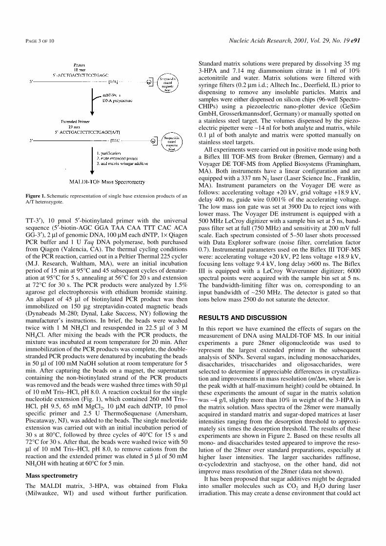
TT-3′), 10 pmol 5′-biotinylated primer with the universalsequence (5′-biotin-AGC GGA TAA CAA TTT CAC ACAGG-3′), 2 µl of genomic DNA, 100 µM each dNTP, 1× QiagenPCR buffer and 1 U Taq DNA polymerase, both purchasedfrom Qiagen (Valencia, CA). The thermal cycling conditionsof the PCR reaction, carried out in a Peltier Thermal 225 cycler(M.J. Research, Waltham, MA), were an initial incubationperiod of 15 min at 95°C and 45 subsequent cycles of denatur-ation at 95°C for 5 s, annealing at 56°C for 20 s and extensionat 72°C for 30 s. The PCR products were analyzed by 1.5%agarose gel electrophoresis with ethidium bromide staining.An aliquot of 45 µl of biotinylated PCR product was thenimmobilized on 150 µg streptavidin-coated magnetic beads(Dynabeads M-280; Dynal, Lake Success, NY) following themanufacturer’s instructions. In brief, the beads were washedtwice with 1 M NH4Cl and resuspended in 22.5 µl of 3 MNH4Cl. After mixing the beads with the PCR products, themixture was incubated at room temperature for 20 min. Afterimmobilization of the PCR products was complete, the double-stranded PCR products were denatured by incubating the beadsin 50 µl of 100 mM NaOH solution at room temperature for 5min. After capturing the beads on a magnet, the supernatantcontaining the non-biotinylated strand of the PCR productswas removed and the beads were washed three times with 50 µlof 10 mM Tris–HCl, pH 8.0. A reaction cocktail for the singlenucleotide extension (Fig. 1), which contained 260 mM Tris–HCl, pH 9.5, 65 mM MgCl2, 10 µM each ddNTP, 10 pmolspecific primer and 2.5 U ThermoSequenase (Amersham,Piscataway, NJ), was added to the beads. The single nucleotideextension was carried out with an initial incubation period of30 s at 80°C, followed by three cycles of 40°C for 15 s and72°C for 30 s. After that, the beads were washed twice with 50µl of 10 mM Tris–HCl, pH 8.0, to remove cations from thereaction and the extended primer was eluted in 5 µl of 50 mMNH4OH with heating at 60°C for 5 min.
Mass spectrometry
The MALDI matrix, 3-HPA, was obtained from Fluka(Milwaukee, WI) and used without further purification.
Standard matrix solutions were prepared by dissolving 35 mg3-HPA and 7.14 mg diammonium citrate in 1 ml of 10%acetonitrile and water. Matrix solutions were filtered withsyringe filters (0.2 µm i.d.; Alltech Inc., Deerfield, IL) prior todispensing to remove any insoluble particles. Matrix andsamples were either dispensed on silicon chips (96-well Spectro-CHIPs) using a piezoelectric nano-plotter device (GeSimGmbH, Grosserkmannsdorf, Germany) or manually spotted ona stainless steel target. The volumes dispensed by the piezo-electric pipetter were ∼14 nl for both analyte and matrix, while0.1 µl of both analyte and matrix were spotted manually onstainless steel targets.
All experiments were carried out in positive mode using botha Biflex III TOF-MS from Bruker (Bremen, Germany) and aVoyager DE TOF-MS from Applied Biosystems (Framingham,MA). Both instruments have a linear configuration and areequipped with a 337 nm N2 laser (Laser Science Inc., Franklin,MA). Instrument parameters on the Voyager DE were asfollows: accelerating voltage +20 kV, grid voltage +18.9 kV,delay 400 ns, guide wire 0.001% of the accelerating voltage.The low mass ion gate was set at 3900 Da to reject ions withlower mass. The Voyager DE instrument is equipped with a500 MHz LeCroy digitizer with a sample bin set at 5 ns, band-pass filter set at full (750 MHz) and sensitivity at 200 mV fullscale. Each spectrum consisted of 5–50 laser shots processedwith Data Explorer software (noise filter, correlation factor0.7). Instrumental parameters used on the Biflex III TOF-MSwere: accelerating voltage +20 kV, P2 lens voltage +18.9 kV,focusing lens voltage 9.4 kV, long delay >600 ns. The BiflexIII is equipped with a LeCroy Waverunner digitizer; 6000spectral points were acquired with the sample bin set at 5 ns.The bandwidth-limiting filter was on, corresponding to aninput bandwidth of ∼250 MHz. The detector is gated so thations below mass 2500 do not saturate the detector.
RESULTS AND DISCUSSION
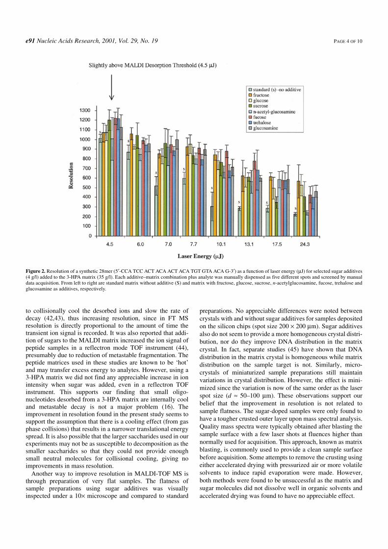
In this report we have examined the effects of sugars on themeasurement of DNA using MALDI-TOF MS. In our initialexperiments a pure 28mer oligonucleotide was used torepresent the largest extended primer in the subsequentanalysis of SNPs. Several sugars, including monosaccharides,disaccharides, trisaccharides and oligosaccharides, wereselected to determine if appreciable differences in crystalliza-tion and improvements in mass resolution (m/∆m, where ∆m isthe peak width at half-maximum height) could be obtained. Inthese experiments the amount of sugar in the matrix solutionwas ∼4 g/l, slightly more than 10% in weight of the 3-HPA inthe matrix solution. Mass spectra of the 28mer were manuallyacquired in standard matrix and sugar-doped matrices at laserintensities ranging from the desorption threshold to approxi-mately six times the desorption threshold. The results of theseexperiments are shown in Figure 2. Based on these results allmono- and disaccharides tested appeared to improve the reso-lution of the 28mer over standard preparations, especially athigher laser intensities. The larger saccharides raffinose,α-cyclodextrin and stachyose, on the other hand, did notimprove mass resolution of the 28mer (data not shown).
It has been proposed that sugar additives might be degradedinto smaller molecules such as CO2 and H2O during laserirradiation. This may create a dense environment that could act
Figure 1. Schematic representation of single base extension products of anA/T heterozygote.
e91 Nucleic Acids Research, 2001, Vol. 29, No. 19 PAGE 4 OF 10
to collisionally cool the desorbed ions and slow the rate ofdecay (42,43), thus increasing resolution, since in FT MSresolution is directly proportional to the amount of time thetransient ion signal is recorded. It was also reported that addi-tion of sugars to the MALDI matrix increased the ion signal ofpeptide samples in a reflectron mode TOF instrument (44),presumably due to reduction of metastable fragmentation. Thepeptide matrices used in these studies are known to be ‘hot’and may transfer excess energy to analytes. However, using a3-HPA matrix we did not find any appreciable increase in ionintensity when sugar was added, even in a reflectron TOFinstrument. This supports our finding that small oligo-nucleotides desorbed from a 3-HPA matrix are internally cooland metastable decay is not a major problem (16). Theimprovement in resolution found in the present study seems tosupport the assumption that there is a cooling effect (from gasphase collisions) that results in a narrower translational energyspread. It is also possible that the larger saccharides used in ourexperiments may not be as susceptible to decomposition as thesmaller saccharides so that they could not provide enoughsmall neutral molecules for collisional cooling, giving noimprovements in mass resolution.
Another way to improve resolution in MALDI-TOF MS isthrough preparation of very flat samples. The flatness ofsample preparations using sugar additives was visuallyinspected under a 10× microscope and compared to standard
preparations. No appreciable differences were noted betweencrystals with and without sugar additives for samples depositedon the silicon chips (spot size 200 × 200 µm). Sugar additivesalso do not seem to provide a more homogeneous crystal distri-bution, nor do they improve DNA distribution in the matrixcrystal. In fact, separate studies (45) have shown that DNAdistribution in the matrix crystal is homogeneous while matrixdistribution on the sample target is not. Similarly, micro-crystals of miniaturized sample preparations still maintainvariations in crystal distribution. However, the effect is mini-mized since the variation is now of the same order as the laserspot size (d ≈ 50–100 µm). These observations support ourbelief that the improvement in resolution is not related tosample flatness. The sugar-doped samples were only found tohave a tougher crusted outer layer upon mass spectral analysis.Quality mass spectra were typically obtained after blasting thesample surface with a few laser shots at fluences higher thannormally used for acquisition. This approach, known as matrixblasting, is commonly used to provide a clean sample surfacebefore acquisition. Some attempts to remove the crusting usingeither accelerated drying with pressurized air or more volatilesolvents to induce rapid evaporation were made. However,both methods were found to be unsuccessful as the matrix andsugar molecules did not dissolve well in organic solvents andaccelerated drying was found to have no appreciable effect.
Figure 2. Resolution of a synthetic 28mer (5′-CCA TCC ACT ACA ACT ACA TGT GTA ACA G-3′) as a function of laser energy (µJ) for selected sugar additives(4 g/l) added to the 3-HPA matrix (35 g/l). Each additive–matrix combination plus analyte was manually dispensed as five different spots and screened by manualdata acquisition. From left to right are standard matrix without additive (S) and matrix with fructose, glucose, sucrose, n-acetylglucosamine, fucose, trehalose andglucosamine as additives, respectively.
PAGE 5 OF 10 Nucleic Acids Research, 2001, Vol. 29, No. 19 e91
As noted in Figure 2, the highest resolution (>1000) wasobtained at a laser energy (4.5 µJ) slightly above the MALDIdesorption threshold in samples prepared with the sugar-dopedmatrix. In general, resolutions measured for the 28mer in thestandard matrix were lower than those obtained in the sugar-doped matrix. With the use of sugar additives, the enhancingeffect on resolution was more obvious at laser energies>13.1 µJ. Under these conditions the resolution was ∼400–800and signal-to-noise (S/N) ratios ranged from 50 to 90 for the28mer in the sugar-doped matrices, while the resolution wastypically <325 and the S/N ratio <50 in the standard matrix.Although most monosaccharides produced similar results, thehighest quality and most consistent mass spectra were obtainedwhen fructose or fucose was present in the matrix. Therefore,these two sugars were used exclusively in the followingstudies.
After the initial manual screening for suitable sugar addi-tives, the amounts of sugar added to the matrix solution werevaried in order to determine the effective sugar concentrationfor automatic data acquisition and to improve MALDI samplepreparation. These studies were performed in automatic acqui-sition mode where five shots were summed after an initialblasting of the sample surface with eight high intensity lasershots (10.1 µJ). In automated acquisition the spectral peaks areevaluated for absolute intensity, resolution and S/N ratio usingcommercial fuzzy logic software. After the spectrum is saved,the sample translation stage in the ion source brings anothersample spot into the focal point of the laser for interrogationand the process is repeated. It was found that high concentra-tions of sugar (>4 g/l) added to the matrix interfered with both
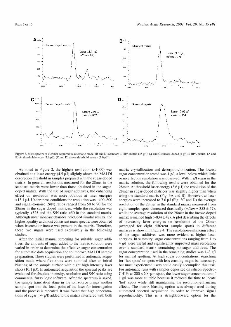
matrix crystallization and desorption/ionization. The lowestsugar concentration tested was 1 g/l, a level below which littleor no effect on resolution was observed. With 1 g/l sugar in thematrix solution, the following results were obtained for the28mer. At threshold laser energy (3.6 µJ) the resolution of the28mer in sugar-doped matrices was slightly higher than whenusing the standard matrix (Fig. 3A and B). However, as laserenergies were increased to 7.0 µJ (Fig. 3C and D) the averageresolution of the 28mer in the standard matrix measured fromeight samples spots decreased drastically (m/∆m ≈ 353 ± 57),while the average resolution of the 28mer in the fucose-dopedmatrix remained high (∼834 ± 42). A plot describing the effectsof increasing laser energies on resolution of the 28mer(averaged for eight different sample spots) in differentmatrices is shown in Figure 4. The resolution-enhancing effectof the sugar additives was more evident at higher laserenergies. In summary, sugar concentrations ranging from 1 to4 g/l were useful and significantly improved mass resolutionover a standard matrix containing no sugar additives. Thesugar concentration used in the remaining studies was 1–3 g/lfor manual spotting. At high sugar concentrations, searchingfor ‘hot spots’ or spots with less crusting might be necessary,but most experienced users could easily accomplish this task.For automatic runs with samples deposited on silicon Spectro-CHIPs as 200 × 200 µm spots, the lower sugar concentration of1 g/l was more suitable because it reduced the time to locate‘hot’ spots while still maintaining the resolution-enhancingeffects. The matrix blasting option was always used duringautomated spectral acquisition in order to improve signalreproducibility. This is a straightforward option for the
Figure 3. Mass spectra of a 28mer acquired in automatic mode. (B and D) Standard 3-HPA matrix (35 g/l); (A and C) fucose-doped (1 g/l) 3-HPA matrix. (A andB) At threshold energy (3.6 µJ); (C and D) above threshold energy (7.0 µJ).
e91 Nucleic Acids Research, 2001, Vol. 29, No. 19 PAGE 6 OF 10
automatic mode settings of commercial MALDI-TOF instru-ments and only adds a fraction of a second to each acquisition.For example, usually eight laser shots at high fluence were
used to blast the sample surface. At the laser repetition rate of20 Hz, matrix blasting takes only an additional 0.4 s, which isonly a fraction of the entire acquisition event, usually 3–4 s. Bycomparison, optimizing the laser energy for each sample spotduring an automated run requires between three and ten timesthe standard acquisition time because the software evaluatesseveral laser settings for each raster position.
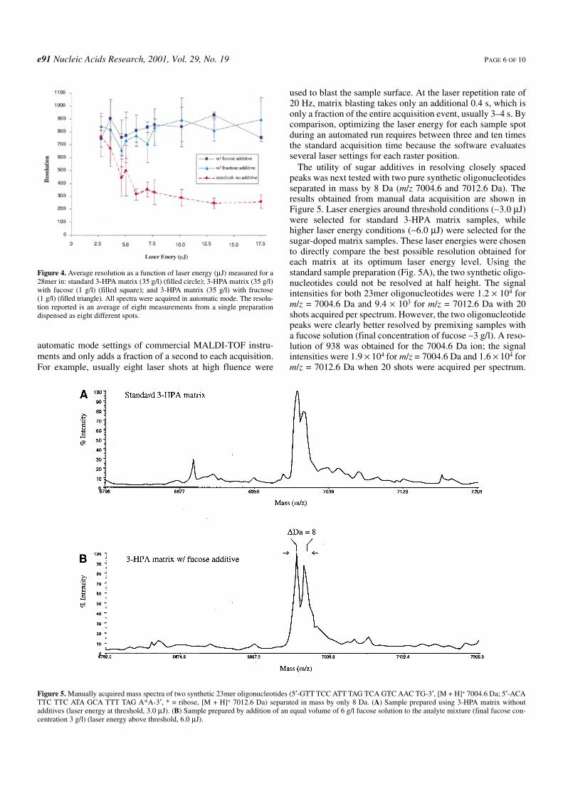
The utility of sugar additives in resolving closely spacedpeaks was next tested with two pure synthetic oligonucleotidesseparated in mass by 8 Da (m/z 7004.6 and 7012.6 Da). Theresults obtained from manual data acquisition are shown inFigure 5. Laser energies around threshold conditions (∼3.0 µJ)were selected for standard 3-HPA matrix samples, whilehigher laser energy conditions (∼6.0 µJ) were selected for thesugar-doped matrix samples. These laser energies were chosento directly compare the best possible resolution obtained foreach matrix at its optimum laser energy level. Using thestandard sample preparation (Fig. 5A), the two synthetic oligo-nucleotides could not be resolved at half height. The signalintensities for both 23mer oligonucleotides were 1.2 × 10⁴ form/z = 7004.6 Da and 9.4 × 10³ for m/z = 7012.6 Da with 20shots acquired per spectrum. However, the two oligonucleotidepeaks were clearly better resolved by premixing samples witha fucose solution (final concentration of fucose ∼3 g/l). A reso-lution of 938 was obtained for the 7004.6 Da ion; the signalintensities were 1.9 × 10⁴ for m/z = 7004.6 Da and 1.6 × 10⁴ form/z = 7012.6 Da when 20 shots were acquired per spectrum.
Figure 4. Average resolution as a function of laser energy (µJ) measured for a28mer in: standard 3-HPA matrix (35 g/l) (filled circle); 3-HPA matrix (35 g/l)with fucose (1 g/l) (filled square); and 3-HPA matrix (35 g/l) with fructose(1 g/l) (filled triangle). All spectra were acquired in automatic mode. The resolu-tion reported is an average of eight measurements from a single preparationdispensed as eight different spots.
Figure 5. Manually acquired mass spectra of two synthetic 23mer oligonucleotides (5′-GTT TCC ATT TAG TCA GTC AAC TG-3′, [M + H]+ 7004.6 Da; 5′-ACATTC TTC ATA GCA TTT TAG A*A-3′, * = ribose, [M + H]+ 7012.6 Da) separated in mass by only 8 Da. (A) Sample prepared using 3-HPA matrix withoutadditives (laser energy at threshold, 3.0 µJ). (B) Sample prepared by addition of an equal volume of 6 g/l fucose solution to the analyte mixture (final fucose con-centration 3 g/l) (laser energy above threshold, 6.0 µJ).
PAGE 7 OF 10 Nucleic Acids Research, 2001, Vol. 29, No. 19 e91
For these samples, the sugar was premixed with the analyteand then dispensed onto the matrix, as this was found to furtherimprove sample homogeneity.
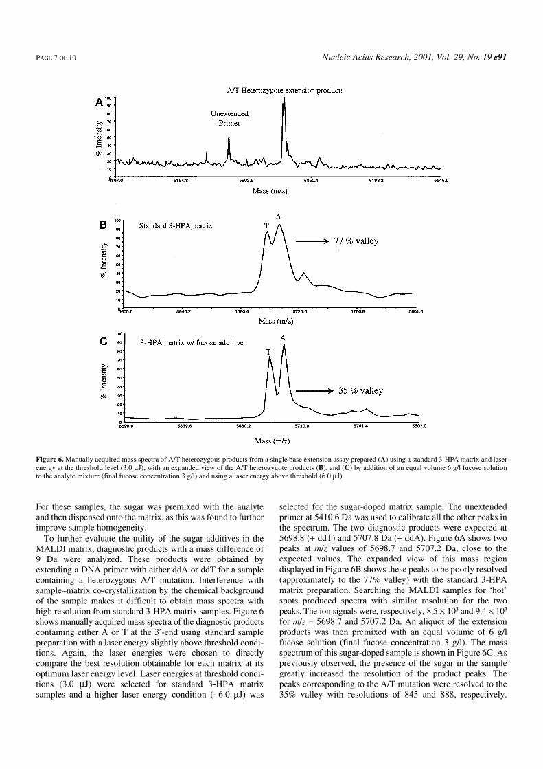
To further evaluate the utility of the sugar additives in theMALDI matrix, diagnostic products with a mass difference of9 Da were analyzed. These products were obtained byextending a DNA primer with either ddA or ddT for a samplecontaining a heterozygous A/T mutation. Interference withsample–matrix co-crystallization by the chemical backgroundof the sample makes it difficult to obtain mass spectra withhigh resolution from standard 3-HPA matrix samples. Figure 6shows manually acquired mass spectra of the diagnostic productscontaining either A or T at the 3′-end using standard samplepreparation with a laser energy slightly above threshold condi-tions. Again, the laser energies were chosen to directlycompare the best resolution obtainable for each matrix at itsoptimum laser energy level. Laser energies at threshold condi-tions (3.0 µJ) were selected for standard 3-HPA matrixsamples and a higher laser energy condition (∼6.0 µJ) was
selected for the sugar-doped matrix sample. The unextendedprimer at 5410.6 Da was used to calibrate all the other peaks inthe spectrum. The two diagnostic products were expected at5698.8 (+ ddT) and 5707.8 Da (+ ddA). Figure 6A shows twopeaks at m/z values of 5698.7 and 5707.2 Da, close to theexpected values. The expanded view of this mass regiondisplayed in Figure 6B shows these peaks to be poorly resolved(approximately to the 77% valley) with the standard 3-HPAmatrix preparation. Searching the MALDI samples for ‘hot’spots produced spectra with similar resolution for the twopeaks. The ion signals were, respectively, 8.5 × 10³ and 9.4 × 10³
for m/z = 5698.7 and 5707.2 Da. An aliquot of the extensionproducts was then premixed with an equal volume of 6 g/lfucose solution (final fucose concentration 3 g/l). The massspectrum of this sugar-doped sample is shown in Figure 6C. Aspreviously observed, the presence of the sugar in the samplegreatly increased the resolution of the product peaks. Thepeaks corresponding to the A/T mutation were resolved to the35% valley with resolutions of 845 and 888, respectively.
Figure 6. Manually acquired mass spectra of A/T heterozygous products from a single base extension assay prepared (A) using a standard 3-HPA matrix and laserenergy at the threshold level (3.0 µJ), with an expanded view of the A/T heterozygote products (B), and (C) by addition of an equal volume 6 g/l fucose solutionto the analyte mixture (final fucose concentration 3 g/l) and using a laser energy above threshold (6.0 µJ).
e91 Nucleic Acids Research, 2001, Vol. 29, No. 19 PAGE 8 OF 10
The ion signals were 1.4 × 10⁴ (m/z = 5699 Da) and 1.7 × 10⁴
(m/z = 5707 Da) when the sugar additive was used. Resultsconclusively indicate that the presence of the sugar additiveimproves the resolution attained in MALDI-TOF mass spectraof these diagnostic products.
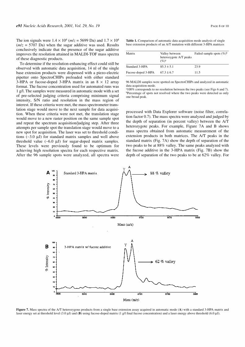
To determine if the resolution-enhancing effect could still beobserved with automatic data acquisition, 14 nl of the singlebase extension products were dispensed with a piezo-electricpipetter onto SpectroCHIPs preloaded with either standard3-HPA or fucose-doped 3-HPA matrix in an 8 × 12 arrayformat. The fucose concentration used for automated runs was1 g/l. The samples were measured in automatic mode with a setof pre-selected judging criteria comprising minimum signalintensity, S/N ratio and resolution in the mass region ofinterest. If these criteria were met, the mass spectrometer trans-lation stage would move to the next sample for data acquisi-tion. When these criteria were not met, the translation stagewould move to a new raster position on the same sample spotand repeat the spectrum acquisition/judging step. After threeattempts per sample spot the translation stage would move to anew spot for acquisition. The laser was set to threshold condi-tions (∼3.0 µJ) for standard matrix samples and well abovethreshold value (∼6.0 µJ) for sugar-doped matrix samples.These levels were previously found to be optimum forachieving high resolution spectra for each respective matrix.After the 96 sample spots were analyzed, all spectra were
processed with Data Explorer software (noise filter, correla-tion factor 0.7). The mass spectra were analyzed and judged bythe depth of separation (in percent valley) between the A/Theterozygote peaks. For example, Figure 7A and B showsmass spectra obtained from automatic measurement of theextension products in both matrices. The A/T peaks in thestandard matrix (Fig. 7A) show the depth of separation of thetwo peaks to be at 88% valley. The same peaks analyzed withthe fucose additive in the 3-HPA matrix (Fig. 7B) show thedepth of separation of the two peaks to be at 62% valley. For
Table 1. Comparison of automatic data acquisition mode analysis of singlebase extension products of an A/T mutation with different 3-HPA matrices
96 MALDI samples were spotted on SpectroCHIPs and analyzed in automaticdata acquisition mode.a100% corresponds to no resolution between the two peaks (see Figs 6 and 7).bPercentage of spots not resolved where the two peaks were detected as onlyone broad peak.
Matrix Valley betweenheterozygote A/T peaks(%)a
Failed sample spots (%)b
Standard 3-HPA 85.3 ± 5.1 23.9
Fucose-doped 3-HPA 67.3 ± 6.7 11.5
Figure 7. Mass spectra of the A/T heterozygous products from a single base extension assay acquired in automatic mode (A) with a standard 3-HPA matrix andlaser energy set at threshold level (3.0 µJ) and (B) using fucose-doped matrix (1 g/l final fucose concentration) and a laser energy above threshold (6.0 µJ).
PAGE 9 OF 10 Nucleic Acids Research, 2001, Vol. 29, No. 19 e91
the 96 samples analyzed the average depths of separationbetween the A/T peaks with both matrices are shown in Table 1.For the standard 3-HPA matrix it was found to be at 85.3 ± 5.1%valley, while for the 3-HPA matrix with fucose additive it wasfound to be lower at 67.3 ± 6.7% valley. These experimentsshowed that the fucose-doped matrices consistently producedspectra of the two peaks with higher resolution in automaticacquisition mode. In addition, it was also found that 23.9% ofthe standard matrix samples failed to resolve the two peaks, asonly one broad peak was detected. That is in comparison toonly 11.5% failure in fucose-doped matrix samples. Insummary, our findings conclusively indicate that fucose-dopedsamples produce higher resolution spectra of A/T extensionproducts compared to samples prepared in the standard matrixformulation in both manual and automated data acquisitionmodes. This is especially important in a high throughputenvironment where manual optimization and manipulation byan experienced user is not feasible. The ability to resolve oligo-nucleotides with very small mass differences demonstrates aviable application of this methodology to the analysis of diag-nostic products and complex mixtures containing DNAoligomers with similar molecular weights.
CONCLUSION
We have demonstrated that the quality of MALDI-TOF massspectra is less dependent on laser fluence when selected sugarsare added during matrix crystallization. The best results wereproduced by adding fructose or fucose to the MALDI matrix.Protocols for both manual and automatic modes of data acqui-sition were developed and used to obtain quality mass spectra.High resolution was maintained even with laser powerssignificantly higher than the desorption threshold. Theimproved formulation is important for routinely resolvingoligonucleotides with small mass differences during automaticdata acquisition, as shown in the selected single base extensionassay for genotyping. The smallest mass change of 9 Da, repre-senting an SNP of a heterogeneous A/T transition, wasresolved and resolution could be maintained with a laser powerhigher than the desorption threshold. Therefore, during auto-matic data acquisition, a laser fluence higher than the desorp-tion threshold could be chosen to guarantee ion generationwithout compromising mass resolution. This technique couldbe very useful for producing high quality mass spectra in caseswhere higher laser energy is needed to overcome low ionsignals generated from weak assays. The increased resolutionof the 3-HPA matrix with sugar additives could also prove tosignificantly improve the high throughput screening of DNAoligomers with similar masses, such as those in single baseextension reactions or multiplex assays.
REFERENCES
1. Ch’ang,L.-Y., Tang,K., Schell,M., Ringelberg,C., Matteson,K.J.,Allman,S.L. and Chen,C.H. (1995) Detection of ∆F508 mutation of thecystic fibrosis gene by matrix-assisted laser desorption/ionization massspectrometry. Rapid Commun. Mass Spectrom., 9, 772–774.
2. Bai,J., Liu,Y., Liang,X., Zhu,Y. and Lubman,D.M. (1995) Procedures fordetection of DNA by matrix-assisted laser desorption ionization massspectrometry using a modified nafion film substrate. Rapid Commun.Mass Spectrom., 9, 1172–1176.
3. Ross,P.L. and Belgrader,P. (1997) Analysis of short tandem repeatpolymorphisms in human DNA by matrix-assisted laser desorption/ionization mass spectrometry. Anal. Chem., 69, 3966–3972.
4. Ross,P.L., Davis,P.A. and Belgrader,P. (1998) Analysis of DNAfragments from conventional and microfabricated PCR devices usingdelayed extraction MALDI-TOF mass spectrometry. Anal. Chem., 70,2067–2073.
5. Taranenko,N.I., Golovlev,V.V., Allman,S.L., Taranenko,N.V.,Chen,C.H., Hong,J. and Chang,L.Y. (1998) Matrix-assisted laserdesorption/ionization for short tandem repeat loci. Rapid Commun. MassSpectrom., 12, 413–418.
6. Roskey,M.T., Juhasz,P., Smirnov,I.P., Takach,E.J., Martin,S.A. andHaff,L.A. (1996) DNA sequencing by delayed extraction-matrix-assistedlaser desorption/ionizaton time of flight mass spectrometry. Proc. NatlAcad. Sci. USA, 93, 4724–4729.
7. Köster,H., Tang,K., Fu,D., Braun,A., van den Boom,D., Smith,C.L.,Cotter,R.J. and Cantor,C.R. (1996) A strategy for rapid and efficient DNAsequencing by mass spectrometry. Nat. Biotechnol., 14, 1123–1128.
8. Fu,D., Tang,K., Braun,A., Reuter,D., Darnhofer-Demar,B., Little,D.L.,O’Donnell,M.J., Cantor,C.R. and Köster,H. (1998) Sequencing exons 5 to8 of the p53 gene by MALDI-TOF mass spectrometry. Nat. Biotechnol.,16, 381–384.
9. Kirpekar,F., Nordhoff,E., Larsen,L.K., Kristiansen,K., Roepstorff,P. andHillenkamp,F. (1998) DNA sequence analysis by MALDI massspectrometry. Nucleic Acids Res., 26, 2554–2559.
10. Braun,A., Little,D.P. and Köster,H. (1997) Detecting CFTR genemutations by using primer oligo base extension and mass spectrometry.Clin. Chem., 43, 1151–1158.
11. Haff,L. and Smirnov,I. (1997) Single-nucleotide polymorphismidentification assays using a thermostable DNA polymerase and delayedextraction MALDI-TOF mass spectrometry. Genome Res., 7, 378–388.
12. Ross,P., Hall,L., Smirnov,I.P. and Haff,L. (1998) High level multiplexgenotyping by MALDI-TOF mass spectrometry. Nat. Biotechnol., 16,1347–1351.
13. Tang,K., Fu,D., Julien,D., Braun,A., Cantor,C.R. and Köster,H. (1999)DNA chip for genotyping by mass spectrometry. Proc. Natl Acad. Sci.USA, 96, 10016–10020.
14. Dai,Y., Whittal,R.M., Weinberger,S.R. and Li,L. (1996) Accurate massmeasurement of oligonucleotides using a time-lag focusing matrix-assisted laser desorption/ionization time-of-flight mass spectrometer.Rapid Commun. Mass Spectrom., 10, 1792–1796.
15. Juhasz,P., Roskey,M.T., Smirnov,I.P., Haff,L.A., Vestal,M.L. andMartin,S.A. (1996) Applications of delayed extraction matrix-assistedlaser desorption ionization time-of-flight mass spectrometry tooligonucleotide analysis. Anal. Chem., 68, 941–946.
16. Li,Y., Tang,K., Little,D.P., Köster,H., Hunter,R.L. and McIver,R.T.(1996) High resolution MALDI Fourier transform mass spectrometry ofoligonucleotides. Anal. Chem., 68, 2090–2096.
17. Guo,B., Wang,S. and Fan,Y. (2000) Improving the performance ofMALDI-TOF in oligonucleotide analysis using a new SDIFA technology.Anal. Chem., 72, 5792–5797.
18. Tang,W., Zhu,L. and Smith,L.M. (1997) Controlling DNA fragmentationin MALDI-MS by chemical modification. Anal. Chem., 69, 302–312.
19. Hunter,J.M., Lin,H. and Becker,C.H. (1997) Cryogenic frozen solutionmatrixes for analysis of DNA by time-of-flight mass spectrometry. Anal.Chem., 69, 3608–3612.
20. Little,D.P., Cornish,T.J., O’Donnell,M.J., Braun,A., Cotter,R.J. andKöster,H. (1997) MALDI on a chip: analysis of arrays of low-femtomoleto subfemtomole quantities of synthetic oligonucleotides and DNAdiagnostic products dispensed by a piezoelectric pipet. Anal. Chem., 69,4540–4546.
21. Hung,K.C., Rashidzadeh,H., Wang,Y. and Guo,B.C. (1998) Use ofparaffin wax film in MALDI-TOF analysis of DNA. Anal. Chem., 70,3088–3093.
22. Berkenkamp,S., Kirpekar,F. and Hillenkamp,F. (1998) Infrared MALDImass spectrometry of large nucleic acids. Science, 281, 260–262.
23. Hung,K.C., Ding,H. and Guo,B. (1999) Use of poly(tetrafluoroethylene)sas a sample support for the MALDI-TOF analysis of DNA and proteins.Anal. Chem., 71, 518–521.
24. Kirpekar,F., Berkenkamp,S. and Hillenkamp,F. (1999) Detection ofdouble-stranded DNA by IR- and UV-MALDI mass spectrometry. Anal.Chem., 71, 2334–2339.
e91 Nucleic Acids Research, 2001, Vol. 29, No. 19 PAGE 10 OF 10
25. Schuerenberg,M., Luebbert,C., Eickhoff,H., Kalkum,M., Lehrach,H. andNordhoff,E. (2000) Prestructured MALDI-MS sample supports. Anal.Chem., 72, 3436–3442.
26. Koomen,J.M., Russell,W.K., Hettick,J.M. and Russell,D.H. (2000)Improvement of resolution, mass accuracy and reproducibility in reflectedmode DE-MALDI-TOF analysis of DNA using fast evaporation-overlayer sample preparations. Anal. Chem., 72, 3860–3866.
27. Nordhoff,E., Cramer,R., Karas,M., Hillenkamp,F., Kirpekar,F.,Kristiansen,K. and Roepstoff,P. (1993) Ion stability of nucleic acids ininfrared matrix-assisted laser desorption/ionization mass spectrometry.Nucleic Acids Res., 21, 3347–3357.
28. Currie,G.J. and Yates,J.R. (1993) Analysis of oligodeoxynucleotides bynegative-ion matrix-assisted laser desorption mass spectrometry. J. Am.Soc. Mass Spectrom., 4, 955–963.
29. Pieles,U., Zürcher,W., Schär,M. and Moser,H.E. (1993) Matrix-assistedlaser desorption ionization time-of-flight mass spectrometry: a powerfultool for the mass and sequence analysis of natural and modifiedoligonucleotides. Nucleic Acids Res., 21, 3191–3196.
30. Zhu,Y.F., Taranenko,N.I., Allman,S.L., Martin,S.A., Haff,L. andChen,C.H. (1996) The effect of ammonium salt and matrix in thedetection of DNA by matrix-assisted laser desorption/ionization time-of-flight mass spectrometry. Rapid Commun. Mass Spectrom., 10,1591–1596.
31. Li,Y.C.L., Cheng,S.-W. and Chan,T.-W.D. (1998) Evaluation ofammonium salts as co-matrices for matrix-assisted laser desorption/ionization mass spectrometry of oligonucleotides. Rapid Commun. MassSpectrom., 12, 993–998.
32. Simmons,T.A. and Limbach,P.A. (1998) Influence of co-matrix protonaffinity on oligonucleotide ion stability in matrix-assisted laserdesorption/ionization time-of-flight mass spectrometry. J. Am. Soc. MassSpectrom., 9, 668–675.
33. Asara,J.M. and Allison,J. (1999) Enhanced detection of oligonucleotidesin UV MALDI MS using the tetraamine spermine as a matrix additive.Anal. Chem., 71, 2866–2870.
34. Vandell,V.E. and Limbach,P.A. (1999) Polyamine co-matrices for matrix-assisted laser desorption/ionization mass spectrometry ofoligonucleotides. Rapid Commun. Mass Spectrom., 13, 2014–2021.
35. Zhu,Y.F., Chung,C.N., Taranenko,N.I., Allman,S.L., Martin,S.A.,Haff,L.A. and Chen,C.H. (1996) The study of
2,3,4-trihydroxyacetophenone and 2,4,6-trihydroxyacetophenone asmatrices for DNA detection in matrix-assisted laser desorption/ionizationtime-of-flight mass spectrometry. Rapid Commun. Mass Spectrom., 10,383–388.
36. Zhu,Y.F., Taranenko,N.I., Allman,S.L., Taranenko,N.V., Martin,S.A.,Haff,L.A. and Chen,C.H. (1997) Oligonucleotide sequencing byfragmentation in matrix-assisted laser desorption/ionization time-of-flightmass spectrometry. Rapid Commun. Mass Spectrom., 11, 897–903.
37. Wu,K.J., Steding,A. and Becker,C.H. (1993) Matrix-assisted laserdesorption time-of-flight mass spectrometry of oligonucleotides using3-hydroxypicolinic acid as an ultraviolet-sensitive matrix. RapidCommun. Mass Spectrom., 7, 142–146.
38. Lecchi,P., Le,H.M.T. and Pannell,L.K. (1995) 6-Aza-2-thiothymine: amatrix for MALDI spectra of oligonucleotides. Nucleic Acids Res., 23,1276–1277.
39. Louris,J.N., Cooks,R.G., Syka,J.E.P., Kelley,P.E., Stafford,G.C.Jr andTodd,J.F.J. (1987) Instrumentation, applications and energy deposition inquadrupole ion trap MS/MS spectrometry. Anal. Chem., 59, 1677–1685.
40. McIver,R.T., Li,Y. and Hunter,R.L. (1994) High-resolution laserdesorption mass spectrometry of peptides and small proteins. Proc. NatlAcad. Sci. USA, 91, 4801–4805.
41. Beavis,R.C., Lindner,J., Grotemeyer,J. and Schlag,E.W. (1988) Sample-matrix effect in infrared laser neutral desorption, multiphoton-ionizationmass spectrometry. Chem. Phys. Lett., 146, 310–313.
42. Köster,C., Castoro,J.A. and Wilkins,C.L. (1992) High-resolution matrix-assisted laser desorption/ionization of biomolecules by Fourier transformmass spectrometry. J. Am. Chem. Soc., 114, 7572–7574.
43. Castoro,J.A. and Wilkins,C.L. (1993) Ultrahigh resolution matrix-assistedlaser desorption/ionization of small proteins by Fourier transform massspectrometry. Anal. Chem., 65, 2621–2627.
44. Gusev,A.I., Wilkinson,W.R., Proctor,A. and Hercules,D.M. (1995)Improvement of ion signal reproducibility and matrix/comatrix effects inMALDI analysis. Anal. Chem., 67, 1034–1041.
45. Horneffer,V., Forsmann,A., Strupat,K., Hillenkamp,F. andKubitscheck,U. (2000) Confocal laser scanning microscopy (CLSM) as asuitable imaging technique for studies of the analyte distribution inMALDI standard preparations. In Proceedings of the 48th ASMSConference on Mass Spectrometry and Allied Topics, Long Beach, CA.
Copyright © 2022 FDOKUMEN