Equine CRISP-3: primary structure and expression in the male genital tract
Upload
aasthahealthcareCategory
view
0download
0
Study Of Metaplastic Lesions Of Different Parts Of The Female Genital Tract: A Prospective Study M Banyameen, I Masood, A Aiman, M Yasir, I Mushtaq, V Verma
Citation
M Banyameen, I Masood, A Aiman, M Yasir, I Mushtaq, V Verma.
Study Of Metaplastic Lesions Of Different Parts Of The Female Genital Tract: A
Prospective Study. The Internet Journal of Pathology. 2009 Volume 10 Number 2.
Abstract
Objective: To study the pattern and relative incidence of metaplastic lesions of
different parts of the female genital tract and the relationship of different
metaplastic lesions with age. Method: A total of 200 female genital tract samples
in the form of hysterectomy specimens, endometrial curettings, pap smears and
cervical biopsies were subjected to gross and microscopic examination and the
metaplastic changes observed were recorded. Among all the samples, 57.5% were
hysterectomy specimens, 20% were pap smears, 15% were endometrial curettings
and 2.5% were cervix biopsies. Maximum number of patient (73%) was in the age
group of 30-49. Results: Among 200 patients enrolled in the study, 120 (60%)
were found to be positive for different types of metaplasias. Among the cervical
metaplasias, 60.2% were showing squamous metaplasia of the cervix, followed by
Ciliated and Microglandular metaplasia with a frequency of 25.5% and 20.4%
respectively with the highest frequency in the age group of 40-59 years. Among
the endometrial metaplasias Ciliated, Mucinous and Eosinophilic metaplasias were
found to be the commonest with a frequency of 28.5%, 25.5% and 25.5%
respectively. In the study, it was also observed that the metaplastic lesions of the
endometrium and that of the cervix co-existed with each other in 74% of the cases
Conclusions: From these observations, squamous metaplasia of the cervix was the
commonest metaplastic lesion observed on histological examinations, followed by
ciliated and microglandular metaplasia, where as in the endometrium ciliated
(tubal) metaplasia was the commonest followed by eosinophilic and mucinous
metaplasia.. A large number of metaplastic lesions of the cervix co-existed with
each other and also with that of the endometrium.
Introduction
The adult nulliparous uterus is a hollow, pear shaped organ that weights 40 – 80
grams and measures 7 to 8 cm along its longest axis. It is divided into the corpus
and the cervix. The cervix is the lower portion of the uterus and is divided into a
portion that protrudes into the vagina (portio vaginalis) and one that lies above the
vaginal vault (supravaginal portion). The outer surface of the portio vaginalis is
known as the exocervix or ectocervix and the portion related to the endocervical
canal corresponds to the endocervix.1
The endometrium lines the uterine cavity above the level of internal os. During the
first half of the menstrual cycle, all the components of the endometrium including
glands, stroma and blood vessels, proliferate under the influence of estrogens and
during the later half, these elements respond to progesterone with the production of
glandular secretions and there are stromal and vascular alterations.2
Most of the exocervix is covered by non-keratinizing squamous epithelium while
as the glandular mucosa of the endocervix is formed by a layer of columnar
mucous secreting cells. The area where the squamous and glandular epithelia meet
is known as the squamo-columnar junction. This is a very unstable region, in
which replacement of one epithelium for another repeatedly occurs, a process that
Robert Meyer referred to as “the fight of the epithelia”. Today this area is more
prosaically known as the transformation zone.1
Metaplasia is a condition in which there is a change of one type of differentiated
tissue into another type of similar differentiated tissue3 or as the abnormal
transformation of an adult, fully differentiated tissue of one kind into a
differentiated tissue of another kind.4
The mullerian derived epithelium which lines
most of the female genital tract is well known for its capacity to differentiate into
various types of epithelium such as, ciliated, mucinous, endometrioid, transitional
and squamous types.5
The metaplasias of the uterine corpus and cervix are the most common sites of
metaplasia. Metaplasia occasionally can occur in other parts of the female genital
tract such as mucosa of the fallopian tube and the vagina. Mucinous lesions of the
fallopian tube mucosa are extremely rare and are of interest because of their
association with other mucinous lesions of the female genital tract and with Peutz-
Jeghers syndrome.6
The abdominal and pelvic peritoneum, part of the secondary mullerian system, can
also undergo metaplasia into the various mullerian epithelia. The secondary
mullerian system comprises the mesothelium and the adjacent mesenchyme of the
pelvis and lower abdomen. Conditions such as endosalpingiosis and endocervicosis
are regarded as examples of mullerian metaplasia.4
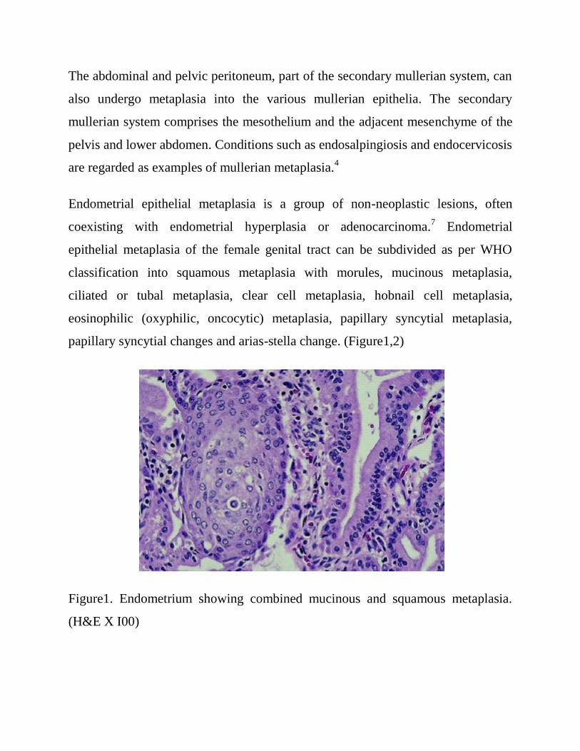
Endometrial epithelial metaplasia is a group of non-neoplastic lesions, often
coexisting with endometrial hyperplasia or adenocarcinoma.7 Endometrial
epithelial metaplasia of the female genital tract can be subdivided as per WHO
classification into squamous metaplasia with morules, mucinous metaplasia,
ciliated or tubal metaplasia, clear cell metaplasia, hobnail cell metaplasia,
eosinophilic (oxyphilic, oncocytic) metaplasia, papillary syncytial metaplasia,
papillary syncytial changes and arias-stella change. (Figure1,2)
Figure1. Endometrium showing combined mucinous and squamous metaplasia.
(H&E X I00)
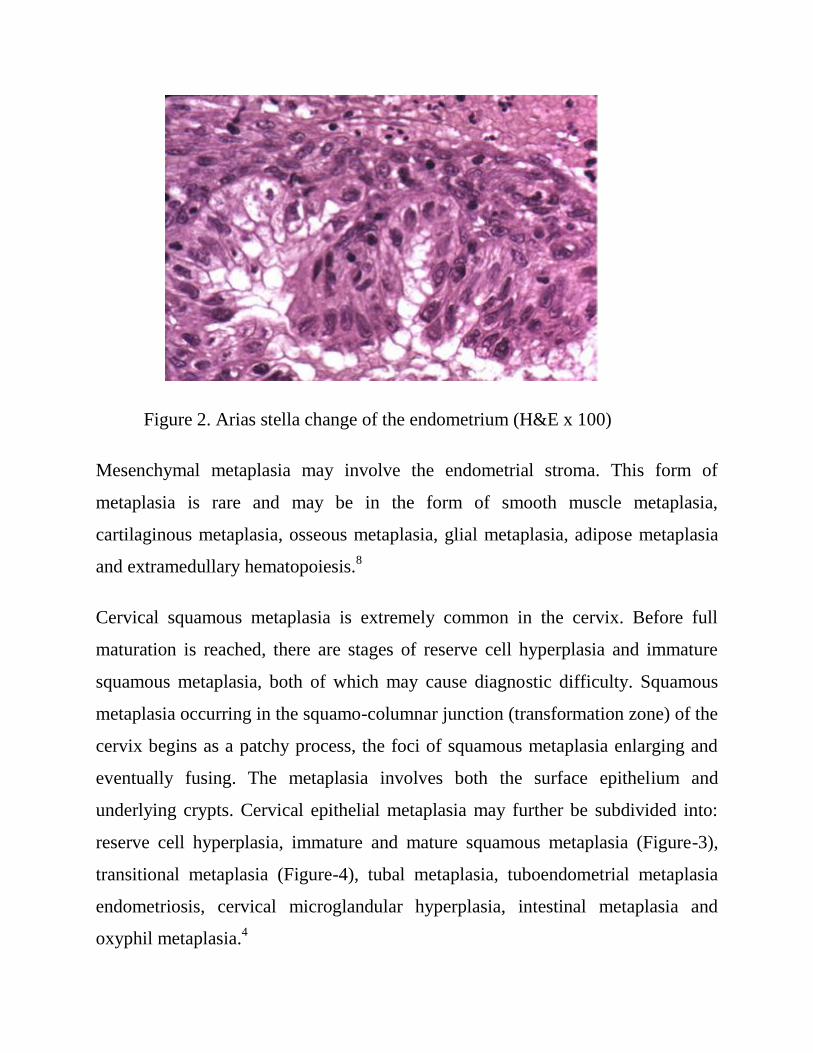
Figure 2. Arias stella change of the endometrium (H&E x 100)
Mesenchymal metaplasia may involve the endometrial stroma. This form of
metaplasia is rare and may be in the form of smooth muscle metaplasia,
cartilaginous metaplasia, osseous metaplasia, glial metaplasia, adipose metaplasia
and extramedullary hematopoiesis.8
Cervical squamous metaplasia is extremely common in the cervix. Before full
maturation is reached, there are stages of reserve cell hyperplasia and immature
squamous metaplasia, both of which may cause diagnostic difficulty. Squamous
metaplasia occurring in the squamo-columnar junction (transformation zone) of the
cervix begins as a patchy process, the foci of squamous metaplasia enlarging and
eventually fusing. The metaplasia involves both the surface epithelium and
underlying crypts. Cervical epithelial metaplasia may further be subdivided into:
reserve cell hyperplasia, immature and mature squamous metaplasia (Figure-3),
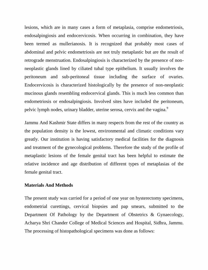
transitional metaplasia (Figure-4), tubal metaplasia, tuboendometrial metaplasia
endometriosis, cervical microglandular hyperplasia, intestinal metaplasia and
oxyphil metaplasia.4
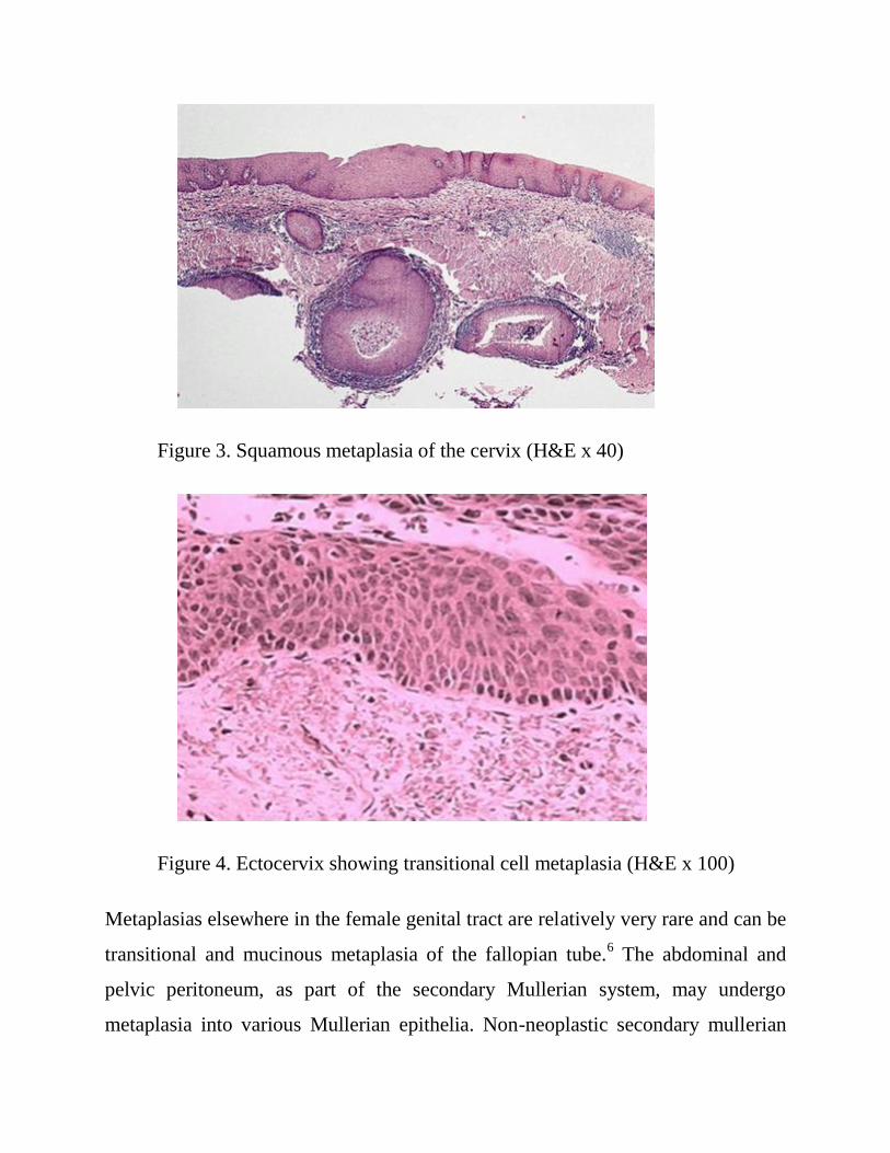
Figure 3. Squamous metaplasia of the cervix (H&E x 40)
Figure 4. Ectocervix showing transitional cell metaplasia (H&E x 100)
Metaplasias elsewhere in the female genital tract are relatively very rare and can be
transitional and mucinous metaplasia of the fallopian tube.6 The abdominal and
pelvic peritoneum, as part of the secondary Mullerian system, may undergo
metaplasia into various Mullerian epithelia. Non-neoplastic secondary mullerian
lesions, which are in many cases a form of metaplasia, comprise endometriosis,
endosalpingiosis and endocervicosis. When occurring in combination, they have
been termed as mullerianosis. It is recognized that probably most cases of
abdominal and pelvic endometriosis are not truly metaplastic but are the result of
retrograde menstruation. Endosalpingiosis is characterized by the presence of non-
neoplastic glands lined by ciliated tubal type epithelium. It usually involves the
peritoneum and sub-peritoneal tissue including the surface of ovaries.
Endocervicosis is characterized histologically by the presence of non-neoplastic
mucinous glands resembling endocervical glands. This is much less common than
endometriosis or endosalpingiosis. Involved sites have included the peritoneum,
pelvic lymph nodes, urinary bladder, uterine serosa, cervix and the vagina.9
Jammu And Kashmir State differs in many respects from the rest of the country as
the population density is the lowest, environmental and climatic conditions vary
greatly. Our institution is having satisfactory medical facilities for the diagnosis
and treatment of the gynecological problems. Therefore the study of the profile of
metaplastic lesions of the female genital tract has been helpful to estimate the
relative incidence and age distribution of different types of metaplasias of the
female genital tract.
Materials And Methods
The present study was carried for a period of one year on hysterectomy specimens,
endometrial curettings, cervical biopsies and pap smears, submitted to the
Department Of Pathology by the Department of Obstetrics & Gynaecology,
Acharya Shri Chander College of Medical Sciences and Hospital, Sidhra, Jammu.
The processing of histopathological specimens was done as follows:
Hysterectomy Specimens
Hysterectomy specimens were grossed and 3-5 mm thin sections were taken for
processing. Endometrial curettings were processed as such. Fixation was done in
10% buffered formol saline. Dehydration was carried out in ascending
concentrations of ethanol. Clearing was done in xylene, and impregnation in
paraffin wax. Tissue blocks were prepared using leukhart’s ‘L’ pieces. 3-5
micrometer thin sections were cut using rotary microtome and were stained with
haematoxylin and eosin.
The staining with Haematoxylin and Eosin was done as follows: Deparaffinization
was done in xylene and the sections were treated in descending concentrations of
ethanol and then brought to water. Staining was done with Harris haematoxylin for
10-40 minutes. Bluing was done under running tap water. Sections were
differentiated in 1% acid alcohol, followed by Washing in water. Counter staining
with 1% aqueous eosin for 2-4 minutes followed by dehydration in ascending
concentrations of ethanol was done. Sections cleared in xylene and were finally
mounted in DPX.10
Cervicovaginal smears
Papanicolaou staining method was used. The smears were wet fixed rapidly in 95%
ethyl alcohol, before any air drying occurred. Polyethylene glycol fixative was
removed in 50% alcohol (wherever coating fixatives were used). Sections were
hydrated in 95% & 70% alcohol for 2 minutes each. Staining was done in Harris
haematoxylin for 5 minutes. Slides were differentiated in 0.5% aqueous
hydrochloric acid for 10 seconds, followed by rinsing in water for 2 minutes.
Bluing was done in Scotts tap water substitute for 2 minutes. Smears were
dehydrated in ascending concentrations of ethanol. Staining with OG 6 (orange G
6) was done for 2 minutes followed by rinsing in 95% alcohol for 2 minutes.
Staining in EA50 for 3 minutes was done followed by rinsing in 95% alcohol for 1
minute. The nuclei should appear blue / black and cytoplasm of non-keratinizing
squamous cells should appear blue/green and that in keratinizing cells,
pink/orange. A detailed histopathological examination of haematoxylin & eosin
stained sections was carried out & the findings were recorded.
Observations
The following observations were made:
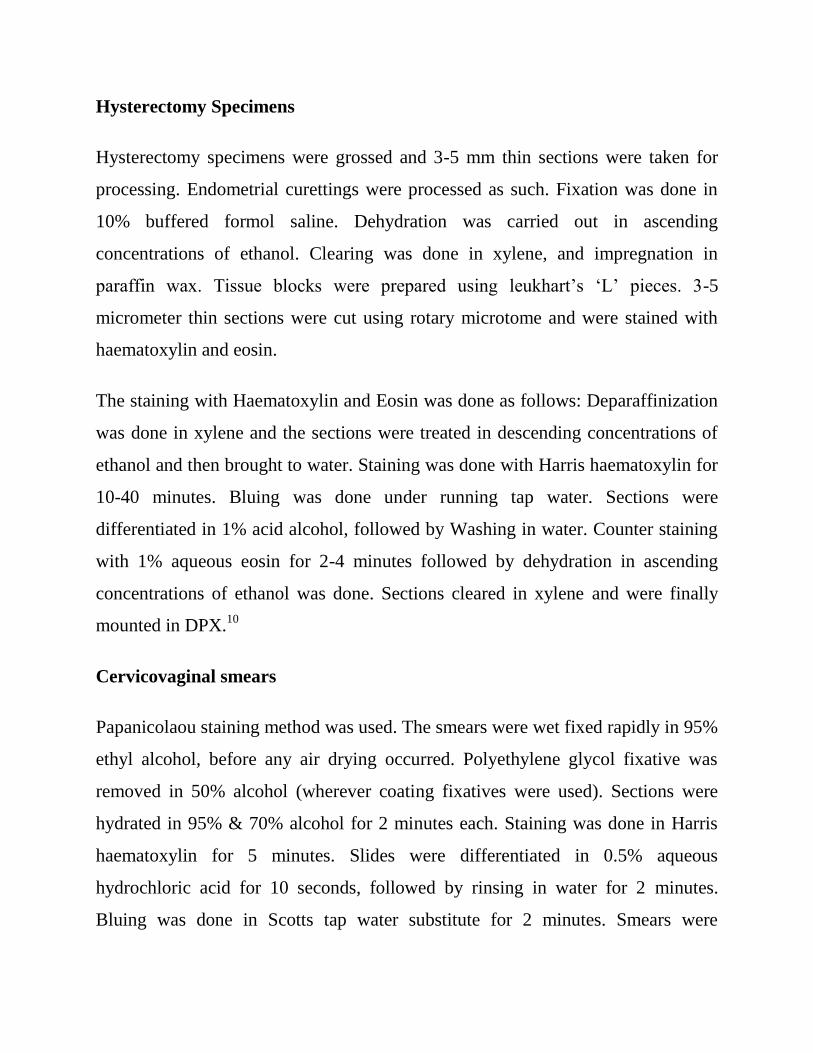
The total number of specimens included 200, out of which 125 (62.5%) specimens
were obtained from hysterectomy (endometrium, myometrium, cervix, fallopian
tubes). Endometrial curettings accounted for 30 (15%) specimens, Pap smears for
40 (20%) specimens and cervical biopsies for 5 (2.5%) specimens. All the above
specimens were unremarkable on gross examination. (Table 1)
Table 1: Showing the types of sample
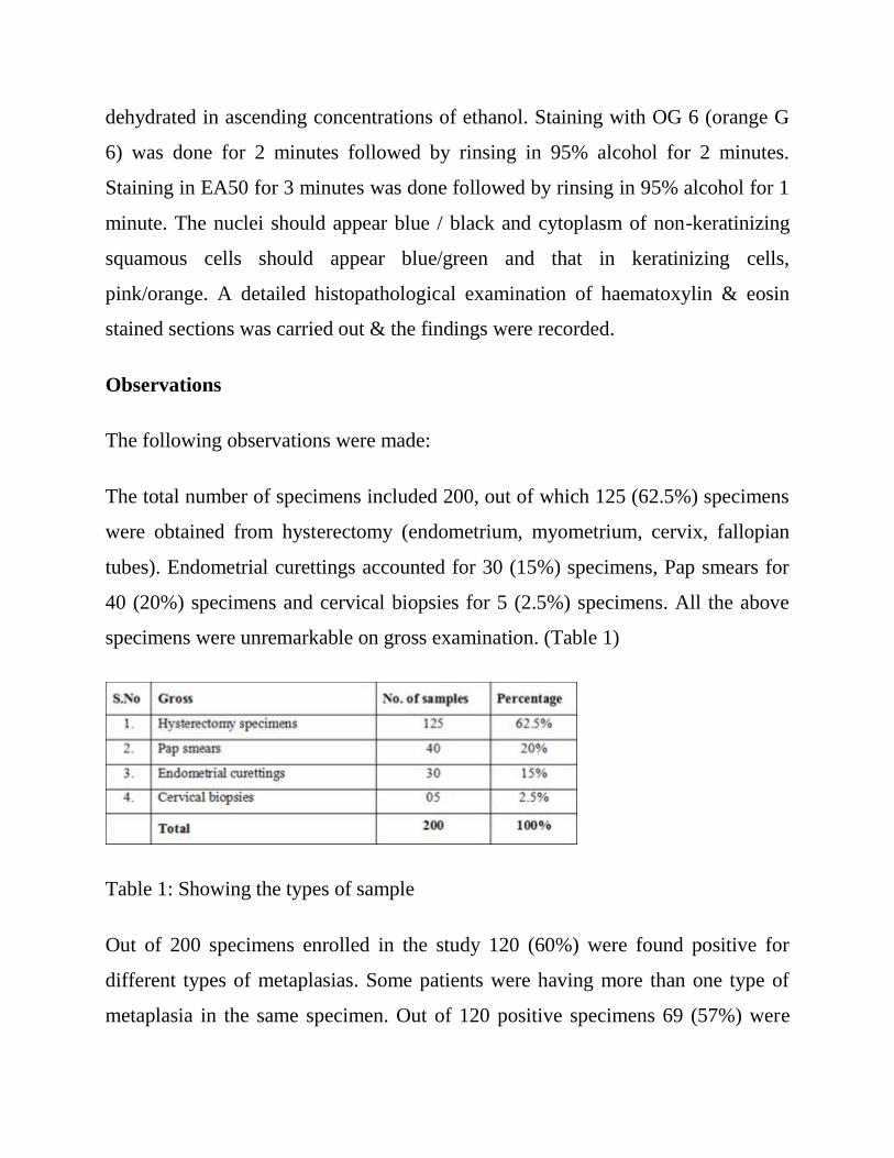
Out of 200 specimens enrolled in the study 120 (60%) were found positive for
different types of metaplasias. Some patients were having more than one type of
metaplasia in the same specimen. Out of 120 positive specimens 69 (57%) were
hysterectomy specimens (endometrium, myometrium, cervix, fallopian tubes), 28
(23.3%) were pap smears (cervix only), 22 (18.3%) were endometrial curettings
(endometrium, myometrium) and 01 (0.12%) specimen was of cervical biopsy
(cervix only). (Table 2)
Table 2: showing the number and percentage of Positive samples
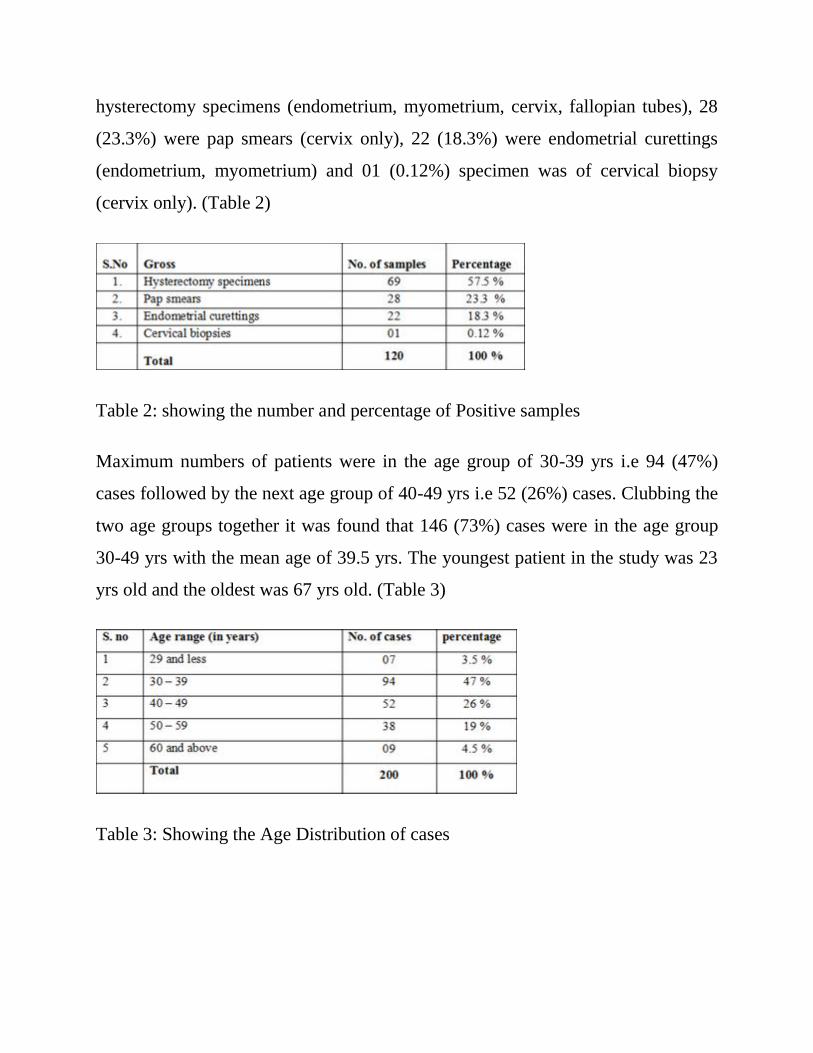
Maximum numbers of patients were in the age group of 30-39 yrs i.e 94 (47%)
cases followed by the next age group of 40-49 yrs i.e 52 (26%) cases. Clubbing the
two age groups together it was found that 146 (73%) cases were in the age group
30-49 yrs with the mean age of 39.5 yrs. The youngest patient in the study was 23
yrs old and the oldest was 67 yrs old. (Table 3)
Table 3: Showing the Age Distribution of cases
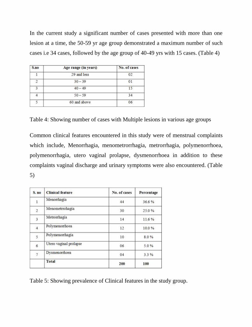
In the current study a significant number of cases presented with more than one
lesion at a time, the 50-59 yr age group demonstrated a maximum number of such
cases i.e 34 cases, followed by the age group of 40-49 yrs with 15 cases. (Table 4)
Table 4: Showing number of cases with Multiple lesions in various age groups
Common clinical features encountered in this study were of menstrual complaints
which include, Menorrhagia, menometrorrhagia, metrorrhagia, polymenorrhoea,
polymenorrhagia, utero vaginal prolapse, dysmenorrhoea in addition to these
complaints vaginal discharge and urinary symptoms were also encountered. (Table
5)
Table 5: Showing prevalence of Clinical features in the study group.

Out of 120 patients found positive for metaplasia 93 (77.5%) had received some
sort of hormone replacement therapy while as 27 (22.5%) were not on any sort of
hormone replacement therapy. Out of 80 patients found negative for metaplasia
only 15 (18.75%) had received some sort of hormone replacement therapy while as
65 (81.25%) were not on any kind of hormone replacement therapy. In the study it
was also seen that the relationship between HRT and metaplasia was not
significant, as all the types of metaplasias were evenly distributed among the
patients taking hormones.
Out of 98 positive specimens of cervical metaplasia, 59 specimens were observed
to show squamous metaplasia. The next common presentation was that of ciliated
(tubal) metaplasia of the cervix in 25 specimens. Next to follow were
microglandular metaplasia of the cervix 20 specimens, tuboendometroid 18
specimens and the least common was Arias Stella change seen in only 05
specimens. (Table 6a)
Table 6a: Showing the Pattern and incidence of Cervical metaplasias
Out of the total of 91 positive specimens of endometrium/myometrium
(hysterectomies, endometrial curettings) 26 were found positive for ciliated
metaplasia. The next common presentation was that of mucinous metaplasia and
eosinophilic cell change 23 cases each. Next to follow was squamous metaplasia in

03 cases and the least common was Arias Stella change of the endometrium in 01
case only. Other rare forms of metaplasia like hobnail cell metaplasia, clear cell
metaplasia, cartilaginous, osseous, glial and smooth muscle metaplasia were not
seen in any of the cases. (Table 6b)
Table 6b: Showing the Pattern and incidence of Endometrial metaplasias
In the present study no metaplasia was observed in any of the specimens of
myometrium, fallopian tubes and vagina. In the study it was also observed that a
significant number of metaplastic lesions of the cervix coexisted with that of the
endometrium. (Table 7)
Table 7: Coexisting lesions
The prevalence of cervical metaplasia was maximum in age group of 50-59 for
squamous, equal between 40-49 and 50-59 for ciliated and 50-59 for both
microglandular and Tuboendometroid subtypes. (Table 8)
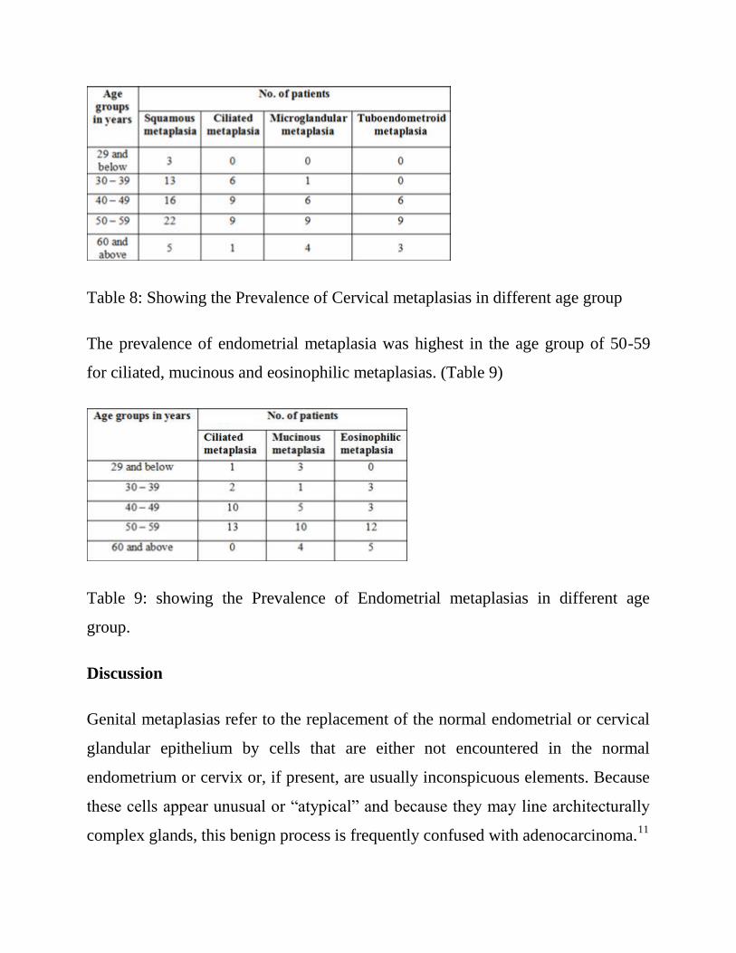
Table 8: Showing the Prevalence of Cervical metaplasias in different age group
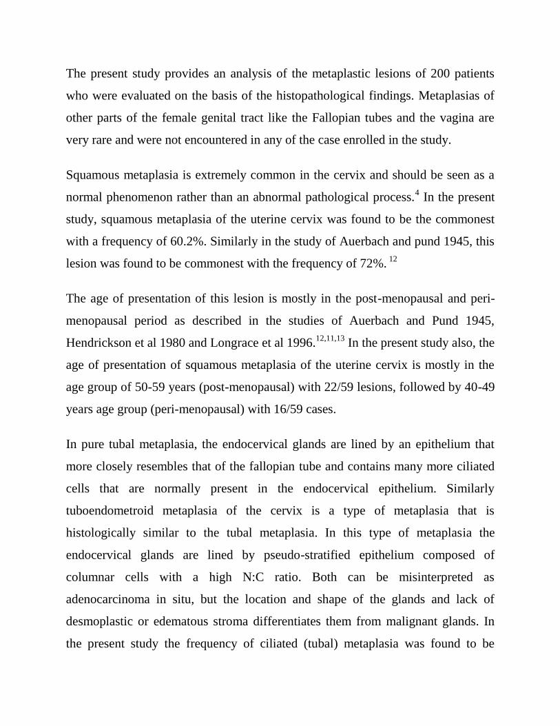
The prevalence of endometrial metaplasia was highest in the age group of 50-59
for ciliated, mucinous and eosinophilic metaplasias. (Table 9)
Table 9: showing the Prevalence of Endometrial metaplasias in different age
group.
Discussion
Genital metaplasias refer to the replacement of the normal endometrial or cervical
glandular epithelium by cells that are either not encountered in the normal
endometrium or cervix or, if present, are usually inconspicuous elements. Because
these cells appear unusual or “atypical” and because they may line architecturally
complex glands, this benign process is frequently confused with adenocarcinoma.11
The present study provides an analysis of the metaplastic lesions of 200 patients
who were evaluated on the basis of the histopathological findings. Metaplasias of
other parts of the female genital tract like the Fallopian tubes and the vagina are
very rare and were not encountered in any of the case enrolled in the study.
Squamous metaplasia is extremely common in the cervix and should be seen as a
normal phenomenon rather than an abnormal pathological process.4 In the present
study, squamous metaplasia of the uterine cervix was found to be the commonest
with a frequency of 60.2%. Similarly in the study of Auerbach and pund 1945, this
lesion was found to be commonest with the frequency of 72%. 12
The age of presentation of this lesion is mostly in the post-menopausal and peri-
menopausal period as described in the studies of Auerbach and Pund 1945,
Hendrickson et al 1980 and Longrace et al 1996.12,11,13
In the present study also, the
age of presentation of squamous metaplasia of the uterine cervix is mostly in the
age group of 50-59 years (post-menopausal) with 22/59 lesions, followed by 40-49
years age group (peri-menopausal) with 16/59 cases.
In pure tubal metaplasia, the endocervical glands are lined by an epithelium that
more closely resembles that of the fallopian tube and contains many more ciliated
cells that are normally present in the endocervical epithelium. Similarly
tuboendometroid metaplasia of the cervix is a type of metaplasia that is
histologically similar to the tubal metaplasia. In this type of metaplasia the
endocervical glands are lined by pseudo-stratified epithelium composed of
columnar cells with a high N:C ratio. Both can be misinterpreted as
adenocarcinoma in situ, but the location and shape of the glands and lack of
desmoplastic or edematous stroma differentiates them from malignant glands. In
the present study the frequency of ciliated (tubal) metaplasia was found to be
25.5% which is almost similar to the study of Jonasson et al 1992, in which the
frequency of ciliated (tubal) metaplasia was 31%.14
Similarly in this study, tuboendometroid metaplasias were seen in 18.3%, which is
in accordance with the study of Ismail 1991, which shows the frequency of
tuboendometroid metaplasia to be 26%.15
Maximum number of patients 15/25
(60%) were seen in the reproductive age group (30-49 years) replicating the studies
of Ducatman et al 1993, and Jonasson et al 1992.16,14
Although not a true metaplasia, microglandular metaplasia is best characterized
with metaplasias. It is common within the cervix and is usually associated with
exogenous hormone use or pregnancy and usually occurs in the reproductive age
group.4 In the present study the frequency of microglandular metaplasia was 20.4%
which lies in close range with the study findings of Brown and wells 1986, in
whose study its frequency was found to be 27%.17
Arias Stella reaction that develops in the endocervical glands during pregnancy is
identical to that which occurs in the endometrium. The Arias Stella reaction can
occasionally be mistaken for clear cell carcinoma or adenocarcinoma in situ of the
cervix. The Arias Stella reaction of the endocervix is usually focal and is more
commonly present in the proximal portion of the endocervix. In our study we
found 5 cases (5.1%) of Arias Stella reaction in the age group of 40-59 years which
lie in close proximity to the results of the study of Schneider 1981, having
frequency of 9% in the same age group.18
Ciliated endometrial epithelial cells are a normal phenomenon, so a diagnosis of
ciliated metaplasia should be made only when one or more endometrial glands are
lined predominantly by ciliated cells. In our study the relative incidence of ciliated
metaplasia was found to be 28.5% out of which 50% were in the reproductive age
group of 29-49 years, which lies in close proximity with the studies of Masterton et
al 1975 and Suzoko et al 2005 with the relative incidence of 20% and 31%
respectively.19,20
Mucinous endometrial metaplasia should be reserved for cases in which the
endometrial epithelial cells are replaced by cells with abundant mucin containing
cytoplasm resembling endocervical cells. Normal endometrial epithelial cells
contain some intracytoplasmic mucin, so abundant mucin is required for the
diagnosis. In our study the relative incidence of mucinous metaplasia of the
endometrium was found to be 25.5 % in which more than 60% were in the
postmenopausal group (50 years and above). The findings were consistent with the
studies of Suzuko et al (2005), with 26% of mucinous metaplasia in the post-
menopausal group and Nucci et al (1999) in whose study 80% of the patients were
in the post-menopausal group.21
Eosinophilic cell change is one of the common endometrial metaplasias occurring
in both non-neoplastic and neoplastic endometrium. It is characterized by the
presence of epithelial cells with abundant eosinophilic cytoplasm. The cytoplasm
may be granular in which case the term oncocytic metaplasia has be used. Some
degree of cytoplasmic eosinophilia is commonly found in endometrial epithelial
cells and does not alone warrant a diagnosis of eosinophilic metaplasia. In the
present study the relative incidence of eosinophilic cell change was 25.5% with the
maximum number of cases (74%) in the post-menopausal age group of 50 years
and above. Suzuko et al (2005) in their study found the relative incidence of this
lesion to be 28% and maximum number of cases were in the post-menopausal age
group with the mean age of 56 years.
Squamous metaplasia of the endometrium may be a focal finding or it may be
widespread and involve most of the endometrium. Microscopically it is composed
of bland squamous cells with eosinophilic cytoplasm. Morules differ from mature
squamous metaplasia in that they lack keratinization and intercellular bridges.
Morules and foci of mature squamous metaplasia often co-exist. The present study
found 3 cases (3.2%) of squamous metaplasia of the endometrium within the age
groups of 40-49, 50-59, 60 & above years respectively, whereas Suzuko et al 2005,
in their study found the frequency of the same lesion to be 7%.
Arias Stella change is almost always seen in pregnancy, trophoblastic diseases and
occasionally with hormone therapy rarely there is no relation. The most important
diagnostic dilemma is its differentiation from clear cell adenocarcinoma but the
diagnosis of Arias Stella change is usually straightforward if there is a history of
pregnancy or other morphological features of pregnancy are present. We in our
study observed 1 (1.09%) case of Arias Stella change of the endometrium in 40-49
years age group, which is similar to the study findings of Suzuko et al (2005) with
the frequency of 1%.
References
1. Rosai J; Uterus-corpus; Rosai and Ackerman’s Surgical Pathology; 9thedition;
volume 2; 2004; Elseiver; 1569-1603.
2. Mutter GL, Ferenczy A. Anatomy and histology of the uterine corpus; Ronnet,
BM, Kurman RJ. Blaustein’s pathology of the female genital tract; 5th edition;
Springer Verlag; New york, Berlin, Heidelberg; 2002; 383-500.
3. Walter and Israel. Disorders of growth, 7 th edition. Churchill livingstone
publishings 1996; p 407-424.
4. McCluggage WG, Desai V and Manek S. Tamoxifen-associated postmenopausal
adenomyosis exhibits stromal fibrosis, glandular dilatation and epithelial
metaplasias. Histopathology. 2000 Oct; 37 (4): 340-6.
5. Lauchlan SC. Metaplasias and neoplasias of Müllerian epithelium.
Histopathology 1984; 8 (4): 543-545.
6. Seidman JD. Mucinous lesions of the fallopian tube. A report of seven cases.
Am J Surg Pathol 1994; 18 (12): 1205-1212.
7. Kaku T, Tsukamoto N, Tsuruchi N, Sugihara K, Kamura T, Nakano H.
Endometrial metaplasia associated with endometrial carcinoma. Obstet Gynecol
1992; 80: 812-816.
8. Sirgi KE, Swanson PE, Gersell DJ. Extramedullary hematopoiesis in the
endometrium. Report of four cases and review of the literature. Am J Clin Pathol
1994; 101 (5): 643-646.
9. Young RH, Treger T, Scully RE.Am J Clin Pathol. Atypical polypoid
adenomyoma of the uterus. A report of 27 cases. 1986 Aug; 86 (2):139- 45.
10. Gamble M, Wilson I; The hematoxylins and eosin; Bancroft JD, Gamble M
(eds). Theory and practice of histological techniques; 5th edition; Churchill
livingstone 2002; 125-138.
11. Hendrickson MR, Kempson RL.Endometrial epithelial metaplasias:
proliferations frequently misdiagnosed as adenocarcinoma. Am J Surg Pathol
1980; 4 (6): 525-542
12. Auerbach SH, Pund ER. Squamous metaplasia of the cervix uteri. American
Journal of Obstetrics and Gynecology, 1945 Jan; 49 (1): 207-213.
13. Longacre TA, Chung MH, Rouse RV, Hendrickson MR. Atypical polypoid
adenomyofibromas (atypical polypoid adenomyomas) of the uterus. A
clinicopathologic study of 55 cases. Am J Surg Pathol. 1996 Jan; 20(1): 1-20.
14. Jonasson JG, Wang HH, Antonioli DA, Ducatman BS. Tubal metaplasia of the
uterine cervix: a prevalence study in patients with gynecologic pathologic findings.
Int J Gynecol Pathol. 1992; 11(2): 89-95.
15. Ismail SM. Cone biopsy causes cervical endometriosis and tubo- endometrioid
metaplasia. Histopathology 1991; 18(2): 107-114.
16. Ducatman BS, Wang HH, Jonasson JG, Hogan CL, Antonioli DA. Tubal
metaplasia: a cytologic study with comparison to other neoplastic and non-
neoplastic conditions of the endocervix. Diagn Cytopathol. 1993; 9(1): 98-103;
discussion 103-5.
17. Brown LJ, Wells M. Cervical glandular atypia associated with squamous
intraepithelial neoplasia: a premalignant lesion? J Clin Pathol. 1986 Jan; 39 (1):
22-8.
18. Schneider V. Arias-stella reaction of the endocervix: frequency and location.
Acta Cytol. 1981 May-Jun; 25(3): 224-8.
19. Masterton R, Armstrong EM, More IA The cyclical variation in the percentage
of ciliated cells in the normal human endometrium. J Reprod Fertil. 1975 Mar;
42(3): 537-40.
20. Suzuko Moritani1, Ryoji Kushima, Shu Ichihara1, Hidetoshi Okabe, Takanori
Hattori,Tadao K Kobayashi and Steven Silverberg. Eosinophilic cell change of the
endometrium: a possible relationship to mucinous differentiation. Modern
Pathology 2005; 18, 1243–1248.
21. Nucci MR, Prasad CJ, Crum CP, Mutter GL. Mucinous endometrial epithelial
proliferations: a morphologic spectrum of changes with diverse clinical
significance. Mod Pathol. 1999 Dec; 12 (12): 1137-42.
Copyright © 2022 FDOKUMEN