Study of different types of Chordomas - A case series
6
STUDY OF DIFFE *Dr. Mohammad Banyameen Iqba Department of P ARTICLE INFO ABST Chordo remnan anatom vertebr chordo histoch Copyright © Dr. Banyameen Mohammad Iqbal et al. Th unrestricted use, distribution, and reproduction in any m INTRODUCTION Chordoma is a rare low grade, primary malig arising from primitive notochord remnants. 4% of all primary skeletal tumours. Sacru more common anatomical site of origin follo region, cervical vertebrae and thoracolumbar in 1858 was the first person to propose th related to the notochord while as Ribbert was coin the term ‘chordoma’ in 1894. Data from (2009) found that annual incidence of chord million individuals and that chordoma wa 17.5% of all primary malignant bone tumors of families have been reported in which mult been affected by chordoma. In four o duplication of the brachyury gene was found for causing chordoma. A possible associat sclerosis complex (TSC1 or TSC2 has been s MATERIAL AND METHODS 2 years retrospective study was done medical institute (SKIMS) from january 2 Data and slides were collected from th section of the hospital 3 cases of chordoma were reported with clivus, sacrum and lumbar vertebrae. Mean age was 50 years. Histopathological spectrum: 1 conventi and 1 de-differentiated chordoma *Corresponding author: Dr. Banyameen Mohammad I Department of Pathology, Dr DY Patil Medical College, ISSN: 0975-833X Article History: Received 18 th October, 2013 Received in revised form 27 th November, 2013 Accepted 07 th December, 2013 Published online 26 th January, 2014 Key words: Malignant tumors of the bone, Rare tumors of the bone, Low grade tumors, Notochord remnant tumors. RESEARCH ARTICLE ERTENT TYPES OF CHORDOMAS, A CASE al, Dr. Tushar Kambale, Dr. Atul K. Jain a Pathology, Dr DY Patil Medical College, Pune, I TRACT oma is a rare, low grade, primary malignant bone tumou nts. It accounts for 1-4% of all primary skeletal tumours. Sa mical site of origin followed by skull base region, cerv rae. Ours is a case series of three patients diagnosed to b oma’s, that is conventional, chondroid and de-differentiated hemistry and radiological findings. his is an open access article distributed under the Creative Common medium, provided the original work is properly cited. gnant bone tumour It accounts for 1- um represents the owed by skull base r vertebrae. Muller hat the tumor was s the first person to m Swedish registry doma was 0.5% per as responsible for rs. A small number tiple relatives have of these families d to be responsible tion with tuberous suggested in a tertiary care 2009 - dec 2010 the histopathology th one each at the ional, 1 chondroid Iqbal e, Pune, India RESULTS Case 1 60 years male admitted u 644914, date of admission: of Difficulty in walking and weeks and history of Consti O/E: Patient ambulatory. Systemic Examination: clin Motor examination: Bulk, S MRI Available online at http://www.journalcra.com International Journal of Current Research Vol. 6, Issue, 01, pp.4589-4594, January, 2014 I SERIES and Dr. Iqra Mushtaq India ur arising from primitive notochord acrum represents the more common vical vertebrae and thoracolumbar be suffering from different types of d based on histopathology, immuno- ns Attribution License, which permits under hospital registration no: 28/01/2009 came with a history d lower back ache for the last 2 ipation for the last 1 year. nically no abnormality detected Sensory and Tone Normal. I spine INTERNATIONAL JOURNAL OF CURRENT RESEARCH
-
Upload
aasthahealthcare -
Category
Documents
-
view
6 -
download
0
Transcript of Study of different types of Chordomas - A case series
z
RESEARCH ARTICLE
STUDY OF DIFFERTENT TYPES OF CHORDOMAS, A CASE SERIES
*Dr. Mohammad Banyameen Iqbal, Dr. Tushar Kambale, Dr. Atul K. Jain and Dr. Iqra MushtaqDepartment of Pathology, Dr DY Patil Medical College, Pune, IndiaARTICLE INFO ABSTRACT
Chordoma is a rare, low grade, primary malignant bone tumour arising from primitive notochordremnants. It accounts for 1-4% of all primary skeletal tumours. Sacrum represents the more commonanatomical site of origin followed by skull base region, cervical vertebrae and thoracolumbarvertebrae. Ours is a case series of three patients diagnosed to be suffering from different types ofchordoma’s, that is conventional, chondroid and de-differentiated based on histopathology, immuno-histochemistry and radiological findings.
Copyright © Dr. Banyameen Mohammad Iqbal et al. This is an open access article distributed under the Creative Commons Attribution License, which permitsunrestricted use, distribution, and reproduction in any medium, provided the original work is properly cited.
INTRODUCTIONChordoma is a rare low grade, primary malignant bone tumourarising from primitive notochord remnants. It accounts for 1-4% of all primary skeletal tumours. Sacrum represents themore common anatomical site of origin followed by skull baseregion, cervical vertebrae and thoracolumbar vertebrae. Mullerin 1858 was the first person to propose that the tumor wasrelated to the notochord while as Ribbert was the first person tocoin the term ‘chordoma’ in 1894. Data from Swedish registry(2009) found that annual incidence of chordoma was 0.5% permillion individuals and that chordoma was responsible for17.5% of all primary malignant bone tumors. A small numberof families have been reported in which multiple relatives havebeen affected by chordoma. In four of these familiesduplication of the brachyury gene was found to be responsiblefor causing chordoma. A possible association with tuberoussclerosis complex (TSC1 or TSC2 has been suggested
MATERIAL AND METHODS 2 years retrospective study was done in a tertiary care
medical institute (SKIMS) from january 2009 - dec 2010 Data and slides were collected from the histopathology
section of the hospital 3 cases of chordoma were reported with one each at the
clivus, sacrum and lumbar vertebrae. Mean age was 50 years. Histopathological spectrum: 1 conventional, 1 chondroid
and 1 de-differentiated chordoma
*Corresponding author: Dr. Banyameen Mohammad IqbalDepartment of Pathology, Dr DY Patil Medical College, Pune, India
RESULTS
Case 1 60 years male admitted under hospital registration no:
644914, date of admission: 28/01/2009 came with a historyof Difficulty in walking and lower back ache for the last 2weeks and history of Constipation for the last 1 year.
O/E: Patient ambulatory. Systemic Examination: clinically no abnormality detected Motor examination: Bulk, Sensory and Tone Normal.
MRI spine
ISSN: 0975-833X
Available online at http://www.journalcra.com
International Journal of Current ResearchVol. 6, Issue, 01, pp.4589-4594, January, 2014
INTERNATIONAL JOURNALOF CURRENT RESEARCH
Article History:Received 18th October, 2013Received in revised form27th November, 2013Accepted 07th December, 2013Published online 26th January, 2014
Key words:
Malignant tumors of the bone, Rare tumorsof the bone, Low grade tumors,Notochord remnant tumors.

Fig. 1. A large expansile lytic lesion involving sacrococcygeal junction withsoft tissue extension
FNAC
Fig. 2. Impression: Tumour cells with abundant vacuolated pale cytoplasm(physaliphorous cells)
Histopathology
Fig. 3. Reveals presence of very tiny bits of tissue, the cells showhistomorphology suggestive of features of a chordoma
IMMUNOHISTOCHEMISTRY
4590 Dr. Mohammad Banyameen Iqbal et al. Study of differtent types of chordomas, a case series

Fig. 4. S-100 and NSE Positivity
Case 2: 55 years old male, admitted under hospital MRD NO:982738 on 15/06/2009 came with a history of Lower backache, Loss of appetite, Weight loss for the last 3 months.Weakness in left lower limbs and Urinary retention 1 week.
O/E: Chest, CVS, P/A: Clinically within normal limitsCNS: BULK– Decreased in all limbs TONE– NormalPower Upper limbs Lower limbs Rt: G 0-1 Rt: G II Lt: G III Lt: G III
MRI SPINE: A large enhanced soft tissue mass lesion seen inrelation to S1-S3 Vertebrae with destruction of underlyingbone involving posterior elements and paravertebral softtissue. Hyperintense on T2w1, Hypointense on T1w1, Anotherlytic lesion destroying c2-c4 vertebrae
IMPRESSION: Metastatic bone disease
HISTOPATHOLOGY
Fig. 5. Impression: Dedifferentiated Sacral chordoma - with metastasis tocervical spine
4591 International Journal of Current Research, Vol. 6, Issue, 01, pp.4589-4594, January, 2014
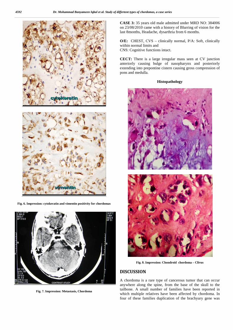
Fig. 6. Impression: cytokeratin and vimentin positivity for chordomas
Fig. 7. Impression: Metastasis, Chordoma
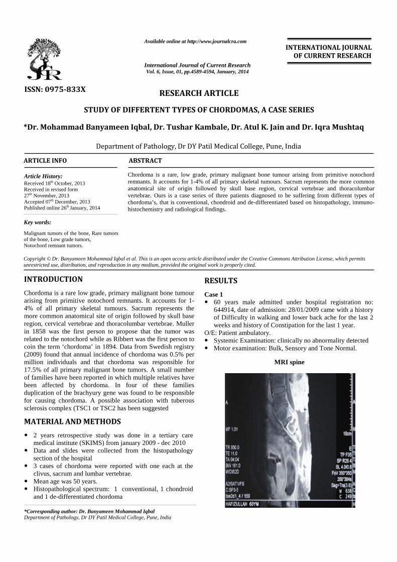
CASE 3: 35 years old male admitted under MRD NO: 384006on 23/08/2010 came with a history of Blurring of vision for thelast 8months, Headache, dysarthria from 6 months.
O/E: CHEST, CVS – clinically normal, P/A: Soft, clinicallywithin normal limits andCNS: Cognitive functions intact.
CECT: There is a large irregular mass seen at CV junctionanteriorly causing bulge of nasopharynx and posteriorlyextending into prepontine cistern causing gross compression ofpons and medulla.
Histopathology
Fig. 8. Impression: Chondroid chordoma – Clivus
DISCUSSION
A chordoma is a rare type of cancerous tumor that can occuranywhere along the spine, from the base of the skull to thetailbone. A small number of families have been reported inwhich multiple relatives have been affected by chordoma. Infour of these families duplication of the brachyury gene was
4592 Dr. Mohammad Banyameen Iqbal et al. Study of differtent types of chordomas, a case series

found to be responsible for causing chordoma. A possibleassociation with tuberous sclerosis complex (TSC1 or TSC2has been suggested
Fig. 9. Alcian blue staining PAS Diastase sensitive
PROGNOSTIC FACTORS
Age Gender Tumour size Positive surgical margins Tumour necrosis Mib-i index Histopathological variant
Fig. 10. Osteocalcin and osteonectin
TREATMENT
Radical enbloc resection. High energy photon/proton beam radiation. There are no drugs currently approved to treat chordoma,
however a clinical trial conducted in Italy using thePDGFR inhibitor Imatinib demonstrated a modest responsein some chordoma patients.
The combination of imatinib and sirolimus caused aresponse in several patients whose tumors progressed onimatinib alone.
Conclusion
1) Low grade primary bone tumour2) Clinically they can present in varied forms3) FNAC has a high sensitivity of diagnosing it.4) Physaliferous cell remains the hallmark of tumor.5) Histopathology & IHC together has high specificity in
diagnosis.6) Recurrence occurs in approximately 40 to 60% of cases.7) Rate of metastasis varies from < 5% to 43 %.8) Radical resection and EBRT remains the gold standard of
treatment
4593 International Journal of Current Research, Vol. 6, Issue, 01, pp.4589-4594, January, 2014

REFERENCES
"Chordoma: current concepts, management, and futuredirections.” Lancet Oncol 13 (2): e69–76. 2012.doi:10.1016/S1470-2045(11)70337-0. PMID 22300861.
"Familial Chordoma Study". Retrieved 2009-02-03."Primary Malignant Bone Tumors: Tumors of Bones and
Joints: Merck Manual Professional". Retrieved 2009-01-04.
Chugh R, Tawbi H, Lucas DR, Biermann JS, Schuetze SM,Baker LH (November 2007). "Chordoma: the nonsarcomaprimary bone tumor". Oncologist 12 (11): 1344–50.doi:10.1634/theoncologist.12-11-1344. PMID 18055855.
Fasig JH, Dupont WD, LaFleur BJ, Olson SJ, Cates JM(February 2008). "Immunohistochemical analysis ofreceptor tyrosine kinase signal transduction activity inchordoma.". Neuropathology and Applied Neurobiology 34(1): 95–104. doi:10.1111/j.1365-2990.2007.00873.x.PMID 17973908.
Han S, Polizzano C, Nielsen GP, Hornicek FJ, Rosenberg AE,Ramesh V (March 2009). "Aberrant Hyperactivation ofAkt and Mammalian Target of Rapamycin Complex 1Signaling in Sporadic Chordomas". Clinical CancerResearch 15 (6): 1940–6. doi:10.1158/1078-0432.CCR-08-2364. PMC 2701205. PMID 19276265.
Lee-Jones L, Aligianis I, Davies PA, et al. (September 2004)."Sacrococcygeal chordomas in patients with tuberoussclerosis complex show somatic loss of TSC1 orTSC2". Genes Chromosomes Cancer 41 (1): 80–5.Doi:10.1002/gcc.20052. PMID 15236319.
Presneau N, Shalaby A, Idowu B, Gikas P, Cannon SR, Gout I,Diss T, Tirabosco R, Flanagan AM (May 2009). "Potentialtherapeutic targets for chordoma: PI3K/AKT/TSC1/TSC2/mTOR pathway". British Journal of Cancer 100 (9):1406–14. doi:10.1038/sj.bjc.6605019. PMC 2694420.PMID 19401700.
*******
4594 Dr. Mohammad Banyameen Iqbal et al. Study of differtent types of chordomas, a case series