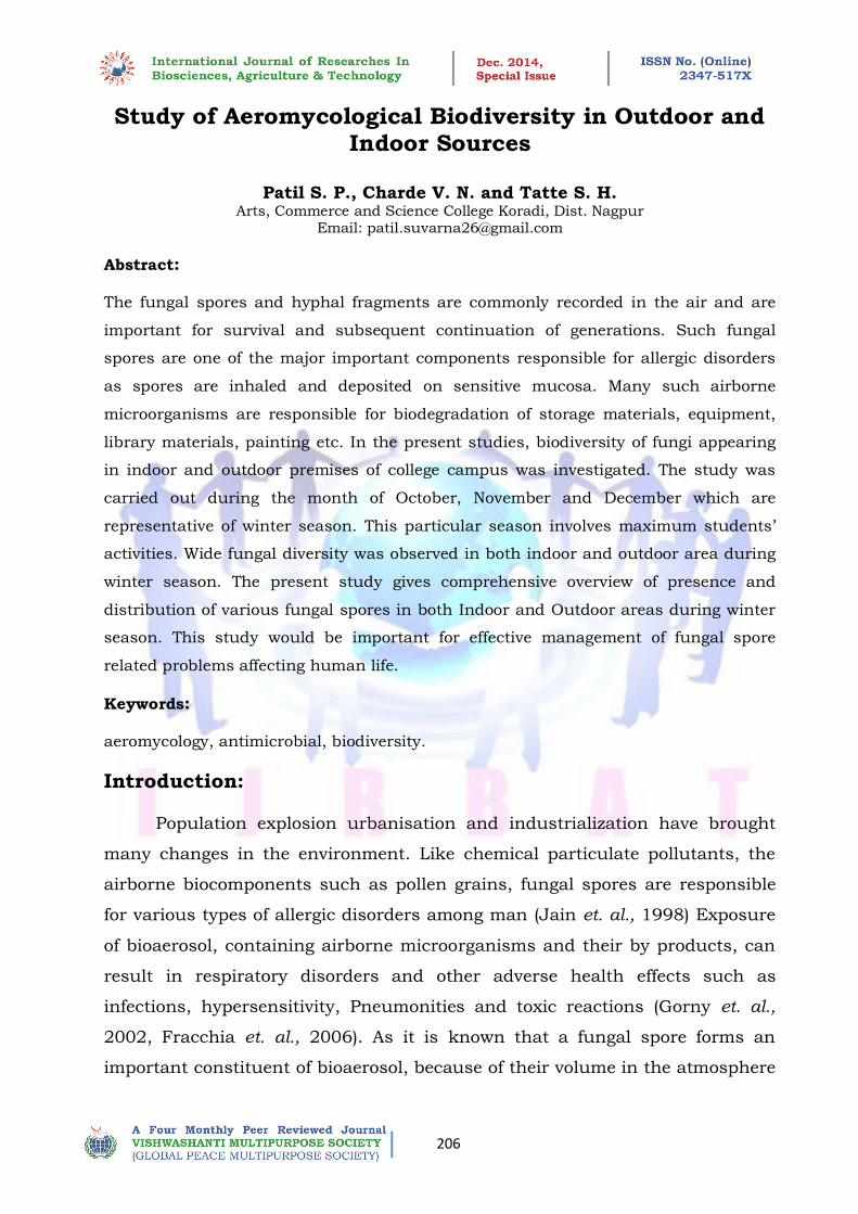
Study of Aeromycological Biodiversity in Outdoor and Indoor Sources
11
| | 206 Study of Aeromycological Biodiversity in Outdoor and Indoor Sources Patil S. P., Charde V. N. and Tatte S. H. Arts, Commerce and Science College Koradi, Dist. Nagpur Email: [email protected] Abstract: The fungal spores and hyphal fragments are commonly recorded in the air and are important for survival and subsequent continuation of generations. Such fungal spores are one of the major important components responsible for allergic disorders as spores are inhaled and deposited on sensitive mucosa. Many such airborne microorganisms are responsible for biodegradation of storage materials, equipment, library materials, painting etc. In the present studies, biodiversity of fungi appearing in indoor and outdoor premises of college campus was investigated. The study was carried out during the month of October, November and December which are representative of winter season. This particular season involves maximum students’ activities. Wide fungal diversity was observed in both indoor and outdoor area during winter season. The present study gives comprehensive overview of presence and distribution of various fungal spores in both Indoor and Outdoor areas during winter season. This study would be important for effective management of fungal spore related problems affecting human life. Keywords: aeromycology, antimicrobial, biodiversity. Introduction: Population explosion urbanisation and industrialization have brought many changes in the environment. Like chemical particulate pollutants, the airborne biocomponents such as pollen grains, fungal spores are responsible for various types of allergic disorders among man (Jain et. al., 1998) Exposure of bioaerosol, containing airborne microorganisms and their by products, can result in respiratory disorders and other adverse health effects such as infections, hypersensitivity, Pneumonities and toxic reactions (Gorny et. al., 2002, Fracchia et. al., 2006). As it is known that a fungal spore forms an important constituent of bioaerosol, because of their volume in the atmosphere
-
Upload
independent -
Category
Documents
-
view
3 -
download
0
Transcript of Study of Aeromycological Biodiversity in Outdoor and Indoor Sources
| |
206
Study of Aeromycological Biodiversity in Outdoor and Indoor Sources
Patil S. P., Charde V. N. and Tatte S. H. Arts, Commerce and Science College Koradi, Dist. Nagpur
Email: [email protected]
Abstract:
The fungal spores and hyphal fragments are commonly recorded in the air and are
important for survival and subsequent continuation of generations. Such fungal
spores are one of the major important components responsible for allergic disorders
as spores are inhaled and deposited on sensitive mucosa. Many such airborne
microorganisms are responsible for biodegradation of storage materials, equipment,
library materials, painting etc. In the present studies, biodiversity of fungi appearing
in indoor and outdoor premises of college campus was investigated. The study was
carried out during the month of October, November and December which are
representative of winter season. This particular season involves maximum students’
activities. Wide fungal diversity was observed in both indoor and outdoor area during
winter season. The present study gives comprehensive overview of presence and
distribution of various fungal spores in both Indoor and Outdoor areas during winter
season. This study would be important for effective management of fungal spore
related problems affecting human life.
Keywords:
aeromycology, antimicrobial, biodiversity.
Introduction:
Population explosion urbanisation and industrialization have brought
many changes in the environment. Like chemical particulate pollutants, the
airborne biocomponents such as pollen grains, fungal spores are responsible
for various types of allergic disorders among man (Jain et. al., 1998) Exposure
of bioaerosol, containing airborne microorganisms and their by products, can
result in respiratory disorders and other adverse health effects such as
infections, hypersensitivity, Pneumonities and toxic reactions (Gorny et. al.,
2002, Fracchia et. al., 2006). As it is known that a fungal spore forms an
important constituent of bioaerosol, because of their volume in the atmosphere
| |
207
and small size, fungal spores play an important role in respiratory allergy and
cause a wide range of symptoms, including allergic rhinitis, asthma, chronic
bronchitis etc. (Tilak S. T. 1991, Vijay et. al., 1991, Hasnain et. al., 1994).
Due to increasing awareness of the relationship of airborne fungi to
allergy scientist began to study the spectrum and incidence of airborne fungi
word wide. Extensive survey conducted in India & abroad on respiratory
allergies indicates that patterns in the incidence of ‘airborne allergens’ differ
considerably from place to place & season to season. Many airborne
microorganisms are responsible for biodegradation of storage materials,
equipment, library materials, painting etc. More than 80 genera of fungi have
been associated with the most commonly identified belonging to three
distinctive fungal groups, Ascomycetes, Basidiomycetes and
Deuteromycetes.(Tilak S.T. 2010) The main types of allergic spores are
Aspergillus, Cladosporium, & Penicillium. The clinical investigation have
provided the significance and utility of treatment is preventing the effect of
aeroallergens to sensitive individual. (Hung et. al., 2011, Chakraborti et.al.,
2012) Therefore it is a need of trained aeromycologist and clinicians.
Materials and Methods:
Media: Dehydrated Potato dextrose agar and Potato dextrose broth medium of
Hi media were used for isolation of fungal species. All media were sterilized in
autoclave at 15lb pressure for 20 minutes.
Reagents: Lacto phenol cotton blue stain solution of Hi media was used for
staining of fungi.
Isolation and identification of Microorganisms:
1) Isolation of fungus species:
1. Three sets of Potato Dextrose Agar (PDA) plates were exposed to air for
10 minutes at five different places, Library, Office, Near Parking, ground
and garden in college premises. The process was repeated in October,
November and December.
2. These exposed plates were incubated at 27 - 30° C for 3 – 4 days.
| |
208
3. Different isolated colonies of fungus were studied further.
2) Identification of Fungus species:
1. Individual colony showing different cultural characteristics was picked
up with sterile loop and streaked on another Potato dextrose agar plates
for obtaining pure culture. These plates were incubated at 27 - 30° C for
2 – 3 days.
2. Cultural characteristics of all fungal isolates were studies after
incubation and loopful of culture was picked up from plates and
streaked on Potato dextrose agar slant and used for further study after
incubation at 27 - 30° C for 2 – 3 days.
Microscopic Identification of fungal morphology:
1. Place a drop of lacto phenol cotton blue on a clean slide.
2. Transfer a small tuff of the fungus preferably with spore and spore
bearing structure into the drop using a flamed cooled needle.
3. Gently tease the material using the mounting needle.
4. Place a cover slip over the preparation taking care that air bubbles do
not trap in.
5. Observed under low and high power objective.
6. Identification of fungal isolates was done microscopically using Lacto
phenol cotton blue stain and their cultural characteristics.
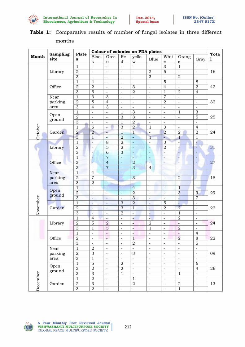
Result and Discussion:
Result showing numbers of fungal isolates differentiated on the basis of
colour of colonies on PDA plates obtained in different areas are given in Table:
1.
Five different sampling sites in college premises were selected i.e.
Library, Office, and area near Parking, Open ground and Garden. The study
was conducted during the winter season, i.e.in the month of October,
November and December. From the above table, it was observed that total 138
| |
209
different coloured colonies were appeared in month of October with maximum
in Office and least in Library. Similarly in the month of November, Library
showed maximum number of fungi growing on plates and minimum in area
near parking with total 127 numbers of isolates. In December there was not
significant variation in no. of fungal isolates appearing on PDA plates in
different areas with total 94 isolates. Wide fungal diversity was observed in
both indoor and outdoor area during winter season.
The isolates were identified on the basis of cultural characteristics on PDA
and microscopic examination. Results of cultural characteristics and
microscopic examination are given in Table 2.
Distribution of different fungal species at different locations and in
different months are given in Table – 3.
In Library area:
In the month of October, the fungal isolates obtained in library were
identified as Penicillium sp., Pythium sp. and Aureobasidium sp. In November
the dominating fungal species were Pythium sp., Trichoderma sp., and
Dreshlera sp., Penicillium sp., Aureobasidium sp., Trichoderma sp. and
Aspergillus sp. were dominantly present in December.
In Office:
In the month of October, the fungal isolates obtained in office were
identified as Pythium sp., Fusarium sp., Aspergillus sp., Aureobasidium sp.,
and Curvularia sp. In November the dominating fungal species were Fusarium
species, Trichoderma species and Penicillium sp., In December, Fusarium sp,
Curvularia, and Aureobasidium sp. were conformed.
Near Parking:
In the month of October, the fungal isolates obtained near Parking were
identified as Pythium sp., Aspergillus sp., Trichoderma sp., Dreshlera sp. In
November the different fungal species were as Aspergillus niger, Fusarium sp.,
Aureobasidium sp. In December, the dominating fungus species were
Aspergillus niger sp., Fusarium sp
| |
210
.Open Ground:
In the month October, fungal isolates obtained in open ground were
Aureobasidium sp., Curvularia sp., Dreshlera sp., Fusarium sp. In November,
the dominating fungal species were Aureobasidium sp., Curvularia sp. In
December, Dreshlera sp., Curvularia sp., Aureobasidium sp., Aspergillus sp.,
fungal spore was obtained.
Garden:
In the month of October, fungal isolates obtained in Garden were
identified as Pythium sp., Curvularia sp., Aureobasidium sp., Aspergillus sp. In
November, the dominating fungal species were Aureobasidium sp., Fusarium
sp., Pythium sp., Dreshlera sp. In December, the dominating fungal were
Fusarium sp., Aureobasidium sp., and Aspergillus niger.
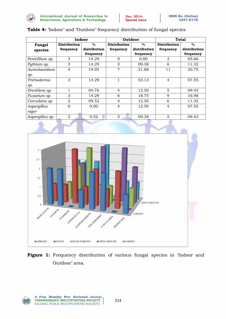
The study was conducted at five different sites; out of these, Library area
and Office area are considered as Indoor area whereas Open Ground and
Garden are considered as Outdoor area. The distribution of fungal spores in
‘Indoor’ and ‘Outdoor’ are important with respect to Aeromycology and their
impact on living things. Frequency distribution of various fungus species in
Indoor and Outdoor are given in Table 4 and also their percent distribution is
represented graphically in figure of 13, 14 and 15 for Indoor and Outdoor and
Total distribution respectively.
The study was conducted in month of October, November and December.
Hence the distribution of fungal spore is representative of winter season.
In Indoor area the order of dominance is Aureobasidium sp. and then
Penicillium sp., Pythium sp, Trichoderma sp., and Fusarium sp, Curvularia sp,
and Aspergillus sp and last Dreshlera sp. In outdoor area, the order of
dominance is Aureobasidium sp and then Fusarium sp. Then Dreshlera sp,
Curvularia sp. and A. niger sp. then Pythium sp. and Aspergillus sp. The most
frequently occurring fungal species in both Indoor and Outdoor was found to
be Aureobasidium sp. Penicillium sp. was found only in Indoor and A.niger sp
was observed only in Outdoor area whereas others were found to present in
both Indoor and Outdoor area.
| |
211
Indoor air is usually exchanged fairly and rapidly by ventilation with
outside air. Due to this exchanged the microbial extent of indoor air tend to
change, however, pollens and spores concentration in indoors is usually lower
than outside. Indoor airs also lower than outside. Indoor air also comprises of
other microbes derived from indoor sources and thrives well due to congenital
environment and organic matter providing suitable substrate. A no. of species
such as Pythium sp, Fusarium sp, Aspergillus sp, Aureobasidium sp,
Trichoderma sp, Curvularia sp, Penicillium sp were observed. Presence of large
no. of pathogenic as well as other species can affect not only official material
but also affects staff members (Tilak S.T. 2009).
In parking areas, no. of fungal spore found to be present depending
upon no. of environmental conditions which is suitable for growth of various
species. A number of species such as Pythium sp. A. niger sp, Fusarium sp,
Aureobasidium sp, Dreshlera sp and Trichoderma sp. was demonstrated.
On ground maximum no. of fungal spores were observed. The percentage
of fungal spores in air approximately 10 times that of pollen dominates. Fungal
spores such as Aureobasidium, Curvularia, Dreshlera, Fusarium, Aspergillus
which are encountered in maximum concentration, However, all these visible
fungal spores in air are not allergic; some may harmless, while some may
cause disease of plants and animals. The occurrence and dominance of fungal
spore in air depends on variety of environmental factors such as rainfall,
humidity, direction etc. According to their abundance which is determined by
environmental parameters, the fungal spores have been identified as wet spora
and dry spora (Tilak S.T. 2009). A no. of fungal spores can be observed in near
plant. In India plant disease forecasting service is at its incipient stage. The
estimation and assessment of inoculums in air forms one of the major bases of
devising an efficient disease forecasting system. A number of fungal spores
such as Pythium sp., Dreshlera sp., Aspergillus sp., Curvularia sp.,
Aureobasidium sp., Fusarium sp., Penicillium sp., A. niger sp. were observed.
Some spores were harmful to various plants, which cause serious damage to
plants (Tilak S.T. 2010).
| |
212
Table 1: Comparative results of number of fungal isolates in three different
months
Month Sampling site
Plates
Colour of colonies on PDA plates Total
Black
Green
Red
yellow
Blue White
Orange
Gray
Octo
ber
Library
1 - - - - - 3 1 -
16 2 - - - - 2 5 - -
3 - - - - 3 - 2 -
Office
1 4 - - - - 5 - 8
42 2 2 - - 3 - 4 - 2
3 5 - - 2 - 1 2 4
Near parking area
1 3 3 - - - 7 - -
32 2 5 4 - - - 2 - -
3 4 3 - - - - - -
Open ground
1 - - 1 3 - - 1 3
25 2 - - 3 3 - - - 5
3 - - 1 2 - - -
Garden
1 6 - 3 2 1 3 - 4
24 2 2 - 1 1 - 2 2 2
3 1 - 2 - 1 - 1 1
Novem
ber
Library
1 - 8 2 - - 3 - -
31 2 - 5 2 - - 2 - -
3 - 6 3 - - - - -
Office
1 - 7 - - - - - -
27 2 - 4 - 2 - - - -
3 - 7 - 3 4 - - -
Near parking area
1 4 - - - - - - -
18 2 7 - - 3 - - 2 -
3 2 - - - - - - -
Open ground
1 - - - 4 - - 1 -
29 2 - - - 2 - - 3 9
3 - - - 3 - - - 7
Garden
1 - - 3 2 - 5 - -
22 2 - - 3 1 - 2 2 -
3 - - 2 - - - 1 -
Decem
ber
Library
1 4 - - - - - 2 -
24 2 5 2 - - 2 - - -
3 1 5 - - 1 - 2 -
Office
1 - - - - - - - 4
22 2 - - - 1 - - 2 8
3 - - - 2 - - - 5
Near parking area
1 2 - - - - - - -
09 2 3 - - 3 - - - -
3 1 - - - - - - -
Open ground
1 5 - 2 - - - - 6
26 2 2 - 2 - - - - 4
3 3 - 1 - - - 1 -
Garden
1 2 - - 1 - - - -
13 2 3 - - 2 - - 2 -
3 2 - - - - - 1 -

| |
213
Table 2: Identification of fungal spores on the basis of colours.
Sr.
No.
Standard Colony Colour
Characteristics on PDA
Colony Colour
Characteristics on
PDA
Identification of
Fungi
1 Blue-green, variously
coloured Green Penicillium sp.
2 Colourless, white White Pythium sp.
3 Pale to bright coloured Pale colour Fusarium sp.
4 Carbon black to deep brown
black Black Colour Aspergillus sp.
5 White or creamy Cream colour Aureobasidium sp.
6 Brown, gray, or black, hairy,
cottony, velvety
Gray to brown colour Curvularia sp.
7 Grey, brown, blackish
brown, hairy, rarely velvety
Blackish brown Dreshlera sp.
8 Typically shades of green of
less often white, grey or
brown
White with green shade Trichoderma sp.
Table 3: Area and Month wise Distribution of different fungal diversity.
Sampling area Fungi isolates
October November December
Library Penicillin sp.
Pythium sp.
Aureobasidium sp.
Pythium sp.
Trichoderma sp.
Dreshlera sp.
Penicillium sp.
Aureobasidium sp.
Trichoderma sp.
Aspergillus sp.
Office Pythium sp.
Fusarium sp.
Aspergillus niger.
Aureobasidium sp.
Curvularia sp.
Fusarium sp.
Trichoderma sp.
Penicillin sp.
Fusarium sp.
Curvularia sp.
Aureobasidium sp.
Near Parking Pythium sp.
Aspergillus sp.
Trichoderma sp.
Aspergillus niger .
Fusarium sp.
Aureobasidium sp.
Aspergillus niger.
Fusarium sp.
Open Ground Aureobasidium sp.
Curvularia sp.
Dreshlera sp.
Fusarium sp.
Aureobasidium sp.
Fusarium sp.
Curvularia sp.
Dreshlera sp.
Curvularia sp.
Aureobasidium sp.
Aspergillus niger sp.
Garden Pythium sp.
Curvularia sp.
Aureobasidium sp.
Aspergillus niger .
Aureobasidium sp.
Fusarium sp.
Pythium sp.
Dreshlera sp.
Fusarium sp.
Aureobasidium sp.
Aspergillus niger .
| |
214
Table 4: ‘Indoor’ and ‘Outdoor’ frequency distribution of fungal species
Indoor Outdoor Total
Fungal
species
Distribution
frequency
%
distribution
frequency
Distribution
frequency
%
distribution
frequency
Distribution
frequency
%
distribution
frequency
Penicillium sp. 3 14.29 0 0.00 3 05.66
Pythium sp. 3 14.29 3 09.38 6 11.32
Aureobasidium
sp.
4 19.05 7 21.88 11 20.75
Trichoderma
sp.
3 14.29 1 03.13 4 07.55
Dreshlera sp. 1 04.76 4 12.50 5 09.43
Fusarium sp. 3 14.29 6 18.75 9 16.98
Curvularia sp. 2 09.52 4 12.50 6 11.32
Aspergillus
niger
0 0.00 4 12.50 4 07.55
Aspergillus sp. 2 9.52 3 09.38 5 09.43
Figure 1: Frequency distribution of various fungal species in ‘Indoor and
Outdoor’ area.
LIBRARY
OPEN GROUND0
0.5
1
1.5
2
2.5
3
LIBRARY OFFICE NEAR PARKING OPEN GROUND GARDEN
| |
215
Conclusion:
Fungal allergen exposure is associated with development and severity of
asthma in sensitized individuals. The contribution of indoor fungal allergens
exposure in allergic disease is still not completely clear. Method to assess
fungal allergens by using immunoassays is still in their infancy. More
traditional methods of exposure assessment with spore counts and
quantitative cultures suggests that indoor fungal exposure indeed contributes
of allergenic airway disease. The presence of fungal growth in home or offices
implies a problematic measure to decrease, the infiltration of air from outdoor
environment control indoor moisture problems and clean or remove
contaminated material may improve health of individuals with fungal induced
allergic diseases. The present study gives comprehensive overview of presence
and distribution of various fungal spores in both Indoor and Outdoor areas
during winter season. This study would definitely pave the way for effective
management of fungal spore related problem affecting human life.
Reference:
Chakaraborti H., Das S. and Bhattachrya S. (2012) Outdoor airborne fungal
spora load in a suburb of Kolkata, India Its variation, metrological determinants & health impact.. Inter. J. Environ. Health Res,Vol 22(1); 37-50.
Francchia L., Pietronave S., Rinaldi M. and Martinotti M.G. (2006). The aassesment of airborne bacterial contaminator in three composting plants
revealed site related biological hazard and seasonal variation. J. Appl. Environ Microbiol. 100 ( 5) ; 973-984.
Gorny R. L., Reponen T. Willeke K., Schnechel D., Robine E., Boissier M
and Grinshpum S. A. (2002) Fungal fragments as indoor air biocontaminants.
J. Appl. Environ. Microbiol. 68(7); 3522 – 3531.
Hasnain S. M., Al- Frayh A. S., Harfi H. A., Gad-el- Rab M.O., Al- Moberik
K. And Al- Sedairy S. T. (1994). Cladosporium as an airborne allergen in
Saudi Arabia, Ann Saudi Med. Vol.14; 142-146.
Hung W., Su S., Shiu, L. and Chang T. C. (2011). Rapid identification of
allergic pathogenic moulds in environmental air by an oligonucleotide array. BMC infectious diseases, Vol. 11; 91.
| |
216
Jain A. K. Datta T. R. and Narang K. (1998). Airborn fungi and
hypersensitivity among allergenic patients at Gwalior. Environment and Ecology (Ed Ashok Jain) research periodicals and book publishing house. Taxes USA.
257 -265.
Tilak S. T. ( 2009) Aeromycology U.S. publication, Pune.
Tilak S. T. (1991) Fungal spores and allergy. J Palynol vol. 27; 32-40.
Tilak S. T. (2010) Aerobiology to astrology. publish by Bharti vidyapeeth
Deemed University, Pune-30
Vijay H. M., Burton M., Young N. M., Copeland D. F. and Corlett M. (1991)
Allergenic components of isolets of cladosporium herbarum, Grana Vol. 30;
161-165.