Regulation of Fructose Metabolism and Polymer Synthesis by ...
doi:10.1016/j.jmb.2005.01.008 J. Mol. Biol. (2005) 347, 135–144
Structure of the Thermolabile Mutant Aldolase B, A149P:Molecular Basis of Hereditary Fructose Intolerance
Ali D. Malay1, Karen N. Allen2* and Dean R. Tolan1*
1Biology Department, BostonUniversity, Boston, MA 02215USA
2Department of Physiology andBiophysics, Boston UniversitySchool of Medicine, BostonMA 02118, USA
0022-2836/$ - see front matter q 2005 E
Abbreviations used: DHAP, dihyphosphate; Fru-1,6-P2, fructose-1,6-bfructose 1-phosphate; HFI, hereditaintolerance; pdb, Protein Data BankE-mail addresses of the correspon
[email protected]; [email protected]
Hereditary fructose intolerance (HFI) is a potentially lethal inborn error inmetabolism caused bymutations in the aldolase B gene, which is critical forgluconeogenesis and fructose metabolism. The most common mutation,which accounts for 53% of HFI alleles identified worldwide, results insubstitution of Pro for Ala at position 149. Structural and functionalinvestigations of human aldolase B with the A149P substitution (AP-aldolase) have shown that the mutation leads to losses in thermal stability,quaternary structure, and activity. X-ray crystallography is used to revealthe structural basis of these perturbations. Crystals of AP-aldolase aregrown at two temperatures (4 8C and 18 8C), and the structure solved to3.0 A resolution, using the wild-type structure as the phasing model. Thestructures reveal that the single residue substitution, A149P, causesmolecular disorder around the site of mutation (residues 148–159), whichis propagated to three adjacent b-strand and loop regions (residues 110–129, 189–199, 235–242). Disorder in the 110–129-loop region, whichcomprises one subunit–subunit interface, provides an explanation for thedisrupted quaternary structure and thermal instability. Greater structuralperturbation, particularly at a Glu189-Arg148 salt bridge in the active-sitearchitecture, is observed in the structure determined at 18 8C, which couldexplain the temperature-dependent loss in activity. The disorder revealedin these structures is far greater than that predicted by homology modelingand underscores the difficulties in predicting perturbations of proteinstructure and function by homology modeling alone. The AP-aldolasestructure reveals the molecular basis of a hereditary disease and representsone of only a few structures known for mutant proteins at the root of thethousands of other inherited disorders.
q 2005 Elsevier Ltd. All rights reserved.
Keywords: unstable protein; X-ray crystallography; thermal stability;molecular disorder; homology modeling
*Corresponding authorsIntroduction
Hereditary fructose intolerance (HFI) is an auto-somal recessive disorder that afflicts approximatelyone in 20,000 individuals.1,2 In those affected, theingestion of fructose from food or other sourcesleads to a multifaceted array of symptoms that mayinclude vomiting, gastrointestinal pain, hypo-glycemia, and liver and kidney failure.2 The failure
lsevier Ltd. All rights reserve
droxyacetoneisphosphate; Fru-1-P,ry fructose.ding authors:u
to diagnose HFI can easily be fatal due to persistentor inadvertent ingestion of fructose.3
HFI is caused by a deficiency in the activity ofaldolase B, the isozyme of class I aldolase expressedin the liver, small intestine, and kidneys.4,5 Amongthe three vertebrate aldolase isozymes (A, B, and C),aldolase B is responsible for cleavage of themetabolic intermediate fructose 1-phosphate(Fru-1-P) into dihydroxyacetone phosphate(DHAP) and glyceraldehyde in the fructosemetabolism pathway. In addition, aldolase B isimportant for glucose homeostasis in the liver viathe synthesis of fructose 1,6-bisphosphate (Fru-1,6-P2) from DHAP and glyceraldehyde 3-phosphate aspart of gluconeogenesis2. Defects in the aldolase Bgene have been found to cause HFI.6 Currently, 24
d.

136 Molecular Basis of Hereditary Fructose Intolerance
mutations in the aldolase B gene are known to beassociated with HFI. The most prevalent alleleencodes an Ala/Pro substitution at position 149and accounts for bout 55% of HFI alleles identifiedworldwide7. Assuming Hardy–Weinberg equi-librium8 of this allele in all HFI patients,approximately 80% of HFI patients carry at leastone copy of A149P.
The overexpression, purification, and character-ization of the aldolase B enzyme carrying the A149Pmutation (AP-aldolase) shows a disruption ofquaternary structure and partial activity, albeitwith a marked temperature sensitivity.9 The specificactivity toward Fru-1,6-P2 and Fru-1-P decreasesfrom 16% to 0.5% of wild-type levels between 10 8Cand 30 8C, with a T1/2 of 25 8C. Circular dichroismexperiments similarly revealed decreased stabilityof the tertiary and secondary protein structures ofAP-aldolase, with T1/2 values of 33 8C and 45 8C,respectively, as compared to the wild-type values of43 8C and 50 8C. On the other hand, changes in theKm values of AP-aldolase with respect to the wild-type enzyme were temperature-independent, butshowed considerably greater effects for the sub-strates DHAP and Fru-1-P, suggesting a structuralperturbation at the C1-phosphate binding site ofAP-aldolase.9 Further evidence shows some linkbetween the temperature-sensitive loss of activityand the loss of quaternary structure. In contrast tothe tetrameric wild-type aldolase B, AP-aldolaseforms dimers in solution from 4 8C to 30 8C, withsome dissociation into monomeric species above30 8C.9 This finding is in agreement with thecanonical role of the quaternary structure ofaldolase in promoting structural stability.10 Inter-estingly, several other mutations of aldolase Bassociated with HFI,11,12 as well as mutations inthe aldolase A isozyme associated with a rare formof hemolytic anemia,13,14 have been shown to affectquaternary structure.
Although the structure of O1000 proteinsinvolved in biological pathways have beendetermined, only a handful are representative ofdysfunctional proteins resulting from geneticmutations.15–18 This is due to the inherentdifficulty of structural analysis of unstableproteins. Here, the structure of one such protein,AP-aldolase, is determined by X-ray crystallo-graphy at 4 8C and 18 8C, revealing the molecularbasis of the structural perturbations and loss ofactivity with increased temperature. The structuresshowed that the point mutation causes extensivestructural perturbation both at the site of substi-tution and in adjacent distal loop regions, providingimportant insights into the mutant phenotype. Theextent of disorder is reflected in the model statistics,as has been reported for other unstable proteins.19
This study lends insight into the molecularprocesses of disease pathogenesis and gives aview of the utility of modeling studies in under-standing the molecular perturbations inherent in“mutant” proteins.
Results
Overall structure
AP-aldolase is a thermolabile form of aldolase B,which exhibits loss in secondary and tertiarystructure at 45 8C and 33 8C, respectively. However,catalytic activity is lost at O15 8C that is notaccounted for by changes in the CD spectra.9 TheX-ray crystal structure of AP-aldolase was solved at4 8C and 18 8C in order to visualize the structuralchanges occurring at a temperature where thedecline in catalytic activity is most acute. It shouldbe noted that the crystals grown at 4 8C weretransferred into cryo conditions at 4 8C and frozenin liquid nitrogen to prevent any exposure to highertemperatures during mounting on the X-ray beam.
The AP-aldolase crystals showed relatively weakdiffraction, although improved resolution wasobtained from larger crystals. In addition to thedata presented here, three to four complete datasetsof AP-aldolase were collected at each temperature.Overall anisotropy in the diffraction pattern wasalways observed, regardless of the crystal growthtemperature or cryoprotectant conditions. The datawere consistently weakest along the l-axis, with aresolution limit for strong reflections of about 4 Aon that axis, while the h and k-axes had resolutionlimits of 3 A or better. The anisotropic alignment ofthe reflections along the crystallographic axessignifies that it is an inherent property of the crystalform, and not merely due to stochastic factors. Inaddition, this interpretation is fully consistent withthe anisotropic distribution of the B-factors. In thestructure of AP-aldolase at 18 8C, for example, theoverall anisotropic temperature factors (B11Z16.20 A2, B22Z12.39 A2, and B33ZK28.59 A2),together with the average B-factor for the structure(60.3 A2), signifies that the effective averageB-factors in the x, y, and z directions are 44.1 A2,47.91 A2, and 88.89 A2, respectively. It should benoted that the scaling together or merging ofseparate datasets failed to improve completenessor dataset statistics.
In both AP-aldolase structures, local effects of thesubstitution and propagated effects on structuralorder were observed, as reflected by the relativelyhigh overall B-factors. Similar disorder wasobserved in a recent study on the structure ofglutathione reductase from Plasmodium falciparum.19
In this investigation, the structure was solved to2.6 A resolution by molecular replacement, with thefinal model having RworkZ25.1%, RfreeZ30.2%, andan average protein B-factor of 80 A2. The structurehad areas of missing density in three differentregions of the protein, corresponding to 27, 32, andfour residues (out of a total of 478 residues in themonomer), similar to observations reported here.The authors argued that the model with thedisordered regions was correct based on obser-vations from annealed omit maps, anisotropy of thecrystals along one crystal axis, a reasonable R-factordistribution versus resolution, and reasonable
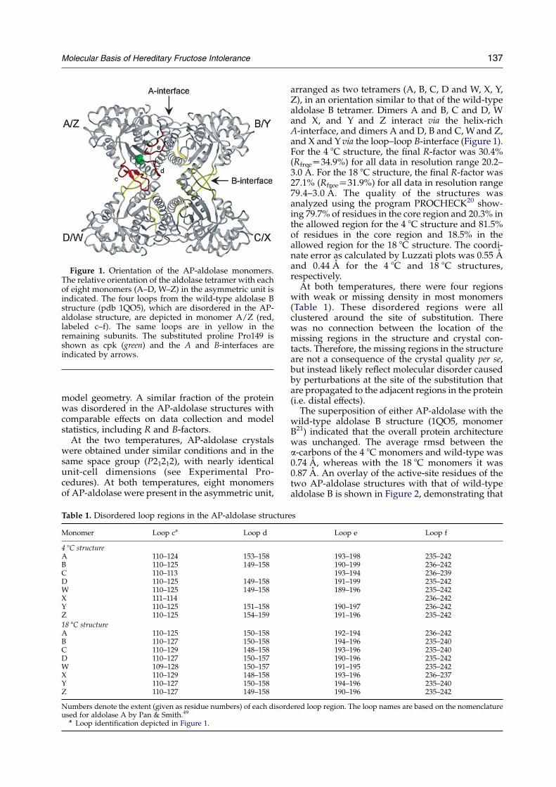
Figure 1. Orientation of the AP-aldolase monomers.The relative orientation of the aldolase tetramer with eachof eight monomers (A–D, W–Z) in the asymmetric unit isindicated. The four loops from the wild-type aldolase Bstructure (pdb 1QO5), which are disordered in the AP-aldolase structure, are depicted in monomer A/Z (red,labeled c–f). The same loops are in yellow in theremaining subunits. The substituted proline Pro149 isshown as cpk (green) and the A and B-interfaces areindicated by arrows.
Molecular Basis of Hereditary Fructose Intolerance 137
model geometry. A similar fraction of the proteinwas disordered in the AP-aldolase structures withcomparable effects on data collection and modelstatistics, including R and B-factors.
At the two temperatures, AP-aldolase crystalswere obtained under similar conditions and in thesame space group (P21212), with nearly identicalunit-cell dimensions (see Experimental Pro-cedures). At both temperatures, eight monomersof AP-aldolase were present in the asymmetric unit,
Table 1. Disordered loop regions in the AP-aldolase structur
Monomer Loop ca Loop d
4 8C structureA 110–124 153–158B 110–125 149–158C 110–113D 110–125 149–158W 110–125 149–158X 111–114Y 110–125 151–158Z 110–125 154–159
18 8C structureA 110–125 150–158B 110–127 150–158C 110–129 148–158D 110–127 150–157W 109–128 150–157X 110–129 148–158Y 110–127 150–158Z 110–127 149–158
Numbers denote the extent (given as residue numbers) of each disordused for aldolase A by Pan & Smith.49
a Loop identification depicted in Figure 1.
arranged as two tetramers (A, B, C, D and W, X, Y,Z), in an orientation similar to that of the wild-typealdolase B tetramer. Dimers A and B, C and D, Wand X, and Y and Z interact via the helix-richA-interface, and dimers A and D, B and C, Wand Z,and X and Yvia the loop–loop B-interface (Figure 1).For the 4 8C structure, the final R-factor was 30.4%(RfreeZ34.9%) for all data in resolution range 20.2–3.0 A. For the 18 8C structure, the final R-factor was27.1% (RfreeZ31.9%) for all data in resolution range79.4–3.0 A. The quality of the structures wasanalyzed using the program PROCHECK20 show-ing 79.7% of residues in the core region and 20.3% inthe allowed region for the 4 8C structure and 81.5%of residues in the core region and 18.5% in theallowed region for the 18 8C structure. The coordi-nate error as calculated by Luzzati plots was 0.55 Aand 0.44 A for the 4 8C and 18 8C structures,respectively.At both temperatures, there were four regions
with weak or missing density in most monomers(Table 1). These disordered regions were allclustered around the site of substitution. Therewas no connection between the location of themissing regions in the structure and crystal con-tacts. Therefore, the missing regions in the structureare not a consequence of the crystal quality per se,but instead likely reflect molecular disorder causedby perturbations at the site of the substitution thatare propagated to the adjacent regions in the protein(i.e. distal effects).The superposition of either AP-aldolase with the
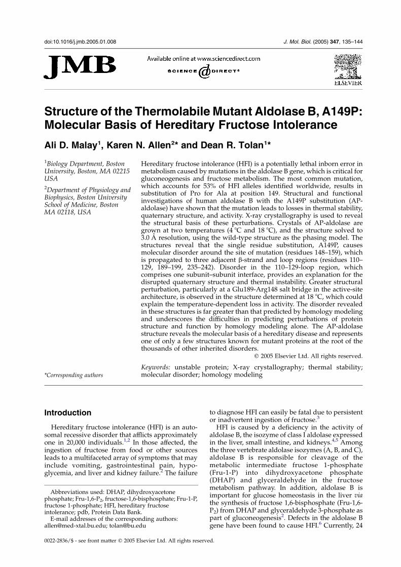
wild-type aldolase B structure (1QO5, monomerB21) indicated that the overall protein architecturewas unchanged. The average rmsd between thea-carbons of the 4 8C monomers and wild-type was0.74 A, whereas with the 18 8C monomers it was0.87 A. An overlay of the active-site residues of thetwo AP-aldolase structures with that of wild-typealdolase B is shown in Figure 2, demonstrating that
es
Loop e Loop f
193–198 235–242190–199 236–242193–194 236–239191–199 235–242189–196 235–242
236–242190–197 236–242191–196 235–242
192–194 236–242194–196 235–240193–196 235–240190–196 235–242191–195 235–242193–196 236–237194–196 235–240190–196 235–242
ered loop region. The loop names are based on the nomenclature
Figure 2. Overlay of the active sites of AP-aldolase andwild-type aldolase B. Overlay of the active-site residuesare depicted for wild-type aldolase B (cyan, pdb 1QO5,chain B), AP-aldolase structure at 4 8C (yellow, monomerX), and AP-aldolase structure at 18 8C (red, monomer B).
138 Molecular Basis of Hereditary Fructose Intolerance
the orientation of active-site residues of AP-aldolasewas intact at the two temperatures. The exceptionwas the side-chain of Arg303, which the electrondensity clearly showed was oriented closer toLys229 (5.8 A compared to 9.4 A in wild-type) inall eight monomers at both temperatures. Arg303 isa conserved residue in the aldolase active site andmay be important in the catalytic mechanism. Thealdolase mechanism proceeds via binding hexose,Schiff-base formation, C3–C4 bond cleavage, releaseof the first triose, Schiff-base hydrolysis, and finally,release of the second triose.22 Arg303 has beenshown to have alternative capacities in binding tothe C6-phosphate of the linear hexose substrate,23
or the C1-phosphate of the covalently boundproduct,24 or to a residue near the C terminus(Glu354) in an enzyme–product complex.25 ThusArg303 switches roles between binding the sub-strate/product and binding the C terminus. Giventhat the rate-limiting step is product release26,27 andlikely involves a conformational change in the Cterminus, this succession of liganded structures
Figure 3. Stereo view of the active site. The 18 8CAP-aldolascontoured at a 1.0s.
indicated that Arg303 may be a “trigger” residue inmodulating the action of the C terminus duringcatalysis.24,28 This new conformation of Arg303in the AP-aldolase structures could explain theOtenfold loss in activity at all temperatures.9 Theelectron density shown in Figure 3 around Arg303and other active-site residues from monomer Wwas typical for all 16 monomers.
AP-aldolase structure at 4 8C
The electron density maps of the AP-aldolasestructure solved at 4 8C, where AP-aldolase hasmaximal activity,9 had four loop regions of weak ormissing density in six of eight monomers. Alsomissing were the first residues at the N terminusand the last 18–21 residues of the C-terminal region,which are not visible in most class I-aldolasestructures.21,23,29,30 The four disordered regions(loops c, d, e, and f; see Figure 1 and Table 1) arelocalized at the C-terminal end of the b-strandcontaining the site of the substitution and in theloop immediately following (residues 149–158), aswell as in two flanking regions (residues 190–197and 236–242) and the large region from residues110–125. Monomers X and C were the exception,with complete density visible at the site of substi-tution (residues 149–158; loop d) as well as for mostor all residues in loops c and e (Table 1). All threeloops had electron density corresponding to thewild-type aldolase B structure, showing that AP-aldolase at 4 8C can adopt a conformation similar tothat of wild-type (Figure 4(a)). An examination ofcrystal contacts showed that monomers X and C arenot stabilized to any greater extent than the othermonomers by packing interactions. Poor electrondensity was visible at some of the regions where amodel was not built. For example, in monomer Drepeated attempts to build a model for residues115–119 and 121–125 resulted in increased R-factorsupon refinement.
In the regions where density was visible, thestructure of AP-aldolase was very similar to that ofwild-type aldolase B. The missing regions werelikely a consequence of molecular disorder in theloops including and flanking the substituted
e structure (monomerW) is depictedwith the 2FoKFc map
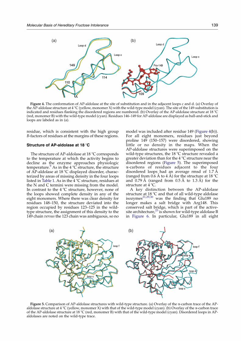
Figure 4. The conformation of AP-aldolase at the site of substitution and in the adjacent loops c and d. (a) Overlay ofthe AP-aldolase structure at 4 8C (yellow, monomer X) with the wild-typemodel (cyan). The site of the 149-substitution isindicated and residues flanking the disordered regions are numbered. (b) Overlay of the AP-aldolase structure at 18 8C(red, monomer B) with the wild-typemodel (cyan). Residues 146–149 for AP-aldolase are displayed as ball-and-stick andloops are labeled as in (a).
Molecular Basis of Hereditary Fructose Intolerance 139
residue, which is consistent with the high groupB-factors of residues at the margins of these regions.
Structure of AP-aldolase at 18 8C
The structure of AP-aldolase at 18 8C correspondsto the temperature at which the activity begins todecline as the enzyme approaches physiologictemperature.9 As in the 4 8C structure, the structureof AP-aldolase at 18 8C displayed disorder, charac-terized by areas of missing density in the four loopslisted in Table 1. As in the 4 8C structure, residues atthe N and C termini were missing from the model.In contrast to the 4 8C structure, however, none ofthe loops showed complete density in any of theeight monomers. Where there was clear density forresidues 148–150, the structure deviated into theregion occupied by residues 123–125 in the wild-type structure, the assignment of this density to the149 chain versus the 123 chain was ambiguous, so no
Figure 5. Comparison of AP-aldolase structures with wild-taldolase structure at 4 8C (yellow, monomer X) with that of theof the AP-aldolase structure at 18 8C (red, monomer B) with thaldolases are noted on the wild-type trace.
model was included after residue 149 (Figure 4(b)).For all eight monomers, residues just beyondproline 149 (150–157) were disordered, showinglittle or no density in the maps. When theAP-aldolase structures were superimposed on thewild-type structures, the 18 8C structure revealed agreater deviation than for the 4 8C structure near thedisordered regions (Figure 5). The superimposeda-carbons of residues adjacent to the fourdisordered loops had an average rmsd of 1.7 A(ranged from 0.6 A to 4 A) for the structure at 18 8Cand 0.79 A (ranged from 0.5 A to 1.3 A) for thestructure at 4 8C.A key distinction between the AP-aldolase
structure at 18 8C and that of all wild-type aldolaseisozymes21,29,30 was the finding that Glu189 nolonger makes a salt bridge with Arg148. Thisconserved salt bridge, which is part of the active-site architecture,23 is shown for wild-type aldolase Bin Figure 6. In particular, Glu189 in all eight
ype structure. (a) Overlay of the a-carbon trace of the AP-wild-type model (cyan). (b) Overlay of the a-carbon traceat of the wild-type model (cyan). Disordered loops in AP-

Figure 6. Loss of a conserved salt-bridge at active site inAP-aldolase. Overlay of the AP-aldolase structure at 18 8C(red, monomer W) with the wild-type model (cyan)rendered as coils with the Glu189/Arg148 salt-bridge andresidue 149 rendered as ball-and stick.
140 Molecular Basis of Hereditary Fructose Intolerance
monomers of the 18 8C structure was oriented in theopposite direction from that in the wild-typestructure. Glu189 appeared to make no contacts inthis swung-out position, however, the formerpartner, Arg148, was within 3.5–3.9 A of Glu187 inmonomers A and W of the 18 8C structure. In theother monomers Arg148 was either not in the modelor was O4.3 A from Glu187, sometimes with anintervening water molecule. Glu187 is important inthe mechanism of Schiff-base chemistry acting as ageneral acid catalyst in the dehydration of thecarbinolamine intermediate24,31 (Choi, K. H.,K.N.A. & D.R.T., unpublished results). A new saltbridge formed with Glu187 could interfere with thenormal catalytic mechanism by hindering acid–basechemistry. Whereas the absence of the Glu189/Arg148 salt bridge was found in all monomers inthe structure at 18 8C, only monomers B and Yshowed this loss in the 4 8C structure. Thus, thealtered conformation of Glu189 may be a likelyexplanation for the temperature-dependent declinein activity at temperatures above 15 8C.
Discussion
Frequently the instability of mutant proteinsimplicated in human disease discourages empiricalexperimental structure/function studies.Homology modeling is used to map the conse-quences of deleterious mutations onto structures ofnative or structurally related proteins in order topredict the structural and functional consequencesof the mutation.32–34 However, the necessity ofusing a modeling approach by its nature makesvalidation difficult without sufficient empiricalstructural studies on similar systems. In one study,a theoretical modeling program was demonstratedto be no better at predicting deleterious effects of
missense mutations than a program that madepredictions based solely on conservation of primarystructure among homologous proteins.35 The utilityof structure determination of mutant proteins isexemplified by the study of the hemoglobinstructure in sickle-cell anemia.36 Despite this fact,only a handful of structures have been determinedfor proteins from missense mutations involved inhuman diseases.16–18 Thus, we have undertaken thestructure of unstable mutant proteins involved inHFI to assess the extent of structural changes andtheir relationship to the loss of function.
Structural consequences of the Ala/Prosubstitution in AP-aldolase
The unique nature of a proline substitutionimposes severe constraints on the polypeptidebackbone. In AP-aldolase the substitution of prolineclearly disrupts the native b-strand structure atposition 149. In addition, this perturbation results ina change in structure at positions that play a role incatalysis, i.e. Arg303 and Glu189/Arg148, and thereare distal effects imposed on the loop regionsflanking both sides of the substitution. Thedeviation of loop d into the region occupied byloop c leads to disorder in loop c and disrupts theconformation necessary for oligomerization via theB-interface.10 In addition, the shift at the site ofsubstitution causes changes in loops e and f. Thus,the structure of AP-aldolase demonstrates that thesingle missense mutation causes extensive distaleffects to the protein structure. Previous studieshave shown that the quaternary structure of AP-aldolase is disrupted, adopting a dimeric state insolution, instead of the wild-type tetramer.9 It islikely that the thermolability displayed by themutant enzyme is a direct consequence of itsinability to properly oligomerize.10,13,14,37
Effects on catalysis
Previous investigations have shown that purifiedAP-aldolase exhibits low levels of catalytic activitytoward the reactants normally utilized by aldolasein vivo. The residual activity is extremely thermo-labile, with a T1/2 of 25 8C and a peak kcatcorresponding to 16% of wild-type activity at astable temperature of 10 8C.9 A comparison of theAP-aldolase structures with wild-type aldolasesuggests that changes in three active-site residues,Arg148, Glu189, and Arg303, could account for thekcat decrease. Arg303 is a multifunctional residuethat has been hypothesized to undergo a confor-mational switch during catalysis,24,28 and could beinvolved in the release of DHAP, the rate-limitingstep for both aldolase A27,38 and aldolase B (Choi,K. H. & D.R.T., unpublished results). Mutations atthis site (R303A in aldolase A and R303W andR303Q in aldolase B) demonstrate an effect on boththe kcat and Km values.23,39,40 In wild-type aldolase,Arg148 and Glu189 form a conserved salt bridgethat constitutes one surface of a cleft leading to the

Molecular Basis of Hereditary Fructose Intolerance 141
active site.21,24,29 Mutations at either of these tworesidues in aldolase A (E189A and R148A) lead to a70–300-fold decrease in kcat value.
41 The change inthe orientation of Glu189 seen predominantly in the18 8C AP-aldolase structure, which was caused bythe perturbation of loop e adjacent to the site ofsubstitution, was consistent with the decreasedcatalytic activity. The disordered loop regions andconsequences near the active site were moreextensive at 18 8C compared to those at 4 8C. Thus,the structural perturbation associated with a localconformational change explains the temperaturedependence of the AP-aldolase structure.
Insights into the viability of homology modelingas a predictive tool
The use of homology modeling for AP-aldolase,along with many other substitution mutations thathave been found as the cause of genetic diseases,have been reviewed.42 The result of homologymodeling predicted that the A149P mutation leadsto backbone strain in the b-strand, possibly causingprotein instability, with no prediction of anypropagated effects. The findings from the AP-aldolase structure raise general issues regardingthe validity of using homology modeling as apredictive tool for the deleterious effects ofmutations on protein function. The AP-aldolasestructures showed “distal effects” of the single-residue substitution, in that three regions (loops c, e,and f) aside from the site of substitution aresignificantly affected in the mutant structure. Thelimitations of a theoretical modeling approach wereespecially underscored by the changes observed inthe quaternary structure of AP-aldolase. Even witha priori knowledge of a quaternary structuralperturbation, a theoretical approach would nothave predicted with any certainty which of thesubunit interfaces were actually disrupted, sincethey are equidistant from the site of the substitution.
X-ray crystallography can reveal the structuralconsequences of disease-causing mutations thatcannot easily be anticipated from homology model-ing. This study will provide a basis for improvingsuch theoretical predictions. Herein, the structure ofAP-aldolase was solved at two temperatures, givinga detailed picture of the temperature-dependentloss of structure. This includes an altered confor-mation for a conserved salt-bridge at the active siteexplaining the loss in activity with increasedtemperature. The disordered regions correspondto loops involved in the dimer–dimer interface inthe wild-type tetramer, thus revealing the basis forthe known loss in quaternary structure of AP-aldolase.9 The prevalence of the A149P mutation inHFI makes AP-aldolase a very good potential targetfor therapies that would aim to restore stability andfunctionality to the thermolabile enzyme usingsmall molecule ligands. The structure makespossible the search for such ligands via screeningof chemical libraries in combination with structure-based ligand design.
Experimental Procedures
Purification and crystallization
AP-aldolase was produced by heterologous expressionin Escherichia coli DH5a as a glutathione-S-transferase-fusion protein and subsequent cleavage to yield the fulllength, purified, active protein, as described9. The crystalswere obtained by the vapor diffusion method withhanging-drop geometry, by mixing equal volumes of20 mg/ml purified AP-aldolase in 1 mM phosphate-buffered saline (pH 7.4), 1 mM DTT, with 1.8–2.2 M(NH4)2SO4 and 2% (w/v) 1,8-diaminooctane at 4 8C andequilibrating over wells containing the same buffer. Forthe crystals grown at 18 8C, crystallization trays weretransferred to 18 8C after setup and overnight incubationat 4 8C. Crystals appeared within one to two weeks.
Data collection
Data for the 4 8C crystals were collected on beamlineX4A at the National Synchrotron Light Source atBrookhaven National Laboratories. Cryoprotection con-ditions consisted of successive transfers of single crystalsat 4 8C into a drop of well solution containing 15% (w/v)glucose for one minute, and into a drop of well solutioncontaining 30% glucose plus 5% (v/v) glycerol for30 seconds. The crystals were frozen in liquid nitrogenand transferred to a stream of nitrogen gas cooled to 93 K.A full dataset was collected to 2.6 A using a quad4detector, with 0.58 oscillations for 1808 total, and a crystal-to-detector distance of 200 mm. The Denzo and Scalepackpackages43 were used for data indexing, reduction, andscaling. The space group was determined to be P21212,with unit cell dimensions of aZ153.48 A, bZ153.51 A, cZ186.48 A.Prior to data collection, the 18 8C crystals were
dehydrated44,45 at the same temperature by vapordiffusion against 3.5 M (NH4)2SO4 for three hours.Paratone-N was used as cryoprotectant and the crystalswere frozen as for the 4 8C crystals. Data were collected atthe Boston University School of Medicine on a RigakuRU300 X-ray generator equipped with an R-Axis IVCC
area detector. A full dataset was collected at 2.7 A with 18oscillations for 1808 total, and a crystal-to-detectordistance of 200 mm. The space group was determined tobe P21212, with unit cell dimensions of aZ153.42 A, bZ153.48 A, cZ185.55 A. Due to the high Rsym values in theouter shell, both data sets were refined to 3.0 A.
Molecular replacement
The AP-aldolase structure was solved by molecularreplacement using the program AMoRe46 and thestructure of wild-type aldolase B (pdb 1QO5;21) as theinitial search model. The best search model consisted of adimer, as opposed to the tetramer (the A–B dimer10).Eight monomers were identified in the asymmetric unit(designated A–D and W–Z), arranged as two tetramers,consistent with the wild-type oligomeric structure.Orientation of the eight monomers obtained by molecularreplacement was improved by rigid-body refinementusing CNS.47
Structural refinement
The SigmaA-weighted 2FoKFc maps, calculated fromthe model after rigid-body refinement, revealed regions

Table 2. Data collection and refinement statistics
4 8C model 18 8C model
Data collectionResolution (A) N–3.0 N–3.0Total no. of reflections 410,514 371,291No. of unique reflections 91,423 83,595Completeness (overall/outer shella) (%) 94.9/90.2 95.1/88.8Rsym (overall/outer shell) 0.061/0.283 0.067/0.364I/s (overall/outer shell) 18.7/2.6 12.1/2.1
Refinement statisticsNo. of protein atoms (non-hydrogen) 18,879 18,483No. of water molecules 21 35No. of sulfate molecules 5 8Resolution (A) 20.2–3.0 79.4–3.0s cutoff 2.0 2.0Rcryst
b 30.4 27.1Rfree
c 34.9 31.9Mean B-factor (A2) 64.9 60.3
Data were collected to higher resolution (2.6 A and 2.7 A, respectively) but data refer to that with a 3.0 A cutoff.a Values for the outermost shell are for the range 3.11–3.0 A for both the 4 8C and 18 8C structures.b RZS[(IKhIi)2]/SI2.c Rfree was calculated from a random selection constituting 10% of the data.
142 Molecular Basis of Hereditary Fructose Intolerance
of missing or ambiguous electron density in mostmonomers (see Results). These regions were excludedfrom the model and built in, where applicable, duringrounds of refinement using 2FoKFc simulated-annealingomit maps. Iterative rounds of model building were doneusing O48 and positional, group temperature-factor, andsimulated annealing refinement procedures in the pro-gram CNS. For refinement, a test set consisting of 10% ofthe data was used for Rfree calculations. Tight NCSrestraints (weightZ300) were applied between pairs ofmonomers in the asymmetric unit (AZZ; BZY; CZX;DZW), based on the similarity of the refined groupB-factor values. In the final rounds of refinement, 2FoKFcmaps were used to include water molecules using a 3scutoff. Several sulfate molecules were also identified inthe active site. The data collection, refinement, and modelstatistics are summarized in Table 2.
Protein Data Bank accession codes
The coordinates of the refined structures have beendeposited in the Protein Data Bank with accession codes1XCL and 1XCM.
Acknowledgements
We acknowledge the use of the HHMI beamlineX4A at the NSLS, Brookhaven National Laboratory.This work was supported by Public Health Servicegrants DK43521 and DK065089 (D.R.T.) andGM60616 (D.R.T. and K.N.A.).
References
1. James, C. L., Rellos, P., Ali, M., Heeley, A. F. & Cox,T. M. (1996). Neonatal screening for hereditaryfructose intolerance: frequency of the most commonmutant aldolase B allele (A149P) in the Britishpopulation. J. Med. Genet. 33, 837–841.
2. Steinmann, B., Gitzelmann, R. & Van den Berghe, G.
(2001). Disorders of Fructose Metabolism. In TheMetabolic and Molecular Basis of Inherited Disease(Scriver, C., Beaudet, A., Sly, W. & Valle, D., eds),vol. 1, pp. 1489–1520, McGraw-Hill, Inc, New York.
3. Ali, M., Rosien, U. & Cox, T. M. (1993). DNAdiagnosis of fatal fructose intolerance from archivaltissue. Quart. J. Med. 86, 25–30.
4. Lebherz, H. G. & Rutter, W. J. (1969). Distribution offructose diphosphate aldolase variants in biologicalsystems. Biochemistry, 8, 109–121.
5. Rutter, W. J., Blostein, R. E., Woodfin, B. M. & Weber,C. S. (1963). Enzyme variants and metabolic diversi-fication. Adv. Enzyme Regul. 17, 39–56.
6. Hers, H.-G. & Joassin, G. (1961). Anomalie del’aldolase hepatique dans l’intolerance au fructose.Enzymol. Biol. Clin. 1, 4–14.
7. Tolan, D. R. (1995). Molecular basis of hereditaryfructose intolerance: mutations and polymorphismsin the human aldolase B gene.Hum. Mutat. 6, 210–218.
8. Tamarin, R. & Leavitt, R. W. (1991). Principles ofGenetics (3rd edit.), Wm. C. Brown Publishers,Dubuque, IA.
9. Malay, A. D., Procious, S. L. & Tolan, D. R. (2002). Thetemperature dependence of activity and structure forthe most prevalent mutant aldolase B associated withhereditary fructose intolerance. Arch. Biochem.Biophys. 408, 295–304.
10. Beernink, P. T. & Tolan, D. R. (1996). Disruption ofthe aldolase A tetramer into catalytically activemonomers. Proc. Natl Acad. Sci. USA, 93, 5374–5379.
11. Rellos, P., Sygusch, J. & Cox, T. M. (2000). Expression,purification, and characterization of natural mutantsof human aldolase B. Role of quaternary structure incatalysis. J. Biol. Chem. 275, 1145–1151.
12. Esposito, G., Vitagliano, L., Santamaria, R., Viola, A.,Zagari, A. & Salvatore, F. (2002). Structural andfunctional analysis of aldolase B mutants related tohereditary fructose intolerance. FEBS Letters, 531,152–156.
13. Kishi, H., Mukai, T., Hirono, A., Fujii, H., Miwa, S. &Hori, K. (1987). Human aldolase A deficiency associ-ated with a hemolytic anemia: thermolabile aldolasedue to a single base mutation. Proc. Natl Acad. Sci.USA, 84, 8623–8627.

Molecular Basis of Hereditary Fructose Intolerance 143
14. Beernink, P. T. & Tolan, D. R. (1994). Subunit interfacemutants of rabbit muscle aldolase form active dimers.Protein Sci. 3, 1383–1391.
15. Padlan, E. A. & Love, W. E. (1985). Refined crystalstructure of deoxyhemoglobin S.I. Restrained least-squares refinement at 3.0-A resolution. J. Biol. Chem.260, 8272–8279.
16. Phillips, J. D., Parker, T. L., Schubert, H. L., Whitby,F. G., Hill, C. P. & Kushner, J. P. (2001). Functionalconsequences of naturally occurring mutations inhuman uroporphyrinogen decarboxylase. Blood, 98,3179–3185.
17. Hamilton, J. A., Steinrauf, L. K., Liepnieks, J., Benson,M. D., Holmgren, G., Sandgren, O. & Steen, L. (1992).Alteration in molecular structure which results indisease: the Met-30 variant of human plasma trans-thyretin. Biochim. Biophys. Acta, 1139, 9–16.
18. Cardoso, R. M., Thayer, M. M., DiDonato, M., Lo, T. P.,Bruns, C. K., Getzoff, E. D. & Tainer, J. A. (2002).Insights into Lou Gehrig’s disease from the structureand instability of the A4V mutant of human Cu,Znsuperoxide dismutase. J. Mol. Biol. 324, 247–256.
19. Sarma, G. N., Savvides, S. N., Becker, K., Schirmer, M.,Schirmer, R. H. & Karplus, P. A. (2003). Glutathionereductase of the malarial parasite Plasmodiumfalciparum: crystal structure and inhibitor develop-ment. J. Mol. Biol. 328, 893–907.
20. Laskowski, R. A., MacArthur, M. W., Moss, D. S. &Thornton, J. M. (1993). PROCHECK: a program tocheck the sterochemical quality of protein structures.J. Appl. Crystallog. 26, 283–291.
21. Dalby, A., Tolan, D. R. & Littlechild, J. A. (2001).Crystal structure of human liver fructose 1,6-bisphos-phate aldolase. Acta Crystallog. sect. D, 57, 1526–1533.
22. Rose, I. A., O’Connell, E. L. & Mehler, A. H. (1965).Mechanism of the aldolase reaction. J. Biol. Chem. 240,1758–1765.
23. Choi, K. H., Mazurkie, A. S., Morris, A. J., Utheza, D.,Tolan, D. R. & Allen, K. N. (1999). Structure of afructose-1,6-bis(phosphate) aldolase liganded to itsnatural substrate in a cleavage-defective mutant at2.3 A. Biochemistry, 38, 12655–12664.
24. Choi, K. H., Shi, J., Hopkins, C. E., Tolan, D. R. &Allen, K. N. (2001). Snapshots of catalysis: thestructure of fructose-1,6-(bis)phosphate aldolasecovalently bound to the substrate dihydroxyacetonephosphate. Biochemistry, 40, 13868–13875.
25. Blom, N. & Sygusch, J. (1997). Product binding androle of the C-terminal region in class I D-fructose 1,6-bisphosphate aldolase. Nature Struct. Biol. 4, 36–39.
26. Morris, A. J., Davenport, R. C. & Tolan, D. R. (1996). Alysine to arginine substitution at position 146 of rabbitaldolase A shifts the rate-determining step to Schiffbase formation. Protein Eng. 9, 61–67.
27. Rose, I. A., Warms, J. V. B. & Kuo, D. J. (1987).Concentration and partitioning of intermediates inthe fructose bisphosphate aldolase reaction. Com-parison of the muscle and liver enzymes. J. Biol. Chem.262, 692–701.
28. Dalby, A., Dauter, Z. & Littlechild, J. A. (1999). Crystalstructure of human muscle aldolase complexed withfructose 1,6-bisphosphate: mechanistic implications.Protein Sci. 8, 291–297.
29. Sygusch, J., Beaudry, D. & Allaire, M. (1987).Molecular architecture of rabbit skeletal musclealdolase at 2.7-A resolution. Proc. Natl Acad. Sci.USA, 84, 7846–7850.
30. Arakaki, T. L., Pezza, J. A., Cronin, M. A., Hopkins,C. E., Zimmer, D. B., Tolan, D. R., T. & Allen, K. N.
(2004). Structure of human brain fructose 1,6-bisphos-phate aldolase: linking isozyme structure with func-tion. Protein Sci. 13, 3077–3084.
31. Maurady, A., Zdanov, A., de Moissac, D., Beaudry, D.& Sygusch, J. (2002). A conserved glutamate residueexhibits multifunctional catalytic roles in D-fructose-1,6-bisphosphate aldolases. J. Biol. Chem. 277,9474–9483.
32. Erlandsen, H. & Stevens, R. C. (1999). The structuralbasis of phenylketonuria. Mol. Genet. Metab. 68,103–125.
33. Waters, P. J. (2003). How PAH gene mutations causehyper-phenylalaninemia and why mechanismmatters: insights from in vitro expression. Hum.Mutat. 21, 357–369.
34. Wedekind, J. E., Frey, P. A. & Rayment, I. (1995).Three-dimensional structure of galactose-1-phos-phate uridylyltransferase from Escherichia coli at1.8 A resolution. Biochemistry, 34, 11049–11061.
35. Ng, P. C. & Henikoff, S. (2002). Accounting for humanpolymorphisms predicted to affect protein function.Genome Res. 12, 436–446.
36. Harrington, D. J., Adachi, K. & Royer, W. E., Jr (1997).The high resolution crystal structure of deoxyhemo-globin S. J. Mol. Biol. 272, 398–407.
37. Tolan, D. R., Schuler, B., Beernink, P. T. & Jaenicke, R.(2003). Thermodynamic analysis of the dissociation ofthe aldolase tetramer substituted at one or both of thesubunit interfaces. Biol. Chem. 384, 1463–1471.
38. Morris, A. J. & Tolan, D. R. (1994). Lysine-146 of rabbitmuscle aldolase is essential for cleavage andcondensation of the C3–C4 bond of fructose 1,6-bis(phosphate). Biochemistry, 33, 12291–12297.
39. Santamaria, R., Esposito, G., Vitagliano, L., Race, V.,Paglionico, I., Zancan, L. et al. (2000). Functional andmolecular modelling studies of two hereditary fruc-tose intolerance-causing mutations at arginine 303 inhuman liver aldolase. Biochem. J. 350, 823–828.
40. Santamaria, R., Tamasi, S., Del Piano, G., Sebastio, G.,Andria, G., Borrone, C. et al. (1996). Molecular basis ofhereditary fructose intolerance in Italy: identificationof two novel mutations in the aldolase B gene. J. Med.Genet. 33, 786–788.
41. Morris, A. J. (1995). A Proposed Catalytic Mechanismof Rabbit Aldolase A Based Upon Site-DirectedMutagenesis. PhD Dissertation, Boston University.
42. Wang, Z. & Moult, J. (2001). SNPs, protein structure,and disease. Hum. Mutat. 17, 263–270.
43. Otwinowski, Z. & Minor, W. (1997). Processing ofX-ray diffraction data collected in oscillation mode.Methods Enzymol. 276, 307–326.
44. Kuo, A., Bowler, M. W., Zimmer, J., Antcliff, J. F. &Doyle, D. A. (2003). Increasing the diffraction limitand internal order of a membrane protein crystal bydehydration. J. Struct. Biol. 141, 97–102.
45. Heras, B., Edeling, M. A., Byriel, K. A., Jones, A.,Raina, S. & Martin, J. L. (2003). Dehydration convertsDsbG crystal diffraction from low to high resolution.Structure, 11, 139–145.
46. Navaza, J. (1994). AMoRe: an automated package formolecular replacement. Acta Crystallog. sect. A, 50,157–163.
47. Brunger, A. T., Adams, P. D., Clore, G. M., DeLano,W. L., Gros, P., Grosse-Kunstleve, R. W. et al. (1998).Crystallography & NMR system: A new softwaresuite for macromolecular structure determination.Acta Crystallog. sect. D, 54, 905–921.
48. Jones, T. A., Zou, J. Y., Cowan, S. W. & Kjeldgaard, M.(1991). Improved methods for building protein

144 Molecular Basis of Hereditary Fructose Intolerance
models in electron density maps and the location oferrors in these methods. Acta Crystallog. sect. A, 47,110–119.
49. Pan, H. & Smith, D. L. (2003). Quaternary structure ofaldolase leads to differences in its folding andunfolding intermediates. Biochemistry, 42, 5713–5721.
Edited by R. Huber
(Received 4 November 2004; received in revised form 22 December 2004; accepted 3 January 2005)
Copyright © 2022 FDOKUMEN