Structure of H/ACA RNP Protein Nhp2p Reveals Cis/ Trans Isomerization of a Conserved Proline at the...
24
Structure of H/ACA RNP protein Nhp2p reveals cis/trans isomerization of a conserved proline at the RNA and Nop10 binding interface Bon-Kyung Koo 1,2 , Chin-Ju Park 1,2,§ , Cesar F. Fernandez 1 , Nicholas Chim 1,† , Yi Ding 1,‡ , Guillaume Chanfreau 1 , and Juli Feigon 1,* 1 Department of Chemistry and Biochemistry, and the Molecular Biology Institute, PO Box 951569, University of California, Los Angeles, CA 90095-1569, USA Abstract H/ACA small nucleolar and Cajal body ribonucleoproteins (RNPs) function in site-specific pseudouridylation of eukaryotic rRNA and snRNA, rRNA processing, and vertebrate telomerase biogenesis. Nhp2, one of four essential protein components of eukaryotic H/ACA RNPs, forms a core trimer with the pseudouridylase Cbf5 and Nop10 that specifically binds to H/ACA RNAs. Crystal structures of archaeal H/ACA RNPs have revealed how the protein components interact with each other and with the H/ACA RNA. However, in place of Nhp2p, archaeal H/ACA RNPs contain L7Ae, which binds specifically to an RNA K-loop motif absent in eukaryotic H/ACA RNPs, while Nhp2 binds a broader range of RNA structures. We report solution NMR studies of S. cerevisiae Nhp2 (Nhp2p), which reveal that Nhp2p exhibits two major conformations in solution due to cis/trans isomerization of the evolutionarily conserved Pro83. The equivalent proline is in the cis conformation in all reported structures of L7Ae and other homologous proteins. Nhp2p has the expected α-β-α fold, but the solution structures of the major conformation of Nhp2p with trans Pro83 and of Nhp2p-S82W with cis Pro83 reveal that Pro83 cis/trans isomerization affects the positions of numerous residues at the Nop10- and RNA-binding interface. An S82W substitution, which stabilizes the cis conformation, also stabilizes the association of Nhp2p with H/ACA snoRNPs in vivo. We propose that Pro83 plays a key role in the assembly of the eukaryotic H/ACA RNP, with the cis conformation locking in a stable Cbf5- Nop10-Nhp2 ternary complex and positioning the protein backbone to interact with the H/ACA RNA. Keywords Nhp2; L7Ae; NMR; snoRNP; scaRNP © 2011 Elsevier Ltd. All rights reserved. * Corresponding Author: Juli Feigon, Tel:1-310-206-6922, Fax:1-310-825-0982, [email protected]. 2 These authors contributed equally to this work § Current address: Gwangju Institute of Science and Technology, 261 Cheomdan-gwagiro, Buk-gu, Gwangju 500-712 Republic of Korea † Current address: Department of Molecular Biology and Biochemistry, 2212 Natural Sciences 1, University of California, Irvine, CA 92697 ‡ Current address: Galaxy Biotech, LLC, 1230 Bordeaux Dr., Sunnyvale, CA 94089. Publisher's Disclaimer: This is a PDF file of an unedited manuscript that has been accepted for publication. As a service to our customers we are providing this early version of the manuscript. The manuscript will undergo copyediting, typesetting, and review of the resulting proof before it is published in its final citable form. Please note that during the production process errors may be discovered which could affect the content, and all legal disclaimers that apply to the journal pertain. NIH Public Access Author Manuscript J Mol Biol. Author manuscript; available in PMC 2012 September 2. Published in final edited form as: J Mol Biol. 2011 September 2; 411(5): 927–942. doi:10.1016/j.jmb.2011.06.022. NIH-PA Author Manuscript NIH-PA Author Manuscript NIH-PA Author Manuscript
Transcript of Structure of H/ACA RNP Protein Nhp2p Reveals Cis/ Trans Isomerization of a Conserved Proline at the...

Structure of H/ACA RNP protein Nhp2p reveals cis/transisomerization of a conserved proline at the RNA and Nop10binding interface
Bon-Kyung Koo1,2, Chin-Ju Park1,2,§, Cesar F. Fernandez1, Nicholas Chim1,†, Yi Ding1,‡,Guillaume Chanfreau1, and Juli Feigon1,*
1Department of Chemistry and Biochemistry, and the Molecular Biology Institute, PO Box 951569,University of California, Los Angeles, CA 90095-1569, USA
AbstractH/ACA small nucleolar and Cajal body ribonucleoproteins (RNPs) function in site-specificpseudouridylation of eukaryotic rRNA and snRNA, rRNA processing, and vertebrate telomerasebiogenesis. Nhp2, one of four essential protein components of eukaryotic H/ACA RNPs, forms acore trimer with the pseudouridylase Cbf5 and Nop10 that specifically binds to H/ACA RNAs.Crystal structures of archaeal H/ACA RNPs have revealed how the protein components interactwith each other and with the H/ACA RNA. However, in place of Nhp2p, archaeal H/ACA RNPscontain L7Ae, which binds specifically to an RNA K-loop motif absent in eukaryotic H/ACARNPs, while Nhp2 binds a broader range of RNA structures. We report solution NMR studies ofS. cerevisiae Nhp2 (Nhp2p), which reveal that Nhp2p exhibits two major conformations insolution due to cis/trans isomerization of the evolutionarily conserved Pro83. The equivalentproline is in the cis conformation in all reported structures of L7Ae and other homologousproteins. Nhp2p has the expected α-β-α fold, but the solution structures of the major conformationof Nhp2p with trans Pro83 and of Nhp2p-S82W with cis Pro83 reveal that Pro83 cis/transisomerization affects the positions of numerous residues at the Nop10- and RNA-bindinginterface. An S82W substitution, which stabilizes the cis conformation, also stabilizes theassociation of Nhp2p with H/ACA snoRNPs in vivo. We propose that Pro83 plays a key role in theassembly of the eukaryotic H/ACA RNP, with the cis conformation locking in a stable Cbf5-Nop10-Nhp2 ternary complex and positioning the protein backbone to interact with the H/ACARNA.
KeywordsNhp2; L7Ae; NMR; snoRNP; scaRNP
© 2011 Elsevier Ltd. All rights reserved.*Corresponding Author: Juli Feigon, Tel:1-310-206-6922, Fax:1-310-825-0982, [email protected] authors contributed equally to this work§Current address: Gwangju Institute of Science and Technology, 261 Cheomdan-gwagiro, Buk-gu, Gwangju 500-712 Republic ofKorea†Current address: Department of Molecular Biology and Biochemistry, 2212 Natural Sciences 1, University of California, Irvine, CA92697‡Current address: Galaxy Biotech, LLC, 1230 Bordeaux Dr., Sunnyvale, CA 94089.Publisher's Disclaimer: This is a PDF file of an unedited manuscript that has been accepted for publication. As a service to ourcustomers we are providing this early version of the manuscript. The manuscript will undergo copyediting, typesetting, and review ofthe resulting proof before it is published in its final citable form. Please note that during the production process errors may bediscovered which could affect the content, and all legal disclaimers that apply to the journal pertain.
NIH Public AccessAuthor ManuscriptJ Mol Biol. Author manuscript; available in PMC 2012 September 2.
Published in final edited form as:J Mol Biol. 2011 September 2; 411(5): 927–942. doi:10.1016/j.jmb.2011.06.022.
NIH
-PA Author Manuscript
NIH
-PA Author Manuscript
NIH
-PA Author Manuscript
INTRODUCTIONMost H/ACA small nucleolar and small Cajal body ribonucleoproteins (snoRNPs andscaRNPs) function in the pseudouridylation of ribosomal and spliceosomal small nuclearRNAs, respectively, in eukarya and archaea.1 Each H/ACA RNP consists of an H/ACARNA and four evolutionary conserved proteins, Nhp2 (L7Ae in archaea),2 Cbf5 (calledDyskerin3 or Nap574 in mammals), Nop10, and Gar1. In most eukaryotes, each H/ACARNA has two hairpins followed respectively by single stranded regions containing aconserved H-Box (ANANNA) and ACA-box (ACA).5,6 In archaea, the number of hairpinsvaries from 1 to 3, but is most often 1.7–10 Substrate RNAs bind to internal loops in the H/ACA hairpins, which are specific for each substrate, through base pairing11,12 with thetarget U and the adjacent 3′ nucleotide unpaired at the top of the internal loop.13,14 Cbf5functions as the pseudouridylase catalytic subunit, and the other three proteins are essentialfor optimal activity, stability, maturation, and nucleolar localization of the H/ACARNPs.15–17 L7Ae does not interact with other H/ACA RNP proteins in the absence of the H/ACA RNA,18,19 while Nhp2p interacts directly with Cbf5 and Nop10 to form a ternarycomplex prior to binding H/ACA RNA.20 The 3′ half of human telomerase RNA (hTR) is anH/ACA scaRNA that is bound by the H/ACA proteins.21–24 Mutations in the human H/ACARNP proteins Dyskerin, Nop10, and Nhp2 have been found in the genes of some patientswith the bone marrow failure syndrome dyskeratosis congenita (DC).3,25–27
Reconstitution in vitro and crystal structures of archaeal H/ACA protein complexes with andwithout RNA have provided a general structural framework of the molecular interactions inH/ACA RNPs.18,28–31 Cbf5 interacts with Nop10, a small protein that is largelyunstructured in isolation,13,32 which forms an interface between L7Ae and Cbf5 to form anRNP complex that can be assembled independently of Gar1.29 Gar1 binds to Cbf5 withoutcontacts to the other proteins. The crystal structure of H/ACA RNPs from the archaeon P.furiosus (PDB ID: 2HVY), which contains all four proteins with a guide RNA, revealed thatinteractions between the H/ACA guide RNA and Cbf5, Nop10, and L7Ae occur at the twoends of the guide RNA.33 L7Ae interacts with the upper stem-loop of the H/ACA RNA. Inarchaea, the upper stem-loop contains an RNA motif called a K-turn or K-loop,34 withstacked A-G and G-A base pairs and a flipped out U, which interact specifically with L7Ae.At the opposite end, the 3′ ACA tail interacts with the PUA domain of Cbf5. Comparisonwith the crystal structure of substrate RNA-bound H/ACA RNPs without L7Ae (PDB ID:2RFK) and fluorescence spectroscopy revealed that L7Ae plays a key role in re-orienting thesubstrate RNA bound to the H/ACA guide pocket for catalysis.35
To date, no structures of known eukaryotic H/ACA RNP proteins except for Nop1036 havebeen reported. Eukaryotic H/ACA RNAs do not contain the conserved K-loop found inarchaeal H/ACA RNPs, and Nhp2 interacts with irregular stem-loop structures without anyapparent specificity for the K-loop motif.37 Unlike L7Ae, in the absence of the otherproteins Nhp2 binds non-specifically to RNA stem-loops in vitro, and the binding specificityfor H/ACA RNAs appears to be conferred by the Cbf5-Nop10-Nhp2 core trimer, whichforms prior to assembly with the RNA.20,37,38 Nevertheless, Nhp2 has a high level ofhomology with K-loop or K-turn binding proteins such as L7Ae, Snu13p, and 15.5kDprotein, the human homologue of Snu13p.39,40 This raises an interesting paradox on therelationship between structure and function of Nhp2, since it bears sequence similaritieswith these proteins but appears to recognize a broader range of RNA structures.
Here we have investigated the structure of S. cerevisiae Nhp2 (Nhp2p) by solution NMRspectroscopy. Nhp2p adopts the α-β-α fold found in L7Ae and other homologous proteins.Surprisingly, however, the conserved Pro83 exhibits cis/trans isomerization. We report thestructures of the major conformations of Nhp2p-WT (Pro83 trans) and Nhp2p-S82W (Pro83
Koo et al. Page 2
J Mol Biol. Author manuscript; available in PMC 2012 September 2.
NIH
-PA Author Manuscript
NIH
-PA Author Manuscript
NIH
-PA Author Manuscript

cis), which show that Pro83 cis/trans isomerization results in conformational changes on theputative Nop10 and H/ACA RNA binding surface. The S82W mutation that stabilizes thePro83 cis conformation also stabilizes the interaction of Nhp2p with H/ACA snoRNPs invivo. In archaeal H/ACA RNPs, the homologous proline to Pro83 is part of a conserved“proline spine” that traverses the RNP from L7Ae through Nop10 to the active site ofCbf5,41 and interacts with both Nop10 and the flipped out U in the H/ACA RNA K-loop.We propose that Pro83 likely plays a similar role in Nhp2p, positioning the backbone ofresidue 82 to interact with the RNA, in spite of the absence of the K-loop motif. The cis/trans isomerization leading to flexibility at the RNA- and protein-binding interface mayassure pre-assembly of the core Cbf5-Nop10-Nhp2p trimer prior to H/ACA RNA binding.
RESULTSNhp2p-WT has multiple conformations in solution due to proline cis/trans isomerization
Nhp2p contains an additional N-terminal extension (~14–23 residues) compared to otherhomologous 15.5kD family proteins. The full length Nhp2p behaved as a dimer in gelfiltration chromatography and showed poor quality 1H-15N HSQC spectra (data not shown).A version of Nhp2p containing a deletion of the first 23 residues (Nhp2p-24–156) wasmonomeric at concentrations used for NMR studies (~0.8mM). We hereby refer to this N-terminally truncated version of Nhp2p as Nhp2p-WT. An 1H-15N HSQC spectrum ofNhp2p-WT is shown in Figure 1A. Although the cross peaks are for the most part welldispersed, indicative of a folded protein, many residues gave rise to two cross peaks. Thebackbone assignment of Nhp2p-WT revealed that ~36 of the amide signals are duplicated,indicating the presence of (at least) two conformations in slow exchange. We hypothesizedthat the two sets of peaks might arise from propagation of structural changes due to prolinecis/trans isomerization. To test this hypothesis and identify the source of the structuralheterogeneity, we generated six single proline-to-alanine substitutions in Nhp2p separately(P83A, P91A, P100A, P105A, P119A, and P127A, which includes all but the 3 prolines inthe N-terminal region). Of these, P83, P91, and P100 are completely conserved in the Nhp2orthologs 15.5kD, snu13p and L7Ae (Figure 2), as well as Nhp2 from other organisms.The 1H-15N HSQC spectra of the Nhp2p proteins containing mutations of the less conservedprolines (Nhp2p-P105A, Nhp2p-P119A and Nhp2p-P127A), all had doubled peaks similarto Nhp2p-WT (Figure S1), indicating that two (or more) conformations were present inthese mutants as well. The 1H-15N HSQC spectra of Nhp2p-P91A and Nhp2p-P100A(Figure S1) all showed poor chemical shift dispersion and line broadening, indicating thatthe alanine substitutions in these proteins disrupted proper protein folding. Only Nhp2p-P83A gave a well-dispersed 1H-15N HSQC with about one-third the number of doubledpeaks (Figure 1B). Subsequent sequential assignment indicated that the remaining doubledcross peaks corresponded to residues near Pro33 and Pro105. These results indicate that themain source of the conformational heterogeneity in Nhp2p-WT is the presence of cis/transisomerization of Pro83. This gives rise to two major conformations which are present at aratio of about 60:40, based on integration of the 1H-15N HSQC cross peak volumes (Figure1C). The two non-conserved prolines, Pro33 and Pro105, which also show cis/transisomerization have a more local effect on the structure.
Comparison of Nhp2p-WT and Nhp2p-P83ADue to the presence of nine prolines in Nhp2p-WT (24–156) and the increased spectraloverlap caused by the duplicated peaks, it was highly challenging to obtain the backboneassignment of Nhp2p-WT. Therefore, we initially tried to determine the structure of Nhp2p-P83A, assuming it would represent the conformation of Nhp2p-WT with Pro83 in the transorientation. However, although the Pro83 to Ala mutation resulted in a unique conformationfor most residues of the protein, it also induced the loss of NH resonances near the mutated
Koo et al. Page 3
J Mol Biol. Author manuscript; available in PMC 2012 September 2.
NIH
-PA Author Manuscript
NIH
-PA Author Manuscript
NIH
-PA Author Manuscript

residue from Ala84 to Ile90. Based on sequence alignment with homologous proteins andpreliminary structures of Nhp2p-P83A, these residues correspond to the N-terminal half ofhelix α3, which was consequently not well defined. In contrast to the case for Nhp2p-P83A,amide resonances for Asp85 to Ser88 were present in spectra of Nhp2p-WT. Since furtheranalysis indicated that substitution of Pro83 with Ala partially destabilized helix α3, weproceeded to assign the resonances from the major conformation of Nhp2p-WT for structuredetermination. Resonance assignments were greatly aided by comparison of chemical shiftsand assignments of Nhp2p-P83A. The other missing amides in the spectra of Nhp2p-P83Awere also absent in the spectrum of Nhp2p-WT. Ala84 was assigned from the spectra ofNhp2p-S82W as discussed below. Backbone assignments were ~90% completed, and nearlycomplete side-chain assignments were obtained for those residues.
The major conformation of Pro83 is trans in Nhp2p-WT and cis in Nhp2p-S82WBased on comparison with the spectra from Nhp2p-P83A, it appeared that the majorconformation of Nhp2p-WT had Pro83 in the trans conformation. A BLAST42 search of thePDB database and a DALI43 server search predicts that Nhp2p is structurally most similar toH. sapiens 15.5 kD (human homolog of Snu13p), followed by S. cervisiae Snu13p, theputative C. parvum Nhp2, and P. furiosus L7Ae. Sequence homology for these proteins withNhp2p is 63%, 58%, 74%, and 68%, respectively (Figure 2A). The residues that areequivalent to Nhp2p Pro83 have a cis conformation in the solution structure of 15.5kD(2JNB)44 and the crystal structures of Snu13p (2ALE),45 L7Ae (1PXW, 1XBI),46,47 L7Aein complex with K-loop RNA (1SDS),48 15.5kD in complex with RNA (1E7K),39 and L7Aein various H/ACA RNP complexes with RNA (2HVY, 3HJW, 3HAX).28,29,33 We thereforewanted to confirm that the major conformation of Nhp2p-WT had a trans Pro83 and todetermine the structure of Nhp2p with Pro83 in the cis conformation. Since the identity ofthe residue preceding a Pro can influence the conformation of the peptide bond,49–52 weinvestigated the effect of the S82E and S82W amino acid substitutions. These substitutionswere chosen because homologous proteins have an acidic residue (Glu or Asp) at position82, and Trp has been shown to increase the ratio of cis:trans proline conformers in α-hemoglobin stabilizing protein.50 1H-15N HSQC spectra of Nhp2p-S82E showed moredoubled peaks than Nhp2p-WT. Thus, the Glu substitution does not induce Nhp2p to form asingle conformation with cis Pro83 as seen in structures of homologous proteins.The 1H-15N HSQC spectra of Nhp2p-S82W showed a well-dispersed peak pattern with thesame number of doubled peaks as Nhp2p-WT. However, the ratio of the cross peak volumesfor almost all the doubled peaks changed from 60:40 to 40:60 (Figure 1C,D). This indicatesthat the minor conformation in Nhp2p-WT is the major conformation in Nhp2p-S82W.
The spectra of Nhp2p-S82W also allowed us to assign Ala84, whose amide was missing inNhp2p-WT, and therefore confirmed that the major conformation of Pro83 is trans inNhp2p-WT and cis in Nhp2p-S82W. The chemical shift difference δ(13Cβ)-δ(13Cγ) forproline is diagnostic for the conformation of the preceding peptide bond, with averagevalues of 4.5 ± 1.2 and 9.6 ± 1.3 ppm observed for trans and cis isomers,respectively.50,53,54 The chemical shift difference δ(13Cβ)-δ(13Cγ) for the majorconformation of Pro83 in Nhp2-WT was 5.34 ppm, indicating a trans Ser82-Pro83conformation, and 8.11 ppm for Nhp2-S82W, indicating a cis peptidyl conformation.Further evidence for the proline conformations was obtained from 13C-edited NOESYspectra, which show a strong αHn-1-αHn(Pro) NOE cross peak for the cis conformation and aweak or no visible NOE cross peak for the trans conformation.
We were unable to unambiguously determine the peptide bond conformation for Pro119,since we could not assign the Pro119 13Cβ and 13Cγ chemical shifts or the Hα resonance ofArg118. The chemical shift analyses of 13Cβ and 13Cγ show that the conformation of all
Koo et al. Page 4
J Mol Biol. Author manuscript; available in PMC 2012 September 2.
NIH
-PA Author Manuscript
NIH
-PA Author Manuscript
NIH
-PA Author Manuscript
other prolines is trans except for Pro83, Pro33, and Pro105, which show a mixture of cis andtrans as discussed above.
Solution structures of Nhp2p with the cis and trans conformation of Pro83The structure calculations for Nhp2p-WT and Nhp2p-S82W (residues 36–156) wereperformed using 915 and 1001 interproton distance restraints, respectively, and 177 and 178dihedral angle restraints, respectively (Table 1). The N-terminal unstructured part of theconstruct (Met24 to Ala35, 12 a.a.) was not included in the structure calculations due to lackof restraints. For the residues with two sets of peaks, indicative of two conformations, weused restraints associated with the larger populated state only, which corresponded to thetrans conformation of Pro83 in Nhp2p-WT and the cis conformation of Pro83 in Nhp2p-S82W. Due to the spectral overlap from the doubling of many resonances, the backbone andside chain assignments were only about 90% complete for both proteins. Superposition ofthe 20 lowest energy structures for Nhp2p-WT and Nhp2p-S82W for residues 36–156 areshown in Figure 2B,C. While most of the secondary structure of Nhp2p-WT and Nhp2p-S82W is well defined (Table 1) except for the beginning of α3, the β4-α5 loop (residues126–138) and α4-β4 loop (residues 112–118) are less well determined.
The protein structure for both Nhp2p-WT (trans Pro83) and Nhp2p-S82W (cis Pro83) iscomposed of four β-strands and five α-helices organized in a αβαβαβαβα order (Figure 2). Itis a three-layered α-β-α sandwich, as seen in the homologous proteins, with one layercomprising α-helices 4, 1, and 5, the middle layer comprising a four-stranded β-sheet with1(↑), 4(↓), 2(↑), and 3(↑) order, and the other layer comprising α-helices 2 and 3, which areparallel with the β-strands. Based on the sequence alignment with homologous proteins(Figure 2A) Nhp2p has a relatively longer amino acid sequences between β4 and α5 than thehomologous proteins, and in the structure this region contains a short helical element (N130to K133) in the middle. However, the location of α5 is very similar to that of thehomologous proteins (see Figure 5).
In the structure of Nhp2p-WT, the Ser82-Pro83 αH distance is 4.89Å, while in Nhp2p-S82W the equivalent αH distance is 2.31Å. For comparison, the Glu61-Pro62 αH distance in15.5kD is 2.5Å and 1.02Å in the free and RNA bound structures, respectively. Analysis ofthe 1H-15N HSQC spectra of Nhp2p-WT and Nhp2p-P83A shows that Pro83 cis/transisomerization affects the chemical shifts of many residues on helix α2, α3, α4, and the β-sheet (Figure 3A). Remarkably, the residues that have doubled peaks due to cis/transisomerization of Pro83 are localized to the face of the protein that interacts with Nop10 andH/ACA RNA in the homologous L7Ae complexes28,29 (Figure 3A,B). There is a strongcorrespondence between the doubled peaks on helix α3 and helix α2 and the residues thatwould be expected to interact with Nop10 and RNA, respectively. Pro83 is in the β2-α3 turnand caps the N-terminal end of helix α3. Superposition of Nhp2p-WT and Nhp2p-S82Wshows that changing the prolyl peptide bond from trans to cis has a significant effect on theposition of helix α3, which moves closer to helix α2 (Figure 3C,D). The distance between α2and α3 is ~9.5 Å in Nhp2p-WT (trans Pro83) and ~8.0Å for Nhp2p-S82W (cis Pro83).These two helices are separated by ~7.0Å in L7Ae in the H/ACA RNP.33 Helix α3 ofNhp2p-WT is also angled up ~10° relative to the position of α3 of Nhp2p-S82W. Thepositions of the trans and cis proline side chains are substantially different, with cis Pro83much closer to helix α2, and the backbones of the preceding residue 82 point in oppositedirections (Figure 3D). Of the two other prolines that also show cis/trans isomerisation,Pro105 is in the loop between β3 and α4, and is on the opposite side of the protein fromPro83 (Figure 2D,E), and the cis/trans isomerization has only small local effects on thestructure. Pro33 is in the disordered N-terminus, which is not shown in the final structures.
Koo et al. Page 5
J Mol Biol. Author manuscript; available in PMC 2012 September 2.
NIH
-PA Author Manuscript
NIH
-PA Author Manuscript
NIH
-PA Author Manuscript

Although the NMR (and CD) spectra indicate that Nhp2p-WT, Nhp2p-P83A, and Nhp2p-S82W all form folded structures, CD spectra (Figure S2) showed that Nhp2p-P83A is muchmore stable and Nhp2p-S82W is somewhat less stable than Nhp2p-WT. These resultsindicate that even though the absence of the Pro83 in Nhp2p-P83A partially destabilizeshelix α3, overall the trans conformation at position 83 results in a more stable protein fold.
The S82W substitution stabilizes the association of Nhp2p with H/ACA snoRNPs, while theNhp2p-P83A mutant shows reduced interaction with H/ACA snoRNPs
We hypothesized that adoption of the cis conformation is important for Nhp2p function andH/ACA RNP formation in vivo, since L7Ae as well as other homologous proteins all have acis proline at the same position. To test this hypothesis, we constructed plasmids expressingfull-length FLAG-tagged versions of Nhp2p, either wild-type or with the S82W or P83Amutations. These mutations were chosen because Nhp2p-S82W has a higher proportion ofcis Pro83 (60%) than Nhp2p-WT, while position 83 in Nhp2p-P83A is locked in the transconformation. The plasmids were transformed into a wild-type strain and the genomic copyof NHP2 was deleted, such that the only sources of cellular Nhp2p were the plasmid borneversions.
The strains expressing full-length FLAG-tagged Nhp2p, Nhp2p-P83A, and Nhp2p-S82W allshowed comparable growth rates and levels of Nhp2p expression (Figure S3), showing thatthese FLAG-tagged versions of Nhp2p are functional, and that the mutations do not grosslyaffect Nhp2p function or expression in vivo. However, significant differences were observedwhen we investigated the stability of the interaction of Nhp2p mutants with H/ACAsnoRNPs in vivo. As Nhp2p is a stable core component of yeast H/ACA snoRNPs, weassessed the association of the S82W and P83A mutants with H/ACA snoRNPs bymonitoring their interaction with H/ACA snoRNAs in co-immunoprecipitation experiments.We used anti-FLAG antibodies to pull-down FLAG-tagged Nhp2p wild-type, -S82W, or -P83A and assessed the level of Nhp2-associated snoRNAs by Northern blot (Figure 4A).Immunoprecipitation experiments were performed at either physiological salt concentration(150 mM NaCl), or at higher, more stringent salt concentration 500 mM NaCl. As H/ACAsnoRNP complexes are stable, we found that Nhp2p is capable of interaction with H/ACAsnoRNAs at 500 mM NaCl (Figure 4A), consistent with previous reports.17,55 Noimmunoprecipitation of the U1 snRNA was observed in any of these conditions (Figure 4A),confirming the specificity of these co-immunoprecipitations studies. Only small differencesin the level of snoRNA pulled down with Nhp2p, Nhp2p-P83A, and Nhp2p-S82W wereobserved in low salt conditions (Figure 4A). However under more stringent high saltconditions, Nhp2p-S82W showed a higher level of association than the wild-type versionwith snoRNAs, especially for snR37 and snR42 and to a lesser extent snR32. In contrast, atthis salt concentration Nhp2p-P83A exhibited an almost complete loss of interaction with allsnoRNAs. The stronger association of S82W with some specific snoRNAs should beconsidered in light of the observation that Nhp2p-S82W is somewhat less stable thanNhp2p-WT (see above). These results correlate well with the level of cis peptide bondconformation between residues 82 and 83 in the different Nhp2p versions.
To confirm that the RNA pull-downs accurately reflect the association of Nhp2p with theintact H/ACA snoRNPs and not just with the H/ACA snoRNAs, we assessed the interactionof these various versions of Nhp2p with Cbf5p. We performed a second set of co-precipitation experiments using wild-type and mutant Nhp2p constructs expressed in a straincontaining a TAP-tagged version of Cbf5p. After pull down with Calmodulin-Sepharosebeads to precipitate the TAP-tagged Cbf5p, we monitored the association of wild-type andmutant versions of Nhp2p by western blot using anti-FLAG antibodies. While the amount ofCbf5p precipitated was similar for all constructs (Figure 4B), we found that the S82Wversion of Nhp2p associated more robustly with Cbf5p than the wild-type Nhp2p, especially
Koo et al. Page 6
J Mol Biol. Author manuscript; available in PMC 2012 September 2.
NIH
-PA Author Manuscript
NIH
-PA Author Manuscript
NIH
-PA Author Manuscript

at more stringent salt conditions (Figure 4B). This result shows that despite the variability ofassociation with individual snoRNAs observed in Figure 4A, Nhp2-S82W is more stronglyassociated with the bulk of H/ACA snoRNPs than the wild-type version. In contrast theP83A mutant showed a much-reduced association with Cbf5p in these conditions (Figure4B, which is consistent with the results observed in the RNA co-immunoprecipitationexperiments. In conclusion, we found that increasing the conformation of Pro83 to cis usingthe S82W mutation stabilizes the association of Nhp2p with most H/ACA RNPs, while theP83A mutation strongly decreases its association with the RNPs. We cannot rule out that thedeletion of the conserved Pro83 in Nhp2p-P83A, rather than the obligate trans conformationinduced by the alanine substitution, is responsible for the decreased association with H/ACARNPs. Nevertheless, the results with Nhp2p-S82W strongly support the hypothesis that thecis conformation of Pro83 of Nhp2p strengthens the association of Nhp2p with the othercomponents of the H/ACA RNPs in vivo.
DISCUSSIONComparison of the structure of Nhp2p to functional orthologues
Box C/D and H/ACA RNPs function in site-specific 2′O methylation and pseudouridylationof ribosomal and snRNAs.31,56,57 The archaeal L7Ae, S. cerevisiae Snu13p, and H. sapiens15.5 kD proteins bind specifically to K-turn motifs present in box C/D RNAs.38,39,48,58–60
L7Ae also has specificity for the related K-loop motif found in archaeal H/ACARNAs.18,30,34,61 In eukaryotic H/ACA RNPs, which do not contain a K-loop motif, L7Ae isreplaced by Nhp2. In contrast to L7Ae, which binds specifically to K-loop or K-turn RNA inthe absence of other box C/D or box H/ACA proteins, Nhp2p binds non-specifically toirregular stem-loops in vitro.37 In vivo, Nhp2p assembles with Nop10p and Cbf5 (thepseudouridylase), and the ternary complex confers specificity for the H/ACA RNA.20
While the structures of archaeal H/ACA RNPs, proteins and RNA complexes have provideddetailed insight into their architecture and functional roles,41 there is little structuralinformation on eukaryotic H/ACA proteins. Comparison of the structures of Nhp2p and ofthe homologous proteins 15.5 kD, Snu13p, L7Ae, and the putative C. parvum Nhp2 in thefree state (Figure 5A) shows that, on the RNA and protein binding surface, all these proteinshave a similar helix α2 length but there are differences in the length and structure of helixα3. The N-terminal end of helix α3 has a kink or bend in Nhp2p-S82W, Snu13p, C. parvumputative Nhp2, and L7Ae and the helix is shorter in 15.5 kD. The putative Nhp2 from C.parvum, identified by structural genomics,62 has a structure and sequence more similar toL7Ae than Nhp2p. Nhp2p has a higher pI (9.73) than the homologous proteins (Snu13p,L7Ae, C. parvum putative Nhp2p, and 15.5kD have pIs of 6.57, 5.35, 5.85, and 8.36,respectively) and the binding surface is more positively charged (Figure 5B). Comparison ofthe electrostatic potential surface of Nhp2p reveals that the protein-binding region (primarilyhelix α3) is less negatively charged and the RNA binding region (primarily helix α2) is morepositively charged than the homologous proteins. The more positively charged RNA bindingregion on Nhp2p may facilitate stronger electrostatic interactions to compensate for lowerspecificity of interaction with a broader range of RNA substrates that do not have a K-loopor K-turn.
Our structure also provides insights into the effects of two human Nhp2 mutationsassociated with dyskeratosis congenita, V126M and Y139H, which have been shown toaffect RNP assembly.63 Analysis of the structure shows that the equivalent residues, V122and F146 in Nhp2p, are at the beginning of β4 and within α5, respectively. F146 is in thehydrophobic core, and the human mutation to histidine would be expected to destabilize theprotein. V122 is in the RNA binding region and is the first residue in β4 of Nhp2p, so amutation to methionine could destabilize both the hydrophobic core and RNA binding.
Koo et al. Page 7
J Mol Biol. Author manuscript; available in PMC 2012 September 2.
NIH
-PA Author Manuscript
NIH
-PA Author Manuscript
NIH
-PA Author Manuscript

Conformational flexibility at the H/ACA RNA and Nop10 binding interfaceThe structural studies of Nhp2p presented here revealed that three prolines, Pro33, Pro105,and the universally conserved Pro83, are undergoing cis/trans isomerization, leading toconformational flexibility. Pro33 is in the flexible N-terminus of the protein, such that cis/trans isomerization of this proline has little effect on the folded structure. Pro105 is at thebeginning of the β3-α4 loop. It is localized on the opposite surface of the protein from theNop10- and RNA-binding surface (Figure 2), and also has only a local effect on thestructure. In contrast, the cis/trans isomerization of the conserved Pro83 results insignificant chemical shift differences for residues on the protein and RNA binding interface,and a large change in the positions of the proline and adjacent loop residues and of helix α3relative to helix α2 (Figure 3 and 6), as discussed further below.
Among Nhp2p orthologues studied to date, only Nhp2p has been found to show cis/transisomerization of the universally conserved Pro83. Of these proteins, only the structure of15.5kD protein has been solved in solution by NMR. Published 1H-15N HSQC spectra of15.5kD44 and Snu1364 clearly show no indication of cis/trans isomerization of the residueequivalent to Pro83. Nevertheless, the NMR study of 15.5kD protein also showed that helixα3, which interacts with the 61kD protein, is conformationally dynamic, and the flexibilityof α3 was suggested to be an important attribute of the protein interaction interface.44 Asdiscussed above, helix α3 is less well defined and much more variable in length andstructure than helix α2 (Figure 5) among the homologous proteins, perhaps indicatingvariability in the intrinsic flexibility of the free proteins, with L7Ae being the most rigid andNhp2p being the most flexible. Many eukaryotic proteins adopt multiple conformations inthe absence of binding partners.65,66 In the case of Nhp2p, interconversion between themajor conformations due to Pro83 cis/trans isomerization may drive the formation of theternary Cbf5-Nop10-Nhp2 complex prior to assembly with the H/ACA RNA. This in turnassures that Nhp2p binds specifically to H/ACA RNAs rather than non-specifically to anyRNA stem-loop.
The cis conformation of Pro83 plays a key role at the H/ACA RNA and Nop10 bindinginterface
Figure 6 illustrates the positions of the side chains on the putative RNA and Nop10 bindingsurface, which are localized primarily on the helix α2 and helix α3 surface, of Nhp2p-WT(trans) and Nhp2p-S82W (cis) compared with L7Ae in the archaeal H/ACA RNP (PDB ID:2HVY). In Nhp2p-WT with trans Pro83, helix α3 is positioned ~9.5 Å from helix α2, whilein Nhp2p-S82W, with cis Pro83, helix α3 is positioned ~8.0 Å from helix α2. This shifts theside chains of the putative Nop10 binding residues H89, L93 (α3), and V64 (α2) closertogether to form a more compact binding surface, as observed for the equivalent residuesH66, L70, and T41 in L7Ae. The repositioning of helix α3 relative to helix α2 also puts theRNA binding residues, which are on helix α2 and the α4-β4 loop, closer to the Nop10binding surface.
L7Ae interacts with the archaeal RNAs at the K-loop motif, with additional contacts to somephosphates in the stem below the A-G base pairs. The K-loop motif contains tandem G-A,A-G base pairs at the top of a stem-loop with a conserved G/A-N-G/U sequence on the 3′side of the loop.34 These stem–loops are related by sequence and structure to the K-turn,which consists of two RNA stems separated by a short asymmetric loop with a characteristicsharp bend (kink) between the two stems, one with tandem A-G base pairs, the other withWatson-Crick pairs.67 The residues that have side-chain or backbone hydrogen bond orelectrostatic interactions with the RNA K-loop and stem are R34, K35, N38, E39, K42, R46,D59, A96, A98 in L7Ae,33 which correspond to K57, R58, K61, E62, K65, K69, S82, S121,and V123 in Nhp2p. The lysine and arginine residues are conserved, and the other residues
Koo et al. Page 8
J Mol Biol. Author manuscript; available in PMC 2012 September 2.
NIH
-PA Author Manuscript
NIH
-PA Author Manuscript
NIH
-PA Author Manuscript
hydrogen bond via their backbone atoms. Significantly, as discussed in the following, the cisconformation of Pro83 changes the position of residue 82 so that its backbone amide andcarbonyl point toward instead of away from the putative RNA binding surface on helix α2(Figure 3D).
In the archaeal P. furiosus H/ACA RNP, a series of prolines in Cbf5, Nop10, and L7Aestack on each other to form a proline spine (Figure 6D),41 which connects the flipped outnucleotides at the active site to the functionally important L7Ae. The universally conservedPro83 corresponds to L7Ae Pro60, the last proline in the proline spine. Pro60 interacts withPro33 of Nop10 and stacks over the flipped out U of the K-loop. In addition, the cisconformation of Pro60 positions the peptide backbone NH and C=O of Asp59 to hydrogenbond to the C2 carbonyl and N3 imino of the flipped out U (Figure 6D). The amino acid atposition 59 (Ser82 in Nhp2p) is not conserved, but since the hydrogen bonds with theflipped out uracil are from the peptide backbone, the identity of the side chain at thisposition is not important. In fact, in the archaeal M. jannaschii L7Ae the equivalent residueis a lysine and makes identical interactions with the RNA base. The same contacts are seenin the complexes of the homologous proteins 15.5 kD and Snu13 with K-turns in box C/DsnoRNAs.
The Nhp2p-S82W substitution stabilized the binding to H/ACA RNPs, consistent with ourproposal that the stabilization is due to the cis conformation of Pro83, while Nhp2p-P83Areduces binding to the RNP. Remarkably, the cis Pro83 and Ser82 are positioned to interactwith Nop10 and the RNA in the same way as for L7Ae. However, eukaryotic H/ACA RNPsdo not have a K-turn or K-loop motif. Since the universally conserved proline (at position 83in Nhp2p) and the preceding residue appear to have a conserved structure and interactionwith a flipped out U among a diverse set of homologous proteins, we examined thesecondary structure of H/ACA RNAs in S. cerevisiae for which substrates have beenidentified (http://yeastgenome.org andhttp://people.biochem.umass.edu/fournierlab/snornadb/main.php).68 Approximately 73%(27 of 37) have a U, ten or eleven base pairs from the top of the pseudouridylation pocket ator near a loop, that could potentially bulge out to hydrogen bond with the Ser82 backbone.In the K-loop found in archaeal H/ACA RNAs, the universally conserved bulge U is tenbase pairs from the top of the pseudouridylation pocket, counting the two G•A base pairs.We hypothesize that Pro83 in Nhp2p serves the same function as Pro60 in L7Ae, adoptingthe cis conformation in the complex with Nop10-Cbf5, which correctly positions Pro83 nextto Pro33 of Nop10 and the backbone of the Ser82 to hydrogen bond with a bulged out U onthe upper stem-loop of the H/ACA RNA. We conclude that Nhp2p Pro83 may play a keyrole in the assembly of the eukaryotic H/ACA RNP, with the cis conformation locking in astable Cbf5-Nop10-Nhp2 ternary complex and positioning the protein backbone to interactwith the H/ACA RNA, thus stabilizing the overall structure of H/ACA RNPs.
MATERIALS AND METHODSProtein Purification for Structural Studies
The gene encoding S. cerevisiae Nhp2p-WT(Nhp2p24–156) was cloned into the pET24avector as a C-terminal histidine-tagged fusion (Novagen), and the construct was used totransform E. coli strain BL21(DE3) gold cells (Stratagene). Uniformly 15N- and 15N/13Clabeled proteins were obtained by growing the transformed E. coli cells in M9-minimalmedia containing 15NH4Cl (Cambridge Isotopes Inc.) and unlabeled/13C6-labeled-D-glucose(Cambridge Isotope Inc.) as the sole nitrogen and carbon sources, respectively.Selectively 15N-Ile-labeled Nhp2p-S82W was prepared by incorporating 98% [15N]Ile ingrowth medium containing a mixture of the other unlabeled amino acids. A finalconcentration of 0.2 % (v/v) polyethylenimine applied to the crude lysate largely removed
Koo et al. Page 9
J Mol Biol. Author manuscript; available in PMC 2012 September 2.
NIH
-PA Author Manuscript
NIH
-PA Author Manuscript
NIH
-PA Author Manuscript

nucleic acid without precipitating the proteins. The proteins were purified by FPLC columnchromatography with a Ni-NTA affinity column (GE Healthcare) followed by Resource Sion exchange and Superdex-75 (GE Healthcare) gel filtration. The purity and homogeneityof all samples were confirmed by SDS-PAGE.
Site-directed mutagenesis for six single P→A mutants (P83A, P91A, P100A, P105A, P119Aand P127A), S→W mutant (S82W) and S→E mutant (S82E) was performed by PCR usingthe QuikChange site-directed mutagenesis kit (Stratagene). Mutations were confirmed byDNA sequencing. NMR samples for the Nhp2p-P83A and Nhp2p-S82W were prepared withthe same purification procedure of the wild-type protein (Nhp2p-WT).
NMR spectroscopy and solution structure calculationsAll spectra for assignments and structure determination were obtained on Bruker DRX 600and 800 MHz spectrometers at 293 K (Nhp2p-P83A) and 298 K (Nhp2p-WT and Nhp2p-S82W). Backbone assignments of Nhp2p-WT and Nhp2p-S82W were obtained followingstandard procedures69 from CBCANH, HNCACB, CBCA(CO)NH, HNCO, andHN(CA)CO using uniformly 15N,13C-labeled proteins in a 90% H2O/10% D2O solutioncontaining 20 mM HEPES-KOH, pH 7.4, 200 mM NaCl, 0.1 mM EDTA, 1 mM DTT. Thesame series of experiments were done for Nhp2p-P83A in a 90% H2O/10% D2O solutioncontaining 100 mM sodium phosphate, 100 mM NaCl (pH 6). Side-chain assignments wereobtained using HBHA(CO)NH70 and HCCH-TOCSY71 for each protein. Many residueshave two sets of cross-peaks caused by cis/trans isomerization of Pro83 and to a lesserextent Pro33 and Pro105. Sidechain assignments were obtained only for the most populatedconformer in each protein, which corresponds to Pro83 trans for Nhp2p-WT and Nhp2p-P83A and Pro83 cis for Nhp2p-S82W. The proline side chain carbon resonances wereassigned from CC(CO)NH and HNCACB experiments, and the chemical shift differenceδ(13Cβ)-δ(13Cγ) for proline was used to determine the proline peptide bond conformation(average values of 4.5 ± 1.2 and 9.6 ± 1.3 ppm for trans and cis isomers,respectively50,53,54). Side chain assignments for the 7 isoleucines were obtained orconfirmed by analysis of 1H-15N HSQC spectrum of 15N-Ile- labeled Nhp2p-S82W.
The NOE cross-peaks for distance restraints were obtained from 3D 13C-edited NOESY-HSQC (τm=150 msec) and 3D 15N-edited NOESY-HSQC (τm=120 and 150 msec).72 Onlycross peaks from the most populated conformer were used in the structure calculation. NOE-derived interproton distance restraints were classified into three ranges: 1.8–2.8, 1.8–3.4 and1.8–5.0 Å, corresponding to strong, medium, and weak NOE cross-peak intensities,respectively. An additional 0.5 Å was added to the upper bound for NOEs involving methylgroups. The dihedral angle restraints were extracted from TALOS chemical shift analysis.73
The hydrogen bond restraints were determined based on the slowly exchanging amideprotons identified by recording 1H-15N HSQC after a 15N-labeled protein sample had beenexchanged into a D2O buffer. All data were processed with Topspin (Bruker) andNMRPipe,74 and analyzed with the program Sparky (http://www.cgl.ucsf.edu/home/sparky).
Structure calculations were performed with CYANA 2.175 and validated using Pro-CheckNMR.76 Unambiguous side-chain NOE cross-peak assignments were obtained manually andused in an initial round of structure calculations to identify the secondary-structure topology.These initial structures were used to filter possible assignments for previously ambiguouspeaks. Seven cycles of CYANA generated further side-chain assignments, each cycle having10,000 steps of torsion-angle dynamics with a simulated annealing protocol. A set of 100structures was calculated, and the 20 structures with the lowest target function values werechosen for analysis. Structures were visualized using the programs PyMOL(http://www.pymol.org) and MOLMOL.77
Koo et al. Page 10
J Mol Biol. Author manuscript; available in PMC 2012 September 2.
NIH
-PA Author Manuscript
NIH
-PA Author Manuscript
NIH
-PA Author Manuscript

Yeast expression plasmids and immunoprecipitation assaysNHP2 full-length constructs were PCR-amplified for insertion into the centromeric plasmidspUG34 and pUG3678 by the addition of XbaI and EcoRI sites at the 5′ and 3′ end,respectively, of the oligonucleotide primers. The sequence for a FLAG epitope tag flankedby glycine residues (GGCGATTACAAGGATGACGACGATAAGGGT) was inserted at theC-terminus of the NHP2 constructs by inclusion of the complementary sequence in theoligonucleotide 3′ primer. Cloning of the NHP2 constructs into pUG34 and pUG36 withXbaI and EcoRI resulted in removal of eGFP but maintained the MET25-P. Constructs wereconfirmed by sequencing.
Yeast media and manipulations were as described.79 NHP2 plasmid constructs weretransformed in BY4741 and Cbf5TAP (Open Biosystems). NHP2 was disrupted using theKanMX6 marker from pFA6a-kanMx680 amplified with a forward primer containing 49 ntof NHP2 sequence upstream of the 5′ end and a reverse primer containing 56 nt of NHP2sequence downstream of the 3′ end. The amplified DNA was transformed into strainscontaining NHP2 full-length FLAG tagged wild-type and mutant constructs, plated on YPDovernight and replica-plated on YPD-G418 (200 μg/ml). Deletion of NHP2 was confirmedby PCR. We found that the low levels of expression of Nhp2p from the MET25 promoterwhen strains were grown in methionine-containing medium (basal levels of transcriptionfrom the MET25 promoter) were sufficient to complement the deletion of the NHP2 gene.Therefore, to avoid any issues linked to overexpression of Nhp2p, strains were grown in thepresence of methionine to keep the expression of Nhp2p to minimal levels.
Immunoprecipitations, western and northern blotsImmunoprecipitations of Nhp2pFLAG were carried out by growing strains in appropriateselective media at 30°C to OD600 between 0.4 and 0.6, washed once with H2O, andresuspended in NET-2 (40 mM Tris-HCl pH 7.5, 150 mM NaCl, 0.05% IGEPAL) andprotease inhibitor cocktail (complete EDTA-free, Roche). Cell disruption was done byaddition of glass beads and vortexing. Samples were sedimented at 2500 r.p.m. in a micro-centrifuge followed by supernatant sedimentation at 16,000 g for 20 min in a micro-centrifuge. Lysates were added to anti-FLAG M2 agarose beads (Sigma) with NET-2 at afinal concentration of 150 mM or 500 mM NaCl for immunoprecipitations and incubated at4°C for 1 hr. Beads were washed in NET-2 with either 150 mM or 500 mM NaCl finalconcentration. RNAs were extracted by phenol, ethanol precipitated and resuspended inH2O. RNAs were fractionated in 5% polyacrylamide/8 M urea gels. Northern blots andsnoRNA probes were as described.81
Immunoprecipitations of Cbf5pTAP strains were carried out as above but cells wereresuspended in IPP150 without EDTA (10 mM β-Me, 10 mM Tris HCl pH 8.0, 150 mMNaCl, 1 mM MgAcetate, 1 mM Imidizole, and 0.1% IGEPAL) and pull-downs were carriedout with Calmodulin Sepharose 4B (GE Healthcare). After washing, beads were split in halffor RNA and protein sample extraction. Protein samples were heated at 100°C for 10 min inSDS-sample buffer. For western blots, anti-FLAG monoclonal antibodies (Sigma) at 1:5000with goat anti-mouse HRP linked IgG at 1:10000 and Peroxidase anti-peroxidase at 1:1000were used and revealed by Super Signal West Femto (Thermo Scientific) ECL system.
Accession numbersCoordinates and restraints for the 20 lowest energy structures of Nhp2p-WT (residues 36–156) and Nhp2p-S82W (residues 35–156) have been deposited in the Protein Data Bankwith accession numbers 2LBX and 2LBW, respectively. NMR assignments have beendeposited in the Biological Magnetic Resonance Bank (accession numbers 17579 and17578).
Koo et al. Page 11
J Mol Biol. Author manuscript; available in PMC 2012 September 2.
NIH
-PA Author Manuscript
NIH
-PA Author Manuscript
NIH
-PA Author Manuscript

Supplementary MaterialRefer to Web version on PubMed Central for supplementary material.
AcknowledgmentsThis work was supported by NIH grants GM37254 and GM48123 to J.F. and by grants ACS RSG-06-040 and NIHGM61518 to G.C. The authors thank Joel Mackay for helpful advice on making the proline to alanine mutations.
References1. Terns M, Terns R. Noncoding RNAs of the H/ACA family. Cold Spring Harb Symp Quant Biol.
2006; 71:395–405. [PubMed: 17381322]2. Rozhdestvensky TS, Tang TH, Tchirkova IV, Brosius J, Bachellerie JP, Huttenhofer A. Binding of
L7Ae protein to the K-turn of archaeal snoRNAs: a shared RNA binding motif for C/D and H/ACAbox snoRNAs in Archaea. Nucleic Acids Res. 2003; 31:869–77. [PubMed: 12560482]
3. Heiss NS, Knight SW, Vulliamy TJ, Klauck SM, Wiemann S, Mason PJ, Poustka A, Dokal I. X-linked dyskeratosis congenita is caused by mutations in a highly conserved gene with putativenucleolar functions. Nat Genet. 1998; 19:32–8. [PubMed: 9590285]
4. Meier UT, Blobel G. NAP57, a mammalian nucleolar protein with a putative homolog in yeast andbacteria. J Cell Biol. 1994; 127:1505–14. [PubMed: 7798307]
5. Ganot P, Caizergues-Ferrer M, Kiss T. The family of box ACA small nucleolar RNAs is defined byan evolutionarily conserved secondary structure and ubiquitous sequence elements essential forRNA accumulation. Genes Dev. 1997; 11:941–56. [PubMed: 9106664]
6. Balakin AG, Smith L, Fournier MJ. The RNA world of the nucleolus: two major families of smallRNAs defined by different box elements with related functions. Cell. 1996; 86:823–34. [PubMed:8797828]
7. Dennis PP, Omer A. Small non-coding RNAs in Archaea. Curr Opin Microbiol. 2005; 8:685–94.[PubMed: 16256421]
8. Tang TH, Bachellerie JP, Rozhdestvensky T, Bortolin ML, Huber H, Drungowski M, Elge T,Brosius J, Huttenhofer A. Identification of 86 candidates for small non-messenger RNAs from thearchaeon Archaeoglobus fulgidus. Proc Natl Acad Sci U S A. 2002; 99:7536–41. [PubMed:12032318]
9. Thebault P, de Givry S, Schiex T, Gaspin C. Searching RNA motifs and their intermolecularcontacts with constraint networks. Bioinformatics. 2006; 22:2074–80. [PubMed: 16820426]
10. Muller S, Leclerc F, Behm-Ansmant I, Fourmann JB, Charpentier B, Branlant C. Combined insilico and experimental identification of the Pyrococcus abyssi H/ACA sRNAs and their targetsites in ribosomal RNAs. Nucleic Acids Res. 2008; 36:2459–75. [PubMed: 18304947]
11. Zebarjadian Y, King T, Fournier MJ, Clarke L, Carbon J. Point mutations in yeast CBF5 canabolish in vivo pseudouridylation of rRNA. Mol Cell Biol. 1999; 19:7461–72. [PubMed:10523634]
12. Ni J, Tien AL, Fournier MJ. Small nucleolar RNAs direct site-specific synthesis of pseudouridinein ribosomal RNA. Cell. 1997; 89:565–73. [PubMed: 9160748]
13. Wu H, Feigon J. H/ACA small nucleolar RNA pseudouridylation pockets bind substrate RNA toform three-way junctions that position the target U for modification. Proc Natl Acad Sci U S A.2007; 104:6655–60. [PubMed: 17412831]
14. Jin H, Loria JP, Moore PB. Solution structure of an rRNA substrate bound to the pseudouridylationpocket of a box H/ACA snoRNA. Mol Cell. 2007; 26:205–15. [PubMed: 17466623]
15. Girard JP, Lehtonen H, Caizergues-Ferrer M, Amalric F, Tollervey D, Lapeyre B. GAR1 is anessential small nucleolar RNP protein required for pre-rRNA processing in yeast. EMBO J. 1992;11:673–82. [PubMed: 1531632]
16. Henras A, Henry Y, Bousquet-Antonelli C, Noaillac-Depeyre J, Gelugne JP, Caizergues-Ferrer M.Nhp2p and Nop10p are essential for the function of H/ACA snoRNPs. EMBO J. 1998; 17:7078–90. [PubMed: 9843512]
Koo et al. Page 12
J Mol Biol. Author manuscript; available in PMC 2012 September 2.
NIH
-PA Author Manuscript
NIH
-PA Author Manuscript
NIH
-PA Author Manuscript
17. Watkins NJ, Gottschalk A, Neubauer G, Kastner B, Fabrizio P, Mann M, Luhrmann R. Cbf5p, apotential pseudouridine synthase, and Nhp2p, a putative RNA-binding protein, are present togetherwith Gar1p in all H BOX/ACA-motif snoRNPs and constitute a common bipartite structure. RNA.1998; 4:1549–68. [PubMed: 9848653]
18. Baker DL, Youssef OA, Chastkofsky MI, Dy DA, Terns RM, Terns MP. RNA-guided RNAmodification: functional organization of the archaeal H/ACA RNP. Genes Dev. 2005; 19:1238–48.[PubMed: 15870259]
19. Rashid R, Liang B, Baker DL, Youssef OA, He Y, Phipps K, Terns RM, Terns MP, Li H. Crystalstructure of a Cbf5-Nop10-Gar1 complex and implications in RNA-guided pseudouridylation anddyskeratosis congenita. Mol Cell. 2006; 21:249–60. [PubMed: 16427014]
20. Wang C, Meier UT. Architecture and assembly of mammalian H/ACA small nucleolar andtelomerase ribonucleoproteins. EMBO J. 2004; 23:1857–67. [PubMed: 15044956]
21. Lukowiak AA, Narayanan A, Li ZH, Terns RM, Terns MP. The snoRNA domain of vertebratetelomerase RNA functions to localize the RNA within the nucleus. RNA. 2001; 7:1833–44.[PubMed: 11780638]
22. Mitchell JR, Cheng J, Collins K. A box H/ACA small nucleolar RNA-like domain at the humantelomerase RNA 3′ end. Mol Cell Biol. 1999; 19:567–76. [PubMed: 9858580]
23. Pogacic V, Dragon F, Filipowicz W. Human H/ACA small nucleolar RNPs and telomerase shareevolutionarily conserved proteins NHP2 and NOP10. Mol Cell Biol. 2000; 20:9028–40. [PubMed:11074001]
24. Egan ED, Collins K. Specificity and stoichiometry of subunit interactions in the human telomeraseholoenzyme assembled in vivo. Mol Cell Biol. 2010; 30:2775–86. [PubMed: 20351177]
25. Trahan C, Dragon F. Dyskeratosis congenita mutations in the H/ACA domain of human telomeraseRNA affect its assembly into a pre-RNP. RNA. 2009; 15:235–43. [PubMed: 19095616]
26. Vulliamy T, Beswick R, Kirwan M, Marrone A, Digweed M, Walne A, Dokal I. Mutations in thetelomerase component NHP2 cause the premature ageing syndrome dyskeratosis congenita. ProcNatl Acad Sci U S A. 2008; 105:8073–8. [PubMed: 18523010]
27. Walne AJ, Vulliamy T, Marrone A, Beswick R, Kirwan M, Masunari Y, Al-Qurashi FH, Aljurf M,Dokal I. Genetic heterogeneity in autosomal recessive dyskeratosis congenita with one subtypedue to mutations in the telomerase-associated protein NOP10. Hum Mol Genet. 2007; 16:1619–29.[PubMed: 17507419]
28. Duan J, Li L, Lu J, Wang W, Ye K. Structural mechanism of substrate RNA recruitment in H/ACARNA-guided pseudouridine synthase. Mol Cell. 2009; 34:427–39. [PubMed: 19481523]
29. Liang B, Zhou J, Kahen E, Terns RM, Terns MP, Li H. Structure of a functional ribonucleoproteinpseudouridine synthase bound to a substrate RNA. Nat Struct Mol Biol. 2009; 16:740–6.[PubMed: 19478803]
30. Charpentier B, Muller S, Branlant C. Reconstitution of archaeal H/ACA small ribonucleoproteincomplexes active in pseudouridylation. Nucleic Acids Res. 2005; 33:3133–44. [PubMed:15933208]
31. Kiss T, Fayet-Lebaron E, Jady BE. Box H/ACA small ribonucleoproteins. Mol Cell. 2010; 37:597–606. [PubMed: 20227365]
32. Hamma T, Reichow SL, Varani G, Ferre-D’Amare AR. The Cbf5-Nop10 complex is a molecularbracket that organizes box H/ACA RNPs. Nat Struct Mol Biol. 2005; 12:1101–7. [PubMed:16286935]
33. Li L, Ye K. Crystal structure of an H/ACA box ribonucleoprotein particle. Nature. 2006; 443:302–7. [PubMed: 16943774]
34. Nolivos S, Carpousis AJ, Clouet-d’Orval B. The K-loop, a general feature of the Pyrococcus C/Dguide RNAs, is an RNA structural motif related to the K-turn. Nucleic Acids Res. 2005; 33:6507–14. [PubMed: 16293637]
35. Liang B, Xue S, Terns RM, Terns MP, Li H. Substrate RNA positioning in the archaeal H/ACAribonucleoprotein complex. Nat Struct Mol Biol. 2007; 14:1189–1195.
36. Reichow SL, Varani G. Nop10 is a conserved H/ACA snoRNP molecular adaptor. Biochemistry.2008; 47:6148–56. [PubMed: 18473479]
Koo et al. Page 13
J Mol Biol. Author manuscript; available in PMC 2012 September 2.
NIH
-PA Author Manuscript
NIH
-PA Author Manuscript
NIH
-PA Author Manuscript

37. Henras A, Dez C, Noaillac-Depeyre J, Henry Y, Caizergues-Ferrer M. Accumulation of H/ACAsnoRNPs depends on the integrity of the conserved central domain of the RNA-binding proteinNhp2p. Nucleic Acids Res. 2001; 29:2733–46. [PubMed: 11433018]
38. Nottrott S, Hartmuth K, Fabrizio P, Urlaub H, Vidovic I, Ficner R, Luhrmann R. Functionalinteraction of a novel 15.5kD [U4/U6. U5] tri-snRNP protein with the 5′ stem-loop of U4 snRNA.EMBO J. 1999; 18:6119–33. [PubMed: 10545122]
39. Vidovic I, Nottrott S, Hartmuth K, Luhrmann R, Ficner R. Crystal structure of the spliceosomal15.5kD protein bound to a U4 snRNA fragment. Mol Cell. 2000; 6:1331–42. [PubMed: 11163207]
40. Watkins NJ, Segault V, Charpentier B, Nottrott S, Fabrizio P, Bachi A, Wilm M, Rosbash M,Branlant C, Luhrmann R. A common core RNP structure shared between the small nucleoar boxC/D RNPs and the spliceosomal U4 snRNP. Cell. 2000; 103:457–66. [PubMed: 11081632]
41. Hamma T, Ferre-D’Amare AR. The box H/ACA ribonucleoprotein complex: interplay of RNA andprotein structures in post-transcriptional RNA modification. J Biol Chem. 2010; 285:805–9.[PubMed: 19917616]
42. Altschul SF, Gish W, Miller W, Myers EW, Lipman DJ. Basic local alignment search tool. J MolBiol. 1990; 215:403–10. [PubMed: 2231712]
43. Holm L, Sander C. Alignment of three-dimensional protein structures: network server for databasesearching. Methods Enzymol. 1996; 266:653–62. [PubMed: 8743712]
44. Soss SE, Flynn PF. Functional implications for a prototypical K-turn binding protein fromstructural and dynamical studies of 15.5K. Biochemistry. 2007; 46:14979–86. [PubMed:18044964]
45. Dobbyn HC, McEwan PA, Krause A, Novak-Frazer L, Bella J, O’Keefe RT. Analysis of pre-mRNA and pre-rRNA processing factor Snu13p structure and mutants. Biochem Biophys ResCommun. 2007; 360:857–62. [PubMed: 17631273]
46. Suryadi J, Tran EJ, Maxwell ES, Brown BA 2nd. The crystal structure of the Methanocaldococcusjannaschii multifunctional L7Ae RNA-binding protein reveals an induced-fit interaction with thebox C/D RNAs. Biochemistry. 2005; 44:9657–72. [PubMed: 16008351]
47. Charron C, Manival X, Charpentier B, Branlant C, Aubry A. Purification, crystallization andpreliminary X-ray diffraction data of L7Ae sRNP core protein from Pyrococcus abyssii. ActaCrystallogr D Biol Crystallogr. 2004; 60:122–4. [PubMed: 14684904]
48. Hamma T, Ferre-D’Amare AR. Structure of protein L7Ae bound to a K-turn derived from anarchaeal box H/ACA sRNA at 1.8 A resolution. Structure. 2004; 12:893–903. [PubMed:15130481]
49. Jakob RP, Schmid FX. Molecular determinants of a native-state prolyl isomerization. J Mol Biol.2009; 387:1017–31. [PubMed: 19232524]
50. Gell DA, Feng L, Zhou S, Jeffrey PD, Bendak K, Gow A, Weiss MJ, Shi Y, Mackay JP. A cis-proline in alpha-hemoglobin stabilizing protein directs the structural reorganization of alpha-hemoglobin. J Biol Chem. 2009; 284:29462–9. [PubMed: 19706593]
51. Jakob RP, Schmid FX. Energetic coupling between native-state prolyl isomerization andconformational protein folding. J Mol Biol. 2008; 377:1560–75. [PubMed: 18325533]
52. Reimer U, Scherer G, Drewello M, Kruber S, Schutkowski M, Fischer G. Side-chain effects onpeptidyl-prolyl cis/trans isomerisation. J Mol Biol. 1998; 279:449–60. [PubMed: 9642049]
53. Andreotti AH. Native state proline isomerization: an intrinsic molecular switch. Biochemistry.2003; 42:9515–24. [PubMed: 12911293]
54. Schubert M, Labudde D, Oschkinat H, Schmieder P. A software tool for the prediction of Xaa-Propeptide bond conformations in proteins based on 13C chemical shift statistics. J Biomol NMR.2002; 24:149–54. [PubMed: 12495031]
55. Henras AK, Capeyrou R, Henry Y, Caizergues-Ferrer M. Cbf5p, the putative pseudouridinesynthase of H/ACA-type snoRNPs, can form a complex with Gar1p and Nop10p in absence ofNhp2p and box H/ACA snoRNAs. RNA. 2004; 10:1704–12. [PubMed: 15388873]
56. Ye K. H/ACA guide RNAs, proteins and complexes. Curr Opin Struct Biol. 2007; 17:287–92.[PubMed: 17574834]
Koo et al. Page 14
J Mol Biol. Author manuscript; available in PMC 2012 September 2.
NIH
-PA Author Manuscript
NIH
-PA Author Manuscript
NIH
-PA Author Manuscript

57. Kiss T, Fayet E, Jady BE, Richard P, Weber M. Biogenesis and intranuclear trafficking of humanbox C/D and H/ACA RNPs. Cold Spring Harb Symp Quant Biol. 2006; 71:407–17. [PubMed:17381323]
58. Moore T, Zhang Y, Fenley MO, Li H. Molecular basis of box C/D RNA-protein interactions;cocrystal structure of archaeal L7Ae and a box C/D RNA. Structure. 2004; 12:807–18. [PubMed:15130473]
59. Charron C, Manival X, Clery A, Senty-Segault V, Charpentier B, Marmier-Gourrier N, Branlant C,Aubry A. The archaeal sRNA binding protein L7Ae has a 3D structure very similar to that of itseukaryal counterpart while having a broader RNA-binding specificity. J Mol Biol. 2004; 342:757–73. [PubMed: 15342235]
60. Marmier-Gourrier N, Clery A, Senty-Segault V, Charpentier B, Schlotter F, Leclerc F, Fournier R,Branlant C. A structural, phylogenetic, and functional study of 15.5-kD/Snu13 protein binding onU3 small nucleolar RNA. RNA. 2003; 9:821–38. [PubMed: 12810916]
61. Gagnon KT, Zhang X, Qu G, Biswas S, Suryadi J, Brown BA 2nd, Maxwell ES. Signature aminoacids enable the archaeal L7Ae box C/D RNP core protein to recognize and bind the K-loop RNAmotif. RNA. 2010; 16:79–90. [PubMed: 19926724]
62. Vedadi M, Lew J, Artz J, Amani M, Zhao Y, Dong A, Wasney GA, Gao M, Hills T, Brokx S, QiuW, Sharma S, Diassiti A, Alam Z, Melone M, Mulichak A, Wernimont A, Bray J, Loppnau P,Plotnikova O, Newberry K, Sundararajan E, Houston S, Walker J, Tempel W, Bochkarev A,Kozieradzki I, Edwards A, Arrowsmith C, Roos D, Kain K, Hui R. Genome-scale proteinexpression and structural biology of Plasmodium falciparum and related Apicomplexan organisms.Mol Biochem Parasitol. 2007; 151:100–10. [PubMed: 17125854]
63. Trahan C, Martel C, Dragon F. Effects of dyskeratosis congenita mutations in dyskerin, NHP2 andNOP10 on assembly of H/ACA pre-RNPs. Hum Mol Genet. 2010; 19:825–36. [PubMed:20008900]
64. Workman H, Skalicky JJ, Flynn PF. Assignment of 1H, 13C, and 15N resonances of the RNAbinding protein Snu13p from Saccharomyces cerevisiae. Biomol NMR Assign. 2008; 2:1–3.[PubMed: 19636910]
65. Boehr DD, Nussinov R, Wright PE. The role of dynamic conformational ensembles inbiomolecular recognition. Nat Chem Biol. 2009; 5:789–96. [PubMed: 19841628]
66. Chao JA, Prasad GS, White SA, Stout CD, Williamson JR. Inherent protein structural flexibility atthe RNA-binding interface of L30e. J Mol Biol. 2003; 326:999–1004. [PubMed: 12589748]
67. Klein DJ, Schmeing TM, Moore PB, Steitz TA. The kink-turn: a new RNA secondary structuremotif. EMBO J. 2001; 20:4214–21. [PubMed: 11483524]
68. Piekna-Przybylska D, Decatur WA, Fournier MJ. New bioinformatic tools for analysis ofnucleotide modifications in eukaryotic rRNA. RNA. 2007; 13:305–12. [PubMed: 17283215]
69. Cavanagh, J.; Fairbrother, WJ.; Palmer, AG., III; Rance, M.; Skelton, NJ. Protein NMRSpectroscopy: Principles and Practice. 2. Academic Press, Inc; San Diego: 2007.
70. Grzesiek S, Ikura M, Clore GM, Gronenborn AM, Bax A. A 3D Triple-Resonance NMRTechnique For Qualitative Measurement of Carbonyl-H-Beta J Couplings in Isotopically EnrichedProteins. J Magn Reson. 1992; 96:215–221.
71. Kay LE, Ikura M, Bax A. Proton-proton correlation via carbon-carbon couplings: a three-dimensional NMR approach for the assignment of aliphatic resonances in proteins labeled withcarbon-13. J Am Chem Soc. 1990; 112:888–889.
72. Kay LE, Ikura M, Tschudin R, Bax A. 3-dimensional triple-resonance NMR spectroscopy ofisotopically enriched proteins. J Magn Reson. 1990; 89:496–514.
73. Cornilescu G, Delaglio F, Bax A. Protein backbone angle restraints from searching a database forchemical shift and sequence homology. J Biomol NMR. 1999; 13:289–302. [PubMed: 10212987]
74. Delaglio F, Grzesiek S, Vuister GW, Zhu G, Pfeifer J, Bax A. NMRPipe: a multidimensionalspectral processing system based on UNIX pipes. J Biomol NMR. 1995; 6:277–93. [PubMed:8520220]
75. Guntert P. Automated NMR structure calculation with CYANA. Methods Mol Biol. 2004;278:353–78. [PubMed: 15318003]
Koo et al. Page 15
J Mol Biol. Author manuscript; available in PMC 2012 September 2.
NIH
-PA Author Manuscript
NIH
-PA Author Manuscript
NIH
-PA Author Manuscript
76. Laskowski RA, Rullmannn JA, MacArthur MW, Kaptein R, Thornton JM. AQUA andPROCHECK-NMR: programs for checking the quality of protein structures solved by NMR. JBiomol NMR. 1996; 8:477–86. [PubMed: 9008363]
77. Koradi R, Billeter M, Wuthrich K. MOLMOL: a program for display and analysis ofmacromolecular structures. J Mol Graph. 1996; 14:51–5. 29–32. [PubMed: 8744573]
78. Niedenthal RK, Riles L, Johnston M, Hegemann JH. Green fluorescent protein as a marker forgene expression and subcellular localization in budding yeast. Yeast. 1996; 12:773–86. [PubMed:8813764]
79. Sherman F. Getting started with yeast. Methods Enzymol. 1991; 194:3–21. [PubMed: 2005794]80. Longtine MS, McKenzie A 3rd, Demarini DJ, Shah NG, Wach A, Brachat A, Philippsen P, Pringle
JR. Additional modules for versatile and economical PCR-based gene deletion and modification inSaccharomyces cerevisiae. Yeast. 1998; 14:953–61. [PubMed: 9717241]
81. Chanfreau G, Legrain P, Jacquier A. Yeast RNase III as a key processing enzyme in smallnucleolar RNAs metabolism. J Mol Biol. 1998; 284:975–88. [PubMed: 9837720]
82. Poirot O, Suhre K, Abergel C, O’Toole E, Notredame C. 3DCoffee@igs: a web server forcombining sequences and structures into a multiple sequence alignment. Nucleic Acids Res. 2004;32:W37–40. [PubMed: 15215345]
83. Larkin MA, Blackshields G, Brown NP, Chenna R, McGettigan PA, McWilliam H, Valentin F,Wallace IM, Wilm A, Lopez R, Thompson JD, Gibson TJ, Higgins DG. Clustal W and Clustal Xversion 2.0. Bioinformatics. 2007; 23:2947–8. [PubMed: 17846036]
84. Gouet P, Courcelle E, Stuart DI, Metoz F. ESPript: analysis of multiple sequence alignments inPostScript. Bioinformatics. 1999; 15:305–8. [PubMed: 10320398]
Koo et al. Page 16
J Mol Biol. Author manuscript; available in PMC 2012 September 2.
NIH
-PA Author Manuscript
NIH
-PA Author Manuscript
NIH
-PA Author Manuscript
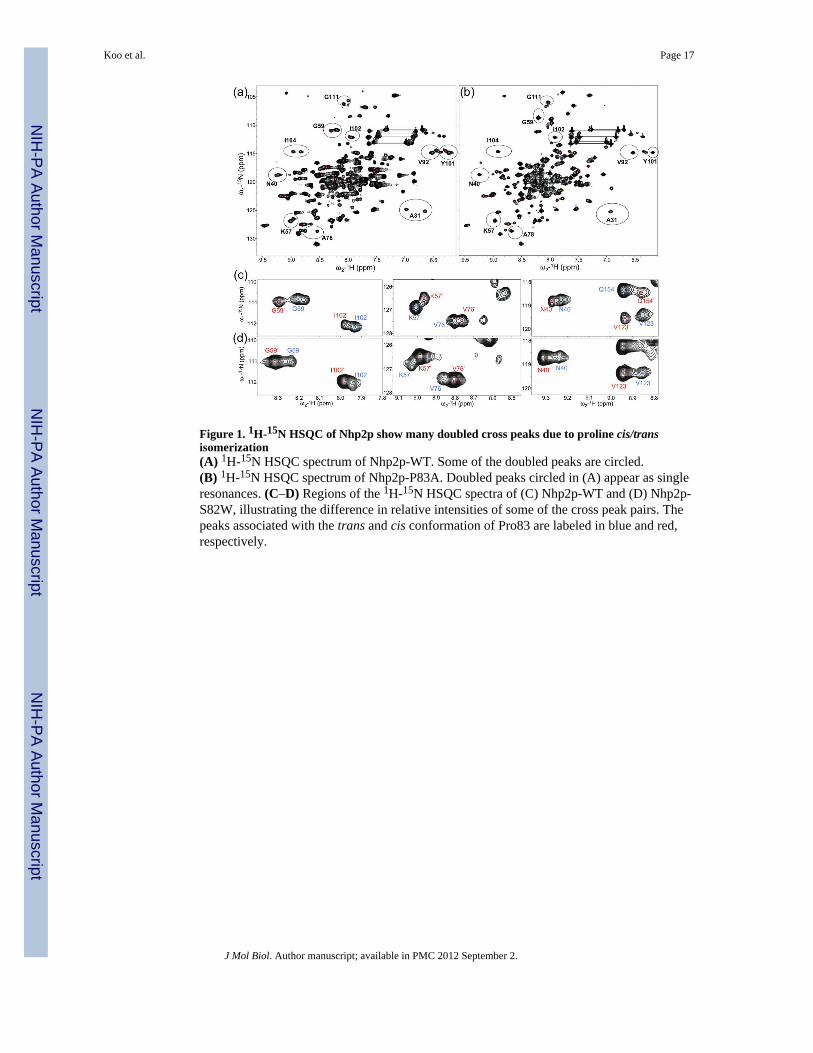
Figure 1. 1H-15N HSQC of Nhp2p show many doubled cross peaks due to proline cis/transisomerization(A) 1H-15N HSQC spectrum of Nhp2p-WT. Some of the doubled peaks are circled.(B) 1H-15N HSQC spectrum of Nhp2p-P83A. Doubled peaks circled in (A) appear as singleresonances. (C–D) Regions of the 1H-15N HSQC spectra of (C) Nhp2p-WT and (D) Nhp2p-S82W, illustrating the difference in relative intensities of some of the cross peak pairs. Thepeaks associated with the trans and cis conformation of Pro83 are labeled in blue and red,respectively.
Koo et al. Page 17
J Mol Biol. Author manuscript; available in PMC 2012 September 2.
NIH
-PA Author Manuscript
NIH
-PA Author Manuscript
NIH
-PA Author Manuscript
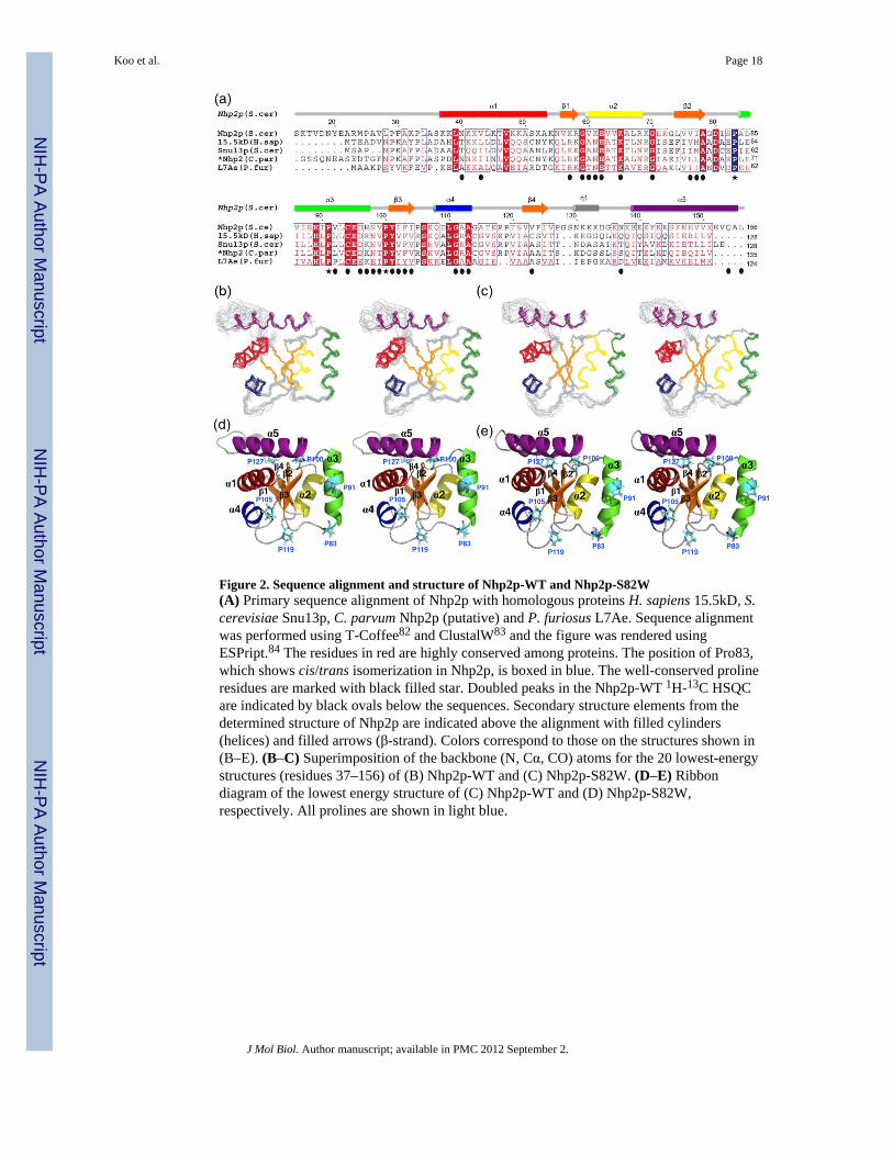
Figure 2. Sequence alignment and structure of Nhp2p-WT and Nhp2p-S82W(A) Primary sequence alignment of Nhp2p with homologous proteins H. sapiens 15.5kD, S.cerevisiae Snu13p, C. parvum Nhp2p (putative) and P. furiosus L7Ae. Sequence alignmentwas performed using T-Coffee82 and ClustalW83 and the figure was rendered usingESPript.84 The residues in red are highly conserved among proteins. The position of Pro83,which shows cis/trans isomerization in Nhp2p, is boxed in blue. The well-conserved prolineresidues are marked with black filled star. Doubled peaks in the Nhp2p-WT 1H-13C HSQCare indicated by black ovals below the sequences. Secondary structure elements from thedetermined structure of Nhp2p are indicated above the alignment with filled cylinders(helices) and filled arrows (β-strand). Colors correspond to those on the structures shown in(B–E). (B–C) Superimposition of the backbone (N, Cα, CO) atoms for the 20 lowest-energystructures (residues 37–156) of (B) Nhp2p-WT and (C) Nhp2p-S82W. (D–E) Ribbondiagram of the lowest energy structure of (C) Nhp2p-WT and (D) Nhp2p-S82W,respectively. All prolines are shown in light blue.
Koo et al. Page 18
J Mol Biol. Author manuscript; available in PMC 2012 September 2.
NIH
-PA Author Manuscript
NIH
-PA Author Manuscript
NIH
-PA Author Manuscript

Figure 3. Effect of proline isomerization on the structure of Nhp2p(A) Ribbon representation of Nhp2p-WT with the positions of the residues whose amidesthat show doubled peaks in Nhp2p-WT but not in Nhp2p-P83A highlighted in purple andresidues whose amides which are doubled in both Nhp2p-WT and Nhp2p-P83A highlightedin light violet. (B) Ribbon representation of Nhp2p-WT with the positions of residuesequivalent to those in L7Ae which bind to Nop10 and H/ACA RNA highlighted in yellowand cyan, respectively. (C) Superimposition of structures of Nhp2p-WT (gray) with Nhp2p-S82W (blue). Pro83 and the peptide backbone of Ser82 or Trp82 are highlighted in green forNhp2p-WT and magenta for Nhp2p-S82W. (D) Close-up view of the region of Nhp2p-WTand Nhp2p-S82W around P83A (region boxed in (A), 90° rotated). In the cis conformation,Pro83 and the backbone amide and carbonyl of residue 82 point toward the helix α2, whilein the trans conformation they point away.
Koo et al. Page 19
J Mol Biol. Author manuscript; available in PMC 2012 September 2.
NIH
-PA Author Manuscript
NIH
-PA Author Manuscript
NIH
-PA Author Manuscript

Figure 4. Nhp2-P83A shows decreased association with H/ACA snoRNA and proteins, whileNhp2p-S82W stabilizes the association with H/ACA snoRNPs(A) Northern blot analysis of RNAs co-immunoprecipitated with the various versions ofNhp2p. Extracts from a wild-type strain containing the pUG34 vector (lanes 1, 5, 9) andnhp2Δ strains containing the pNHP2FLAG WT (lanes 2, 6, 10), pNHP2FLAG S82W (lanes3, 7, 11) or pNHP2FLAG P83A (lanes 4, 8, 12) plasmid were subjected toimmunoprecipitation with anti-FLAG agarose beads. RNAs were extracted with phenol,precipitated, fractionated on a 5% polyacrylamide/8 M urea gel and subjected to Northernanalysis to detect the H/ACA snoRNAs snR10, snR32, snR37 and snR42 and the U1snRNA. Quantifications of the levels of associations of the different mutants with individualsnoRNAs is provided in Table S1. The snR37 northern blot was obtained from a separate
Koo et al. Page 20
J Mol Biol. Author manuscript; available in PMC 2012 September 2.
NIH
-PA Author Manuscript
NIH
-PA Author Manuscript
NIH
-PA Author Manuscript

experiment than for the other snoRNAs, therefore the U1 snRNA control is shown twice.(B) Western blot analysis of the association of the various versions of Nhp2p with Cbf5p.Extracts from a wild-type strain containing the pUG36 vector (lanes 1, 7) or Nhp2FLAGWT in pUG36 (lanes 2, 8) and a CBF5TAP strain containing pUG36 (lanes 3, 9) orNhp2FLAG WT (lanes 4, 10), Nhp2FLAG S82W (lanes 5, 11) or Nhp2FLAG P83A (lanes6, 12) plasmids in pUG36 were subjected to affinity precipitation using Calmodulinsepharose beads. Proteins were extracted from the beads by heating at 100°C in SDS samplebuffer were fractionated on either a (a) 12% or (b) 6% SDS-polyacrylamide gel and probedby western blot analysis with (a) anti-FLAG antibodies to detect Nhp2pFLAG or (b) PAP todetect Cbf5pTAP.
Koo et al. Page 21
J Mol Biol. Author manuscript; available in PMC 2012 September 2.
NIH
-PA Author Manuscript
NIH
-PA Author Manuscript
NIH
-PA Author Manuscript
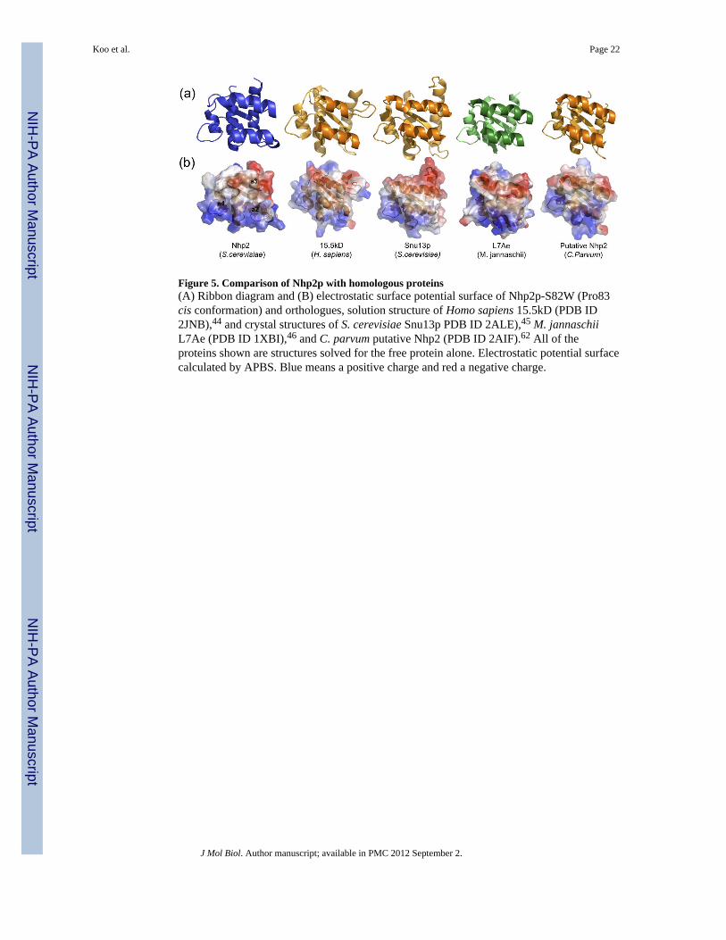
Figure 5. Comparison of Nhp2p with homologous proteins(A) Ribbon diagram and (B) electrostatic surface potential surface of Nhp2p-S82W (Pro83cis conformation) and orthologues, solution structure of Homo sapiens 15.5kD (PDB ID2JNB),44 and crystal structures of S. cerevisiae Snu13p PDB ID 2ALE),45 M. jannaschiiL7Ae (PDB ID 1XBI),46 and C. parvum putative Nhp2 (PDB ID 2AIF).62 All of theproteins shown are structures solved for the free protein alone. Electrostatic potential surfacecalculated by APBS. Blue means a positive charge and red a negative charge.
Koo et al. Page 22
J Mol Biol. Author manuscript; available in PMC 2012 September 2.
NIH
-PA Author Manuscript
NIH
-PA Author Manuscript
NIH
-PA Author Manuscript

Figure 6. Comparison of the Nop10 and RNA binding interface of Nhp2p-WT, Nhp2p-S82W,and L7AE(A–C) Ribbon diagrams of (A) Nhp2p-WT (gray), (B) Nhp2p-S82W (blue), and (C) L7Ae(light green) from the archaeal P. furious H/ACA RNP with Cbf5, Nop10, H/ACA RNA,and substrate RNA (PDB ID 2HVY).29 The residues in L7Ae that interact with Nop10 andRNA, and the homologous residues in Nhp2p, are highlighted in yellow and aqua,respectively. The RNA binding residues in L7Ae are R34, K35, T37, E39, K42, R46, D59,(N38, I93, V95, A96, A98 have van der Waals contacts),33 which correspond to K57, R58,K61, E62, K65, K69, S82, (V60, T116, T120, S121, V123) in Nhp2p. The Nop10p bindingresidues in L7Ae are T41 in α2, A65, H66, P69, L70 and E73 in α3, which correspond toV64, P83, S88, H89, V92, L93 and D96 in Nhp2p. (D) Zoomed in view of the P. furious H/ACA RNP showing the residues comprising the proline spine. Pro60 (equivalent residue toNhp2p Pro83) stacks over the flipped out U in the K-loop, and the backbone of Asp59 hastwo hydrogen bonds with the U base (red). The H/ACA RNA is orange, substrate RNA ismagenta, L7Ae is light green, Nop10 is cyan, Cbf5 is tan.
Koo et al. Page 23
J Mol Biol. Author manuscript; available in PMC 2012 September 2.
NIH
-PA Author Manuscript
NIH
-PA Author Manuscript
NIH
-PA Author Manuscript

NIH
-PA Author Manuscript
NIH
-PA Author Manuscript
NIH
-PA Author Manuscript
Koo et al. Page 24
Table 1
Structural statistics for Nhp2p-WT and Nhp2p-S82W
Nhp2p-WT (Residues 36–156) Nhp2p-S82W (Residues 36–156)
NOE restraints 915 1001
Short-range (|i−j| <= 1) 567 599
Medium-range (1<|i−j| <5) 140 161
Long-range (|i−j| >= 5) 208 241
Hydrogen bonds distance restraintsa 114 116
Dihedral Angle Restraints 177 178
Phi 89 89
Psi 88 89
Violations
Distances > 0.5 Å None None
Dihedral angles > 5° None 3
Average target function (Å2) 3.64 ± 0.31 10.94 ± 0.54
Ramachandran analysis (%)
Most favored regions 83.7 85.9
Additional allowed regions 16.0 12.3
Generously allowed regions 0.2 1.8
Disallowed regions 0.0 0.0
RMS deviations from the mean (Å)
Backbone atoms (N,Cα,C′,O) 0.74 ± 0.16 0.75 ± 0.14
Heavy atoms 1.30 ± 0.17 1.39 ± 0.15
aTwo restraints per one hydrogen bond
J Mol Biol. Author manuscript; available in PMC 2012 September 2.


![Revis�o das II Diretrizes da Sociedade Brasileira de Cardiologia para o Diagn�stico e Tratamento da Insufici�ncia Card�aca]]>](https://static.fdokumen.com/doc/165x107/633912bd4d48a32016081fc6/cdatabreviso-das-ii-diretrizes-da-sociedade-brasileira-de-cardiologia.jpg)


