Structure and Functional Studies of the CS Domain of the Essential H/ACA Ribonucleoparticle Assembly...
12
Structure and Functional Studies of the CS Domain of the Essential H/ACA Ribonucleoparticle Assembly Protein SHQ1 * Received for publication, September 22, 2008, and in revised form, November 10, 2008 Published, JBC Papers in Press, November 19, 2008, DOI 10.1074/jbc.M807337200 Mahavir Singh ‡1 , Fernando A. Gonzales ‡1 , Duilio Cascio § , Nathanael Heckmann ‡ , Guillaume Chanfreau ‡§ , and Juli Feigon ‡§2 From the ‡ Department of Chemistry and Biochemistry and the § Molecular Biology Institute, UCLA, Los Angeles, California 90095 H/ACA ribonucleoprotein particles are essential for riboso- mal RNA and telomerase RNA processing and metabolism. Shq1p has been identified as an essential eukaryotic H/ACA small nucleolar (sno) ribonucleoparticle (snoRNP) biogenesis and assembly factor. Shq1p is postulated to be involved in the early biogenesis steps of H/ACA snoRNP complexes, and Shq1p depletion leads to a specific decrease in H/ACA small nucleolar RNA levels and to defects in ribosomal RNA processing. Shq1p contains two predicted domains as follows: an N-terminal CS (named after CHORD-containing proteins and SGT1) or HSP20-like domain, and a C-terminal region of high sequence homology called the Shq1 domain. Here we report the crystal structure and functional studies of the Saccharomyces cerevisiae Shq1p CS domain. The structure consists of a compact anti- parallel -sandwich fold that is composed of two -sheets con- taining four and three -strands, respectively, and a short -he- lix. Deletion studies showed that the CS domain is required for the essential functions of Shq1p. Point mutations in residues Phe-6, Gln-10, and Lys-80 destabilize Shq1p in vivo and induce a temperature-sensitive phenotype with depletion of H/ACA small nucleolar RNAs and defects in rRNA processing. Although CS domains are frequently found in co-chaperones of the Hsp90 molecular chaperone, no interaction was detected between the Shq1p CS domain and yeast Hsp90 in vitro. These results show that the CS domain is essential for Shq1p function in H/ACA snoRNP biogenesis in vivo, possibly in an Hsp90- independent manner. Modification of uridine to pseudouridine in ribosomal RNA and some spliceosomal RNAs is catalyzed by highly specialized ribonucleoparticle (RNP) 3 complexes called box H/ACA RNPs (1–5). Depending on their site of maturation and action H/ACA RNPs are classified into two classes, small nucleolar RNPs (snoRNPs) and small Cajal body RNPs. In Saccharomyces cerevisiae, H/ACA snoRNPs contain four proteins: Nhp2p (L7ae in archaea (6) and Cbf5p, also called dyskerin, in humans (7)), Nop10p, Gar1p, and a single small nucleolar RNA (snoRNA), specific to each snoRNP (8 –11). Cbf5p provides the pseudouridylase activity to the complex, and the snoRNA component provides the “guide RNA” for position- ing the substrate RNA for modification (8, 10, 12–15). The 3 end of human telomerase RNA (hTR) contains an H/ACA scaRNA domain that binds the H/ACA proteins and is required for 3 end processing, accumulation, and localiza- tion of hTR to Cajal bodies (16 –19). In archaea, the assembly of H/ACA snoRNP appears to proceed by assembly of the protein components, followed by the incorporation of the H/ACA RNA (8, 20–23). In eukaryotes, the assembly and final maturation of the holoenzyme RNP are more complicated, possibly because of subcellular compartmentalization, and require accessory proteins (22, 24). Two proteins specifically found in eukaryotes, Naf1p and Shq1p, were initially identified in yeast as factors involved in the assembly of H/ACA snoRNPs (23–25). Both Shq1p and Naf1p are essential proteins, and their depletion leads to the loss of H/ACA snoRNAs (22, 24). Shq1p and Naf1p interact with the H/ACA RNP components Cbf5p and Nhp2p as shown by high throughput proteomic approaches and by directed protein interaction studies (24, 26 –28). Both Naf1p and Gar1p contain a central domain that forms a six-stranded -barrel fold and interact competitively with Cbf5p using this “core-Gar1” domain (29). Although Shq1p was first identified in yeast (24), orthologues have been found in all eukaryotic genomes investigated, includ- ing human (22). Shq1p is not associated with the precursor or mature RNPs and is localized in the nucleoplasm (24) rather than in the nucleolus or Cajal bodies where mature H/ACA RNPs reside. It was therefore proposed that Shq1p is involved in the early biogenesis steps of H/ACA snoRNPs. However, Shq1p does not share any homology with either Naf1p or Gar1p, and its mode of action remains unclear. Based on the Saccharomyces Genome Data base annotations and domain predictions, Shq1p seems to be a modular protein with two predicted domains in its sequence (Fig. 1A). The C-terminal half contains a region of high sequence homology with other * This work was supported, in whole or in part, by National Institutes of Health Grants GM37254 and GM48123 (to J. F.). This work was also supported by American Cancer Society Research Scholar Grant RSG-06-040 (to G. C.). The costs of publication of this article were defrayed in part by the payment of page charges. This article must therefore be hereby marked “advertise- ment” in accordance with 18 U.S.C. Section 1734 solely to indicate this fact. The atomic coordinates and structure factors (code 3EUD) have been deposited in the Protein Data Bank, Research Collaboratory for Structural Bioinformatics, Rutgers University, New Brunswick, NJ (http://www.rcsb.org/). 1 Both authors contributed equally to this work. 2 To whom correspondence should be addressed: Dept. of Chemistry and Biochemistry, P.O. Box 951569, UCLA, Los Angeles, CA 90095-1569. Tel.: 310-206-6922; Fax: 310-825-0982; E-mail: [email protected]. 3 The abbreviations used are: RNP, ribonucleoparticle; snoRNP, small nucleo- lar ribonucleoparticle; snoRNA, small nucleolar RNA; hTR, human telomer- ase RNA; GFP, green fluorescent protein; BisTris, 2-[bis(2-hydroxyethyl)- amino]-2-(hydroxymethyl)propane-1,3-diol; HSP, heat shock protein; sHSP, small HSP; WT, wild type; HSQC, heteronuclear single quantum coherence. THE JOURNAL OF BIOLOGICAL CHEMISTRY VOL. 284, NO. 3, pp. 1906 –1916, January 16, 2009 © 2009 by The American Society for Biochemistry and Molecular Biology, Inc. Printed in the U.S.A. 1906 JOURNAL OF BIOLOGICAL CHEMISTRY VOLUME 284 • NUMBER 3 • JANUARY 16, 2009 at USC Norris Medical Library on April 9, 2015 http://www.jbc.org/ Downloaded from
Transcript of Structure and Functional Studies of the CS Domain of the Essential H/ACA Ribonucleoparticle Assembly...
Structure and Functional Studies of the CS Domain of theEssential H/ACA Ribonucleoparticle Assembly Protein SHQ1*
Received for publication, September 22, 2008, and in revised form, November 10, 2008 Published, JBC Papers in Press, November 19, 2008, DOI 10.1074/jbc.M807337200
Mahavir Singh‡1, Fernando A. Gonzales‡1, Duilio Cascio§, Nathanael Heckmann‡, Guillaume Chanfreau‡§,and Juli Feigon‡§2
From the ‡Department of Chemistry and Biochemistry and the §Molecular Biology Institute, UCLA, Los Angeles, California 90095
H/ACA ribonucleoprotein particles are essential for riboso-mal RNA and telomerase RNA processing and metabolism.Shq1p has been identified as an essential eukaryotic H/ACAsmall nucleolar (sno) ribonucleoparticle (snoRNP) biogenesisand assembly factor. Shq1p is postulated to be involved in theearly biogenesis steps of H/ACA snoRNP complexes, and Shq1pdepletion leads to a specific decrease in H/ACA small nucleolarRNA levels and to defects in ribosomal RNA processing. Shq1pcontains two predicted domains as follows: an N-terminal CS(named after CHORD-containing proteins and SGT1) orHSP20-like domain, and a C-terminal region of high sequencehomology called the Shq1 domain. Here we report the crystalstructure and functional studies of the Saccharomyces cerevisiaeShq1p CS domain. The structure consists of a compact anti-parallel �-sandwich fold that is composed of two �-sheets con-taining four and three �-strands, respectively, and a short �-he-lix. Deletion studies showed that the CS domain is required forthe essential functions of Shq1p. Point mutations in residuesPhe-6, Gln-10, and Lys-80 destabilize Shq1p in vivo and inducea temperature-sensitive phenotype with depletion of H/ACAsmall nucleolar RNAs and defects in rRNA processing.Although CS domains are frequently found in co-chaperones ofthe Hsp90 molecular chaperone, no interaction was detectedbetween the Shq1p CS domain and yeast Hsp90 in vitro. Theseresults show that the CS domain is essential for Shq1p functionin H/ACA snoRNP biogenesis in vivo, possibly in an Hsp90-independent manner.
Modification of uridine to pseudouridine in ribosomal RNAand some spliceosomal RNAs is catalyzed by highly specializedribonucleoparticle (RNP)3 complexes called box H/ACA RNPs
(1–5). Depending on their site of maturation and actionH/ACA RNPs are classified into two classes, small nucleolarRNPs (snoRNPs) and small Cajal body RNPs. In Saccharomycescerevisiae, H/ACA snoRNPs contain four proteins: Nhp2p(L7ae in archaea (6) and Cbf5p, also called dyskerin, in humans(7)), Nop10p, Gar1p, and a single small nucleolar RNA(snoRNA), specific to each snoRNP (8–11). Cbf5p providesthe pseudouridylase activity to the complex, and thesnoRNA component provides the “guide RNA” for position-ing the substrate RNA for modification (8, 10, 12–15). The 3�end of human telomerase RNA (hTR) contains an H/ACAscaRNA domain that binds the H/ACA proteins and isrequired for 3� end processing, accumulation, and localiza-tion of hTR to Cajal bodies (16–19). In archaea, the assemblyof H/ACA snoRNP appears to proceed by assembly of theprotein components, followed by the incorporation of theH/ACA RNA (8, 20–23). In eukaryotes, the assembly andfinalmaturation of the holoenzymeRNParemore complicated,possibly because of subcellular compartmentalization, andrequire accessory proteins (22, 24). Two proteins specificallyfound in eukaryotes, Naf1p and Shq1p, were initially identifiedin yeast as factors involved in the assembly of H/ACA snoRNPs(23–25). Both Shq1p andNaf1p are essential proteins, and theirdepletion leads to the loss of H/ACA snoRNAs (22, 24). Shq1pand Naf1p interact with the H/ACA RNP components Cbf5pand Nhp2p as shown by high throughput proteomicapproaches and by directed protein interaction studies (24,26–28). Both Naf1p and Gar1p contain a central domain thatforms a six-stranded �-barrel fold and interact competitivelywith Cbf5p using this “core-Gar1” domain (29).Although Shq1pwas first identified in yeast (24), orthologues
have been found in all eukaryotic genomes investigated, includ-ing human (22). Shq1p is not associated with the precursor ormature RNPs and is localized in the nucleoplasm (24) ratherthan in the nucleolus or Cajal bodies where mature H/ACARNPs reside. It was therefore proposed that Shq1p is involvedin the early biogenesis steps of H/ACA snoRNPs. However,Shq1p does not share any homology with either Naf1p orGar1p, and its mode of action remains unclear. Based on theSaccharomyces Genome Data base annotations and domainpredictions, Shq1p seems to be a modular protein with twopredicted domains in its sequence (Fig. 1A). The C-terminalhalf contains a region of high sequence homology with other
* This work was supported, in whole or in part, by National Institutes of HealthGrants GM37254 and GM48123 (to J. F.). This work was also supported byAmerican Cancer Society Research Scholar Grant RSG-06-040 (to G. C.). Thecosts of publication of this article were defrayed in part by the payment ofpage charges. This article must therefore be hereby marked “advertise-ment” in accordance with 18 U.S.C. Section 1734 solely to indicate this fact.
The atomic coordinates and structure factors (code 3EUD) have been deposited inthe Protein Data Bank, Research Collaboratory for Structural Bioinformatics,Rutgers University, New Brunswick, NJ (http://www.rcsb.org/).
1 Both authors contributed equally to this work.2 To whom correspondence should be addressed: Dept. of Chemistry and
Biochemistry, P.O. Box 951569, UCLA, Los Angeles, CA 90095-1569. Tel.:310-206-6922; Fax: 310-825-0982; E-mail: [email protected].
3 The abbreviations used are: RNP, ribonucleoparticle; snoRNP, small nucleo-lar ribonucleoparticle; snoRNA, small nucleolar RNA; hTR, human telomer-ase RNA; GFP, green fluorescent protein; BisTris, 2-[bis(2-hydroxyethyl)-amino]-2-(hydroxymethyl)propane-1,3-diol; HSP, heat shock protein;
sHSP, small HSP; WT, wild type; HSQC, heteronuclear single quantumcoherence.
THE JOURNAL OF BIOLOGICAL CHEMISTRY VOL. 284, NO. 3, pp. 1906 –1916, January 16, 2009© 2009 by The American Society for Biochemistry and Molecular Biology, Inc. Printed in the U.S.A.
1906 JOURNAL OF BIOLOGICAL CHEMISTRY VOLUME 284 • NUMBER 3 • JANUARY 16, 2009
at USC
Norris M
edical Library on A
pril 9, 2015http://w
ww
.jbc.org/D
ownloaded from
Shq1 proteins called the Shq1 domain, but this region shows noidentified folding motif. The N-terminal region of the proteinhas a predicted CS (named after CHORD-containing proteinsand SGT1) or HSP20-like domain, which is found in a numberof co-chaperones for heat shock protein 90 (Hsp90) (30–34),and hence is presumed to be an Hsp90 (Hsp82p in yeast) bind-ing domain. The CS, HSP20-like, and p23-like domains belongto the HSP20 domain superfamily.We have determined the x-ray crystal structure of the CS
domain of Shq1p and investigated its importance for Shq1pfunction. The structure consists of a �-sandwich immunoglob-ulin light chain fold (35). Like otherCS domains, theCS domainof Shq1p is primarily composed of two �-sheets (30, 31, 33, 36),but it has an additional short helix at the C terminus. We showthat the Shq1pCS domain is essential for growth in S. cerevisiaeand that the integrity of the CS domain is required for the func-tion of Shq1p in vivo. Based on the structure and sequenceconservation among Shq1proteins, we investigated the effect ofthree single point mutations, F6A, Q10A, and K80A, on Shq1pstructure and function.All threemutations destabilized the ter-tiary structure of the CS domain, albeit to differing extents.Incorporation of these mutations in Shq1p resulted in a tem-perature-sensitive growth phenotype, specific depletion ofH/ACA snoRNAs, and rRNA processing defects. NMR chemi-cal shift mapping showed no interaction between the Shq1p CSdomain and Hsp82p, suggesting that the Shq1p CS domaindoes not have a canonical role as an Hsp82p co-chaperoneprotein.
EXPERIMENTAL PROCEDURES
Protein Expression and Purification for Structural Studies—CS (residues 1–101) andCS-L (residues 1–145) constructswerePCR-amplified using yeast genomic DNA as a template andcloned into pET46 LIC/Ek (Novagen). F6A, Q10A, and K80Amutant constructs of the CS domain were made by site-di-rected mutagenesis (Stratagene), and plasmids were trans-formed into Escherichia coli BL21 (DE3) cells for proteinexpression. For selenomethionine labeling, E. coli B834 (DE3)cells (Novagen) were used. Cells were grown either in LBmedium (for unlabeled proteins) orM9medium (for selenome-thionine, 13C,15N-labeled, and 15N-labeled protein). Cultureswere grown at 37 °C until an A600 of 0.6 was reached, and thenthe temperature was lowered to 28 °C (22 °C for the F6A andQ10Amutants) and the cells were inducedwith 1mM isopropyl1-thio-�-D-galactopyranoside for 16 h. Cells were harvested bycentrifugation, and the pellets were resuspended in the bindingbuffer (30 ml; buffer A: 50 mM NaH2PO4, pH 8, 300 mM NaCl,10 mM �-mercaptoethanol). Cells were lysed by sonication onice and centrifuged to remove cell debris. The supernatant wasapplied to a nickel-nitrilotriacetic acid-agarose column (5 ml),which had been pre-equilibrated with buffer A. The columnwas washed with buffer A, followed by buffer B (buffer A sup-plemented with 20 mM imidazole) and were eluted with bufferC (buffer A supplemented with 250 mM imidazole). Proteinswere further purified by Superdex S75 preparative gel filtration.Yeast Hsp82p was PCR-amplified and cloned into pET30 LIC/Xa. Protein was expressed in E. coli at room temperature andpurified over a nickel-nitrilotriacetic acid-agarose column fol-
lowed by gel filtration on Superdex S75 column. All proteinswere �95% purified as judged by SDS-PAGE. The buffer forNMR and CD spectroscopy was 20 mM NaH2PO4, pH 7.2, 100mMNaCl, and 2mM dithiothreitol, 0.02%NaN3 and for crystal-lization was 20 mM Tris base, pH 7.2, 100 mM NaCl, 2 mM
dithiothreitol, and 0.02% NaN3.NMR Spectroscopy—Uniformly 13C,15N-labeled samples of
CS(WT) were used for NMR assignments, and 15N-labeledsamples of WT and mutant CS domains were used for otherNMR experiments. NMR spectra were recorded on BrukerDRX 600-MHz and DRX 800-MHz spectrometers equippedwith cryoprobes. NMR samples contained 0.3 to 0.8 mM pro-tein. For the 1H-15N HSQC spectra, a total of 1024 complexpoints in t2, and 256 complex points at t1 increments wereacquired. The backbone assignments for CS(WT) wereobtained by standard methods using the CBCACONH/CBCANH and HNCACO/HNCO pairs of triple resonanceexperiments at 22 °C (37). NMR titrations were carried out byadding unlabeled purified Hsp82p to 15N-labeled CS or CS-Land recording 1H-15N TROSY-HSQC spectra. For the 1H-15NTROSY-HSQC spectra, a total of 1024 complex points in t2,and 256 complex points at t1 increments were acquired. All thetitrations were carried out at 22 °C at 600MHz.NMRdata wereprocessed using the Bruker program Xwin-NMR version 3.5and assigned using the Sparky software program (T. D. God-dard and D. G. Kneller, SPARKY version 3, University of Cali-fornia, San Francisco).Crystallization, Data Collection, and Structure Determina-
tion—Crystallization was carried out with a hanging drop,vapor-diffusion method by mixing equal volumes of proteinand reservoir solutions (0.1 M BisTris, pH 6.0, 0.2 M ammoniumsulfate, 25% w/v PEG 3350). Crystals appeared under the sameconditions for both normal and selenomethionine derivativeswithin 24 h. Crystals grew to maximum size of 0.2 � 0.2 � 0.3mm in 4 days and belonged to space group P21212. Unit celldimensions and other parameters are given in Table 1.Datawere collected at theAdvanced Photon Source at beam-
line 24-ID-C. Data were processed with the DENZO/SCALE-PACK package (38). The selenomethionine derivative wasmeasured at peak wavelength of 0.97949 Å. Datasets were ofhigh quality and showed a strong anomalous signal. Macromo-lecular phasing was carried out by HKL2MAP (39). The result-ing phases were improved by using the DM program (40) andused for an automatedmodel building with the Arp/Warp soft-ware (40). The resulting model of about 90% completeness wasinspected and finished manually with the program Coot (41).Restrained refinement enforced by the Refmac5 software wasthen performed and was followed by the addition of water mol-ecules by using Arp/Warp (42). Final iterative rounds of modelbuilding and refinement were carried out using Coot (41) andPHENIX (43). Final data collection, phasing, and refinementstatistics are presented in Table 1.Most of themodel has a clearelectron density, with the exception of the N-terminal His tag(15 residues) and 3–4 C-terminal residues. Additionally, somesolvent-exposed side chains had no interpretable electron den-sity. These parts were omitted in the final model. The R-factorof the presented structure is 22.5%, and Rfree is 24.6%. All struc-
Structure and Functional Analysis of Shq1 CS Domain
JANUARY 16, 2009 • VOLUME 284 • NUMBER 3 JOURNAL OF BIOLOGICAL CHEMISTRY 1907
at USC
Norris M
edical Library on A
pril 9, 2015http://w
ww
.jbc.org/D
ownloaded from
ture figures were drawn using PyMOL (DeLano Scientific, SanCarlos, CA).CD Spectroscopy—CD spectra of the proteins were obtained
on a J-715 spectropolarimeter (Jasco J715model). Spectra wererecorded using 1-mm path length quartz cuvettes. Each spec-trum was recorded as an average of four scans. For thermaldenaturation experiments, spectra were monitored at 215 nm,and the sample temperature was increased at a rate of 30 °C/h.Cloning—DNA cloning and DNA manipulations were per-
formed as described (44). Plasmids expressing the GFP fusionsin yeast were constructed by insertingDNA fragments contain-ing the PCR-amplified SHQ1 open reading frame into pUG23and pUG35 (45) using oligonucleotides with XbaI and EcoRIrestriction sites. Constructs expressing Shq1p residues 1–483,1–491, 1–500, 9–508, 18–508, and 26–508 and point mutantsF6A, Q10A, K18A, S22A, and K80A were amplified by PCRusing specific oligonucleotides and inserted into pUG23.Yeast Transformation andMaintenance—Yeast strains were
maintained in synthetic medium (YNB) complemented withthe appropriate amino acid mix. Glucose was added as carbonsource to a final concentration of 2% (w/v) as indicated. Yeastcells were transformed using the lithium acetate method asdescribed (46).The shq1� knock-out strain was obtained using a PCR-based
gene deletion strategy described in Ref. 47, by homologousrecombination of a DNA fragment containing the codingregion of TRP marker flanked by the last 40 and first 40 nucle-otides of the 5�- and 3�-untranslated region, respectively, ofSHQ1. The TRP cassette was co-transformed with a plasmidcontaining a wild-type SHQ1 gene and URA3 auxotrophy
marker (pUG35-SHQ1) in a wild-type yeast strain BMA64(ura3-1; trp1; ade2-1; leu2-3,112; his3-11,15).Western Blots—Whole-cell lysates were prepared at 4 °C
from equal A600 amounts of exponentially growing yeast cul-tures. Cells were resuspended in a buffer containing 1.85 M
NaOH, 7.4% �-mercaptoethanol, and trichloroacetic acid at afinal concentration of 5.48%. Lysates were centrifuged, the pel-lets resuspended, and the proteins fractionated by SDS-PAGE.Immunoblotting was performed with anti-GFP (1:10,000) andanti-hexokinase (1:10,000) antibodies and revealed by ECL sys-tem using the Super Signal� West Femto from Thermo Scien-tific Pierce Protein Research Products.
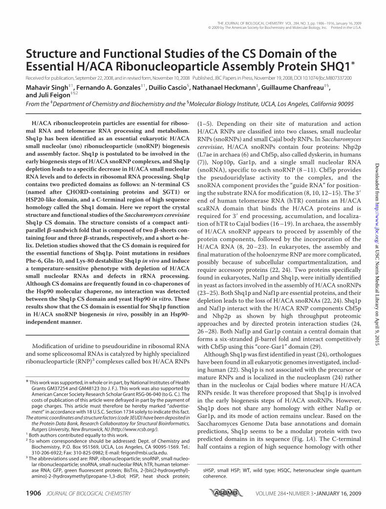
FIGURE 1. Deletion analysis and stability of Shq1p truncated mutants.A, domain organization of Shq1p and complementation of the shq1� strainby the various truncation mutants. Numbers refer to SHQ1 amino acid resi-dues. Cells were grown in plates containing 5-fluoroacetic acid to counters-elect the wild-type (wt) SHQ1 copy borne by the URA3-containing plasmidand in the absence of methionine to express Shq1p, and cell growth wasassessed. B, example of growth assay used to generate the previous figure.Shown is the growth of strains expressing the indicated deletions of Shq1p ona plate containing 5-fluoroacetic acid to counterselect the wild-type SHQ1copy borne by the URA3-containing plasmid. C, stability of truncatedmutants. Cells containing the endogenous SHQ1 copy and the indicated con-structs were grown on liquid medium in the absence of methionine. Proteinswere expressed under the control of the Met25 promoter, which is highlyinduced in the absence of Met. Numbers refer to SHQ1 amino acid residues.Shq1p proteins were detected by immunoblotting with anti-GFP. Hexokinase(Hxk) was used as a loading control.
TABLE 1Data collection and refinement statistics for Shq1p CS domainRcryst � �hkl�i�Ii(hkl) � I(hkl�/�hkl�iIi(hkl). Rwork � ��Fo � Fc�/�Fo. Rfree � � �Fo �Fc�/�Fo, calculated using a random set containing 5% reflections that were notincluded throughout refinement.
Data collection Se-Met: peakSpace group P21212Cell constants a � 141.94 Å, b � 146.29 Å, c � 48.51 ÅResolution range (last shell) 39–2.33 Å (2.44–2.4 Å)Wavelength 0.97949 ÅObserved reflections 502,217Unique reflections 72,994Completeness (%) (last shell) 95.2 (61.5)Rcryst (last shell) 6.7 (39.8)I/�(I) (last shell) 24.62 (2.55)SAD phasingNo. of selenium sites found 12Figure of merit (afterdensity modification)
0.603 (0.746)
RefinementNo. of reflections 39,269Resolution 35.42 to 2.4 ÅR-factor/Rfree 22.5/24.6%Average B 51.6 Å2
Root mean square deviationbond length/angles
0.01 Å/1.3°
Content of asymmetric unitNo. of protein molecules 6No. of proteinresidues/atoms
577/4853
No. of solvent atoms 110Ramachandran statisticsAllowed/generous/disallowed (%)
98.5/0.4/1.1
Structure and Functional Analysis of Shq1 CS Domain
1908 JOURNAL OF BIOLOGICAL CHEMISTRY VOLUME 284 • NUMBER 3 • JANUARY 16, 2009
at USC
Norris M
edical Library on A
pril 9, 2015http://w
ww
.jbc.org/D
ownloaded from
RNA Analysis—Total RNA extractions and Northern blotanalyses were performed as described (48). Oligonucleotidesused for snoRNA probes are described in Ref. 48. Total RNAwas extracted from cells incubated at 37 °C for different timeintervals and hybridized against 32P-labeled oligonucleotideprobes. The 35 S pre-rRNAwas detected using 5� ETS and 18 Sprobes; the 33 S pre-rRNA was detected using 18 S probe; the27 S pre-rRNA was detected with the ITS2 probe; and the 20 Spre-rRNA was detected with A1–A2 probe as described (48).Coordinate Deposition—The structure factors and coordi-
nates for Shq1p CS domain have been deposited in the ProteinData Bank (accession code 3EUD).
RESULTS AND DISCUSSION
Integrity of the CS Domain of Shq1p Is Required for Shq1pEssential Functions—To assess the importance of the putativeCS domain of Shq1p in vivo, a series of constructs harboringN-terminal deletions was designed (Fig. 1A) and expressed asGFP fusion proteins in S. cerevisiae. The CS or alternativelynamedHSP20-like domain is predicted to start at the precise Nterminus of Shq1p and extends approximately to residue 145(Fig. 1A). We also expressed a series of Shq1p C-terminal dele-tions (Fig. 1A) as controls. Strikingly, proteins deleted of only afew amino acids from the N terminus of Shq1p (Shq1p 9–508)could not complement the SHQ1 knock-out and resulted in alethal phenotype (Fig. 1,A and B). In contrast, C-terminal dele-tions of up to 17 residues did not affect cell growth (Fig. 1, Aand B).To investigate whether these phenotypes were because of
instability of themutant proteins, wemonitored the expressionlevels of these constructs in a wild-type strain expressing theseconstructs in addition to the endogenous wild-type SHQ1 copy(Fig. 1C). The mutant proteins were detected using anti-GFPantibodies because these constructs were expressed as GFP
fusions. Constructs expressing non-functional versions of Shq1p such as9–508, 18–508, and 26–508 wereexpressed to levels similar to wildtype (Fig. 1C). Further deletions inthe N-terminal domain resulted inprotein instability (Fig. 1C), pre-cluding us from drawing conclu-sions about these constructs. Over-all the data obtained with the9–508, 18–508, and 26–508 con-structs show that although stablyexpressed, proteins lacking the firstN-terminal amino acids of theShq1p CS domain cannot function-ally complement the absence ofShq1p. These data strongly suggestthat the CS domain is required forShq1p essential functions in vivo.Structure of the Shq1p CS Do-
main—These results led us to carryout further functional and struc-tural characterization of the CSdomain. Based on the predicted
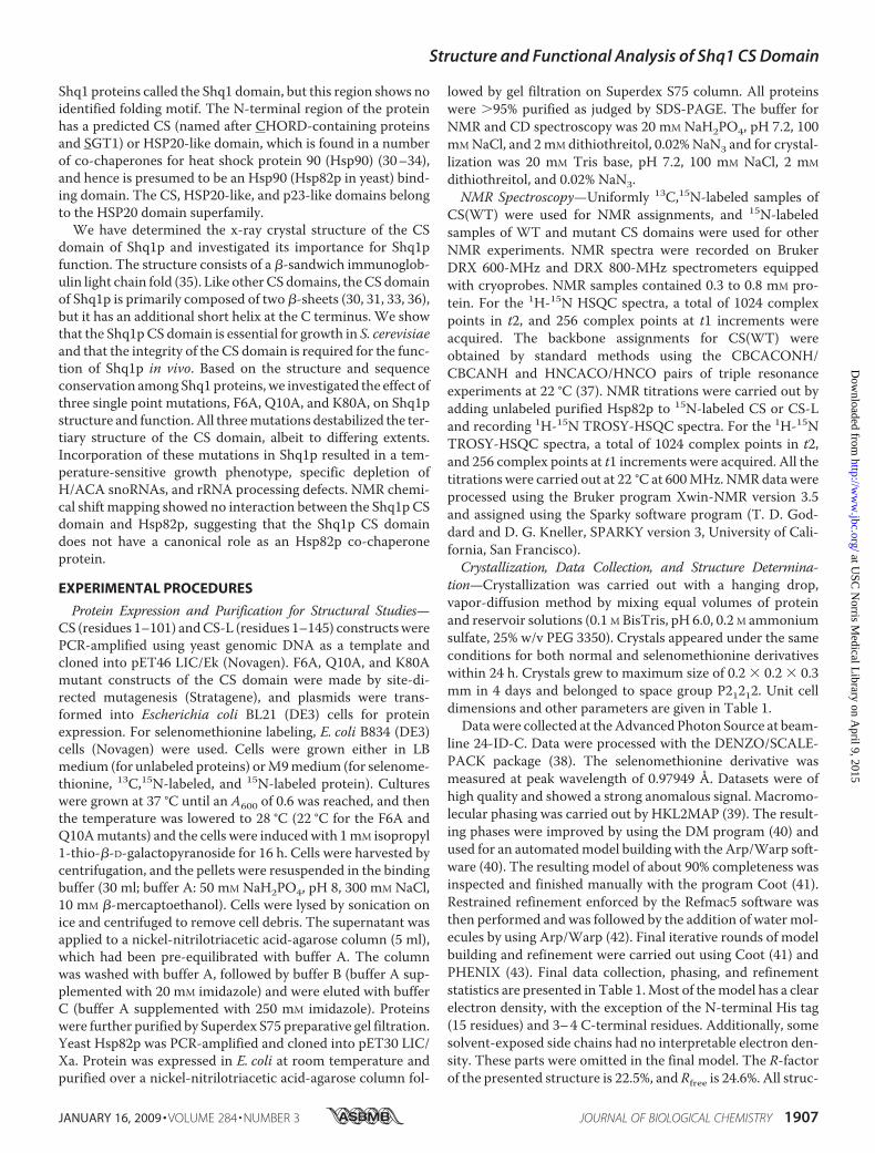
domain boundaries and secondary structure for the CS orHSP20-like domain of Shq1p, we cloned, expressed, and puri-fied two constructs, CS (residues 1–101) and the longer CS-L(residues 1–145) which encompasses the entire N-terminalregion containing the HSP20-like and CS domain (Fig. 1A).15N-1H two-dimensional HSQC NMR spectra show that bothprotein constructs contain a well folded domain (Fig. 2). Thebackbone amides of the CS domain constructs were assignedusing CBCACONH/CBCANH and HNCA/HNCOCA sets oftriple resonance NMR spectra (see “Experimental Proce-dures”). Comparison of the spectra of CS and CS-L reveals thatall of the resonance peaks in the CS-L spectrum that are fromthe additional 44C-terminal residues are in the region generallycorresponding to unstructured or unfolded residues. In addi-tion, the amide resonances from the last �25 residues of CS-Lwere not visible in the 1H-15N HSQC spectra, consistent withthis region being unstructured (Fig. 2).We recorded the 1H-15NHSQC of the CS-L construct under different salt, pH, and tem-perature values; however, we did not observe these C-terminalamide resonances under any of these conditions. Thus, thestructured region of CS-L is entirely contained in CS. Crystal-lization screens were set up for both CS and CS-L; however,crystals appeared only for CS. CS-L did not crystallize, possiblybecause of the flexible C-terminal region in the protein.The CS domain crystallized in the P21212 space group with
six copies of themolecule in one asymmetric unit. However, theCS domain, as well as full-length Shq1p, purified as monomerson a gel filtration column (data not shown). The NMR linewidths are also consistent with what would be expected for amonomer. Attempts to solve the structure by molecularreplacement using the structure of p23 (33) as a template werenot successful. Therefore, the structure was solved by the singleanomalous diffractionmethod using a selenomethionine deriv-ative. The final resolution of the structure was 2.4 Å (Fig. 3, A
FIGURE 2. 1H-15N HSQC spectra of the Shq1p CS domain. Overlay of 1H-15N HSQC spectra of Shq1p CS (red)and CS-L (cyan) proteins. Assignments of amide resonances of CS are indicated (K0 is a residue from the tag).
Structure and Functional Analysis of Shq1 CS Domain
JANUARY 16, 2009 • VOLUME 284 • NUMBER 3 JOURNAL OF BIOLOGICAL CHEMISTRY 1909
at USC
Norris M
edical Library on A
pril 9, 2015http://w
ww
.jbc.org/D
ownloaded from
and B). We did not observe electrondensity for the N-terminal His tag(first 15 residues) and the last fourresidues at the C terminus, andthese were therefore omitted in thefinal refinement and structure.The overall fold of Shq1p CS is a
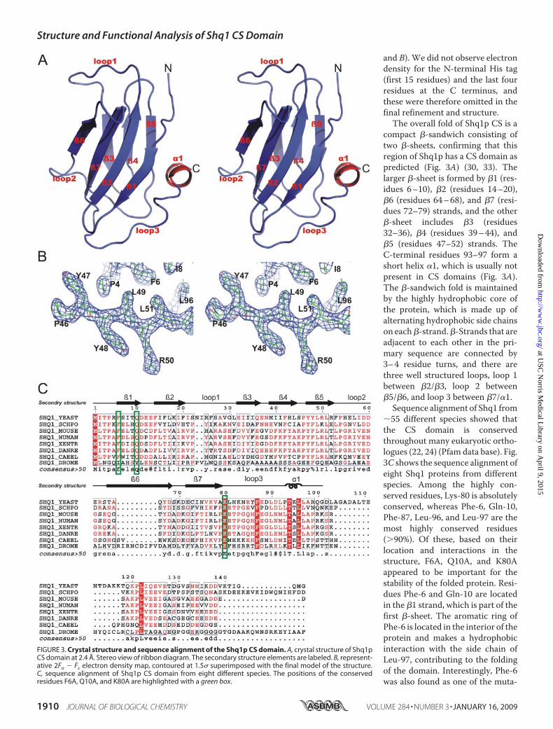
compact �-sandwich consisting oftwo �-sheets, confirming that thisregion of Shq1p has a CS domain aspredicted (Fig. 3A) (30, 33). Thelarger �-sheet is formed by �1 (res-idues 6–10), �2 (residues 14–20),�6 (residues 64–68), and �7 (resi-dues 72–79) strands, and the other�-sheet includes �3 (residues32–36), �4 (residues 39–44), and�5 (residues 47–52) strands. TheC-terminal residues 93–97 form ashort helix �1, which is usually notpresent in CS domains (Fig. 3A).The �-sandwich fold is maintainedby the highly hydrophobic core ofthe protein, which is made up ofalternating hydrophobic side chainson each�-strand.�-Strands that areadjacent to each other in the pri-mary sequence are connected by3–4 residue turns, and there arethree well structured loops, loop 1between �2/�3, loop 2 between�5/�6, and loop 3 between �7/�1.Sequence alignment of Shq1 from
�55 different species showed thatthe CS domain is conservedthroughout many eukaryotic ortho-logues (22, 24) (Pfam data base). Fig.3C shows the sequence alignment ofeight Shq1 proteins from differentspecies. Among the highly con-served residues, Lys-80 is absolutelyconserved, whereas Phe-6, Gln-10,Phe-87, Leu-96, and Leu-97 are themost highly conserved residues(�90%). Of these, based on theirlocation and interactions in thestructure, F6A, Q10A, and K80Aappeared to be important for thestability of the folded protein. Resi-dues Phe-6 and Gln-10 are locatedin the �1 strand, which is part of thefirst �-sheet. The aromatic ring ofPhe-6 is located in the interior of theprotein and makes a hydrophobicinteraction with the side chain ofLeu-97, contributing to the foldingof the domain. Interestingly, Phe-6was also found as one of the muta-
FIGURE 3. Crystal structure and sequence alignment of the Shq1p CS domain. A, crystal structure of Shq1pCS domain at 2.4 Å. Stereo view of ribbon diagram. The secondary structure elements are labeled. B, represent-ative 2Fo � Fc electron density map, contoured at 1.5� superimposed with the final model of the structure.C, sequence alignment of Shq1p CS domain from eight different species. The positions of the conservedresidues F6A, Q10A, and K80A are highlighted with a green box.
Structure and Functional Analysis of Shq1 CS Domain
1910 JOURNAL OF BIOLOGICAL CHEMISTRY VOLUME 284 • NUMBER 3 • JANUARY 16, 2009
at USC
Norris M
edical Library on A
pril 9, 2015http://w
ww
.jbc.org/D
ownloaded from
tions in a screen for temperature-sensitivemutants using a ran-dom mutagenesis approach.4 The Gln-10 side chain O-�1 andN-�2 make hydrogen bonds with the backbone amide N andcarbonyl O of Phe-87, which is also a highly conserved residue.The side chain of the conserved Lys-80 is almost completelyburied inside the hydrophobic interior of the protein, and itsbackbone amide N makes a hydrogen bond with the backbonecarbonyl oxygen of Glu-13. Based on their location in the struc-ture (Fig. 4F) and strong sequence conservation, we character-ized the effect of alanine substitutions of these residues on theCS domain structure and stability as described below.Biophysical Characterization of CS Domain Mutants—Ex-
pression of the wild-type CS domain in E. coli was efficient,yielding �10 mg of purified protein per liter of LB or minimal
media. However, the expression lev-els of the CS(F6A) and CS(Q10A)mutants were �10-fold lower thanthe wild-type CS domain, and(K80A) did not express in all of ourexpression attempts. We reasonedthat CS(K80A) may have beenunfolded and degraded, and there-fore there was no detectable expres-sion of the full-length domain inE. coli.To assess the effect of the amino
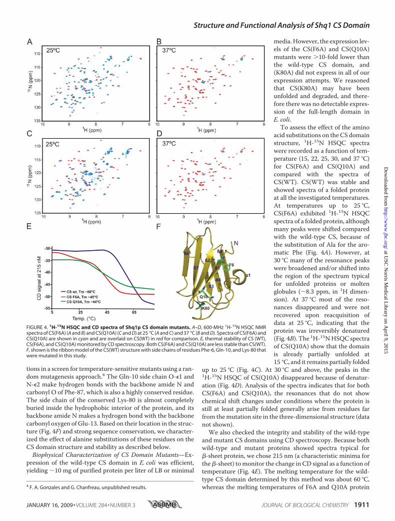
acid substitutions on theCS domainstructure, 1H-15N HSQC spectrawere recorded as a function of tem-perature (15, 22, 25, 30, and 37 °C)for CS(F6A) and CS(Q10A) andcompared with the spectra ofCS(WT). CS(WT) was stable andshowed spectra of a folded proteinat all the investigated temperatures.At temperatures up to 25 °C,CS(F6A) exhibited 1H-15N HSQCspectra of a folded protein, althoughmany peaks were shifted comparedwith the wild-type CS, because ofthe substitution of Ala for the aro-matic Phe (Fig. 4A). However, at30 °C many of the resonance peakswere broadened and/or shifted intothe region of the spectrum typicalfor unfolded proteins or moltenglobules (�8.3 ppm, in 1H dimen-sion). At 37 °C most of the reso-nances disappeared and were notrecovered upon reacquisition ofdata at 25 °C, indicating that theprotein was irreversibly denatured(Fig. 4B). The 1H-15NHSQCspectraof CS(Q10A) show that the domainis already partially unfolded at15 °C, and it remains partially folded
up to 25 °C (Fig. 4C). At 30 °C and above, the peaks in the1H-15N HSQC of CS(Q10A) disappeared because of denatur-ation (Fig. 4D). Analysis of the spectra indicates that for bothCS(F6A) and CS(Q10A), the resonances that do not showchemical shift changes under conditions where the protein isstill at least partially folded generally arise from residues farfrom themutation site in the three-dimensional structure (datanot shown).We also checked the integrity and stability of the wild-type
and mutant CS domains using CD spectroscopy. Because bothwild-type and mutant proteins showed spectra typical for�-sheet protein, we chose 215 nm (a characteristic minima forthe�-sheet) tomonitor the change in CD signal as a function oftemperature (Fig. 4E). The melting temperature for the wild-type CS domain determined by this method was about 60 °C,whereas the melting temperatures of F6A and Q10A protein4 F. A. Gonzales and G. Chanfreau, unpublished results.
FIGURE 4. 1H-15N HSQC and CD spectra of Shq1p CS domain mutants. A–D, 600-MHz 1H-15N HSQC NMRspectra of CS(F6A) (A and B) and CS(Q10A) (C and D) at 25 °C (A and C) and 37 °C (B and D). Spectra of CS(F6A) andCS(Q10A) are shown in cyan and are overlaid on CS(WT) in red for comparison. E, thermal stability of CS (WT),CS(F6A), and CS(Q10A) monitored by CD spectroscopy. Both CS(F6A) and CS(Q10A) are less stable than CS(WT).F, shown is the ribbon model of the CS(WT) structure with side chains of residues Phe-6, Gln-10, and Lys-80 thatwere mutated in this study.
Structure and Functional Analysis of Shq1 CS Domain
JANUARY 16, 2009 • VOLUME 284 • NUMBER 3 JOURNAL OF BIOLOGICAL CHEMISTRY 1911
at USC
Norris M
edical Library on A
pril 9, 2015http://w
ww
.jbc.org/D
ownloaded from
were 44 and 45 °C, respectively (Fig. 4E). In the case ofCS(Q10A), an early melting transition was observed in the15–30 °C range (Fig. 4E), suggesting that at least one region ofthe protein is undergoing a structural transition even at roomtemperature. These findings agree with our NMR results thatshow partial unfolding of CS(Q10A) at a lower temperaturethan for CS(F6A).Point Mutations within the Shq1 CS Domain Destabilize
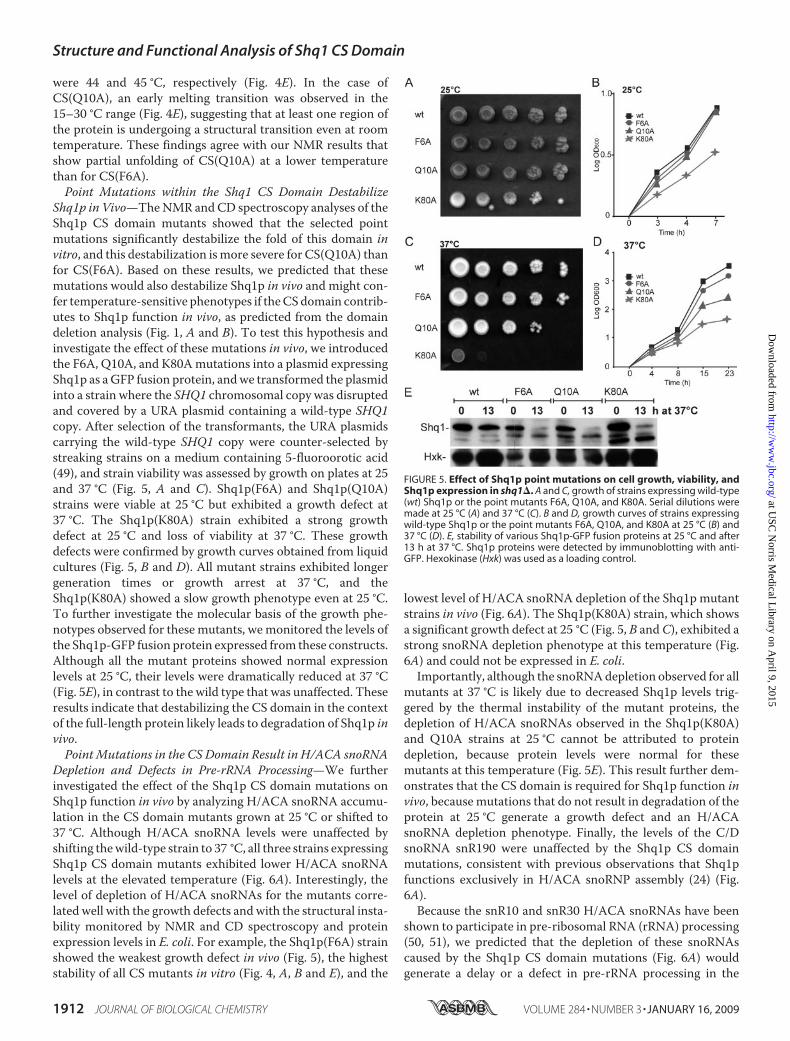
Shq1p in Vivo—TheNMR andCD spectroscopy analyses of theShq1p CS domain mutants showed that the selected pointmutations significantly destabilize the fold of this domain invitro, and this destabilization ismore severe for CS(Q10A) thanfor CS(F6A). Based on these results, we predicted that thesemutations would also destabilize Shq1p in vivo and might con-fer temperature-sensitive phenotypes if theCSdomain contrib-utes to Shq1p function in vivo, as predicted from the domaindeletion analysis (Fig. 1, A and B). To test this hypothesis andinvestigate the effect of these mutations in vivo, we introducedthe F6A, Q10A, and K80Amutations into a plasmid expressingShq1p as aGFP fusion protein, andwe transformed the plasmidinto a strain where the SHQ1 chromosomal copy was disruptedand covered by a URA plasmid containing a wild-type SHQ1copy. After selection of the transformants, the URA plasmidscarrying the wild-type SHQ1 copy were counter-selected bystreaking strains on a medium containing 5-fluoroorotic acid(49), and strain viability was assessed by growth on plates at 25and 37 °C (Fig. 5, A and C). Shq1p(F6A) and Shq1p(Q10A)strains were viable at 25 °C but exhibited a growth defect at37 °C. The Shq1p(K80A) strain exhibited a strong growthdefect at 25 °C and loss of viability at 37 °C. These growthdefects were confirmed by growth curves obtained from liquidcultures (Fig. 5, B and D). All mutant strains exhibited longergeneration times or growth arrest at 37 °C, and theShq1p(K80A) showed a slow growth phenotype even at 25 °C.To further investigate the molecular basis of the growth phe-notypes observed for these mutants, wemonitored the levels ofthe Shq1p-GFP fusion protein expressed from these constructs.Although all the mutant proteins showed normal expressionlevels at 25 °C, their levels were dramatically reduced at 37 °C(Fig. 5E), in contrast to the wild type that was unaffected. Theseresults indicate that destabilizing the CS domain in the contextof the full-length protein likely leads to degradation of Shq1p invivo.Point Mutations in the CS Domain Result in H/ACA snoRNA
Depletion and Defects in Pre-rRNA Processing—We furtherinvestigated the effect of the Shq1p CS domain mutations onShq1p function in vivo by analyzing H/ACA snoRNA accumu-lation in the CS domain mutants grown at 25 °C or shifted to37 °C. Although H/ACA snoRNA levels were unaffected byshifting thewild-type strain to 37 °C, all three strains expressingShq1p CS domain mutants exhibited lower H/ACA snoRNAlevels at the elevated temperature (Fig. 6A). Interestingly, thelevel of depletion of H/ACA snoRNAs for the mutants corre-lated well with the growth defects and with the structural insta-bility monitored by NMR and CD spectroscopy and proteinexpression levels in E. coli. For example, the Shq1p(F6A) strainshowed the weakest growth defect in vivo (Fig. 5), the higheststability of all CS mutants in vitro (Fig. 4, A, B and E), and the
lowest level of H/ACA snoRNA depletion of the Shq1p mutantstrains in vivo (Fig. 6A). The Shq1p(K80A) strain, which showsa significant growth defect at 25 °C (Fig. 5, B andC), exhibited astrong snoRNA depletion phenotype at this temperature (Fig.6A) and could not be expressed in E. coli.Importantly, although the snoRNAdepletion observed for all
mutants at 37 °C is likely due to decreased Shq1p levels trig-gered by the thermal instability of the mutant proteins, thedepletion of H/ACA snoRNAs observed in the Shq1p(K80A)and Q10A strains at 25 °C cannot be attributed to proteindepletion, because protein levels were normal for thesemutants at this temperature (Fig. 5E). This result further dem-onstrates that the CS domain is required for Shq1p function invivo, because mutations that do not result in degradation of theprotein at 25 °C generate a growth defect and an H/ACAsnoRNA depletion phenotype. Finally, the levels of the C/DsnoRNA snR190 were unaffected by the Shq1p CS domainmutations, consistent with previous observations that Shq1pfunctions exclusively in H/ACA snoRNP assembly (24) (Fig.6A).Because the snR10 and snR30 H/ACA snoRNAs have been
shown to participate in pre-ribosomal RNA (rRNA) processing(50, 51), we predicted that the depletion of these snoRNAscaused by the Shq1p CS domain mutations (Fig. 6A) wouldgenerate a delay or a defect in pre-rRNA processing in the
FIGURE 5. Effect of Shq1p point mutations on cell growth, viability, andShq1p expression in shq1�. A and C, growth of strains expressing wild-type(wt) Shq1p or the point mutants F6A, Q10A, and K80A. Serial dilutions weremade at 25 °C (A) and 37 °C (C). B and D, growth curves of strains expressingwild-type Shq1p or the point mutants F6A, Q10A, and K80A at 25 °C (B) and37 °C (D). E, stability of various Shq1p-GFP fusion proteins at 25 °C and after13 h at 37 °C. Shq1p proteins were detected by immunoblotting with anti-GFP. Hexokinase (Hxk) was used as a loading control.
Structure and Functional Analysis of Shq1 CS Domain
1912 JOURNAL OF BIOLOGICAL CHEMISTRY VOLUME 284 • NUMBER 3 • JANUARY 16, 2009
at USC
Norris M
edical Library on A
pril 9, 2015http://w
ww
.jbc.org/D
ownloaded from
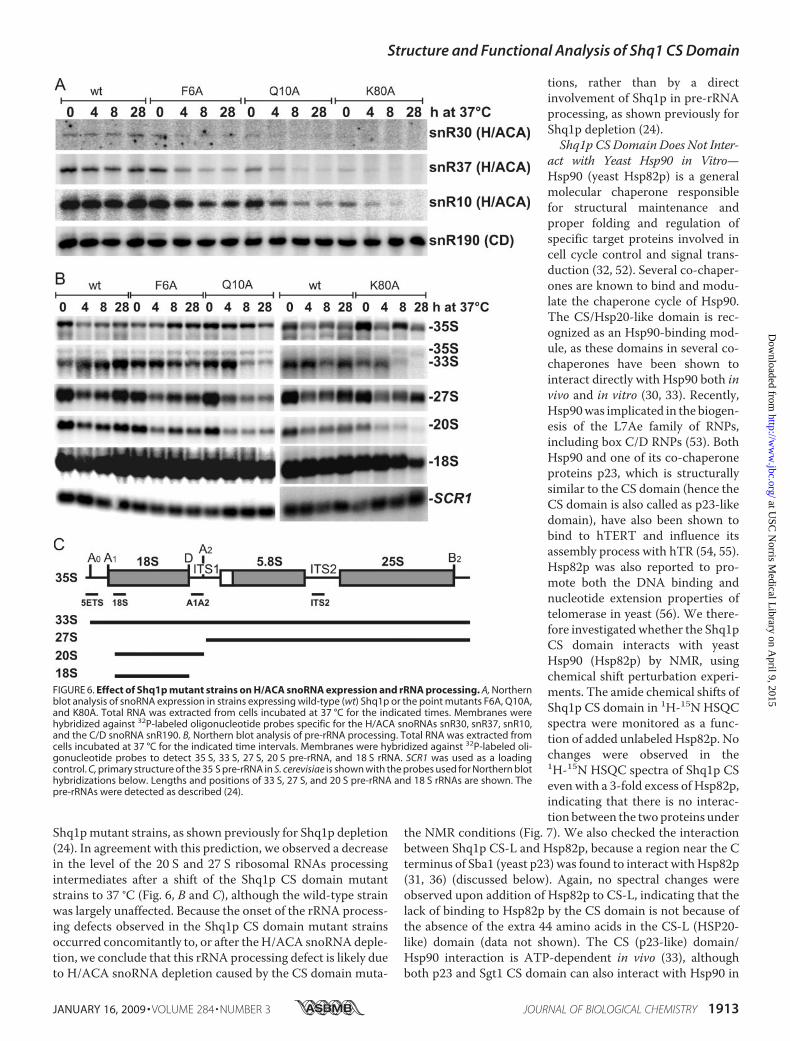
Shq1pmutant strains, as shown previously for Shq1p depletion(24). In agreement with this prediction, we observed a decreasein the level of the 20 S and 27 S ribosomal RNAs processingintermediates after a shift of the Shq1p CS domain mutantstrains to 37 °C (Fig. 6, B and C), although the wild-type strainwas largely unaffected. Because the onset of the rRNA process-ing defects observed in the Shq1p CS domain mutant strainsoccurred concomitantly to, or after theH/ACA snoRNAdeple-tion, we conclude that this rRNA processing defect is likely dueto H/ACA snoRNA depletion caused by the CS domain muta-
tions, rather than by a directinvolvement of Shq1p in pre-rRNAprocessing, as shown previously forShq1p depletion (24).Shq1p CS Domain Does Not Inter-
act with Yeast Hsp90 in Vitro—Hsp90 (yeast Hsp82p) is a generalmolecular chaperone responsiblefor structural maintenance andproper folding and regulation ofspecific target proteins involved incell cycle control and signal trans-duction (32, 52). Several co-chaper-ones are known to bind and modu-late the chaperone cycle of Hsp90.The CS/Hsp20-like domain is rec-ognized as an Hsp90-binding mod-ule, as these domains in several co-chaperones have been shown tointeract directly with Hsp90 both invivo and in vitro (30, 33). Recently,Hsp90was implicated in the biogen-esis of the L7Ae family of RNPs,including box C/D RNPs (53). BothHsp90 and one of its co-chaperoneproteins p23, which is structurallysimilar to the CS domain (hence theCS domain is also called as p23-likedomain), have also been shown tobind to hTERT and influence itsassembly process with hTR (54, 55).Hsp82p was also reported to pro-mote both the DNA binding andnucleotide extension properties oftelomerase in yeast (56). We there-fore investigated whether the Shq1pCS domain interacts with yeastHsp90 (Hsp82p) by NMR, usingchemical shift perturbation experi-ments. The amide chemical shifts ofShq1p CS domain in 1H-15N HSQCspectra were monitored as a func-tion of added unlabeledHsp82p. Nochanges were observed in the1H-15N HSQC spectra of Shq1p CSeven with a 3-fold excess of Hsp82p,indicating that there is no interac-tion between the twoproteins under
the NMR conditions (Fig. 7). We also checked the interactionbetween Shq1p CS-L and Hsp82p, because a region near the Cterminus of Sba1 (yeast p23) was found to interact withHsp82p(31, 36) (discussed below). Again, no spectral changes wereobserved upon addition of Hsp82p to CS-L, indicating that thelack of binding to Hsp82p by the CS domain is not because ofthe absence of the extra 44 amino acids in the CS-L (HSP20-like) domain (data not shown). The CS (p23-like) domain/Hsp90 interaction is ATP-dependent in vivo (33), althoughboth p23 and Sgt1 CS domain can also interact with Hsp90 in
FIGURE 6. Effect of Shq1p mutant strains on H/ACA snoRNA expression and rRNA processing. A, Northernblot analysis of snoRNA expression in strains expressing wild-type (wt) Shq1p or the point mutants F6A, Q10A,and K80A. Total RNA was extracted from cells incubated at 37 °C for the indicated times. Membranes werehybridized against 32P-labeled oligonucleotide probes specific for the H/ACA snoRNAs snR30, snR37, snR10,and the C/D snoRNA snR190. B, Northern blot analysis of pre-rRNA processing. Total RNA was extracted fromcells incubated at 37 °C for the indicated time intervals. Membranes were hybridized against 32P-labeled oli-gonucleotide probes to detect 35 S, 33 S, 27 S, 20 S pre-rRNA, and 18 S rRNA. SCR1 was used as a loadingcontrol. C, primary structure of the 35 S pre-rRNA in S. cerevisiae is shown with the probes used for Northern blothybridizations below. Lengths and positions of 33 S, 27 S, and 20 S pre-rRNA and 18 S rRNAs are shown. Thepre-rRNAs were detected as described (24).
Structure and Functional Analysis of Shq1 CS Domain
JANUARY 16, 2009 • VOLUME 284 • NUMBER 3 JOURNAL OF BIOLOGICAL CHEMISTRY 1913
at USC
Norris M
edical Library on A
pril 9, 2015http://w
ww
.jbc.org/D
ownloaded from
the absence of ATP in vitro (30, 31, 33). To check whether ATPwas required for interaction between Shq1p CS domain andHsp82p, we also performed the NMR titration in the presenceof ATP. Addition of ATP did not result in formation of anShq1p CS domain-Hsp82p complex (data not shown). In con-
trast to these results, bindingbetween Hsp90/Hsp82p and thep23, human Sgt1, and plant (Arabi-dopsis thaliana) Sgt1a CS domains,without and with ATP, has beendetected using NMR chemical shiftperturbation studies even at sub-stoichiometric ratios of HSP90/82to CS domain (30, 57).We compared the structure of the
Shq1p CS domain with those ofthree proteins, Sgt1 CS domain,p23, and Sba1, which have beenshown to interact with Hsp90 usingNMR or crystallography and otherbiochemical methods (30, 31, 33,36). Sgt1 is a subunit of the kineto-chore and SCF (Skp1-Cul1-F-box)ubiquitin ligase complexes (58, 59),and p23 is a highly conserved pro-tein that acts as a co-chaperone forHsp90 and modulates its activity(33, 36, 60). Sequence alignment ofthe Shq1p CS domain, p23, humanSgt1 CS domain, and Sba1 (Fig. 8A)showed low homology (overallhomology15%, identity2%), butall have a similar tertiary fold (Fig.8B). However, there are significantdifferences among all of the proteinsin the lengths of the �-strands, theloops, and the C-terminal region.TheCS domain of Shq1p has a small�-helix in theC-terminal part that isreplaced by a short �-strand in Sba1and p23 and no structural elementin Sgt1 CS domain (30, 31, 33, 36)(Fig. 8B). In Sba1 the lengths of the�-strands are shorter in comparisonwith the Shq1pCS domain (Fig. 8B).The interactions between p23,
Sgt1 CS domain, and plant Sgt1a CSdomain and Hsp90 have been stud-ied by NMR (30, 31). Chemical shiftmapping of resonances that shiftedor showed line broadening in thepresence of Hsp90 was reported forp23 and a homology model of plantSgt1a (30, 57). A more detailedinteraction surface has been pre-sented in the co-crystal structure ofthe complex of Hsp90 in an ATP-bound closed conformation with
Sba1 (36). All of these studies localize the binding surface forHsp90 on the CS domains to the face of the protein containingthe �1�2�6�7 sheet.
Fig. 8A shows the residues of the p23 (blue box) and Sba1(cyan box) that were shown to be involved in the complex
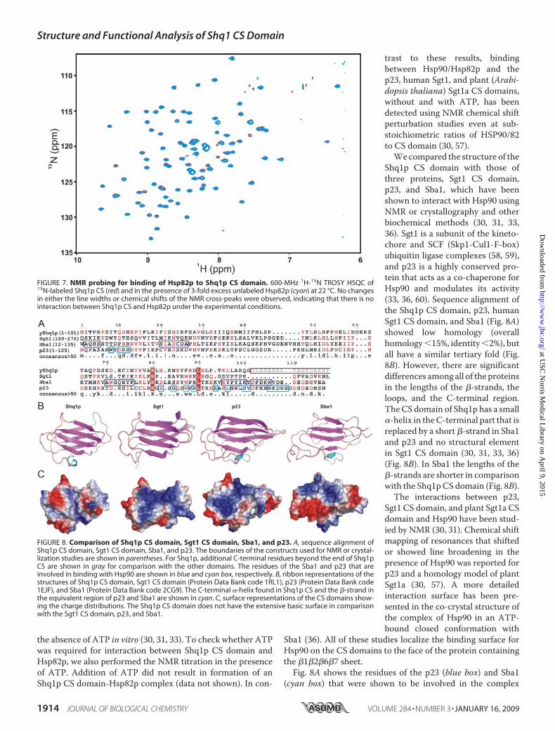
FIGURE 7. NMR probing for binding of Hsp82p to Shq1p CS domain. 600-MHz 1H-15N TROSY HSQC of15N-labeled Shq1p CS (red) and in the presence of 3-fold excess unlabeled Hsp82p (cyan) at 22 °C. No changesin either the line widths or chemical shifts of the NMR cross-peaks were observed, indicating that there is nointeraction between Shq1p CS and Hsp82p under the experimental conditions.
FIGURE 8. Comparison of Shq1p CS domain, Sgt1 CS domain, Sba1, and p23. A, sequence alignment ofShq1p CS domain, Sgt1 CS domain, Sba1, and p23. The boundaries of the constructs used for NMR or crystal-lization studies are shown in parentheses. For Shq1p, additional C-terminal residues beyond the end of Shq1pCS are shown in gray for comparison with the other domains. The residues of the Sba1 and p23 that areinvolved in binding with Hsp90 are shown in blue and cyan box, respectively. B, ribbon representations of thestructures of Shq1p CS domain, Sgt1 CS domain (Protein Data Bank code 1RL1), p23 (Protein Data Bank code1EJF), and Sba1 (Protein Data Bank code 2CG9). The C-terminal �-helix found in Shq1p CS and the �-strand inthe equivalent region of p23 and Sba1 are shown in cyan. C, surface representations of the CS domains show-ing the charge distributions. The Shq1p CS domain does not have the extensive basic surface in comparisonwith the Sgt1 CS domain, p23, and Sba1.
Structure and Functional Analysis of Shq1 CS Domain
1914 JOURNAL OF BIOLOGICAL CHEMISTRY VOLUME 284 • NUMBER 3 • JANUARY 16, 2009
at USC
Norris M
edical Library on A
pril 9, 2015http://w
ww
.jbc.org/D
ownloaded from
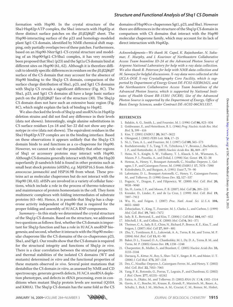
formation with Hsp90. In the crystal structure of theSba1�Hsp82p�ATP complex, the Sba1 interacts with Hsp82p atthree distinct surface patches on the �1�2�6�7 sheet. TheHsp90-interacting surface of the p23 and homology-modeledplant Sgt1 CS domain, identified by NMR chemical shift map-ping, only partially overlaps two of these patches. Furthermore,based on an Hsp90-Nter�Sgt1-CS crystal structure and model-ing of an Hsp90�Sgt1-CS�Sba1 complex, it has very recentlybeen proposed that Sba1 (p23) and the Sgt1aCS domain bind atdifferent sites on Hsp90 (61, 62). Although it is therefore diffi-cult to identify specific differences in residues on the�1�2�6�7surface of the CS domain that may account for the absence ofHsp90 binding to the Shq1p CS domain, comparison of thesurface charge distribution of Sba1, p23, and Sgt1 CS domainswith Shq1p CS reveals a significant difference (Fig. 8C). TheSba1, p23, and Sgt1 CS domains all have a large basic surfacepatch on the �1�2�6�7 face of the structure (30). The Shq1pCS domain does not have such an extensive basic region (Fig.8C), which might explain the lack of binding to Hsp82.We also checked the levels of Shq1p and snoRNAs inHsp82p
deletion strains and did not find any difference in their levels(data not shown). Interestingly, single alanine substitutions inCS surface residues Lys-18 and Ser-22 did not show any phe-notype in vivo (data not shown). The equivalent residues in theSba1�Hsp82p�ATP complex are in the binding interface. Basedon these observations it appears unlikely that the Shq1p CSdomain binds to and functions as a co-chaperone for Hsp90.However, we cannot rule out the possibility that other regionsof Shq1 or accessory proteins may interact with Hsp90.AlthoughCSdomains generally interactwithHsp90, theHsp20superfamily �-sandwich fold is found in other proteins such assmall heat shock proteins (sHSPs), e.g.MjHSP16.5 fromMeth-anococcus jannaschii and HSP16.9B from wheat. These pro-teins act as molecular chaperones but do not interact with theHsp90 (30, 63). sHSPs are involved in a variety of cellular func-tions, which include a role in the process of thermo-toleranceand maintenance of protein homeostasis in the cell. They formmultimeric complexes with folding intermediates of substrateproteins (63–66). Hence, it is possible that Shq1p has a chap-erone activity independent of Hsp90 that is required for theproper folding and assembly of H/ACA RNP components.Summary—In this study we determined the crystal structure
of the Shq1p CS domain. Based on the structure, we addressedtwo questions as follows: first, whether theCS domain is impor-tant for Shq1p function and has a role in H/ACA snoRNP bio-genesis; and second, whether it interacts with theHsp90molec-ular chaperone like the CS domains of the co-chaperones p23,Sba1, and Sgt1.Our results show that theCS domain is requiredfor the structural integrity and functions of Shq1p in vivo.There is a clear correlation between the structural propertiesand thermal stabilities of the isolated CS domains (WT andmutants) determined in vitro and the functional properties ofthese mutants observed in vivo. Several point mutations thatdestabilize the CS domain in vitro, as assessed by NMR and CDspectroscopy, generate growth defects, H/ACA snoRNAdeple-tion phenotypes, and defects in rRNA processing, even in con-ditions when mutant Shq1p protein levels are normal (Q10Aand K80A). The Shq1p CS domain has the same fold as the CS
domains ofHsp90 co-chaperones Sgt1, p23, and Sba1.Howeverthere are differences in the structure of the Shq1pCS domain incomparison with CS domains that interact with the Hsp90molecular chaperone family, which may account for its lack ofdirect interaction with Hsp82p.
Acknowledgments—We thank M. Capel, K. Rajashankar, N. Suku-mar, F. Murphy, and I. Kourinov of Northeastern CollaborativeAccess Team beamline ID-24 at the Advanced Photon Source ofArgonne National Laboratory for help with x-ray data collection.We also thank R. Peterson for help with NMR data collection andM. Sawaya for helpful discussions. X-ray data were collected at theUCLA-DOE X-ray Crystallography Core Facility, which is sup-ported by Department of Energy Grant DE-FC02-02ER63421, andthe Northeastern Collaborative Access Team beamlines of theAdvanced Photon Source, which is supported by National Insti-tutes of Health Grant RR-15301 (NCRR). Use of the AdvancedPhoton Source is supported by the Department of Energy, Office ofBasic Energy Sciences, under Contract DE-AC02-06CH11357.
REFERENCES1. Balakin, A. G., Smith, L., and Fournier, M. J. (1996) Cell 86, 823–8342. Goldwasser, E., and Heinrikson, R. L. (1966) Prog. Nucleic Acids Res. Mol.
Biol. 5, 399–4163. Kiss, T. (2001) EMBO J. 20, 3617–36224. Ofengand, J. (2002) FEBS Lett. 514, 17–255. Ni, J., Tien, A. L., and Fournier, M. J. (1997) Cell 89, 565–5736. Rozhdestvensky, T. S., Tang, T.H., Tchirkova, I. V., Brosius, J., Bachellerie,
J. P., and Huttenhofer, A. (2003) Nucleic Acids Res. 31, 869–8777. Heiss, N. S., Knight, S. W., Vulliamy, T. J., Klauck, S. M., Wiemann, S.,
Mason, P. J., Poustka, A., and Dokal, I. (1998) Nat. Genet. 19, 32–388. Henras, A., Henry, Y., Bousquet-Antonelli, C., Noaillac-Depeyre, J., Gel-
ugne, J. P., and Caizergues-Ferrer, M. (1998) EMBO J. 17, 7078–70909. Kolodrubetz, D., and Burgum, A. (1991) Yeast 7, 79–9010. Lafontaine, D. L., Bousquet-Antonelli, C., Henry, Y., Caizergues-Ferrer,
M., and Tollervey, D. (1998) Genes Dev. 12, 527–53711. Pogacic, V., Dragon, F., and Filipowicz, W. (2000) Mol. Cell. Biol. 20,
9028–904012. Jin, H., Loria, J. P., and Moore, P. B. (2007)Mol. Cell 26, 205–21513. Kressler, D., Linder, P., and de La Cruz, J. (1999) Mol. Cell. Biol. 19,
7897–791214. Wu, H., and Feigon, J. (2007) Proc. Natl. Acad. Sci. U. S. A. 104,
6655–666015. Zebarjadian, Y., King, T., Fournier, M. J., Clarke, L., and Carbon, J. (1999)
Mol. Cell. Biol. 19, 7461–747216. Jady, B. E., Bertrand, E., and Kiss, T. (2004) J. Cell Biol. 164, 647–65217. Mitchell, J. R., and Collins, K. (2000)Mol. Cell 6, 361–37118. Theimer, C. A., Jady, B. E., Chim,N., Richard, P., Breece, K. E., Kiss, T., and
Feigon, J. (2007)Mol. Cell 27, 869–88119. Zhu, Y., Tomlinson, R. L., Lukowiak, A. A., Terns, R. M., and Terns, M. P.
(2004)Mol. Biol. Cell 15, 81–9020. Baker, D. L., Youssef, O. A., Chastkofsky,M. I., Dy, D. A., Terns, R.M., and
Terns, M. P. (2005) Genes Dev. 19, 1238–124821. Charpentier, B., Muller, S., and Branlant, C. (2005) Nucleic Acids Res. 33,
3133–314422. Darzacq, X., Kittur, N., Roy, S., Shav-Tal, Y., Singer, R. H., andMeier, U. T.
(2006) J. Cell Biol. 173, 207–21823. Dez, C., Noaillac-Depeyre, J., Caizergues-Ferrer, M., and Henry, Y. (2002)
Mol. Cell. Biol. 22, 7053–706524. Yang, P. K., Rotondo, G., Porras, T., Legrain, P., and Chanfreau, G. (2002)
J. Biol. Chem. 277, 45235–4524225. Fatica, A., Dlakic, M., and Tollervey, D. (2002) RNA (N. Y.) 8, 1502–151426. Gavin, A. C., Bosche, M., Krause, R., Grandi, P., Marzioch, M., Bauer, A.,
Schultz, J., Rick, J. M., Michon, A. M., Cruciat, C. M., Remor, M., Hofert,
Structure and Functional Analysis of Shq1 CS Domain
JANUARY 16, 2009 • VOLUME 284 • NUMBER 3 JOURNAL OF BIOLOGICAL CHEMISTRY 1915
at USC
Norris M
edical Library on A
pril 9, 2015http://w
ww
.jbc.org/D
ownloaded from
C., Schelder, M., Brajenovic, M., Ruffner, H., Merino, A., Klein, K., Hudak,M., Dickson, D., Rudi, T., Gnau, V., Bauch, A., Bastuck, S., Huhse, B.,Leutwein, C., Heurtier, M. A., Copley, R. R., Edelmann, A., Querfurth, E.,Rybin, V., Drewes, G., Raida, M., Bouwmeester, T., Bork, P., Seraphin, B.,Kuster, B., Neubauer, G., and Superti-Furga, G. (2002) Nature 415,141–147
27. Ho, Y., Gruhler, A., Heilbut, A., Bader, G. D., Moore, L., Adams, S. L.,Millar, A., Taylor, P., Bennett, K., Boutilier, K., Yang, L., Wolting, C.,Donaldson, I., Schandorff, S., Shewnarane, J., Vo, M., Taggart, J., Goud-reault, M., Muskat, B., Alfarano, C., Dewar, D., Lin, Z., Michalickova, K.,Willems, A. R., Sassi, H., Nielsen, P. A., Rasmussen, K. J., Andersen, J. R.,Johansen, L. E., Hansen, L. H., Jespersen, H., Podtelejnikov, A., Nielsen, E.,Crawford, J., Poulsen, V., Sorensen, B. D., Matthiesen, J., Hendrickson,R. C., Gleeson, F., Pawson, T., Moran, M. F., Durocher, D., Mann, M.,Hogue, C. W., Figeys, D., and Tyers, M. (2002) Nature 415, 180–183
28. Ito, T., Chiba, T., Ozawa, R., Yoshida, M., Hattori, M., and Sakaki, Y.(2001) Proc. Natl. Acad. Sci. U. S. A. 98, 4569–4574
29. Leulliot, N., Godin, K. S., Hoareau-Aveilla, C., Quevillon-Cheruel, S., Va-rani, G., Henry, Y., and Van Tilbeurgh, H. (2007) J. Mol. Biol. 371,1338–1353
30. Lee, Y. T., Jacob, J.,Michowski,W., Nowotny,M., Kuznicki, J., andChazin,W. J. (2004) J. Biol. Chem. 279, 16511–16517
31. Martinez-Yamout, M. A., Venkitakrishnan, R. P., Preece, N. E., Kroon, G.,Wright, P. E., and Dyson, H. J. (2006) J. Biol. Chem. 281, 14457–14464
32. Pearl, L. H., Prodromou, C., and Workman, P. (2008) Biochem. J. 410,439–453
33. Weaver, A. J., Sullivan,W. P., Felts, S. J., Owen, B. A., andToft, D.O. (2000)J. Biol. Chem. 275, 23045–23052
34. Zhu, S., and Tytgat, J. (2004) FASEB J. 18, 940–94735. Garcia-Ranea, J. A., Mirey, G., Camonis, J., and Valencia, A. (2002) FEBS
Lett. 529, 162–16736. Ali, M. M., Roe, S. M., Vaughan, C. K., Meyer, P., Panaretou, B., Piper,
P. W., Prodromou, C., and Pearl, L. H. (2006) Nature 440, 1013–101737. Cavanagh, J., Fairbrother, W. J., Skelton, N. J., Rance, M., and Palmer,
I. A. G. (2006) Protein NMR Spectroscopy: Principles and Practices,Elsevier Science Publishers B.V., Amsterdam
38. Otwinowski, Z., and Minor, W. (1997)Methods Enzymol. 276, 307–32639. Pape, T., and Schneider, T. R. (2004) J. Appl. Crystallogr. 37, 843–84440. Collaborative Computational Project No. 4 (1994) Acta Crystallogr. Sect.
D Biol. Crystallogr. 50, 760–76341. Emsley, P., and Cowtan, K. (2004) Acta Crystallogr. Sect. D Biol. Crystal-
logr. 60, 2126–213242. Lamzin, V. S., and Wilson, K. S. (1993) Acta Crystallogr. Sect. D Biol.
Crystallogr. 49, 129–14743. Adams, P. D., Grosse-Kunstleve, R. W., Hung, L. W., Ioerger, T. R.,
McCoy, A. J., Moriarty, N. W., Read, R. J., Sacchettini, J. C., Sauter, N. K.,
and Terwilliger, T. C. (2002)Acta Crystallogr. Sect. D Biol. Crystallogr. 58,1948–1954
44. Sambrook, J., and Russell, D. W. (2001) Molecular Cloning, Cold SpringHarbor Laboratory Press, Cold Spring Harbor, NY
45. Niedenthal, R. K., Riles, L., Johnston,M., andHegemann, J. H. (1996)Yeast12, 773–786
46. Sherman, F., Fink, G. R., and Lawrence, C. W. (1974) Methods in YeastGenetics, Cold Spring Harbor Laboratory Press, Cold Spring Harbor, NY
47. Longtine, M. S., McKenzie, A., III, Demarini, D. J., Shah, N. G., Wach, A.,Brachat, A., Philippsen, P., and Pringle, J. R. (1998) Yeast 14, 953–961
48. Chanfreau, G., Legrain, P., and Jacquier, A. (1998) J. Mol. Biol. 284,975–988
49. Guthrie, C. (1991) Science 253, 157–16350. Tollervey, D. (1987) EMBO J. 6, 4169–417551. Morrissey, J. P., and Tollervey, D. (1993)Mol. Cell. Biol. 13, 2469–247752. Wandinger, S. K., Richter, K., and Buchner, J. (2008) J. Biol. Chem. 283,
18473–1847753. Boulon, S., Marmier-Gourrier, N., Pradet-Balade, B., Wurth, L., Verheg-
gen, C., Jady, B. E., Rothe, B., Pescia, C., Robert,M. C., Kiss, T., Bardoni, B.,Krol, A., Branlant, C., Allmang, C., Bertrand, E., andCharpentier, B. (2008)J. Cell Biol. 180, 579–595
54. Forsythe, H. L., Jarvis, J. L., Turner, J. W., Elmore, L. W., and Holt, S. E.(2001) J. Biol. Chem. 276, 15571–15574
55. Holt, S. E., Aisner, D. L., Baur, J., Tesmer, V. M., Dy, M., Ouellette, M.,Trager, J. B., Morin, G. B., Toft, D. O., Shay, J. W., Wright, W. E., andWhite, M. A. (1999) Genes Dev. 13, 817–826
56. Toogun, O. A., Dezwaan, D. C., and Freeman, B. C. (2008)Mol. Cell. Biol.28, 457–467
57. Boter, M., Amigues, B., Peart, J., Breuer, C., Kadota, Y., Casais, C., Moore,G., Kleanthous, C., Ochsenbein, F., Shirasu, K., and Guerois, R. (2007)Plant Cell 19, 3791–3804
58. Takahashi, A., Casais, C., Ichimura, K., and Shirasu, K. (2003) Proc. Natl.Acad. Sci. U. S. A. 100, 11777–11782
59. Kitagawa, K., Skowyra, D., Elledge, S. J., Harper, J.W., andHieter, P. (1999)Mol. Cell 4, 21–33
60. Buchner, J. (1999) Trends Biochem. Sci. 24, 136–14161. Zhang, M., Boter, M., Li, K., Kadota, Y., Panaretou, B., Prodromou, C.,
Shirasu, K., and Pearl, L. H. (2008) EMBO J. 27, 2789–279862. Kadota, Y., Amigues, B., Ducassou, L.,Madaoui, H., Ochsenbein, F., Guer-
ois, R., and Shirasu, K. (2008) EMBO Rep. 9, 1209–121563. Kim, K. K., Kim, R., and Kim, S. H. (1998) Nature 394, 595–59964. Haslbeck, M., Braun, N., Stromer, T., Richter, B., Model, N., Weinkauf, S.,
and Buchner, J. (2004) EMBO J. 23, 638–64965. Haslbeck, M., and Buchner, J. (2002) Prog. Mol. Subcell. Biol. 28, 37–5966. Haslbeck,M., Franzmann, T.,Weinfurtner, D., and Buchner, J. (2005)Nat.
Struct. Mol. Biol. 12, 842–846
Structure and Functional Analysis of Shq1 CS Domain
1916 JOURNAL OF BIOLOGICAL CHEMISTRY VOLUME 284 • NUMBER 3 • JANUARY 16, 2009
at USC
Norris M
edical Library on A
pril 9, 2015http://w
ww
.jbc.org/D
ownloaded from
Chanfreau and Juli FeigonCascio, Nathanael Heckmann, Guillaume Mahavir Singh, Fernando A. Gonzales, Duilio Ribonucleoparticle Assembly Protein SHQ1Domain of the Essential H/ACA Structure and Functional Studies of the CSRNA: Processing and Catalysis:
doi: 10.1074/jbc.M807337200 originally published online November 19, 20082009, 284:1906-1916.J. Biol. Chem.
10.1074/jbc.M807337200Access the most updated version of this article at doi:
.JBC Affinity SitesFind articles, minireviews, Reflections and Classics on similar topics on the
Alerts:
When a correction for this article is posted•
When this article is cited•
to choose from all of JBC's e-mail alertsClick here
http://www.jbc.org/content/284/3/1906.full.html#ref-list-1
This article cites 63 references, 30 of which can be accessed free at
at USC
Norris M
edical Library on A
pril 9, 2015http://w
ww
.jbc.org/D
ownloaded from