Structure and functions of the liver
33
Training workshop on screening, diagnosis and treatment of hepatitis B and C 1
-
Upload
khangminh22 -
Category
Documents
-
view
1 -
download
0
Transcript of Structure and functions of the liver

Training workshop onscreening, diagnosis and treatment
of hepatitis B and C
1

Session 2
Structure and functions of the liver
2

Learning objectives
At the end of this session, participants shall be able to understand the following concept
• Gross anatomy of the liver and its blood supply • Microanatomy of the liver and its vasculature • Liver fibrosis and its grading• Functions of liver
I welcome all the participants to this first session of the entire training programme.
In this session, we will learn about the basics of the liver in terms of its anatomy,microscopic structure, and a few very important physiological functions that are mostrelevant in the context of viral hepatitis. We will also learn about acute and chronic liverinjuries, and development of liver fibrosis in response to longstanding ongoing chronicinjury such as chronic viral hepatitis. By the end of the session, we will be able tounderstand the pathological effect of liver injury and liver fibrosis on the human body.
3

Quadrants of abdomen
We all know that the abdomen extends vertically from the xiphisternum (above) to pelvic bone (below).
4
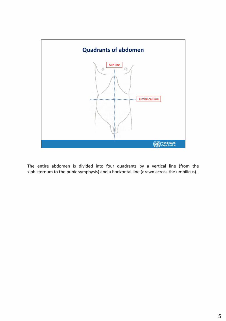
Quadrants of abdomen
Midline
Umbilical line
The entire abdomen is divided into four quadrants by a vertical line (from thexiphisternum to the pubic symphysis) and a horizontal line (drawn across the umbilicus).
5

Quadrants of abdomen
Midline
Umbilical line
Left upperRight upper
Left lowerRight lower
The four quadrants of the abdomen are: right upper, right lower, left upper and left lower.
6

Quadrants of abdomen
Midline
Umbilical line
Left upperRight upper
Left lowerRight lower
Located in the right upper abdomen
The liver is located in the right upper quadrant of the abdomen.
7

Position of liver
• Located in the right upper abdomen• Protected by the right rib cage• Measures: 12–15 cm in vertical direction• Weight: ~1500 g
• Two lobes– Right: Extends till near the right costal
margin(the lower edge of the rib cage)
– Left: Extends midway between xiphisternum and umbilicus
Midline
The liver is located deep in the right upper quadrant and is well protected by the right ribcage. Its size, as measured in the right midclavicular line, is about 12–15 cm and itsweight is about 1500 g. The weight of the liver is approximately 2.5% of the body weight.
The right lobe of the healthy liver is not usually palpable. The left lobe may be palpableup to midway between the xiphisternum and umbilicus. This means that a palpable leftlobe, in isolation, is not of clinical importance. In a patient, the consistency (normalconsistency is firm), surface (normal is smooth, non-tender) and margins (normal isregular) of the liver are much more important features than the liver size alone.
8

Blood supply of liver
• Blood flow ~ 1500 mL/min~ 25% of cardiac output (what the heart pumps)
• Dual blood supply
• Nature of blood flowHepatic artery 1/3 Venous blood (portal vein) 2/3
from the intestine
The liver is a very vascular organ. About 1500 mL of blood passes through the liver everyminute, which is approximately 25% of the cardiac output (normal cardiac output is 5L/min). Compared to its weight (which is about 2.5% of the body weight), it receives amassive blood supply.
It is important to realize that the majority (about 65%) of the blood supplied to the liveris deoxygenated venous blood (which carries much less oxygen than arterial blood) fromthe small and large intestine. Only one third of the supply is oxygenated arterial bloodand carries a high level of oxygen. This dual blood supply serves three importantfunctions. First, the dual blood supply gives a safety cushion to the liver and keeps italive even if one supply is terminated because of some pathological state. Second, thevenous blood carries several harmful substances, toxins and biological products derivedfrom food and gut bacteria present in the large intestine; the liver acts as a filter thatprevents the systemic circulation from exposure to these substances; when this filterfunction of the liver is impaired, such as in patients with liver failure, these harmfulsubstances reach to the brain and the patient becomes unconscious. Third, venousblood carries a lot of nutrients from the small intestine; these nutrients, if releasedunchecked into the circulation, will produce metabolic imbalance. The liver acts as atemporary warehouse to store excessive amounts of these nutrients and releases themat the time of need (such as fasting).
9

Normal circulation
Blood from all the tissues reaches the right heart and then goes to the lungs
Left heartRight heart
Arteries
Capillaries
Veins
Lung capillaries
During normal blood circulation, deoxygenated blood is collected from all over the bodyby the venous system and is pumped by the right side of the heart into the lungs. In thelungs, oxygenation of blood takes place and oxygen-rich blood is returned to the left sideof the heart and pumped through the arteries throughout the body.
Capillaries connect arteries to veins. Oxygen and carbon dioxide is exchanged in arteries,and blood collected from the capillaries returns to the lungs through veins.
10

Normal circulation
Venous blood goes to another organ instead of to the right heart
Left heartRight heart
Arteries
Capillaries
Veins
Lung capillaries
Another organ
During normal blood circulation, deoxygenated blood is collected from all over the bodyby the venous system and is pumped by the right side of the heart into the lungs. In thelungs, oxygenation of blood takes place and oxygen-rich blood is returned to the left sideof the heart and pumped through the arteries. During normal circulation, the bloodcollected from the capillaries is returned to the lungs through veins.
In the portal system, the blood returning from the capillaries is not directly returned tothe venous system but is again passed through another set of capillaries in anotherorgan or tissue. There are two portal systems in the human body: the pituitary–hypophyseal system in the brain and the second in the liver. The objective of this portalsystem is to provide the liver with extra circulation time and expose the blood to theextensive network of hepatocyte plates. It helps the liver to perform its metabolic andfiltering activities more efficiently.
11
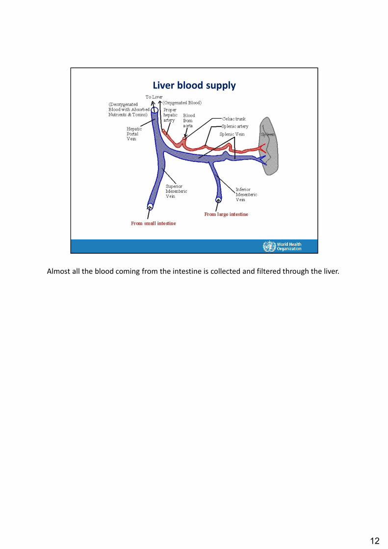
Liver blood supply
Almost all the blood coming from the intestine is collected and filtered through the liver.
12

Liver portal system
This picture shows the venous drainage into the portal venous system and the venousdrainage of the liver. The portal vein is formed by the superior mesenteric vein (whichcollect the nutrients and toxin-rich deoxygenated blood from the intestine) and splenicvein (which carries the immune-activated lymphocytes) from the spleen.
Inside the liver, the portal venous blood is first distributed to the extensive network ofsinusoids and is then collected by three hepatic veins: right, middle and left hepaticveins. These hepatic veins drain into the inferior vena cava, which drains into the rightside of the heart for oxygenation.
13

Liver: Microscopic anatomy
Now we will learn the microanatomy of the liver. We have to take out a piece of liver(liver biopsy) with the help of a Tru-Cut biopsy needle and examine it under themicroscope. Liver biopsy is a risky procedure and needs hospitalization, skill to performthe biopsy, and a trained pathologist to interpret the findings under the microscope. Itcarries a finite, though small, risk of death following the procedure. Hence, these days,non-invasive methods are used to study the liver tissue, which we will learn during thiscourse.
14

Liver: Microscopic anatomy
If we see the liver tissue under the microscope, we will find that the hepatocytes (liverparenchymal cells) are organized in honeycomb-like structures. These structure arecalled hepatic lobules. Such lobules are three-dimensional structures and are commonlyhexagonal in shape.
Within each lobule, the individual liver cells (hepatocytes) are well organized. We cancompare the liver with a large school, liver lobule with a classroom, and each hepatocytewith every single chair inside the classroom. In a well-organized classroom, the chairsare organized in rows and placed at a definite distance, which facilitates easy movementof the students/teachers inside the classroom.
Similarly, inside a liver lobule, hepatocytes are organized in the form of plates (akin torows in a classroom), which are separated by sinusoids through which blood flows easilywithout much resistance.
15

Liver: Microscopic anatomy
Organized as hepatic lobules
Lobules are penta- to hexagonal structures, located closely together
Each liver lobule has the following 3 structures in each corner of its hexagon: bile duct(green), portal vein (blue) and hepatic artery (pink). Usually there is one of each of thesebut sometimes there may be 2–4 bile ducts and sometimes only 2 structures. In thecentre of each lobule there is one central vein (blue), which drains into the hepatic veins.
16

Liver: Microscopic anatomy
Organized as hepatic lobules
Lobules are penta- to hexagonal structures, with portal tracts at each corner and central vein in the middle
Portal tract
The three structures, along with the surrounding plate of hepatocytes, at the corner ofthe hepatic lobule are collectively called a portal tract.
17

Liver: Microscopic anatomy
Organized as hepatic lobules
Lobules are penta- to hexagonal structures, with portal tracts at each corner and central vein in the middle
Portal tract
In each lobule, blood from the branches of the portal vein and hepatic artery entersfrom the corner and flows in a centripetal direction to drain into the central vein. Thisflow of blood is slow and under low pressure, which gives adequate time for exchange ofmaterial between the blood and surrounding hepatocytes.
18

Liver: Microscopic anatomy
Organized as hepatic lobules
Lobules are penta- to hexagonal structures, with portal tracts at each corner and central vein in the middle
Portal tract
Bile is produced by hepatocytes. The bile produced by hepatocytes flows in a centrifugaldirection (opposite to the flow of blood) to drain into a branch of the bile duct located inthe corner of the lobule.
19

Microscopic anatomy of liver
This is a three-dimensional picture of a hexagonal liver lobule. Each lobule is a three-dimensional structure, which shows the portal tract at each of the corner. Each portaltract has a branch of the portal vein (blue), hepatic artery (red) and bile duct (lightgreen). The entire lobule is packed with hepatocytes organized in the form of plates(brown), which are separated by blood-filled sinusoids (purple).
20

Causes of liver injury
• The liver can be damaged by many insults (separate talk)
• Major causes
– Infections, especially viruses• Hepatitis viruses – A to E
– Non-infectious causes• Alcohol• Drugs• Non-alcoholic fatty liver disease
Till now we have learnt about the normal structure of the liver.
The liver can be injured by any number of agents but the major causes are viralinfections (hepatitis viruses such as hepatitis B or C), toxins (alcohol) or drugs (anti-tuberculosis drugs such as INH, rifampicin, pyrazinamide; antiepileptic drugs –phenytoin; paracetamol; oral contraceptive pills, etc.).
21

Prolonged liver injury leads to scarring (fibrosis)
F1 F2
F3 F4
Fibrosis starts around the portal area and extends gradually into the lobularparenchyma. There are four grades of fibrosis: F1, F2, F3, F4.
22

Response of the liver to injury
• Acute (short-term) injury– Inflammation– Swelling and mild enlargement– Death of cells and impairment of liver function
• Chronic (long-term) injury– Chronic inflammation– Fibrosis (scarring)
Regardless of the cause, liver injury manifests in form of hepatitis which meansinflammation of the liver. There are five components of inflammation. What are these?(The 5 components are: rubor – redness, calor – heat, dolor – pain, tumor – swelling,and loss of function).
Liver inflammation (or hepatitis) could be either acute or chronic.
Acute hepatitis is characterized by sudden and massive death of hepatocytes over ashort period of time and is characterized by all the five components of inflammation.Clinically we find liver enlargement (tumor); tenderness on palpation (dolor); jaundicewith or without coagulopathy/encephalopathy (loss of function).
In contrast, chronic hepatitis is caused by slow but long-standing injury, which leads toan ongoing process of cell death and healing. Healing during chronic hepatitis is see asfibrosis.
23

Prolonged liver injury leads to scarring (fibrosis)
F1 F2
F3 F4
Central vein
Portal tract
Scarring: particularly affects the central vein and portal tracts (blood vessels)
F1 indicates fibrosis in the portal area; the fibrosis has not extended beyond the limitingplate of the portal tract.
F2 indicates portal fibrosis with fibrous septae; thin septae are developing, which havestarted extending from the portal tract to the liver parenchyma; very few thin septaemight be seen joining two adjacent portal tracts.
F3 indicates numerous septae without cirrhotic nodules; a number of thick fibrousbands can be seen connecting adjacent portal tracts, which convert each liver lobule intoa single nodule surrounded by a thick fibrous band; there will be no or very few thinbands from the portal tract to the central vein.
F4 indicates cirrhosis, nodule formation or findings suggestive of nodule formation;there will be a number of thick bands extending from the portal tract to both adjacentportal tracts as well as the central veins. Hence, the entire lever lobule is converted intoa clump of multiple smaller nodules each surrounded by thick fibrous septae.
24

Prolonged liver injury leads to scarring (fibrosis)
F1 F2
F3 F4
Central vein
Portal tract
Fibrosis affects the flow of blood through the portal tracts and hepatic lobules
25

Progression of liver fibrosis
Metavir fibrosis stages: F0-F4
Cirrhosis: An advanced stage of liver fibrosis characterized by – extensive fibrosis– nodular regeneration– distortion of liver architecture = F4 fibrosis
The spectrum of liver disease ranges from minimal fibrosis to cirrhosis.
Without antiviral therapy, chronic hepatitis gradually progresses to cirrhosis in 20–30years.
The Metavir fibrosis staging system is a scoring system for assessing liver fibrosis basedon pathological findings.
F4 Metavir fibrosis stage is also known as cirrhosis.
26

Effect of lobular disorganization
Distortion of the architecture of the liver lobule leads to hindrance in blood flow, just asthe difficulty caused in trying to move about in a classroom with disorganized chairs.
The fibrosis converts the non-turbulent, low-pressure blood flow in the lobule sinusoidsinto a turbulent high-pressure flow. This stage is known as portal hypertension.
27

Effect of liver injury
Two effects• Impairment of liver function• Impairment of blood circulation through the liver
As a part of the inflammation of liver injury, there are two types of adverse effects:impairment of liver function and impairment of blood circulation through itsparenchyma.
28

Functions of the liver
Several functions, including some of the following
Glucose metabolism• During the feeding phase Move glucose into glycogen stores• During fasting Move sugar from stores to the blood
Excretory function• Bile pigments (bilirubin)• Bile salts Important for absorption of fats• Other harmful substances
Synthetic function• Albumin• Coagulation factors
There are three major functions of the liver: (i) glucose metabolism, which maintains theblood glucose within an acceptable range; (ii) excretion of waste substances from thebody in the bile; and (iii) synthesis of important body proteins such as albumin andcoagulation factors.
Role of albumin: maintains the oncotic pressure; the half-life is 21 days, which isimportant to know in cases of liver dysfunction.
29

Liver disease: Effect on liver function
Several functions, including some of the following
Glucose metabolism• During the feeding phase Blood sugar too high (hyperglycaemia)• During fasting Blood sugar too low (hypoglycaemia)
Excretory function• Bile pigments (bilirubin) Jaundice• Bile salts Poor absorption of fats• Other harmful substances Unconsciousness
Synthetic function• Albumin Edema, (ascites)• Coagulation factors Deranged coagulation, bleeding
Impaired glucose metabolism results in postprandial hyperglycaemia and post-fastinghypoglycaemia.
Impaired excretion of bilirubin results in jaundice. Impaired clearance of toxic wastesmay lead to unconsciousness.
Albumin is the main protein that maintains the oncotic pressure and maintains the bodyvascular volume. If albumin is not synthesized then fluid will move out from the bloodvessels to the third spaces such as the peritoneal cavity and pleural cavities. It results inascites and pleural effusion.
Impaired synthesis of coagulation factors will lead to bleeding manifestations.
30

Effect of cirrhosis on liver circulation
Decreased blood flowthrough the liver leads to
high pressure in the portal venous system
or portal hypertension
Blood vessels proximal to the liver get congested, which is known as portalhypertension.
31

Liver disease: Effect on blood circulation
• Obstruction of blood flow through the hepatic lobules leads to increased pressure in the portal vein
• Portal hypertension = increased pressure in the portal vein
• Manifestations– Development of collateral veins Abnormally enlarged veins
(varices)– Exudation of fluid into abdomen Ascites – Congestion of venous system Splenomegaly >pancytopenia
Portal hypertension results in congestive splenomegaly, ascites, and formation ofcollaterals at various places. The newly formed collateral vessels manifest as esophagealvarices or gastric varices. Hypersplenism results in pancytopenia, in particular,thrombocytopenia.
32
Summary
• An organ located in the right upper abdomen
• Has dual blood supply, including via the portal vein
• Is metabolically highly active, with many functions
• Continuing liver injury, irrespective of the cause, leads to liver fibrosis of varying degrees (F0 to F4)
• Advanced fibrosis (e.g. F4 or cirrhosis) is associated with – Impaired liver function– Impaired blood flow through the liver, leading to increased
pressure in the portal vein (= portal hypertension)
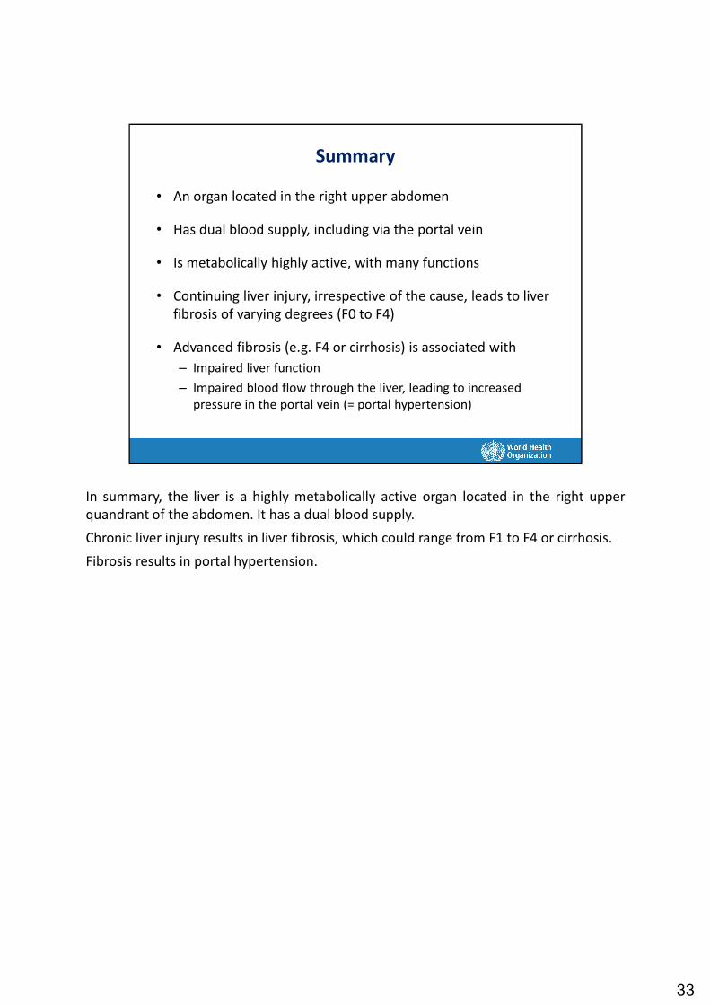
In summary, the liver is a highly metabolically active organ located in the right upperquandrant of the abdomen. It has a dual blood supply.
Chronic liver injury results in liver fibrosis, which could range from F1 to F4 or cirrhosis.
Fibrosis results in portal hypertension.
33