Structural plasticity of climbing fibers and the growth-associated protein GAP-43
7
PERSPECTIVE ARTICLE published: 21 February 2013 doi: 10.3389/fncir.2013.00025 Structural plasticity of climbing fibers and the growth-associated protein GAP-43 Giorgio Grasselli 1 * and Piergiorgio Strata 2 1 Department of Neurobiology, University of Chicago, Chicago, IL, USA 2 National Institute of Neuroscience-Italy, University of Turin, Turin, Italy Edited by: Chris I. De Zeeuw, Erasmus MC, Netherlands Reviewed by: David Linden, Johns Hopkins University, USA Patricia C. Salinas, University College London, UK *Correspondence: Giorgio Grasselli, Department of Neurobiology, University of Chicago, 947 E. 58th Street, MC0928, Chicago, IL 60637, USA. e-mail: [email protected] Structural plasticity occurs physiologically or after brain damage to adapt or re-establish proper synaptic connections. This capacity depends on several intrinsic and extrinsic determinants that differ between neuron types. We reviewed the significant endogenous regenerative potential of the neurons of the inferior olive (IO) in the adult rodent brain and the structural remodeling of the terminal arbor of their axons, the climbing fiber (CF), under various experimental conditions, focusing on the growth-associated protein GAP-43. CFs undergo remarkable collateral sprouting in the presence of denervated Purkinje cells (PCs) that are available for new innervation. In addition, severed olivo-cerebellar axons regenerate across the white matter through a graft of embryonic Schwann cells. In contrast, CFs undergo a regressive modification when their target is deleted. In vivo knockdown of GAP-43 in olivary neurons, leads to the atrophy of their CFs and a reduction in the ability to sprout toward surrounding denervated PCs. These findings demonstrate that GAP-43 is essential for promoting denervation-induced sprouting and maintaining normal CF architecture. Keywords: climbing fiber, GAP-43, sprouting, atrophy, branching INTRODUCTION Structural plasticity is limited in the central nervous system (CNS) of adult mammals, constituting a significant impediment to recovery from injuries such as those caused by trauma, stroke, and neurodegenerative and demyelinating diseases (Duffau, 2006; Wieloch and Nikolich, 2006; Landi and Rossini, 2010). Nevertheless, a relatively high degree of structural plasticity is retained by certain areas of brain, such as the cerebellum (Carulli et al., 2004; Cesa and Strata, 2009). The cerebellar climbing fiber (CF), the terminal arbor of the olivo-cerebellar axons, has provided the first example, in the mammalian CNS, of individually observed fibers undergoing sprouting after brain injury (Rossi et al., 1991a,b). In 6-weeks-old Wistar rats, CFs normally encompass approximately 1000 μm of dendritic length and bear an average of 544 ± 23 varicosities that express the vesicular glutamate transporter VGLUT2 (Grasselli et al., 2011). CFs constitute a suitable model that can be used to investi- gate axonal structural plasticity, based on their significant plastic potential and morphological hallmarks. In fact, they have a one- to-one relationship with their target Purkinje cell (PC). CFs undergo lesion-induced sprouting, activity-dependent remodel- ing, expansion of their area of innervation in response to an enlarged target territory, and regressive modifications after elim- ination of their target (Rossi and Strata, 1995; Strata and Rossi, 1998; Cesa and Strata, 2009). STRUCTURAL PLASTICITY OF CLIMBING FIBERS Neurons differ widely in regard to their response to axonal injuries (Carulli et al., 2004; Dusart et al., 2005). For example, in the cerebellum, PCs respond to injury with little upregulation of plasticity-related genes in the cell body, no axonal regeneration after axotomy, and weak sprouting; most PCs survive, but they usually do not increase the expression of plasticity-related genes, except when the axotomy occurs near the cell body (Rossi et al., 1995; Bravin et al., 1997; Zagrebelsky et al., 1998; Wehrle et al., 2001; Morel et al., 2002; Gianola and Rossi, 2004). Further, axonal sprouting is limited and might be induced only following proper manipulation of intrinsic and environmental factors (Buffo et al., 1997, 2000; Zagrebelsky et al., 1998; Zhang et al., 2005, 2007). In contrast, neurons in the inferior olive (IO) respond dra- matically to axonal injury. The resection of olivo-cerebellar axons leads to the regression of the remaining stump and the death of many axotomized neurons in the IO during the first few weeks after injury (Buffo et al., 1998). Concurrently, olivary neurons upregulate several intrinsic factors, including nitric oxide syn- thase (NOS), c-Jun, JunD, the early growth response protein EGR1/Krox-24 (Rossi and Strata, 1995; Bravin et al., 1997; Buffo et al., 1998, 2003; Wehrle et al., 2001). As a result of this upregulation and the high constitutive levels of growth-associated factors in olivary neurons, such as GAP-43, MARCKS, EGR-1/KROX-24, L1CAM, and PSA-NCAM in the olivary neurons (Kruger et al., 1993; Herdegen et al., 1995; McNamara and Lenox, 1997; Fernandez et al., 1999; Horinouchi et al., 2005), lesioned olivo-cerebellar axons can elongate and innervate their target PCs when an appropriate per- missive environment is provided, such as neonatal Schwann cells that have been inserted at the site of axotomy (Bravin et al., 1997). Lesioned olivo-cerebellar fibers can also elongate into a transplant of embryonic cerebellum, where they innervate the Frontiers in Neural Circuits www.frontiersin.org February 2013 | Volume 7 | Article 25 | 1 NEURAL CIRCUITS
Transcript of Structural plasticity of climbing fibers and the growth-associated protein GAP-43
PERSPECTIVE ARTICLEpublished: 21 February 2013
doi: 10.3389/fncir.2013.00025
Structural plasticity of climbing fibers and thegrowth-associated protein GAP-43Giorgio Grasselli 1* and Piergiorgio Strata 2
1 Department of Neurobiology, University of Chicago, Chicago, IL, USA2 National Institute of Neuroscience-Italy, University of Turin, Turin, Italy
Edited by:
Chris I. De Zeeuw, Erasmus MC,Netherlands
Reviewed by:
David Linden, Johns HopkinsUniversity, USAPatricia C. Salinas, UniversityCollege London, UK
*Correspondence:
Giorgio Grasselli, Department ofNeurobiology, University of Chicago,947 E. 58th Street, MC0928,Chicago, IL 60637, USA.e-mail: [email protected]
Structural plasticity occurs physiologically or after brain damage to adapt or re-establishproper synaptic connections. This capacity depends on several intrinsic and extrinsicdeterminants that differ between neuron types. We reviewed the significant endogenousregenerative potential of the neurons of the inferior olive (IO) in the adult rodent brainand the structural remodeling of the terminal arbor of their axons, the climbing fiber (CF),under various experimental conditions, focusing on the growth-associated protein GAP-43.CFs undergo remarkable collateral sprouting in the presence of denervated Purkinje cells(PCs) that are available for new innervation. In addition, severed olivo-cerebellar axonsregenerate across the white matter through a graft of embryonic Schwann cells. Incontrast, CFs undergo a regressive modification when their target is deleted. In vivoknockdown of GAP-43 in olivary neurons, leads to the atrophy of their CFs and a reductionin the ability to sprout toward surrounding denervated PCs. These findings demonstratethat GAP-43 is essential for promoting denervation-induced sprouting and maintainingnormal CF architecture.
Keywords: climbing fiber, GAP-43, sprouting, atrophy, branching
INTRODUCTIONStructural plasticity is limited in the central nervous system(CNS) of adult mammals, constituting a significant impedimentto recovery from injuries such as those caused by trauma, stroke,and neurodegenerative and demyelinating diseases (Duffau,2006; Wieloch and Nikolich, 2006; Landi and Rossini, 2010).Nevertheless, a relatively high degree of structural plasticity isretained by certain areas of brain, such as the cerebellum (Carulliet al., 2004; Cesa and Strata, 2009).
The cerebellar climbing fiber (CF), the terminal arbor of theolivo-cerebellar axons, has provided the first example, in themammalian CNS, of individually observed fibers undergoingsprouting after brain injury (Rossi et al., 1991a,b). In 6-weeks-oldWistar rats, CFs normally encompass approximately 1000 µm ofdendritic length and bear an average of 544 ± 23 varicosities thatexpress the vesicular glutamate transporter VGLUT2 (Grasselliet al., 2011).
CFs constitute a suitable model that can be used to investi-gate axonal structural plasticity, based on their significant plasticpotential and morphological hallmarks. In fact, they have a one-to-one relationship with their target Purkinje cell (PC). CFsundergo lesion-induced sprouting, activity-dependent remodel-ing, expansion of their area of innervation in response to anenlarged target territory, and regressive modifications after elim-ination of their target (Rossi and Strata, 1995; Strata and Rossi,1998; Cesa and Strata, 2009).
STRUCTURAL PLASTICITY OF CLIMBING FIBERSNeurons differ widely in regard to their response to axonalinjuries (Carulli et al., 2004; Dusart et al., 2005). For example,
in the cerebellum, PCs respond to injury with little upregulationof plasticity-related genes in the cell body, no axonal regenerationafter axotomy, and weak sprouting; most PCs survive, but theyusually do not increase the expression of plasticity-related genes,except when the axotomy occurs near the cell body (Rossi et al.,1995; Bravin et al., 1997; Zagrebelsky et al., 1998; Wehrle et al.,2001; Morel et al., 2002; Gianola and Rossi, 2004). Further, axonalsprouting is limited and might be induced only following propermanipulation of intrinsic and environmental factors (Buffo et al.,1997, 2000; Zagrebelsky et al., 1998; Zhang et al., 2005, 2007).
In contrast, neurons in the inferior olive (IO) respond dra-matically to axonal injury. The resection of olivo-cerebellar axonsleads to the regression of the remaining stump and the death ofmany axotomized neurons in the IO during the first few weeksafter injury (Buffo et al., 1998). Concurrently, olivary neuronsupregulate several intrinsic factors, including nitric oxide syn-thase (NOS), c-Jun, JunD, the early growth response proteinEGR1/Krox-24 (Rossi and Strata, 1995; Bravin et al., 1997; Buffoet al., 1998, 2003; Wehrle et al., 2001).
As a result of this upregulation and the high constitutivelevels of growth-associated factors in olivary neurons, such asGAP-43, MARCKS, EGR-1/KROX-24, L1CAM, and PSA-NCAMin the olivary neurons (Kruger et al., 1993; Herdegen et al.,1995; McNamara and Lenox, 1997; Fernandez et al., 1999;Horinouchi et al., 2005), lesioned olivo-cerebellar axons canelongate and innervate their target PCs when an appropriate per-missive environment is provided, such as neonatal Schwann cellsthat have been inserted at the site of axotomy (Bravin et al.,1997). Lesioned olivo-cerebellar fibers can also elongate into atransplant of embryonic cerebellum, where they innervate the
Frontiers in Neural Circuits www.frontiersin.org February 2013 | Volume 7 | Article 25 | 1
NEURAL CIRCUITS
Grasselli and Strata CF structural plasticity and GAP-43
grafted PCs, forming new CFs (Gardette et al., 1988; Rossi et al.,1995).
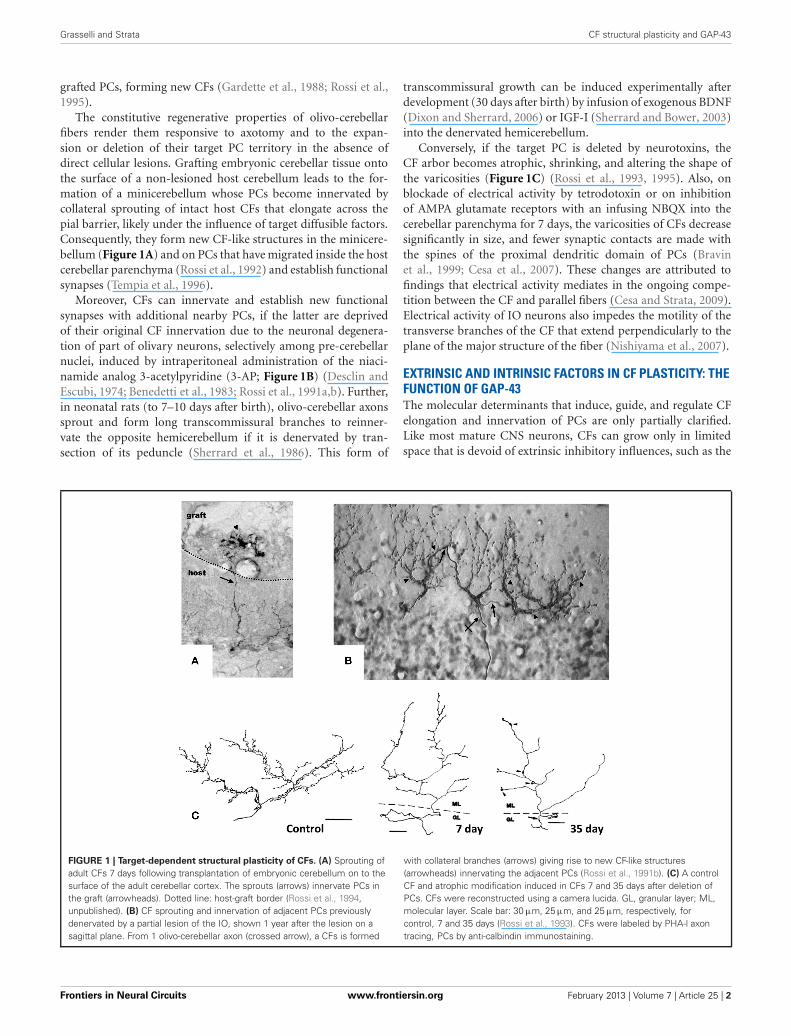
The constitutive regenerative properties of olivo-cerebellarfibers render them responsive to axotomy and to the expan-sion or deletion of their target PC territory in the absence ofdirect cellular lesions. Grafting embryonic cerebellar tissue ontothe surface of a non-lesioned host cerebellum leads to the for-mation of a minicerebellum whose PCs become innervated bycollateral sprouting of intact host CFs that elongate across thepial barrier, likely under the influence of target diffusible factors.Consequently, they form new CF-like structures in the minicere-bellum (Figure 1A) and on PCs that have migrated inside the hostcerebellar parenchyma (Rossi et al., 1992) and establish functionalsynapses (Tempia et al., 1996).
Moreover, CFs can innervate and establish new functionalsynapses with additional nearby PCs, if the latter are deprivedof their original CF innervation due to the neuronal degenera-tion of part of olivary neurons, selectively among pre-cerebellarnuclei, induced by intraperitoneal administration of the niaci-namide analog 3-acetylpyridine (3-AP; Figure 1B) (Desclin andEscubi, 1974; Benedetti et al., 1983; Rossi et al., 1991a,b). Further,in neonatal rats (to 7–10 days after birth), olivo-cerebellar axonssprout and form long transcommissural branches to reinner-vate the opposite hemicerebellum if it is denervated by tran-section of its peduncle (Sherrard et al., 1986). This form of
transcommissural growth can be induced experimentally afterdevelopment (30 days after birth) by infusion of exogenous BDNF(Dixon and Sherrard, 2006) or IGF-I (Sherrard and Bower, 2003)into the denervated hemicerebellum.
Conversely, if the target PC is deleted by neurotoxins, theCF arbor becomes atrophic, shrinking, and altering the shape ofthe varicosities (Figure 1C) (Rossi et al., 1993, 1995). Also, onblockade of electrical activity by tetrodotoxin or on inhibitionof AMPA glutamate receptors with an infusing NBQX into thecerebellar parenchyma for 7 days, the varicosities of CFs decreasesignificantly in size, and fewer synaptic contacts are made withthe spines of the proximal dendritic domain of PCs (Bravinet al., 1999; Cesa et al., 2007). These changes are attributed tofindings that electrical activity mediates in the ongoing compe-tition between the CF and parallel fibers (Cesa and Strata, 2009).Electrical activity of IO neurons also impedes the motility of thetransverse branches of the CF that extend perpendicularly to theplane of the major structure of the fiber (Nishiyama et al., 2007).
EXTRINSIC AND INTRINSIC FACTORS IN CF PLASTICITY: THEFUNCTION OF GAP-43The molecular determinants that induce, guide, and regulate CFelongation and innervation of PCs are only partially clarified.Like most mature CNS neurons, CFs can grow only in limitedspace that is devoid of extrinsic inhibitory influences, such as the
FIGURE 1 | Target-dependent structural plasticity of CFs. (A) Sprouting ofadult CFs 7 days following transplantation of embryonic cerebellum on to thesurface of the adult cerebellar cortex. The sprouts (arrows) innervate PCs inthe graft (arrowheads). Dotted line: host-graft border (Rossi et al., 1994,unpublished). (B) CF sprouting and innervation of adjacent PCs previouslydenervated by a partial lesion of the IO, shown 1 year after the lesion on asagittal plane. From 1 olivo-cerebellar axon (crossed arrow), a CFs is formed
with collateral branches (arrows) giving rise to new CF-like structures(arrowheads) innervating the adjacent PCs (Rossi et al., 1991b). (C) A controlCF and atrophic modification induced in CFs 7 and 35 days after deletion ofPCs. CFs were reconstructed using a camera lucida. GL, granular layer; ML,molecular layer. Scale bar: 30 µm, 25 µm, and 25 µm, respectively, forcontrol, 7 and 35 days (Rossi et al., 1993). CFs were labeled by PHA-l axontracing, PCs by anti-calbindin immunostaining.
Frontiers in Neural Circuits www.frontiersin.org February 2013 | Volume 7 | Article 25 | 2
Grasselli and Strata CF structural plasticity and GAP-43
cerebellar molecular layer, which lacks inhibitory myelin growthfactors.
More is known about the intrinsic factors that confer highlyplastic properties to CFs. The well-characterized plasticity ofmature IO neurons is associated with high, constitutive expres-sion of the growth-associated proteins GAP-43, EGR-1/KROX-24,MARCKS, L1CAM, and PSA-NCAM, and with the upregulationof c-Jun, JunD, Krox-24, and NOS in response to axonal lesions.However, the contribution of each factor is still not clear.
GAP-43 was one of the first of these proteins to be studiedextensively and described for its abundance in axonal growthcones (Zwiers et al., 1976; Skene and Willard, 1981); thus it isused widely as a marker of axonal sprouting (Oestreicher et al.,1997). GAP-43 (also known as neuromodulin and B-50) mediatesaxonal growth, branching, and pathfinding during development.Mice that lack this protein have a low survival rate in the earlypostnatal period (Strittmatter et al., 1995; Maier et al., 1999).In humans, heterozygous chromosomal deletions comprising thelocus for Gap-43 gene (3q13.10–3q13.21) are linked to agenesisof the corpus callosum and severe mental retardation (Genuardiet al., 1994; Mackie Ogilvie et al., 1998).
GAP-43 plays a pivotal role not only during developmentbut also in axonal remodeling in the adult brain. Its expressionrises in several conditions that induce neuronal rewiring, suchas the disruption of the neuronal networks due to pathologicalor traumatic lesions (Benowitz et al., 1990; Oestreicher et al.,1997; Buffo et al., 2003): it is upregulated in the motoneurons ofdystrophin-deficient mice (mdx mice), a model of human mus-cular dystrophy, in which degeneration-regeneration events inmuscle fibers are accompanied by remodeling of intramuscularterminal nerve fibers (Verzè et al., 1996), and after the inductionof robust neuronal activity, for example due to seizure or electricalstimulation (McNamara and Routtenberg, 1995; Cantallops andRouttenberg, 1996; Miyake et al., 2002; Sharma et al., 2010).
Complex alterations in GAP-43 expression are frequentlyobserved in human neuropathologies and their animal mod-els, suggesting axonal damage or attempts of regenerative axonalsprouting. For instance GAP-43 expression declines in the frontalcortex and certain areas of the hippocampus in Alzheimerpatients but is robust in association with senile-like plaques(Bogdanovic et al., 2000). Moreover, GAP-43 levels decrease inmost lesions in the white matter of patients with multiple sclerosisand increase in some remyelinated white matter tracts (Teunissenet al., 2006).
In several experimental conditions, GAP-43 overexpressionin vivo increases axonal sprouting. In transgenic mice that over-express GAP-43, motoneurons undergo axonal sprouting, evenspontaneously in the absence of injuries, and increased sprout-ing after lesion. These mice experience prominent, spontaneoussprouting of mossy fibers in the dentate gyrus (Aigner et al.,1995). As discussed, when GAP-43 was overexpressed in PCs,their axons sprout profusely along their length and at their stumpeven at sites that are covered by myelin demonstrating that itsoverexpression is sufficient to induce sprouting in the absence ofany injury and promote lesion-induced sprouting in PCs (Buffoet al., 1997; Gianola and Rossi, 2004). In a recent report, aftersilencing the expression of GAP-43 in IO neurons of juvenile
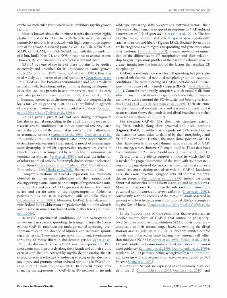
wild-type rats using shRNA-expressing lentiviral vectors, theirCFs were virtually unable to sprout in response to 3-AP-induceddenervation of PCs (Figure 2A) (Grasselli et al., 2011). The fewCFs that were, however, still able to sprout were significantlysmaller than control fibers (Figures 2B,C). Because IO neuronsare heterogeneous with regards to sprouting and gene expressionafter axotomy (Buffo et al., 2003), a more in-depth examina-tion of the differences in CF morphology and their relation-ship to gene expression profiles of their neurons should providegreater insight into the function of the factors that regulate CFmorphology.
GAP-43 is not only necessary for CF sprouting but plays alsoa crucial role for normal neuronal morphology in non-traumaticconditions. The mere silencing of GAP-43 destabilizes CF struc-ture in the absence of any insult (Figures 2D–G) (Grasselli et al.,2011). Control CFs normally comprises a thick axonal stalk fromwhich many thin collaterals emerge (namely tendrils), forming anet-like structure around the PC dendrite and bearing varicosi-ties (Rossi et al., 1991b; Sugihara et al., 1999). Their structurehas been examined quantitatively and a recent complete digitalreconstruction shows that tendrils and distal branches are richerin varicosities (Brown et al., 2012).
On silencing GAP-43, CFs alter their structure, extend-ing fewer tendrils along their proximal and distal portions(Figures 2D–G), quantified as a significant 17% reduction inthe density of varicosities, as defined by their morphology andVGLUT2 expression. Further, the most distal portions of CFs,which have fewer tendrils and a thinner stalk, are affected by GAP-43 silencing, which shortens CF length by 33%. These data havebeen confirmed in 2–3-months-old mice (Grasselli et al., 2011).
Several lines of evidence support a model in which GAP-43is needed for proper interaction of the axon with its target neu-ron and organization of the molecular machinery that supportsaxonal structures during axonal growth. In GAP-43 knockoutmice, the axons of retinal ganglion cells fail to cross the opticchiasm properly (Strittmatter et al., 1995), instead assumingabnormal trajectories in the chiasm (Sretavan and Kruger, 1998).Moreover, these mice fail to form the anterior commissure, hip-pocampal commissure, and corpus callosum (Shen et al., 2002),consistently with the agenesis of the corpus callosum observed inpatients who bear heterozygous chromosomal deletions compris-ing the Gap-43 locus (Genuardi et al., 1994; Mackie Ogilvie et al.,1998).
In the hippocampus of transgenic mice that overexpress aninactive mutant form of GAP-43 that cannot be phosphory-lated (with an amino acid substitution S42A), mossy fibers growectopically to their normal target layer, innervating the distalstratum oriens (Holahan et al., 2010). Notably, similar ectopicgrowth was observed in mice lacking the neuronal cell adhe-sion molecule NCAM (Cremer et al., 1997; Bukalo et al., 2004).L1CAM, another adhesion molecule that mediates commissuralaxon guidance (Kamiguchi et al., 1998; Demyanenko et al., 1999),regulates GAP-43 pathway, acting synergistically with it promot-ing axon growth and regeneration when overexpressed in PCsin vivo (Zhang et al., 2005).
L1CAM and NCAM are expressed at constitutively high lev-els in the IO (Horinouchi et al., 2005; Quartu et al., 2010), and
Frontiers in Neural Circuits www.frontiersin.org February 2013 | Volume 7 | Article 25 | 3

Grasselli and Strata CF structural plasticity and GAP-43
FIGURE 2 | Silencing of GAP-43 in CFs prevents its sprouting and
induces atrophy. (A) Sprouting in GFP-positive CFs, induced by asub-total lesion of the IO, is dramatically reduced to nearly null levelsin rats treated with GAP-43-silencing vectors (siGAP) 3 weeks beforethe lesion compared with controls as observed on coronal sections(N = 3 and 5 animals, respectively; ∗p < 0.05; mean ± SEM). (B,C) Thetotal extension of CFs that are still able to grow sprouts followingGAP-43 silencing was also significantly reduced as assessed on coronal
sections. (D,E) Representative confocal images of rat CFs in sagittalsections under normal conditions 3 weeks after treatment with controlor silencing vectors. (F,G) Details of the most distal segment and firstmain branching point of CFs shown in (D,E), showing a reduction innumber and length of tendrils and consequent decrease in the densityof varicosities. Arrows: thick axonal stalks; arrowheads: examples oftendrils [GFP, green; VGLUT2, red; calbindin, blue; modified fromGrasselli et al. (2011)].
GAP-43 responds to the NCAM pathway by being phosphorylatedby protein kinase C (PKC), ultimately binding the actin fila-ments and other scaffolding proteins stabilizing their cytoskeletalcomplexes (Oestreicher et al., 1997; Riederer and Routtenberg,1999; Mosevitsky, 2005; Denny, 2006; Chakravarthy et al., 2008;Ditlevsen et al., 2008). These findings suggests that, in CFs,GAP-43 synergizes with cell adhesion molecules to transducetarget-dependent signals and stabilize the cytoskeleton.
In addition to maintaining of CF structure, GAP-43 might alsogovern the organization of the presynaptic terminal and, conse-quently, neurotransmitter release. When GAP-43 is silenced, CFvaricosities undergo alteration in morphology, becoming rounderand larger compared with control varicosities (Grasselli et al.,2011), which are often irregularly shaped and smaller, mirroringthe phenotype observed after blockade of AMPA receptor (Cesaet al., 2007). These changes might be related to GAP-43 calcium-and PKC-dependent control of the cytoskeleton.
Several studies have also established the involvement of GAP-43 in neurotransmitter release and synaptic plasticity (Dekkeret al., 1989; Gianotti et al., 1992; Ramakers et al., 1995, 1999,
2000; Biewenga et al., 1996; Kantor and Gnegy, 1998; Routtenberget al., 2000; Hulo et al., 2002; Denny, 2006; Powell, 2006; Holahanand Routtenberg, 2008; Holahan et al., 2010), reporting directcalcium-dependent interactions with components of the synap-tic machinery, such as SNAP-25, syntaxin, and VAMP (Harutaet al., 1997), and with rabaptin-5, which regulate the recycling ofsynaptic vesicle (Neve et al., 1998).
Thus, increasing evidence suggest that GAP-43 has a doublerole in mature CFs in sustaining both injury-induced sproutingand the maintaining their structure under normal conditions,possibly by mediating cytoskeletal reorganization that is triggeredby cell adhesion molecules and CFs interactions with their target.In addition GAP-43 appears to regulate the organization of CFpresynaptic terminal and neurotransmitter release.
Emerging technologies, such as 2-photon microscopy and laseraxotomy, will allow us to monitor cells during injury and repair inlive mammalian brains and induce microscopic lesions, enablingus to determine the sequence of structural remodeling events thatoccur in single fibers after axotomy (Holtmaat and Svoboda, 2009;Allegra Mascaro et al., 2010).
Frontiers in Neural Circuits www.frontiersin.org February 2013 | Volume 7 | Article 25 | 4
Grasselli and Strata CF structural plasticity and GAP-43
REFERENCESAigner, L., Arber, S., Kapfhammer, J.
P., Laux, T., Schneider, C., Botteri,F., et al. (1995). Overexpression ofthe neural growth-associated pro-tein GAP-43 induces nerve sprout-ing in the adult nervous system oftransgenic mice. Cell 83, 269–278.
Allegra Mascaro, A. L., Sacconi, L.,and Pavone, F. S. (2010). Multi-photon nanosurgery in live brain.Front. Neuroenergetics 2:21. doi:10.3389/fnene.2010.00021
Benedetti, F., Montarolo, P. G., Strata,P., and Tosi, L. (1983). “Collateralreinnervation in the olivocerebel-lar pathway in the rat,” in BirthDefects Original Article Series, eds P.-P. J. Haber, B. Hashim, G. Giuffrida-Stella Am (New York, NY: Alan Liss,Inc.), 461–464.
Benowitz, L. I., Rodriguez, W. R., andNeve, R. L. (1990). The pattern ofGAP-43 immunostaining changes inthe rat hippocampal formation dur-ing reactive synaptogenesis. BrainRes. Mol. Brain Res. 8, 17–23.
Biewenga, J. E., Schrama, L. H., andGispen, W. H. (1996). Presynapticphosphoprotein B-50/GAP-43 inneuronal and synaptic plasticity.Acta Biochim. Pol. 43, 327–338.
Bogdanovic, N., Davidsson, P.,Volkmann, I., Winblad, B., andBlennow, K. (2000). Growth-associated protein GAP-43 in thefrontal cortex and in the hippocam-pus in Alzheimer’s disease: animmunohistochemical and quan-titative study. J. Neural Trans. 107,463–478.
Bravin, M., Morando, L., Vercelli, A.,Rossi, F., and Strata, P. (1999).Control of spine formation by elec-trical activity in the adult rat cere-bellum. Proc. Natl. Acad. Sci. U.S.A.96, 1704–1709.
Bravin, M., Savio, T., Strata, P., andRossi, F. (1997). Olivocerebellaraxon regeneration and target rein-nervation following dissociatedSchwann cell grafts in surgicallyinjured cerebella of adult rats. Eur.J. Neurosci. 9, 2634–2649.
Brown, K. M., Sugihara, I., Shinoda,Y., and Ascoli, G. A. (2012).Digital morphometry of rat cere-bellar climbing fibers revealsdistinct branch and bouton types.J. Neurosci. 32, 14670–14684.
Buffo, A., Carulli, D., Rossi, F., andStrata, P. (2003). Extrinsic regula-tion of injury/growth-related geneexpression in the inferior olive ofthe adult rat. Eur. J. Neurosci. 18,2146–2158.
Buffo, A., Fronte, M., Oestreicher,A. B., and Rossi, F. (1998).Degenerative phenomena and
reactive modifications of the adultrat inferior olivary neurons fol-lowing axotomy and disconnectionfrom their targets. Neuroscience 85,587–604.
Buffo, A., Holtmaat, A. J., Savio,T., Verbeek, J. S., Oberdick, J.,Oestreicher, A. B., et al. (1997).Targeted overexpression of the neu-rite growth-associated protein B-50/GAP-43 in cerebellar Purkinjecells induces sprouting after axo-tomy but not axon regenerationinto growth-permissive transplants.J. Neurosci. 17, 8778–8791.
Buffo, A., Zagrebelsky, M., Huber,A. B., Skerra, A., Schwab, M. E.,Strata, P., et al. (2000). Applicationof neutralizing antibodies againstNI-35/250 myelin-associated neu-rite growth inhibitory proteins tothe adult rat cerebellum inducessprouting of uninjured Purkinje cellaxons. J. Neurosci. 20, 2275–2286.
Bukalo, O., Fentrop, N., Lee, A. Y.,Salmen, B., Law, J. W., Wotjak, C. T.,et al. (2004). Conditional ablationof the neural cell adhesion moleculereduces precision of spatial learn-ing, long-term potentiation, anddepression in the CA1 subfield ofmouse hippocampus. J. Neurosci.24, 1565–1577.
Cantallops, I., and Routtenberg, A.(1996). Rapid induction by kainicacid of both axonal growth andF1/GAP-43 protein in the adult rathippocampal granule cells. J. Comp.Neurol. 366, 303–319.
Carulli, D., Buffo, A., and Strata, P.(2004). Reparative mechanismsin the cerebellar cortex. Prog.Neurobiol. 72, 373–398.
Cesa, R., Scelfo, B., and Strata, P.(2007). Activity-dependent presy-naptic and postsynaptic structuralplasticity in the mature cerebellum.J. Neurosci. 27, 4603–4611.
Cesa, R., and Strata, P. (2009). Axonalcompetition in the synaptic wiringof the cerebellar cortex duringdevelopment and in the maturecerebellum. Neuroscience 162,624–632.
Chakravarthy, B., Rashid, A., Brown,L., Tessier, L., Kelly, J., and Menard,M. (2008). Association of Gap-43(neuromodulin) with microtubule-associated protein MAP-2 in neu-ronal cells. Biochem. Biophys. Res.Commun. 371, 679–683.
Cremer, H., Chazal, G., Goridis, C., andRepresa, A. (1997). NCAM is essen-tial for axonal growth and fascicula-tion in the hippocampus. Mol. Cell.Neurosci. 8, 323–335.
Dekker, L. V., De Graan, P. N.,Oestreicher, A. B., Versteeg, D.H., and Gispen, W. H. (1989).
Inhibition of noradrenaline releaseby antibodies to B-50 (GAP-43).Nature 342, 74–76.
Demyanenko, G. P., Tsai, A. Y., andManess, P. F. (1999). Abnormalitiesin neuronal process extension,hippocampal development, andthe ventricular system of L1knockout mice. J. Neurosci. 19,4907–4920.
Denny, J. B. (2006). Molecularmechanisms, biological actions,and neuropharmacology of thegrowth-associated protein GAP-43. Curr. Neuropharmacol. 4,293–304.
Desclin, J. C., and Escubi, J. (1974).Effects of 3-acetylpyridine on thecentral nervous system of the rat,as demonstrated by silver methods.Brain Res. 77, 349–364.
Ditlevsen, D. K., Povlsen, G. K.,Berezin, V., and Bock, E. (2008).NCAM-induced intracellular sig-naling revisited. J. Neurosci. Res. 86,727–743.
Dixon, K. J., and Sherrard, R.M. (2006). Brain-derived neu-rotrophic factor induces post-lesiontranscommissural growth of olivaryaxons that develop normal climbingfibers on mature Purkinje cells. Exp.Neurol. 202, 44–56.
Duffau, H. (2006). Brain plasticity:from pathophysiological mecha-nisms to therapeutic applications.J. Clin. Neurosci. 13, 885–897.
Dusart, I., Ghoumari, A., Wehrle, R.,Morel, M. P., Bouslama-Oueghlani,L., Camand, E., et al. (2005). Celldeath and axon regeneration ofPurkinje cells after axotomy: chal-lenges of classical hypotheses ofaxon regeneration. Brain Res. BrainRes. Rev. 49, 300–316.
Fernandez, A. M., Gonzalez De LaVega, A. G., Planas, B., and Torres-Aleman, I. (1999). Neuroprotectiveactions of peripherally adminis-tered insulin-like growth factor Iin the injured olivo-cerebellar path-way. Eur. J. Neurosci. 11, 2019–2030.
Gardette, R., Alvarado-Mallart, R. M.,Crepel, F., and Sotelo, C. (1988).Electrophysiological demonstra-tion of a synaptic integration oftransplanted Purkinje cells into thecerebellum of the adult Purkinjecell degeneration mutant mouse.Neuroscience 24, 777–789.
Genuardi, M., Calvieri, F., Tozzi,C., Coslovi, R., and Neri, G.(1994). A new case of intersti-tial deletion of chromosome 3q,del(3q)(q13.12q21.3), with agen-esis of the corpus callosum. Clin.Dysmorphol. 3, 292–296.
Gianola, S., and Rossi, F. (2004).GAP-43 overexpression in adult
mouse Purkinje cells overridesmyelin-derived inhibition ofneurite growth. Eur. J. Neurosci. 19,819–830.
Gianotti, C., Nunzi, M. G., Gispen,W. H., and Corradetti, R. (1992).Phosphorylation of the presynapticprotein B-50 (GAP-43) is increasedduring electrically induced long-term potentiation. Neuron 8,843–848.
Grasselli, G., Mandolesi, G., Strata,P., and Cesare, P. (2011). Impairedsprouting and axonal atrophy incerebellar climbing fibres followingin vivo silencing of the growth-associated protein GAP-43. PLoSONE 6:e20791. doi: 10.1371/jour-nal.pone.0020791
Haruta, T., Takami, N., Ohmura, M.,Misumi, Y., and Ikehara, Y. (1997).Ca2+-dependent interaction ofthe growth-associated proteinGAP-43 with the synaptic corecomplex. Biochem. J. 325(Pt 2),455–463.
Herdegen, T., Kovary, K., Buhl, A.,Bravo, R., Zimmermann, M., andGass, P. (1995). Basal expressionof the inducible transcriptionfactors c-Jun, JunB, JunD, c-Fos,FosB, and Krox-24 in the adultrat brain. J. Comp. Neurol. 354,39–56.
Holahan, M., and Routtenberg, A.(2008). The protein kinase Cphosphorylation site on GAP-43differentially regulates informa-tion storage. Hippocampus 18,1099–1102.
Holahan, M. R., Honegger, K. S., andRouttenberg, A. (2010). Ectopicgrowth of hippocampal mossyfibers in a mutated GAP-43 trans-genic mouse with impaired spatialmemory retention. Hippocampus20, 58–64.
Holtmaat, A., and Svoboda, K. (2009).Experience-dependent structuralsynaptic plasticity in the mam-malian brain. Nat. Rev. Neurosci 10,647–658.
Horinouchi, K., Nakamura, Y.,Yamanaka, H., Watabe, T., andShiosaka, S. (2005). Distribution ofL1cam mRNA in the adult mousebrain: in situ hybridization andNorthern blot analyses. J. Comp.Neurol. 482, 386–404.
Hulo, S., Alberi, S., Laux, T., Muller,D., and Caroni, P. (2002).A point mutant of GAP-43induces enhanced short-termand long-term hippocampalplasticity. Eur. J. Neurosci. 15,1976–1982.
Kamiguchi, H., Hlavin, M. L.,Yamasaki, M., and Lemmon, V.(1998). Adhesion molecules and
Frontiers in Neural Circuits www.frontiersin.org February 2013 | Volume 7 | Article 25 | 5
Grasselli and Strata CF structural plasticity and GAP-43
inherited diseases of the human ner-vous system. Annu. Rev. Neurosci.21, 97–125.
Kantor, L., and Gnegy, M. E. (1998).Protein kinase C inhibitorsblock amphetamine-mediateddopamine release in rat striatalslices. J. Pharmacol. Exp. Ther. 284,592–598.
Kruger, L., Bendotti, C., Rivolta, R., andSamanin, R. (1993). Distributionof GAP-43 mRNA in the adultrat brain. J. Comp. Neurol. 333,417–434.
Landi, D., and Rossini, P. M. (2010).Cerebral restorative plasticityfrom normal ageing to braindiseases: a “never ending story.”Restor. Neurol. Neurosci. 28,349–366.
Mackie Ogilvie, C., Rooney, S. C.,Hodgson, S. V., and Berry, A. C.(1998). Deletion of chromosome3q proximal region gives rise to avariable phenotype. Clin. Genet. 53,220–222.
Maier, D. L., Mani, S., Donovan,S. L., Soppet, D., Tessarollo, L.,McCasland, J. S., et al. (1999).Disrupted cortical map and absenceof cortical barrels in growth-associated protein (GAP)-43knockout mice. Proc. Natl. Acad.Sci. U.S.A. 96, 9397–9402.
McNamara, R. K., and Lenox, R. H.(1997). Comparative distribution ofmyristoylated alanine-rich C kinasesubstrate (MARCKS) and F1/GAP-43 gene expression in the adultrat brain. J. Comp. Neurol. 379,48–71.
McNamara, R. K., and Routtenberg,A. (1995). NMDA receptor block-ade prevents kainate induction ofprotein F1/GAP-43 mRNA in hip-pocampal granule cells and subse-quent mossy fiber sprouting in therat. Brain Res. Mol. Brain Res. 33,22–28.
Miyake, K., Yamamoto, W., Tadokoro,M., Takagi, N., Sasakawa, K., Nitta,A., et al. (2002). Alterations inhippocampal GAP-43, BDNF,and L1 following sustained cere-bral ischemia. Brain Res. 935,24–31.
Morel, M. P., Dusart, I., and Sotelo, C.(2002). Sprouting of adult Purkinjecell axons in lesioned mousecerebellum: “non-permissive”versus “permissive” environment.J. Neurocytol. 31, 633–647.
Mosevitsky, M. I. (2005). Nerve end-ing “signal” proteins GAP-43,MARCKS, and BASP1. Int. Rev.Cytol. 245, 245–325.
Neve, R. L., Coopersmith, R., McPhie,D. L., Santeufemio, C., Pratt, K. G.,Murphy, C. J., et al. (1998). The
neuronal growth-associated proteinGAP-43 interacts with rabaptin-5 and participates in endocytosis.J. Neurosci. 18, 7757–7767.
Nishiyama, H., Fukaya, M., Watanabe,M., and Linden, D. J. (2007). Axonalmotility and its modulation byactivity are branch-type specific inthe intact adult cerebellum. Neuron56, 472–487.
Oestreicher, A. B., De Graan, P. N.,Gispen, W. H., Verhaagen, J., andSchrama, L. H. (1997). B-50, thegrowth associated protein-43:modulation of cell morphologyand communication in the ner-vous system. Prog. Neurobiol. 53,627–686.
Powell, C. M. (2006). Gene targetingof presynaptic proteins in synapticplasticity and memory: across thegreat divide. Neurobiol. Learn. Mem.85, 2–15.
Quartu, M., Serra, M. P., Boi, M.,Melis, T., Ambu, R., and DelFiacco, M. (2010). Brain-derivedneurotrophic factor (BDNF) andpolysialylated-neural cell adhesionmolecule (PSA-NCAM): codistri-bution in the human brainstemprecerebellar nuclei from prena-tal to adult age. Brain Res. 1363,49–62.
Ramakers, G. M., De Graan, P. N.,Urban, I. J., Kraay, D., Tang, T.,Pasinelli, P., et al. (1995). Temporaldifferences in the phosphorylationstate of pre- and postsynaptic pro-tein kinase C substrates B-50/GAP-43 and neurogranin during long-term potentiation. J. Biol. Chem.270, 13892–13898.
Ramakers, G. M., Heinen, K., Gispen,W. H., and De Graan, P. N.(2000). Long term depression inthe CA1 field is associated witha transient decrease in pre- andpostsynaptic PKC substrate phos-phorylation. J. Biol. Chem. 275,28682–28687.
Ramakers, G. M., McNamara, R. K.,Lenox, R. H., and De Graan, P.N. (1999). Differential changes inthe phosphorylation of the proteinkinase C substrates myristoylatedalanine-rich C kinase substrate andgrowth-associated protein-43/B-50following Schaffer collateral long-term potentiation and long-termdepression. J. Neurochem. 73,2175–2183.
Riederer, B. M., and Routtenberg, A.(1999). Can GAP-43 interact withbrain spectrin? Brain Res. Mol. BrainRes. 71, 345–348.
Rossi, F., Borsello, T., and Strata,P. (1992). Embryonic Purkinjecells grafted on the surface of thecerebellar cortex integrate in the
adult unlesioned cerebellum. Eur. J.Neurosci. 4, 589–593.
Rossi, F., Borsello, T., and Strata, P.(1994). Embryonic Purkinje cellsgrafted on the surface of the adultuninjured rat cerebellum migrate inthe host parenchyma and inducesprouting of intact climbing fibres.Eur. J. Neurosci. 6, 121–136.
Rossi, F., Borsello, T., Vaudano, E.,and Strata, P. (1993). Regressivemodifications of climbing fibresfollowing Purkinje cell degener-ation in the cerebellar cortex ofthe adult rat. Neuroscience 53,759–778.
Rossi, F., Jankovski, A., and Sotelo,C. (1995). Differential regener-ative response of Purkinje celland inferior olivary axons con-fronted with embryonic grafts:environmental cues versus intrinsicneuronal determinants. J. Comp.Neurol. 359, 663–677.
Rossi, F., and Strata, P. (1995).Reciprocal trophic interactions inthe adult climbing fibre-Purkinjecell system. Prog. Neurobiol. 47,341–369.
Rossi, F., Van Der Want, J. J.,Wiklund, L., and Strata, P. (1991a).Reinnervation of cerebellar Purkinjecells by climbing fibres survivinga subtotal lesion of the inferiorolive in the adult rat. II. Synapticorganization on reinnervatedPurkinje cells. J. Comp. Neurol. 308,536–554.
Rossi, F., Wiklund, L., Van DerWant, J. J., and Strata, P. (1991b).Reinnervation of cerebellar Purkinjecells by climbing fibres surviving asubtotal lesion of the inferior olivein the adult rat. I. Developmentof new collateral branches andterminal plexuses. J. Comp. Neurol.308, 513–535.
Routtenberg, A., Cantallops, I., Zaffuto,S., Serrano, P., and Namgung, U.(2000). Enhanced learning aftergenetic overexpression of a braingrowth protein. Proc. Natl. Acad. Sci.U.S.A. 97, 7657–7662.
Sharma, N., Marzo, S. J., Jones, K.J., and Foecking, E. M. (2010).Electrical stimulation and testos-terone differentially enhanceexpression of regeneration-associated genes. Exp. Neurol.223, 183–191.
Shen, Y., Mani, S., Donovan, S. L.,Schwob, J. E., and Meiri, K. F.(2002). Growth-associated protein-43 is required for commissural axonguidance in the developing verte-brate nervous system. J. Neurosci.22, 239–247.
Sherrard, R. M., and Bower, A. J.(2003). IGF-1 induces neonatal
climbing-fibre plasticity in themature rat cerebellum. Neuroreport14, 1713–1716.
Sherrard, R. M., Bower, A. J., andPayne, J. N. (1986). Innervation ofthe adult rat cerebellar hemisphereby fibres from the ipsilateral inferiorolive following unilateral neona-tal pedunculotomy: an autoradio-graphic and retrograde fluorescentdouble-labelling study. Exp. BrainRes. 62, 411–421.
Skene, J. H., and Willard, M. (1981).Characteristics of growth-associatedpolypeptides in regeneratingtoad retinal ganglion cell axons.J. Neurosci. 1, 419–426.
Sretavan, D. W., and Kruger, K. (1998).Randomized retinal ganglion cellaxon routing at the optic chiasmof GAP-43-deficient mice: asso-ciation with midline recrossingand lack of normal ipsilateralaxon turning. J. Neurosci. 18,10502–10513.
Strata, P., and Rossi, F. (1998). Plasticityof the olivocerebellar pathway.Trends Neurosci. 21, 407–413.
Strittmatter, S. M., Fankhauser, C.,Huang, P. L., Mashimo, H., andFishman, M. C. (1995). Neuronalpathfinding is abnormal in micelacking the neuronal growthcone protein GAP-43. Cell 80,445–452.
Sugihara, I., Wu, H., and Shinoda,Y. (1999). Morphology of sin-gle olivocerebellar axons labeledwith biotinylated dextran aminein the rat. J. Comp. Neurol. 414,131–148.
Tempia, F., Bravin, M., and Strata, P.(1996). Postsynaptic currents andshort-term synaptic plasticity inPurkinje cells grafted onto an unin-jured adult cerebellar cortex. Eur. J.Neurosci. 8, 2690–2701.
Teunissen, C. E., Dijkstra, C.D., Jasperse, B., Barkhof, F.,Vanderstichele, H., Vanmechelen,E., et al. (2006). Growth-associatedprotein 43 in lesions and cere-brospinal fluid in multiple sclerosis.Neuropathol. App. Neurobiol. 32,318–331.
Verzè, L., Buffo, A., Rossi, F.,Oestreicher, A. B., Gispen, W.H., and Strata, P. (1996). Increase ofB-50/GAP-43 immunoreactivity inuninjured muscle nerves of MDXmice. Neuroscience 70, 807–815.
Wehrle, R., Caroni, P., Sotelo, C., andDusart, I. (2001). Role of GAP-43in mediating the responsiveness ofcerebellar and precerebellar neuronsto axotomy. Eur. J. Neurosci. 13,857–870.
Wieloch, T., and Nikolich, K. (2006).Mechanisms of neural plasticity
Frontiers in Neural Circuits www.frontiersin.org February 2013 | Volume 7 | Article 25 | 6
Grasselli and Strata CF structural plasticity and GAP-43
following brain injury. Curr. Opin.Neurobiol. 16, 258–264.
Zagrebelsky, M., Buffo, A., Skerra,A., Schwab, M. E., Strata, P., andRossi, F. (1998). Retrograde reg-ulation of growth-associated geneexpression in adult rat Purkinje cellsby myelin-associated neurite growthinhibitory proteins. J. Neurosci. 18,7912–7929.
Zhang, Y., Bo, X., Schoepfer, R.,Holtmaat, A. J., Verhaagen,J., Emson, P. C., et al. (2005).Growth-associated protein GAP-43
and L1 act synergistically topromote regenerative growthof Purkinje cell axons in vivo.Proc. Natl. Acad. Sci. U.S.A. 102,14883–14888.
Zhang, Y., Zhang, X., Yeh, J.,Richardson, P., and Bo, X. (2007).Engineered expression of polysialicacid enhances Purkinje cell axonalregeneration in L1/GAP-43 doubletransgenic mice. Eur. J. Neurosci. 25,351–361.
Zwiers, H., Veldhuis, H. D., Schotman,P., and Gispen, W. H. (1976).
ACTH, cyclic nucleotides, and brainprotein phosphorylation in vitro.Neurochem. Res. 1, 669–677.
Conflict of Interest Statement: Theauthors declare that the researchwas conducted in the absence of anycommercial or financial relationshipsthat could be construed as a potentialconflict of interest.
Received: 18 September 2012; accepted:03 February 2013; published online: 21February 2013.
Citation: Grasselli G and Strata P (2013)Structural plasticity of climbing fibersand the growth-associated protein GAP-43. Front. Neural Circuits 7:25. doi:10.3389/fncir.2013.00025Copyright © 2013 Grasselli and Strata.This is an open-access article dis-tributed under the terms of the CreativeCommons Attribution License, whichpermits use, distribution and reproduc-tion in other forums, provided the origi-nal authors and source are credited andsubject to any copyright notices concern-ing any third-party graphics etc.
Frontiers in Neural Circuits www.frontiersin.org February 2013 | Volume 7 | Article 25 | 7