Structural Insights into the Allosteric Effects of 4EBP1 on the Eukaryotic Translation Initiation...
17
Structural insights into the allosteric effects of 4EBP1 on the eukaryotic translation initiation factor eIF4E Nadeem Siddiqui 1 , Wolfram Tempel 2 , Lucy Nedyalkova 2 , Laurent Volpon 1 , Amy K. Wernimont 2 , Michael J. Osborne 1 , Hee-Won Park 2 , and Katherine L.B. Borden 1,* 1 Institute for Research in Immunology and Cancer & Department of Pathology and Cell Biology, Université de Montréal, Montréal, QC, H3T 1J4, Canada 2 Structural Genomics Consortium and Department of Pharmacology, University of Toronto, Toronto, Ontario M5G 1L7, Canada Abstract The eukaryotic translation initiation factor eIF4E plays key roles in cap dependent translation and mRNA export. These functions rely on binding the 7-methylguanosine moiety (5′cap) to the 5′-end of all mRNAs. eIF4E is regulated by proteins such as eIF4G and eIF4E binding proteins (4EBPs) that bind the dorsal surface of eIF4E, distal to the cap binding site, and modulate cap binding activity. Both proteins increase the affinity of eIF4E for 5′cap. Our understanding of the allosteric effects and structural underpinnings of 4EBP1 or eIF4G binding can be advanced by structural data on cap-free eIF4E bound to one of these proteins. Here, we report the crystal structure of apo- eIF4E and cap-free eIF4E in complex with a 4EBP1 peptide. We also monitored 4EBP1 binding to cap free eIF4E in solution using NMR. Together, these studies suggest that 4EBP1 transforms eIF4E into a cap receptive state. NMR methods were also used to compare the allosteric routes activated by 4EBP1, eIF4G, and the arenavirus Z protein, a negative regulator of cap binding. We observed chemical shift perturbation at the dorsal binding site leading to alterations in the core of the protein which were ultimately communicated to the unoccupied cap binding site of eIF4E. There were notable similarities between the routes taken by 4EBP1 and eIF4G and differences from the negative regulator Z. Thus, binding of 4EBP1 or eIF4G allosterically drives alterations throughout the protein that increase the affinity of eIF4E for the 5′cap. Keywords eIF4E; eIF4E binding protein (4EBP1); m 7 G cap binding; X-ray Crystallography; NMR Spectroscopy © 2011 Elsevier Ltd. All rights reserved. * Corresponding author: Katherine L.B. Borden, IRIC, Université de Montréal, Montréal, QC, H3T 1J4, Canada. Telephone: 1-514-343-6291. [email protected]. Accession Numbers Atomic coordinates and structure factors have been deposited in the Protein Data Bank for human Apo-eIF4E (3TF2) and cap free 4EBP1-eIF4E (3U7X). Publisher's Disclaimer: This is a PDF file of an unedited manuscript that has been accepted for publication. As a service to our customers we are providing this early version of the manuscript. The manuscript will undergo copyediting, typesetting, and review of the resulting proof before it is published in its final citable form. Please note that during the production process errors may be discovered which could affect the content, and all legal disclaimers that apply to the journal pertain. NIH Public Access Author Manuscript J Mol Biol. Author manuscript; available in PMC 2013 February 3. Published in final edited form as: J Mol Biol. 2012 February 3; 415(5): 781–792. doi:10.1016/j.jmb.2011.12.002. NIH-PA Author Manuscript NIH-PA Author Manuscript NIH-PA Author Manuscript
-
Upload
independent -
Category
Documents
-
view
5 -
download
0
Transcript of Structural Insights into the Allosteric Effects of 4EBP1 on the Eukaryotic Translation Initiation...

Structural insights into the allosteric effects of 4EBP1 on theeukaryotic translation initiation factor eIF4E
Nadeem Siddiqui1, Wolfram Tempel2, Lucy Nedyalkova2, Laurent Volpon1, Amy K.Wernimont2, Michael J. Osborne1, Hee-Won Park2, and Katherine L.B. Borden1,*
1Institute for Research in Immunology and Cancer & Department of Pathology and Cell Biology,Université de Montréal, Montréal, QC, H3T 1J4, Canada2Structural Genomics Consortium and Department of Pharmacology, University of Toronto,Toronto, Ontario M5G 1L7, Canada
AbstractThe eukaryotic translation initiation factor eIF4E plays key roles in cap dependent translation andmRNA export. These functions rely on binding the 7-methylguanosine moiety (5′cap) to the 5′-endof all mRNAs. eIF4E is regulated by proteins such as eIF4G and eIF4E binding proteins (4EBPs)that bind the dorsal surface of eIF4E, distal to the cap binding site, and modulate cap bindingactivity. Both proteins increase the affinity of eIF4E for 5′cap. Our understanding of the allostericeffects and structural underpinnings of 4EBP1 or eIF4G binding can be advanced by structuraldata on cap-free eIF4E bound to one of these proteins. Here, we report the crystal structure of apo-eIF4E and cap-free eIF4E in complex with a 4EBP1 peptide. We also monitored 4EBP1 bindingto cap free eIF4E in solution using NMR. Together, these studies suggest that 4EBP1 transformseIF4E into a cap receptive state. NMR methods were also used to compare the allosteric routesactivated by 4EBP1, eIF4G, and the arenavirus Z protein, a negative regulator of cap binding. Weobserved chemical shift perturbation at the dorsal binding site leading to alterations in the core ofthe protein which were ultimately communicated to the unoccupied cap binding site of eIF4E.There were notable similarities between the routes taken by 4EBP1 and eIF4G and differencesfrom the negative regulator Z. Thus, binding of 4EBP1 or eIF4G allosterically drives alterationsthroughout the protein that increase the affinity of eIF4E for the 5′cap.
KeywordseIF4E; eIF4E binding protein (4EBP1); m7G cap binding; X-ray Crystallography; NMRSpectroscopy
© 2011 Elsevier Ltd. All rights reserved.*Corresponding author: Katherine L.B. Borden, IRIC, Université de Montréal, Montréal, QC, H3T 1J4, Canada. Telephone:1-514-343-6291. [email protected] NumbersAtomic coordinates and structure factors have been deposited in the Protein Data Bank for human Apo-eIF4E (3TF2) and cap free4EBP1-eIF4E (3U7X).Publisher's Disclaimer: This is a PDF file of an unedited manuscript that has been accepted for publication. As a service to ourcustomers we are providing this early version of the manuscript. The manuscript will undergo copyediting, typesetting, and review ofthe resulting proof before it is published in its final citable form. Please note that during the production process errors may bediscovered which could affect the content, and all legal disclaimers that apply to the journal pertain.
NIH Public AccessAuthor ManuscriptJ Mol Biol. Author manuscript; available in PMC 2013 February 3.
Published in final edited form as:J Mol Biol. 2012 February 3; 415(5): 781–792. doi:10.1016/j.jmb.2011.12.002.
NIH
-PA Author Manuscript
NIH
-PA Author Manuscript
NIH
-PA Author Manuscript

IntroductionThe eukaryotic translation initiation factor (eIF4E) modulates the expression of genesinvolved in cell growth, proliferation, and apoptosis via two major pathways: cap dependenttranslation and mRNA export. In both nuclear and cytoplasmic compartments, eIF4Eassociates with the 7-methylguanosine moiety (5′cap) on the 5′ end of mRNA which is aprerequisite for its function. In the nucleus, eIF4E plays an important role in the export of asubset of growth promoting mRNAs containing the eIF4E-sensitivity element. While in thecytoplasm, eIF4E (along with eIF4G and eIF4A) forms the 5′-cap binding complex, eIF4F,which acts as a scaffold to recruit other factors to promote cap dependent translation. eIF4Gbinds to the dorsal surface of eIF4E, which is located at the opposite face of the 5′capbinding site. The same dorsal surface can be accessed by multiple factors, such as RINGproteins (promyelocytic leukemia protein (PML) and areanviral Z), select homeodomains(HOXA9, EMX2, PRH), and the family of translational inhibiting eIF4E-binding proteins(4EBPs) [8]. These dorsal surface binding proteins can act through two mechanisms. Forexample, 4EBP can sterically block the interaction with eIF4G thereby preventingrecruitment of other translation factors and consequently downregulating translation [8, 9].Whereas other proteins, such as PML and arenaviral Z, can also cause allosteric changes ineIF4E leading to reduction in cap affinity or for the case of eIF4G and 4EBP1, an increase inthe same activity.
Many structural studies have elucidated the basic features of cap recognition. In general,eIF4E maintains a conserved fold comprising eight anti-parallel β-strands and three α-helices. The structure adopts the shape of a glove with the 5′cap binding site situated on theconcave surface of the β-strands. Three α-helices on the convex face that serve as a proteinbinding interface. According to the structure of eIF4E in complex with either m7GDP,m7GTP or m7GpppA, the core binding site contains three principal regions that interact withthe 5′cap. The indolyl moieties from two tryptophan residues W56 and W102, located withinloop regions at the extremities of the ‘glove’ stacks against the guanine base within the5′cap. A second region containing charged residues R112, R157, K159 and K162 whichmake contacts with the phosphates in the cap structure further stabilizing the complex.Finally, the A203-T211 loop located at the C-terminal end of the protein interacts with thesecond nucleotide from the m7GpppA cap analog. Together these contacts contribute to theoverall conformational stability of the cap binding site.
Structural details of the molecular recognition of 4EBP1 and eIF4G peptides are wellunderstood for the cap bound eIF4E. Peptides from eIF4G and 4EBP1 contain a commoneIF4E binding motif (YXXXXLΦ; where X is any residue and Φ any hydrophobic) and inboth cases interact to the same area on the dorsal surface in eIF4E. Notably, the peptidesbinding site on eIF4E is approximately 35Å away from the cap binding pocket with nodirect contact between the peptide and the 5′cap. Fragments corresponding to the eIF4Ebinding region from eIF4G and 4EBP1 lack secondary structure in the free state, butundergo a disorder-to-order transition upon binding to eIF4E. The binding segment ofapproximately fifteen residues form a reversed L-shaped structure consisting of an extendedchain region and a short α-helix which interacts with the N-terminal region and dorsalhelices (α1 and α2) of eIF4E though a mixture of hydrophobic and hydrophilic interactions.Given that the peptides manifest a comparable affinity for eIF4E to that of full length 4EBP1or eIF4G, the major determinants of specificity must be within the conserved binding motif[9]. Functionally, peptides derived from eIF4G or 4EBP1 are sufficient to impair translationof reporter mRNA by obstructing the association of dorsal binding partners.
Several biophysical studies describe a cooperative link between the association of 4EBP1 oreIF4G binding on the dorsal surface of mammalian eIF4E and 5′cap binding affinity. For
Siddiqui et al. Page 2
J Mol Biol. Author manuscript; available in PMC 2013 February 3.
NIH
-PA Author Manuscript
NIH
-PA Author Manuscript
NIH
-PA Author Manuscript

instance, interactions with a 100 residue eIF4G fragment or full length 4EBP1 can enhancethe association of eIF4E to the 5′cap. Although there are some discrepancies in the literature,eIF4G or 4EBP peptides were shown to enhance 5′cap affinity of eIF4E by approximatelytwo fold, suggesting that the peptides have an allosteric effect on eIF4E. Negative regulatorsof cap binding activity, such as the promyelocytic leukemia (PML) and the arenaviral Zproteins, can also bind the dorsal surface and alter the conformation of eIF4E. This leads toa reduction in 5′cap affinity by about two orders of magnitude and consequentlydownregulation of eIF4E’s mRNA export and in vitro translation activity [7,16]. Overall,investigating how eIF4E regulatory factors influence cap affinity has been an importantaspect in understanding the control of eIF4E dependent mRNA export and translation.
To understand the mechanism of increased cap affinity induced by 4EBP1 or eIF4G,multiple crystallographic studies of cap bound eIF4E structures in the presence or absence ofthe peptides were completed, but no complexes were reported without the cap. Theseanalyses showed no significant structural changes in eIF4E-cap complexes upon binding toeither 4EBP1 or eIF4G peptides. To fully comprehend the mechanism induced by peptidebinding, it is essential to elucidate the set of structures in the absence of the 5′ cap. To thisend, we solved the first crystal structures of cap free eIF4E in the absence and in complexwith a 4EBP1 peptide. In addition, to resolve the allosteric effects of peptide binding, wecarried out solution studies monitoring the interaction of 4EBP1 with cap free eIF4E usingNMR spectroscopy.
Results and DiscussionComparison of Apo-eIF4E and cap free eIF4E-4EBP1 peptide complex
The crystal structures of human apo-eIF4E and cap free eIF4E-4EBP1 peptide complexwere solved at a resolution of 2.1Å (Table 1). Although we previously solved the apo-eIF4Esolution structure by NMR, a comparison with the crystal structure should confirm thatdifferences between structures in the crystal lattice and solution did not confound ourinterpretation of conformational affects induced by 4EBP1. Globally, both the apo-eIF4Eand eIF4E-4EBP1 cap-free crystal structures display the characteristic fold (Figure 1A and1B) seen in the family of published crystal and solution structures for human eIF4E (2GPQ,1IPC, 1IPB, 2V8W, 2W97 and 1WKW). The apo-eIF4E and cap-free eIF4E-4EBP1 crystalstructures were unexpectedly similar to each other (Figure 1C; RMSD 0.585Å). Seemingly,binding of the 4EBP1 peptide does not appear to affect the cap-binding pocket of eIF4E.This was surprising given our NMR data (details described below) which shows regionsoutside the peptide binding site, including the unoccupied 5′cap binding pocket, areperturbed upon binding of 4EBP1 to apo-eIF4E. Our previous NMR data also indicate thatthe W56 and W102 loops of apo-eIF4E are dynamic in solution. Consistent with this, thesame regions were also mobile in the crystal apo-eIF4E structure as reflected by their highB-factor values. Studies have shown that the conformation of flexible loops observed in thecrystal lattice may be the result of conformational selection triggered by crystal packing. Wetherefore analyzed the effects of crystal packing within the apo-eIF4E crystal structures.
Affects of crystal packing on the apo-eIF4E crystal structureWhile the global fold was preserved between solution and crystal apo-eIF4E structures(RMSD 2.4Å), important differences are observed in part of the 5′cap binding site. TheW56-loop region in the crystal falls into a similar position within the ensemble of apo-eIF4Esolution structures (Figure 2A). On the other hand, the W102-loop, which is in an extendedconformation in the solution structure, is shifted closer to the cap binding pocket in thecrystal structure. Further analysis of the apo-eIF4E crystal lattice shows four molecules inthe asymmetric unit containing two similar dimers (Figure 2B). Each pair displays the dorsal
Siddiqui et al. Page 3
J Mol Biol. Author manuscript; available in PMC 2013 February 3.
NIH
-PA Author Manuscript
NIH
-PA Author Manuscript
NIH
-PA Author Manuscript

surface of the protein to be in close proximity with each other. Specifically residues V69,E70, W73, and N77 form contacts (2.7–4.3Å) with residues L131, L135, E132, R186, andE185 on the other eIF4E molecule (Figure 2B). For comparison, the 4EBP1 peptide contactsa similar set of residues on the dorsal surface of eIF4E including V69, W73, L131, E132,L135, but the interactions are not as extensive and less close in space than the apo-apoeIF4E contacts (Figure 2B). Thus, it is possible that the crystal contacts on the dorsal surfaceof apo-eIF4E mediate similar conformational changes to those caused by 4EBP1 association(see below). Further, the motions observed for the apo-eIF4E have higher B-factors for theW56 and W102 loops indicating that the static snapshot presented by the crystal structure islikely not fully reflective of the dynamic nature of these loops. It is also possible that thesolution apo-eIF4E structure exists in an equilibrium state with the W102 residue rotatedinto the cap binding site at a low frequency, and this state has been captured in the crystalstructure. These features likely underlie the differences we observe between the apo-eIF4Ecrystal and NMR structures leading us to use the solution NMR apo-eIF4E structure forcomparison below.
Comparison of 4EBP1-eIF4E cap free and cap bound structuresThe 4EBP1-eIF4E crystal structure is highly similar to the ternary 4EBP1-eIF4E-capcomplex (e.g. 1WKW; RMSD of 0.890 Å; Figure 2C). The 4EBP1 peptide binds to thedorsal surface of cap free eIF4E with identical interactions as those previously observed inthe human ternary eIF4E-4EBP1-cap complex structures (1WKW, 2V8W, 1EJ4). The N-terminus remains flexible, based on the absence of electron density, in the eIF4E-4EBP1structure as is observed in eIF4E-cap-4EBP1 ternary complexes. In terms of resolving theallosteric effects of 4EBP1, we were most interested in examining features around the capbinding site. Comparisons of our cap free eIF4E-4EBP1 and cap bound eIF4E complexes(with our without 4EBP1) (e.g. 1WKW, 2V8W, 1IPC, 1IPB), show differences in the capbinding site. First, the W56-loop is further away from the cap binding site in the cap freeeIF4E-4EBP1 complex relative to the cap bound forms and is more flexible than other partsof the protein (B-factor; 60–100 Å2) (Figure 2C). The prior and preceding strands of theW56-loop (β1 and β2) show no changes in position suggesting that the loop moves as ahinge relative to the structural core when it binds the 5′cap. This is consistent with solutionstudies of cap binding to the apo-eIF4E protein. Second, the backbone conformation of theW102 loop (K95-G111) is similar to those of the cap bound structures reported with theexception that the side chain of E103 is repositioned and the aromatic W102 is at an angleoblique to the plane of the 5′cap rather than parallel in the cap bound structure (Figure 2C).The phosphate binding region of the cap pocket (N155-K162) remains in a similarconformation between structures, however the K159 side chain which contacts thephosphates in the cap bound structure is only partially resolved in the eIF4E-4EBP1 cap freestructure suggesting that it is mobile (data not shown). Finally, the C-terminal region (A203-T211), changes from a short-helix in the cap free eIF4E-4EBP1-bound structure to anextended loop which interacts with the second base of m7GpppA in eIF4E-dinucleotidecomplexes, relative to mononucleotide complexes (Figure 2C). Given the potential crystalcontact issues with the apo-eIF4E crystal structure; we also examined the crystal contacts inthe eIF4E-4EBP1 cap-free complex. There are two molecules in the asymmetric unit and thecontacts are observed between the W102 of both molecules. Thus, one cannot rule out thepossibility that the W102 side-chain is rotated into the cap binding site due to these crystalcontacts. In summary, our comparative analysis between the cap-free and cap-boundstructures show the changes induced by the binding of the cap analogue to eIF4E.
Conformational changes induced by the 4EBP1 peptide in cap free eIF4EA comparison of the solution apo-eIF4E structure and the eIF4E-4EBP1 cap free crystalstructure revealed important similarities and differences. The W56 loop is in a similar
Siddiqui et al. Page 4
J Mol Biol. Author manuscript; available in PMC 2013 February 3.
NIH
-PA Author Manuscript
NIH
-PA Author Manuscript
NIH
-PA Author Manuscript
position and highly flexible in both structures. The W56 hinge appears not to be alteredrelative to the cap free eIF4E-4EBP1 structure indicating that this part of the binding site isnot altered by 4EBP1 peptide, but is considerably altered by cap binding. A major differenceis seen where W102 is extended away from the cap binding site in apo-eIF4E and rotatedinto the site in the eIF4E-4EBP1 cap free form. This suggests that 4EBP1 binding can causea major alteration in the cap binding site of eIF4E, principally leading to rotation of W102into the ligand binding pocket and possibly leading the formation of a cap receptive state ineIF4E. However, the W102 loop is highly flexible as evidenced by heteronuclear NOEexperiments and with higher than average B-factors in this region.
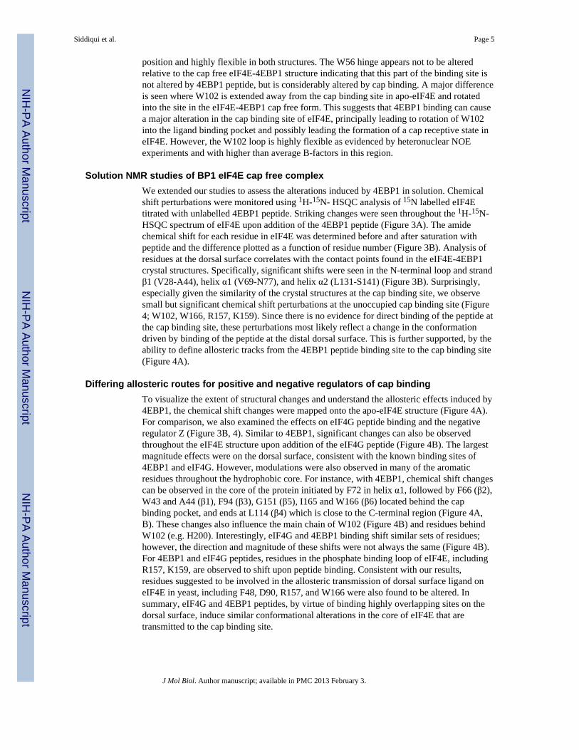
Solution NMR studies of BP1 eIF4E cap free complexWe extended our studies to assess the alterations induced by 4EBP1 in solution. Chemicalshift perturbations were monitored using 1H-15N- HSQC analysis of 15N labelled eIF4Etitrated with unlabelled 4EBP1 peptide. Striking changes were seen throughout the 1H-15N-HSQC spectrum of eIF4E upon addition of the 4EBP1 peptide (Figure 3A). The amidechemical shift for each residue in eIF4E was determined before and after saturation withpeptide and the difference plotted as a function of residue number (Figure 3B). Analysis ofresidues at the dorsal surface correlates with the contact points found in the eIF4E-4EBP1crystal structures. Specifically, significant shifts were seen in the N-terminal loop and strandβ1 (V28-A44), helix α1 (V69-N77), and helix α2 (L131-S141) (Figure 3B). Surprisingly,especially given the similarity of the crystal structures at the cap binding site, we observesmall but significant chemical shift perturbations at the unoccupied cap binding site (Figure4; W102, W166, R157, K159). Since there is no evidence for direct binding of the peptide atthe cap binding site, these perturbations most likely reflect a change in the conformationdriven by binding of the peptide at the distal dorsal surface. This is further supported, by theability to define allosteric tracks from the 4EBP1 peptide binding site to the cap binding site(Figure 4A).
Differing allosteric routes for positive and negative regulators of cap bindingTo visualize the extent of structural changes and understand the allosteric effects induced by4EBP1, the chemical shift changes were mapped onto the apo-eIF4E structure (Figure 4A).For comparison, we also examined the effects on eIF4G peptide binding and the negativeregulator Z (Figure 3B, 4). Similar to 4EBP1, significant changes can also be observedthroughout the eIF4E structure upon addition of the eIF4G peptide (Figure 4B). The largestmagnitude effects were on the dorsal surface, consistent with the known binding sites of4EBP1 and eIF4G. However, modulations were also observed in many of the aromaticresidues throughout the hydrophobic core. For instance, with 4EBP1, chemical shift changescan be observed in the core of the protein initiated by F72 in helix α1, followed by F66 (β2),W43 and A44 (β1), F94 (β3), G151 (β5), I165 and W166 (β6) located behind the capbinding pocket, and ends at L114 (β4) which is close to the C-terminal region (Figure 4A,B). These changes also influence the main chain of W102 (Figure 4B) and residues behindW102 (e.g. H200). Interestingly, eIF4G and 4EBP1 binding shift similar sets of residues;however, the direction and magnitude of these shifts were not always the same (Figure 4B).For 4EBP1 and eIF4G peptides, residues in the phosphate binding loop of eIF4E, includingR157, K159, are observed to shift upon peptide binding. Consistent with our results,residues suggested to be involved in the allosteric transmission of dorsal surface ligand oneIF4E in yeast, including F48, D90, R157, and W166 were also found to be altered. Insummary, eIF4G and 4EBP1 peptides, by virtue of binding highly overlapping sites on thedorsal surface, induce similar conformational alterations in the core of eIF4E that aretransmitted to the cap binding site.
Siddiqui et al. Page 5
J Mol Biol. Author manuscript; available in PMC 2013 February 3.
NIH
-PA Author Manuscript
NIH
-PA Author Manuscript
NIH
-PA Author Manuscript

As mentioned, both the eIF4G and 4EBP1 peptides increase the affinity of eIF4E for thecap. We then compared allosteric tracks induced by these peptides with the arenavirus Zprotein, a repressor of cap binding. Arenavirus Z (Z) is a ninety residue RING protein thatassociates with eIF4E on a surface overlapping but distinct from eIF4G and 4EBP1. Thebinding of Z is more centred on α-helix 1 and induces not only chemical shift perturbations,but also substantial line broadening in regions distal to its binding site. This underlies itsability to increase conformational exchange in the cap binding site of eIF4E, contributing toits ability to reduce cap binding. A comparison of the chemical shift perturbation pattern ofZ with the eIF4G and 4EBP1 peptides indicated important findings. First, there wassubstantial overlap in the allosteric pathways that were activated in both cases includingalterations in the phosphate binding region of the cap binding surface of eIF4E (K159,R157) and alterations in the main chain of W102, suggesting that similar changes observedin the crystal structure can also be observed in solution. Comparing the areas with chemicalshift perturbation revealed that the region corresponding to β-sheet 3, residues L81-M101,was perturbed much more upon Z binding than either peptide eIF4G or 4EBP1 peptide(Figure 3B, 4A). Given that Z binding centres on binding helix α1 which is adjacent to β3and ends at W102, it seems likely that this route is specific to Z and thus uses a uniquecommunication route to the cap binding site. Unlike Z, both eIF4G and 4EBP1 inducechemical shift perturbation in region E171-A201 which corresponds to areas that bind thesecond nucleotide of the m7GpppA complex (Figure 4). Taken together, the distinct effectsof eIF4G and 4EBP1 relative to Z on cap affinity appear to arise, at least in part, from theallosteric pathways activated by binding distinct regions on the dorsal surface.
ConclusionsIn summary, these are the first structural studies to elucidate the basis for the allostericregulation 4EBP1 or eIF4G on the eIF4E protein. Given that eIF4E-cap and 4EBP1-eIF4E-cap complex structure have been solved, it was important to determine the structure of thecap-free 4EBP1-eIF4E complex. Our first approach was to solve the crystal structure of4EBP1-eIF4E cap free complex and compare this to the crystal structure of cap free eIF4E.Unfortunately, crystal contacts in the cap free form of eIF4E possibly drove allostericchanges in the cap binding site, confounding interpretation of our crystallographic data.However, the studies with apo-eIF4E crystals do highlight the effects of dorsal surfaceligands, including itself, on the cap binding site. Relative to the solution apo-eIF4Estructure, the 4EBP1-eIF4E cap free complex shows that W102 rotates partially into the capbinding site while the W56 loop remains flexible. However, again there are crystal contactsthat could contribute to the W102 loop conformation. We extended our studies to assess ifsimilar changes were induced by 4EBP1 in solution and to characterize the allosteric modesused. These studies revealed that 4EBP1 and eIF4G peptides activate very similar allostericpathways in eIF4E to communicate changes in the phosphate binding loop, W102, and theC-terminal nucleotide binding region. It is possible that the changes observed would beamplified if larger or full length constructs were used. This would correlate with the higheraffinity of eIF4E for the 5′cap in the presence of larger fragments as described in previousstudies. In contrast, arenavirus Z binding on helix α1 of the dorsal surface is communicateddown the β3 -sheet to the cap binding site. This route appears to be less prevalent, in eIF4Gor 4EBP1. Overall, the analysis of cap free eIF4E have given insight, at least in part, to theallosteric mechanisms driving ligand induced conformational changes in eIF4E thatultimately impact its biological activities. Clearly, alterations in protein dynamics by ligandswill likely play a significant role in this process and is an important area of future work.
Siddiqui et al. Page 6
J Mol Biol. Author manuscript; available in PMC 2013 February 3.
NIH
-PA Author Manuscript
NIH
-PA Author Manuscript
NIH
-PA Author Manuscript

AcknowledgmentsCrystal structures shown in this report are derived from work performed at Argonne National Laboratory, StructuralBiology Center at the Advanced Photon Source. Argonne is operated by University of Chicago Argonne, LLC, forthe U.S. Department of Energy, Office of Biological and Environmental Research under contract DE-AC02-06CH11357. 600MHz NMR experiments were recorded at the Biophysical platform at IRIC. 800 MHz NMRexperiments were recorded with the assistance of Dr. Tara Sprules at the Québec/Eastern Canada High Field NMRFacility, supported by grants from the Canada Foundation for Innovation, the Natural Sciences and EngineeringCouncil of Canada, the Québec ministère de la recherché en science et technologie, and McGill University. IRICreceives infrastructure support from CIHR and FRSQ. Research was supported by NIH RO1 98571 to K.L.B.B.K.L.B.B. holds a Canada Research Chair in Molecular Biology of the Cell Nucleus. N.S. is a fellow of the ColeFoundation for Research in Leukemia and Lymphoma.
Abbreviations
eIF eukaryotic translation initiation factor
4EBP eIF4E binding protein
NMR nuclear magnetic resonance
NOE nuclear Overhauser effect
PDB Protein Data Bank
PML promyelocytic leukemia, Arenavirus Z (Z)
References1. Sonenberg N, Hinnebusch AG. Review Regulation of Translation Initiation in Eukaryotes:
Mechanisms and Biological Targets. Cell. 2009; 136:731–745. [PubMed: 19239892]2. Rousseau D, Kaspar R, Rosenwald I, Gehrke L, Sonenberg N. Translation initiation of ornithine
decarboxylase and nucleocytoplasmic transport of cyclin D1 mRNA are increased in cellsoverexpressing eukaryotic initiation factor 4E. Proceedings of the National Academy of Sciences ofthe United States of America. 1996; 93:1065–70. [PubMed: 8577715]
3. Culjkovic B, Topisirovic I, Skrabanek L, Ruiz-Gutierrez M, Borden KLB. eIF4E promotes nuclearexport of cyclin D1 mRNAs via an element in the 3′UTR. The Journal of cell biology. 2005;169:245–56. [PubMed: 15837800]
4. Culjkovic B, Topisirovic I, Skrabanek L, Ruiz-Gutierrez M, Borden KLB. eIF4E is a central node ofan RNA regulon that governs cellular proliferation. The Journal of cell biology. 2006; 175:415–26.[PubMed: 17074885]
5. Mader S, Lee H, Pause A, Sonenberg N. The translation initiation factor eIF-4E binds to a commonmotif shared by the translation factor eIF-4 gamma and the translational repressors 4E-bindingproteins. Molecular and cellular biology. 1995; 15:4990–7. [PubMed: 7651417]
6. Volpon L, Osborne MJ, Capul AA, Torre JC, de la Borden KLB. Structural characterization of the ZRING-eIF4E complex reveals a distinct mode of control for eIF4E. Proceedings of the NationalAcademy of Sciences of the United States of America. 2010; 107:5441–6. [PubMed: 20212144]
7. Topisirovic I, Borden KLB. Homeodomain proteins and eukaryotic translation initiation factor 4E(eIF4E): an unexpected relationship. Histology and histopathology. 2005; 20:1275–84. [PubMed:16136508]
8. Marcotrigiano J, Gingras AC, Sonenberg N, Burley SK. Cocrystal structure of the messenger RNA5′ cap-binding protein (eIF4E) bound to 7-methyl-GDP. Cell. 1997; 89:951–61. [PubMed: 9200613]
9. Volpon L, Osborne MJ, Topisirovic I, Siddiqui N, Borden KLB. Cap-free structure of eIF4Esuggests a basis for conformational regulation by its ligands. The EMBO journal. 2006; 25:5138–49. [PubMed: 17036047]
10. Marcotrigiano J, Gingras AC, Sonenberg N, Burley SK. Cap-dependent translation initiation ineukaryotes is regulated by a molecular mimic of eIF4G. Molecular cell. 1999; 3:707–16.[PubMed: 10394359]
Siddiqui et al. Page 7
J Mol Biol. Author manuscript; available in PMC 2013 February 3.
NIH
-PA Author Manuscript
NIH
-PA Author Manuscript
NIH
-PA Author Manuscript

11. Tomoo K, Matsushita Y, Fujisaki H, et al. Structural basis for mRNA Cap-Binding regulation ofeukaryotic initiation factor 4E by 4E-binding protein, studied by spectroscopic, X-ray crystalstructural, and molecular dynamics simulation methods. Biochimica et biophysica acta. 2005;1753:191–208. [PubMed: 16271312]
12. Brown CJ, Verma CS, Walkinshaw MD, Lane DP. Crystallization of eIF4E complexed witheIF4GI peptide and glycerol reveals distinct structural differences around the cap-binding site. Cellcycle (Georgetown, Tex). 2009; 8:1905–11.
13. Matsuo H, Li H, McGuire AM, et al. Structure of translation factor eIF4E bound to m7GDP andinteraction with 4E-binding protein. Nature structural biology. 1997; 4:717–24.
14. Tomoo K, Shen X, Okabe K, et al. Crystal structures of 7-methylguanosine 5′-triphosphate(m(7)GTP)- and P(1)-7-methylguanosine-P(3)-adenosine-5′,5′-triphosphate (m(7)GpppA)-boundhuman full-length eukaryotic initiation factor 4E: biological importance of the C-terminal flexibleregion. The Biochemical journal. 2002; 362:539–44. [PubMed: 11879179]
15. Zuberek J, Jemielity J, Niedzwiecka A, et al. Influence of the length of the phosphate chain inmRNA 5′ cap analogues on their interaction with eukaryotic initiation factor 4E. Nucleosides,nucleotides & nucleic acids. 22:1707–10.
16. Gross, JD.; Moerke, NJ.; Haar, TVD., et al. Ribosome Loading onto the mRNA Cap Is Driven byConformational Coupling between eIF4G and eIF4E. Vol. 115. University of California; Berkeley:2003. p. 739-750.
17. Gosselin P, Oulhen N, Jam M, et al. The translational repressor 4E-BP called to order by eIF4E:new structural insights by SAXS. Nucleic acids research. 2011; 39:3496–503. [PubMed:21183464]
18. Fletcher CM, Wagner G. The interaction of eIF4E with 4E-BP1 is an induced fit to a completelydisordered protein. Protein science: a publication of the Protein Society. 1998; 7:1639–42.[PubMed: 9684899]
19. Moerke NJ, Aktas H, Chen H, et al. Small-Molecule Inhibition of the Interaction between theTranslation Initiation Factors eIF4E and eIF4G. Cell. 2007:257–267. [PubMed: 17254965]
20. Haghighat A, Sonenberg N. eIF4G dramatically enhances the binding of eIF4E to the mRNA 5′-cap structure. The Journal of biological chemistry. 1997; 272:21677–80. [PubMed: 9268293]
21. Ptushkina M, von der Haar T, Karim MM, Hughes JM, McCarthy JE. Repressor binding to adorsal regulatory site traps human eIF4E in a high cap-affinity state. The EMBO journal. 1999;18:4068–75. [PubMed: 10406811]
22. Ptushkina M, von der Haar T, Vasilescu S, et al. Cooperative modulation by eIF4G of eIF4E-binding to the mRNA 5′ cap in yeast involves a site partially shared by p20. The EMBO journal.1998; 17:4798–808. [PubMed: 9707439]
23. von Der Haar T, Ball PD, McCarthy JE. Stabilization of eukaryotic initiation factor 4E binding tothe mRNA 5′-Cap by domains of eIF4G. The Journal of biological chemistry. 2000; 275:30551–5.[PubMed: 10887196]
24. von der Haar T, Oku Y, Ptushkina M, et al. Folding transitions during assembly of the eukaryoticmRNA cap-binding complex. Journal of molecular biology. 2006; 356:982–92. [PubMed:16405910]
25. Slepenkov SV, Korneeva NL, Rhoads RE. Kinetic mechanism for assembly of them7GpppG.eIF4E. eIF4G complex. The Journal of biological chemistry. 2008; 283:25227–37.[PubMed: 18614538]
26. Goss DJ, Carberry SE, Dever TE, Merrick WC, Rhoads RE. A fluorescence study of theinteraction of protein synthesis initiation factors 4A, 4E, and 4F with mRNA and oligonucleotideanalogs. Biochimica et biophysica acta. 1990; 1050:163–6. [PubMed: 2207140]
27. Friedland DE, Wooten WNB, LaVoy JE, Hagedorn CH, Goss DJ. A mutant of eukaryotic proteinsynthesis initiation factor eIF4E(K119A) has an increased binding affinity for both m7G capanalogues and eIF4G peptides. Biochemistry. 2005; 44:4546–50. [PubMed: 15766285]
28. Shen X, Tomoo K, Uchiyama S, Kobayashi Y, Ishida T. Structural and thermodynamic behavior ofeukaryotic initiation factor 4E in supramolecular formation with 4E-binding protein 1 and mRNAcap analogue, studied by spectroscopic methods. Chemical & pharmaceutical bulletin. 2001;49:1299–303.
Siddiqui et al. Page 8
J Mol Biol. Author manuscript; available in PMC 2013 February 3.
NIH
-PA Author Manuscript
NIH
-PA Author Manuscript
NIH
-PA Author Manuscript

29. Niedzwiecka A, Marcotrigiano J, Stepinski J, et al. Biophysical studies of eIF4E cap-bindingprotein: recognition of mRNA 5′ cap structure and synthetic fragments of eIF4G and 4E-BP1proteins. Journal of molecular biology. 2002; 319:615–35. [PubMed: 12054859]
30. Youtani T, Tomoo K, Ishida T, Miyoshi H, Miura K. Regulation of human eIF4E by 4E-BP1:binding analysis using surface plasmon resonance. IUBMB life. 2000; 49:27–31. [PubMed:10772338]
31. Cohen N, Sharma M, Kentsis A, et al. PML RING suppresses oncogenic transformation byreducing the affinity of eIF4E for mRNA. The EMBO journal. 2001; 20:4547–59. [PubMed:11500381]
32. Kentsis A, Dwyer EC, Perez JM, et al. The RING domains of the promyelocytic leukemia proteinPML and the arenaviral protein Z repress translation by directly inhibiting translation initiationfactor eIF4E. Journal of molecular biology. 2001; 312:609–23. [PubMed: 11575918]
33. Brown CJ, McNae I, Fischer PM, Walkinshaw MD. Crystallographic and mass spectrometriccharacterisation of eIF4E with N7-alkylated cap derivatives. Journal of molecular biology. 2007;372:7–15. [PubMed: 17631896]
34. Tomoo K, Shen X, Okabe K, et al. Structural features of human initiation factor 4E, studied by X-ray crystal analyses and molecular dynamics simulations. Journal of molecular biology. 2003;328:365–83. [PubMed: 12691746]
35. Rapp CS, Pollack RM. Crystal packing effects on protein loops. Proteins. 2005; 60:103–9.[PubMed: 15852307]
36. Layten M, Hornak V, Simmerling C. The open structure of a multi-drug-resistant HIV-1 proteaseis stabilized by crystal packing contacts. Journal of the American Chemical Society. 2006;128:13360–1. [PubMed: 17031940]
37. Miura T, Shiratori Y, Shimma N. Backbone resonance assignment of human eukaryotic translationinitiation factor 4E (eIF4E) in complex with 7-methylguanosine diphosphate (m7GDP) and a 17-amino acid peptide derived from human eIF4GII. Journal of biomolecular NMR. 2003; 27:279–80.[PubMed: 12975586]
38. Kentsis A, Topisirovic I, Culjkovic B, Shao L, Borden KLB. Ribavirin suppresses eIF4E-mediatedoncogenic transformation by physical mimicry of the 7-methyl guanosine mRNA cap. Proceedingsof the National Academy of Sciences of the United States of America. 2004; 101:18105–10.[PubMed: 15601771]
39. Hope H. Cryocrystallography of biological macromolecules: a generally applicable method. Actacrystallographica Section B, Structural science. 1988; 44 (Pt 1):22–6.
40. Minor W, Cymborowski M, Otwinowski Z, Chruszcz M. HKL-3000: the integration of datareduction and structure solution--from diffraction images to an initial model in minutes. Actacrystallographica Section D, Biological crystallography. 2006; 62:859–66.
41. McCoy AJ, Grosse-Kunstleve RW, Adams PD, et al. Phaser crystallographic software. Journal ofapplied crystallography. 2007; 40:658–674. [PubMed: 19461840]
42. Berman HM, Westbrook J, Feng Z, et al. The Protein Data Bank. Nucleic acids research. 2000;28:235–42. [PubMed: 10592235]
43. Brünger AT. Free R value: a novel statistical quantity for assessing the accuracy of crystalstructures. Nature. 1992; 355:472–5. [PubMed: 18481394]
44. Murshudov GN, Vagin AA, Dodson EJ. Refinement of macromolecular structures by themaximum-likelihood method. Acta crystallographica Section D, Biological crystallography. 1997;53:240–55.
45. Emsley P, Lohkamp B, Scott WG, Cowtan K. Features and development of Coot. Actacrystallographica Section D, Biological crystallography. 2010; 66:486–501.
46. Davis IW, Murray LW, Richardson JS, Richardson DC. MOLPROBITY: structure validation andall-atom contact analysis for nucleic acids and their complexes. Nucleic acids research. 2004;32:W615–9. [PubMed: 15215462]
47. Yang H, Guranovic V, Dutta S, et al. Automated and accurate deposition of structures solved by X-ray diffraction to the Protein Data Bank. Acta crystallographica Section D, Biologicalcrystallography. 2004; 60:1833–9.
Siddiqui et al. Page 9
J Mol Biol. Author manuscript; available in PMC 2013 February 3.
NIH
-PA Author Manuscript
NIH
-PA Author Manuscript
NIH
-PA Author Manuscript

48. Krissinel E, Henrick K. Secondary-structure matching (SSM), a new tool for fast protein structurealignment in three dimensions. Acta crystallographica Section D, Biological crystallography.2004; 60:2256–68.
49. Delaglio F, Grzesiek S, Vuister GW, et al. NMRPipe: a multidimensional spectral processingsystem based on UNIX pipes. Journal of biomolecular NMR. 1995; 6:277–93. [PubMed: 8520220]
50. Lee W, Westler WM, Bahrami A, Eghbalnia HR, Markley JL. PINE-SPARKY: graphical interfacefor evaluating automated probabilistic peak assignments in protein NMR spectroscopy.Bioinformatics (Oxford, England). 2009; 25:2085–7.
Siddiqui et al. Page 10
J Mol Biol. Author manuscript; available in PMC 2013 February 3.
NIH
-PA Author Manuscript
NIH
-PA Author Manuscript
NIH
-PA Author Manuscript

Highlights
• 1. Cap free eIF4E-4EBP1 complex and apo-eIF4E crystal structures solved at2.1Å.
• 2. 4EBP1 induces allosteric changes in eIF4E converging at the cap binding site.
• 3. Ligand specific allosteric routes on eIF4E underlie different effects on capaffinity.
Siddiqui et al. Page 11
J Mol Biol. Author manuscript; available in PMC 2013 February 3.
NIH
-PA Author Manuscript
NIH
-PA Author Manuscript
NIH
-PA Author Manuscript

Figure 1. Crystal structures of Apo-eIF4E and cap-free eIF4E in complex with the 4EBP1peptideBoth structures were solved at a resolution of 2.1Å. (A) Ribbon representation Apo-eIF4E(3TF2, blue) and (B) eIF4E in a cap-free state (3U7X, green) bound with a 20 residue4EBP1 peptide (red) with the W56 loop not resolved in the latter structure. 4EBP1 makescontacts with helix α1 and -α2 located at the dorsal surface of eIF4E. The cap bindingpocket, highlighted by W56 and W102 (yellow, orange) is distal to the peptide binding site.(C) An overlay of both structures displays high similarity (RMSD 0.585Å). The regioncorresponding to the eIF4E binding site from human 4EBP1 (GI: 34921508, 47-PGGTRIIYDRKFLMECRNSP-66) was synthesized by Fmoc N-(9-fluorenyl)-methoxycarbonyl solid-phase peptide synthesis (Sheldon Biotechnology Center) andpurified by reverse-phase chromatography on a Vydac C18 column. The composition andpurity of the peptide was verified by ion-spray quadrupole mass spectroscopy. All structurefigures were generated using Pymol (The PyMOL Molecular Graphics System, Version1.23pre, Schrödinger, LLC). Structural alignment and comparisons were completed bySUPERPOSE software.
Siddiqui et al. Page 12
J Mol Biol. Author manuscript; available in PMC 2013 February 3.
NIH
-PA Author Manuscript
NIH
-PA Author Manuscript
NIH
-PA Author Manuscript
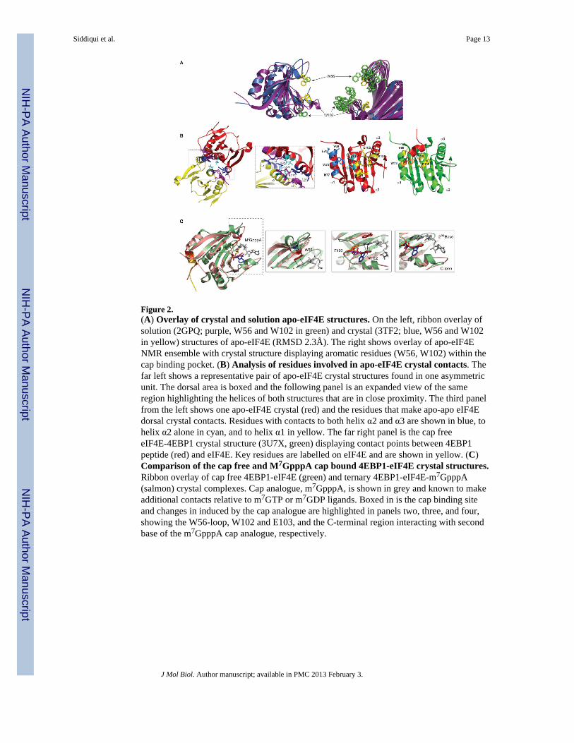
Figure 2.(A) Overlay of crystal and solution apo-eIF4E structures. On the left, ribbon overlay ofsolution (2GPQ; purple, W56 and W102 in green) and crystal (3TF2; blue, W56 and W102in yellow) structures of apo-eIF4E (RMSD 2.3Å). The right shows overlay of apo-eIF4ENMR ensemble with crystal structure displaying aromatic residues (W56, W102) within thecap binding pocket. (B) Analysis of residues involved in apo-eIF4E crystal contacts. Thefar left shows a representative pair of apo-eIF4E crystal structures found in one asymmetricunit. The dorsal area is boxed and the following panel is an expanded view of the sameregion highlighting the helices of both structures that are in close proximity. The third panelfrom the left shows one apo-eIF4E crystal (red) and the residues that make apo-apo eIF4Edorsal crystal contacts. Residues with contacts to both helix α2 and α3 are shown in blue, tohelix α2 alone in cyan, and to helix α1 in yellow. The far right panel is the cap freeeIF4E-4EBP1 crystal structure (3U7X, green) displaying contact points between 4EBP1peptide (red) and eIF4E. Key residues are labelled on eIF4E and are shown in yellow. (C)Comparison of the cap free and M7GpppA cap bound 4EBP1-eIF4E crystal structures.Ribbon overlay of cap free 4EBP1-eIF4E (green) and ternary 4EBP1-eIF4E-m7GpppA(salmon) crystal complexes. Cap analogue, m7GpppA, is shown in grey and known to makeadditional contacts relative to m7GTP or m7GDP ligands. Boxed in is the cap binding siteand changes in induced by the cap analogue are highlighted in panels two, three, and four,showing the W56-loop, W102 and E103, and the C-terminal region interacting with secondbase of the m7GpppA cap analogue, respectively.
Siddiqui et al. Page 13
J Mol Biol. Author manuscript; available in PMC 2013 February 3.
NIH
-PA Author Manuscript
NIH
-PA Author Manuscript
NIH
-PA Author Manuscript

Figure 3. Solution NMR experiments with eIF4E and 4EBP1 and comparative chemical shiftanalysis of eIF4E binding partner eIF4G and arenavirus Z(A) 1H-15N HSQC overlay of cap-free eIF4E (red) and eIF4E bound with 4EBP1 peptide(blue). 4EBP1 peptide was added at a 5:1 molar ratio to eIF4E. (B) 1H-15N chemical shiftperturbation analyses are shown for eIF4E with 4EBP1 (top) and eIF4G (middle) peptideand the RING protein Z (bottom). The change between apo-eIF4E and peptide bound formswere calculated and plotted as a function of residue number. Residue numbers correspond tothe human eIF4E sequence (GI: 4503535). Cartoon diagram seen below chart represent β-sheets (blue) and α-helices (red). The W56, W102, and phosphate binding loops are alsolabeled. Labeled human eIF4E was expressed in Escherichia coli BL21 (DE3) and purifiedas previously described. NMR samples contained ~0.25–0.3 mM protein in 7% D2O, 50mMsodium phosphate buffer (pH 7.3), 100 mM NaCl 250 μM100 μM TCEP, and 1mM NaN3.NMR experiments were completed at 20°C on either a 600 or 800 MHz Varian INOVAspectrometer equipped with an HCN cold probe. Data were processed with NMRPipe andanalyzed with Sparky.
Siddiqui et al. Page 14
J Mol Biol. Author manuscript; available in PMC 2013 February 3.
NIH
-PA Author Manuscript
NIH
-PA Author Manuscript
NIH
-PA Author Manuscript

Figure 4. Mapping of chemical shift changes on the apo-eIF4E structure and select residuesfrom 1H-15N HSQC experiments(A) Chemical shifts induced by 4EBP1 peptide (left), eIF4G (middle), and arenavirus Z(right) on the dorsal surface of eIF4E are shown in blue, green, and red, respectively.Residues affected away from the dorsal surface are highlighted in orange and yellowrepresenting relative strong and medium chemical shifts. In cyan are residues that show linebroadening. Overall, contacts on the dorsal surface lead to alterations in the core of theprotein which are ultimately communicated to the cap binding site of eIF4E. (B) 1H-15NHSQC overlay of apo-eIF4E (red) bound with 4EBP1 (blue) and eIF4G (green) peptides.From left to right, each column displays residues tracing the allosteric tracks as shown in theabove panel. These include residues from the core of the protein, phosphate binding loop,and C-terminal region.
Siddiqui et al. Page 15
J Mol Biol. Author manuscript; available in PMC 2013 February 3.
NIH
-PA Author Manuscript
NIH
-PA Author Manuscript
NIH
-PA Author Manuscript

NIH
-PA Author Manuscript
NIH
-PA Author Manuscript
NIH
-PA Author Manuscript
Siddiqui et al. Page 16
Table 1Crystallography data and refinement statistics
Crystallization and structure determinationThe plasmid containing the full-length cDNA for human eIF4E was a gift from Dr N. Shimma, ChugaiPharmaceutical Co., Japan). Human eIF4E was expressed in Escherichia coli BL21 (DE3) and purified aspreviously described. The protein was purified from the soluble fraction and solution conditions used werepreviously optimized to ensure the proper folding of eIF4E. For crystallization trials, eIF4E protein sampleswere then exchanged into 20mM HEPES, 100mM KCl, 1mM TCEP, pH 7.3 using an Amicon UltraCentrifugal Filter device (10K MWCO, Ultra Centrifugal Filter Device, Millipore) and concentrated to 8–10mg/ml prior to plating (320–400 μM). Apo-eIF4E protein and solution was mixed in a 1:1 ratio with 20%polyethelyne glycol 3350 and 0.2M di-ammonium citrate. For the complex, 4EBP1 peptide (900μM-1mM)and 4mM ribavirin were mixed with eIF4E and incubated at 4°C for 1 hour. The sample was then mixed in a1:1 ratio with 2M ammonium sulfate, 2% PEG400, 0.1M Na HEPES, pH 7.5. Even though the eIF4E-4EBP1peptide complex was crystallized in the presence of ribavirin, we did not observe plausible electron density forthe putative ribosyl and triazolyl moieties of ribavirin supporting that the eIF4E-4EBP1 is in its cap free state.The high affinity analogue is the triphosphorylated form of ribavirin, which would be more suitable for futurestudies. Both samples were placed at 16°C and crystallized using the sitting drop vapour diffusion method.Thin needle like crystals grew to mountable size within 10–14 days. Prior to data collection, crystals wereflash frozen in liquid nitrogen. For each structure, 180 continuous 1° diffraction images were collected at APSbeamline 19ID and reduced using the HKL3000 program suite. Initial molecular replacements were performedwith the program PHASER and coordinates derived from PDB entry 1IPB. Subsets of reflections wereselected in thin resolution shells using SFTOOLS (Dr. B. Hazes, University of Alberta) and used forcalculation of the “free” residual (Rfree). For both structures, model refinement and rebuilding wereperformed with REFMAC and COOT, respectively. Model geometry was monitored on the MOLPROBITYserver. Model depositions in the PDB were prepared with PDB_EXTRACT.
Apo-eIF4E eIF4E - 4EBP1
PDB code 3TF2 3U7X
X-ray wavelength (Å) 0.9793 0.9792
Space group P1 P21
a, b, c (Å) 39.24, 56.74, 100.31 86.50, 38.26, 93.53
α, β, γ (°) 103.61, 91.36, 110.14 90.00, 97.97, 90.00
Resolution (outer shell, Å) 40.00-2.10 (2.14-2.10) 50.00-2.10 (2.14-2.10)
Unique observed HKLs 44808 (2190) 35859 (1728)
Completeness (%) 97.8 (97.0) 99.7 (96.9)
Friedel Redundancy 1.9 (2.0) 3.6 (3.4)
Rsym 0.050 (0.374) 0.089 (0.640)
<I>/<σ> 15.9 (2.1) 16.4 (1.8)
Refinement resolution (Å) 30.00-2.10 30.00-2.10
Number of reflections (of which free set) 43329 (1342) 35692 (1503)
Rwork/Rfree 0.215/0.260 0.198/0.224
Number of atoms/mean B factor (Å2) 5823/36.1 3427/50.3
Protein (eIF4E) 5654/36.2 3068/49.1
Peptide (4EBP1) none 216/67.4
Water 169/32.9 124/47.3
J Mol Biol. Author manuscript; available in PMC 2013 February 3.

NIH
-PA Author Manuscript
NIH
-PA Author Manuscript
NIH
-PA Author Manuscript
Siddiqui et al. Page 17
Apo-eIF4E eIF4E - 4EBP1
RMSD bond lengths (Å)/angles (°) 0.013/1.3 0.014/1.3
Ramachandran Plot (%)
Most favored regions (%) 94.1 91.7
Generously allowed none 0.6
Disallowed 0.7a none
aResidues Asp-143 in protein chains A through D with ϕ of approx. 60° and ψ of approx. −115°.
J Mol Biol. Author manuscript; available in PMC 2013 February 3.