Functional consequences of the binding of MHC class ll-derived peptides to MHC class II
Upload
jamiamilliaislamiaCategory
view
2download
0
Comp. by: PG1397SNijam Stage: Revises1 ChapterID: 0001257665APCSB978-0-12-381262-9 Date:7/4/11 Time:09:56:45File Path:\\pchns1002z\WOMAT\Production\PRODENV\0000000001\0000025497\0000000016\0001257665.3dAcronym:APCSB Volume:83006
CORRECTEDPROOF
c0006 STRUCTURAL DIVERSITY OF CLASS I MHC-LIKEMOLECULES AND ITS IMPLICATIONS IN
BINDING SPECIFICITIES
By MD. IMTAIYAZ HASSAN AND FAIZAN AHMAD
Centre for Interdisciplinary Research in Basic Sciences, Jamia Millia Islamia, New Delhi, India
I. Introduction . . . . . . . . . . . . . . . . . . . . . . . . . . . . . . . . . . . . . . . . . . . . . . . . . . . . . . . . . . . . . . . . . . . . . . . 224II. Sequence Analysis. . . . . . . . . . . . . . . . . . . . . . . . . . . . . . . . . . . . . . . . . . . . . . . . . . . . . . . . . . . . . . . . . . 227
A. Ligand-Binding Residues . . . . . . . . . . . . . . . . . . . . . . . . . . . . . . . . . . . . . . . . . . . . . . . . . . . . . . 232B. TCR-Binding Residues. . . . . . . . . . . . . . . . . . . . . . . . . . . . . . . . . . . . . . . . . . . . . . . . . . . . . . . . . 236C. Light Chain-Binding Residues . . . . . . . . . . . . . . . . . . . . . . . . . . . . . . . . . . . . . . . . . . . . . . . . 238
III. Structure Analysis . . . . . . . . . . . . . . . . . . . . . . . . . . . . . . . . . . . . . . . . . . . . . . . . . . . . . . . . . . . . . . . . . . 239A. Structure of Ligand-Binding Groove. . . . . . . . . . . . . . . . . . . . . . . . . . . . . . . . . . . . . . . . . . 244B. Structural Basis of TCR Binding . . . . . . . . . . . . . . . . . . . . . . . . . . . . . . . . . . . . . . . . . . . . . . 251C. Structural Basis of Light Chain Binding . . . . . . . . . . . . . . . . . . . . . . . . . . . . . . . . . . . . . . 255
IV. Glycosyalation . . . . . . . . . . . . . . . . . . . . . . . . . . . . . . . . . . . . . . . . . . . . . . . . . . . . . . . . . . . . . . . . . . . . . . 258V. Conclusion . . . . . . . . . . . . . . . . . . . . . . . . . . . . . . . . . . . . . . . . . . . . . . . . . . . . . . . . . . . . . . . . . . . . . . . . . 261
References. . . . . . . . . . . . . . . . . . . . . . . . . . . . . . . . . . . . . . . . . . . . . . . . . . . . . . . . . . . . . . . . . . . . . . . . . . 262
Abstract
sp0050 The binding groove of class I major histocompatibility complex (MHC)class is essentially important for antigen binding and presentation onT cells. There are several molecules that have analogous conformationsto class I MHC. However, they bind specifically to varying types of ligandsand cell-surface receptors in order to elicit an immune response. Toelucidate how such recognition is achieved in classical MHC-I like mole-cules, we have extensively analyzed the structure of human leukocyteantigen (HLA-1), neonatal Fc receptor (FcRn), hereditary hemochroma-tosis protein (HFE), cluster of differentiation 1 (CD1), gamma delta T cellreceptor ligand (22), zinc-a2-glycoprotein (ZAG), and MHC class I chain-related (MIC-A) proteins. All these molecules have analogous structuralanatomy, divided into three distinct domains, where a1�a2 superdomainsform a groove-like structure that potentially bind to certain ligand, whilethe a3 domain adopts a fold resembling immunoglobulin constantdomains, and holds this a1�a2 platform and the light chain. We haveobserved many remarkable features of a1�a2 platform, which provide
ADVANCES IN PROTEIN CHEMISTRY AND 223 Copyright 2011, Elsevier Inc.STRUCTURAL BIOLOGY, Vol. 83 All rights reserved.DOI: 10.1016/B978-0-12-381262-9.00006-9

Comp. by: PG1397SNijam Stage: Revises1 ChapterID: 0001257665APCSB978-0-12-381262-9 Date:7/4/11 Time:09:56:45File Path:\\pchns1002z\WOMAT\Production\PRODENV\0000000001\0000025497\0000000016\0001257665.3dAcronym:APCSB Volume:83006
CORRECTEDPROOF
specificities to these proteins toward a particular class of ligands. Therelative orientation of a1, a2, and a3 domains is primarily responsible forthe specificity to the light chain. Interestingly, light chain of all theseproteins is b2-microglobulin (b2M), except ZAG which has prolactin-induced protein (PIP). However, MIC-A is devoid of any light chain.Residues on b2M recognize a sequence motif on the a3 domain that isessentially restricted to specific heavy chain of MHC class I molecules. Ouranalysis suggests that the structural features of class I molecules determinethe recognition of different ligands and light chains, which are responsiblefor their corresponding functions through an inherent mechanism.
s0005I. Introduction
p0005 Major histocompatibility complex I (MHC-I) is a set of molecules an-chored to cell surfaces and are essentially important for lymphocyte recog-nition and presentation through the T cell receptor (TCR; Bjorkman andParham, 1990; Madden, 1995). The class I molecules consist of two polypep-tide chains including one heavy chain while the light chain is b2-microglo-bulin (b2M; Vitiello et al., 1990). Class I MHC molecules usually bind topeptides (8–10 residues long;Madden, 1995) and subsequently interactwiththe TCR of CD8þT cells leading to the CD8þT cell activation, proliferation,differentiation, and effector function (Salter et al., 1990). The sequence andstructure analysis of class I MHCmolecules suggests that it is a heterodimer,consisting of a heavy alpha chain of �45 kDa and a light chain, b2M of�12 kDa (Bjorkmanet al., 1987b; Bjorkman andParham, 1990). In additionto the transmembrane and cytoplasmic domains, the MHC heavy chaincomprises three extracellular domains, a1, a2, and a3. The a1 anda2 domains are highly polymorphic and this polymorphism is mostly loca-lized around the peptide-binding groove. The a1–a2 domain groove isoccupied by a peptide or mixture of peptides in all class I MHC structuressolved to date, and continuous electron-density representing peptide(s) isalways seen in the a1–a2 groove. The a3 domain is highly conserved andclosely interacts with b2M. Both a3 domain and b2M are homologous to theCH3 domain of human immunoglobulin (Ig; Bjorkman and Parham, 1990;Madden, 1995).
p0010 The various isoforms of HLA are encoded by a small number of genes,that is, HLA-A, -B, and -C in man and H2-K, -D, and -L in mouse, locatedwithin the MHC on human chromosome 6p21.3 and murine chromosome
224 HASSAN AND AHMAD

Comp. by: PG1397SNijam Stage: Revises1 ChapterID: 0001257665APCSB978-0-12-381262-9 Date:7/4/11 Time:09:56:45File Path:\\pchns1002z\WOMAT\Production\PRODENV\0000000001\0000025497\0000000016\0001257665.3dAcronym:APCSB Volume:83006
CORRECTEDPROOF
17 (The MHC Sequencing Consortium, 1999). MHCs are further classifiedinto classical (MHC-Ia) and nonclassical genes (MHC-Ib), such as HLA-E,HLA-F, and HLA-G in man and the H2-Q, -T, and -M loci in mice (Peaseet al., 1991). All these show a restricted pattern of tissue expression andunique immunologic functions. These functions include presentation ofN-formylated peptides by H2-M3 and leader sequence peptides by HLA-Eand many others (Lindahl et al., 1997). Despite these apparently specialfeatures, the high degree of sequence similarity between these moleculesdefines them as members of the same gene lineage and suggests that theyare derived from a common ancestor through gene duplication andminimal diversification (Riegert et al., 1998).
p0015 Despite of antigen presenting MHC-I, a large number of proteins havebeen identified to have a close sequence and structural similarity with classI molecules and hence these members are considered as a MHC class-Irelated proteins (Ojcius et al., 2002). All these molecules have structurallysimilar groove but chemical character of the groove is different anddepends upon the nature of their ligands (Table I). For example, thelargely hydrophobic grooves of CD1 (Zeng et al., 1997; Porcelli andModlin, 1999), class Ib MHC molecules Qa-2 (He et al., 2001), andHLA-E (O’Callaghan et al., 1998) allow binding of lipids (includingCD1) and hydrophobic peptides (class Ib proteins). HFE is also anMHC-related protein that is mutated in the iron overload disease heredi-tary hemochromatosis. HFE binds to transferrin receptor (TfR) andreduces its affinity for iron-loaded transferrin, implicating HFE in theiron metabolism (Feder et al., 1996; Lebron et al., 1998). The othermember, neonatal Fc receptor (FcRn) for IgG, expressed in monocytes,macrophages, and dendritic cells, originally characterized as a transportreceptor involved in the uptake of maternal IgG by an intestinal route inrodents (Simister and Mostov, 1989). Its major function is to transport IgGacross polarized epithelial cells and protect IgG from degradation(Burmeister et al., 1994a,b). Many MHC class I-like molecules have distinctligand-binding capabilities, suggesting that they have evolved for specifictasks that are distinct from those of MHC class. The zinc-a2-glycoprotein(ZAG) is involved in the lipid mobilization and is overexpressed in certainhuman malignant tumors to stimulate lipolysis in adipocytes (Bing et al.,2004; Hassan et al., 2008b). Further, ZAG directly influences the uncou-pling protein and in turn enhances the lipid utilization during cancercachexia (Sanders and Tisdale, 2004). Murine T10 and T22 are highly
COMPLEXITY OF CLASS I MHC MOLECULES 225

Comp. by: PG1397SNijam Stage: Revises1 ChapterID: 0001257665APCSB978-0-12-381262-9 Date:7/4/11 Time:09:56:45File Path:\\pchns1002z\WOMAT\Production\PRODENV\0000000001\0000025497\0000000016\0001257665.3dAcronym:APCSB Volume:83006
CORRECTEDPROOF
TableI
t0005
Importan
tFeaturesofMHCClass
Ian
dClass
I-relatedProteins
Protein
nam
ePDBco
de
Ligan
dRecep
tor
Light
chain
Functions
Referen
ces
HLAA2
1HLA
Pep
tides
ab-TCR
b 2M
Antige
npresentation
Bjorkman
etal.(19
87a,b)
HLAb27
1HSA
Pep
tides
ab-TCR
b 2M
Antige
npresentation
Mad
den
etal.(19
92)
HLAH-2Dd
1BII
Pep
tides
ab-TCR,L
y94
b 2M
Activation/inhibitionofCD8
þTorNKcells
Ach
ouret
al.(19
98)
HLA-IH-2Kb
2VAA
Pep
tides
ab-TCR
b 2M
Antige
npresentation
Fremontet
al.(19
92)
HLA-E
3BZE
Pep
tides
CD94
b 2M
Activation/inhibitionofNK
cells
Hoare(200
8)
HLA-G
3KYO
Pep
tides
LIR
2b 2M
InhibitionofNKcells
Walpole
(201
0)Mouse
CD1
1CD1
Glyco
lipids
Va14,Ja28
1TCR
b 2M
Lipidican
tige
npresentation
Zen
get
al.(19
97)
Rat
FcR
n3F
RU
FcofIgG
b 2M
IgGtran
sport
Burm
eister
etal.(19
94a,b)
Human
HFE
1A6Z
TfR
b 2M
Ironmetab
olism
Leb
ronet
al.(19
98)
MurineT10
/T22
1c16
gd-TCR
b 2M
Sign
alingforintracellular
infections
Wingren
etal.(20
00)
Human
ZAG
1ZAG
3ES6
Fattyacids
b 3-Adrenoge
nic
receptor
PIP
Lipid
mobilization
Sanch
ezet
al.(19
99a,b),
Hassanet
al.(20
08a,b)
MIC
-A1B
3Jgd-TCRNKG2D
–Sign
alforstress,activationof
NKcells
Liet
al.(19
99)
MIC
-B1JE6
gd-TCRNKG2D
–Sign
alforstress,activationof
NKcells
Holm
eset
al.(20
02)
RAE-1
1JFM
NKD2D
–ActivationofNKcells
Liet
al.(20
02)

Comp. by: PG1397SNijam Stage: Revises1 ChapterID: 0001257665APCSB978-0-12-381262-9 Date:7/4/11 Time:09:56:45File Path:\\pchns1002z\WOMAT\Production\PRODENV\0000000001\0000025497\0000000016\0001257665.3dAcronym:APCSB Volume:83006
CORRECTEDPROOF
related nonclassical MHC class Ib proteins that bind to certain gd-TCR inthe absence of other components (Wingren et al., 2000). Recognition andstimulation by T22 and T10 presumably do not require peptide or anyother ligand for cell-surface expression or recognition by gd-T cells (Schildet al., 1994). Another gd-TCR-binding MHC class I homolog is MIC-Awhich functions as a stress-inducible antigen and its recognition is inde-pendent of b2M and bound peptides (Li et al., 1999).
p0020 Despite of the fact that all these proteins have class I MHC-like foldstructure and sequence, they perform various unrelated functions(Table I). We searched protein data bank (PDB) for finding proteinsthat belongs to class I family, and observed a large number of structuresof class I molecules and its complexes with peptide, potential ligands,TCR, and CD8 receptor have been determined (Table II). Interestingly, allthese proteins have b2M as a light chain except MIC-A and ZAG. Recently,we have observed that ZAG accommodates prolactin-induced protein(PIP), a molecule which is identical to b2M in their size and fold despiteof least sequence identity (3%; Hassan et al., 2008a). All these findingsreveal that a minor change in the sequences of a1, a2, and a3 domains ofMHC class I protein leads to a significant change in their functions. Themain function of these proteins is binding/transport and is directly asso-ciated with the sequences of a1 and a2 domains. However, the a3 domainis solely responsible for binding to the light chains, b2M (PIP in ZAG), andfor holding the a1–a2 platform. In this section, our aim is to focus on theimportance of sequence and three-dimensional structure of class I pro-teins in their ligands and light chain binding, in order to get an insight tostructure–function relationship in class I molecules.
s0010II. Sequence Analysis
p0025 In the past few years, more than 500 primate MHC genes or partsthereof have been sequenced. The extraordinary sequence informationis used here to draw conclusions about structure, function, diversity, andevolution of class I proteins (Bjorkman and Parham, 1990; Klein et al.,1993). The sequence analysis of class-I mutants have provided a modelsystem for understanding the generation of diversity of the genes encod-ing various histocompatibility molecules, and the relationship of theirstructure to function (Nathenson et al., 1986; Bjorkman and Parham,1990). We have aligned the sequences of all heavy chain from MHC-I-
COMPLEXITY OF CLASS I MHC MOLECULES 227

Comp. by: PG1397SNijam Stage: Revises1 ChapterID: 0001257665APCSB978-0-12-381262-9 Date:7/4/11 Time:09:56:45File Path:\\pchns1002z\WOMAT\Production\PRODENV\0000000001\0000025497\0000000016\0001257665.3dAcronym:APCSB Volume:83006
CORRECTEDPROOF
Table II
t0010 List of MHC Class I Molecules and Class I-Like Molecules in Protein Data Bank
S. no. MHC molecules PDB code Ligand Receptor
1 HLA-A1 1W72, 3BO8 Peptide2 HLA-AW68 2HLA, 1HSB Peptide3 HLA-B*1402 3BVN, 3BXN Peptide4 HLA-B*1501 1XR8, 1XR9 Peptide5 HLA-A*1101 2HN7, 1X7Q, 1Q94, 1QVO Peptide6 HLA-B21 3BEV, 3BEW Peptide7 HLA-B*27 1HSA Peptide8 HLA-B*2705 3LV3, 1OGT, 1UXS, 3B6S,
3DTX, 3BP4, 2A83, 1W0V,1JGE
Peptide
9 HLA-B*2709 3HCV, 1OF2, 3B3I, 3BP7,1JGD, 1K5N, 1UXW, 3CZF,1W0W
Peptide
10 HLA-B*4103 3LN4 Peptide11 HLA-B*4104 3LN5 Peptide12 HLA B*3501 2CIK, 1ZSD, 1A1N Peptide13 HLA B*3501 2NX5 Peptide TCR14 HLA B*3508 3KWW, 2NW3 Peptide15 HLA-B*3508 2AK4, 3KXF Peptide TCR16 HLA, B*4403 1SYS Peptide17 HLA-B*4405 1SYV Peptide18 HLA-B*5703 2BVO, 2BVP, 2BVQ Peptide19 HLA-DP2 3LQZ20 HLA B*44 1N2R Peptide21 HLA-B*4402 3L3D, 1M6O, 3L3G, 3L3I,
3L3J, 3L3K, 3KPL, 3KPMPeptide
22 HLA-B*4403 3KPN, 3KPO Peptide23 HLA B*4405 3KPP, 3KPQ, 3KPS Peptide24 HLA B*4405 3KPR Peptide TCR25 HLA-B*5301 1A1M, 1A1O, 2VLJ, 2VLK,
2VLR, 2J8U26 HLA-B*8 1M05 Peptide27 HLA-A2.1 2X4N, 2X4O, 2X4P, 2X4Q,
2X4R, 2X4S, 2X4T, 2X4U,2V2W, 2V2X, 1EEY, 1I1Y
Peptide
28 HLA-A2.1 3HLA29 HLA-A2.1 2JCC, 2P5E, 2P5W, 2PYE,
2UWE, 2BNQ, 2BNR,1OGA, 1G6R, 1FO0, 2CKB
Peptide TCR
30 HLA-A2.1 2C7U CD8
228 HASSAN AND AHMAD

Comp. by: PG1397SNijam Stage: Revises1 ChapterID: 0001257665APCSB978-0-12-381262-9 Date:7/4/11 Time:09:56:45File Path:\\pchns1002z\WOMAT\Production\PRODENV\0000000001\0000025497\0000000016\0001257665.3dAcronym:APCSB Volume:83006
CORRECTEDPROOF
TABLE II (Continued)
S. no. MHC molecules PDB code Ligand Receptor
31 HLA-A2 2GUO, 2GIT, 2FO4, 1G7Q,2AV1, 2AV7, 1S8D, 1T1W,1T1X, 1T1Y, 1T1Z, 1T20,1T21, 1T22, 1I7T, 1I7U,1IM3, 2VLL
Peptide
32 HLA-A2 1P7Q Peptide LIR33 HLA-A*0201 1DUZ, 1JF1, 1JHT, 1I4F,
1HHJPeptide
34 HLA-Ib M10.5 1ZS835 H2-M3 1MHC Peptide36 HLA-E 3BZE, 3BZF, 1KPR, 1KTL,
1MHEPeptide
37 HLA-E 2ESV Peptide TCR38 HLA-H2 3CVH Peptide TCR39 H2-DB 2VE6, 1BZ9, 2CII, 1YN6,
1YN7, 1ZHB, 1WBX, 1WBY,1WBZ, 1N5A, 1FFN, 1FFO,1FFP, 1QLF, 1HOC
Peptide
40 H2-DD 1DDH, 1BII Peptide41 H2-LD 1LDP, 1LD9 Peptide42 HLA-DR 1H15, 1FV1, 1IIE Peptide43 HLA-DR2a 1ZGL Peptide TCR44 H-2Kb 3C8K Peptide L49C45 H-2Kb 1BQH, Peptide TCR46 H-2Kb 1S7Q, 1S7R, 1S7S, 1S7T,
1KPU, 1KPV, 1N59, 1LEG,1LEK, 1KJ3, 1FZJ, 1FZK,1FZM, 1FZO, 1KBG, 1OSZ,1VAA, 1VAB, 1VAD, 1VAC,2MHA
Peptide
47 H-2KbM3 1MWA, 1JTR, Peptide TCR48 H-2Kb 3C8J L49C49 H-2Kd 1VGK Peptide50 H-2Db 1S7U, 1S7V, 1S7W, 1S7X Peptide51 HLA-DR1 1SJE, 1SJH, 1T5X Peptide52 HLA-G 3KYN, 3KYO, 2D31 Peptide53 HLA-G 2DYP Peptide LIR54 H-2Dd 3ECB, 3DMM Peptide CD855 HLA-T10 1R3H56 HLA-T22 1C1657 HLA-T22 1YPZ Peptide gd-TCR
(Continued)
COMPLEXITY OF CLASS I MHC MOLECULES 229

Comp. by: PG1397SNijam Stage: Revises1 ChapterID: 0001257665APCSB978-0-12-381262-9 Date:7/4/11 Time:09:56:45File Path:\\pchns1002z\WOMAT\Production\PRODENV\0000000001\0000025497\0000000016\0001257665.3dAcronym:APCSB Volume:83006
CORRECTEDPROOF
like proteins and observed a remarkable sequence identity especially inthe sequence of functional relevance evident from crystal structure analy-sis (Fig. 1; Bjorkman et al., 1987b; Bjorkman and Parham, 1990; Fremontet al., 1992; Burmeister et al., 1994a,b; Madden, 1995; Gao et al., 1997;Zeng et al., 1997; Lebron et al., 1998; O’Callaghan et al., 1998; Li et al.,1999; Sanchez et al., 1999a; Burdin et al., 2000; Zajonc et al., 2003; Kochet al., 2005; Hassan et al., 2008a). The residues binding to light chain aswell as the ligands are found to be highly conserved in all members of classI MHC proteins. These finding suggests that all these proteins belong to acommon ancestor and adapted different conformations in order to bindsuitable ligands to perform corresponding functions, including antigen
TABLE II (Continued)
S. no. MHC molecules PDB code Ligand Receptor
58 ZAG 3ES6, 1ZAG, 1T7V, 1T7W,1T7X, 1T7Y, 1T7Z, 1T80
59 FcRn 3M17, 3M1B Peptideinhibitor
60 FcRn 1I1A, 1FRT Fc61 FcRn 3FRU62 CD1 1CD1, 3JVG, 3DBX63 CD1a 1ONQ Sulfatide64 CD1a 1XZ0 Peptide65 CD1b 1UQS, 2H26, 1GZP, 1GZQ Glycolipids66 CD1d 2GAZ, 3ILQ, 3GML, 3GMN,
3GMM, 3GMP, 3GMQ,3GMR, 2Q7Y, 2FIK, 2AKR,1Z5L, 1ZHN, 1ZT4
Lipid
67 CD1d 3HE6, 3HE7, 2CDF, 2CDG Galactoceramide TCR68 CD1d 2PO6, 2EYR, 2EYS, 2EYT,
2CDENK cells
69 HFE 1A6Z70 HFE 1DE4 TfR71 MIC-A 1B3J72 MIC-A 1HYR NK receptor73 MIC-B 1JE674 MIC-B 2WY3 UL1675 RAE-1 BETA 1JFM76 RAE-1 BETA 1JSK NKG2D77 ULBP3 1KCG NKG2D
230 HASSAN AND AHMAD

Comp. by: PG1397SNijam Stage: Revises1 ChapterID: 0001257665APCSB978-0-12-381262-9 Date:7/4/11 Time:09:56:45File Path:\\pchns1002z\WOMAT\Production\PRODENV\0000000001\0000025497\0000000016\0001257665.3dAcronym:APCSB Volume:83006
CORRECTEDPROOF
presentation (Nathenson et al., 1986; Bjorkman and Parham, 1990; Kleinet al., 1993; Madden, 1995). The localization of nonconserved amino acidsin the various MHC molecules further provides in insight to complemen-tary recognition regions in the MHC sequences that play a major role indetermining their ligands and light chain associative recognition (Fig. 1).A comprehensive sequence analysis may be useful to answer how do thesemolecules interact with ligands, TCR, CD8/CD4, and b2M.
FIG. 1.f0005 Multiple sequence alignment of class I MHC proteins showing conservedresidues. The sequence was taken from protein data bank with their PDB codes asHLA_A2, 2CLR; T22, 1C16; HFE, 1A6Z; ZAG, 1ZAG; FcRn, 3FRU; MIC_A, 1B3J; andCD1, 1CD1. The identical and homologous residues are highlighted in dark and lightgray, respectively. Conserved cysteine residues are shaded in yellow. The secondarystructure elements are shown at the top of sequences (taken from PDB code 2CLR)where loop is represented by black line, b-strand by filled arrow (green), and a helicesare in filled cylinder (red). Residues of HLA_A2 showing interaction to peptide and b2Mare marked by blue and red arrow (#), respectively, on the top of sequences. (Forinterpretation of the references to color in this figure legend, the reader is referred tothe Web version of this chapter.)
COMPLEXITY OF CLASS I MHC MOLECULES 231

Comp. by: PG1397SNijam Stage: Revises1 ChapterID: 0001257665APCSB978-0-12-381262-9 Date:7/4/11 Time:09:56:45File Path:\\pchns1002z\WOMAT\Production\PRODENV\0000000001\0000025497\0000000016\0001257665.3dAcronym:APCSB Volume:83006
CORRECTEDPROOF
s0015 A. Ligand-Binding Residuesp0030 The antigen-binding cleft of MHC class I molecules is formed by a1 and
a2 domains, in which two a-helices overlie the floor of eight antiparallel b-stranded sheets (residues 1�180), which is positioned on top of theimmunoglobulin (Ig) constant-like domain, a3 (residues 181–275) andb2M (Nathenson et al., 1986; Bjorkman et al., 1987b; Bjorkman andParham, 1990; Madden, 1995; Fig. 2). It has been observed that thesubstitution of a few amino acids in the proposed antigen (ligand) recog-nition regions located in the a1 domain (residues 70–90) and/or a2 do-main (residues 150–180) can have marked effects on biological functionsof class I molecules (Nathenson et al., 1986). Structurally the proposedantigen-binding site of MHC-I (Bjorkman et al., 1987a,b) is similar tothose of FcRn (Burmeister et al., 1994a,b), HFE (Bennett et al., 2000),ZAG (Sanchez et al., 1999a), T22 (Wingren et al., 2000), CD1 (Zeng et al.,1997), and MIC-A (Li et al., 1999). Structure analysis of peptide–MHC(pMHC) complex (Fremont et al., 1992; Collins et al., 1994; Madden,1995; Achour et al., 1998) demonstrates that the peptide-bindingsequences can be categorized into two types of residues: constant andvariable (polymorphic or dimorphic). The constant residues are identicalin all MHC class I, which provides a common conserved framework for thebinding to the peptide in a gene- or allele-independent manner. Theseconstant residues are Tyr59, Tyr84, Tyr123, and Tyr159 (Reche andReinherz, 2003). In fact, these residues are involved in forming a networkof H bonds to the N- and C-termini of the peptide. It has been observed ina complex structure of HLA with the HIV-1-derived peptide(RGPGRAFVTI) that the N-terminus of peptide binds strongly throughhydrogen bonds to the side chains of Tyr171 and Tyr7 in the A pocket,while the C-terminal is anchored by a network of hydrogen bonds formedby Tyr84, Thr143, and Lys146 in the F pocket (Achour et al., 1998), similarto other peptides in MHC class I complexes (Madden, 1995). Moreover,many hydrophobic residues are present in the antigen-binding clef whichoffers hydrophobic interactions to hold the peptide strongly. The hydro-phobic residues are Tyr123, Phe116, Ile24, Trp147, Trp97, Ala99, Trp114,Tyr159, and Leu95 (Fremont et al., 1992; Achour et al., 1998).
p0035 The FcRn is structurally similar to the class I molecules, involved intransporting IgG from mother to her fetus, and providing the passivehumoral immunity (Roopenian and Akilesh, 2007). Further, it protects
232 HASSAN AND AHMAD

Comp. by: PG1397SNijam Stage: Revises1 ChapterID: 0001257665APCSB978-0-12-381262-9 Date:7/4/11 Time:09:56:45File Path:\\pchns1002z\WOMAT\Production\PRODENV\0000000001\0000025497\0000000016\0001257665.3dAcronym:APCSB Volume:83006
CORRECTEDPROOF
IgG from degradation, thereby providing increase in half-life of this classof antibody in the serum. As evident from its crystal structure, the firstcontact zone of the heavy chain of the rat FcRn molecule can be found atthe end of the a1 domain (residues 84–86 and 90). The second and thirdcontact zones involve residues of a2 domains (113–119 and 131–137)(Burmeister et al., 1994a,b). The second contact zone, which is part of
TCR-a
a1–a2 domain
a3 domainb2M
TCR-b
FIG. 2.f0010 Architecture of MHC-like molecules showing anatomy of complex formedbetween MHC class I proteins and ab-TCR. Class I molecules consist of a heavy chain(light green) and a light b2M chain (orange). The peptide-binding site is formedexclusively by elements of the heavy chain formed by a1�a2 platform, whereas the a3domain is mainly responsible for b2M binding. A view from the TCR binding onto theclass I peptide-binding groove where peptide is shown in ball and stick model (reddish).The a (sky blue) and b (yellow) chains of TCR are shown in ribbon diagram. Thestructure was drawn in PyMOL using the atomic coordinates of pMHC–TCR complexwith PDB code 2CKB (Garcia et al., 1998). (See color plate x).
COMPLEXITY OF CLASS I MHC MOLECULES 233

Comp. by: PG1397SNijam Stage: Revises1 ChapterID: 0001257665APCSB978-0-12-381262-9 Date:7/4/11 Time:09:56:46File Path:\\pchns1002z\WOMAT\Production\PRODENV\0000000001\0000025497\0000000016\0001257665.3dAcronym:APCSB Volume:83006
CORRECTEDPROOF
the a2 domain, is well conserved, emphasizing its importance in IgGbinding (Burmeister et al., 1994b). Zhou et al. (2003) performed anexperiment to investigate the molecular basis for the difference in bindingspecificity between human and mouse FcRn. They suggested that a fewresidues of FcRn are of great importance in the IgG binding, such asLeu137, Asp132, loop121–132, loop79–89, and loop79–89.
p0040 The HFE does not bind peptides or perform any known immunefunction because it has a narrower and shallower version of the class Ipeptide-binding groove (Lebron et al., 1998; Bennett et al., 2000). How-ever, HFE binds to the TfR, a macromolecule associated with iron homeo-stasis (Townsend and Drakesmith, 2002). Crystal structure analysissuggests that Val78 and Trp81 from HFE pairs Leu619 and Val622 fromTfR (Bennett et al., 2000). This finding was further strengthened by site-directed mutants of HFE at positions 78 and 81, for either shows nodetectable binding to TfR (Barton et al., 1999). Further, binding may beaffected by two residues near this position in hereditary hemochromatosis(Ile83Thr and Gly71Arg). Another important residue that takes part inTfR binding is Leu22, showing a decrease in binding affinity upon muta-tion (Lebron and Bjorkman, 1999). TfR binds to HFE in a pH-dependentmanner, and subsequently facilitates release of iron from Fe–Tf complex.Thus, residues on HFE and/or TfR must be responsible for the sharp pHdependence. It was suggested that His74 and His150 of HFE are essentiallyimportant in hydrophobic contacts with TfR residues (Bennett et al.,2000). Lebron and Bjorkman (1999) carried out site-directed mutagenesisstudies to determine the critically important residues in HFE. Theyobserved many important residues which are quite important for TfRbinding, such as Leu22, Val78, Trp81, His87, His89, His94, and His123.However, there are many residues such as His41, Arg44, Ser43, Trp141,and Pro166 which do not strongly affect the TfR binding on mutation.
p0045 CD1 is also an antigen-presenting molecule; especially the lipidic anti-gen is distantly related to class I MHC molecules (Porcelli and Modlin,1999; Zajonc et al., 2003). It appears from sequence similarity and domaintopology that CD1 is more closely related to MHC class Ia and Ib mole-cules (Zeng et al., 1997; Koch et al., 2005). The CD1 family can be dividedinto two groups by sequence homology, that is, group I (CD1a, b, and c)and group II (CD1d, and e), suggesting that each group of CD1 moleculescould have a distinct function (Porcelli and Modlin, 1999). The groove ofCD1 is slightly narrower and deeper as compared to other class I
234 HASSAN AND AHMAD

Comp. by: PG1397SNijam Stage: Revises1 ChapterID: 0001257665APCSB978-0-12-381262-9 Date:7/4/11 Time:09:56:46File Path:\\pchns1002z\WOMAT\Production\PRODENV\0000000001\0000025497\0000000016\0001257665.3dAcronym:APCSB Volume:83006
CORRECTEDPROOF
molecules (Moody et al., 2005). Three highly conserved residues Phe49,Trp40, and Phe18 appear to be responsible for the elevation of helices ofb-sheet platform (Zeng et al., 1997). CD1 groove is predominated byhydrophobic residues, Val28, His38, Leu114, Leu162, and Phe169 in theA0 pocket. However, the F0 pocket contains Trp14, Ile96, and Leu148, andit becomes wider at the surface of the binding groove due to the presenceof Val98, Leu116, Phe144, and Val147 (Zajonc et al., 2003). Further, threeconserved residues (Tyr7, Tyr59, and Tyr171) in MHC class I form hydro-gen-bonding network with the NH2-terminus of the bound peptide(Bjorkman et al., 1987b; Bjorkman and Parham, 1990; Fremont et al.,1992). While, in the case of CD1d1, the same position is occupied byentirely hydrophobic residues (Phe10, Trp63, and Phe171), this indicatespreferential binding with lipidic antigen instead of peptide in MHC-I(Fig. 1; Zeng et al., 1997). Moreover, the residues that interact with thecarboxy terminus of class I peptides (Thr80, Tyr84, Thr143, Lys146, andTrp147) are also substituted by hydrophobic residues (Asp83, Met87,Pro146, Val149, and Leu150) in CD1. All these findings strengthen thepossibility of binding of lipid or glycolipid antigens to the CD1 groovewhich is compatible due to the hydrophobic character.
p0050 The closely related nonclassical MHC class I molecules, T10 and T22,have been identified as a ligand for gd-TCR (Moriwaki et al., 1993). Theincreased expression of T22 and/or T10 might trigger immunoregulatorygd-T cells during immune responses (Crowley et al., 2000). T22 and T10are specifically recognized by gd-T cells, which do not require any peptideor ligand for cell-surface expression (Schild et al., 1994). A deletion ofthree amino acid in the a1 domain (residues 46–48) was observed withrespect to class Ia molecules (Collins et al., 1994). The a2 domain has theleast structural similarity to any known classical or nonclassical MHC-I-likemolecule (Fig. 1). T22 has two potential N-linked glycosylation sites at(Asn86 and Asn150) in the a1�a2 domains. Interestingly, Asn86 highlyconserved MHC class Ia molecules, while the second site is not conserved(Collins et al., 1994). Residues Arg6, Tyr7, Val25, and Arg35 are highlyconserved in the sequence of T22b and that of MHC class Ia (Fig. 1). Thestructure of T22b is stabilized by two potential salt bridges formed betweenArg6 and Asp102, and Arg35 and Glu40/Asp53. Further, position of a fewresidues is critically important and mutation of these residues (Leu5, Arg6,Tyr7, Val25, or Arg35) may cause a local disruption of the T22 structure(Wingren et al., 2000).
COMPLEXITY OF CLASS I MHC MOLECULES 235
Comp. by: PG1397SNijam Stage: Revises1 ChapterID: 0001257665APCSB978-0-12-381262-9 Date:7/4/11 Time:09:56:46File Path:\\pchns1002z\WOMAT\Production\PRODENV\0000000001\0000025497\0000000016\0001257665.3dAcronym:APCSB Volume:83006
CORRECTEDPROOF
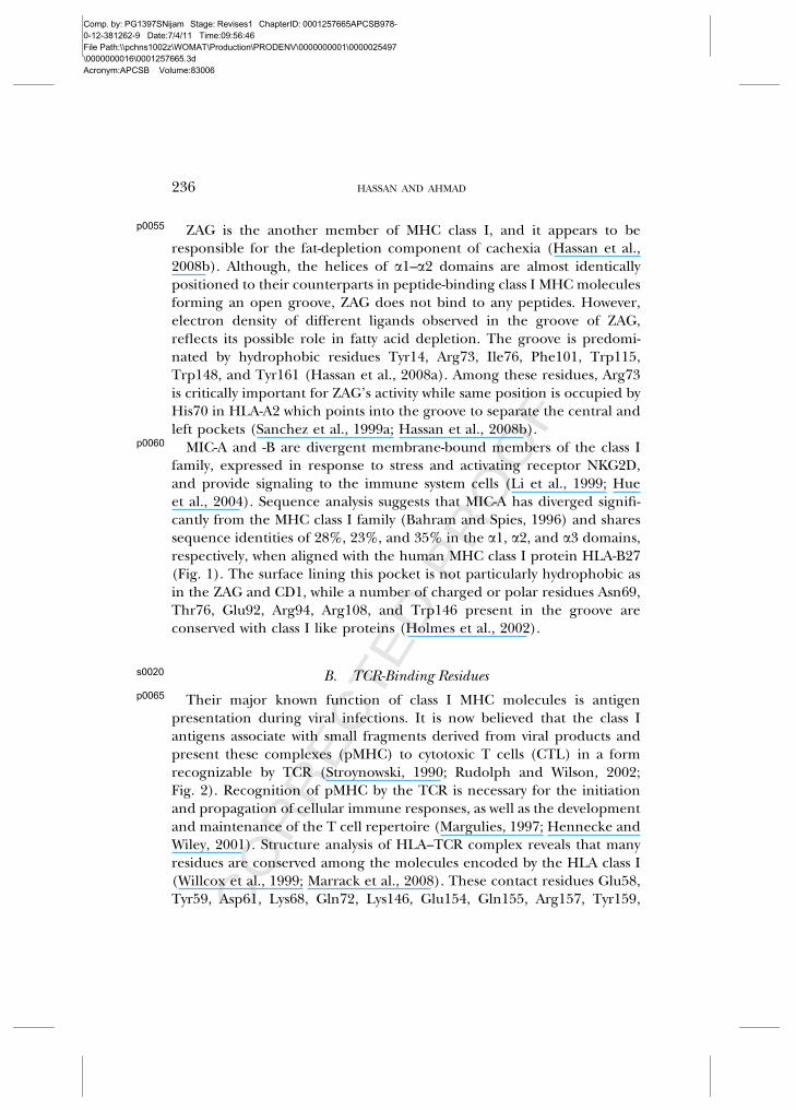
p0055 ZAG is the another member of MHC class I, and it appears to beresponsible for the fat-depletion component of cachexia (Hassan et al.,2008b). Although, the helices of a1–a2 domains are almost identicallypositioned to their counterparts in peptide-binding class I MHC moleculesforming an open groove, ZAG does not bind to any peptides. However,electron density of different ligands observed in the groove of ZAG,reflects its possible role in fatty acid depletion. The groove is predomi-nated by hydrophobic residues Tyr14, Arg73, Ile76, Phe101, Trp115,Trp148, and Tyr161 (Hassan et al., 2008a). Among these residues, Arg73is critically important for ZAG’s activity while same position is occupied byHis70 in HLA-A2 which points into the groove to separate the central andleft pockets (Sanchez et al., 1999a; Hassan et al., 2008b).
p0060 MIC-A and -B are divergent membrane-bound members of the class Ifamily, expressed in response to stress and activating receptor NKG2D,and provide signaling to the immune system cells (Li et al., 1999; Hueet al., 2004). Sequence analysis suggests that MIC-A has diverged signifi-cantly from the MHC class I family (Bahram and Spies, 1996) and sharessequence identities of 28%, 23%, and 35% in the a1, a2, and a3 domains,respectively, when aligned with the human MHC class I protein HLA-B27(Fig. 1). The surface lining this pocket is not particularly hydrophobic asin the ZAG and CD1, while a number of charged or polar residues Asn69,Thr76, Glu92, Arg94, Arg108, and Trp146 present in the groove areconserved with class I like proteins (Holmes et al., 2002).
s0020 B. TCR-Binding Residuesp0065 Their major known function of class I MHC molecules is antigen
presentation during viral infections. It is now believed that the class Iantigens associate with small fragments derived from viral products andpresent these complexes (pMHC) to cytotoxic T cells (CTL) in a formrecognizable by TCR (Stroynowski, 1990; Rudolph and Wilson, 2002;Fig. 2). Recognition of pMHC by the TCR is necessary for the initiationand propagation of cellular immune responses, as well as the developmentand maintenance of the T cell repertoire (Margulies, 1997; Hennecke andWiley, 2001). Structure analysis of HLA–TCR complex reveals that manyresidues are conserved among the molecules encoded by the HLA class I(Willcox et al., 1999; Marrack et al., 2008). These contact residues Glu58,Tyr59, Asp61, Lys68, Gln72, Lys146, Glu154, Gln155, Arg157, Tyr159,
236 HASSAN AND AHMAD

Comp. by: PG1397SNijam Stage: Revises1 ChapterID: 0001257665APCSB978-0-12-381262-9 Date:7/4/11 Time:09:56:46File Path:\\pchns1002z\WOMAT\Production\PRODENV\0000000001\0000025497\0000000016\0001257665.3dAcronym:APCSB Volume:83006
CORRECTEDPROOF
Gly162, and Arg170 are constant and conserved in all three subgroups ofHLA molecules (Reche and Reinherz, 2003). Baxter et al. (2004) showedthat HLA-A2 have a ‘‘functional hot spot’’ that comprises Arg65 and Lys66and is involved in recognition by most peptide-specific HLA-A2-restrictedTCRs. They showed that the Lys66 is involved in the dual recognitions ofboth peptides and TCRs. Mutations in the a2 domain of HLA class Imolecules can also interfere with CD8a/a binding suggesting the possibil-ity that a2 domain on class I may be involved in CD8 interaction, perhapsincluding activated CD8 binding (Sun et al., 1995).
p0070 CD1 isotypes are meant for the presentation of specialized sets of lipidicantigens. Residues on the exposed surfaces of the a1H2, a2H1, and a2H2of a helices are among the most variable in the collection of CD1sequences and may confer T cell specificity for different isotypes(Burdin et al., 2000). It has been reported that a single mutation ofArg79Glu reduces proliferation of some mouse iNKT hybridomas(Porcelli and Modlin, 1999; Burdin et al., 2000). A similar result wasobserved in the double mutation (Arg79Glu79 and Asp80Ala) of CD1(Sidobre et al., 2002). Another mutation of Ser76 to Gly showed a reducedability of a few iNKT hybridomas to recognize a-GalCer, a ligand of CD1(Burdin et al., 2000). Koch et al. (2005) suggested that these residues onthe a1 helix are exposed in the putative TCR recognition surface and areprobably involved in maintaining contact with the TCR.
p0075 The structural differences between T22b and MHC class Ia moleculesclearly indicate that gd- and ab-TCRs recognize antigens differently (Schildet al., 1994). Moreover, the gd-TCRs may be more like immunoglobulins intheir recognition properties. To know the importance of T22b residuesinvolved in TCR binding, Moriwaki et al. (1993) performed Ala mutationstudies followed by KN6-stimulatory assay. They observed that four muta-tions (Tyr9Ala, Val25Ala, Ile23Ala, and Gln115Ala) did not affect theKN6-stimulatory activity appreciably. However, another four mutations(Tyr7Ala, Leu95Ala, Leu98Ala, and Leu116Ala) showed a drastic decreasein the KN6-stimulatory activity. Finally, it was suggested that mutations atthe floor of the proposed antigen-binding groove of T22b affect recogni-tion by the gd-TCR. It is also evident from the crystal structure analysis thatpatches on the exposed floor of T22b may be in direct contact site for acomplementarity determining region (CDR) loop of the gd-TCR (Wingrenet al., 2000). The ZAG andMHC share high sequence homology (sequenceidentities: ZAG with HLA-A2, 36%; FcRn, 27%; mouse CD1d, 23%; HFE,
COMPLEXITY OF CLASS I MHC MOLECULES 237

Comp. by: PG1397SNijam Stage: Revises1 ChapterID: 0001257665APCSB978-0-12-381262-9 Date:7/4/11 Time:09:56:46File Path:\\pchns1002z\WOMAT\Production\PRODENV\0000000001\0000025497\0000000016\0001257665.3dAcronym:APCSB Volume:83006
CORRECTEDPROOF
36%, and MIC-A, 29%). However, ZAG is unlikely to associate with theT cell coreceptor CD8 because, of 15 CD8-binding residues of class I heavychain, only one is conserved between class I and ZAG sequences (class IAsp122, ZAG Asp123).
p0080 The classical function of MHC molecule is antigen processing andpresentation. However, the MIC-A and -B are ligands for the C-typelectin-like activating immunoreceptor, NKG2D (Bahram et al., 1994;Bahram and Spies, 1996). These proteins are expressed on the surfaceof cells in response to stress where they are recognized by gd-T cells, CD8þab-T cells, and NK cells through the NKG2D receptor (Bauer et al., 1999;Wu et al., 1999). Interestingly, NKG2D-MIC recognition events stimulateeffectors response and serving a costimulatory function because NK cellsand gd-T cells positively modulates CD8þ ab-T cell responses (Groh et al.,1998; Bauer et al., 1999). The exact mode of recognition at the sequenceand structure level are not described.
s0025 C. Light Chain-Binding Residuesp0085 X-ray structural studies suggest that the a3 domain of class I molecules
and b2M are structurally related to immunoglobulin domains and interactwith each other, specifically having a role in enhancing peptide-bindingin vitro (Bjorkman et al., 1987a,b; Vitiello et al., 1990; Fig. 2). The a3domain is showing high conservation of sequences with classical moleculessuch as HFE, FcRn, and CD1 (Fig. 1). Functional constraints imposed bythe binding of b2M to the a3 domain, appear to be responsible for theamino acid sequence conservation (Govindarajan et al., 2003). Other site-directed mutagenesis studies suggest that the negatively charged loop(residues 222–229) in the highly conserved a3 domain of MHC class Imolecules plays an important role in CD8-dependent CTL recognitionand activation (Potter et al., 1989; Salter et al., 1990). Interestingly, in theabsence of CD8 binding, this charged loop is highly flexible (Madden,1995), whereas in the CD8aa/HLA-A2/peptide complex, the loop isclamped between the CDR1- and CDR3-like loops from both of the CD8subunits (Gao et al., 1997). This observation was further supported by theinability of murine CD8-dependent CTL response by the a3 mutated classI molecules, which was attributed to the disruption of CD8 interaction withclass I (Potter et al., 1989; Salter et al., 1990).
238 HASSAN AND AHMAD

Comp. by: PG1397SNijam Stage: Revises1 ChapterID: 0001257665APCSB978-0-12-381262-9 Date:7/4/11 Time:09:56:46File Path:\\pchns1002z\WOMAT\Production\PRODENV\0000000001\0000025497\0000000016\0001257665.3dAcronym:APCSB Volume:83006
CORRECTEDPROOF
p0090 In the FcRn, the critical residues of the a3 domains (amino acids Lue216,Lys242, His248, and His249) involved in the FcRn/Fc interaction are con-served among the different species thus far analyzed (Burmeister et al.,1994b). HFE is also associated with b2M. However, in the case of hereditaryhemochromatosis, the mutation (Gly845Ala) leads to similar structuralchanges as in Cys260Tyr mutation which eliminates a disulphide bond inthe a3 domain of HFE and prevents association with b2M and cell-surfaceexpression incell-culturemodels (Lebronetal., 1998). In class Imolecules,a3domain contains a highly acidic loop comprising residues 220–229, which isessentially important for CD8 binding (Salter et al., 1990). The similar struc-ture organization is observed in CD1 (on change) and even in FcRn whichdoes not bind accessory molecules (Bjorkman and Burmeister, 1994;Burmeister et al., 1994a). The a3 domain of human ZAG contains an RGDsequence (Arg231, Gly232, and Asp233) thought to be involved in cell adhe-sion (Sanchezet al., 1999a). ZAGdoesnot bindwithb2M. Instead it bindswithnew protein, PIP has been recently discovered in the cleft of ZAG (Hassanet al., 2008a). The displacement of the ZAG a3 domain compared to its class Icounterpart results in the inabilityofb2M tooptimally contact thea3anda1–a2domainsofZAGwhichcontributes to lackofaffinityofZAGfor theb2M.Themost important interactions between ZAG and PIP occur through theiradjacent b4 strands. These two strands are almost perfectly aligned in theantiparallel manner and generate a number of interactions between them.The potential backbone hydrogen bonds include residues Glu229, Arg231,Asp233, andHis236 fromZAGwith residues Lys41, Lys57, Thr59, Thr60, andLys68 fromPIP. These also include two salt bridges between pairs of residues,Lys41–Asp233 and Lys68–Glu229. Similarly, MIC proteins do not requireeither peptide or b2M for folding, stability, or cell-surface expression (Liet al., 1999). Structurally, the a3 domains of MIC-A are joined through aflexible linker to a1�a2 platform, allowing considerable interdomain flexi-bility. This unique feature of MIC-A among MHC class I proteins and homo-logsmay be a reason for its existencewithout b2M (Tysoe-Calnonet al., 1991).
s0030III. Structure Analysis
p0095 The MHC superfamily comprised class I MHC and MHC-like proteinsthat are involved in a large variety of biological processes. The crystalstructure determination of classical and nonclassical MHCs has been a
COMPLEXITY OF CLASS I MHC MOLECULES 239

Comp. by: PG1397SNijam Stage: Revises1 ChapterID: 0001257665APCSB978-0-12-381262-9 Date:7/4/11 Time:09:56:46File Path:\\pchns1002z\WOMAT\Production\PRODENV\0000000001\0000025497\0000000016\0001257665.3dAcronym:APCSB Volume:83006
CORRECTEDPROOF
prime focus of structure biologists as well as immunologists to explain thestructural basis of antigen recognition and presentation. Structural basis ofantigen recognition by various HLAs has been reviewed by many authorsindependently (Nathenson et al., 1986; Bjorkman and Parham, 1990;Stroynowski, 1990; Janeway, 1992; Germain and Margulies, 1993; Kleinet al., 1993; Shawar et al., 1994; Madden, 1995; Lindahl et al., 1997). Class IMHC molecules acts as a peptide receptor and are capable of bindingdifferent peptides. However, each MHC molecule has a sequence andstructure specific for a set of peptides. On the other hand are variousmolecules such as ZAG, HFE, FcRn, CD1, etc., which have class I MHC-likefold and considerable r.m.s. deviations (Table III) but cannot bind pep-tide; instead, they bind several specific ligands. Hence there is still a needto compare the structural features of all these proteins which have MHC-I-like fold, to gain an insight for the structural basis of protein and peptideinteraction.
p0100 Crystallographic studies of all these molecules reveal that the two mem-brane distal domains (a and a2) of MHC form a superdomain comprisingtwo a helices supported on a platform of eight b-pleated sheets, forming agroove-like structure, which has been postulated to be the antigen/ligand-binding function (Fig. 2; Bjorkman et al., 1987a,b; Madden, 1995). Theenormous sequence variability has been reported in this region and aretherefore likely to be involved in the selection of specific sets of peptides/ligand that are bound to the specific MHC-I-like molecules (Fig. 1). Thesefindings provide a basis for the known diversity in immune responsivenessby different molecules (Stroynowski, 1990; Madden, 1995). Further,ligands should assume a specific conformation in the groove, and theavailability of space for the ligand and the presence of charged, polar, andhydrophobic amino acids at certain positions in the groove would deter-mine whether a given peptide or ligand can fit (Figs. 3A, B and 4A, B;Garrett et al., 1989). Another important features of class I molecule isbinding with b2M which binds noncovalently to the a1, a2, and a3domains of the heavy chain of class I molecules. There are in fact fourdistinct regions that form the contact points between the a-chain and b2M:one on each of the a, a2 and two on the a3 domain (Fig. 2). Both a3domain and b2M resembles to the Ig constant domain. b2M does not bindto MHC until peptide is first bound to the heavy chain in order to facilitatethe formation of b2M-binding site on it (Ljunggren et al., 1990). Thirdimportant point is the binding of pMHC complex to the TCR, which is a
240 HASSAN AND AHMAD

Comp. by: PG1397SNijam Stage: Revises1 ChapterID: 0001257665APCSB978-0-12-381262-9 Date:7/4/11 Time:09:56:46File Path:\\pchns1002z\WOMAT\Production\PRODENV\0000000001\0000025497\0000000016\0001257665.3dAcronym:APCSB Volume:83006
CORRECTEDPROOF
TableIII
t0015
StructuralComparisonsofClass
IMHCMolecu
les,byCalcu
latingRootMeanSq
uareDeviations(R
MSD
)(A
2)forTheir
Backb
oneCarbonAtoms
Theco
mparisonsareshownin
bluea
rowforwhole
Ach
ain,whiterowfora1
–a2superdomains,an
dgree
nrowfora3
domain.
Thesequen
ceiden
tities
aregivenin
yellowbox.
RMSD
andsequen
ceiden
titywerecalculatedbySw
issPDBViewer
andclustalW,
respectively.
aForinterpretationofthereferencesto
colorin
thisfigu
relege
nd,theread
erisreferred
totheWeb
versionofthisch
apter.

Comp. by: PG1397SNijam Stage: Revises1 ChapterID: 0001257665APCSB978-0-12-381262-9 Date:7/4/11 Time:09:56:47File Path:\\pchns1002z\WOMAT\Production\PRODENV\0000000001\0000025497\0000000016\0001257665.3dAcronym:APCSB Volume:83006
CORRECTEDPROOF
11.63
5.76
16.15
13.30
11.0615.86
8.11
16.396.48
8.568.94 11.10 9.95
6.70
14.97
14.43
(G) (H)
(E) (F)
(C) (D)
(A) (B)
15.86 5.63
FIG. 3.f0015 Cartoon diagram showing top view of the ligand binding a1 anda2 domains of various MHC-like molecules. (A) HLA-A2 is involved in the presentationof peptides (orange); (B) HLA-B27 also binds to peptides (cyan); (C) a hydrophobicbinding groove of the CD1 (pink); (D) the nonclassical MHC molecule T22, which is agd-T cell ligand (light green); (E) Fc transported molecules FcRn (green); (F) TfR-binding protein HFE (yellow); (G) a view of the a1 and a2 domains of ZAG showing theproposed site for fatty acid binding (sky blue); and (H) the a1 and a2 domains ofnonclassical MHC-like molecule MICA showing that its groove is structurally analogousto a class I molecule. The distance between the helices was calculated in PyMol. Thestructure was drawn using atomic coordinates of PDB code, 2CLR (HLA_A2), 3HLA(HLA_B27), 1C16 (T22), 1A6Z (HFE), 1ZAG (ZAG), 3FRU (FcRn), 1B3J (MIC_A), and1CD1 (CD1). (See color plate x).
242 HASSAN AND AHMAD

Comp. by: PG1397SNijam Stage: Revises1 ChapterID: 0001257665APCSB978-0-12-381262-9 Date:7/4/11 Time:09:56:48File Path:\\pchns1002z\WOMAT\Production\PRODENV\0000000001\0000025497\0000000016\0001257665.3dAcronym:APCSB Volume:83006
CORRECTEDPROOF
(A) (B)
(C) (D)
(E) (F)
(G) (H)
FIG. 4.f0020 Comparison of molecular surfaces of the ligand-binding grooves formedby a1 and a2 domains of MHC class I and class I-like proteins. The electrostatic potentialsmapped to the surface of a1–a2 superdomains. (A) Size and charge distribution of HLA-A2 groove showing it is sufficient to accommodate peptide; (B) HLA-B27 is also havingidentical groove but the charge distribution is different; (C) the groove molecularsurface of CD1 is neutral and hydrophobic compared to the grooves of the otherMHC-like molecules (largest and deepest groove of all molecules) showing its efficiencyin lipidic antigen binding; (D) T22 is not having proper groove hence it does not bind toany peptide or ligand; (E) there is essentially no groove in FcRn; (F) HFE binds TfR and
COMPLEXITY OF CLASS I MHC MOLECULES 243

Comp. by: PG1397SNijam Stage: Revises1 ChapterID: 0001257665APCSB978-0-12-381262-9 Date:7/4/11 Time:09:56:50File Path:\\pchns1002z\WOMAT\Production\PRODENV\0000000001\0000025497\0000000016\0001257665.3dAcronym:APCSB Volume:83006
CORRECTEDPROOF
crucial step for antigen processing (Madden, 1995; Margulies, 1997).Structure analysis of pMHC–TCR complex reveals that CDR forms manycontacts with highly conserved residues of helices of a1 and a2 domains ofMHC-I (Fig. 2). Structurally, CDRs 2a and 2b lie directly on top of thea2 and a1 helices, respectively, and, therefore, interact exclusively with theMHC heavy chain (Garcia et al., 1998). The analysis of all three mentionedfeatures of class I MHC-like molecules showed many striking differences intheir conformation and topology. Here, many significant structural fea-tures of these molecules will be discussed in order to relate functions ofthese molecules with their structures.
s0035 A. Structure of Ligand-Binding Groovep0105 The classical MHC molecules are highly polymorphic, especially in the
al and a2 domains. The hypervariable residues found on the top of helicesof the groove attain significant peptide- and TCR-binding properties(Fig. 2). A given antigen-binding groove is capable of binding hundredsor thousands of different peptides that may be identical or homologous atonly a few side chain positions (Engelhard, 1994a,b). The size of groove issufficient to accommodate peptide antigen (Fig. 3A and B). A collection ofhydrogen and van der Waals contacts offered by the main chain and sidechain atoms of peptide as well as groove help to stabilize the binding ofany peptide in the corresponding groove (Madden, 1995). The size limi-tation of peptides binding to MHC class I is mainly determined by theamino- and carboxy-terminal ends of the antigen-binding cleft (Fremontet al., 1992; Madden et al., 1992). Structurally, the antigen-binding groovecan be divided into three distinct regions: a narrow deep cleft responsiblefor binding to the N-terminal part of the peptide, a more shallow broadmid-cleft depression over which 5–9 amino acid residues of the peptide are
regulates iron homeostasis having a shallow groove; (G) the molecular surface of thecentral portion of the ZAG groove is nearly neutral except for the contribution of Arg73(positively charged); and (H) MIC is showing a big shallow groove essentially negative incharge that may be suitable to bind NK cell receptor NKG2D. Electrostatics potentialswere calculated in PyMol with full formal charge. Positive potential (�15 mV), neutralpotential (0 mV), and negative potential (�210 mV) are shown in blue, gray, and red,respectively. The atomic coordinates are same as used in Fig. 3. (For interpretation of thereferences to color in this figure legend, the reader is referred to the Web version of thischapter.)
244 HASSAN AND AHMAD

Comp. by: PG1397SNijam Stage: Revises1 ChapterID: 0001257665APCSB978-0-12-381262-9 Date:7/4/11 Time:09:56:50File Path:\\pchns1002z\WOMAT\Production\PRODENV\0000000001\0000025497\0000000016\0001257665.3dAcronym:APCSB Volume:83006
CORRECTEDPROOF
binding, and the characteristic deep hydrophobic F pocket which binds tothe carboxy terminal of peptide (Achour et al., 1998). It is evident fromthe crystal structure and sequence analysis that certain residues in theantigen-binding cleft found to be highly conserved, such as Tyr7, Tyr59,and Tyr171, form a network of hydrogen bonds directly to the N-terminusof peptide (Fig. 1). However, Tyr84, Thr143, and Lys146 interact withC-terminus of peptide. Hence these are considered as essential residuesfor holding the peptide in the cleft despite of sequence specificity(Engelhard, 1994a,b; Madden, 1995). Moreover, these conserved residuesare found to be present in two relatively shallow and open pockets(A and F) that are specific for binding to the amino- and carboxy-terminalof peptides. In addition, the central part of the cleft contains manypockets (B, C, D, and E), which presumably bind to the side chain ofcomplementary peptides (Madden et al., 1991, 1992; Fremont et al., 1992;Matsumura et al., 1992). Further, residues contributing to these pocketsare polymorphic, and therefore, these molecules are characterized bydifferences in the precise nature (charge) and position in the pockets(Fig. 4A and B; Matsumura et al., 1992). Interestingly, a single-residuepolymorphism distinguishes HLA-B*3508 (Arg156) from HLA-B*3501(Leu156) and controls differential peptide selection, even though HLA-B*3501 can bind to the LPEP epitope in vitro (Tynan et al., 2005a). The a1and a2 helices of free and bound pMHC superimpose closely (0.72 A r.m.s.deviation between Ca atoms; Garcia et al., 1998).
p0110 CD1 may have evolved to present antigens that are predominantly foundin microbial pathogens (Porcelli and Modlin, 1999). It is evident from thecrystal structure that CD1 folds very much like MHC class I moleculesincluding arrangements of domain and secondary structure elements(Fig. 3C). However, a notable difference in the groove was observed asthe first a1 helix (H1) adopts an irregular, extended conformation in CD1(Zeng et al., 1997). Interestingly, the a1 domain of CD1 is structurallymore similar to MHC class I molecules (r.m.s. deviation ¼ 1.9 A) and FcRn(r.m.s. deviation ¼1.8 A), whereas the a2 domain is more similar to thestructure of FcRn (r.m.s. deviation ¼0.9 A) than to class I molecules (r.m.s. deviation ¼1.5 A; see Table III). For the combined a1�a2 domain, ther.m.s. deviation is 2.6 A between FcRn or class Ia molecules (Zeng et al.,1997). The putative ligand binding site of CD1 is quite distinct from otherMHC-like molecules (Fig. 4C). The binding groove of CD1 is narrower ascompared to class I molecule (14 A) and is broader than FcRn (�12 A;
COMPLEXITY OF CLASS I MHC MOLECULES 245

Comp. by: PG1397SNijam Stage: Revises1 ChapterID: 0001257665APCSB978-0-12-381262-9 Date:7/4/11 Time:09:56:50File Path:\\pchns1002z\WOMAT\Production\PRODENV\0000000001\0000025497\0000000016\0001257665.3dAcronym:APCSB Volume:83006
CORRECTEDPROOF
Fig. 3C). The groove of CD1 is 4–6 A deeper, presumably due to thepresence of bulkier residues (Phe49, Trp40, and Phe18). The A0 pocketin CD1a, CD1b, and mouse CD1d is the more conserved region of thelipid-antigen-binding groove (Gadola et al., 2002; Zajonc et al., 2003; Kochet al., 2005). The pocket A0 of MHC class I contains a cluster of tyrosineresidues (Tyr7, Tyr59, and Tyr171) to form hydrogen bonds with theamino-terminus of the bound peptide. However, in mouse CD1d1, thecorresponding pocket wall contains hydrophobic residues (Phe10, Trp63,and Phe171), emphasizing the role of CD1 in the binding of lipidicantigen (Fig. 4A; Bjorkman et al., 1987b; Zeng et al., 1997). Similarly,the F9 end of class I molecule at the end of the groove usually have polarresidues (Thr80, Tyr84, Thr143, Lys146, and Trp147) which interact withthe carboxy-terminus of the peptide. However, the same are substituted bymostly hydrophobic residues (Asp83, Met87, Pro146, Val149, Leu150) inCD1. In addition to the hydrophobic residues, CD1 has least number ofresidues in the groove, which have capacity to form hydrogen bonds to thebound ligands. These residues are Gln14, Ser28, Arg74 (at A0 pocket),Tyr73, Ser76, Asp80, and Thr156 (at the entrance of pocket; Fig. 4C; Zenget al., 1997). The structure analysis of CD1a in the complex with a sulfatideself antigen (Zajonc et al., 2003) reveals that A0 pocket of CD1a forms asemicircular curve between the a1–a2-helices, limited by Val28, His38,Leu114, Leu162, and Phe169 and a clearly defined terminus. In contrast,the F0 pocket is long and straight, delineated by Trp14, Ile96, and Leu148.It becomes wider as it approaches the surface of the binding groove due tothe presence of Val98, Leu116, Phe144, and Val147. The sulfogalactosylunit is located between Glu154 (a2-helix) and Arg76 (a1-helix). Thegalactose moiety is stabilized by hydrogen bonding to Arg73 and Ser77,whereas the 30 sulfate group hydrogen bonds to Arg76 and Glu154 and toa water molecule that is in complex between Arg73 and Glu154. The fattyacid chain of the sulfatide protrudes at the T cell recognition surface andmakes van der Waals contacts with Ile81, Phe144, and Val147 of the F0
pocket. The a1�a2 domain of human CD1a has the smallest ligand-binding capacity of the CD1 molecules, with a volume of 1300 A3, whichis the most distinctive and has only one pocket (A0) that is similar in lengthand orientation to those of the other CD1 molecules (Koch et al., 2005).
p0115 Another MHC-like molecule does not seem to present any antigen suchas gd-TCR ligands T10 and T22 (Wingren et al., 2000). The a1 of MHCclass I molecules comprises two segments, H1 (residues 49–54) and H2
246 HASSAN AND AHMAD

Comp. by: PG1397SNijam Stage: Revises1 ChapterID: 0001257665APCSB978-0-12-381262-9 Date:7/4/11 Time:09:56:50File Path:\\pchns1002z\WOMAT\Production\PRODENV\0000000001\0000025497\0000000016\0001257665.3dAcronym:APCSB Volume:83006
CORRECTEDPROOF
(residues 56–85). Interestingly, T22 is devoid of H1 segment and the H2 isshorter (residues 60–86). The a2 domain of T22 has the least structuralresemblance to any known classical or nonclassical MHC-like molecule(Fig. 3D). The a2 of MHC class I molecules contains three segments,named H1 (residues 138–150), H2a (residues 153–162), and H2b (residues164–180). Similarly, T22 does not have the H1 segment and very short H2a,whereas H2b is approximately same with a r.m.s. deviation (residues 163–180) of �0.35 between T22 and class Ia molecules (Wingren et al., 2000;see Table III). Further, the minimum distance between the a1 and a2 he-lical structure is only about 6–10 A (Ca to Ca), compared with 12–20 A(class Ia and II), 10–13 A (FcRn), and 7–10 A (MIC-A);(Fig. 3). The grooveformed by a1�a2 domains are not as precise to accommodate any peptideor ligand (Fig. 4D). These evidences are sufficient to describe the inabilityof T10 and T22 to bind a peptide.
p0120 FcRn transfers IgG from the mother to the fetus across the placenta andthe proximal small intestine (Ghetie and Ward, 2000; Roopenian andAkilesh, 2007). In order to understand the mechanism of IgG transportby FcRn, the crystal structure was determined in the native (Burmeisteret al., 1994a) and the complex (FcRn–IgG) forms (Burmeister et al.,1994b; Vaughn and Bjorkman, 1998). Like class I MHC molecules, thea1 and a2 domains groove-like structure, where the width of cleft is verylow, hence FcRn forms narrowest groove and is devoid of any peptide-binding capability (Fig. 3E). The narrowing of groove is due to the Pro162,conserved in all FcRn, which causes a dramatic movement of the a2 H2a
and H1 helices toward a1, leading to an effective collapsing of the grooveat one end (Burmeister et al., 1994a). Although, the groove of FcRnprimarily consists of Tyr9, Tyr60, Phe156, His168, and Arg164 as similarto the class I molecules, it cannot accommodate any peptide because ofArg164 which effectively obstructs the pocket A (Fig. 4D). It is evidentfrom the FcRn–Fc cocrystal structure that FcRn binds to the Fc portion ofIgG at a site that is distinct from the binding sites of the classical compo-nent of the complement such as FcgRs or the C1q (Burmeister et al.,1994b). The FcRn binds to the CH2–CH3 hinge region of IgG, a versatileregion of Fc responsible to bind with various microbial antigens (Derrickand Wigley, 1992; Roopenian and Akilesh, 2007). Further, FcRn binds tothe Fc in a pH-dependent manner (Vaughn and Bjorkman, 1998). Inter-estingly, at physiological pH 7.4, FcRn does not bind IgG, and at acidic pHof the endosome (pH 6–6.5), FcRn strongly binds to the Fc region of IgG.
COMPLEXITY OF CLASS I MHC MOLECULES 247

Comp. by: PG1397SNijam Stage: Revises1 ChapterID: 0001257665APCSB978-0-12-381262-9 Date:7/4/11 Time:09:56:50File Path:\\pchns1002z\WOMAT\Production\PRODENV\0000000001\0000025497\0000000016\0001257665.3dAcronym:APCSB Volume:83006
CORRECTEDPROOF
Moreover, FcRn does not undergo any significant conformational changewhen it binds IgG (Vaughn and Bjorkman, 1998). It has been observedfrom structure analysis (Martin et al., 2001) and site-directed mutationalstudies (Vaughn et al., 1997) that Glu115 and Asp130 in human FcRn andGlu117 and Glu132 in rat FcRn are responsible for the pH-dependentbinding to residues His310 and His435 on Fc (Shields et al., 2001). Apartfrom hydrogen bonds, the hydrophobic interaction offered by Ile253 ofIgG to the Trp133 of FcRn is equally important. Another importantinteraction between Ile1 of b2M with the hydrophobic residue at position309 of Fc can also be useful in the Fc–RcRn complex formation. Recently,the crystal structure of FcRn with the peptides which inhibit the binding ofthe natural ligand IgG has been determined (Mezo et al., 2010). Structureanalysis reveals that the peptide acts as competitive inhibitors of the IgG–FcRn interactions and it binds to the same site on FcRn as does Fc.
p0125 A considerable sequence and structure similarities between HFE andclass I MHC molecules have been noted. However, HFE neither bindspeptides and TCR nor serves any known function in the immune system(Beutler, 2006). The crystal structure analysis of HFE clearly explains thereason for HFE unable to bind any peptide. As compared to the groove ofclass I MHC, its groove is narrowed by a translation of the a1 helixbringing it to � 4 A closer to the a2 helix, which is almost identicallypositioned (Fig. 3F). Each domain of HFE exactly superimposed upon thecorresponding domains in HLA-A2 (Bjorkman et al., 1987b), FcRn(Burmeister et al., 1994a), and CD1 (Zeng et al., 1997) with r.m.s. devia-tions �1.5 A for most Ca atoms (see Table III). Amino acid residues in theA pocket of class I molecules are Tyr7, Tyr59, Tyr159, and Tyr171 that aremeant to interact with the N terminus of the peptide, including Trp167.Interestingly, only two of the corresponding tyrosines are conserved(Tyr10 and Tyr160) in HFE while the Tyr10 is buried (Lebron et al.,1998). Further, the side chain of Gln168 of HFE points into the grooveto occlude pocket A in the same way as Arg164 in FcRn (Burmeister et al.,1994a). The structures of various HLA molecules with the peptide havebeen superimposed in order to predict the clashing residues of HFE whichinterfere with the peptide binding. It was observed that a total of 10 sidechains (eight from a1 helix and two from the a2 helix) are stericallyclashed with the peptide in the binding groove (Lebron et al., 1998).This occurs due to the translation of the a1 helix toward the a2 helix andthe presence of larger side chains in HFE as compared to class I (HFE:
248 HASSAN AND AHMAD

Comp. by: PG1397SNijam Stage: Revises1 ChapterID: 0001257665APCSB978-0-12-381262-9 Date:7/4/11 Time:09:56:50File Path:\\pchns1002z\WOMAT\Production\PRODENV\0000000001\0000025497\0000000016\0001257665.3dAcronym:APCSB Volume:83006
CORRECTEDPROOF
Leu69, Trp72, Met75, and Arg153 vs. class I Val67, His70, Thr73, andVal152). In addition, Trp114 on the HFE groove floor is also interferingwith peptide binding. Surface area measurements reveal that total surfacearea of the groove in HFE (415 A2) is intermediate between those in FcRn(235 A2) and class I molecules (760 A2; Lebron et al., 1998; Fig. 4F).However, CD1 has a narrower but deeper groove than class I grooves,with the most extensive surface area of 1440 A2 (Zeng et al., 1997). Allthese findings clearly suggest the inability of HFE toward peptide binding(Lebron et al., 1998). However, it is reported that HFE binds to the TfR atthe pH of the cell surface (pH 7.4; Feder et al., 1998), a macromoleculeinvolved in the iron homeostasis (Aisen et al., 1999). The HFE–TfRstructure was determined and observed that one HFE molecule isbound to each chain of the TfR homodimer to form a twofold symmetriccomplex (Bennett et al., 2000). The primary intermolecular contacts areformed between HFE a1�a2 domains and the helical domain of TfR,whereas the a3 and b2M domains of HFE do not interact with TfR. Theinteraction between HFE and TfR covers a large interface; a total of2000 A2 of solvent accessible surface area is buried upon binding betweenthese two molecules. It has been demonstrated that HFE binds TfR withhigh affinity in a pH-dependent manner (Lebron et al., 1999), presumablybecause of the critical histidine residue. It was postulated that His74 andHis150 of HFE are involved in hydrophobic contacts with TfR residues andgot disrupted on changing the pH (Lebron et al., 1998; Lebron andBjorkman, 1999). Other possibility may be arisen due to the histidineresidues from TfR which has two histidine (His684 and His475) in eachpolypeptide chain, which are found to be present on the TfR dimerinterface (Bennett et al., 2000).
p0130 The a1�a2 superdomain of the heavy chain of ZAG forms a groove-likestructure which serves as the binding site, an analogous to the antigen-binding site in class I MHC and CD1 (Fig. 3G; Madden, 1995). The crystalstructures of ZAG in the native form (Sanchez et al., 1999a) as well as incomplex with PIP (Hassan et al., 2008a) have been determined indepen-dently, and in both the studies, it was proven that the groove is open andoccupied by some ligand (unidentified electron density) presumably cru-cial for the biological function of ZAG. The molecular surface of thecentral part of the groove appears to be neutral in charge, due to thepresence of 3 Trp, 4 Tyr, 2 Phe, and 1 Ile residues. However, one Arg73protrudes its side chain from one side of the groove. Interestingly,
COMPLEXITY OF CLASS I MHC MOLECULES 249

Comp. by: PG1397SNijam Stage: Revises1 ChapterID: 0001257665APCSB978-0-12-381262-9 Date:7/4/11 Time:09:56:50File Path:\\pchns1002z\WOMAT\Production\PRODENV\0000000001\0000025497\0000000016\0001257665.3dAcronym:APCSB Volume:83006
CORRECTEDPROOF
replacing Arg73 to alanine has abolished ligand binding (McDermottet al., 2006). The groove of ZAG is similar to the ligand-binding site ofCD1 which is also hydrophobic in nature, suggesting that ZAG may bindhydrophobic ligand although its groove is not as much deeper as CD1groove (Fig. 4G). Structure studies showed that ZAG, CD1, HFE, and FcRncontain prolines in their a2 domain at a position corresponding to Val165in classical class I MHC molecules (Bjorkman and Parham, 1990). Thisproline in FcRn and CD1 causes a kink at a position near their prolineresidues. However, helices in ZAG and HFE are similar to the a2 domainhelices of class I molecules without any kink. In addition to crystal struc-ture determination (Sanchez et al., 1999a), various studies have beencarried by Bjrokman and coworker (Kennedy et al., 2001; Delker et al.,2004; McDermott et al., 2006) in order to determine the critical residuesof ZAG responsible for binding to a particular fatty acid. Therefore, it maybe possible to manipulate the biological effects of ZAG with knowledge ofan appropriate ZAG-binding ligand. Despite of low sequence similaritywith class I molecules, residues closest to the ZAG ligand are completelyconserved in between ZAG and class I molecules (Sanchez et al., 1999a).
p0135 There are many cell-surface receptors which serve as general stresssensors. Usually they do not present any peptide antigens and are inde-pendent of transporter associated with antigen processing (Stephens,2001). These nonclassical MHC molecules comprising ligands for theNK cell lead to the activation of NKG2D receptor that apparently lacksaffinity for small molecule antigens or peptide (Wolan et al., 2001). Thesemolecules are human MIC-A, MIC-B, ULBP, murine Rae1, and H60 (Vivieret al., 2002). Crystal structures of MIC-A (Li et al., 1999) and Rae-1b (Liet al., 2002) indicated that the loss of peptide (or any other ligand)binding was due to the reduced distance between the a1and a2 helices(7–10 A) which do not form any appropriate groove (Fig. 3H). On super-imposition of this groove over class I proteins, many structural differencesare observed, especially in the loop between the first two strands of the b-sheet (residues 13–20) and the hairpin connecting the third and fourth b-strands (residues 37–40; Li et al., 1999). Further, the helix 1 (a1 domain:residues 45–54) is much longer than any other class I homolog, whereasthe a2 domain of MIC-A is quite similar to that of FcRn. The groove is alsonot sufficient to hold a ligand (Fig. 4H). In Rae-1b, these helices comeclose enough to permit formation of a noncanonical disulfide bond with aleucine-rich interface filling the former ligand-binding cavity.
250 HASSAN AND AHMAD

Comp. by: PG1397SNijam Stage: Revises1 ChapterID: 0001257665APCSB978-0-12-381262-9 Date:7/4/11 Time:09:56:50File Path:\\pchns1002z\WOMAT\Production\PRODENV\0000000001\0000025497\0000000016\0001257665.3dAcronym:APCSB Volume:83006
CORRECTEDPROOF
s0040 B. Structural Basis of TCR Bindingp0140 MHC–peptide–TCR recognition is the fundamental process required to
trigger the cellular immune response. The ability of a pMHC complex toactivate a T cell generally correlates with the strength and duration of TCRbinding (Davis et al., 1998). An important question is: Does the uniquestructural properties of TCR–pMHC interface facilitate specificity? Toanswer this question, residues of MHC bound to the TCR have beenextensively studied (Rudolph et al., 2006; Marrack et al., 2008). Fromcrystal structure analysis of TCR/pMHC complexes (Garcia et al., 1998),it was suggested that this interaction is likely to be similar to the well-characterized interactions between antibodies and antigens (Henneckeand Wiley, 2001). Typically up to three upward-facing, solvent-exposedresidues from the antigenic peptide contribute directly to the TCR inter-action (Rudolph and Wilson, 2002). The TCR binds pMHC in a conserveddiagonal orientation that positions the CDR1 and CDR2 loops mainly overthe MHC and the CDR3 loops over peptide. TCR recognition is highlysensitive to small numbers of ligands among thousands of different pMHCcomplexes, and yet it is often highly specific for a particular pMHCcombination (Germain and Stefanova, 1999). It is evident from the struc-tures of TCR–pMHC that the architecture of a given peptide-bindinggroove can impose a recurrent structural signature on the peptides andhence it accommodates and thereby restricts the repertoire of CDR3s withwhich it can cooperate (Housset and Malissen, 2003). The flexibility inCDR3 and variability in Va–Vb pairing angles facilitates the adaptation ofthe TCR to conformational constrain on pMHC surface (Willcox et al.,1999). Thermodynamic analyses of TCR–pMHC interactions further sug-gest that the TCR and/or pMHC possess flexible binding surfaces that arestabilized on complexation (Boniface et al., 1999).
p0145 Large conformational changes in three of the TCR CDR loops areinduced upon binding, providing a mechanism of structural plasticity toaccommodate a variety of different peptide antigens (Garcia et al., 1998).It is reported that a structural plasticity or flexibility is essential for therecognition of pMHC (Kuzushima et al., 1995) in response to ligand(Janeway, 1992), and properties for the survival and function of T cells.In order to determine structural alterations in either the TCR or pMHCupon complexation, Garcia et al. (1998) reported the crystal structure ofthe mouse 2C TCR in complex with mouse MHC class I H-2Kb bound to
COMPLEXITY OF CLASS I MHC MOLECULES 251

Comp. by: PG1397SNijam Stage: Revises1 ChapterID: 0001257665APCSB978-0-12-381262-9 Date:7/4/11 Time:09:56:50File Path:\\pchns1002z\WOMAT\Production\PRODENV\0000000001\0000025497\0000000016\0001257665.3dAcronym:APCSB Volume:83006
CORRECTEDPROOF
the self-peptide dEV8 (EQYKFYSV) and H-2Kb–dEV8 unliganded (Garciaet al., 1996). The structure analysis has clearly explained the degeneratespecificity of TCR–pMHC interaction in terms of a structural plasticity inthe TCR–pMHC interface. About 1876 A2 of surface is buried in the 2C-Kbinterface, of which 900 A2 is contributed by the TCR and 976 A2 by thepMHC (Garcia et al., 1996, 1998). Overall, the Va CDR1 and CDR2 contactresidues appear to be more highly conserved, not only within Kb-restrictedTCRs but also across other TCRs compared with the correspondingb-chain contact residues. In particular, Ser27 of CDR1a, that forms hydro-gen bonds to the conserved Kb residue Glu58, and Ser51 of CDR2a, thatcontacts Glu166, are the most frequently occurring residues to occur in Vagene sequences at these positions (Garcia et al., 1996, 1998). Theseinteractions between conserved residues are consistent with recent dataindicating a critical role for Va residues at 27th and 51st in the restrictionof particular murine TCRs (Sim et al., 1996). Other potentially conservedcontacts appear between a (Tyr31) and b (His29, Glu56) chain of TCRresidues to the MHC helices. The TCR interaction with the peptide ismediated directly and indirectly by hydrogen bonds to the functionalgroups of the upward-facing side chains (P1, P4, P6, and P7) from CDRs1a, 1b, 3a, and 3b, which are in simultaneous contact with the a1 anda2 helices of the MHC. It was observed that when the 2C TCR in thecomplex is compared with the unliganded 2C, the individual a and bchains superimpose with r.m.s. deviation of 1.2 and 0.7 A, respectively, forback bone carbon atoms (Garcia et al., 1996, 1998).
p0150 Longer peptides can either bind by extension at the C terminus (Speiret al., 2001) or due to the fixing of their termini, bulge out of the bindinggroove, providing additional surface area for TCR recognition (Tynanet al., 2005a). It is evident from the crystal structure analysis of pMHCand TCR complex that the CDR1a, CDR2a, and CDR3a loops of the Vadomain of TCR provided the main contacts with HLA molecule onto thea2-helix-spanning residues (150–158: Tynan et al., 2005b). More precisely,Asn50a and Phe52a of TCR fill the void between the TCR and the MHCbecause the Phe52a has been packed against Arg157, Glu154, and Ala158of HLA, whereas Asn50a forms many interactions with Glu154. Moreover,Phe95a and Tyr96a form a contact site to interact with the Gln155 of HLA.This Gln155 have multifunctional role in switching of its interactionsbetween peptide and TCR (Ding et al., 1998), suggesting that this residuemay act as a critical residue for guiding MHC class I-restricted T cell
252 HASSAN AND AHMAD

Comp. by: PG1397SNijam Stage: Revises1 ChapterID: 0001257665APCSB978-0-12-381262-9 Date:7/4/11 Time:09:56:50File Path:\\pchns1002z\WOMAT\Production\PRODENV\0000000001\0000025497\0000000016\0001257665.3dAcronym:APCSB Volume:83006
CORRECTEDPROOF
recognition (Kjer-Nielsen et al., 2003). Further, Leu98b from the CDR3bloop forms many van der Waals interactions with Gln155 and Arg151,whereas Glu101b forms a salt bridge to the Arg151 of the a2-helix of MHC.Taken together, these findings provide a structural basis for the highlyconstrained ab-TCR restriction toward the pMHC complex.
p0155 However, in the nonclassical MHC molecule such as CD1, the ligand-binding groove is deeper, narrower, and more hydrophobic than in class IMHCs (Zeng et al., 1997). The groove formed in such a way that the lipidtails of glycolipids and lipopeptides are bound in the groove and theirpolar moieties presented to T cells (Zajonc et al., 2003; Koch et al., 2005;Moody et al., 2005). The structure analysis (Koch et al., 2005) has con-firmed that three hydrogen bonds offered by key residues (Asp80, Asp151,and Thr154) to the a-galactoceramide are seemingly crucial for maintain-ing a-galactoceramide in the correct position and orientation for recogni-tion by the TCR (Marrack et al., 2008). Moreover, the hydrophilic residuesof ligands are necessary for the specific TCR-mediated recognition, where-as the hydrophobic moiety is for the presentation by CD1 (Porcelli andModlin, 1999). The highly acidic loop, a3 residues 220–229 from class Iimplicated in CD8 binding (Salter et al., 1990), retains some structuresimilarity in CD1 as well as in FcRn (does not bind antigen) (Burmeisteret al., 1994a; Zeng et al., 1997). However, a single residue deletion occursin the putative binding loop of CD1 compared with other MHC class Imolecules (Konig et al., 1992).
p0160 Structure analysis revealed that T22b adopts modified MHC-like fold,whereas it lacks an appropriate classical peptide-binding groove (Wingrenet al., 2000). Hence, it was suggested that T22b and T10 do not require anypeptide for recognition by stimulation of gd-T cells (Schild et al., 1994).The structural differences between ab- and gd-TCR and between T22b andMHC class I molecules support the idea that gd- and ab-TCRs recognizeantigens differently (Schild et al., 1994; Chien et al., 1996; Allison et al.,2001). Moreover, the gd-TCRs may be more like immunoglobulins in theirrecognition properties (Li et al., 1998), not restricted in their choice ofligands or epitopes (Chien et al., 1996). CD8 primarily binds to residues ofa3 domain, especially residues from 223 to 229, similar to the class Imolecules. The r.m.s. deviation calculated for this region was found tobe 0.8–1.2. In addition to that, three residues (Gln115, Asp122, andGlu128) in the a2 domain is also responsible for binding of CD8 (Gaoet al., 1997) and are conserved in T22b and class I molecules.
COMPLEXITY OF CLASS I MHC MOLECULES 253

Comp. by: PG1397SNijam Stage: Revises1 ChapterID: 0001257665APCSB978-0-12-381262-9 Date:7/4/11 Time:09:56:50File Path:\\pchns1002z\WOMAT\Production\PRODENV\0000000001\0000025497\0000000016\0001257665.3dAcronym:APCSB Volume:83006
CORRECTEDPROOF
p0165 The a1 and a2 domain of HFE are involved in TfR binding in the samefashion as class I MHC molecules bind to TCRs (Bennett et al., 2000).However, many significant variations are observed in the complex of HFE–TfR as compared to the class I MHC interactions with TCRs. Despite the factthat TCRs and TfR bind across the a1�a2 domain, class I-restricted TCRsbind with a diagonal interaction with a1–a2 domains in order to avoid thekink of the helices (Lebron et al., 1998). However, the a2 helix of HFE,havingakink, is amajor contactpoint forTfR. Further,TCRs recognizea1�a2 helices along with the peptide bound in the groove, whereas the samegroove in HFE is narrowed and devoid of any peptide or other ligand.Interestingly, the distance between a1 and a2 helices of HFE is critical forits interaction with TfR. Hence it was suggested that if the HFE groove gotbroadened, may be due to the binding of peptide, it presumably wouldprevent the interaction between HFE and TfR (Bennett et al., 2000).
p0170 Despite of a considerable sequence and structure similarity betweenZAG and class I molecules, structural features of the ZAG a3 domainmake it unlikely to associate with the T cell coreceptor CD8. Out of 15residues responsible for CD8 binding in the HLA-A2/CD8 cocrystal struc-ture (Gao et al., 1997), only one is conserved between class I and ZAGsequences (class I Asp122, ZAG Asp123). Further, any interaction betweenZAG and TCR can be possible, because the class I MHC residues thatcontact these receptors are not particularly conserved with respect to theZAG (Ding et al., 1998; Garcia et al., 1998). However, it may be suggestedthat ZAG has a completely different and unidentified b3-adrenergic recep-tor which plays a significant role in lipid metabolism especially in cancercachexia (Ojcius et al., 2002).
p0175 MIC-A and MIC-B are compatible with gd-TCRs (Bahram et al., 1994;Chien et al., 1996), instead of ab-TCR (Davis et al., 1998), and areproposed to mediate antimicrobial responses and regulate innate andacquired immunity. The mode of interaction between gd-TCRs and theirligands are different from that of interaction between ab-TCRs and theirligands (Germain and Margulies, 1993). Unlike conventional MHC class Iproteins, MIC-A requires neither peptide nor b2M for stability or cell-surface expression (Groh et al., 1998). However, MIC-A is known toform an unrelated complex of the NK cell receptor NKG2D (Li et al.,2001). Interestingly, MIC-A refolds upon receptor binding from its par-tially unwound a2-helix structure, whereas, this kind of refolding is notobserved with T22 which also binds to gd-TCR does (Rudolph et al., 2006).
254 HASSAN AND AHMAD

Comp. by: PG1397SNijam Stage: Revises1 ChapterID: 0001257665APCSB978-0-12-381262-9 Date:7/4/11 Time:09:56:50File Path:\\pchns1002z\WOMAT\Production\PRODENV\0000000001\0000025497\0000000016\0001257665.3dAcronym:APCSB Volume:83006
CORRECTEDPROOF
s0045 C. Structural Basis of Light Chain Bindingp0180 b2M is noncovalently bound to class I MHC proteins and is required for
their surface expression and function (Vitiello et al., 1990). However,MHC-I-like proteins may or may not interact with b2M. The membraneproximal end of the class I molecules is formed by the a3 and b2Mdomains, which adopts a standard immunoglobulin-like fold, character-ized by a b-sandwich formed by close packing together of a three-strandedand a four-stranded b-sheet (Madden, 1995). It is required to predict theb2M binding (or nonbinding) to the various members of class I MHCsuperfamily in order to decipher their function and to understand themolecular recognition mechanisms. Structure analysis of class I moleculesreveals that the a3 and b2M residues that are involved in the contactinterface are within their respective four-stranded sheets. There aremore extensive contacts between b2M and the a1�a2 platform thanbetween a3 and the platform (Bjorkman et al., 1987b), because b2Msupports the a1�a2 platform. Finally, a3 domain interacts extensivelyonly to the b2M, whereas b2M interacts with all three domains of theheavy chain (Bjorkman and Parham, 1990). It is evident from the structureanalysis that 19 residues of the heavy chain form 44 contacts with 18different residues on human b2M (Tysoe-Calnon et al., 1991). In betweena1 domain and b2M, several side chains pointed into a central pocket,where Asp53 of b2M forms two ionic and two hydrogen bonds with Gln32,Arg35, and Arg48 on the a1 domain. Further, Ser55 and Tyr63 of b2Moffer hydrogen bonds to the hydroxy group of Tyr27 and Tyr63. Similarly,between b2M and the a2 domain, many well-defined hydrogen bonds areobserved, connecting His31, Trp60, and Phe62 of the b2M and Gln96 onthe a2 domain. Further, there are two contact zones observed in the caseof a3 and b2M interactions. The upper patch (residues near to a1–a2 domains) was formed by a large number of closely spaced interactivecontacts between these domains (Tysoe-Calnon et al., 1991). There are sixinteractions formed by the main chain atoms of a3 domains (Gln232,Pro235, Ala36, and Gly37) to the side chains of Gln8, Tyr10, Arg12, andAsn24 of the b2M. However, the lower patch involves the two C-terminalresidues Asp98 and Met99 of b2M and His192, Arg202, Arg234, andTrp244 of the a3 domain (Tysoe-Calnon et al., 1991).
p0185 Although CD1 molecule has identical structure assembly, the relativeposition of the b2M and the CD1 a1–a2 domains have a slight rotation and
COMPLEXITY OF CLASS I MHC MOLECULES 255

Comp. by: PG1397SNijam Stage: Revises1 ChapterID: 0001257665APCSB978-0-12-381262-9 Date:7/4/11 Time:09:56:50File Path:\\pchns1002z\WOMAT\Production\PRODENV\0000000001\0000025497\0000000016\0001257665.3dAcronym:APCSB Volume:83006
CORRECTEDPROOF
translation as compared to the arrangement in class I molecules(Matsumura et al., 1992; Zeng et al., 1997). After aligning all homologousmolecules on their b2M domains, the transformation necessary to align thea1�a2 domains with CD1 is a 0.4–1� rotation and 3.9–11.4 A translation forclass I molecules, and 1.0� rotation and 5.3 A translation for FcRn(Burmeister et al., 1994a). The total buried surface area between b2Mand the heavy chain is slightly less than for class I molecules, but it is morehydrophobic, which may account for the CD1–b2M complex being morestable to thermal denaturation. Further, a notable difference in quaterna-ry structure has been observed in a3–b2M association. Interestingly, thepseudodyadic rotation necessary to superimpose a3 onto b2M in CD1 ismore similar to class II than to class I molecules. In the crystal structure ofmouse CD1, the outermost a3 S1strand now comes closer to b2M andresults in a slightly different pattern of interactions.
p0190 The quaternary arrangement of the a1�a2 platform, a3 domain, andb2M in the case of T10 and T22 are within the range of orientationsobserved for MHC class Ia molecules (19). The total buried surface areaand the number of hydrogen bonds between b2M and the heavy chain isslightly less (845 A2 and 5 for T22) on comparison to class I molecules(850–1036 A2 and 7–21) that may explain the observation that a closelyrelated T10–b2M complex is less stable to thermal denaturation than classIa molecules.
p0195 Apart from slight differences between class I molecules and FcRn, thereare many significant differences reported. Most of the interactions formedbetween heavy chain and light chain of FcRn and MHC class molecules areconserved except the interaction formed between b-strand (residues 52–55) with a1�a2 domain of FcRn. Furthermore, the heavy chain (residues16–21) dips downward to contact b2M and forms many extra contacts(Burmeister et al., 1994a).
p0200 In HFE, as in class I and class I-related proteins, a3 and b2M interactwith a symmetry that deviates from the pseudodyad symmetry relatingantibody constant domains. Specifically, the HFE domains are related bya 161� rotation and a 13 A translation (Lebron et al., 1998). This relativeorientation is in the range seen for the a3–b2M relationship in class Imolecules (146–160� rotation, 13–14 A translation; Madden, 1995), CD1(170� rotation, 12 A translation; Zeng et al., 1997), and FcRn (157�
rotation, 13 A translation; Burmeister et al., 1994a). Site-directed muta-genesis studies confirmed that on mutating Cys282Tyr mutant protein
256 HASSAN AND AHMAD

Comp. by: PG1397SNijam Stage: Revises1 ChapterID: 0001257665APCSB978-0-12-381262-9 Date:7/4/11 Time:09:56:50File Path:\\pchns1002z\WOMAT\Production\PRODENV\0000000001\0000025497\0000000016\0001257665.3dAcronym:APCSB Volume:83006
CORRECTEDPROOF
cannot form disulfide bond with b2M, such mutation was observed inhereditary haemochromatosis (Feder et al., 1996; Waheed et al., 1997).Hence in absence of the b2M, HFE does not properly fold (Parkkila et al.,1997). However, His63Asp mutant is capable of interaction with b2M, butit is unable to regulate cellular iron uptake like the wild-type HFE protein(Waheed et al., 1997).
p0205 The quaternary arrangement of the ZAG a3 domain with respect to thea1�a2 platform differs from that found in b2M-binding class I and relatedproteins. The spatial relationship between its three domains is slightlydifferent from that found in class I MHC proteins, which may explain thelack of affinity of ZAG for b2M. Structural features that prevent ZAG frombinding b2M include the residues such as Ile13, Thr15, Leu30, Arg40,Gln98, Tyr118, Lys122, Val234, His236, Trp245, which clash with b2Mwhen it is positioned on the ZAG structure either by interacting with a3or with a1�a2 domains (Sanchez et al., 1999a). However, ZAG accom-modates a molecules of PIP which has similar scaffold to the b2M in thesame cleft but in opposite orientation (Hassan et al., 2008a). The inter-actions between ZAG and PIP are highly specific. There are 12 hydrogenbonds and three well-defined salt bridges formed between ZAG and PIP.The two salt bridges in the ZAG–PIP complex act as anchors at the twoends of vertically aligned b-strands of the a3 domain of ZAG and b-strandsof PIP (Hassan et al., 2008a). Further, there are dissimilar charge distribu-tions at the interface of ZAG and HLA in the two respective complexes,which further highlight the specificity of ZAG–PIP and HLA–b2M.
p0210 The a3 domain of MIC-A adopted a C-type immunoglobulin conforma-tion and is highly similar to the structure to a3 domains of MHC class Iproteins, as well as b2M itself (Li et al., 1999). Interestingly, the relativepositions of the two domains are significantly different in MIC-A and otherMHC class I proteins (Li et al., 1999). The a3 domains of MIC-A contains alinker between the platform, consists of four residues (Leu178-Thr181)leading to an extended conformation that is different from the additionalturn of helix found in place of the linkers in other MHC class I homologs(Madden, 1995). Interestingly, there are no contacts observed in betweenthe two domains, whereas the linker is good enough to allow for consider-able interdomain flexibility (Li et al., 1999). The important point to bediscussed here is the lack of affinity of MIC-A from the b2M. The crystalstructure analysis reveals that the b2M-binding interfaces are oriented inincompatible directions in the extended structure of MIC-A (Li et al.,
COMPLEXITY OF CLASS I MHC MOLECULES 257
Comp. by: PG1397SNijam Stage: Revises1 ChapterID: 0001257665APCSB978-0-12-381262-9 Date:7/4/11 Time:09:56:51File Path:\\pchns1002z\WOMAT\Production\PRODENV\0000000001\0000025497\0000000016\0001257665.3dAcronym:APCSB Volume:83006
CORRECTEDPROOF
1999, 2001). Moreover, N-linked oligosaccharide site (Asn8) is locatednear the center of the b2M-binding surface on the underside of theplatform, which presumably would prevent b2M binding in MIC-A. It isnoteworthy that the interface residues of class I proteins (Gln96, Ala117,and Gly120, numbered as in HLA-B27) and of the b2M (His31, Phe56,Trp60, and Phe-62) are highly conserved. Interestingly, the residuescorresponding to these positions in MIC-A are also conserved except forTyr111 (corresponds to Ala117 in HLA-B27; Li et al., 1999). Structureanalysis suggests that the side chain of Tyr111 sterically clashes with Trp60of b2M when MIC-A is superimposed onto a class I structure. This furthersuggests the possibility of inability of b2M binding to MIC-A. In addition tothat, Asp122, a highly conserved residue in class I molecules, forms ahydrogen bond with Trp60 of b2M. Interestingly, substitution of thisresidue by either Leu116 or Arg116, further support the preventing factorof MIC-A against b2M binding (Collins et al., 1995).
s0050IV. Glycosyalation
p0215 Glycosylation is a major feature of proteins expressed on the surfaces ofcells, including molecules involved in the cellular immune system such asMHC class I molecule, and class I-like molecules (Rudd et al., 1999b).Glycosylation of proteins or MHC-I molecules provides a unique structureto modulate its selectivity and to control their interactions with theircomplementary proteins or ligands (Rudd et al., 1999a). It has beenreported that the conformational stability of glycoproteins increasesupon addition of glycan chain (Shental-Bechor and Levy, 2008). Although,glycosylation does not affect the average backbone conformation of afolded protein, it has a significant impact on protein stability by decreasingthe flexibility of the protein backbone (Shental-Bechor and Levy, 2008).Further, in the immunological interactions, glycosylation is one of theimportant factors that could affect the protein–protein interactions signif-icantly by conferring rigidity to the protein, which could lead to anincrease in affinity and selectivity of proteins for their correspondingreceptors (Rudd et al., 1999b; Wormald and Dwek, 1999). Further, ithas been observed that glycans are equally important at all three proposedstages such as formation of the cell junction, recruitment of pMHC by theTCRs, and stabilizing the conformation of the complexes involved in theT cell recognition of antigen-presenting cells (Grakoui et al., 1999).
258 HASSAN AND AHMAD

Comp. by: PG1397SNijam Stage: Revises1 ChapterID: 0001257665APCSB978-0-12-381262-9 Date:7/4/11 Time:09:56:51File Path:\\pchns1002z\WOMAT\Production\PRODENV\0000000001\0000025497\0000000016\0001257665.3dAcronym:APCSB Volume:83006
CORRECTEDPROOF
p0220 Sequence analysis suggests that there are three highly conserved glyco-sylation sites in class I molecules, that is, Asn86, Asn176 (a2-domain), andAsn256 (a3-domain) (Stroynowski, 1990). Interestingly, the class Ia mole-cules have glycosylation at all three sites, whereas in the class Ib, theposition 176 is devoid of glycosylation (Shawar et al., 1994). However,the function of several human class I molecules lacking this site areapparently unaffected (Watts et al., 1989). The Asn86 is present in theloop connecting the a1 domain to a2 domain (Bjorkman et al., 1987b).Biochemical analysis suggests that glycosylation is really important for thefunction of class I proteins. MHC class I heavy chains that bind to calnexinthrough the terminal glucose moiety, which is required for protein foldingand assembly pathway, because calnexin and calreticulin are lectins andacts as a chaperone. Subsequently, after association with b2M, the fullyassembled MHC is released from calnexin and subsequently binds tocalreticulin through the same glucose residue. This event is necessaryfor the loading of antigenic peptide onto MHC class I molecules fromthe transporter associated with antigen processing (Sadasivan et al., 1996).
p0225 Murine CD1 sequence contains five potential N-glycosylation sites:Asn25, Asn38, Asn60, Asn128, and Asn183. However, in the crystal struc-ture, only three sites showed glycan chain electron density. Structurally,glycan chain at a1 helix contributes to stabilization of the H2 helix byinteraction with conserved Arg74 (Zeng et al., 1997). It was proposed thatcarbohydrate moiety at these sites provides protection against endosomalproteases. Moreover, glycosylation also plays an important role in theorganization and spacing of CD1 on the cell surface by prevention ofnonspecific aggregation (Rudd et al., 1999b).
p0230 Structure and biochemical analysis suggest that T22b has two potentialN-linked glycosylation sites (Asn86 and Asn150) in the a1�a2 superdo-mains, as evident from the surface potential analysis that a negativelycharged patch is present on a completely neutral surface (Fig. 4D). Nota-bly, the Asn86 is highly conserved in MHC class I molecules (Madden,1995), however, Asn150 is not conserved and present in the middle ofprotruding loop of a2 domain (Wingren et al., 2000). Crystal structuresuggests that carbohydrate plays a significant role in folding of the hetero-dimer in MHC class I (Asn86; Parham, 1996). Although, glycan moietydoes not provide any specificity in the T22b–gd-TCR interaction (Crowleyet al., 1997), but the bulky glycan chains may be important in guiding thegd- T cell to its binding site.
COMPLEXITY OF CLASS I MHC MOLECULES 259

Comp. by: PG1397SNijam Stage: Revises1 ChapterID: 0001257665APCSB978-0-12-381262-9 Date:7/4/11 Time:09:56:51File Path:\\pchns1002z\WOMAT\Production\PRODENV\0000000001\0000025497\0000000016\0001257665.3dAcronym:APCSB Volume:83006
CORRECTEDPROOF
p0235 Sequence analysis suggests that N-linked glycosylation site at Asn128 isfound in rodent but not human or bovine forms of FcRn (Simister andMostov, 1989; Story et al., 1994). Moreover, in the case of mouse FcRn,differential glycosylation affects the receptor/ligand stoichiometry fromtypical 1:1 FcRn/Fc complexes (high-mannose forms of FcRn) to 2:1complexes (complex carbohydrates). Crystal structure analysis revealedthat Asn128 is critically important for FcRn/Fc complex formation asevident from surface area calculation that 10–15% of the buried surfacearea in the FcRn/Fc interface is contributed by three glycans attached toAsn128 form close contact to four Fc residues (Martin et al., 2001).Moreover, it was suggested that maximal Fc binding affinity requiresbulkier glycan attachment to FcRn at Asn128 (Sanchez et al., 1999b).
p0240 The protein sequence of humanHFE contains three putative N-glycosyla-tion sites at residues Asn110, Asn130, and Asn234 (Beutler, 2006), andAsn110 is highly conserved with class I proteins. It has been suggested thatthe glycosylation is important for the normal intracellular trafficking andfunctional activity of HFE (Bhatt et al., 2010). It has been shown thatglycosylation ofHFE provides different conformation as compared to ungly-cosylated fromand subsequently affect their associationwith theTfR (Salter-Cidet al., 1999). Structurally, Val78andTrp81ofHFEaremainly responsibleforTfRbinding, corresponding toahelical and loopregion in thea1domainthat possesses both the Asn110 and Asn130 residues. It was presumed thatN-glycosylation sites in a region are of functional importance for TfR1binding. Therefore, it may be possible that the 110/130 double mutationsmay directly disrupt regulation of TfR1 (Bhatt et al., 2010). It was furtherconfirmed that Asn-linked glycosylation is necessary for the association ofHFE with TfR by HeLa cells transfection with HFE (Gross et al., 1998).Studies based on site-directed mutagenesis and biochemical characteriza-tion revealed that all three potential glycosylation sites contain glycan chainas evident from their electrophoreticmobility (Bhatt et al., 2010). The triplemutant (functionally identical to Cys282Tyr HFE) showed significantly de-creased interactions to the b2M, which is believed to be important for HFEfolding and stabilization. Moreover, the proper folding of HFE is alsoessential for transport andexit of theprotein fromtheER(Bhatt et al., 2010).
p0245 Extensive carbohydrate density is found at Asn239 (nine ordered carbo-hydrate residues) and to amuch lesser extent atAsn89 andAsn108 in all fourZAG molecules (Sanchez et al., 1999a). Crystal structures of glycoproteinsrarely show more than three ordered carbohydrate residues at each
260 HASSAN AND AHMAD

Comp. by: PG1397SNijam Stage: Revises1 ChapterID: 0001257665APCSB978-0-12-381262-9 Date:7/4/11 Time:09:56:51File Path:\\pchns1002z\WOMAT\Production\PRODENV\0000000001\0000025497\0000000016\0001257665.3dAcronym:APCSB Volume:83006
CORRECTEDPROOF
glycosylation site (Vaughn and Bjorkman, 1998). The Asn in the fourthpotential N-linked glycosylation site (Asn92) does not show densitycorresponding to carbohydrate. The bond between Asn92 and Gly93 canbe broken by hydroxylamine, confirming that Asn92 is not glycosylated.There are eight N-linked glycosylation sites in MIC-A at Asn8, Asn56,Asn102, Asn187, Asn197, Asn211, Asn238, and Asn266 as evident fromsequence and structure analysis. However, no glycan moiety has been ob-served at Asn56 andAsn197 during crystal structure analysis (Li et al., 1999).It was suggested that inMIC-A, glycosylationwasnot essential for its structurebut it enhances the complex formation with NKG2D (Li et al., 2001).
s0055V. Conclusion
p0250 In the past 2 decades, enormous number of crystal structures of class IMHC-likemolecules have been determined, which led to a great progress inour understanding of the structure of the class I proteins and their bindingaffinity for ligands and TCR. Structural studies showed that all class I MHCmolecules have identical fold and forming a groove-like structure by a1�a2domain,which takepart in ligandbinding.Variation in thegroove residuesleads to the different binding specificities for their corresponding ligands.Further, pMHC complex binds to the TCR by the residues of a1�a2 domainalong with few residues from peptides. A notable difference has beenobserved in the molecules bind to ab- or gd-TCR. Except ZAG and MIC-A,allmolecules are stabilizedbyb2M,which is essential for loading thepeptidesin the case of class I MHC molecules. A new molecule, PIP, has beenidentified in the cleft ofZAG, in the sameplaceofb2M,but in totallydifferentorientation. Glycosylation of class I molecules is essentially important fortheir corresponding functions as well as their specific binding and stabiliza-tion. Sequence analysis of class Imolecules suggests the role critically impor-tant residues in the folding, stability, and function. Our comprehensiveanalysis may be useful for better understanding of mechanism of ligandbinding by class I MHC molecules.
Acknowledgments
Authors are grateful to the funding agencies, Department of Science and Technology,Council of Scientific and Industrial Research, and Indian National Science Academy forfinancial assistance.
COMPLEXITY OF CLASS I MHC MOLECULES 261

Comp. by: PG1397SNijam Stage: Revises1 ChapterID: 0001257665APCSB978-0-12-381262-9 Date:7/4/11 Time:09:56:51File Path:\\pchns1002z\WOMAT\Production\PRODENV\0000000001\0000025497\0000000016\0001257665.3dAcronym:APCSB Volume:83006
CORRECTEDPROOF
References
Achour, A., Persson, K., Harris, R. A., Sundback, J., Sentman, C. L., et al. (1998). Thecrystal structure of H-2Dd MHC class I complexed with the HIV-1-derived peptideP18-I10 at 2.4 A resolution: implications for T cell and NK cell recognition. Immu-nity 9, 199–208.
Aisen, P., Wessling-Resnick, M., Leibold, E. A. (1999). Iron metabolism. Curr. Opin.Chem. Biol. 3, 200–206.
Allison, T. J., Winter, C. C., Fournie, J. J., Bonneville, M., Garboczi, D. N. (2001).Structure of a human gammadelta T-cell antigen receptor. Nature 411, 820–824.
Bahram, S., Bresnahan, M., Geraghty, D. E., Spies, T. (1994). A second lineage ofmammalian major histocompatibility complex class I genes. Proc. Natl. Acad. Sci.USA 91, 6259–6263.
Bahram, S., Spies, T. (1996). The MIC gene family. Res. Immunol. 147, 328–333.Barton, J. C., Sawada-Hirai, R., Rothenberg, B. E., Acton, R. T. (1999). Twonovelmissense
mutations of the HFE gene (I105T and G93R) and identification of the S65C muta-tion in Alabama hemochromatosis probands. Blood Cells Mol. Dis. 25, 147–155.
Bauer, S., Groh, V., Wu, J., Steinle, A., Phillips, J. H., et al. (1999). Activation of NK cellsand T cells by NKG2D, a receptor for stress-inducible MICA. Science 285, 727–729.
Baxter, T. K., Gagnon, S. J., Davis-Harrison, R. L., Beck, J. C., Binz, A. K., et al. (2004).Strategic mutations in the class I major histocompatibility complex HLA-A2 inde-pendently affect both peptide binding and T cell receptor recognition. J. Biol.Chem. 279, 29175–29184.
Bennett, M. J., Lebron, J. A., Bjorkman, P. J. (2000). Crystal structure of the hereditaryhaemochromatosis protein HFE complexed with transferrin receptor. Nature 403,46–53.
Beutler, E. (2006). Hemochromatosis: genetics and pathophysiology. Annu. Rev. Med.57, 331–347.
Bhatt, L., Murphy, C., O’Driscoll, L. S., Carmo-Fonseca, M., McCaffrey, M. W., et al.(2010). N-glycosylation is important for the correct intracellular localization ofHFE and its ability to decrease cell surface transferrin binding. FEBS J. 277,3219–3234.
Bing, C., Bao, Y., Jenkins, J., Sanders, P., Manieri, M., et al. (2004). Zinc-alpha2-glycoprotein, a lipid mobilizing factor, is expressed in adipocytes and is up-regu-lated in mice with cancer cachexia. Proc. Natl. Acad. Sci. USA 101, 2500–2505.
Bjorkman, P. J., Burmeister, W. P. (1994). Structures of two classes of MHC moleculeselucidated: crucial differences and similarities. Curr. Opin. Struct. Biol. 4, 852–856.
Bjorkman, P. J., Parham, P. (1990). Structure, function, and diversity of class I majorhistocompatibility complex molecules. Annu. Rev. Biochem. 59, 253–288.
Bjorkman, P. J., Saper, M. A., Samraoui, B., Bennett, W. S., Strominger, J. L., et al.(1987a). The foreign antigen binding site and T cell recognition regions of class Ihistocompatibility antigens. Nature 329, 512–518.
Bjorkman, P. J., Saper, M. A., Samraoui, B., Bennett, W. S., Strominger, J. L., et al.(1987b). Structure of the human class I histocompatibility antigen, HLA-A2. Nature329, 506–512.
262 HASSAN AND AHMAD

Comp. by: PG1397SNijam Stage: Revises1 ChapterID: 0001257665APCSB978-0-12-381262-9 Date:7/4/11 Time:09:56:51File Path:\\pchns1002z\WOMAT\Production\PRODENV\0000000001\0000025497\0000000016\0001257665.3dAcronym:APCSB Volume:83006
CORRECTEDPROOF
Boniface, J. J., Reich, Z., Lyons, D. S., Davis, M. M. (1999). Thermodynamics of T cellreceptor binding to peptide-MHC: evidence for a general mechanism of molecularscanning. Proc. Natl. Acad. Sci. USA 96, 11446–11451.
Burdin, N., Brossay, L., Degano, M., Iijima, H., Gui, M., et al. (2000). Structuralrequirements for antigen presentation by mouse CD1. Proc. Natl. Acad. Sci. USA97, 10156–10161.
Burmeister, W. P., Gastinel, L. N., Simister, N. E., Blum, M. L., Bjorkman, P. J. (1994a).Crystal structure at 2.2 A resolution of the MHC-related neonatal Fc receptor.Nature 372, 336–343.
Burmeister, W. P., Huber, A. H., Bjorkman, P. J. (1994b). Crystal structure of thecomplex of rat neonatal Fc receptor with Fc. Nature 372, 379–383.
Chien, Y. H., Jores, R., Crowley, M. P. (1996). Recognition by gamma/delta T cells.Annu. Rev. Immunol. 14, 511–532.
Collins, E. J., Garboczi, D. N., Karpusas, M. N., Wiley, D. C. (1995). The three-dimen-sional structure of a class I major histocompatibility complex molecule missing thealpha 3 domain of the heavy chain. Proc. Natl. Acad. Sci. USA 92, 1218–1221.
Collins, E. J., Garboczi, D. N., Wiley, D. C. (1994). Three-dimensional structure of apeptide extending from one end of a class I MHC binding site. Nature 371,626–629.
Crowley, M. P., Fahrer, A. M., Baumgarth, N., Hampl, J., Gutgemann, I., et al. (2000). Apopulation of murine gammadelta T cells that recognize an inducible MHC classIb molecule. Science 287, 314–316.
Crowley, M. P., Reich, Z., Mavaddat, N., Altman, J. D., Chien, Y. (1997). The recogni-tion of the nonclassical major histocompatibility complex (MHC) class I molecule,T10, by the gammadelta T cell, G8. J. Exp. Med. 185, 1223–1230.
Davis, M. M., Boniface, J. J., Reich, Z., Lyons, D., Hampl, J., et al. (1998). Ligandrecognition by alpha beta T cell receptors. Annu. Rev. Immunol. 16, 523–544.
Delker, S. L., West, A. P., Jr., McDermott, L., Kennedy, M. W., Bjorkman, P. J. (2004).Crystallographic studies of ligand binding by Zn-alpha2-glycoprotein. J. Struct. Biol.148, 205–213.
Derrick, J. P., Wigley, D. B. (1992). Crystal structure of a streptococcal protein Gdomain bound to an Fab fragment. Nature 359, 752–754.
Ding, Y. H., Smith, K. J., Garboczi, D. N., Utz, U., Biddison, W. E., et al. (1998). Twohuman T cell receptors bind in a similar diagonal mode to the HLA-A2/Taxpeptide complex using different TCR amino acids. Immunity 8, 403–411.
Engelhard, V. H. (1994a). Structure of peptides associated with class I and class II MHCmolecules. Annu. Rev. Immunol. 12, 181–207.
Engelhard, V. H. (1994b). Structure of peptides associated with MHC class I molecules.Curr. Opin. Immunol. 6, 13–23.
Feder, J. N., Gnirke, A., Thomas, W., Tsuchihashi, Z., Ruddy, D. A., et al. (1996). Anovel MHC class I-like gene is mutated in patients with hereditary haemochroma-tosis. Nat. Genet. 13, 399–408.
Feder, J. N., Penny, D. M., Irrinki, A., Lee, V. K., Lebron, J. A., et al. (1998). Thehemochromatosis gene product complexes with the transferrin receptor and low-ers its affinity for ligand binding. Proc. Natl. Acad. Sci. USA 95, 1472–1477.
COMPLEXITY OF CLASS I MHC MOLECULES 263

Comp. by: PG1397SNijam Stage: Revises1 ChapterID: 0001257665APCSB978-0-12-381262-9 Date:7/4/11 Time:09:56:51File Path:\\pchns1002z\WOMAT\Production\PRODENV\0000000001\0000025497\0000000016\0001257665.3dAcronym:APCSB Volume:83006
CORRECTEDPROOF
Fremont, D. H., Matsumura, M., Stura, E. A., Peterson, P. A., Wilson, I. A. (1992).Crystal structures of two viral peptides in complex with murine MHC class I H-2Kb.Science 257, 919–927.
Gadola, S. D., Zaccai, N. R., Harlos, K., Shepherd, D., Castro-Palomino, J. C., et al.(2002). Structure of human CD1b with bound ligands at 2.3 A, a maze for alkylchains. Nat. Immunol. 3, 721–726.
Gao, G. F., Tormo, J., Gerth, U. C., Wyer, J. R., McMichael, A. J., et al. (1997). Crystalstructure of the complex between human CD8alpha(alpha) and HLA-A2. Nature387, 630–634.
Garcia, K. C., Degano, M., Pease, L. R., Huang, M., Peterson, P. A., et al. (1998).Structural basis of plasticity in T cell receptor recognition of a self peptide-MHCantigen. Science 279, 1166–1172.
Garcia, K. C., Degano, M., Stanfield, R. L., Brunmark, A., Jackson, M. R., et al. (1996).An alphabeta T cell receptor structure at 2.5 A and its orientation in the TCR-MHCcomplex. Science 274, 209–219.
Garrett, T. P., Saper, M. A., Bjorkman, P. J., Strominger, J. L., Wiley, D. C. (1989).Specificity pockets for the side chains of peptide antigens in HLA-Aw68. Nature342, 692–696.
Germain, R. N., Margulies, D. H. (1993). The biochemistry and cell biology of antigenprocessing and presentation. Annu. Rev. Immunol. 11, 403–450.
Germain, R. N., Stefanova, I. (1999). The dynamics of T cell receptor signaling:complex orchestration and the key roles of tempo and cooperation. Annu. Rev.Immunol. 17, 467–522.
Ghetie, V., Ward, E. S. (2000). Multiple roles for the major histocompatibility complexclass I-related receptor FcRn. Annu. Rev. Immunol. 18, 739–766.
Govindarajan, K. R., Kangueane, P., Tan, T. W., Ranganathan, S. (2003). MPID: MHC-Peptide Interaction Database for sequence-structure-function information on pep-tides binding to MHC molecules. Bioinformatics 19, 309–310.
Grakoui, A., Bromley, S. K., Sumen, C., Davis, M. M., Shaw, A. S., et al. (1999). Theimmunological synapse: a molecular machine controlling T cell activation. Science285, 221–227.
Groh, V., Steinle, A., Bauer, S., Spies, T. (1998). Recognition of stress-induced MHCmolecules by intestinal epithelial gammadelta T cells. Science 279, 1737–1740.
Gross, C. N., Irrinki, A., Feder, J. N., Enns, C. A. (1998). Co-trafficking of HFE, a nonclas-sical major histocompatibility complex class I protein, with the transferrin receptorimplies a role in intracellular iron regulation. J. Biol. Chem. 273, 22068–22074.
Hassan, M. I., Bilgrami, S., Kumar, V., Singh, N., Yadav, S., et al. (2008a). Crystal structureof the novel complex formed between zinc alpha2-glycoprotein (ZAG) and prolactin-inducible protein (PIP) from human seminal plasma. J. Mol. Biol. 384, 663–672.
Hassan, M. I., Waheed, A., Yadav, S., Singh, T. P., Ahmad, F. (2008b). Zinc alpha 2-glycoprotein: a multidisciplinary protein. Mol. Cancer Res. 6, 892–906.
He, X., Tabaczewski, P., Ho, J., Stroynowski, I., Garcia, K. C. (2001). Promiscuousantigen presentation by the nonclassical MHC Ib Qa-2 is enabled by a shallow,hydrophobic groove and self-stabilized peptide conformation. Structure 9,1213–1224.
264 HASSAN AND AHMAD

Comp. by: PG1397SNijam Stage: Revises1 ChapterID: 0001257665APCSB978-0-12-381262-9 Date:7/4/11 Time:09:56:51File Path:\\pchns1002z\WOMAT\Production\PRODENV\0000000001\0000025497\0000000016\0001257665.3dAcronym:APCSB Volume:83006
CORRECTEDPROOF
Hennecke, J., Wiley, D. C. (2001). T cell receptor-MHC interactions up close. Cell 104,1–4.
Hoare, H. L., Sullivan, L. C., Clements, C. S., Ely, L. K., Beddoe, T., Henderson,K. N., et al. (2008). Subtle changes in peptide conformation profoundly affectrecognition of the non-classical MHC class I molecule HLA-E by the CD94-NKG2natural killer cell receptors. J. Mol. Biol. 377, 1297–1303.
Holmes, M. A., Li, P., Petersdorf, E. W., Strong, R. K. (2002). Structural studies ofallelic diversity of the MHC class I homolog MIC-B, a stress-inducible ligand for theactivating immunoreceptor NKG2D. J. Immunol. 169, 1395–1400.
Housset, D., Malissen, B. (2003). What do TCR-pMHC crystal structures teach us aboutMHC restriction and alloreactivity? Trends Immunol. 24, 429–437.
Hue, S., Mention, J. J., Monteiro, R. C., Zhang, S., Cellier, C., et al. (2004). A direct rolefor NKG2D/MICA interaction in villous atrophy during celiac disease. Immunity 21,367–377.
Janeway, C. A., Jr. (1992). The T cell receptor as a multicomponent signalling machine:CD4/CD8 coreceptors and CD45 in T cell activation. Annu. Rev. Immunol. 10,645–674.
Kennedy, M. W., Heikema, A. P., Cooper, A., Bjorkman, P. J., Sanchez, L. M. (2001).Hydrophobic ligand binding by Zn-alpha 2-glycoprotein, a soluble fat-depletingfactor related to major histocompatibility complex proteins. J. Biol. Chem. 276,35008–35013.
Kjer-Nielsen, L., Clements, C. S., Purcell, A. W., Brooks, A. G., Whisstock, J. C., et al.(2003). A structural basis for the selection of dominant alphabeta T cell receptorsin antiviral immunity. Immunity 18, 53–64.
Klein, J., Satta, Y., O’HUigin, C., Takahata, N. (1993). The molecular descent of themajor histocompatibility complex. Annu. Rev. Immunol. 11, 269–295.
Koch, M., Stronge, V. S., Shepherd, D., Gadola, S. D., Mathew, B., et al. (2005). Thecrystal structure of human CD1d with and without alpha-galactosylceramide. Nat.Immunol. 6, 819–826.
Konig, R., Huang, L. Y., Germain, R. N. (1992). MHC class II interaction with CD4mediated by a region analogous to the MHC class I binding site for CD8. Nature356, 796–798.
Kuzushima, K., Sun, R., van Bleek, G. M., Vegh, Z., Nathenson, S. G. (1995). The role ofself peptides in the allogeneic cross-reactivity of CTLs. J. Immunol. 155, 594–601.
Lebron, J. A., Bennett, M. J., Vaughn, D. E., Chirino, A. J., Snow, P. M., et al. (1998).Crystal structure of the hemochromatosis protein HFE and characterization of itsinteraction with transferrin receptor. Cell 93, 111–123.
Lebron, J. A., Bjorkman, P. J. (1999). The transferrin receptor binding site on HFE, theclass I MHC-related protein mutated in hereditary hemochromatosis. J. Mol. Biol.289, 1109–1118.
Lebron, J. A., West, A. P., Jr., Bjorkman, P. J. (1999). The hemochromatosis proteinHFE competes with transferrin for binding to the transferrin receptor. J. Mol. Biol.294, 239–245.
COMPLEXITY OF CLASS I MHC MOLECULES 265

Comp. by: PG1397SNijam Stage: Revises1 ChapterID: 0001257665APCSB978-0-12-381262-9 Date:7/4/11 Time:09:56:51File Path:\\pchns1002z\WOMAT\Production\PRODENV\0000000001\0000025497\0000000016\0001257665.3dAcronym:APCSB Volume:83006
CORRECTEDPROOF
Li, H., Lebedeva, M. I., Llera, A. S., Fields, B. A., Brenner, M. B., et al. (1998). Structureof the Vdelta domain of a human gammadelta T-cell antigen receptor. Nature 391,502–506.
Li, P., McDermott, G., Strong, R. K. (2002). Crystal structures of RAE-1beta and itscomplex with the activating immunoreceptor NKG2D. Immunity 16, 77–86.
Li, P., Morris, D. L., Willcox, B. E., Steinle, A., Spies, T., et al. (2001). Complexstructure of the activating immunoreceptor NKG2D and its MHC class I-like ligandMICA. Nat. Immunol. 2, 443–451.
Li, P., Willie, S. T., Bauer, S., Morris, D. L., Spies, T., et al. (1999). Crystal structure ofthe MHC class I homolog MIC-A, a gammadelta T cell ligand. Immunity 10,577–584.
Lindahl, K. F., Byers, D. E., Dabhi, V. M., Hovik, R., Jones, E. P., et al. (1997). H2-M3, afull-service class Ib histocompatibility antigen. Annu. Rev. Immunol. 15, 851–879.
Ljunggren, H. G., Stam, N. J., Ohlen, C., Neefjes, J. J., Hoglund, P., et al. (1990). EmptyMHC class I molecules come out in the cold. Nature 346, 476–480.
Madden, D. R. (1995). The three-dimensional structure of peptide-MHC complexes.Annu. Rev. Immunol. 13, 587–622.
Madden, D. R., Gorga, J. C., Strominger, J. L., Wiley, D. C. (1991). The structure ofHLA-B27 reveals nonamer self-peptides bound in an extended conformation.Nature 353, 321–325.
Madden, D. R., Gorga, J. C., Strominger, J. L., Wiley, D. C. (1992). The three-dimen-sional structure of HLA-B27 at 2.1 A resolution suggests a general mechanism fortight peptide binding to MHC. Cell 70, 1035–1048.
Margulies, D. H. (1997). Interactions of TCRs with MHC-peptide complexes: a quanti-tative basis for mechanistic models. Curr. Opin. Immunol. 9, 390–395.
Marrack, P., Scott-Browne, J. P., Dai, S., Gapin, L., Kappler, J. W. (2008). Evolutionarilyconserved amino acids that control TCR-MHC interaction. Annu. Rev. Immunol. 26,171–203.
Martin, W. L., West, A. P., Jr., Gan, L., Bjorkman, P. J. (2001). Crystal structure at 2.8 Aof an FcRn/heterodimeric Fc complex: mechanism of pH-dependent binding. Mol.Cell 7, 867–877.
Matsumura, M., Fremont, D. H., Peterson, P. A., Wilson, I. A. (1992). Emergingprinciples for the recognition of peptide antigens by MHC class I molecules. Science257, 927–934.
McDermott, L. C., Freel, J. A., West, A. P., Bjorkman, P. J., Kennedy, M. W. (2006). Zn-alpha2-glycoprotein, an MHC class I-related glycoprotein regulator of adiposetissues: modification or abrogation of ligand binding by site-directed mutagenesis.Biochemistry 45, 2035–2041.
Mezo, A. R., Sridhar, V., Badger, J., Sakorafas, P., Nienaber, V. (2010). X-ray crystalstructures of monomeric and dimeric peptide inhibitors in complex with thehuman neonatal Fc receptor, FcRn. J. Biol. Chem. 285, 27694–27701.
Moody, D. B., Zajonc, D. M., Wilson, I. A. (2005). Anatomy of CD1-lipid antigencomplexes. Nat. Rev. Immunol. 5, 387–399.
266 HASSAN AND AHMAD

Comp. by: PG1397SNijam Stage: Revises1 ChapterID: 0001257665APCSB978-0-12-381262-9 Date:7/4/11 Time:09:56:51File Path:\\pchns1002z\WOMAT\Production\PRODENV\0000000001\0000025497\0000000016\0001257665.3dAcronym:APCSB Volume:83006
CORRECTEDPROOF
Moriwaki, S., Korn, B. S., Ichikawa, Y., van Kaer, L., Tonegawa, S. (1993). Amino acidsubstitutions in the floor of the putative antigen-binding site of H-2 T22 affectrecognition by a gamma delta T-cell receptor. Proc. Natl. Acad. Sci. USA 90,11396–11400.
Nathenson, S. G., Geliebter, J., Pfaffenbach, G. M., Zeff, R. A. (1986). Murine majorhistocompatibility complex class-I mutants: molecular analysis and structure-func-tion implications. Annu. Rev. Immunol. 4, 471–502.
O’Callaghan, C. A., Tormo, J., Willcox, B. E., Braud, V. M., Jakobsen, B. K., et al.(1998). Structural features impose tight peptide binding specificity in the nonclas-sical MHC molecule HLA-E. Mol. Cell 1, 531–541.
Ojcius, D. M., Delarbre, C., Kourilsky, P., Gachelin, G. (2002). MHC and MHC-relatedproteins as pleiotropic signal molecules. FASEB J. 16, 202–206.
Parham, P. (1996). Functions for MHC class I carbohydrates inside and outside the cell.Trends Biochem. Sci. 21, 427–433.
Parkkila, S., Waheed, A., Britton, R. S., Bacon, B. R., Zhou, X. Y., et al. (1997).Association of the transferrin receptor in human placenta with HFE, the proteindefective in hereditary hemochromatosis. Proc. Natl. Acad. Sci. USA 94,13198–13202.
Pease, L. R., Horton, R. M., Pullen, J. K., Cai, Z. L. (1991). Structure and diversity ofclass I antigen presenting molecules in the mouse. Crit. Rev. Immunol. 11, 1–32.
Porcelli, S. A., Modlin, R. L. (1999). The CD1 system: antigen-presenting molecules forT cell recognition of lipids and glycolipids. Annu. Rev. Immunol. 17, 297–329.
Potter, T. A., Rajan, T. V., Dick, R. F., II, Bluestone, J. A. (1989). Substitution at residue227 of H-2 class I molecules abrogates recognition by CD8-dependent, but notCD8-independent, cytotoxic T lymphocytes. Nature 337, 73–75.
Reche, P. A., Reinherz, E. L. (2003). Sequence variability analysis of human class I andclass II MHC molecules: functional and structural correlates of amino acid poly-morphisms. J. Mol. Biol. 331, 623–641.
Riegert, P., Wanner, V., Bahram, S. (1998). Genomics, isoforms, expression, andphylogeny of the MHC class I-related MR1 gene. J. Immunol. 161, 4066–4077.
Roopenian, D. C., Akilesh, S. (2007). FcRn: the neonatal Fc receptor comes of age. Nat.Rev. Immunol. 7, 715–725.
Rudd, P. M., Wormald, M. R., Harvey, D. J., Devasahayam, M., McAlister, M. S., et al.(1999a). Oligosaccharide analysis and molecular modeling of soluble forms ofglycoproteins belonging to the Ly-6, scavenger receptor, and immunoglobulinsuperfamilies expressed in Chinese hamster ovary cells. Glycobiology 9, 443–458.
Rudd, P. M., Wormald, M. R., Stanfield, R. L., Huang, M., Mattsson, N., et al. (1999b).Roles for glycosylation of cell surface receptors involved in cellular immune recog-nition. J. Mol. Biol. 293, 351–366.
Rudolph, M. G., Stanfield, R. L., Wilson, I. A. (2006). How TCRs bind MHCs, peptides,and coreceptors. Annu. Rev. Immunol. 24, 419–466.
Rudolph, M. G., Wilson, I. A. (2002). The specificity of TCR/pMHC interaction. Curr.Opin. Immunol. 14, 52–65.
COMPLEXITY OF CLASS I MHC MOLECULES 267

Comp. by: PG1397SNijam Stage: Revises1 ChapterID: 0001257665APCSB978-0-12-381262-9 Date:7/4/11 Time:09:56:51File Path:\\pchns1002z\WOMAT\Production\PRODENV\0000000001\0000025497\0000000016\0001257665.3dAcronym:APCSB Volume:83006
CORRECTEDPROOF
Sadasivan, B., Lehner, P. J., Ortmann, B., Spies, T., Cresswell, P. (1996). Roles forcalreticulin and a novel glycoprotein, tapasin, in the interaction of MHC class Imolecules with TAP. Immunity 5, 103–114.
Salter, R. D., Benjamin, R. J., Wesley, P. K., Buxton, S. E., Garrett, T. P., et al. (1990). Abinding site for the T-cell co-receptor CD8 on the alpha 3 domain of HLA-A2.Nature 345, 41–46.
Salter-Cid, L., Brunmark, A., Li, Y., Leturcq, D., Peterson, P. A., et al. (1999). Transfer-rin receptor is negatively modulated by the hemochromatosis protein HFE: impli-cations for cellular iron homeostasis. Proc. Natl. Acad. Sci. USA 96, 5434–5439.
Sanchez, L. M., Chirino, A. J., Bjorkman, P. (1999a). Crystal structure of human ZAG, afat-depleting factor related to MHC molecules. Science 283, 1914–1919.
Sanchez, L. M., Penny, D. M., Bjorkman, P. J. (1999b). Stoichiometry of the interactionbetween the major histocompatibility complex-related Fc receptor and its Fcligand. Biochemistry 38, 9471–9476.
Sanders, P. M., Tisdale, M. J. (2004). Effect of zinc-alpha2-glycoprotein (ZAG) onexpression of uncoupling proteins in skeletal muscle and adipose tissue. CancerLett. 212, 71–81.
Schild, H., Mavaddat, N., Litzenberger, C., Ehrich, E. W., Davis, M. M., et al. (1994).The nature of major histocompatibility complex recognition by gamma delta Tcells. Cell 76, 29–37.
Shawar, S. M., Vyas, J. M., Rodgers, J. R., Rich, R. R. (1994). Antigen presentation bymajorhistocompatibility complex class I-B molecules. Annu. Rev. Immunol. 12, 839–880.
Shental-Bechor, D., Levy, Y. (2008). Effect of glycosylation on protein folding: a closelook at thermodynamic stabilization. Proc. Natl. Acad. Sci. USA 105, 8256–8261.
Shields, R. L., Namenuk, A. K., Hong, K., Meng, Y. G., Rae, J., et al. (2001). Highresolution mapping of the binding site on human IgG1 for Fc gamma RI, Fcgamma RII, Fc gamma RIII, and FcRn and design of IgG1 variants with improvedbinding to the Fc gamma R. J. Biol. Chem. 276, 6591–6604.
Sidobre, S., Naidenko, O. V., Sim, B. C., Gascoigne, N. R., Garcia, K. C., et al. (2002).The V alpha 14 NKT cell TCR exhibits high-affinity binding to a glycolipid/CD1dcomplex. J. Immunol. 169, 1340–1348.
Sim, B. C., Zerva, L., Greene, M. I., Gascoigne, N. R. (1996). Control of MHC restric-tion by TCR Valpha CDR1 and CDR2. Science 273, 963–966.
Simister, N. E., Mostov, K. E. (1989). An Fc receptor structurally related to MHC class Iantigens. Nature 337, 184–187.
Speir, J. A., Stevens, J., Joly, E., Butcher, G. W., Wilson, I. A. (2001). Two different,highly exposed, bulged structures for an unusually long peptide bound to rat MHCclass I RT1-Aa. Immunity 14, 81–92.
Stephens, H. A. (2001). MICA and MICB genes: can the enigma of their polymorphismbe resolved? Trends Immunol. 22, 378–385.
Story, C. M., Mikulska, J. E., Simister, N. E. (1994). A major histocompatibility complexclass I-like Fc receptor cloned from human placenta: possible role in transfer ofimmunoglobulin G from mother to fetus. J. Exp. Med. 180, 2377–2381.
Stroynowski, I. (1990). Molecules related to class-I major histocompatibility complexantigens. Annu. Rev. Immunol. 8, 501–530.
268 HASSAN AND AHMAD

Comp. by: PG1397SNijam Stage: Revises1 ChapterID: 0001257665APCSB978-0-12-381262-9 Date:7/4/11 Time:09:56:51File Path:\\pchns1002z\WOMAT\Production\PRODENV\0000000001\0000025497\0000000016\0001257665.3dAcronym:APCSB Volume:83006
CORRECTEDPROOF
Sun, J., Leahy, D. J., Kavathas, P. B. (1995). Interaction between CD8 and majorhistocompatibility complex (MHC) class I mediated by multiple contact surfacesthat include the alpha 2 and alpha 3 domains of MHC class I. J. Exp. Med. 182,1275–1280.
The MHC Sequencing Consortium (1999). Complete sequence and gene map of ahuman major histocompatibility complex. Nature 401, 921–923.
Townsend, A., Drakesmith, H. (2002). Role of HFE in iron metabolism, hereditaryhaemochromatosis, anaemia of chronic disease, and secondary iron overload.Lancet 359, 786–790.
Tynan, F. E., Borg, N. A., Miles, J. J., Beddoe, T., El-Hassen, D., et al. (2005a). Highresolution structures of highly bulged viral epitopes bound to major histocompati-bility complex class I. Implications for T-cell receptor engagement and T-cellimmunodominance. J. Biol. Chem. 280, 23900–23909.
Tynan, F. E., Burrows, S. R., Buckle, A. M., Clements, C. S., Borg, N. A., et al. (2005b). Tcell receptor recognition of a ‘super-bulged’ major histocompatibility complexclass I-bound peptide. Nat. Immunol. 6, 1114–1122.
Tysoe-Calnon, V. A., Grundy, J. E., Perkins, S. J. (1991). Molecular comparisons of thebeta 2-microglobulin-binding site in class I major-histocompatibility-complexalpha-chains and proteins of related sequences. Biochem. J. 277(Pt 2), 359–369.
Vaughn, D. E., Bjorkman, P. J. (1998). Structural basis of pH-dependent antibodybinding by the neonatal Fc receptor. Structure 6, 63–73.
Vaughn, D. E., Milburn, C. M., Penny, D. M., Martin, W. L., Johnson, J. L., et al. (1997).Identification of critical IgG binding epitopes on the neonatal Fc receptor. J. Mol.Biol. 274, 597–607.
Vitiello, A., Potter, T. A., Sherman, L. A. (1990). The role of beta 2-microglobulin inpeptide binding by class I molecules. Science 250, 1423–1426.
Vivier, E., Tomasello, E., Paul, P. (2002). Lymphocyte activation via NKG2D: towards anew paradigm in immune recognition? Curr. Opin. Immunol. 14, 306–311.
Waheed, A., Parkkila, S., Zhou, X. Y., Tomatsu, S., Tsuchihashi, Z., et al. (1997).Hereditary hemochromatosis: effects of C282Y and H63D mutations on associationwith beta2-microglobulin, intracellular processing, and cell surface expression ofthe HFE protein in COS-7 cells. Proc. Natl. Acad. Sci. USA 94, 12384–12389.
Walpole, N. G., Kjer-Nielsen, L., Kostenko, L., McCluskey, J., Brooks, A. G.,Rossjohn, J., Clements, C. S. (2010) The structure and stability of the monomor-phic HLA-G are influenced by the nature of the bound peptide. J. Mol. Biol. 397,467–480.
Watts, S., Wheeler, C., Morse, R., Goodenow, R. S. (1989). Amino acid comparison of theclass I antigens of mouse major histocompatibility complex. Immunogenetics 30,390–392.
Willcox, B. E., Gao, G. F., Wyer, J. R., Ladbury, J. E., Bell, J. I., et al. (1999). TCR binding topeptide-MHC stabilizes a flexible recognition interface. Immunity 10, 357–365.
Wingren, C., Crowley, M. P., Degano, M., Chien, Y., Wilson, I. A. (2000). Crystalstructure of a gammadelta T cell receptor ligand T22: a truncated MHC-like fold.Science 287, 310–314.
COMPLEXITY OF CLASS I MHC MOLECULES 269

Comp. by: PG1397SNijam Stage: Revises1 ChapterID: 0001257665APCSB978-0-12-381262-9 Date:7/4/11 Time:09:56:51File Path:\\pchns1002z\WOMAT\Production\PRODENV\0000000001\0000025497\0000000016\0001257665.3dAcronym:APCSB Volume:83006
CORRECTEDPROOF
Wolan, D. W., Teyton, L., Rudolph, M. G., Villmow, B., Bauer, S., et al. (2001). Crystalstructure of the murine NK cell-activating receptor NKG2D at 1.95 A. Nat. Immunol.2, 248–254.
Wormald, M. R., Dwek, R. A. (1999). Glycoproteins: glycan presentation and protein-fold stability. Structure 7, R155–R160.
Wu, J., Song, Y., Bakker, A. B., Bauer, S., Spies, T., et al. (1999). An activatingimmunoreceptor complex formed by NKG2D and DAP10. Science 285, 730–732.
Zajonc, D. M., Elsliger, M. A., Teyton, L., Wilson, I. A. (2003). Crystal structure of CD1a incomplex with a sulfatide self antigen at a resolution of 2.15A.Nat. Immunol. 4, 808–815.
Zeng, Z., Castano, A. R., Segelke, B. W., Stura, E. A., Peterson, P. A., et al. (1997).Crystal structure of mouse CD1: an MHC-like fold with a large hydrophobicbinding groove. Science 277, 339–345.
Zhou, J., Johnson, J. E., Ghetie, V., Ober, R. J., Ward, E. S. (2003). Generation ofmutated variants of the human form of the MHC class I-related receptor, FcRn,with increased affinity for mouse immunoglobulin G. J. Mol. Biol. 332, 901–913.
270 HASSAN AND AHMAD
Comp. by: PG1397SNijam Stage: Revises1 ChapterID: 0001257665APCSB978-0-12-381262-9 Date:7/4/11 Time:09:56:51File Path:\\pchns1002z\WOMAT\Production\PRODENV\0000000001\0000025497\0000000016\0001257665.3dAcronym:APCSB Volume:83006
CORRECTEDPROOF
Non-Print Items
Keywords: Class I MHCmolecules, Structural genomics, T cell receptor, b2-microglobulin, Human leukocyte antigen, MHC-restricted recognition andantigen presentation
Copyright © 2022 FDOKUMEN