Utilization of Agricultural Wastes in the Manufacture of ...
Ss
Ca
b
c
md
P
a
ARRAA
KABAC
1
smAstK&aHmtcm
sg
r
0d
Carbohydrate Polymers 84 (2011) 96–102
Contents lists available at ScienceDirect
Carbohydrate Polymers
journa l homepage: www.e lsev ier .com/ locate /carbpol
tructural characterization of bacterial cellulose produced by Gluconacetobacterwingsii sp. from Colombian agroindustrial wastes
ristina Castroa,1, Robin Zuluagab,∗, Jean-Luc Putauxc, Gloria Caroa, Inaki Mondragond, Piedad Ganána
School of Engineering, Chemical Engineering Program, New Materials Research Group, Pontificia Bolivariana University, Circular 1 # 70-01, Medellín, ColombiaSchool of Engineering, Agro-Industrial Engineering Program, New Materials Research Group, Pontificia Bolivariana University, Circular 1 # 70-01, Medellín, ColombiaCentre de Recherches sur les Macromolécules Végétales (CERMAV-CNRS), BP 53, F-38041 Grenoble cedex 9, France (affiliated with Université Joseph Fourier andember of the Institut de Chimie Moléculaire de Grenoble)“Materials + Technologies” Group, Chemical & Environmental Engineering Department, Polytechnic School, Universidad del País Vasco/Euskal Herriko Unibertsitatea,za. Europa, 1, 20018 Donostia – San Sebastián, Spain
r t i c l e i n f o
rticle history:eceived 20 September 2010eceived in revised form 23 October 2010ccepted 27 October 2010
a b s t r a c t
Bacterial cellulose microfibrils from non-conventional sources were produced by Gluconacetobacterswingsii sp. Agroindustrial residues such as pineapple peel juice and sugar cane juice were used as culturemedia. Hestrin and Schramm’s medium was used as a reference. The production of bacterial cellulosefrom pineapple peel juice (2.8 g/L) was higher than that produced from Hestrin and Schramm’s medium
vailable online 4 November 2010
eywords:cetobacter xylinumacterial cellulosegroindustrial residues
(2.1 g/L). The carbon and nitrogen resources in pineapple peel and sugar cane juice were sufficient forthe microorganism development. Ribbon-like microfibrils with a width of 20–70 nm were observed inall media. Changes in crystallinity and mass fraction of the I� allomorph were observed. The aggregationof cellulose chains into microfibrils was slightly hindered by other polysaccharides in the agroindustrialwaste that adhered to the surface of the subfibrils. In conclusion, agroindustrial residues can be used as
uce b
rystal structure a culture medium to prod. Introduction
Cellulose, the most abundant biopolymer in Nature, can beynthesized by plants, some animals and a large number oficroorganisms, as is the case with Gluconacetobacter (formerly
cetobacter) (Brown, 1886a,b). This is a gram-negative bacterium,trictly aerobic, capable of producing cellulose extracellularly atemperatures between 25 and 30 ◦C and pH from 3 to 7 (Bielecki,rystynowicz, Turkiewicz, & Kalinowska, 2005; Iguchi, Yamanaka,
Budhiono, 2000), using glucose, fructose, sucrose, mannitol,mong others, as carbon sources (Ramanaka, Tomar, & Singh, 2000;eo & Son, 2002). The bacteria synthesize cellulose as a primaryetabolite. This synthesis mechanism helps the aerobic bacteria
o move to the oxygen-rich surface. Moreover, the cellulose pelli-le is produced to protect the cells from ultraviolet light and retain
oisture (Klemm, Shumann, Udhardt, & Marsch, 2001).Bacterial cellulose is synthesized in three stages. In the firsttage, glucose molecules are polymerized (formation of �-1,4-lucosidic linkages) between the outer and cytoplasm membranes,
∗ Corresponding author. Tel.: +57 4 3544532; fax: +57 4 3544532.E-mail addresses: [email protected] (C. Castro),
[email protected] (R. Zuluaga).1 Tel.: +57 4 3544532; fax: +57 4 35445432.
144-8617/$ – see front matter © 2010 Elsevier Ltd. All rights reserved.oi:10.1016/j.carbpol.2010.10.072
acterial cellulose with low cost for large-scale industrial production.© 2010 Elsevier Ltd. All rights reserved.
forming cellulose changes. 10–15 parallel chains form a 1.5 nm-wide protofibril. In a second step, several protofibrils are assembledinto 2–4 nm wide microfibrils, and, in a third step a bundle ofmicrofibrils are assembled into a 20–100 nm-wide ribbon. A matrixof interwoven ribbons constitutes the bacterial cellulose pellicle(Iguchi et al., 2000; Klemm et al., 2001). The formation of the pelli-cle can be modified by strong aeration during agitated cultures or bythe presence of certain substances that can affect the supramolec-ular organization of microfibrils by disrupting the formation ofhydrogen bonds between cellulose chains (Bootten, Harris, Melton,& Newman, 2008; Hirai, Tsuji, Yamamoto, & Horii, 1998; Tokoh,Takabe, Fujita, & Saiki, 1998; Tokoh, Takabe, Sujiyama, & Fujita,2002; Watanabe, Tabuchi, Moringa, & Yoshinaga, 1998; Whitney,Brigham, Darke, Reid, & Gidley, 1998; Yamamoto & Horii, 1994,1996).
In terms of chemical structure, bacterial cellulose is identicalto that produced by plants. However, it exhibits higher crys-tallinity, water-holding capacity, mechanical strength and purity.It contains no lignin, hemicellulose or other natural components.These features make it an interesting raw material for applications
as nutritional component (Bielecki et al., 2005), artificial skin(Fontana et al., 1990), composite reinforcement, electronic paper(Jonas & Farah, 1998), flexible display screens (Nakagaito, Nogi, &Yano, 2010) and in traditional applications where plant celluloseis used. However, due to the high cost of carbon sources for
C. Castro et al. / Carbohydrate Polymers 84 (2011) 96–102 97
cterial
ll
lcvK&fctab
orsiwimtC
2
2
mS(
cs
Fig. 1. Scheme for the production of ba
arge-scale industrial production, the use of bacterial cellulose isimited (Vandamme, De Baets, Vanbaelen, Joris, & De Wulf, 1998).
In recent years, in order to decrease the costs of bacterial cel-ulose production, there has been a growing concern to developulture media based on other sources of sugars like fruits andegetables (Keshk, Razek, & Sameshima, 2006; Kongruang, 2008;urosumi, Sasaki, Yamashita, & Nakamura, 2009; Moon, Park, Chun,
Kim, 2006). In Colombia, there are a lot of organic wastesrom different stages of agroindustrial productions that, in manyases, cannot be marketed due to their poor quality. However,hey are rich in sugars such as glucose, fructose and sucrose,s well as nitrogen and vitamins that are useful for celluloseiosynthesis.
This work aimed at studying the morphology and structuref bacterial cellulose produced from Colombian agroindustrialesidues like pineapple peel and sugar cane juice (i.e., no refinedugar sources) as carbon sources. Bacterial cellulose producednto Hestrin and Schramm’s medium (Hestrin & Schramm, 1954)
as used for comparison. The cellulose microfibrils synthesizedn different media were characterized by transmission electron
icroscopy (TEM), X-ray diffraction (XRD), attenuated total reflec-ion Fourier transform infrared spectroscopy (ATR-FT-IR) andP/MAS 13C nuclear magnetic resonance (NMR).
. Materials and methods
.1. Materials
The Gluconacetobacter strain was first isolated from home-ade vinegar culture and identified by 16S rRNA method (Arahal,
ánchez, Marcián, & Garay, 2008) as Gluconacetobacter swingsii sp.Dellaglio et al., 2005)
Culture media used for bacterial cellulose production were sugarane juice (0.008%, w/v, glucose, 0.066%, w/v, fructose, 8.57%, w/v,ucrose, 0.23%, w/v, total nitrogen), pineapple peel juice (2.14%,
cellulose from different culture media.
w/v, glucose, 2.4%, w/v, fructose, 2.10%, w/v, sucrose, 0.31%, w/v,total nitrogen) and Hestrin–Schramm (2%, w/v, glucose, 0.5%, w/v,peptone, 0.5%, w/v, yeast extract, 0.27%, w/v, Na2HPO4). Peptoneand yeast extract are important in the HS medium as nitrogensource, the quantification of total nitrogen in sugar cane andpineapple peel juice showed that it was not necessary to addany source of nitrogen. The different culture media were filteredbefore being used to avoid the presence of fibers, acidified to pH3.5 by the addition of citric acid and autoclaved at 121 ◦C. Thecellulose samples produced from the three media above will bereferred to as SC-MFC, PP-MFC and HS-MFC, respectively, in thefollowing.
Experiments were prepared by adding 10 vol.% inoculums to thedifferent media and statically incubating at 28 ◦C for 13 days. Thecollected pellicles were washed with water and homogenized ina Waring Blender for 5 min to obtain microfibrils. The microfibrilswere treated for 14 h in a 5 wt% KOH solution, rinsed until pH 7, andhomogenized in water for 5 min. The treatments and codificationof the cellulose obtained from different media are summarized inFig. 1.
Finally, films of constant thickness (0.1 mm) were preparedfrom a suspension of different cellulose microfibrils (0.3 wt%) byvacuum filtration and oven-dried at 70 ◦C for 48 h between glassplates.
2.2. Scanning electron microscopy (SEM)
SEM was used to observe the microorganism morphology andits distribution in the membrane. The membranes were removedfrom the homemade vinegar medium, dehydrated at room temper-
ature in a graded ethanol series up to 100 vol.% and embedded inparaffin wax. Sections were cut using a rotary microtome until themembrane was seen on the surface. The sample was coated withgold/palladium using an ion sputter coater and observed with a JeolJSM 5910 LV microscope operated at 20 kV.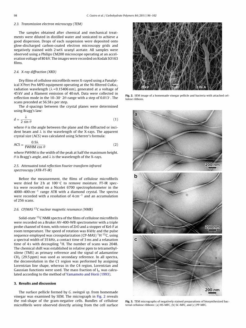
9 ate Polymers 84 (2011) 96–102
2
mggnoefi
2
ir4rs
u
d
wdc
A
w�
2s
wt4wo
2
wprsatTsCtLGl
3
vtm
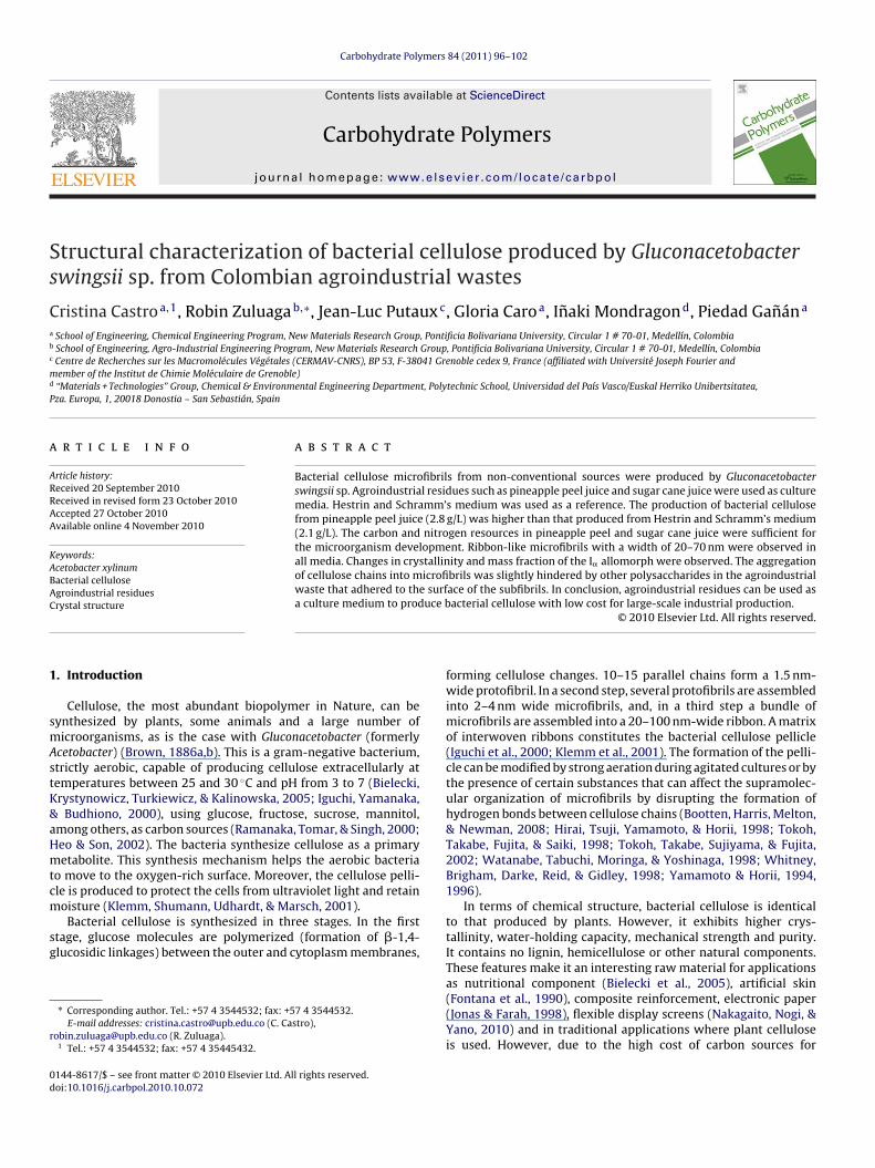
Fig. 2. SEM image of a homemade vinegar pellicle and bacteria with attached cel-lulose ribbons.
8 C. Castro et al. / Carbohydr
.3. Transmission electron microscopy (TEM)
The samples obtained after chemical and mechanical treat-ents were diluted in distilled water and sonicated to achieve a
ood dispersion. Drops of each suspension were deposited ontolow-discharged carbon-coated electron microscopy grids andegatively stained with 2 wt% uranyl acetate. All samples werebserved using a Philips CM200 microscope operating at an accel-ration voltage of 80 kV. The images were recorded on Kodak SO163lms.
.4. X-ray diffraction (XRD)
Dry films of cellulose microfibrils were X-rayed using a Panalyt-cal X’Pert Pro MPD equipment operating at the Ni-filtered CuK�1adiation wavelength (� = 0.15406 nm), generated at a voltage of5 kV and a filament emission of 40 mA. Data were collected ineflection mode in the 10–30◦ 2�-range with a step of 0.013◦. Thecans preceded at 56.58 s per step.
The d-spacings between the crystal planes were determinedsing Bragg’s law:
= �
2 sin �(1)
here � is the angle between the plane and the diffracted or inci-ent beam and � is the wavelength of the X-rays. The apparentrystal size (ACS) was calculated using Scherrer’s formula:
CS = 0.9�
FWHM cos �(2)
here FWHM is the width of the peak at half the maximum height.is Bragg’s angle, and � is the wavelength of the X-rays.
.5. Attenuated total reflection Fourier transform infraredpectroscopy (ATR-FT-IR)
Before the measurement, the films of cellulose microfibrilsere dried for 2 h at 100 ◦C to remove moisture. FT-IR spec-
ra were recorded on a Nicolet 6700 spectrophotometer in the000–400 cm−1 range ATR with a diamond crystal. The spectraere recorded with a resolution of 4 cm−1 and an accumulation
f 256 scans.
.6. CP/MAS 13C nuclear magnetic resonance (NMR)
Solid-state 13C NMR spectra of the films of cellulose microfibrilsere recorded on a Bruker AV-400-WB spectrometer with a triplerobe channel of 4 mm, with rotors of ZrO and a stopper of Kel-F atoom temperature. The speed of rotation was 8 kHz and the pulseequence employed was crosspolarization (CP-MAS) 1H 13C, usingspectral width of 35 kHz, a contact time of 3 ms and a relaxation
ime of 4 s with decoupling 1H. The number of scans was 2048.he chemical shift was established in relative ppm to tetramethyl-ilene (TMS) as primary reference and the signal of adamantineH2 (29.5 ppm) was used as secondary reference. In all spectra,he deconvolution in the C1 region was performed by assigningorentzian line shape, whereas in the C4 region, Lorentzian andaussian functions were used. The mass fraction of I� was calcu-
ated according to the method of Yamamoto and Horii (1993).
. Results and discussion
The surface pellicle formed by G. swingsii sp. from homemadeinegar was examined by SEM. The micrograph in Fig. 2 revealshe rod-shape of the gram-negative cells. Bundles of cellulose
icrofibrils were observed directly arising from the cell surfaceFig. 3. TEM micrographs of negatively stained preparations of biosynthesized bac-terial cellulose ribbons: (a) HS-MFC, (b) SC-MFC, and (c) PP-MFC.
C. Castro et al. / Carbohydrate Polymers 84 (2011) 96–102 99
F crofibi crossc Table
aa
sttoSl
pOaPtttTp(iednlas
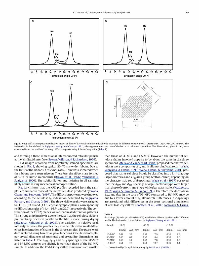
d100 and d110 in the case of PP-MFC compared to HS-MFC may bedue to a lower amount of I� allomorph. Differences in d-spacingsare associated with differences in the cross-sectional dimensionsof cellulose crystallites (Bootten et al., 2008; Ioelovich & Larina,
Table 1d-spacings (d) and crystallite size (ACS) in cellulose ribbons synthesized in differentmedia. The indexation is that defined in Sugiyama, Vuong, et al. (1991).
Sample (1 0 0) (0 1 0) (1 1 0)
d (nm) ACS (nm) d (nm) ACS (nm) d (nm) ACS (nm)
ig. 4. X-ray diffraction spectra (reflection mode) of films of bacterial cellulose mindexation is that defined in Sugiyama, Vuong, and Chanzy (1991), (d) suggestedalculated from the width of the X-ray diffraction peaks using Scherrer’s equation (
nd forming a three-dimensional interconnected reticular pelliclet the air–liquid interface (Brown, Willison, & Richardson, 1976).
TEM images recorded from negatively stained specimens arehown in Fig. 3, showing typical 20–70 nm-wide ribbons. Due tohe twist of the ribbons, a thickness of 6–8 nm was estimated whenhe ribbons were seen edge on. Therefore, the ribbons are formedf 3–11 cellulose microfibrils (Brown et al., 1976; Yamanaka &ugiyama, 2000). The subfibrillation and twisting in all samplesikely occurs during mechanical homogenization.
Fig. 4a–c shows that the XRD profiles recorded from the sam-les are similar to those of the native cellulose produced by Wada,kano, and Sugiyama (1997). The diffraction patterns were indexedccording to the cellulose I� indexation described by Sugiyama,ersson, and Chanzy (1991). The three visible peaks were assignedo (1 0 0), (0 1 0) and (1 1 0) crystallographic planes, correspondingo diffraction angles of 14.4◦, 16.7◦ and 22.7◦, respectively. The con-ribution of the (1 1 2) planes was absent in all diffraction patterns.his strong uniplanarity is due to the fact that the cellulose ribbonsreferentially oriented parallel to the film surface during dryingElazzouzi-Hafraoui et al., 2008). The variation in relative peakntensity between the profiles may also be related to small differ-nces in orientation of chains in the three samples. The peaks were
econvoluted using Lorenzian peak functions. Calculated interpla-ar crystal distances (d-spacings) and crystallite dimensions areisted in Table 1. The d100, d010 and d110 spacings of the SC-MFCnd PP-MFC samples are slightly lower than those of the HS-MFCample. In addition, the PP-MFC crystallite dimensions are smaller
rils produced in different culture media: (a) HS-MFC, (b) SC-MFC, (c) PP-MFC. The-section of the bacterial cellulose crystallites. The dimensions, given in nm, were1).
than those of SC-MFC and HS-MFC. However, the number of cel-lulose chains involved appears to be about the same in the threespecimens. Atalla and Vanderhart (1984) proposed that native cel-luloses were composites of I� and I� allomorphs. Wada et al. (Wada,Sugiyama, & Okano, 1995; Wada, Okano, & Sugiyama, 2001) pro-posed that native cellulose I could be classified into a I�-rich group(algae-bacteria) and a I�-rich group (cotton-ramie) depending onthe characteristic set of d-spacings. Wada et al. (1997) observedthat the d100 and d110 spacings of algal-bacterial type were largerthan those of cotton-ramie type while d010 was smaller (Wada et al.,1997; Wada, Sugiyama, & Okano, 1993). Therefore, the decrease in
HS-MFC 0.61 5.9 0.53 7.9 0.39 6.3SC-MFC 0.60 6.0 0.52 8.4 0.39 6.5PP-MFC 0.60 5.6 0.52 7.8 0.39 6.0HS-REFa 0.61 7.1 0.53 10.7 0.39 8.1
a Determined by X-ray diffractometry by Tokoh et al. (2002b).
1 ate Polymers 84 (2011) 96–102
1grt8mPctcewgm&g
fscc1c1sabi1r
aaIaSlfpsM
bFl(t&teCt&ttCmaHCccmpaI
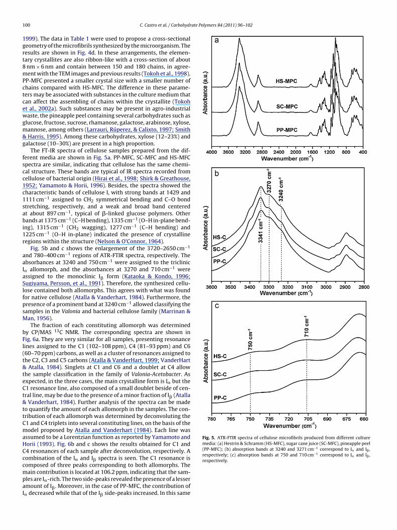
Fig. 5. ATR-FTIR spectra of cellulose microfibrils produced from different culturemedia: (a) Hestrin & Schramm (HS-MFC), sugar cane juice (SC-MFC), pineapple peel(PP-MFC); (b) absorption bands at 3240 and 3271 cm−1 correspond to I� and I� ,respectively; (c) absorption bands at 750 and 710 cm−1 correspond to I� and I� ,respectively.
00 C. Castro et al. / Carbohydr
999). The data in Table 1 were used to propose a cross-sectionaleometry of the microfibrils synthesized by the microorganism. Theesults are shown in Fig. 4d. In these arrangements, the elemen-ary crystallites are also ribbon-like with a cross-section of aboutnm × 6 nm and contain between 150 and 180 chains, in agree-ent with the TEM images and previous results (Tokoh et al., 1998).
P-MFC presented a smaller crystal size with a smaller number ofhains compared with HS-MFC. The difference in these parame-ers may be associated with substances in the culture medium thatan affect the assembling of chains within the crystallite (Tokoht al., 2002a). Such substances may be present in agro-industrialaste, the pineapple peel containing several carbohydrates such as
lucose, fructose, sucrose, rhamanose, galactose, arabinose, xylose,annose, among others (Larrauri, Rúperez, & Calixto, 1997; SmithHarris, 1995). Among these carbohydrates, xylose (12–23%) and
alactose (10–30%) are present in a high proportion.The FT-IR spectra of cellulose samples prepared from the dif-
erent media are shown in Fig. 5a. PP-MFC, SC-MFC and HS-MFCpectra are similar, indicating that cellulose has the same chemi-al structure. These bands are typical of IR spectra recorded fromellulose of bacterial origin (Hirai et al., 1998; Shirk & Greathouse,952; Yamamoto & Horii, 1996). Besides, the spectra showed theharacteristic bands of cellulose I, with strong bands at 1429 and111 cm−1 assigned to CH2 symmetrical bending and C–O bondtretching, respectively, and a weak and broad band centeredt about 897 cm−1, typical of �-linked glucose polymers. Otherands at 1375 cm−1 (C–H bending), 1335 cm−1 (O–H in-plane bend-
ng), 1315 cm−1 (CH2 wagging), 1277 cm−1 (C–H bending) and225 cm−1 (O–H in-plane) indicated the presence of crystallineegions within the structure (Nelson & O’Connor, 1964).
Fig. 5b and c shows the enlargement of the 3720–2650 cm−1
nd 780–400 cm−1 regions of ATR-FTIR spectra, respectively. Thebsorbances at 3240 and 750 cm−1 were assigned to the triclinic
� allomorph, and the absorbances at 3270 and 710 cm−1 weressigned to the monoclinic I� form (Kataoka & Kondo, 1996;ugiyama, Persson, et al., 1991). Therefore, the synthesized cellu-ose contained both allomorphs. This agrees with what was foundor native cellulose (Atalla & Vanderhart, 1984). Furthermore, theresence of a prominent band at 3240 cm−1 allowed classifying theamples in the Valonia and bacterial cellulose family (Marrinan &an, 1956).The fraction of each constituting allomorph was determined
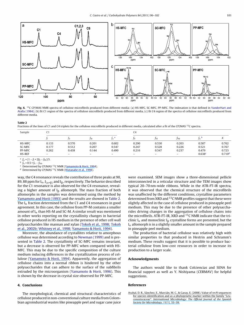
y CP/MAS 13C NMR. The corresponding spectra are shown inig. 6a. They are very similar for all samples, presenting resonanceines assigned to the C1 (102–108 ppm), C4 (81–93 ppm) and C660–70 ppm) carbons, as well as a cluster of resonances assigned tohe C2, C3 and C5 carbons (Atalla & VanderHart, 1999; VanderHart
Atalla, 1984). Singlets at C1 and C6 and a doublet at C4 allowhe sample classification in the family of Valonia-Acetobacter. Asxpected, in the three cases, the main crystalline form is I� but the1 resonance line, also composed of a small doublet beside of cen-ral line, may be due to the presence of a minor fraction of I� (Atalla
Vanderhart, 1984). Further analysis of the spectra can be madeo quantify the amount of each allomorph in the samples. The con-ribution of each allomorph was determined by deconvoluting the1 and C4 triplets into several constituting lines, on the basis of theodel proposed by Atalla and Vanderhart (1984). Each line was
ssumed to be a Lorentzian function as reported by Yamamoto andorii (1993). Fig. 6b and c shows the results obtained for C1 and4 resonances of each sample after deconvolution, respectively. Aombination of the I� and I� spectra is seen. The C1 resonance is
omposed of three peaks corresponding to both allomorphs. Theain contribution is located at 106.2 ppm, indicating that the sam-les are I�-rich. The two side-peaks revealed the presence of a lessermount of I�. Moreover, in the case of PP-MFC, the contribution of
� decreased while that of the I� side-peaks increased. In this same
C. Castro et al. / Carbohydrate Polymers 84 (2011) 96–102 101
Fig. 6. 13C CP/MAS NMR spectra of cellulose microfibrils produced from different media: (a) HS-MFC, SC-MFC, PP-MFC. The indexation is that defined in VanderHart andAtalla (1984); (b) fit C1 region of the spectra of cellulose microfibrils produced from different media, (c) fit C4 region of the spectra of cellulose microfibrils produced fromdifferent media.
Table 2Fractions of the lines of C1 and C4 triplets for the cellulose microfibrils produced in different media, calculated after a fit of the CP/MAS 13C spectra.
Sample C1 C4
fI fII fIII f� a fV fVI fVII f� b X
HS-MFC 0.133 0.570 0.201 0.602 0.290 0.530 0.203 0.587 0.762SC-MFC 0.177 0.512 0.207 0.547 0.247 0.528 0.226 0.521 0.767PP-MFC 0.262 0.438 0.144 0.490 0.216 0.547 0.237 0.479 0.723HS-REF – – – – – – – 0.638c 0.710d
w8fiaYTaaicpe
csbMmlcpei
4
cb
a f� = (1 − fI + 2fII − fIII)/3.b f� = 0.5 + fV − fVII.c Determined by CP/MAS 13C NMR (Yamamoto & Horii, 1994).d Determined by CP/MAS 13C NMR (Watanabe et al., 1998).
ay, the C4 resonance reveals the contribution of three peaks at 90,9, 88 ppm for I�, I(�+�) and I�, respectively. The behavior describedor the C1 resonance is also observed for the C4 resonance, reveal-ng a higher amount of I� allomorph. The mass fraction of bothllomorphs in the samples was determined using the method byamamoto and Horii (1993) and the results are showed in Table 2.he I� fraction determined from the C1 and C4 resonances in goodgreement. In this case, the cellulose from PP-M contains a smallermount of I� than HS-M and SC-M. A similar result was mentionedn other works reporting on the crystallinity changes in bacterialellulose produced in HS medium in the presence of other cell wallolysaccharides like mannan and xylan (Tokoh et al., 1998; Tokoht al., 2002b; Whitney et al., 1998; Yamamoto & Horii, 1994).
Moreover, the abundance of crystallites relative to amorphousellulose was determined according to Newman (1999) and is pre-ented in Table 2. The crystallinity of SC-MFC remains invariant,ut a decrease is observed for PP-MFC when compared with HS-FC. This may be due to the specific composition of the cultureedium inducing differences in the crystallization process of cel-
ulose (Yamamoto & Horii, 1994). Apparently, the aggregation ofellulose chains into a normal ribbon is hindered by the otherolysaccharides that can adhere to the surface of the subfibrilsxtruded by the microorganism (Yamamoto & Horii, 1996). Thiss shown by the decrease in crystal size observed for PP-MFC.
. Conclusions
The morphological, chemical and structural characteristics ofellulose produced in non-conventional culture media from Colom-ian agroindustrial wastes like pineapple peel and sugar cane juice
were examined. SEM images show a three-dimensional pellicleinterconnected in a reticular structure and the TEM images showtypical 20–70 nm-wide ribbons. While in the ATR-FT-IR spectra,it was observed that the chemical structure of the microfibrilswas unaffected by the different conditions, crystalline parametersdetermined from XRD and 13C NMR profiles suggest that these wereslightly affected in the case of cellulose produced in pineapple peelmedium. This may be due to the presence of other polysaccha-rides driving changes in the aggregation of cellulose chains intothe microfibrils. ATR-FT-IR, XRD and 13C NMR indicate that the tri-clinic I� and monoclinic I� crystalline forms are presented, but theI� allomorph is in a slightly smaller amount in the sample preparedin pineapple peel medium.
The production of bacterial cellulose was relatively high withsimilar properties to that produced in Hestrin and Schramm’smedium. These results suggest that it is possible to produce bac-terial cellulose from low-cost resources in order to increase itsproduction to a larger scale.
Acknowledgments
The authors would like to thank Colciencias and SENA forfinancial support as well as Y. Nishiyama (CERMAV) for helpfulsuggestions.
References
Arahal, D. R., Sánchez, E., Marcián, M. C., & Garay, E. (2008). Value of recN sequencesfor species identification and as a phylogenetic marker within the family “Leu-conostocaceae”. International Microbiology: The Official Journal of the SpanishSociety for Microbiology, 11(1), 33–39.
1 ate Po
A
A
B
B
B
B
B
D
E
F
H
H
H
I
I
J
K
K
K
K
K
L
M
02 C. Castro et al. / Carbohydr
talla, R. H., & Vanderhart, D. L. (1984). Native cellulose: A composite of two distinctcrystalline forms. Science, 223(4633), 283–285.
talla, R. H., & VanderHart, D. L. (1999). The role of solid state 13C NMR spectroscopyin studies of the nature of native celluloses. Solid State Nuclear Magnetic Reso-nance, 15(1), 1–19.
ielecki, S., Krystynowicz, A., Turkiewicz, M., & Kalinowska, H. (2005). Bacterialcellulose. In Alexander Steinbüchel, & Yoshiharu Doi (Eds.), Biotechnology of poly-mer: From synthesis to patents (pp. 381–434). Weinheim, Germany: Wiley-VCH.Chapter 14.
ootten, T. J., Harris, P. J., Melton, L. D., & Newman, R. H. (2008). WAXS and 13CNMR study of Glucoacetobacter xylinus cellulose in composites with tamarindxyloglucan. Carbohydrate Research, 343(2), 221–229.
rown, A. J. (1886a). On an acetic ferment which forms cellulose. Journal of theChemical Society, 49, 432–439.
rown, A. J. (1886b). The chemical action of pure cultivations of bacterium aceti.Journal of the Chemical Society, 49, 172–187.
rown, R. M., Jr., Willison, J. H. M., & Richardson, C. L. (1976). Cellulose biosynthesisin Acetobacter xylinum: Visualization of the site of synthesis and direct measure-ment of the in vivo process. Proceedings of the National Academy of Science of theUnited States of America, 73(12), 4565–4569.
ellaglio, F., Cleenwerck, I., Felis, G. E., Engelbeen, K., Janssens, D., & Marzotto, M.(2005). Description of Gluconacetobacter Swingsii sp. Nov. and Gluconacetobac-ter rhaeticus sp. nov., isolated from Italian apple fruit. International Journal ofSystematic and Evolutionary Microbiology, 55(6), 2365–2370.
lazzouzi-Hafraoui, S., Nishiyama, Y., Putaux, J.-L., Heux, L., Dubreuil, F., &Rochas, C. (2008). The shape and size distribution of crystalline nanoparti-cles prepared by acid hydrolysis of native cellulose. Biomacromolecules, 9(1),57–65.
ontana, J. D., De Souza, A. M., Fontana, C. K., Torriani, I. L., Moresch, J. C., Gallotti,B. J., et al. (1990). Acetobacter cellulose pellicle as a temporary skin substitute.Applied Biochemistry and Biotechnology, 24(25), 253–264.
eo, M., & Son, H. (2002). Development of an optimized, simple chemically definedmedium for bacterial cellulose production by Acetobacter sp. A9 in shaking cul-tures. Biotechnology and Applied Biochemistry, 36(1), 41–45.
estrin, S., & Schramm, M. (1954). Synthesis of cellulose by Acetobacter xylinum. 2.Preparation of freeze-dried cells capable of polymerizing glucose to cellulose.Biochemical Journal, 58(2), 345–352.
irai, A., Tsuji, M., Yamamoto, H., & Horii, F. (1998). In situ crystallization of bac-terial cellulose. III. Influences of different polymeric additives on the formationof microfibrils as revealed by transmission electron microscopy. Cellulose, 5(3),201–213.
guchi, M., Yamanaka, S., & Budhiono, A. (2000). Review: Bacterial cellulose—A mas-terpiece of nature’s arts. Journal of Materials Science, 35(2), 261–270.
oelovich, M., & Larina, E. (1999). Parameters of crystalline structure and their influ-ence on the reactivity of cellulose I. Cellulose Chemistry & Technology, 33(1–2),3–12.
onas, R., & Farah, L. (1998). Production and application of microbial cellulose. Poly-mer Degradation and Stability, 59(1–3), 101–106.
ataoka, Y., & Kondo, T. (1996). Changing cellulose crystalline structure in formingwood cell walls. Macromolecules, 29(19), 6356–6358.
eshk, S., Razek, T., & Sameshima, K. (2006). Bacterial cellulose production from beetmolasses. African Journal of Biotechnology, 5(17), 1519–1523.
lemm, D., Shumann, D., Udhardt, U., & Marsch, S. (2001). Bacterial synthesizedcellulose—Artificial blood vessels for microsurgery. Progress in Polymer Science,26(9), 1561–1603.
ongruang, S. (2008). Bacterial cellulose production by Acetobacter xylinum strainsfrom agricultural waste products. Applied Biochemistry and Biotechnology,148(1–3), 245–256.
urosumi, A., Sasaki, C., Yamashita, Y., & Nakamura, Y. (2009). Utilization of variousfruit juices as carbon source for production of bacterial cellulose by Acetobacter
xylinum NBRC 13693. Carbohydrate Polymers, 76(2), 333–335.arrauri, J. A., Rúperez, P., & Calixto, F. S. (1997). Pineapple shell as a source of dietaryfiber with associated polyphenols. Journal of Agricultural and Food Chemistry,45(10), 4028–4031.
arrinan, H., & Mann, J. (1956). Infrared spectra of the crystalline modification ofcellulose. Journal of Polymer Science, 21(98), 301–311.
lymers 84 (2011) 96–102
Moon, S., Park, J., Chun, H., & Kim, S. (2006). Comparisons of physical properties ofbacterial cellulose produced in different culture conditions using saccharifiedfood wastes. Biotechnology and Bioprocess Engineering, 11(1), 26–31.
Nakagaito, A. N., Masaya Nogi, M., & Yano, H. (2010). Displays from transparent filmsof natural nanofibers. MRS Bulletin, 35(3), 214–218.
Nelson, M., & O’Connor, R. (1964). Relation of certain infrared bands to cellulosecrystallinity and crystal lattice type. Part I. Spectra of lattice types I, II, III and ofamorphous cellulose. Journal of Applied Polymer Science, 8(3), 1311–1324.
Newman, R. (1999). Estimation of the lateral dimensions of cellulose crystallitesusing 13C NMR signal strengths. Solid State Nuclear Magnetic Resonance, 15(1),21–29.
Ramanaka, K., Tomar, A., & Singh, L. (2000). Effect of various carbon and nitro-gen sources on cellulose synthesis by Acetobacter xylinum in world. Journal ofMicrobiology and Biotechnology, 16(3), 245–248.
Shirk, H., & Greathouse, G. (1952). Infrared spectra of bacterial cellulose. AnalyticalChemistry, 24(11), 1774–1775.
Smith, B. G., & Harris, P. J. (1995). Polysaccharide composition of unlignified cellwalls of pineapple [Ananas comosus (L.) Merr.] fruit. Plant Physiology, 107(4),1399–1409.
Sugiyama, J., Persson, J., & Chanzy, H. (1991). Combined infrared and electrondiffraction study of the polymorphism of native cellulose. Macromolecules, 24(9),2461–2466.
Sugiyama, J., Vuong, R., & Chanzy, H. (1991). Electron diffraction study on the twocrystalline phases occurring in native cellulose from an algal cell wall. Macro-molecules, 24(14), 4168–4175.
Tokoh, C., Takabe, K., Sujiyama, J., & Fujita, M. (2002a). CP/MAS 13C NMR and electrondiffraction study of bacterial cellulose structure affected by cell wall polysaccha-rides. Cellulose, 9(3–4), 351–360.
Tokoh, C., Takabe, K., Sujiyama, J., & Fujita, M. (2002b). Cellulose synthesized byAcetobacter xylinum in the presence of plant cell wall polysaccharides. Cellulose,9(1), 65–74.
Tokoh, C., Takabe, K., Fujita, M., & Saiki, H. (1998). Cellulose synthesized by Aceto-bacter xylinum in presence of acetyl glucomannan. Cellulose, 5(4), 249–261.
Vandamme, E., De Baets, S., Vanbaelen, A., Joris, K., & De Wulf, P. (1998). Improvedproduction of bacterial cellulose and its application potential. Polymer Degrada-tion and Stability, 59(1–3), 93–99.
VanderHart, D., & Atalla, R. (1984). Studies of microstructure in native cellulosesusing solid-state 13C NMR. Macromolecules, 17(8), 1465–1472.
Wada, M., Sugiyama, J., & Okano, T. (1993). Native cellulose on the basis of two crys-talline phase (I/I) system. Journal of Applied Polymer Science, 49(8), 1491–1496.
Wada, M., Sugiyama, J., & Okano, T. (1995). Two crystalline phases (I�/I�) systemof native celluloses in relation to plant phylogenesis. Mokusai Gakkaishi, 41(2),186–192.
Wada, M., Okano, T., & Sugiyama, J. (1997). Synchrotron-radiated X-ray and neutrondiffraction study of native cellulose. Cellulose, 4(3), 221–232.
Wada, M., Okano, T., & Sugiyama, J. (2001). Allomorphs of native cellulose I evaluatedby two equatorial d-spacing. Journal of Wood Science, 47(2), 124–128.
Watanabe, K., Tabuchi, M., Moringa, Y., & Yoshinaga, F. (1998). Structural featuresand properties of bacterial cellulose produced in agitated culture. Cellulose, 5(3),187–200.
Whitney, S., Brigham, J., Darke, A., Reid, J., & Gidley, M. (1998). Structural aspectsof the interaction of mannan-based polysaccharides with bacterial cellulose.Carbohydrate Research, 307(3–4), 299–309.
Yamamoto, H., & Horii, F. (1993). CP/MAS 13C NMR analysis of the crystal trans-formation induced for Valonia cellulose by annealing at high temperature.Macromolecules, 26(6), 1313–1317.
Yamamoto, H., & Horii, F. (1994). In situ crystallization of bacterial cellulose I.Influences of polymeric Additives, stirring and temperature on the forma-tion cellulose I� and I� as revealed by cross polarization/magic angle spinning(CP/MAS) 13C NMR spectroscopy. Cellulose, 1(1), 57–66.
Yamamoto, H., & Horii, F. (1996). In situ crystallization of bacterial celluloseII. Influences of polymeric Additives, stirring and temperature on the for-mation cellulose I� and I� at the early stage of incubation. Cellulose, 3(1),229–242.
Yamanaka, S., & Sugiyama, J. (2000). Structural modification of bacterial cellulose.Cellulose, 7(3), 213–225.
Copyright © 2022 FDOKUMEN