Structural Brain Connectivity in School-Age Preterm Infants Provides Evidence for Impaired Networks...
13
Structural Brain Connectivity in School-Age Preterm Infants Provides Evidence for Impaired Networks Relevant for Higher Order Cognitive Skills and Social Cognition Elda Fischi-Gómez 1,2,† , Lana Vasung 1,† , Djalel-Eddine Meskaldji 2,3 , Fançois Lazeyras 3 , Cristina Borradori-Tolsa 1 , Patric Hagmann 4 , Koviljka Barisnikov 5 , Jean-Philippe Thiran 2,4 and Petra S. Hüppi 1 1 Division of Development and Growth, Department of Pediatrics, University of Geneva, Geneva, Switzerland, 2 Signal Processing Laboratory 5 (LTS5), Ecole Polytechnique Fédérale de Lausanne (EPFL), Lausanne, Switzerland, 3 Department of Radiology, University Hospital of Geneva, Geneva, Switzerland, 4 Department of Radiology, University Hospital Center (CHUV) and University of Lausanne (UNIL), Lausanne, Switzerland and 5 Child Clinical Neuropsychology Unit, Department of Psychology, University of Geneva, Geneva, Switzerland † Co-First Authors; Elda Fischi-Gómez and Lana Vasung contributed equally to the work. Address correspondence Dr Lana Vasung. Email: [email protected]. Extreme prematurity and pregnancy conditions leading to intrauter- ine growth restriction (IUGR) affect thousands of newborns every year and increase their risk for poor higher order cognitive and social skills at school age. However, little is known about the brain struc- tural basis of these disabilities. To compare the structural integrity of neural circuits between prematurely born controls and children born extreme preterm (EP) or with IUGR at school age, long-ranging and short-ranging connections were noninvasively mapped across corti- cal hemispheres by connection matrices derived from diffusion tensor tractography. Brain connectivity was modeled along fiber bundles connecting 83 brain regions by a weighted characterization of structural connectivity (SC). EP and IUGR subjects, when com- pared with controls, had decreased fractional anisotropy-weighted SC (FAw-SC) of cortico-basal ganglia-thalamo-cortical loop connec- tions while cortico-cortical association connections showed both decreased and increased FAw-SC. FAw-SC strength of these connec- tions was associated with poorer socio-cognitive performance in both EP and IUGR children. Keywords: brain connectivity, connectomics, extreme prematurity, human brain development, intrauterine growth restriction, social cognition Introduction Every year, both in the United States of America and in Europe, ∼60 000 children are born extremely preterm (EP) or with intrauterine growth restriction (IUGR). Survival of these infants has increased steadily due to improvement in perinatal care (World Health Organisation 2012). Nevertheless, the long- term neurodevelopmental impairments seen in these survivors have been recognized as a global burden to society (World Health Organisation 2012). Despite improved survival, the lit- erature reports an increasing number of children with impaired physical health, impaired cognitive capacities, and more re- cently, emotional and behavioral deficits (Bhutta 2002; Ander- son 2004; Clark et al. 2008). Higher rates in externalizing and internalizing behaviors often lead to lower social competence (Ross et al. 1990). Emotional regulation problems with emotional outbursts lead to school and peer relation problems. Preterm infants also have a 3 times odds of meeting criteria for psychiatric diagnoses (Treyvaud et al. 2013). Quality-of-life as- sessments in ex-preterm adolescents reveal rates of poor socia- lization skills due to difficulties in learning and nonverbal communication. As shown in a recent twin study, lower birth weight (BW) with poor intrauterine growth adds an additional risk for these impairments (Edmonds et al. 2010). Therefore, the identification of neuroimaging biomarkers in these chil- dren at school age is crucial for understanding their cognitive and behavioral impairments (Ment et al. 2009). In the absence of focal brain lesions, most neuroimaging studies of prematurely born children (with or without IUGR) have primarily focused on the assessment of cerebral cortex maturation in terms of thickness (de Bie et al. 2010), total cortical volume (Tolsa et al. 2004), and cortical surface com- plexity (Dubois, Benders, Borradori-Tolsa, et al. 2008; Dubois, Benders, Cachia et al. 2008); on volumes of deep gray matter structures (Ball et al. 2012), hippocampus (Lodygensky et al. 2008), on the cerebellum (Limperopoulos et al. 2005), and on white matter (WM) volume (for review see Ment et al. (2009)). Early diffusion magnetic resonance imaging (dMRI) studies have further associated premature birth with changes in WM at term equivalent age (Huppi et al. 1998; Partridge et al. 2004; Counsell et al. 2008). Recent advances in dMRI and operator-independent tracto- graphy enable the noninvasive mapping of WM cortico-cortical and cortico-subcortical connections at high spatial resolution (Hagmann, Cammoun, Gigandet, Gerhard, et al. 2010). This technique has been successfully applied to the study of brain development (Hagmann, Sporns, et al. 2010; Gao et al. 2011; Yap et al. 2011; Dennis et al. 2013). Nevertheless, the effects of prematurity and low BW on the maturation of structural neural networks remain still unexplored. This study presents a whole-brain connectomic analysis of the effects of extremely premature birth and IUGR on chil- dren’s brain connectivity at 6 years of age that might underlie their poorer social and higher order cognitive functioning. Materials and Methods Subjects Sixty prematurely born infants at 6 years (mean age at scan 6.7 ± 0.66 years) were recruited from the Child Development Units of the Centre Hospitalier Universitaire Vaudois (CHUV), Lausanne, and the Hôpitaux Universitaires de Genève (HUG), Switzerland. All studies were per- formed with informed parental and child consent after approval by the medical ethical board. After preprocessing and quality evaluation of the MRI images, 52 subjects were finally included in the analysis (Sup- plementary Table 1). Infant growth parameters (i.e., weight and head circumference measures) and perinatal data, including BW, gestational age (GA), gender, APGAR score, and the presence of perinatal morbid- ities were also collected (Supplementary Table 1). None of the children © The Author 2014. Published by Oxford University Press. All rights reserved. For Permissions, please e-mail: [email protected] Cerebral Cortex doi:10.1093/cercor/bhu073 Cerebral Cortex Advance Access published May 2, 2014 at Université & EPFL Lausanne on October 14, 2014 http://cercor.oxfordjournals.org/ Downloaded from
-
Upload
independent -
Category
Documents
-
view
1 -
download
0
Transcript of Structural Brain Connectivity in School-Age Preterm Infants Provides Evidence for Impaired Networks...
Structural Brain Connectivity in School-Age Preterm Infants Provides Evidence for ImpairedNetworks Relevant for Higher Order Cognitive Skills and Social Cognition
Elda Fischi-Gómez1,2,†, Lana Vasung1,†, Djalel-Eddine Meskaldji2,3, Fançois Lazeyras3, Cristina Borradori-Tolsa1, Patric Hagmann4,Koviljka Barisnikov5, Jean-Philippe Thiran2,4 and Petra S. Hüppi1
1Division of Development and Growth, Department of Pediatrics, University of Geneva, Geneva, Switzerland, 2Signal ProcessingLaboratory 5 (LTS5), Ecole Polytechnique Fédérale de Lausanne (EPFL), Lausanne, Switzerland, 3Department of Radiology,University Hospital of Geneva, Geneva, Switzerland, 4Department of Radiology, University Hospital Center (CHUV) and Universityof Lausanne (UNIL), Lausanne, Switzerland and 5Child Clinical Neuropsychology Unit, Department of Psychology, University ofGeneva, Geneva, Switzerland
†Co-First Authors; Elda Fischi-Gómez and Lana Vasung contributed equally to the work.
Address correspondence Dr Lana Vasung. Email: [email protected].
Extreme prematurity and pregnancy conditions leading to intrauter-ine growth restriction (IUGR) affect thousands of newborns everyyear and increase their risk for poor higher order cognitive and socialskills at school age. However, little is known about the brain struc-tural basis of these disabilities. To compare the structural integrity ofneural circuits between prematurely born controls and children bornextreme preterm (EP) or with IUGR at school age, long-ranging andshort-ranging connections were noninvasively mapped across corti-cal hemispheres by connection matrices derived from diffusiontensor tractography. Brain connectivity was modeled along fiberbundles connecting 83 brain regions by a weighted characterizationof structural connectivity (SC). EP and IUGR subjects, when com-pared with controls, had decreased fractional anisotropy-weightedSC (FAw-SC) of cortico-basal ganglia-thalamo-cortical loop connec-tions while cortico-cortical association connections showed bothdecreased and increased FAw-SC. FAw-SC strength of these connec-tions was associated with poorer socio-cognitive performance inboth EP and IUGR children.
Keywords: brain connectivity, connectomics, extreme prematurity, humanbrain development, intrauterine growth restriction, social cognition
Introduction
Every year, both in the United States of America and in Europe,∼60 000 children are born extremely preterm (EP) or withintrauterine growth restriction (IUGR). Survival of theseinfants has increased steadily due to improvement in perinatalcare (World Health Organisation 2012). Nevertheless, the long-term neurodevelopmental impairments seen in these survivorshave been recognized as a global burden to society (WorldHealth Organisation 2012). Despite improved survival, the lit-erature reports an increasing number of children with impairedphysical health, impaired cognitive capacities, and more re-cently, emotional and behavioral deficits (Bhutta 2002; Ander-son 2004; Clark et al. 2008). Higher rates in externalizing andinternalizing behaviors often lead to lower social competence(Ross et al. 1990). Emotional regulation problems withemotional outbursts lead to school and peer relation problems.Preterm infants also have a 3 times odds of meeting criteria forpsychiatric diagnoses (Treyvaud et al. 2013). Quality-of-life as-sessments in ex-preterm adolescents reveal rates of poor socia-lization skills due to difficulties in learning and nonverbalcommunication. As shown in a recent twin study, lower birthweight (BW) with poor intrauterine growth adds an additional
risk for these impairments (Edmonds et al. 2010). Therefore,the identification of neuroimaging biomarkers in these chil-dren at school age is crucial for understanding their cognitiveand behavioral impairments (Ment et al. 2009).
In the absence of focal brain lesions, most neuroimagingstudies of prematurely born children (with or without IUGR)have primarily focused on the assessment of cerebral cortexmaturation in terms of thickness (de Bie et al. 2010), totalcortical volume (Tolsa et al. 2004), and cortical surface com-plexity (Dubois, Benders, Borradori-Tolsa, et al. 2008; Dubois,Benders, Cachia et al. 2008); on volumes of deep gray matterstructures (Ball et al. 2012), hippocampus (Lodygensky et al.2008), on the cerebellum (Limperopoulos et al. 2005), and onwhite matter (WM) volume (for review see Ment et al. (2009)).Early diffusion magnetic resonance imaging (dMRI) studieshave further associated premature birth with changes in WM atterm equivalent age (Huppi et al. 1998; Partridge et al. 2004;Counsell et al. 2008).
Recent advances in dMRI and operator-independent tracto-graphy enable the noninvasive mapping of WM cortico-corticaland cortico-subcortical connections at high spatial resolution(Hagmann, Cammoun, Gigandet, Gerhard, et al. 2010). Thistechnique has been successfully applied to the study of braindevelopment (Hagmann, Sporns, et al. 2010; Gao et al. 2011;Yap et al. 2011; Dennis et al. 2013). Nevertheless, the effects ofprematurity and low BW on the maturation of structural neuralnetworks remain still unexplored.
This study presents a whole-brain connectomic analysis ofthe effects of extremely premature birth and IUGR on chil-dren’s brain connectivity at 6 years of age that might underlietheir poorer social and higher order cognitive functioning.
Materials and Methods
SubjectsSixty prematurely born infants at 6 years (mean age at scan 6.7 ± 0.66years) were recruited from the Child Development Units of the CentreHospitalier Universitaire Vaudois (CHUV), Lausanne, and the HôpitauxUniversitaires de Genève (HUG), Switzerland. All studies were per-formed with informed parental and child consent after approval by themedical ethical board. After preprocessing and quality evaluation ofthe MRI images, 52 subjects were finally included in the analysis (Sup-plementary Table 1). Infant growth parameters (i.e., weight and headcircumference measures) and perinatal data, including BW, gestationalage (GA), gender, APGAR score, and the presence of perinatal morbid-ities were also collected (Supplementary Table 1). None of the children
© The Author 2014. Published by Oxford University Press. All rights reserved.For Permissions, please e-mail: [email protected]
Cerebral Cortexdoi:10.1093/cercor/bhu073
Cerebral Cortex Advance Access published May 2, 2014 at U
niversité
& E
PFL L
ausanne on October 14, 2014
http://cercor.oxfordjournals.org/D
ownloaded from
had any sign of prematurity-associated brain lesions on MRI at termequivalent age, as assessed by preterm brain injury scores (Woodwardet al. 2006). Furthermore, at 6 years, their MRI scans were read asnormal by experienced neuroradiologists. All of the recruited childrenwere free from medication and from psychiatric or neurologicaldisease (Supplementary Table 1).
Children were classified as 1) moderately premature controls, 2)extreme premature (EP), or 3) moderately premature with IUGR (seeSupplementary Table 1). Extreme prematurity was defined by GA atbirth of <28 weeks. IUGR was defined as BW below 10th percentile(adjusted for GA and gender) and on criteria of placental insufficiencyaccording to intrauterine growth assessment, prenatal ultrasound, andDoppler measurement within the umbilical artery. Infants consideredas controls were born with BW appropriate for GA without extremeprematurity, as moderately premature infants with normal growth gen-erally compare with children born full term in their structural brain de-velopment (Zacharia et al. 2006).
Cognitive assessment at time of MRI scan was carried out in all sub-jects using the French version of Kaufmann Assessment Battery forChildren (Kaufman AS and Kaufman NL 1993). Furthermore, the adap-tive and problematic behavior was assessed by administering theFrench version (d’Acremont and Linden 2008) of the Strengths and Dif-ficulties Questionnaire (SDQ) (Goodman 1997) in 45 subjects (8 con-trols, 20 EP, and 17 IUGR subjects). Four types of problematicbehavior: conduct problems, hyperactivity/inattention, peer relation-ship problems, emotional symptoms, and prosocial behavior wereassessed using 5 SDQ scales. Social reasoning abilities were assessedadministering the Social Resolution Task (SRT) designed to assess theability to judge, identify, and reason about moral and conventionalrules (Barisnikov and Hippolyte 2011). Only Judgment scores (SRTQ1) and Identification scores (SRT Q2) from SRT were used in analysis,since they were considered appropriate for this age group (Barisnikovand Hippolyte 2011).
Socio-economic status (SES) was assessed for each group using theLargo scale (Largo et al. 1989).
Exploratory regression analysis (linear regression models) was usedto estimate the effect of the GA and IUGR on achieved cognitive scoreswhen adjusting for SES and sex.
Data AcquisitionChildren underwent MRI examinations on a 3T Siemens TrioTimsystem (Siemens Medical Solutions, Erlangen, Germany). For eachsubject, a high-resolution T1-weighted image was acquired using a 3Dmagnetization prepared rapid acquisition gradient echo (MPRAGE)sequence (TR/TE = 2500/2.91 [ms/ms]; TI = 1100 ms; resolution = 1 ×1 × 1 mm; FOV = 160 × 256 × 208 mm3). Following a nondiffusion-weighted image (DWI) acquisition (b = 0), DWIs were acquired with asingle-shot, spin-echo planar imaging (SE-EPI) sequence covering 30diffusion directions with a maximum b-value of 1000 s/mm2, providingwhole-brain coverage (TR/TE = 10200/107 [ms/ms]; in-planeresolution = 1.8 × 1.8 mm2; slice thickness = 2 mm; FOV = 230 × 230 ×256 mm3). None of the subjects were sedated during the acquisition.
Data Processing
Connectome Construction and AnalysisThe extraction of the whole-brain structural connectomes was per-formed using the Connectome Mapping Toolkit (CMTK), a python-based open-source software that implements a full diffusion MRIprocessing pipeline, from raw diffusion/T1/T2 data to multiresolutionconnection matrices (www.cmtk.org (Daducci et al. 2012)). The mainsteps and the methods used to construct the connection matrix are 1)WM-GM surface extraction and cortical and subcortical parcellation(performed using Freesurfer software (http://surfer.nmr.mgh.harvard.edu/)), 2) streamline tractography (done using an in-house builtmethod fully implemented in the CMTK pipeline), 3) estimation of con-nection density; the ratio between the sum of all virtual streamlinesconnecting each pair of regions of interest (ROI) and their individuallength (see Supplementary Material for more detailed informationabout the procedure used).
Following (Hagmann, Sporns, et al. 2010), the structural connec-tivity (SC) between cortical regions was defined as the product of 2components (see Supplementary Material). The first component wasthe group connection density (gCD), computed as the following. Theindividual CD matrices were normalized 1) by the size of the corticalregions and 2) by the length of the fiber bundle connecting 2 regions.The groups CD matrices were computed as the mean of all normalizedconnection matrices in a given group. Since the mean is non-null if atleast one of the elements is non-null, these group average connectivitymatrices consist a support of the connectivity in each group and rep-resents the maximum grid for each group. Comparing these supportgCD matrices, no statistically significant differences in average strengthand network density were observed, which was expected due to theabsence of gross brain pathology. The second component was the con-nection efficacy, considered to be subject-dependent. It was computedas the mean fractional anisotropy (FA) value of the bundle connecting2 regions (in the FA-weighted analysis) and as the mean of the inverseapparent diffusion coefficient (ADC) in the ADC-weighted analysis.
As we do not consider connections for which the connectivity is 0for one group and >0 for the other group in the second-levelconnection-wise analysis, we can say that, in this situation, we areusing the minimum grid as support for this specific analysis.
The resulting connectomes were compared globally and locallyusing a Mann–Whitney (a nonparametric test) test, and the resultingP-values were corrected for multiple comparisons. This later step wasperformed using a two-step methodology specially designed for con-nectomes (Meskaldji et al. 2011; Meskaldji 2013). This new statisticalcorrection exploits the information data structure and positive depen-dence of the data to increase the power of testing and is based ongrouping tests into subsets in which tests are supposed to be positivelydependent. In our analysis, tests were grouped in meaningful disjointanatomical groups of ROIs (Supplementary Table 12) based on arecent study (Chen et al. 2012). In their work, Chen et al. grouped thecortical areas that share similar genetic patterns. This parcellationsystem reflects shared genetic influence in regional differentiation inhumans, demonstrating a biologically sensible organization of thestructure of the human cortex. We define subsets of connections byeither the intraconnectivity or the interconnectivity between thegroups of ROIs defined by the Chen decomposition.
In each of the subsets, a screening of the data using a summary stat-istic (in our analysis, the averaged SC) at a predefined threshold α = 0.05.This first step was followed by a local investigation of the statistically sig-nificant subsets in such a way that the global family-wise error rate(FWER) was controlled at a significance level α (Meskaldji et al. 2011;Meskaldji, Fischi-Gomez, et al. 2013) (see Supplementary Material).
FA-weighted (FAw) and ADC-weighted (ADCw) global mean con-nectivity was calculated for 1) whole brain (mean FA-weighted SC[FAw-SC] and mean ADC-weighted SC [ADCw-SC] of all edges in allsubjects in each group), 2) intrahemispheric left and right (meanFAw-SC and mean ADCw-SC of all intrahemispheric edges in all sub-jects in each group), and 3) interhemispheric connections (meanFAw-SC and mean ADCw-SC of all interhemispheric edges in all sub-jects in each group) and compared between groups using Mann–Whitney test and Bonferroni correction.
Finally, in order to relate the differences found in brain’s biologicalsubstrate with the complex changes in cognition and behavior, connec-tions that show significantly altered FAw-SC were correlated with thesocio-cognitive scores (KABC, SDQ an SRT) using Pearson’s corre-lations with P-values corrected using a false discovery rate (FDR)control procedure.
Graph Network Construction and AnalysisThe structural connectomes can be seen as adjacency matrices thatdefine global brain networks. This graphical representation can help tounderstand the large-scale structural topology of brain connectivity(Bullmore and Sporns 2009). Thus, in order to complement the infor-mation obtained from a pairwise connection study, we computedseveral global network measures (small-world index, normalized bythe random equivalent network with same degree distribution of eachsubject; average node degree; average strength; average efficiency;
2 Structural Brain Connectivity in School-Age Preterm Infants • Fischi-Gómez et al.
at UniversitÃ
© &
EPFL
Lausanne on O
ctober 14, 2014http://cercor.oxfordjournals.org/
Dow
nloaded from
average betweenness centrality; and averaged shortest path) basedonly on the FAw-SC connection matrices. These measures were com-puted using the brain connectivity toolbox (http://www.brain-connectivity-toolbox.net (Rubinov and Sporns 2010)), and werecompared statistically using the Mann–Whitney test. The final goal wasto characterize network structure and function by measuring changesrelated to the refinement in specific metrics of networks topology (see(Rubinov and Sporns 2010) for definitions and detailed information ofthe network measures).
Results
Global ConnectivityWhen compared with control subjects, both EP and IUGR sub-jects showed alterations in FAw-SC in terms of mean connec-tivity in the whole brain, left, and right intrahemisphericconnections and interhemispheric connections (Supplemen-tary Tables 3 and 4).
The global ADCw-SC analysis showed significant differencesbetween EPs and controls in the interhemispheric mean con-nectivity (Supplementary Table 3) while IUGR subjects, whencompared with controls, showed differences in whole-brainmean connectivity as well as intrahemispheric mean connec-tivity (both right and left) (see Supplementary Table 4).
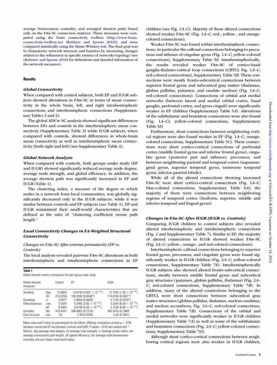
Global Network AnalysisWhen compared with controls, both groups under study (EPand IUGR) showed significantly reduced average node degree,average node strength, and global efficiency. In addition, theaverage shortest path was significantly increased in EP andIUGR (Table 1).
The clustering index, a measure of the degree to whichnodes in a network form local communities, was globally sig-nificantly decreased only in the IUGR subjects, while it wassimilar between controls and EP subjects (see Table 1). EP andIUGR maintained their small-world characteristics that aredefined as the ratio of “clustering coefficient versus pathlength.”
Local Connectivity Changes in FA-Weighted StructuralConnectivity
Changes in FAw-SC After extreme prematurity (EP vs.Controls)The local analysis revealed pairwise FAw-SC alterations in bothinterhemispheric and intrahemispheric connections in EP
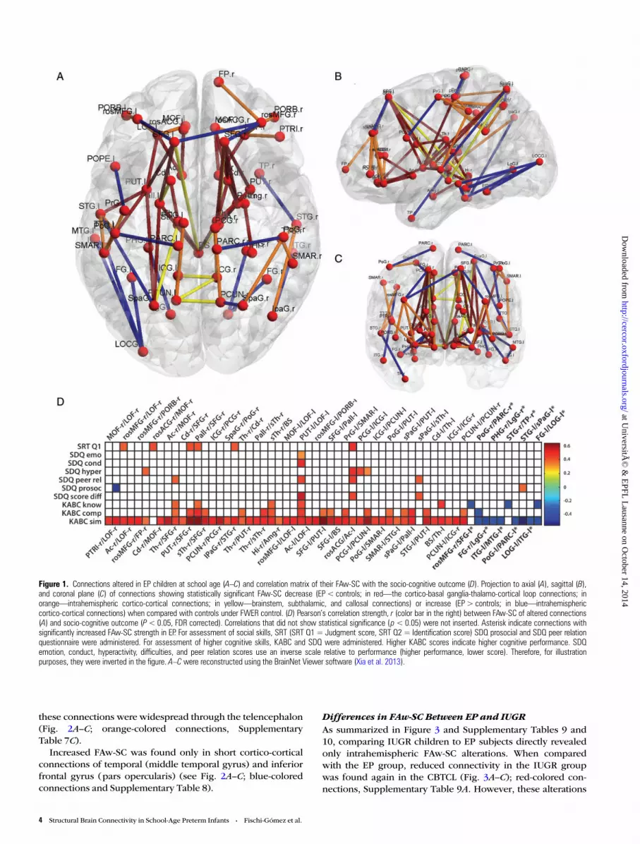
children (see Fig. 1A–C). Majority of these altered connectionsshowed weaker FAw-SC (Fig. 1A–C; red-, yellow-, and orange-colored connections).
Weaker FAw-SC was found within interhemispheric connec-tions, in particular the callosal connections belonging to precu-neus and isthmus of cingulate gyrus (Fig. 1A–C; yellow-coloredconnections), Supplementary Table 5E. Intrahemispherically,the results revealed weaker FAw-SC of cortico-basalganglia-thalamo-cortical loop connections (CBTCL Fig. 1A–C;red-colored connections), Supplementary Table 5B. These con-nections were mostly fronto-subcortical connections betweensuperior frontal gyrus and subcortical gray matter (thalamus,globus pallidus, putamen, and caudate nucleus) (Fig. 1A–C;red-colored connections). Connections of orbital and medialnetworks (between lateral and medial orbital cortex, basalganglia, prefrontal cortex, and gyrus cinguli) were significantlyweaker in EP children (Supplementary Table 5A). Alterationsof the subthalamic and brainstem connections were also found(Fig. 1A–C); yellow-colored connections, SupplementaryTable 5D.
Furthermore, short connections between neighboring corti-cal regions were also found weaker in EP (Fig. 1A–C; orange-colored connections, Supplementary Table 5C). These connec-tions were short cortico-cortical connections of prefrontalcortex (middle frontal gyrus and inferior frontal gyrus), cingu-late gyrus (posterior part and isthmus), precuneus, andbetween neighboring parietal and temporal cortex (supramar-ginal gyrus, superior temporal gyrus, transverse temporalgyrus, inferior parietal lobule).
While all of the altered connections showing increasedFAw-SC were short cortico-cortical connections (Fig. 1A–C;blue-colored connections, Supplementary Table 6A), themajority of them were connections between neighboringregions of temporal cortex (fusiform, superior, middle andinferior temporal and lingual gyrus).
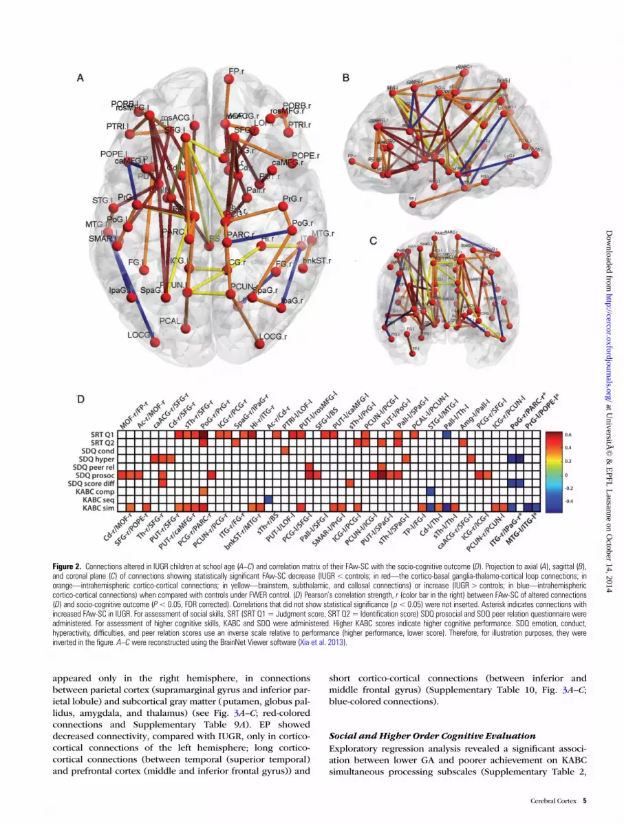
Changes in FAw-SC After IUGR (IUGR vs. Controls)Comparing IUGR children to control subjects also revealedaltered interhemispheric and intrahemispheric connections(Fig. 2 and Supplementary Table 7). Similar to EP, the majorityof altered connections in IUGR showed weaker FAw-SC(Fig. 2A–C; yellow-, orange-, and red-colored connections).
Interhemispheric callosal connections belonging to superiorfrontal gyrus, precuneus, and cingulate gyrus were found sig-nificantly weaker in IUGR children (Fig. 2A–C; yellow-coloredconnections, Supplementary Table 7E). IntrahemisphericallyIUGR subjects also showed altered fronto-subcortical connec-tions, mostly between middle frontal gyrus and subcorticalgray structures (putamen, globus pallidus, thalamus) (Fig. 2A–C; red-colored connections, Supplementary Table 7B). Inaddition, many of the altered connections belonging to theCBTCL were short connections between subcortical graymatter structures (globus pallidus, thalamus, nucleus caudatus,and nucleus accumbens, Fig. 2A–C; red-colored connections,Supplementary Table 7B). Connections of the orbital andmedial networks were significantly weaker in IUGR children(Supplementary Table 7A) as well as some of the subthalamicand brainstem connections (Fig. 2A–C; yellow-colored connec-tions, Supplementary Table 7D).
Although short cortico-cortical connections between neigh-boring cortical regions were also weaker in IUGR children,
Table 1Global network metrics comparison for each group under study
Global networkmeasures
Control EP IUGR
Similarity Deg 15.9804 14.8743 (4.887 × 10⁻⁵*) 15.7292 (1.28 × 10⁻⁴*)Str 213.0259 193.2092 (0.0048*) 174.8194 (0.0001*)
Clustering ci 5.9477 5.9050 (0.9820) 5.1150 (0.0233*)Paths/distances Lam 0.2024 0.2465 (3.65 × 10⁻⁵*) 0.2554 (6.08 × 10⁻⁵*)
eff 6.9464 6.0749 (4.39 × 10⁻⁵*) 5.7536 (6.98 × 10⁻⁵*)Centrality bet 418.4307 396.6862 (0.2316) 393.5479 (0.1960)Core structure swi 2.9 2.39 (0.4295) 2.36 (0.2941)
Mean value and P-value (in parenthesis) for the Mann–Whitney comparison at level α= 0.05between control and EP and between controls and IUGR. P-values <0.05 are marked with *.Metrics: deg (average node degree), str (average node strength), ci (average cluster index), lam(average characteristic path length), eff (global efficiency), bet (average node betweennesscentrality, and swi (mean small-world index).
Cerebral Cortex 3
at UniversitÃ
© &
EPFL
Lausanne on O
ctober 14, 2014http://cercor.oxfordjournals.org/
Dow
nloaded from
these connections were widespread through the telencephalon(Fig. 2A–C; orange-colored connections, SupplementaryTable 7C).
Increased FAw-SC was found only in short cortico-corticalconnections of temporal (middle temporal gyrus) and inferiorfrontal gyrus (pars opercularis) (see Fig. 2A–C; blue-coloredconnections and Supplementary Table 8).
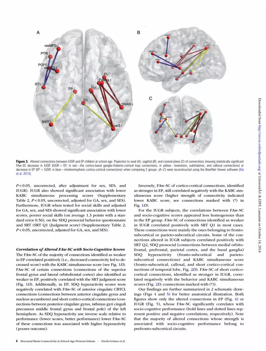
Differences in FAw-SC Between EP and IUGRAs summarized in Figure 3 and Supplementary Tables 9 and10, comparing IUGR children to EP subjects directly revealedonly intrahemispheric FAw-SC alterations. When comparedwith the EP group, reduced connectivity in the IUGR groupwas found again in the CBTCL (Fig. 3A–C); red-colored con-nections, Supplementary Table 9A. However, these alterations
Figure 1. Connections altered in EP children at school age (A–C) and correlation matrix of their FAw-SC with the socio-cognitive outcome (D). Projection to axial (A), sagittal (B),and coronal plane (C) of connections showing statistically significant FAw-SC decrease (EP < controls; in red—the cortico-basal ganglia-thalamo-cortical loop connections; inorange—intrahemispheric cortico-cortical connections; in yellow—brainstem, subthalamic, and callosal connections) or increase (EP > controls; in blue—intrahemisphericcortico-cortical connections) when compared with controls under FWER control. (D) Pearson’s correlation strength, r (color bar in the right) between FAw-SC of altered connections(A) and socio-cognitive outcome (P< 0.05, FDR corrected). Correlations that did not show statistical significance (p< 0.05) were not inserted. Asterisk indicate connections withsignificantly increased FAw-SC strength in EP. For assessment of social skills, SRT (SRT Q1= Judgment score, SRT Q2 = Identification score) SDQ prosocial and SDQ peer relationquestionnaire were administered. For assessment of higher cognitive skills, KABC and SDQ were administered. Higher KABC scores indicate higher cognitive performance. SDQemotion, conduct, hyperactivity, difficulties, and peer relation scores use an inverse scale relative to performance (higher performance, lower score). Therefore, for illustrationpurposes, they were inverted in the figure. A–C were reconstructed using the BrainNet Viewer software (Xia et al. 2013).
4 Structural Brain Connectivity in School-Age Preterm Infants • Fischi-Gómez et al.
at UniversitÃ
© &
EPFL
Lausanne on O
ctober 14, 2014http://cercor.oxfordjournals.org/
Dow
nloaded from
appeared only in the right hemisphere, in connectionsbetween parietal cortex (supramarginal gyrus and inferior par-ietal lobule) and subcortical gray matter (putamen, globus pal-lidus, amygdala, and thalamus) (see Fig. 3A–C; red-coloredconnections and Supplementary Table 9A). EP showeddecreased connectivity, compared with IUGR, only in cortico-cortical connections of the left hemisphere; long cortico-cortical connections (between temporal (superior temporal)and prefrontal cortex (middle and inferior frontal gyrus)) and
short cortico-cortical connections (between inferior andmiddle frontal gyrus) (Supplementary Table 10, Fig. 3A–C;blue-colored connections).
Social and Higher Order Cognitive EvaluationExploratory regression analysis revealed a significant associ-ation between lower GA and poorer achievement on KABCsimultaneous processing subscales (Supplementary Table 2,
Figure 2. Connections altered in IUGR children at school age (A–C) and correlation matrix of their FAw-SC with the socio-cognitive outcome (D). Projection to axial (A), sagittal (B),and coronal plane (C) of connections showing statistically significant FAw-SC decrease (IUGR< controls; in red—the cortico-basal ganglia-thalamo-cortical loop connections; inorange—intrahemispheric cortico-cortical connections; in yellow—brainstem, subthalamic, and callosal connections) or increase (IUGR> controls; in blue—intrahemisphericcortico-cortical connections) when compared with controls under FWER control. (D) Pearson’s correlation strength, r (color bar in the right) between FAw-SC of altered connections(D) and socio-cognitive outcome (P<0.05, FDR corrected). Correlations that did not show statistical significance (p< 0.05) were not inserted. Asterisk indicates connections withincreased FAw-SC in IUGR. For assessment of social skills, SRT (SRT Q1= Judgment score, SRT Q2 = Identification score) SDQ prosocial and SDQ peer relation questionnaire wereadministered. For assessment of higher cognitive skills, KABC and SDQ were administered. Higher KABC scores indicate higher cognitive performance. SDQ emotion, conduct,hyperactivity, difficulties, and peer relation scores use an inverse scale relative to performance (higher performance, lower score). Therefore, for illustration purposes, they wereinverted in the figure. A–C were reconstructed using the BrainNet Viewer software (Xia et al. 2013).
Cerebral Cortex 5
at UniversitÃ
© &
EPFL
Lausanne on O
ctober 14, 2014http://cercor.oxfordjournals.org/
Dow
nloaded from
P < 0.05, uncorrected, after adjustment for sex, SES, andIUGR). IUGR also showed significant association with lowerKABC simultaneous processing scores (SupplementaryTable 2, P < 0.05, uncorrected, adjusted for GA, sex, and SES).Furthermore, IUGR when tested for social skills and adjustedfor GA, sex, and SES showed significant association with lowerscores, poorer social skills (on average 1.3 points with a stan-dard error 0.56), on the SDQ prosocial behavior questionnaireand SRT (SRT Q1 (Judgment score) (Supplementary Table 2,P < 0.05, uncorrected, adjusted for GA, sex, and SES).
Correlation of Altered FAw-SC with Socio-Cognitive ScoresThe FAw-SC of the majority of connections identified as weakerin EP correlated positively (i.e., decreased connectivity led to de-creased score) with the KABC simultaneous score (see Fig. 1D).FAw-SC of certain connections (connections of the superiorfrontal gyrus and lateral orbitofrontal cortex) also identified asweaker in EP, positively correlated with the SRT judgment score(Fig. 1D). Additionally, in EP, SDQ hyperactivity scores werenegatively correlated with FAw-SC of anterior cingulate CBTCLconnections (connections between anterior cingulate gyrus andnucleus accumbens) and short cortico-cortical connections (con-nections between posterior cingulate gyrus, isthmus gyri cinguliprecuneus middle frontal gyrus and frontal pole) of the lefthemisphere. As SDQ hyperactivity use inverse scale relative toperformance (lower scores, better performance) lower FAw-SCof these connections was associated with higher hyperactivity(poorer outcome).
Inversely, FAw-SC of cortico-cortical connections, identifiedas stronger in EP, still correlated negatively with the KABC sim-ultaneous score (higher strength of connectivity indicatedlower KABC score, see connections marked with (*) inFig. 1D).
For the IUGR subjects, the correlations between FAw-SCand socio-cognitive scores appeared less homogenous thanin the EP group. FAw-SC of connections identified as weakerin IUGR correlated positively with SRT Q1 in most cases.These connections were mainly the ones belonging to fronto-subcortical or parieto-subcortical circuits. Some of the con-nections altered in IUGR subjects correlated positively withSRT Q2, SDQ prosocial (connections between medial orbito-frontal, prefrontal, parietal cortex, and the basal ganglia)SDQ hyperactivity (fronto-subcortical and parieto-subcortical connections) and KABC simultaneous score(fronto-subcortical, callosal, and short cortico-cortical con-nections of temporal lobe, Fig. 2D). FAw-SC of short cortico-cortical connections, identified as stronger in IUGR, corre-lated negatively with the behavior and KABC simultaneousscores (Fig. 2D; connections marked with (*)).
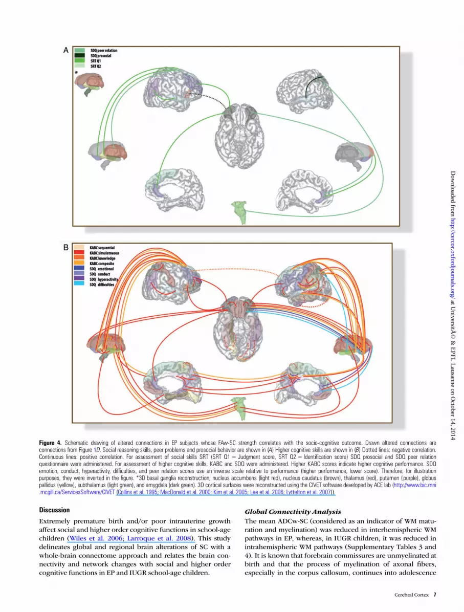
Our findings are further summarized in 2 schematic draw-ings (Figs 4 and 5) for better anatomical illustration. Bothfigures show only the altered connections in EP (Fig. 4) orIUGR (Fig. 5), whose FAw-SC significantly correlates withsocio-cognitive performance (bold lines and dotted lines rep-resent positive and negative correlations, respectively). Notethat the majority of altered connections whose strength isassociated with socio-cognitive performance belong toprefronto-subcortical circuits.
Figure 3. Altered connections between IUGR and EP children at school age. Projection to axial (A), sagittal (B), and coronal plane (C) of connections showing statistically significantFAw-SC decrease in IUGR (IUGR< EP; in red—the cortico-basal ganglia-thalamo-cortical loop connections; in yellow—brainstem, subthalamic, and callosal connections) ordecrease in EP (EP < IUGR; in blue—intrahemispheric cortico-cortical connections) when comparing 2 groups. (A–C) were reconstructed using the BrainNet Viewer software (Xiaet al. 2013).
6 Structural Brain Connectivity in School-Age Preterm Infants • Fischi-Gómez et al.
at UniversitÃ
© &
EPFL
Lausanne on O
ctober 14, 2014http://cercor.oxfordjournals.org/
Dow
nloaded from
Discussion
Extremely premature birth and/or poor intrauterine growthaffect social and higher order cognitive functions in school-agechildren (Wiles et al. 2006; Larroque et al. 2008). This studydelineates global and regional brain alterations of SC with awhole-brain connectome approach and relates the brain con-nectivity and network changes with social and higher ordercognitive functions in EP and IUGR school-age children.
Global Connectivity AnalysisThe mean ADCw-SC (considered as an indicator of WM matu-ration and myelination) was reduced in interhemispheric WMpathways in EP, whereas, in IUGR children, it was reduced inintrahemispheric WM pathways (Supplementary Tables 3 and4). It is known that forebrain commissures are unmyelinated atbirth and that the process of myelination of axonal fibers,especially in the corpus callosum, continues into adolescence
Figure 4. Schematic drawing of altered connections in EP subjects whose FAw-SC strength correlates with the socio-cognitive outcome. Drawn altered connections areconnections from Figure 1D. Social reasoning skills, peer problems and prosocial behavior are shown in (A) Higher cognitive skills are shown in (B) Dotted lines: negative correlation.Continuous lines: positive correlation. For assessment of social skills SRT (SRT Q1= Judgment score, SRT Q2 = Identification score) SDQ prosocial and SDQ peer relationquestionnaire were administered. For assessment of higher cognitive skills, KABC and SDQ were administered. Higher KABC scores indicate higher cognitive performance. SDQemotion, conduct, hyperactivity, difficulties, and peer relation scores use an inverse scale relative to performance (higher performance, lower score). Therefore, for illustrationpurposes, they were inverted in the figure. *3D basal ganglia reconstruction; nucleus accumbens (light red), nucleus caudatus (brown), thalamus (red), putamen (purple), globuspallidus (yellow), subthalamus (light green), and amygdala (dark green). 3D cortical surfaces were reconstructed using the CIVET software developed by ACE lab (http://www.bic.mni.mcgill.ca/ServicesSoftware/CIVET (Collins et al. 1995; MacDonald et al. 2000; Kim et al. 2005; Lee et al. 2006; Lyttelton et al. 2007)).
Cerebral Cortex 7
at UniversitÃ
© &
EPFL
Lausanne on O
ctober 14, 2014http://cercor.oxfordjournals.org/
Dow
nloaded from
(Keshavan 2002). Thus, our results suggest that reduced meanADCw-SC of interhemispheric connections in EP reflectchanges in the maturation of callosal fibers. These results arein agreement with previously reported DTI studies showingmicrostructural WM changes of major WM fiber tracts associ-ated with premature birth (for review see Ment et al. 2009).
Global mean FA was significantly altered for interhemi-spheric as well as for intrahemispheric SC in both groups (EPand IUGR, Supplementary Tables 3 and 4). This provides newevidence that alterations of WM maturation associated withpremature birth are present not only, as previously demon-strated, at term equivalent age (Huppi et al. 1998; Hüppi and
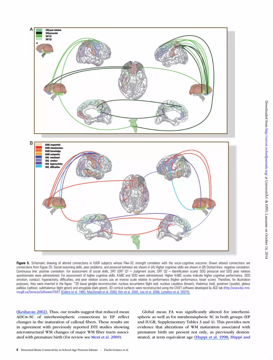
Figure 5. Schematic drawing of altered connections in IUGR subjects whose FAw-SC strength correlates with the socio-cognitive outcome. Drawn altered connections areconnections from Figure 2D. Social reasoning skills, peer problems, and prosocial behavior are shown in (A) Higher cognitive skills are shown in (B) Dotted lines: negative correlation.Continuous line: positive correlation. For assessment of social skills, SRT (SRT Q1= Judgment score, SRT Q2 = Identification score) SDQ prosocial and SDQ peer relationquestionnaire were administered. For assessment of higher cognitive skills, KABC and SDQ were administered. Higher KABC scores indicate higher cognitive performance. SDQemotion, conduct, hyperactivity, difficulties, and peer relation scores use an inverse scale relative to performance (higher performance, lower score). Therefore, for illustrationpurposes, they were inverted in the figure. *3D basal ganglia reconstruction; nucleus accumbens (light red), nucleus caudatus (brown), thalamus (red), putamen (purple), globuspallidus (yellow), subthalamus (light green) and amygdala (dark green). 3D cortical surfaces were reconstructed using the CIVET software developed by ACE lab (http://www.bic.mni.mcgill.ca/ServicesSoftware/CIVET (Collins et al. 1995; MacDonald et al. 2000; Kim et al. 2005; Lee et al. 2006; Lyttelton et al. 2007)).
8 Structural Brain Connectivity in School-Age Preterm Infants • Fischi-Gómez et al.
at UniversitÃ
© &
EPFL
Lausanne on O
ctober 14, 2014http://cercor.oxfordjournals.org/
Dow
nloaded from
Dubois 2006), but also persist upon reaching school age. Fur-thermore, our results show that poor fetal conditions leadingto IUGR provoke structural abnormalities similar to ones seenin EP, with high impact on their socio-cognitive potential.
Global Network PropertiesComplex systems such as the brain show remarkably similarmacroscopic behavior, despite profound differences at themicroscopic level (Alexander-Bloch et al. 2013). Neverthe-less, altered maturation of axonal pathways could lead tochanges in global brain network properties. Indeed, evenif the graph analysis demonstrated that both EP and IUGRmaintained the network small-world characteristic, theyshowed distinct differences in networks properties. Signifi-cantly lower average node metrics (average node degree andaverage node strength (Table 1)) suggest that anatomicalregions in EP and IUGR subjects generally have less well-organized global networks leading to lower efficiency(Table 1). Additionally, the longer shortest path lengths in EPand IUGR (Table 1) subjects confirm the loss of efficiency anddelay in maturation, as shortest path length measures are ex-pected to decrease with age during childhood (Hagmann,Sporns, et al. 2010). Moreover, IUGR children showed a lowerclustering index, indicating less local communities or hubs,and therefore less segregation. Since the clustering indexdoes not usually change during development, this indicates apermanent change in IUGR subjects, rather than delayedmaturation only.
Throughout development, the human brain tends to locallyfavor dense communication, minimize path lengths, and in-crease specialization and segregation of brain areas (Khun-drakpam et al. 2013). Our graph analysis results show thatextreme premature birth and/or prenatal growth restrictionreduce this aforementioned tendency.
Local Connectivity AnalysisDuring development, major WM fiber systems display specificspatio-temporal maturation (Vasung et al. 2010). The matu-ration of thalamo-cortical fibers and striatal fibers of theCBTCL is followed by the maturation of the associational long(intrahemispheric and interhemispheric) and short cortico-cortical fibers (Kostovic and Jovanov-Milosevic 2006; Vasunget al. 2010; Kostovic et al. 2014).
Our results indicate that early maturing fiber systems (e.g.,thalamo-cortical fibers, striatal fibers) have decreased FAw-SC atschool age in EP and IUGR children (Figs 1 and 2). Contrarily,late maturing fiber systems (mostly the associational cortico-cortical fibers) have both decreased (Figs 1A–C and 2A–C;orange-colored connections) and increased FAw-SC (Figs 1A–Cand 2A–C; blue-colored connections). Such differences in FAw-SC strength suggest different vulnerability and developmentaltrajectories of the aforementioned fiber systems.
Connections with Weaker FAw-SC
Cortico-Basal Ganglia-Thalamo-Cortical LoopThe CBTCL is a model of the functional and anatomical organ-ization of cortico-basal ganglia-thalamo-cortical pathways(Cummings 1993) that play an important role in human behav-ior. In this study, connectivity alterations in the CBTCL connec-tions were common in both groups (EP and IUGR) whencompared with controls (see Figs 1A–C and 2A–C; red-colored
connections, and Supplementary Tables 5A,B and 7A,B) andwere linked with the level of their socio-cognitive performance(Figs 4 and 5).
The relationship between the cortex, the basal ganglia, andthe thalamus in terms of information processing is complexand has been an area of scientific debate for years (Goldman-Rakic and Selemon 1990). Basal ganglia receive inputs fromthe majority of cortical areas. This flux of information is thentransferred to the thalamus that serves as a relay for the basalganglia projections back to the cortex (Alexander et al. 1986;Cummings 1993, 1995, 1998; Mega and Cummings 1994;Parent and Hazrati 1995; Takada et al. 1998; Gallay et al. 2008).Recent imaging data using probabilistic tractography demon-strated the structural basis of the CBTCL at the global level(Draganski et al. 2008). The parallel organization of cortico-striatal circuits belonging to the CBTCL allows simultaneousprocessing of cognitive, sensorimotor, and motivational infor-mation (Alexander and Crutcher 1990; Groenewegen et al.2009). Our results suggest that weaker connections withinthis circuit might be a biological blueprint of less efficient sim-ultaneous information processing seen in EP and IUGR chil-dren (Figs 1D, 2D, 4B and 5B). In addition, the connectivityalterations that we found in EP and IUGR suggest that the mostvulnerable part of this loop consists of the components belong-ing to the prefronto-subcortical and limbic circuits (Figs 1A–Cand 2A–C; red-colored connections, Supplementary Tables 5A,B and 7A,B). The prefronto-subcortical circuits are referred toas major mediators of the executive, social, and motivatedhuman behavior (Cummings 1993, 1995, 1998; Parent andHazrati 1995), shown to be affected by premature birth(Nosarti et al. 2010). Moreover, these circuits have also beenimplicated in psychiatric disorders (Mega and Cummings1994) which are known to occur with higher prevalence inprematurely born children (Nosarti et al. 2010) and individualswith low BW.
Orbitofrontal Circuits of the CBTCLDifferent connections belonging to orbital and medial networkcircuitry were found to be weaker in EP and IUGR children(Figs 1A–C and 2A–C; red-colored connections, SupplementaryTables 5A and 7A).
The orbital network serves as a system for sensory integrationwhile the medial prefrontal network plays an important role inreward-related learning, extinction, and reality check and fanta-sies, or to keep thought and behavior in phase with reality(Ongur and Price 2000; Schnider 2008). A neuroimaging studyin adolescents associated the alteration of orbitofrontal sulcaldepth with prematurity (Gimenez et al. 2006), suggestingaltered maturation of orbitofrontal cortex after premature birth.In recent years, alterations of the orbitofrontal cortex havefurther been associated with impaired social behavior, poorreward-based decision making and disturbances in emotionalprocessing. Our results support these findings since in EP andIUGR children weaker strength of connections belonging tomedial and orbital networks (Figs 1A–C and 2A–C) correlatedwith deficits in recognition of social context, social recognitionabilities, simultaneous processing, and hyperactivity (Figs 4and 5). Therefore, our results suggest that these networks mightbe responsible for lower social competences seen in prematurelyborn children (Ross et al. 1990).
Cerebral Cortex 9
at UniversitÃ
© &
EPFL
Lausanne on O
ctober 14, 2014http://cercor.oxfordjournals.org/
Dow
nloaded from
CBTCL Alteration in Relation to InjuryPrimary lesions from acquired insults that are associated withpremature birth were extensively studied, for review see Volpe(2009). The predilection sights of these lesions are periventri-cular regions. “Periventricular crossroad areas”, situated inperiventricular regions, have been recently described as beingcrucial for proper navigation and growth of axonal pathwaysduring human brain development (Judas et al. 2005). Duringfetal development, an abundant periventricular fiber pathway(PVP) (Vasung et al. 2011) is situated within these areas. Sincethe PVP is made of the fronto-occipital fascicle, the corpus cal-losum, the fronto-pontine pathway, and the Muratoff fascicle(major route for cortico-striatal fibers (Schmahmann andPandya 2009)) (Vasung et al. 2011), we suspect that discreteinjury in these “periventricular crossroad areas” might lead toaltered callosal (Supplementary Tables 5E and 7E), brainstem(Supplementary Tables 5D and 7D), and cortico-striatal con-nectivity (CBTC, Supplementary Tables 5A,B and 7A,B).
Additionally, Pierson et al. (2007) reported that primaryinjury of the WM does not happen in isolation, but it is associ-ated with significant neuronal loss and/or gliosis within thebasal ganglia and thalamus. Neuroimaging studies have re-cently shown decreased volumes of thalamus and weakerthalamo-cortical connections in prematurely born infants (Ballet al. 2012). Therefore, discrete injury of periventricularregions affecting the major route for cortico-striatal fibres (theMurratoff fascicle) and neuronal loss within basal gangliacould partially explain the weaker connectivity within theCBTCL that we found in EP and IUGR preterm children(Figs 1A–C and 2A–C; red-colored connections, SupplementaryTables 5 and 7B).
Short Associational Cortico-Cortical Connections
Language-Associated AreasComparing IUGR to EP revealed that EP children displayweaker FAw-SC in connections belonging to regions associatedwith language processing, namely pars opercularis and parstriangularis of inferior frontal gyrus (Fig. 3A–C; blue-coloredconnections, Supplementary Table 10).
It has been shown that low BW and extreme prematurity areassociated with poorer language skills at almost all levels, fromexpressive to receptive language, phonological awareness, dis-course, semantics, and pragmatics (Foster-Cohen et al. 2007;Marston et al. 2007; Wolke et al. 2008; Barre et al. 2011; Ramon-Casas et al. 2013). Furthermore, recent imaging findings,showing connectivity alterations of similar areas as in thecurrent study, suggest that these children recruit alternate corti-cal regions for language processing (Gozzo et al. 2009). Thus,our results provide further evidence of altered (decreased) SCbetween cortical areas relevant for language processing(Figs 1A–C and 2A–C; orange-colored connections, Supplemen-tary Tables 5C and 7C).
PrecuneusOne of the most striking alterations was found in cortico-cortical connections between the precuneus and neighboringregions (Figs 1A–C and 2A–C; orange-colored connections,Supplementary Tables 5C and 7C). The precuneus has been re-ported to have a major role in a broad spectrum of complexcognitive tasks, including visuo-spatial imagery, episodicmemory retrieval, and self-processing.
However, during the resting state of the brain, the precu-neus and the posterior regions of the medio-sagittal cortexshow synchronous activity patterns and have been referred toas being part of the “default mode network” (DMN) function-ally linked to self-consciousness (Cavanna and Trimble 2006).DMN is known to appear later in development of preterminfants (Smyser et al. 2010). Our results of reduced connec-tivity between these 2 major hubs of the DMN at 6 years of life(posterior cingulate gyrus and posterior precuneus) suggestsalteration of this basic structural and functional networkimportant for functional integration of information processing(Hagmann et al. 2007, 2008). Significant correlation with thecognitive outcome provided evidence that weaker strength ofconnections belonging to these circuits might contribute totheir less efficient simultaneous information processing, poorrecognition of social context, and poor prosocial behavior(Figs 1D, 2D, 4, and 5).
Connections with Stronger FAw-SC
Short Associational Cortico-Cortical ConnectionsAlthough human brain develops in distinct stages of geneticactivity (Pletikos et al. 2014), experience-dependent structuralchanges are generally described within the GM. Thesechanges, associated with synaptogenesis and dendritic arbori-zation (Petanjek et al. 2011), contribute to functional plasticity(Volkmar and Greenough 1972; Turner and Greenough 1985).Training and rehabilitation are further known to affect thecortico-cortical rewiring, and recent DTI data show that train-ing induces changes in WM architecture (Scholz et al. 2009). Inour study, all connections with significantly increased FAw-SCin EP and IUGR were short cortico-cortical connections(Figs 1A–C and 2A–C; blue-colored connections). Nevertheless,none of these connections were associated with improvedmeasures of the socio-cognitive outcome. On the contrary,they were associated with less efficient simultaneous proces-sing and higher rates of hyperactivity (Figs 1D, 2D, 4, and 5).Thus, our results indicate that this increase in connectivitystrength does not necessarily mean a better socio-cognitiveperformance but might occur in order to compensate weakerparts of network circuitry.
Implications for Understanding Cognition and BehaviorWhen reaching school age, EP children show deficits in motor,sensory and executive functioning (Marlow et al. 2007). More-over, they display problems in processing complex infor-mation that needs logical reasoning and spatial orientationabilities (Larroque et al. 2008). As these cognitive deficits havea similar incidence in EP and IUGR preterm infants (bornbetween 29 and 32 weeks) (Guellec et al. 2011), similar con-nectivity alterations found in the EP and IUGR subjects speaksin favor of their similar neural substrate. The current analysistherefore suggests that the alterations in the CBTCL and theshort cortico-cortical connections following preterm birth andIUGR might contribute to poorer prosocial behavior, recog-nition of social context, and simultaneous information proces-sing (Figs 4 and 5).
Strengths and Limitations of the StudySome limitations of the current study need to be taken intoconsideration. First, the small cohort of subjects, the highervariability between individuals, and the large number of
10 Structural Brain Connectivity in School-Age Preterm Infants • Fischi-Gómez et al.
at UniversitÃ
© &
EPFL
Lausanne on O
ctober 14, 2014http://cercor.oxfordjournals.org/
Dow
nloaded from
comparisons were overcome using a new two-step method-ology that exploits the data structure and information of posi-tive dependence to increase power of testing. Second, DTI waschosen instead of other dMRI acquisition schemes (HARDI orDSI) because subjects were young children requiring shorteracquisition time. Third, DTI reconstruction and tractographyare sensitive to motion artifacts and both present limitationsfor reconstruction of complex fiber configurations, likecrossing-fibers or kissing-fibers. A threshold on the densityvalues of each bundle (the mean FA value) was used to mini-mize possible artifacts due to the acquisition and tractographynoise. In that way, we reduced the number of residual connec-tion values. Validation of the tractography method and the con-struction of connectomes are not discussed here as bothaspects are extensively discussed elsewhere (Hagmann et al.2007; Cammoun et al. 2012).
Given the relatively small local cohort of extreme prematureinfants, the linear regression model testing the link betweenclinical risk factors and cognitive and behavioral measures hasthe inherent risk of Type I and Type II errors. Nevertheless,our results concur with published data from larger cohorts.
Although connectomics still present technical challenges,they remain an ideal complementary tool to assess structuralco-variance between brain regions on a global brain networkscale (Alexander-Bloch et al. 2013).
Conclusion
The new approach of whole-brain connectome analysis pro-posed here allowed to comprehensively study both long-ranging and short-ranging connectivity in preterm school-agechildren. The results suggest that EP and IUGR have altered SCof CBTCL connections that start their maturation early in devel-opment. SC alterations of the orbitofrontal cortex circuitry(responsible for reward learning, reality check, and socio-emotional processing) were evidenced by the whole-brain con-nectome approach. Specific short associational cortico-corticalconnections, developing later and most likely influenced byenvironmental sensory inputs, showed both diminished andincreased strength of connectivity between areas known toplay an important role in language, problem solving, andsocial behavior. The strength of altered connections wasassociated with their socio-cognitive performance. In con-clusion, the whole-brain connectivity analysis provides a linkbetween early life events (extreme prematurity and IUGR), cir-cuitry development, and specific socio-cognitive disabilities.
Supplementary MaterialSupplementary material can be found at: http://www.cercor.oxfordjournals.org/.
Funding
This work was supported by the Center for BiomedicalImaging (CIBM) of the Geneva and Lausanne Universities, theEcole Polytechnique Fédérale de Lausanne (EPFL) in Switzer-land, and the foundations Leenards and Louis-Jeantet, as wellas by the Swiss National Science Foundation (33CM30_140334and 32473B_135817) and Leenards Foundation Grant N° 2667to P.S.H. Methodological support was provided by the Clinical
Research Center, University of Geneva and Geneva UniversityHospitals (Combescure C, PhD).
NotesWe are grateful to the families that took part in the study and themedical staff who participated in the study. We thank Prof. Ivica Kosto-vic from Croatian Institute for Brain Research, Univesity of Zagreb,Croatia, and Prof. Dimitri Van de Ville from GR-VDV, EPFL, Switzer-land, for critical comments and suggestions; Laura Gui, PhD, for proof-reading and improving the quality of the manuscript. Conflict ofInterest: None declared.
ReferencesAlexander GE, Crutcher MD. 1990. Functional architecture of basal
ganglia circuits: neural substrates of parallel processing. TrendsNeurosci. 13:266–271.
Alexander GE, DeLong MR, Strick PL. 1986. Parallel organization offunctionally segregated circuits linking basal ganglia and cortex.Annu Rev Neurosci. 9(2):357–381.
Alexander-Bloch A, Giedd JN, Bullmore E. 2013. Imaging structuralco-variance between human brain regions. Nat Rev Neurosci.14:322–336.
Anderson PJ. 2004. Executive functioning in school-aged children whowere born very preterm or with extremely low birth weight in the1990s. Pediatrics. 114(1):50–57.
Ball G, Boardman JP, Rueckert D, Aljabar P, Arichi T, Merchant N,Gousias IS, Edwards AD, Counsell SJ. 2012. The effect of pretermbirth on thalamic and cortical development. Cereb Cortex.22:1016–1024.
Barisnikov K, Hippolyte L. 2011. Batterie d’évaluation de la cognitionsociale et émotionnelle. In: Nader-Grosbois N, editor. Théorie del’esprit: Entre cognition, émotion et adaptation sociale chez despersonnes typiques et atypiques Bruxelles (Belgium): De Boeck.pp 135–151.
Barre N, Morgan A, Doyle LW, Anderson PJ. 2011. Language abilities inchildren who were very preterm and/or very low birth weight: ameta-analysis. J Pediatr. 158:766–774 e761.
Bhutta AT. 2002. Cognitive and behavioural outcomes following verypreterm birth. A meta-analysis. JAMA. 288(6):728–736.
Bullmore E, Sporns O. 2009. Complex brain networks: graph theoreti-cal analysis of structural and functional systems. Nat Rev Neurosci.10:186–198.
Cammoun L, Gigandet X, Meskaldji D, Thiran JP, Sporns O, Do KQ,Maeder P, Meuli R, Hagmann P. 2012. Mapping the human connec-tome at multiple scales with diffusion spectrum MRI. J NeurosciMethods. 203:386–397.
Cavanna AE, Trimble MR. 2006. The precuneus: a review of its func-tional anatomy and behavioural correlates. Brain. 129:564–583.
Chen CH, Gutierrez ED, Thompson W, Panizzon MS, Jernigan TL, EylerLT, Fennema-Notestine C, Jak AJ, Neale MC, Franz CE et al. 2012.Hierarchical genetic organization of human cortical surface area.Science. 335:1634–1636.
Clark CA, Woodward LJ, Horwood LJ, Moor S. 2008. Development ofemotional and behavioral regulation in children born extremelypreterm and very preterm: biological and social influences. ChildDev. 79:1444–1462.
Collins D, Holmes C, Peters T, Evans A. 1995. Automatic 3D modelbased neuroanatomical segmentation. Hum Brain Mapp.3:190–208.
Counsell SJ, Edwards AD, Chew AT, Anjari M, Dyet LE, Srinivasan L,Boardman JP, Allsop JM, Hajnal JV, Rutherford MA et al. 2008.Specific relations between neurodevelopmental abilities and whitematter microstructure in children born preterm. Brain.131:3201–3208.
Cummings JL. 1995. Anatomic and behavioral aspects of frontal-subcortical circuits. Ann N Y Acad Sci. 769:1–13.
Cummings JL. 1993. Frontal-subcortical circuits and human behavior.Arch Neurol. 50(8):873–880.
Cerebral Cortex 11
at UniversitÃ
© &
EPFL
Lausanne on O
ctober 14, 2014http://cercor.oxfordjournals.org/
Dow
nloaded from
Cummings JL. 1998. Frontal-subcortical circuits and human behavior. JPsychosom Res. 44(6):627–682.
d’Acremont M, van der Linden M. 2008. Confirmatory factor analysis ofthe Strengths and Difficulties Questionnaire in a community sampleof French-speaking adolescents. Eur J Psychol Asses. 24:1.
Daducci A, Gerhard S, Griffa A, Lemkaddem A, Cammoun L, GigandetX, Meuli R, Hagmann P, Thiran J-P. 2012. The Connectome Mapper:an open-source processing pipeline to map connectomes with MRI.PLoS One. 7:e48121.
de Bie HMA, Oostrom KJ, de Wall HAD-v. 2010. Brain development, in-telligence and cognitive outcome in children born small for gesta-tional age. Horm Res Pediatr. 73:6–14.
Dennis EL, Jahanshad N, McMahon KL, de Zubicaray GI, Martin NG,Hickie IB, Toga AW, Wright MJ, Thompson PM. 2013. Developmentof brain structural connectivity between ages 12 and 30: a 4-Tesladiffusion imaging study in 439 adolescents and adults. Neuro-image. 64:671–684.
Draganski B, Kherif F, Kloppel S, Cook PA, Alexander DC, Parker GJ,Deichmann R, Ashburner J, Frackowiak RS. 2008. Evidence for seg-regated and integrative connectivity patterns in the human BasalGanglia. J Neurosci. 28:7143–7152.
Dubois J, Benders M, Borradori-Tolsa C, Cachia A, Lazeyras F, LeuchterRH-V, Sizonenko SV, Warfield SK, Mangin JF, Hüppi PS. 2008.Primary cortical folding in the human newborn: an early marker oflater functional development. Brain. 131(8):2028–2041.
Dubois J, Benders M, Cachia A, Lazeyras F, Ha-Vinh Leuchter R, Sizo-nenko SV, Borradori-Tolsa C, Mangin JF, Huppi PS. 2008. Mappingthe early cortical folding process in the preterm newborn brain.Cereb Cortex. 18:1444–1454.
Edmonds CJ, Isaacs EB, Cole TJ, Rogers MH, Lanigan J, Singhal A,Birbara T, Gringras P, Denton J, Lucas A. 2010. The effect of intrau-terine growth on verbal IQ scores in childhood: a study of monozy-gotic twins. Pediatrics. 126:e1095–e1101.
Foster-Cohen S, Edgin JO, Champion PR, Woodward LJ. 2007. Earlydelayed language development in very preterm infants: evidencefrom the MacArthur-Bates CDI. J Child Lang. 34:655–675.
Gallay MN, Jeanmonod D, Liu J, Morel A. 2008. Human pallidothalamicand cerebellothalamic tracts: anatomical basis for functional stereo-tactic neurosurgery. Brain Struct Funct. 212:443–463.
Gao W, Gilmore JH, Giovanello KS, Smith JK, Shen D, Zhu H, Lin W.2011. Temporal and spatial evolution of brain network topologyduring the first two years of life. PLoS One. 6:e25278.
Gimenez M, Junque C, Vendrell P, Narberhaus A, Bargallo N, Botet F,Mercader JM. 2006. Abnormal orbitofrontal development due toprematurity. Neurology. 67:1818–1822.
Goldman-Rakic PS, Selemon LD. 1990. New frontiers in basal gangliaresearch. Introduction. Trends Neurosci. 13:241–244.
Goodman R. 1997. The Strengths and Difficulties Questionnaire: a re-search note. J Child Psychol Psychiatry. 38:581–586.
Gozzo Y, Vohr B, Lacadie C, Hampson M, Katz KH, Maller-KesselmanJ, Schneider KC, Peterson BS, Rajeevan N, Makuch RW et al. 2009.Alterations in neural connectivity in preterm children at school age.Neuroimage. 48:458–463.
Groenewegen HJ, Voorn P, Berendse HW, Mulder AB, Cools AR. 2009.The Basal Ganglia IX. New York (NY): Springer.
Guellec I, Lapillonne A, Renolleau S, Charlaluk ML, Roze JC, Marret S,Vieux R, Monique K, Ancel PY. 2011. Neurologic outcomes atschool age in very preterm infants born with severe or mild growthrestriction. Pediatrics. 127:e883–e891.
Hagmann P, Cammoun L, Gigandet X, Gerhard S, Grant PE, Wedeen V,Meuli R, Thiran JP, Honey CJ, Sporns O. 2010. MR connectomics:Principles and challenges. J Neurosci Methods. 194:34–45.
Hagmann P, Cammoun L, Gigandet X, Meuli R, Honey CJ. 2008.Mapping human whole-brain structural networks with diffusionMRI. PLoS One. 6(7):e159.
Hagmann P, Kurant M, Gigandet X, Thiran P, Wedeen VJ, Meuli R,Thiran J-P. 2007. Mapping the structural core of human cerebralcortex. PLoS Biol. 7:e597.
Hagmann P, Sporns O, Madan N, Pieenar LCR, Weeden VJ, Meuli R,Thiran J-P, Grant PE. 2010. White matter maturation reshapes
structural connectivity in the late develop in human brain. Proc NatlAcad Sci USA. 107(44):19067–19072.
Hüppi PS, Dubois J. 2006. Diffusion tensor imaging of brain develop-ment. Semin Fetal Neonatal Med. 11:656–660.
Huppi PS, Maier SE, Peled S, Zientara GP, Barnes PD, Jolesz FA, VolpeJJ. 1998. Microstructural development of human newborn cerebralwhite matter assessed in vivo by diffusion tensor magnetic reson-ance imaging. Pediatr Res. 44:584–590.
Judas M, Rados M, Jovanov-Milosevic N, Hrabac P, Stern-Padovan R,Kostovic I. 2005. Structural, immunocytochemical, and MR imagingproperties of periventricular crossroads of growing cortical path-ways in preterm infants. AJNR Am J Neuroradiol. 26:2671–2684.
Kaufman AS, Kaufman NL. 1993. K-ABC: batterie pour l’examen psy-chologique de l’enfant. Paris (FR): Editions du Centre de Psycholo-gie Appliquée.
Keshavan MS, DeBellis M, Dick E, Kotwal R, Rosenberg DR, SweeneyJA, Minshew N, Pettegrew JW. 2002. Development of the corpuscallosum in childhood, adolescence and early adulthood. Life Sci.70(16):1909–1922.
Khundrakpam BS, Reid A, Brauer J, Carbonell F, Lewis J, Ameis S,Karama S, Lee J, Chen Z, Das S et al. Brain Development Coopera-tive G. 2013. Developmental changes in organization of structuralbrain networks. Cereb Cortex. 23:2072–2085.
Kim J, Singh V, MacDonald D, Lee J, Kim S, Evans A. 2005. Automated3D extraction and evaluation of the outer cortical surface using aLaplacian map and partial volume effect classification. Neuro-Image. 27:210–221.
Kostovic I, Jovanov-Milosevic N. 2006. The development of cerebralconnections during the first 20–45 weeks’ gestation. Semin FetalNeonatal Med. 11(6):415–422.
Kostovic I, Jovanov-Milosevic N, Rados M, Sedmak G, Benjak V,Kostovic-Srzentic M, Vasung L, Culjat M, Rados M, Huppi P et al.2014. Perinatal and early postnatal reorganization of the subplateand related cellular compartments in the human cerebral wall as re-vealed by histological and MRI approaches. Brain Struct Funct.219:231–253.
Largo R, Pfister D, Molinari L, Kundu S, Lipp A, Due G. 1989. Signifi-cance of prenatal, perinatal and postnatal factors in the develop-ment of AGA preterm infants at five to seven years. Dev Med ChildNeurol. 31:440–456.
Larroque B, Ancel PY, Marret S, Marchand L, Andre M, Arnaud C,Pierrat V, Roze JC, Messer J, Thiriez G et al. group ES. 2008. Neuro-developmental disabilities and special care of 5-year-old childrenborn before 33 weeks of gestation (the EPIPAGE study): a longi-tudinal cohort study. Lancet. 371:813–820.
Lee J, Lee J, Kim J, Kim I, Evans A, Kim S. 2006. A novel quantitativecross-validation of different cortical surface reconstructions algor-ithms using MRI phantom. NeuroImage. 31:572–584.
Limperopoulos C, Soul JS, Gauvreau K, Huppi PS, Warfield SK, BassanH, Robertson RL, Volpe JJ, du Plessis AJ. 2005. Late gestation cer-ebellar growth is rapid and impeded by premature birth. Pediatrics.115:688–695.
Lodygensky GA, Seghier ML, Warfield SK, Borradori-Tolsa C, SizonenkoS, Lazeyras F, Hüppi PS. 2008. Intrauterine growth restriction affectsthe preterm infant’s hippocampus. Pediatr Res. 63(4):438–443.
Lyttelton O, Boucher M, Robbins S, Evans AC. 2007. An unbiasediterative group registration template for cortical surface analysis.NeuroImage. 34:1535–1544.
Marlow N, Hennessy EM, Bracewell MA, Wolke D. 2007. Motor andexecutive function at 6 years of age after extremely preterm birth.Pediatrics. 120(4):793–804.
Marston L, Peacock JL, Calvert SA, Greenough A, Marlow N. 2007.Factors affecting vocabulary acquisition at age 2 in children bornbetween 23 and 28 weeks’ gestation. Dev Med Child Neurol.49:591–596.
MacDonald D, Kabani N, Avis D, Evans AC. 2000. Automated 3Dextraction of inner and outer surfaces of cerebral cortex from MRI.NeuroImage. 13:340–356.
Mega MS, Cummings JL. 1994. Frontal-subcortical circuits and neuropsy-chiatric disorders. J Neuropsychiatry Clin Neurosci. 6(4):358–370.
12 Structural Brain Connectivity in School-Age Preterm Infants • Fischi-Gómez et al.
at UniversitÃ
© &
EPFL
Lausanne on O
ctober 14, 2014http://cercor.oxfordjournals.org/
Dow
nloaded from
Ment LR, Hirtz D, Hüppi PS. 2009. Imaging biomarkers of outcome inthe developing preterm brain. Lancet Neurol. 8(11):1042–1055.
Meskaldji DE, Fischi-Gomez E, Griffa A, Hagmann P, Morgenthaler S,Thiran JP. 2013. Comparing connectomes across subjects andpopulations at different scales. Neuroimage. 80:416–425.
Meskaldji DE. 2013. Multiple comparison procedures for large corre-lated data with application to brain connectivity analysis. [PhDthesis], [Lausanne (Switzerland)]: Ecole Polytechnique Fédérale deLausanne.
Meskaldji DE, Ottet M-C, Cammoun L, Hagmann P, Meuli R, Eliez S,Thiran J-P, Morgenthaler S. 2011. Adaptive strategy for the statisti-cal analysis of connectomes. PLOS One. 6(8):e23009.
Nosarti C, Murray RM, Hack M. 2010. Neurodevelopmental outcomesof preterm birth: From childhood to adult life. Cambridge (GB):Cambridge University Press.
Ongur D, Price JL. 2000. The organization of networks within theorbital and medial prefrontal cortex of rats, monkeys and humans.Cereb Cortex. 10(3):206–219.
Parent A, Hazrati LN. 1995. Functional anatomy of the basalganglia. I. The cortico-basal ganglia-thalamo-cortical loop. BrainRes Brain Res Rev. 20(1):91–127.
Partridge SC, Mukherjee P, Henry RG, Miller SP, Berman JI, Jin H, Lu Y,Glenn OA, Ferriero DM, Barkovich AJ et al. 2004. Diffusion tensorimaging: serial quantitation of white matter tract maturity in prema-ture newborns. Neuroimage. 22:1302–1314.
Petanjek Z, Judas M, Simic G, Rasin MR, Uylings HB, Rakic P, KostovicI. 2011. Extraordinary neoteny of synaptic spines in the human pre-frontal cortex. Proc Natl Acad Sci US A. 108:13281–13286.
Pierson CR, Folkerth RD, Billiards SS, Trachtenberg FL, DrinkwaterME, Volpe JJ, Kinney HC. 2007. Gray matter injury associated withperiventricular leukomalacia in the premature infant. Acta Neuro-pathol. 114:619–631.
Pletikos M, Sousa AM, Sedmak G, Meyer KA, Zhu Y, Cheng F, Li M,Kawasawa YI, Sestan N. 2014. Temporal specification andbilaterality of human neocortical topographic gene expression.Neuron. 81:321–332.
Ramon-Casas M, Bosch L, Iriondo M, Krauel X. 2013. Word recognitionand phonological representation in very low birth weight preterms.Early Hum Dev. 89:55–63.
Ross G, Lipper EG, Auld PA. 1990. Social competence and behavior pro-blems in premature children at school age. Pediatrics. 86:391–397.
Rubinov M, Sporns O. 2010. Complex network measures of brain con-nectivity: uses and interpretations. Neuroimage. 52:1059–1069.
Schmahmann JD, Pandya D. 2009. Fiber pathways of the brain. Oxford(GB): Oxford University Press.
Schnider A. 2008. The confabulating mind: how the brain createsreality. New York: Oxford University Press.
Scholz J, Klein MC, Behrens TE, Johansen-Berg H. 2009. Traininginduces changes in white-matter architecture. Nat Neurosci.12:1370–1371.
Smyser CD, Indeer TE, Shimony JS, Hill JE, Degnan AJ, Snyder AZ, NeilJJ. 2010. Longitudinal analysisof neural network development inpreterm infants. Cereb Cortex. 20(12):2852–2862.
Takada M, Tokuno H, Nambu A, Inase M. 1998. Corticostriatal projec-tions from the somatic motor areas of the frontal cortex in themacaque monkey: segregation versus overlap of input zones fromthe primary motor cortex, the supplementary motor area, and thepremotor cortex. Exp Brain Res. 120:114–128.
Tolsa CB, Zimine S, Warfield SK, Freschi M, Rossignol AS, Lazeyras F,Hanquinet S, Pfizenmaier M, Hüppi PS. 2004. Early alteration ofstructural and functional brain development in premature infantsborn with intrauterine growth restriction. Pediatric Res. 56(1):132–138.
Treyvaud K, Ure A, Doyle LW, Lee KJ, Rogers CE, Kidokoro H, InderTE, Anderson PJ. 2013. Psychiatric outcomes at age seven for verypreterm children: rates and predictors. J Child Psychol Psychiatry.54:772–779.
Turner AM, Greenough WT. 1985. Differential rearing effects on ratvisual cortex synapses. I. Synaptic and neuronal density and sy-napses per neuron. Brain Res. 329:195–203.
Vasung L, Huang H, Jovanov-Milosevic N, Pletikos M, Mori S, KostovicI. 2010. Development of axonal pathways in the human fetalfronto-limbic brain: histochemical characterization and diffusiontensor imaging. J Anat. 217:400–417.
Vasung L, Jovanov-Milosevic N, Pletikos M, Mori S, Judas M, Kostovic I.2011. Prominent periventricular fiber system related to ganglioniceminence and striatum in the human fetal cerebrum. Brain StructFunct. 215:237–253.
Volkmar FR, Greenough WT. 1972. Rearing complexity affectsbranching of dendrites in the visual cortex of the rat. Science.172:1445–1447.
Volpe JJ. 2009. The encephalopathy of prematurity—brain injury andimpaired brain development inextricably intertwined. SeminPediatr Neurol. 16:167–178.
Wiles NJ, Peters TJ, Heron J, Gunnell D, Emond A, Lewis G. 2006. Fetalgrowth and childhood behavioral problems: results from theALSPAC cohort. Am J Epidemiol. 163:829–837.
Wolke D, Samara M, Bracewell M, Marlow N, Group EPS. 2008. Specificlanguage difficulties and school achievement in children born at 25weeks of gestation or less. J Pediatr. 152:256–262.
Woodward LJ, Anderson PJ, Austin NC, Howard K, Inder TE. 2006.Neonatal MRI to predict neurodevelopmental outcomes in preterminfants. N Engl J Med. 355:685–694.
World Health Organisation W. 2012. Born too soon: the global actionreport on preterm birth. In World Health Organisation Report.Geneva, Switzerland: World Health Organization. p 126.
Xia M, Wang J, He Y. 2013. BrainNet Viewer: a network visualizationtool for human brain connectomics. PLoS One. 8:e68910.
Yap PT, Fan Y, Chen Y, Gilmore JH, Lin W, Shen D. 2011. Developmenttrends of white matter connectivity in the first years of life. PLoSOne. 6:e24678.
Zacharia A, Ziminea S, Lovblada KO, Warfield S, Thoenyd H,Ozdobab C, Bossie E, Kreisf R, Boeschf C, Schrothb G et al. 2006.Early assessment of brain maturation by MR imaging segmenta-tion in neonates and premature infants. AJNR Am J Neuroradiol.27:972–977.
Cerebral Cortex 13
at UniversitÃ
© &
EPFL
Lausanne on O
ctober 14, 2014http://cercor.oxfordjournals.org/
Dow
nloaded from