Structural Asymmetries in the Infant Language and Sensori-Motor Networks
10
Cerebral Cortex doi:10.1093/cercor/bhn097 Structural Asymmetries in the Infant Language and Sensori-Motor Networks J. Dubois 1,2 , L. Hertz-Pannier 2,3,4,5 , A. Cachia 2,6,7,8 , J.F. Mangin 2,9 , D. Le Bihan 2,10 and G. Dehaene-Lambertz 2,11 1 CEA, UNAF, CEA/DSV/I2BM/Service Hospitalier Fre´de´ric Joliot, 91403 Orsay, France, 2 IFR49, 75013 Paris, France, 3 CEA, Laboratoire de recherche biome´dicale, CEA/SAC/DSV/I2BM/ NeuroSpin, 91191 Gif-sur-Yvette, France, 4 AP-HP, Radiologie Pe´diatrique, Hoˆpital Necker-Enfants Malades, 75013 Paris, France, 5 INSERM, U663, 75013 Paris, France; Universite´ Paris, 75013 Paris, France, 6 INSERM-CEA, U797, CEA/DSV/I2BM/ Service Hospitalier Fre´de´ric Joliot, 91403 Orsay, France, 7 Universite´ Paris-Sud, 91403 Orsay, France, 8 Universite´ Paris Descartes, 75013 Paris, France, 9 CEA, Laboratoire de neuroimagerie assiste´e par ordinateur, CEA/SAC/DSV/I2BM/ NeuroSpin, 91191 Gif-sur-Yvette, France, 10 CEA/SAC/DSV/I2BM/ NeuroSpin, 91191 Gif-sur-Yvette, France and 11 INSERM, U562, CEA/SAC/DSV/I2BM/NeuroSpin, 91191 Gif-sur-Yvette, France Both language capacity and strongly lateralized hand preference are among the most intriguing particularities of the human species. They are associated in the adult brain with functional and anatomical hemispheric asymmetries in the speech perception-- production network and in the sensori-motor system. Only studies in early life can help us to understand how such asymmetries arise during brain development, and to which point structural left--right differences are the source or the consequence of functional lateralization. In this study, we aimed to provide new in vivo structural markers of hemispheric asymmetries in infants from 1 to 4 months of age, with diffusion tensor imaging. We used 3 complementary analysis methods based on local diffusion indices and spatial localizations of tracts. After a prospective approach over the whole brain, we demonstrated early leftward asymmetries in the arcuate fasciculus and in the cortico-spinal tract. These results suggest that the early macroscopic geometry, microscopic organization, and maturation of these white matter bundles are related to the development of later functional lateralization. Keywords: development, DTI, infant, language, laterality, maturation, motor, white matter Introduction Among the most intriguing particularities of humans are their language capacity and strongly lateralized hand preference. These behavioral features are sustained by functional hemi- spheric asymmetries in the speech perception--production network and in the sensori-motor system. They are also associated in the adult brain with structural differences between the left and right hemispheres (for review Toga and Thompson 2003). However such studies cannot disentangle whether structural differences arise from functional asymmetries or whether they are biologically rooted tendencies that lead to specific behavioral features in the human species. Only de- velopmental studies can help us to decipher the temporal relationship between structural and functional asymmetries, by taking advantage of the progressive development during child- hood of lateralized functions such as language and handedness. Early infancy is thus an ideally suited time window to search for brain structural asymmetries that would precede the later behavioral expression of the corresponding lateralized functions. Infants initially use both hands indifferently (Corbetta and Thelen 1999; Ronnqvist and Domellof 2006), then preference for one hand becomes clear generally from 18 months of age on (Fagard and Marks 2000) and is more and more pronounced during the following years (Ingram 1975a). Lateralization of linguistic capacities is behaviorally assessed with dichotic listening. When different syllables are simultaneously presented to both ears, adults are better able to report the syllables presented in the right ear, which is interpreted as reflecting the left hemisphere advantage to process speech (Kimura 1967). As for handedness, asymmetry becomes more obvious with age and a right-ear advantage is robustly observed in children after 3 years of age (Ingram 1975b). Earlier behavioral signs of lateralization have been looked for in both domains. For example, Ottaviano et al. (1989) observed a right-hand preference in spontaneous activity of full-term newborns, and Bertoncini et al. (1989) reported a right-ear advantage for speech stimuli in neonates. However, these studies are sparse and their results were not always replicated (Best 1988). This can be due to difficulties to obtain behavioral evidence at a young age or to an initial fragile lateralization. The few existing brain imaging studies also suggest some early functional asymmetry (Dehaene-Lambertz, Hertz-Pannier, Dubois, 2006). The amplitude of event-related potentials to auditory stimuli is larger over the left hemisphere than the right in 2-month-old infants (Dehaene-Lambertz 2000), and the hemodynamic response to sentences of the native language is significantly asymmetric in the posterior superior temporal region in neonates (Pen˜a et al. 2003) and 3 month olds (Dehaene-Lambertz et al. 2002; Dehaene-Lambertz, Hertz- Pannier, Dubois, Meriaux, et al. 2006). Thus, although the asymmetry toward the left hemisphere for linguistic and motor tasks becomes more pronounced during childhood, some behavioral and brain imaging studies point out toward early functional differences between the left and right hemispheres. Are these early functional left-right differences sustained by corresponding structural differences? Actually, macroscopic left- right differences are present from the fetal life on. Post-mortem studies have shown that some cortical gyri, such as the superior frontal gyrus, the superior temporal gyrus and Heschl’s gyrus appear in the right hemisphere 1 or 2 weeks earlier than in the left (Chi et al. 1977), and a recent neuroimaging study in preterm newborns has revealed a right temporal sulcus larger than the left (Dubois et al. 2007). By contrast, the planum temporale and Heschl’s gyrus are larger on the left side in fetuses and infants (Witelson and Pallie 1973; Chi et al. 1977; Galaburda and Ó The Author 2008. Published by Oxford University Press. All rights reserved. For permissions, please e-mail: [email protected] Cerebral Cortex Advance Access published June 17, 2008
-
Upload
independent -
Category
Documents
-
view
0 -
download
0
Transcript of Structural Asymmetries in the Infant Language and Sensori-Motor Networks

Cerebral Cortex
doi:10.1093/cercor/bhn097
Structural Asymmetries in the InfantLanguage and Sensori-Motor Networks
J. Dubois1,2, L. Hertz-Pannier2,3,4,5, A. Cachia2,6,7,8, J.F. Mangin2,9,
D. Le Bihan2,10 and G. Dehaene-Lambertz2,11
1CEA, UNAF, CEA/DSV/I2BM/Service Hospitalier Frederic
Joliot, 91403 Orsay, France, 2IFR49, 75013 Paris, France, 3CEA,
Laboratoire de recherche biomedicale, CEA/SAC/DSV/I2BM/
NeuroSpin, 91191 Gif-sur-Yvette, France, 4AP-HP, Radiologie
Pediatrique, Hopital Necker-Enfants Malades, 75013 Paris,
France, 5INSERM, U663, 75013 Paris, France; Universite Paris,
75013 Paris, France, 6INSERM-CEA, U797, CEA/DSV/I2BM/
Service Hospitalier Frederic Joliot, 91403 Orsay, France,7Universite Paris-Sud, 91403 Orsay, France, 8Universite Paris
Descartes, 75013 Paris, France, 9CEA, Laboratoire de
neuroimagerie assistee par ordinateur, CEA/SAC/DSV/I2BM/
NeuroSpin, 91191 Gif-sur-Yvette, France, 10CEA/SAC/DSV/I2BM/
NeuroSpin, 91191 Gif-sur-Yvette, France and 11INSERM, U562,
CEA/SAC/DSV/I2BM/NeuroSpin, 91191 Gif-sur-Yvette, France
Both language capacity and strongly lateralized hand preferenceare among the most intriguing particularities of the human species.They are associated in the adult brain with functional andanatomical hemispheric asymmetries in the speech perception--production network and in the sensori-motor system. Only studiesin early life can help us to understand how such asymmetries ariseduring brain development, and to which point structural left--rightdifferences are the source or the consequence of functionallateralization. In this study, we aimed to provide new in vivostructural markers of hemispheric asymmetries in infants from 1 to4 months of age, with diffusion tensor imaging. We used 3complementary analysis methods based on local diffusion indicesand spatial localizations of tracts. After a prospective approachover the whole brain, we demonstrated early leftward asymmetriesin the arcuate fasciculus and in the cortico-spinal tract. Theseresults suggest that the early macroscopic geometry, microscopicorganization, and maturation of these white matter bundles arerelated to the development of later functional lateralization.
Keywords: development, DTI, infant, language, laterality, maturation,motor, white matter
Introduction
Among the most intriguing particularities of humans are their
language capacity and strongly lateralized hand preference.
These behavioral features are sustained by functional hemi-
spheric asymmetries in the speech perception--production
network and in the sensori-motor system. They are also
associated in the adult brain with structural differences between
the left and right hemispheres (for review Toga and Thompson
2003). However such studies cannot disentangle whether
structural differences arise from functional asymmetries or
whether they are biologically rooted tendencies that lead to
specific behavioral features in the human species. Only de-
velopmental studies can help us to decipher the temporal
relationship between structural and functional asymmetries, by
taking advantage of the progressive development during child-
hood of lateralized functions such as language and handedness.
Early infancy is thus an ideally suited time window to search for
brain structural asymmetries that would precede the later
behavioral expression of the corresponding lateralized functions.
Infants initially use both hands indifferently (Corbetta and
Thelen 1999; Ronnqvist and Domellof 2006), then preference
for one hand becomes clear generally from 18 months of age on
(Fagard and Marks 2000) and is more and more pronounced
during the following years (Ingram 1975a). Lateralization of
linguistic capacities is behaviorally assessed with dichotic
listening. When different syllables are simultaneously presented
to both ears, adults are better able to report the syllables
presented in the right ear, which is interpreted as reflecting the
left hemisphere advantage to process speech (Kimura 1967).
As for handedness, asymmetry becomes more obvious with age
and a right-ear advantage is robustly observed in children after
3 years of age (Ingram 1975b). Earlier behavioral signs of
lateralization have been looked for in both domains. For
example, Ottaviano et al. (1989) observed a right-hand
preference in spontaneous activity of full-term newborns, and
Bertoncini et al. (1989) reported a right-ear advantage for
speech stimuli in neonates. However, these studies are sparse
and their results were not always replicated (Best 1988). This
can be due to difficulties to obtain behavioral evidence at
a young age or to an initial fragile lateralization. The few
existing brain imaging studies also suggest some early
functional asymmetry (Dehaene-Lambertz, Hertz-Pannier,
Dubois, 2006). The amplitude of event-related potentials to
auditory stimuli is larger over the left hemisphere than the
right in 2-month-old infants (Dehaene-Lambertz 2000), and the
hemodynamic response to sentences of the native language is
significantly asymmetric in the posterior superior temporal
region in neonates (Pena et al. 2003) and 3 month olds
(Dehaene-Lambertz et al. 2002; Dehaene-Lambertz, Hertz-
Pannier, Dubois, Meriaux, et al. 2006). Thus, although the
asymmetry toward the left hemisphere for linguistic and motor
tasks becomes more pronounced during childhood, some
behavioral and brain imaging studies point out toward early
functional differences between the left and right hemispheres.
Are these early functional left-right differences sustained by
corresponding structural differences? Actually, macroscopic left-
right differences are present from the fetal life on. Post-mortem
studies have shown that some cortical gyri, such as the superior
frontal gyrus, the superior temporal gyrus and Heschl’s gyrus
appear in the right hemisphere 1 or 2 weeks earlier than in the
left (Chi et al. 1977), and a recent neuroimaging study in preterm
newborns has revealed a right temporal sulcus larger than the left
(Dubois et al. 2007). By contrast, the planum temporale and
Heschl’s gyrus are larger on the left side in fetuses and infants
(Witelson and Pallie 1973; Chi et al. 1977; Galaburda and
� The Author 2008. Published by Oxford University Press. All rights reserved.
For permissions, please e-mail: [email protected]
Cerebral Cortex Advance Access published June 17, 2008

Geschwind 1981), as also shown in adults (Hochberg and Le May
1975; Rademacher et al. 1993; Penhune et al. 1996; Rademacher,
Morosan, et al. 2001). In neonates, gray and white matter volumes
are larger in the left hemisphere, contrarily to adults (Gilmore
et al. 2007), and anatomical and physiological asymmetries
continue to evolve during childhood and adolescence (Chiron
et al. 1997; Paus et al. 1999; Sowell et al. 2002).
To progress beyond gross regional differences into the early
asymmetrical local organization of the linguistic and motor
networks, we aimed in this study to identify new structural
markers of brain asymmetry in infants. Diffusion tensor imaging
(DTI) (see for a review Le Bihan et al. 2001) is currently the
gold standard to explore white matter organization and
maturation in vivo in newborns and children (see for reviews
Beaulieu 2002; Neil et al. 2002). Even in weakly myelinated
brains, the main fiber tracts are clearly identifiable (Dubois et al.
2006), and DTI indices have been shown to be differentially
affected by bundles maturation with age (Table 1; Dubois et al.
2008). A better organization and stronger coherence of fibers
in parallel bundles leads to higher fractional anisotropy (FA)
and longitudinal diffusivity (k//) and to a lower transverse
diffusivity (k?) without change in mean diffusivity ( <D >).Myelination by itself also affects these indices. During
‘‘premyelination’’ stages, the isotropic proliferation of cells,
prolongations, intracellular compartments and membranes
uniformly reduces all diffusivity indices ( <D >, k?, k//) with
no effect on anisotropy. Then, subsequent ‘‘true’’ myelination of
fiber fascicles (corresponding to the ensheathment of oligo-
dendroglial processes around the axons) increases FA, associ-
ated with lower <D > and k?, and without difference in k//.In adults, DTI has highlighted numerous interhemispheric
differences (Cao et al. 2003; Park et al. 2004; Fabiano et al. 2005;
Gong et al. 2005). Diffusion in both the left arcuate fasciculus and
the precentral gyrus contra-lateral to the dominant hand appeared
more anisotropic than their counterparts (Buchel et al. 2004),
which may imply a higher coherence in tissue organization of
these bundles. The auditory-language network delineated by 3D
tractography (see for a review Mori and van Zijl 2002) is also
asymmetrical (Parker et al. 2005; Powell et al. 2006). We thus
evaluated in this study whether structural asymmetries could be
detected by DTI in the infant language-related and sensori-motor
networks. Left--right differences were assessed in healthy infants
from 1 to 4 months of age, with 3 different analysis methods based
on local diffusion indices and spatial localization of tracts. We
demonstrated early asymmetries of the arcuate fasciculus and the
cortico-spinal tract which suggest that the macro- and micro-
scopic structural organization and maturation of these networks
underlie the setting up of brain functional lateralization.
Materials and Methods
SubjectsTwenty-three healthy full-term infants, born between January 2004 and
July 2005 (mean maturational age, that is, chronological age corrected
for the gestational age at birth: 10.3 ± 3.8 weeks, range: 3.9--18.4 weeks;
12 boys, 11 girls), were included in this study after their parents gave
written informed consent. Recruitment was based on the civil lists of
births in the south districts of Paris: parents of newborns were notified
by mail about the study, and, if interested, further details were
explained to them by phone. No sedation was used and the infants
were spontaneously asleep during magnetic resonance imaging (MRI).
Particular care was taken to minimize the noise exposure, by using
customized headphones and by covering the magnet bore with a noise
protection foam. The study was approved by the regional ethical
committee for biomedical research.
Data AcquisitionDTI acquisition has been reported in a previous paper (Dubois et al.
2006). Briefly, the acquisition was performed on a 1.5T MRI system
(Signa LX, GEMS, Milwaukee, WI), using a birdcage head coil and
a diffusion weighted-spin echo-echo planar imaging technique (b =700 s/mm2, time echo/time repetition = 89.6 ms/13.8 s, 14--30
encoding orientations diffusion gradient to keep a similar signal-to-
noise ratio across subjects, acquisition time = 3 min 40 s to 7 min 20 s).
Thirty axial slices covering the whole brain were imaged, with a spatial
resolution interpolated to 0.94 3 0.94 3 2.5 mm3 at reconstruction
(field of view = 24 3 24 cm2, matrix = 256 3 256). Conventional MR
images were acquired with a T2-weighted fast spin-echo sequence
which yields the best anatomical contrast between gray and white
matter in the infants’ brain (spatial resolution 1.04 3 1.04 3 2 mm3).
Data PostprocessingData processing was performed using BrainVISA software for DTI data
preparation and white matter tract reconstruction (Cointepas et al.
2003, http://brainvisa.info/), and Statistical Parameter Mapping soft-
ware for image normalization and voxel-based analyses (SPM5, FIL,
http://www.fil.ion.ucl.ac.uk/spm/).
Image Preparation
The DW images were corrected for the geometric distortions due to
eddy currents (Mangin et al. 2002), and the diffusion tensor was
estimated on a voxel-by-voxel basis. Maps of FA and mean ( <D >),transverse (k?), and longitudinal (k//) diffusivities were calculated
(Pierpaoli and Basser 1996).
Creation of FA Template and Image Normalization
To normalize all DTI scalar maps into a common space, we first created,
with an optimized procedure, a FA template which was specific to the
infants group. Individual T2-weighted conventional images were
nonlinearly normalized to an anatomical infant template (Dehaene-
Lambertz et al. 2002, http://www.unicog.org/main/pages.php?page-
=Infants). The transformation parameters were applied to each
corresponding coregistered FA map, and these normalized FA maps
were averaged over the group to create a native FA template (spatial
resolution: 2 3 2 3 2 mm3), which retains existing left-right differences.
To create a FA template that includes these differences, all native FA
maps (N) and corresponding FA maps flipped on the left-right axis (F)
were subsequently nonlinearly normalized to the native template, and
the resulting maps were averaged. For the analyses, both native and
flipped individual FA maps were normalized to this final FA template,
and the resulting transformations were applied to diffusivity maps
( <D >, k?, k//) in each infant.
Bundles Tractography
The arcuate fasciculus and the cortico-spinal tract were reconstructed
in each infant by tractography using a nonlinear algorithm based on the
regularization of a particle trajectory (Perrin et al. 2005; Dubois et al.
2008). A mask to exclude voxels belonging to the cortex or cortico-
spinal fluid was defined (FA smaller than 0.15 or <D > higher than 2.10–3
mm2/s), and different segments of the tracts were identified (Dubois
et al. 2006) by using regions of interest (detailed below) that were both
tractography seeds, regions of selection and regions of split.
The study of the arcuate fasciculus required some methodological
adaptations because of its particularly low maturation in the infant
Table 1Model of relationships between asymmetries of white matter microstructure and diffusion
indices during brain development (also see Dubois et al. 2008): side A is compared with side B
A--B asymmetries FA \D[ k? k//
Higher organization FAA [ FAB — k?A \ k?B k//A [ k//B
Advanced ‘‘pre’’-myelination — \D[A \\D[B k?A \ k?B k//A \ k//B
Advanced ‘‘true’’ myelination FAA [ FAB \D[A \\D[B k?A \ k?B —
Page 2 of 10 Structural Asymmetries in the Infant Brain d Dubois et al.

brain. First, the fasciculus could not be tracked continuously between
the fronto-parietal and temporal lobes in all infants, because of the
sharp curvature and low anisotropy of the parieto-temporal loop fibers
at these ages. Such a limitation has been described in children from 6 to
17 years old (Eluvathingal et al. 2007). Second, it was not possible to
reconstruct consistently the frontal portion of the tract, because of
insufficient diffusion anisotropy in this age range (Zhang et al. 2007).
Hence, 3 tractography seeds were placed in the parietal and temporal
lobes, and at the level of the parieto-temporal loop, which resulted in 2
segments (parietal and temporal, Fig. 1a).
By contrast, the cortico-spinal tract was isolated as a whole, using
tractography seeds localized at the level of the cerebral peduncles, the
posterior limb of the internal capsule, and the low and high centrum
semiovale. Four segments were studied (Fig. 1b): segment 1 in the
midbrain below the cerebral peduncles, segment 2 between the
cerebral peduncles and the posterior limb of the internal capsule,
segment 3 between the posterior limb of the internal capsule and the
low centrum semiovale, and segment 4 between the low and the high
centrum semiovale.
Individual binary masks of the arcuate fasciculus and cortico-spinal
tract were generated by considering all voxels crossed by tracked
fibers, and masks were flipped on the left-right axis. Then both the
native and flipped masks were normalized using the transformations
created for the corresponding FA maps.
Study of Interhemispheric AsymmetriesAs a first approach, we used a whole brain ‘‘voxel-based’’ analysis of FA
index to examine where structural asymmetries, if any, were present
(Analysis A). Similar analyses conducted for diffusivities ( <D >, k?, andk//) were performed but are not presented, as equivalent results were
obtained for FA and k//, and no asymmetry was found for <D > and k?.As asymmetries observed with Analysis A could be related to genuine
left-right differences in tracts microstructure, but also to residual
differences in the localization or geometry of the tracts, still present
despite normalization, we conducted 2 complementary analyses
focused on the arcuate fasciculus and cortico-spinal tract isolated by
tractography: we first compared their localization and geometry on
both sides using a voxel-based analysis of the reconstructed segments
(Analysis B), and then analyzed the left-right differences of diffusion
indices (FA, <D >, k?, k//) as measured within the tracts (Analysis C).
Whole Brain Voxel-Based Analysis of FA Asymmetry (Analysis A)
An asymmetry map between the normalized native and flipped FA maps
(N – F)/(N + F) was calculated for each infant and smoothed with a 5-
mm Gaussian filter. The filter size was adapted according to the small
size of cerebral structures in infants and to the image spatial resolution.
Interhemispheric asymmetries were evaluated by testing the nullity of
these maps over the group with a one-tailed paired t-test on a voxel-by-
voxel basis. The analysis mask (volume: 156 513 voxels) excluded
voxels with low FA ( <0.15) or high <D > ( >2.10–3 mm2/s). Statistical
thresholds were considered at the voxel level at PFDR-corr < 0.05 after
correction for multiple comparisons with ‘‘false discovery rate’’ (FDR)
approach, and at the cluster level at Pcorr < 0.001 (clusters of at least 55
voxels).
Voxel-Based Analysis of the Asymmetry of Tracts Localization and
Geometry (Analysis B)
To study the localization and geometry of the arcuate fasciculus and the
cortico-spinal tract on both sides across infants, we performed a voxel-
based analysis on the corresponding normalized tract segments. This
isolates the tracts from surrounding tissue and avoids partial volume
effects. An asymmetry map between the normalized native and flipped
tract maps (N – F)/(N + F) was computed in each infant for all segments,
and we tested the nullity of these maps over the infants group (one-
tailed paired t-test, statistical thresholds: at the voxel level after FDR
correction PFDR-corr < 0.05, at the cluster level PFDR-corr < 0.001, clusters
of at least 10 voxels). As the analysis mask was the sum of all individual
tract masks, its volume depended on the tract segment (arcuate
fasciculus: 2472 and 1317 voxels for temporal and parietal segments
respectively; cortico-spinal tract: 526, 754, 648, and 1336 voxels for
segment 1, 2, 3, and 4, respectively).
To provide a visual outline of the tracts localization and geometry
over the group, we calculated their probability maps, which corre-
spond to the average of the normalized tract masks over the infants
group (Ciccarelli et al. 2003). The probability P in each voxel thus
means that this voxel belongs to the tract in (total number of infants) 3
P infants (Burgel et al. 2006). Furthermore, left-right differences in the
tracts volumes were evaluated over the group through asymmetry
ratios (L – R)/(L + R) (one-tailed paired t-test, P < 0.05 after correction
for multiple comparisons with FDR approach).
Analysis of Diffusion Indices Asymmetry within the Tracts
(Analysis C)
To analyze differences in the tracts microstructure, we computed the
diffusion indices (FA, <D >, k?, k//) in each infant tract segment (Glenn
et al. 2003; Dubois et al. 2006), and an asymmetry ratio (L – R)/(L + R)
was computed for each index (FA, <D >, k?, k//). We tested the nullity
of this ratio over the infants group using a one-tailed paired t-test (P <
0.05 after correction for multiple comparisons with FDR approach).
Age-Related Effects
Because the brain is rapidly growing and diffusion indices are
dramatically changing during infancy (Dubois et al. 2006), we also
studied whether asymmetries between the left and the right hemi-
spheres evolved with age. Using analyses similar to above for asymmetry
maps and ratios, we computed age-related linear regressions for all left-
right differences (difference maps between normalized native and
flipped maps (N – F) for Analyses A and B, or differences between left
and right volumes or diffusion indices (L – R) for Analysis C). Contrarily
to asymmetry maps and ratios, such differences are not normalized for
interindividual variability, related notably to age. As no significant effect
Figure 1. Localization of the studied fascicles segments. Bundles reconstructed bytractography, showing the parietal and temporal portions of the arcuate fasciculus (a)and the 4 segments of the cortico-spinal tract (b), are superimposed to the corticalsurface and head of a 15.7-week-old infant. Abbreviations: cp: cerebral peduncles,plic: posterior limb of the internal capsule, lcs/hcs: low/high centrum semiovale.
Cerebral Cortex Page 3 of 10

of age was observed in any case, these analyses are not further
considered.
Results
The voxel-based analysis of FA asymmetry over the whole brain
(Analysis A) revealed 7 significantly asymmetrical clusters
(Table 2, Fig. 2). We will consider in turn the arcuate fasciculus,
the cortico-spinal tract, and finally the other clusters.
Asymmetry of the Arcuate Fasciculus
On Analysis A, the highest t value was observed in the
temporal white matter where FA was higher on the left side
(Fig. 2a, cluster no. 1 of 309 voxels, t = 17.12/PFDR-corr <
0.001). Another cluster, more dorsal and posterior, was also
asymmetrical but with higher FA on the right side (Fig. 2b,
cluster no. 3 of 133 voxels, t = 8.26/PFDR-corr < 0.001). The
superposition of these clusters over the arcuate fasciculus
probability map showed that the first cluster was located
inside the tract, whereas the second one was located on its
superior aspect (Fig. 2a,b). Such close localizations with
effects of opposite sign suggested that the left and right
temporal segments of the arcuate fasciculus might not be
perfectly realigned by the normalization process.
Indeed, the superposition of the left and right probability
maps visually showed that the localization and geometry of the
temporal segment of the arcuate fasciculus was different
between both hemispheres (Fig. 3a). The left fasciculus was
more posterior and significantly larger (left/right volumes:
1310 ± 367 mm3/822 ± 372 mm3; t = 4.75/P < 0.001).
Moreover, the presence of 2 branches in the temporal segment
on axial views was more frequent on the left side than on the
right (15/23 and 5/23, respectively).
This asymmetry in localization and geometry of the temporal
segment was subsequently confirmed by the voxel-based
analysis conducted on the bundle masks (Analysis B), which
showed an extended left fasciculus in comparison with the
right (Fig. 3b; cluster of 79 voxels, t = 6.42/PFDR-corr = 0.002).
This cluster had the same localization than cluster 1 in Analysis
A (Fig. 2a), but was smaller. Conversely, no left-right difference
was observed for the parietal part of the arcuate fasciculus.
Finally, the analysis of diffusion indices over the arcuate
fasciculus segments (Analysis C, Table 3, Fig. 4a) showed a
higher FA in the left parietal segment, which suggests either a
higher organization or an advanced ‘‘true’’ myelination (Table 1).
In the left temporal segment, <D >, and k// were higher, relying
probably on a delay in the ‘‘pre’’-myelination stages on that side
(Table 1).
Asymmetry of the Cortico-Spinal Tract
The voxel-based analysis of FA asymmetry (Analysis A) also
revealed that the left cortico-spinal tract between the cerebral
peduncles and the posterior limb of the internal capsule, had
a higher anisotropy than its right counterpart (Fig. 2c, cluster
no. 7 of 69 voxels, t = 5.21/PFDR-corr = 0.008). This cluster was
precisely located in the fascicle reconstructed by tractography
(Fig. 2c).
The voxel-based analysis on tract localization (Analysis B) did
not detect asymmetry for any segment of the cortico-spinal
tract, suggesting that these tracts have the same geometry in
both hemispheres.
The analysis of diffusion indices (Analysis C) in the 4
segments uncovered several asymmetries (Table 3, Fig. 4b),
which can reflect asymmetries in the tract organization and
maturation (Table 1). In the midbrain segment, FA was higher
and k? was lower in the left hemisphere, which suggests either
a higher organization of the fibers or an advanced ‘‘true’’
myelination. In the left segment between the cerebral
peduncles and the posterior limb of the internal capsule, FA
was higher and <D > and k? were lower, which is likely to rely
on an advanced ‘‘true’’ myelination. In the following segment,
below the low centrum semiovale, <D > was lower on the left,
as the result of an advance in the first stages of myelination or
in the ‘‘true’’ myelination. No asymmetry was observed in the
most superior segment, between the low and high centrum
semiovale. To sum up, a more advanced maturation stage was
observed in the left cortico-spinal tract between the midbrain
and the low part of centrum semiovale.
Other Asymmetries in the Developing Brain
The voxel-based analysis of FA (Analysis A) showed 4 other
significant asymmetrical regions (Fig. 2d). The clusters with
higher FA were located in the left deep frontal white matter
(cluster no. 2 of 405 voxels, t = 9.22/PFDR-corr < 0.001), the left
anterior part of the calcarine fissure (cluster no. 4 of 67 voxels,
t = 6.83/PFDR-corr = 0.001), the left thalamus ventral or anterior
ventral nucleus (cluster no. 5 of 58 voxels, t = 6.47/PFDR-corr =0.001) and the right anterior insula (cluster no. 6 of 78 voxels,
t = 5.74/PFDR-corr = 0.004).
Discussion
In this in vivo DTI study of the developing brain, we highlighted
interhemispheric structural asymmetries in different cerebral
areas, but most notably in 2 white matter networks that sustain
asymmetrical functions in human adults, the language-related
and sensori-motor networks. Both a global comparison using
Table 2Whole brain voxel-based analysis of FA asymmetry (Analysis A)
Cluster Hemispheric side Localization Cluster level: no. of voxels Voxel level: t value (PFDR-corr)
1 L Temporal arcuate fasciculus 309 17.12 (\0.001)2 L Frontal white matter 405 9.22 (\0.001)3 R Superior temporal white matter 133 8.26 (\0.001)4 L Anterior calcarine fissure 67 6.83 (0.001)5 L Anterior thalamus 58 6.47 (0.001)6 R Anterior insula 78 5.74 (0.004)7 L Inferior cortico-spinal tract 69 5.21 (0.008)
Note: The statistically asymmetrical clusters (PFDR-corr \ 0.05 at the voxel level, Pcorr \ 0.001 at the cluster-level, clusters of at least 55 voxels), which have a higher FA in the left (L) or right (R, italic
font) hemisphere, are outlined in order of significance, with their localization, number of voxels, t value at local maxima, with P value in parenthesis after correction for multiple comparisons with FDR
approach.
Page 4 of 10 Structural Asymmetries in the Infant Brain d Dubois et al.

voxel-based analysis of FA and specific comparisons of
localization and of DTI indices on the individualized tracts
revealed left--right differences in these 2 networks already
during the first postnatal weeks, with no evolution in the
amplitude of these differences during the considered time
period (3.9--18.4 weeks).
Figure 2. Whole brain voxel-based analysis of FA asymmetry (Analysis A). Asymmetrical regions with higher FA (Table 2) are shown at the level of the left temporal arcuatefasciculus (a), right superior temporal white matter (b), left inferior cortico-spinal tract (c) and the other clusters (d). For each localization in (a--c), the 3 left images show thestatistically asymmetrical clusters superimposed to the anatomical images of a 8.6-week-old infant (the color bar represents the t value from 0 to 8), and, for a better delineation,the right image shows the cluster of interest (in red) superimposed to the FA template and to the corresponding bundle probability map (arcuate fasciculus [af], or cortico-spinaltract [cst], in blue scale from 0.1 to 1). (d) Clusters are superimposed to axial anatomical images.
Cerebral Cortex Page 5 of 10
Arcuate Fasciculus Asymmetry and Development of theLanguage-Related Network
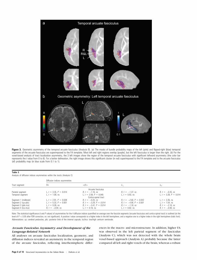
All analyses on arcuate fasciculus localization, geometry, and
diffusion indices revealed an asymmetry in the temporal region
of the arcuate fasciculus, reflecting interhemispheric differ-
ences in the macro- and microstructure. In addition, higher FA
was observed in the left parietal segment of the fasciculus
(Analysis C), which was not detected with the whole brain
voxel-based approach (Analysis A) probably because the latter
compared all left and right voxels of the brain, whereas a robust
Figure 3. Geometric asymmetry of the temporal arcuate fasciculus (Analysis B). (a) The masks of bundle probability maps of the left (pink) and flipped-right (blue) temporalsegments of the arcuate fasciculus are superimposed to the FA template. Most left and right regions overlap (purple), but the left fasciculus is larger than the right. (b) For thevoxel-based analysis of tract localization asymmetry, the 3 left images show the region of the temporal arcuate fasciculus with significant leftward asymmetry (the color barrepresents the t value from 0 to 8). For a better delineation, the right image shows this significant cluster (in red) superimposed to the FA template and to the arcuate fasciculus(af) probability map (in blue scale from 0.1 to 1).
Table 3Analysis of diffusion indices asymmetries within the tracts (Analysis C)
Diffusion indices asymmetries
Tract segment FA \D[ k? k//
Arcuate fasciculusParietal segment L, t 5 3.35, P 5 0.014 R, t 5 �1.16, ns R, t 5 �1.37, ns R, t 5 �0.35, nsTemporal segment L, t 5 1.09, ns L, t 5 2.59, P 5 0.045 L, t 5 0.93, ns L, t 5 3.28, P 5 0.014
Cortico-spinal tractSegment 1 (midbrain) L, t 5 2.91, P 5 0.028 R, t 5 �0.25, ns R, t 5 �2.66, P 5 0.043 L, t 5 2.26, nsSegment 2 (cp--plic) L, t 5 5.33, P 5 0.001 R, t 5 �3.29, P 5 0.014 R, t 5 �4.96, P 5 0.001 L, t 5 1.54, nsSegment 3 (plic--lcs) L, t 5 0.28, ns R, t 5 �3.41, P 5 0.014 R, t 5 �1.18, ns R, t 5 �2.16, nsSegment 4 (lcs--hcs) R, t 5 �0.94, ns L, t 5 0.18, ns L, t 5 0.82, ns R, t 5 �0.90, ns
Note: The statistical significance (t and P values) of asymmetries for the 4 diffusion indices quantified on average over the fascicle segments (arcuate fasciculus and cortico-spinal tract) is outlined (at the
level of P\ 0.05 after FDR correction, ns: not significant). A positive t value corresponds to a higher index in the left hemisphere, and a negative one to a higher index in the right hemisphere (italic font).
Abbreviations: cp: cerebral peduncles, plic: posterior limb of the internal capsule, lcs/hcs: low/high centrum semiovale.
Page 6 of 10 Structural Asymmetries in the Infant Brain d Dubois et al.

averaging of the indices over the segment may be needed to
uncover subtle differences.
In the whole brain voxel-based analysis of FA asymmetry
(Analysis A), the most asymmetrical cluster, detected in the left
temporal region of the arcuate fasciculus, appeared very similar
to the results observed in adults (Buchel et al. 2004). However,
in our study, this asymmetry may reflect the combined effects
of higher longitudinal diffusivity (Analysis C) and of left--right
differences in tract anatomical localization and geometry
(Analysis B), as probability maps revealed a more posterior
and larger left segment.
The early macroscopic asymmetry in the temporal arcuate
fasciculus is probably related to asymmetries of the superior
temporal sulcus, sylvian fissure, and planum temporale
observed in the fetal and newborn brain (Witelson and Pallie
1973). During prenatal life, right sulci, among which the
superior temporal sulcus, usually appear before their left
counterpart (Chi et al. 1977). Furthermore, the surface of the
right superior temporal sulcus is larger in preterm newborns
(Dubois et al. 2007), the sylvian fissure is shorter and steeper
on the right (Sowell et al. 2002), and the planum temporale is
larger on the left side already in fetuses (Witelson and Pallie
1973; Chi et al. 1977). Thus, in the developing brain, the
superior and posterior temporal regions are at the epicenter of
major left--right differences.
Beyond macroscopic geometrical asymmetries, we also
observed left--right differences in the microstructure of the
arcuate fasciculus. In the left temporal segment, higher <D >,and k// (Analysis C) suggested a delayed ‘‘pre’’-myelination
stage on that side. This contrasts with the higher FA detected in
the left parietal segment of the tract, which is rather related to
a higher organization of parallel fibers than to an advanced
‘‘true’’ myelination, as this region matures slowly during early
childhood (Pujol et al. 2006; Zhang et al. 2007) until late
adolescence (Paus et al. 1999). These assumptions on the
mechanisms underlying asymmetries should nevertheless be
confirmed over a larger cohort of infants as congruent results
were not obtained over all diffusion indices (Table 1). Our
observations are however in agreement with a recent study,
which showed a higher FA in the left fronto-temporal segment
and right fronto-parietal segment of the fasciculus in children
between 6 and 17 years old (Eluvathingal et al. 2007).
In summary, the asymmetry of the arcuate fasciculus is
already present in early life, in terms of localization, organization,
and maturation. Do these early structural asymmetries of the
arcuate fasciculus sustain the developing functional lateraliza-
tion of language perception? Although many DTI studies have
underlined asymmetries of the arcuate fasciculus in adults, with
a more extensive connectivity on the left side (Nucifora et al.
2005; Parker et al. 2005; Powell et al. 2006; Catani et al. 2007;
Vernooij et al. 2007), the correlations with functional laterali-
zation are still controversial (Powell et al. 2006, Vernooij et al.
2007). Dichotic listening (Bertoncini et al. 1989) and functional
brain imaging studies in infants (Dehaene-Lambertz 2000;
Dehaene-Lambertz et al. 2002; Pena et al. 2003; Dehaene-
Lambertz, Hertz-Pannier, Dubois, Meriaux, et al. 2006) suggest
that speech processing is biased toward the left hemisphere
early on. Furthermore, the left frontal area, although immature,
is already activated by speech perception at this age especially
when short-term verbal memory is solicited by the experimental
task (Dehaene-Lambertz, Hertz-Pannier, Dubois, Meriaux, et al.
2006). These findings suggest an early efficient temporo-parieto-
frontal connection through the left arcuate fasciculus. The
particular organization and maturation of the left fasciculus may
thus sustain the progressive development of the speech
perception--production network.
Cortico-Spinal Tract Asymmetry and Development ofSensori-Motor Functions
Although the whole cortico-spinal tract appeared to have
a strictly similar geometry, size and localization on both sides
(Analysis B), higher FA was revealed in the left relative to the
right inferior region between the cerebral peduncles and the
posterior limb of the internal capsule (Analyses A and C). In the
midbrain and in the segment between the internal capsule and
the low centrum semiovale, the analysis of DTI indices within
the tract (Analysis C) also detected asymmetries which were not
detected with the FA voxel-based method (Analysis A), probably
because of lower sensitivity of this technique to subtle differ-
ences, as discussed for the arcuate fasciculus. Because the
cortico-spinal tract reconstructed by tractography was larger
than the image spatial resolution, and because similar tract
volumes were observed in the left and right hemispheres,
asymmetries of diffusion indices cannot be attributed to
Figure 4. FA asymmetry within tract segments (Analysis C). Asymmetry ratios in FAare presented for the segments of the arcuate fasciculus (a) and the cortico-spinaltract (b), with statistical significance of the one-tailed paired t-tests (see Table 3, ns:not significant). Abbreviations: cp: cerebral peduncles, plic: posterior limb of theinternal capsule, lcs/hcs: low/high centrum semiovale.
Cerebral Cortex Page 7 of 10

asymmetrical tract thickness triggering different partial volume
effects between the tract and surrounding tissue.
Asymmetries in the 3 lower segments of the cortico-spinal
tract concern different diffusion indices and can be explained
by various biophysical phenomena as outlined in Table 1:
higher fibers organization or advanced ‘‘true’’ myelination in the
left midbrain, advanced ‘‘true’’ myelination between the left
cerebral peduncles and posterior limb of the internal capsule,
and advanced ‘‘pre-’’ or ‘‘true’’ myelination in the left centrum
semiovale. As the tract is made of continuous fibers, the cause
of the asymmetries at different levels of the tract is likely to be
the same and related to an advanced myelination on the left in
comparison with the right.
It is interesting to note that asymmetry may have a specific
spatial spread over the cortico-spinal tract, as we respectively
detected 1) an asymmetry in FA, but not in <D > in the
midbrain, 2) asymmetries in <D > and FA between the cerebral
peduncles and the internal capsule, 3) only an asymmetry in
<D > between the internal capsule and the low centrum
semiovale, and 4) no asymmetry in the upper portion of the
tract. Post-mortem studies have described that the myelination
sequence extends between the third trimester of gestation and
the first postnatal months in the cortico-spinal tract (Brody
et al. 1987; Kinney et al. 1988). Furthermore, because
myelination progresses from the neuron body to the periphery
(McCart and Henry 1994) and starts in sensory pathways before
motor pathways (Yakovlev and Lecours 1967), changes in
white matter intensity are observed in T1-weighted MRI images
in the midbrain and posterior limb of the internal capsule
before the centrum semiovale and subcortical white matter
(McArdle et al. 1987). As we showed in a previous paper that
mean diffusivity reflects white matter maturation earlier than
FA (Dubois et al. 2008), our present results likely suggest that
the maturation of the somatosensory fibers in the cortico-spinal
tract is observed in this age range, and that the asymmetry in
myelination in favor of the left side progresses from the inferior
to the superior regions: myelination on the right begins to
catch up the left one in the midbrain, where no more
asymmetry in <D > was observed, whereas the asymmetry is
only starting above the posterior limb of the internal capsule,
where only an asymmetry in <D > was observed.
Previous studies already described structural asymmetries of
the cortico-spinal tract in the more mature brain. In children
from 6 to 17 years old, FA measured over the whole tract is
higher on the left side (Eluvathingal et al. 2007). In adults,
significant differences in DTI indices (higher FA and lower
<D > on the left) have been observed at the level of the
posterior limb of the internal capsule (Westerhausen et al.
2007). The cortico-spinal tract also appears thicker (Rade-
macher, Burgel, et al. 2001) and the subcortical white matter
more anisotropic in the precentral region contra-lateral to the
predominant hand (Buchel et al. 2004), but the overall DTI-
based relative fiber density is not different between both
hemispheres (Nucifora et al. 2005).
It is not possible to conclude whether this observed advance
of maturation in the left cortico-spinal tract is at the origin of
behavioral asymmetry. Although individual children do not
show reliable evidence of hand preference in voluntary
movements before the second postnatal year, some studies
have revealed a slight right preference for spontaneous
movements at a group level from the last trimester of gestation
on (Hepper et al. 1991), but these results have not been
consistently reproduced. This right side preference might be
related to structural differences between the left and right
sensori-motor cortical organization, and/or to an asymmetrical
position within the womb which frees the right side (Hepper
et al. 2005), and/or to an environment in which heavily right
handed biased parents favor one side (Leconte and Fagard
2004). Using functional MRI (fMRI), Erberich et al. (2006)
found no difference between ipsi- and contra-lateral responses
during passive hand stimulations in newborns, contrarily to
older infants who display asymmetrical responses favoring the
contra-lateral side. In this study, we did not observe an increase
of structural asymmetry in the 15-weeks age range studied,
suggesting no obvious change at least during the first postnatal
weeks, in contrast with functional asymmetry. More data are
needed in order to understand the relationship between these
different behavioral, functional and structural markers. Be that
as it may, our results confirm an indisputable early structural
asymmetry in the human sensori-motor network, which is
present before a robust asymmetrical use of the right hand.
Other Asymmetries of the Developing Brain
Using the whole brain voxel-based analysis in FA, other cerebral
regions were found to be significantly asymmetrical in infants,
but we here focused on the language-related and sensori-motor
networks. Further analyses should be considered to precise the
localization of these regions, their functional implication and
significance in the developing brain, and to understand the
underlying mechanisms of such asymmetries in terms of
maturation, organization or geometry processes.
Methodological Issues
In this study, complementary methods of asymmetries analysis
in local diffusion indices and fascicles localization were used in
order to reach a reliable interpretation of the interhemispheric
differences of the arcuate fasciculus and cortico-spinal tract.
Each method presented distinct advantages and drawbacks.
The FA voxel-based analysis (Analysis A) was used to
underline interhemispheric asymmetries without a priori
hypotheses on their anatomical localization (Barnea-Goraly
et al. 2005). However, the DTI normalization procedure is still
controversial (Jones et al. 2005) and challenging in infants
because of brain growth. Here we used a special procedure
optimized for the infants group, based on the creation of
a specific FA template, which enabled to globally take into
account macroscopic interhemispheric differences, such as
frontal and occipital petalias. The insufficient realignment of
the left and right arcuate fascicles by the normalization process
probably results from 3 factors. First, the tract was larger, with
2 branches, on the left side. Second, the localization of the
posterior part of the sylvian fissure is asymmetrical, i.e. more
superior on the right side in adults and children (Sowell et al.
2002), implying a posterior shift of the left temporal region,
including the arcuate fasciculus. Third, immature white matter
bundles with low FA are difficult to realign precisely, as close
FA values in both the tract and the surrounding tissues result in
a low local contrast in the FA template. On the contrary, the
normalization was sufficient to precisely realign the cortico-
spinal tract, because of its stable localization in the brain
central regions and its relatively high FA compared with
surrounding tissue. In this perspective, applying more refined
registration approaches like Tract-Based Spatial Statistics
(Smith et al. 2006; Anjari et al. 2007) could have been
Page 8 of 10 Structural Asymmetries in the Infant Brain d Dubois et al.

considered in the developing brain, but limitations related to
the asymmetrical tract size would have probably remained.
Such issue thus underlined the necessity to correlate comple-
mentary methods before interpreting asymmetry results from
FA voxel-based analysis.
The voxel-based analysis of tract localization and geometry
(Analysis B) was used to outline the 3D macroscopic asymmetry
of the tracts. The individual analysis of diffusion indices within
the tracts (Analysis C) seemed most sensitive for the detection
of asymmetries. However, the tractography method, which
requires manual drawing of regions for seed placement and tract
selection, is time-consuming and user-dependent. Moreover, it
focuses on specific bundles and asymmetries in the frontal
arcuate fasciculus could not be evaluated because this tract
segment was not reliably reconstructed in all infants.
Consequently, all 3 methods were complementary for the
study of brain asymmetries, and provided a reliable evaluation
of the interhemispheric differences in the immature arcuate
fasciculus and cortico-spinal tract. Whereas this study cannot
disentangle whether these structural asymmetries are the
cause or consequence of the developing functional asymme-
tries, it will serve as a basis for further correlations with later
functional scores.
Conclusion
In this DTI study in infants, we demonstrated early interhemi-
spheric leftward asymmetries in the microstructure and
maturation of both the arcuate fasciculus and cortico-spinal
tract, in addition to macroscopic left--right differences in the
former. These results provide new and unique data which
support the hypothesis that the development of functional
lateralization of language-related and sensori-motor networks
during infancy and childhood is related to early structural
processes that take place in the immature brain before
sustained exposure to environmental stimulations.
Funding
McDonnell foundation and ANR for financial support to G.D.L.
Notes
We thank C. Poupon, F. Lethimonnier, D. Riviere, Y. Cointepas, and
M. Perrin for support on DTI acquisition and postprocessing, Mr Brunet
from Ravier-Touzard Company for designing a baby bouncer chair
specifically adapted to the head coil, F. Brunelle for its support. Conflict
of interest : None declared.
Address correspondence to Jessica Dubois, PhD, U663 Hopital
Necker-Enfants malades, 149 rue de Sevres, 75015 Paris, France. Email:
References
Anjari M, Srinivasan L, Allsop JM, Hajnal JV, Rutherford MA, Edwards AD,
Counsell SJ. 2007. Diffusion tensor imaging with tract-based spatial
statistics reveals local white matter abnormalities in preterm infants.
Neuroimage. 35:1021--1027.
Barnea-Goraly N, Menon V, Eckert M, Tamm L, Bammer R,
Karchemskiy A, Dant CC, Reiss AL. 2005. White matter development
during childhood and adolescence: a cross-sectional diffusion tensor
imaging study. Cereb Cortex. 15:1848--1854.
Beaulieu C. 2002. The basis of anisotropic water diffusion in the
nervous system—a technical review. NMR Biomed. 15:435--455.
Bertoncini J, Morais J, Bijeljac-Babic R, McAdams S, Peretz I, Mehler J.
1989. Dichotic perception and laterality in neonates. Brain Lang.
37:591--605.
Best CT. 1988. The emergence of cerebral asymmetries in early human
development: a literature review and a neuroembryological model.
In: Molfese D, Segalowitz SJ, editors. Brain lateralization in children.
New York: The Guilford Press.
Brody BA, Kinney HC, Kloman AS, Gilles FH. 1987. Sequence of central
nervous system myelination in human infancy. I. An autopsy study of
myelination. J Neuropathol Exp Neurol. 46:283--301.
Buchel C, Raedler T, Sommer M, Sach M, Weiller C, Koch MA. 2004.
White matter asymmetry in the human brain: a diffusion tensor MRI
study. Cereb Cortex. 14:945--951.
Burgel U, Amunts K, Hoemke L, Mohlberg H, Gilsbach JM, Ziles K. 2006.
White matter fiber tracts of the human brain: Three-dimensional
mapping at microscopic resolution, topography and intersubject
variability. Neuroimage. 29:1092--1105.
Cao Y, Whalen S, Huang J, Berger KL, DeLano MC. 2003. Asymmetry of
subinsular anisotropy by in vivo diffusion tensor imaging. Hum Brain
Mapp. 20:82--90.
Catani M, Allin MP, Husain M, Pugliese L, Mesulam MM, Murray RM,
Jones DK. 2007. Symmetries in human brain language pathways
correlate with verbal recall. Proc Natl Acad Sci USA. 104:17163--17168.
Chi JG, Dooling EC, Gilles FH. 1977. Left-right asymmetries of the
temporal speech areas of the human fetus. Arch Neurol. 34:346--348.
Chiron C, Jambaque I, Nabbout R, Lounes R, Syrota A, Dulac O. 1997.
The right brain hemisphere is dominant in human infants. Brain.
120:1057--1065.
Ciccarelli O, Toosy AT, Parker GJ, Wheeler-Kingshott CA, Barker GJ,
Miller DH, Thompson AJ. 2003. Diffusion tractography based group
mapping of major white-matter pathways in the human brain.
Neuroimage. 19:1545--1555.
Cointepas Y, Poupon C, Maroy R, Riviere D, Le Bihan D, Mangin JF.
2003. A freely available Anatomist/BrainVISA package for analysis of
diffusion MR data. Proceedings of the 9th HBM Scientific Meeting,
New York, USA. Neuroimage. 19:S810.
Corbetta D, Thelen E. 1999. Lateral biases and fluctuations in infants’
spontaneous arm movements and reaching. Dev Psychobiol.
34:237--255.
Dehaene-Lambertz G. 2000. Cerebral specialization for speech and non-
speech stimuli in infants. J Cogn Neurosci. 12:449--460.
Dehaene-Lambertz G, Dehaene S, Hertz-Pannier L. 2002. Functional
neuroimaging of speech perception in infants. Science.
298:2013--2015.
Dehaene-Lambertz G, Hertz-Pannier L, Dubois J. 2006. Nature and
nurture in language acquisition: anatomical and functional brain-
imaging studies in infants. Trends Neurosci. 29:367--373.
Dehaene-Lambertz G, Hertz-Pannier L, Dubois J, Meriaux S, Roche A,
Sigman M, Dehaene S. 2006. Functional organization of perisylvian
activation during presentation of sentences in preverbal infants.
Proc Natl Acad Sci USA. 103:14240--14245.
Dubois J, Benders M, Cachia A, Lazeyras F, Ha-Vinh Leuchter R,
Sizonenko SV, Borradori-Tolsa C, Mangin JF, Huppi PS. 2007.
Mapping the early cortical folding process in the preterm newborn
brain. Cereb Cortex. 18:1444--1454.
Dubois J, Dehaene-Lambertz G, Perrin M, Mangin JF, Cointepas Y,
Duchesnay E, Le Bihan D, Hertz-Pannier L. 2008. Asynchrony of the
early maturation of white matter bundles in healthy infants:
quantitative landmarks revealed non-invasively by diffusion tensor
imaging. Hum Brain Mapp. 29:14--27.
Dubois J, Hertz-Pannier L, Dehaene-Lambertz G, Cointepas Y, Le
Bihan D. 2006. Assessment of the early organization and maturation
of infants’ cerebral white matter fiber bundles: a feasibility study
using quantitative diffusion tensor imaging and tractography.
Neuroimage. 30:1121--1132.
Eluvathingal TJ, Hasan KM, Kramer L, Fletcher JM, Ewing-Cobbs L. 2007.
Quantitative diffusion tensor tractography of association and pro-
jection fibers in normally developing children and adolescents.
Cereb Cortex. 17:2760--2768.
Erberich SG, Panigrahy A, Friedlich P, Seri I, Nelson MD, Gilles F. 2006.
Somatosensory lateralization in the newborn brain. Neuroimage.
29:155--161.
Cerebral Cortex Page 9 of 10

Fabiano AJ, Horsfield MA, Bakshi R. 2005. Interhemispheric asymmetry
of brain diffusivity in normal individuals: a diffusion-weighted MR
imaging study. Am J Neuroradiol. 26:1089--1094.
Fagard J, Marks A. 2000. Unimanual and bimanual tasks and the
assessment of handedness in toddlers. Dev Sci. 3:137--147.
Galaburda AM, Geschwind N. 1981. Anatomical asymmetries in the
adult and developing brain and their implications for function. Adv
Pediatr. 28:271--292.
Gilmore JH, Lin W, Prastawa MW, Looney CB, Vetsa YS, Knickmeyer RC,
Evans DD, Smith JK, Hamer RM, Lieberman JA, et al. 2007. Regional
gray matter growth, sexual dimorphism, and cerebral asymmetry in
the neonatal brain. J Neurosci. 27:1255--1260.
Glenn OA, Henry RG, Berman JI, Chang PC, Miller SP, Vigneron DB,
Barkovich AJ. 2003. DTI-based three-dimensional tractography
detects differences in the pyramidal tracts of infants and children
with congenital hemiparesis. J Magn Reson Imaging. 18:641--648.
Gong G, Jiang T, Zhu C, Zang Y, Wang F, Xie S, Xiao J, Guo X. 2005.
Asymmetry analysis of cingulum based on scale-invariant parame-
terization by diffusion tensor imaging. Hum Brain Mapp. 24:92--98.
Hepper PG, Shahidullah S, White R. 1991. Handedness in the human
fetus. Neuropsychologia. 29:1107--1111.
Hepper PG, Wells DL, Lynch C. 2005. Prenatal thumb sucking is related
to postnatal handedness. Neuropsychologia. 43:313--315.
Hochberg FH, Le May M. 1975. Arteriographic correlates of handedness.
Neurology. 25:218--222.
Ingram D. 1975a. Motor asymmetries in young children. Neuro-
psychologia. 13:95--102.
Ingram D. 1975b. Cerebral speech lateralization in young children.
Neuropsychologia. 13:103--105.
Jones DK, Symms MR, Cercignani M, Howard RJ. 2005. The effect of filter
size on VBM analyses of DT-MRI data. Neuroimage. 26:546--554.
Kimura D. 1967. Functional asymmetry of the brain in dichotic
listening. Cortex. 3:163--178.
Kinney HC, Brody BA, Kloman AS, Gilles FH. 1988. Sequence of central
nervous system myelination in human infancy 2: Patterns of
myelination in autopsied infants. J Neuropathol Exp Neurol.
47:217--234.
Le Bihan D, Mangin JF, Poupon C, Clark CA, Pappata S, Molko N,
Chabriat H. 2001. Diffusion tensor imaging: concepts and applica-
tions. J Magn Reson Imaging. 13:534--546.
Leconte P, Fagard J. 2004. Influence of object spatial location and task
complexity on children’s use of their preferred hand depending on
their handedness consistency. Dev Psychobiol. 45:51--58.
Mangin JF, Poupon C, Clark C, Le Bihan D, Bloch I. 2002. Distortion
correction and robust tensor estimation for MR diffusion imaging.
Med Image Anal. 6:191--198.
McArdle CB, Richardson CJ, Nicholas DA, Mirfakhraee M, Hayden CK,
Amparo EG. 1987. Developmental features of the neonatal brain: MR
imaging. Part 1, Gray-white matter differentiation and myelination.
Radiology. 162:223--229.
McCart RJ, Henry GH. 1994. Visual corticogeniculate projections in the
cat. Brain Res. 653:351--356.
Mori S, van Zijl PC. 2002. Fiber tracking: principles and strategies—a
technical review. NMR Biomed. 15:468--480.
Neil JJ, Miller J, Mukherjee P, Huppi PS. 2002. Diffusion tensor imaging
of normal and injured developing human brain—a technical review.
NMR Biomed. 15:543--552.
Nucifora PG, Verma R, Melhem ER, Gur RE, Gur RC. 2005. Leftward
asymmetry in relative fiber density of the arcuate fasciculus.
Neuroreport. 16:791--794.
Ottaviano S, Guidetti V, Allemand F, Spinetoli B, Seri S. 1989. Laterality
of arm movement in full-term newborn. Early Hum Dev. 19:3--7.
Park HJ, Westin CF, Kubicki M, Maier SE, Niznikiewicz M, Baer A,
Frumin M, Kikinis R, Jolesz FA, McCarley RW, et al. 2004. White
matter hemisphere asymmetries in healthy subjects and in schizo-
phrenia: a diffusion tensor MRI study. Neuroimage. 23:213--223.
Parker GJ, Luzzi S, Alexander DC, Wheeler-Kingshott CA, Ciccarelli O,
Lambon Ralph MA. 2005. Lateralization of ventral and dorsal
auditory-language pathways in the human brain. Neuroimage.
24:656--666.
Paus T, Zijdenbos A, Worsley K, Collins DL, Blumenthal J, Giedd JN,
Rapoport JL, Evans AC. 1999. Structural maturation of neural
pathways in children and adolescents: in vivo study. Science.
283:1908--1911.
Pena M, Maki A, Kovacic D, Dehaene-Lambertz G, Koizumi H,
Bouquet F, Mehler J. 2003. Sounds and silence: an optical
topography study of language recognition at birth. Proc Natl Acad
Sci USA. 100:11702--11705.
Penhune VB, Zatorre RJ, MacDonald JD, Evans AC. 1996. Interhemi-
spheric anatomical differences in human primary auditory cortex:
probabilistic mapping and volume measurement from magnetic
resonance scans. Cereb Cortex. 6:661--672.
Perrin M, Poupon C, Cointepas Y, Rieul B, Golestani N, Pallier C,
Riviere D, Constantinesco A, Le Bihan D, Mangin JF. 2005. Fiber
tracking in q-ball fields using regularized particle trajectories. Inf
Process Med Imaging. 19:52--63.
Pierpaoli C, Basser PJ. 1996. Toward a quantitative assessment of
diffusion anisotropy. Magn Reson Med. 36:893--906.
Powell HW, Parker GJ, Alexander DC, Symms MR, Boulby PA, Wheeler-
Kingshott CA, Barker GJ, Noppeney U, Koepp MJ, Duncan JS. 2006.
Hemispheric asymmetries in language-related pathways: a combined
functional MRI and tractography study. Neuroimage. 32:388--399.
Pujol J, Soriano-Mas C, Ortiz H, Sebastian-Galles N, Losilla JM, Deus J.
2006. Myelination of language-related areas in the developing brain.
Neurology. 66:339--343.
Rademacher J, Caviness VS, Jr, Steinmetz H, Galaburda AM. 1993.
Topographical variation of the human primary cortices: implications
for neuroimaging, brain mapping, and neurobiology. Cereb Cortex.
3:313--329.
Rademacher J, Burgel U, Geyer S, Schormann T, Schleicher A,
Freund HJ, Zilles K. 2001. Variability and asymmetry in the human
precentral motor system. A cytoarchitectonic and myeloarchitec-
tonic brain mapping study. Brain. 124:2232--2258.
Rademacher J, Morosan P, Schormann T, Schleicher A, Werner C,
Freund HJ, Zilles K. 2001. Probabilistic mapping and volume
measurement of human primary auditory cortex. Neuroimage.
13:669--683.
Ronnqvist L, Domellof E. 2006. Quantitative assessment of right and left
reaching movements in infants: a longitudinal study from 6 to 36
months. Dev Psychobiol. 48:444--459.
Smith SM, Jenkinson M, Johansen-Berg H, Rueckert D, Nichols TE,
Mackay CE, Watkins KE, Ciccarelli O, Cader MZ, Matthews PM, et al.
2006. Tract-based spatial statistics: voxelwise analysis of multi-
subject diffusion data. Neuroimage. 31:1487--1505.
Sowell ER, Thompson PM, Rex D, Kornsand D, Tessner KD, Jernigan TL,
Toga AW. 2002. Mapping sulcal pattern asymmetry and local cortical
surface gray matter distribution in vivo: maturation in perisylvian
cortices. Cereb Cortex. 12:17--26.
Toga AW, Thompson PM. 2003. Mapping brain asymmetry. Nat Rev
Neurosci. 4:37--48.
Vernooij MW, Smits M, Wielopolski PA, Houston GC, Krestin GP, van
der Lugt A. 2007. Fiber density asymmetry of the arcuate fasciculus
in relation to functional hemispheric language lateralization in both
right- and left-handed healthy subjects: a combined fMRI and DTI
study. Neuroimage. 35:1064--1076.
Westerhausen R, Huster RJ, Kreuder F, Wittling W, Schweiger E. 2007.
Corticospinal tract asymmetries at the level of the internal capsule:
is there an association with handedness? Neuroimage. 37:379--386.
Witelson SF, Pallie W. 1973. Left hemisphere specialization for language
in the newborn. Neuroanatomical evidence of asymmetry. Brain.
96:641--646.
Yakovlev PI, Lecours AR. 1967. The myelogenetic cycles of regional
maturation in the brain. In: Minowski A, editor. Regional de-
velopment of the brain in early life. Oxford: Blackwell. p. 3--69.
Zhang J, Evans A, Hermoye L, Lee SK, Wakana S, Zhang W, Donohue P,
Miller MI, Huang H, Wang X, et al. 2007. Evidence of slow
maturation of the superior longitudinal fasciculus in early childhood
by diffusion tensor imaging. Neuroimage. 38:239--247.
Page 10 of 10 Structural Asymmetries in the Infant Brain d Dubois et al.