Plectolyngbya hodgsonii: a novel filamentous cyanobacterium from Antarctic lakes
Upload
oregonstateCategory
view
0download
0
DOI: 10.1021/jo900578j Published on Web 07/02/2009 J. Org. Chem. 2009, 74, 5267–5275 5267r 2009 American Chemical Society
pubs.acs.org/joc
Structural and Synthetic Investigations of Tanikolide Dimer, a SIRT2
Selective Inhibitor, and Tanikolide seco-Acid from the Madagascar
Marine Cyanobacterium Lyngbya majuscula
Marcelino Guti�errez,†,‡ Eric H. Andrianasolo,§,‡ Won Kyo Shin,^,‡ Douglas E. Goeger,§
Alexandre Yokochi, ) J€org Schemies,# Manfred Jung,# Dennis France,3
Susan Cornell-Kennon,3 Eun Lee,*,^ and William H. Gerwick*,†,§
†Center for Marine Biotechnology and Biomedicine, Scripps Institution of Oceanography and The SkaggsSchool of Pharmacy andPharmaceutical Sciences,University ofCalifornia at SanDiego, La Jolla, California92037, §College of Pharmacy and )Department of Chemical Engineering, Oregon State University, Corvallis,Oregon 97331, ^Department of Chemistry, College of Natural Sciences, Seoul National University, Seoul,151-747, Korea, #Institute of Pharmaceutical Sciences, Albert-Ludwigs-Universit€at Freiburg, Albertstrausse
25, 79104 Freiburg, Germany, and 3Novartis Institute for Biomedical Research, Cambridge,Massachusetts 02139. ‡These authors contributed equally to the work (M.G., stereochemical analyses; E.H.
A., isolation and planar structure; W.K.S., synthesis).
[email protected]; [email protected]
Received April 3, 2009
Tanikolide seco-acid 2 and tanikolide dimer 3, the latter a novel and selective SIRT2 inhibitor, wereisolated from the Madagascar marine cyanobacterium Lyngbya majuscula. The structure of 2,isolated as the pureR enantiomer, was elucidated byX-ray experiment in conjunctionwithNMRandoptical rotation data, whereas the depside molecular structure of 3was initially thought to be amesocompound as established byNMR,MS, and chiral HPLC analyses. Subsequent total synthesis of thethree tanikolide dimer stereoisomers 4, 5, and ent-5, followed by chiral GC-MS comparisons withthe natural product, showed it to be exclusively theR,R-isomer 5. Tanikolide dimer 3 (= 5) inhibitedSIRT2 with an IC50= 176 nM in one assay format and 2.4 μM in another. Stereochemicaldetermination of symmetrical dimers such as compound 3 pose intriguing and subtle questions instructure elucidation and, as shown in the current work, are perhaps best answered in conjunctionwith total synthesis.
Introduction
Tanikolide (1) is a biologically active δ-lactone that wasoriginally isolated from the lipid extract of a collection ofLyngbya majuscula fromMadagascar.1 Tanikolide possessesa single chiral center, a quaternary carbonwith hydroxyl and
hydroxymethyl groups, as well as two multicarbon chains
and exhibits potent antifungal activity against Candida
albicans (13mmdiameter zone inhibition using 100 μg/disk).In addition, it shows strong toxicity in the brine shrimp
Artemia salina and schistosomiasis-carrying snail Biompha-
laria glabrata (LC50 =3.6 and 9.0 μg/mL, respectively)
biological assays. Owing to its juxtaposition of func-
tional groups, tanikolide (1) has been the target of numerous
enantioselective syntheses.2 As part of our ongoing search
for structurally and pharmacologically interesting substances
*To whom correspondence should be addressed. Natural products isola-tion and characterization. (W.H.G.) Tel: (858) 534-0578. Chemical synthesis.(E.L.) Tel: þ82-2-880-6646.
(1) Singh, I. P.; Milligan, K. E.; Gerwick, W. H. J. Nat. Prod. 1999, 62,1333–1335.
5268 J. Org. Chem. Vol. 74, No. 15, 2009
JOCArticle Guti�errez et al.
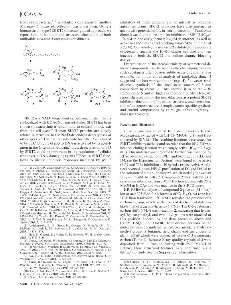
from cyanobacteria,3-5 a detailed exploration of anotherMalagasy L. majuscula collection was undertaken. Using ahuman sirtuin type 2 (SIRT2) bioassay-guided approach, wereport here the isolation and structural elucidation of bothtanikolide seco-acid 2 and tanikolide dimer 3.
SIRT2 is a NADþ-dependent cytoplasmic protein that isco-localized with HDAC6 onmicrotubules. SIRT2 has beenshown to deacetylate R-tubulin and to control mitotic exitfrom the cell cycle.6 Human SIRT2 proteins are closelyrelated in structure to the NAD-dependent deacetylases ofother species.7 The natural substrate for SIRT2 is believedto be p53.8 Binding of p53 to DNA is activated by its acetyl-ation in the C-terminal domain;9 thus, deacetylation of p53by SIRT2 could be important in the regulation of cellularresponses toDNA-damaging agents.10 Because SIRT2 func-tions to silence apoptotic responses mediated by p53,11
inhibitors of these proteins are of interest as potentialanticancer drugs. SIRT2 inhibitors have also emerged asagents with potential utility in neuroprotection.12 Tanikolidedimer 3 was found to be a potent inhibitor of SIRT2 (IC50=176 nM in one assay format; 2.4 μM in another) as well asactive in a sodium channel blocking assay (54% inhibition at5.2 μM).Conversely, the seco-acid 2 exhibited onlymoderatecytotoxicity against the H-460 cancer cell line and wasinactive in both the SIRT2 and sodium channel blockingassays.
Determination of the stereochemistry of symmetrical di-meric compounds can be technically challenging becausesuch substances often possess subtle issues of chirality. Forexample, our initial chiral analysis of tanikolide dimer 3
suggested it to be ameso compound (e.g., 4);13 however, totalchemical synthesis of the three stereoisomers of 3 andcomparison by chiral GC-MS showed it to be the R,R-stereoisomer 5 and of high enantiomeric purity. Here, wereport the isolation of this new dilactone as a potent SIRT2inhibitor, elucidation of its planar structure, and determina-tion of its stereostructure through enantio-specific synthesisand careful comparisons by chiral gas chromatography-mass spectrometry.
Results and Discussion
L. majuscula was collected from near Tanikely Island,Madagascar, extracted with CH2Cl2/MeOH (2:1), and frac-tionated by Si VLC. The resulting fractions were tested forSIRT2 inhibitory activity and revealed that the 40%EtOAc/hexanes eluting fraction was strongly active (IC50=2.5 μg/mL). This material was subjected to further fractionation byRP solid-phase extraction (SPE), and two fractions (D3 andD4, see the Experimental Section) were found to be active(81% and 75% inhibition at 10 μg/mL, respectively). Analy-tical reversed-phase HPLC purification of D3 and D4 led tothe isolation of tanikolide dimer 3, which initially showed anIC50=176 nM to SIRT2. Compound 2 was isolated as acrystalline substance from a VLC fraction eluting with 25%MeOH in EtOAc and was inactive in the SIRT2 assay.
HR FABMS analyses of compound 2 gave an [MþNa]þ
ion at m/z 325.2366 for a formula of C17H34O4Na (one lessDBE than tanikolide). 13C NMR revealed the presence of acarbonyl group, which on the basis of its chemical shift waslikely that of a carboxylic acid (δ 176.8). The C-5 quaternarycarbon shift (δ 74.4) was present in 2, indicating that hydro-xyl, hydroxymethyl, and two alkyl groups were attached atthis position. Indeed, by the data presented above andCOSY, HSQC, and HMBC, four distinct sections of themolecule were formulated: a hydroxy group, a hydroxy-methyl group, a butanoic acid chain, and an undecanylchain, all of which were connected to the C-5 quaternarycarbon (Table 1). Because X-ray quality crystals of 2 weredeposited from a fraction eluting with 25% MeOH inEtOAc, these structural features were confirmed via adiffraction study (see the Supporting Information).
(2) (a)Vichare, P.; Chattopadhyay,A.Tetrahedron: Asymmetry 2008, 19,598–602. (b) Zhang, C.; Hosoda, N.; Asami, M. Tetrahedron: Asymmetry2007, 18, 2185–2189. (c) Fujioka, H.; Matsuda, S.; Horai, M.; Fujii, E.;Morishita, M.; Nishiguchi, N.; Hata, K.; Kita, Y. Chem.;Eur. J. 2007, 13,5238–5248. (d)Wu, F.; Hong, R.; Khan, J.; Liu, X.; Deng, L.Angew. Chem.,Int. Ed. 2006, 45, 4301–4305. (e) Kita, Y.; Matsuda, S.; Fujii, E.; Horai, M.;Hata, K.; Fujioka, H. Angew. Chem., Int. Ed. 2005, 44, 5857–5860. (f)Yajima, T.; Saito, C.; Nagano, H. Tetrahedron 2005, 61, 10203–10215. (g)Ohgiya, T.; Nakamura, K.; Nishiyama, S. Bull. Chem. Soc. Jpn. 2005, 78,1549–1554. (h) Ohgiya, T.; Nishiyama, S. Tetrahedron Lett. 2004, 45, 8273–8275. (i) Arasaki, H.; Iwata,M.;Makida,M.;Masaki, Y.Chem. Pharm. Bull.2004, 52, 848–852. (j) Schomaker, J. M.; Borhan, B. Org. Biomol. Chem.2004, 2, 621–624. (k)Koumbis, A. E.; Dieti, K.M.; Vikentiou,M.G.;Gallos,J. K. Tetrahedron Lett. 2003, 44, 2513–2516. (l) Carda, M.; Rodriguez, S.;Castillo, E.; Bellido, A.; Diaz-Oltra, S.; Alberto, M. J. Tetrahedron 2003, 59,857–864. (m)Mizutani, H.; Watanabe, M.; Honda, T. Tetrahedron 2002, 58,8929–8936. (n) Tanaka, H.; Kozuki, Y.; Ogasawara, K. Tetrahedron Lett.2002, 43, 4175–4178. (o) Kanada, M. R.; Taniguchi, T.; Ogasawara, K.Synlett 2000, 7, 1019–1021.
(3) Andrianasolo, E. H.; Gross, H.; Goeger, D.; Musafija-Girt, M.;McPhail, K.; Leal, R. M.; Mooberry, S. L.; Gerwick, W. H. Org. Lett.2005, 7, 1375–1378.
(4) Han, B.; Goeger, D.; Maier, C. S.; Gerwick, W. H. J. Org. Chem.2005, 70, 3133–3139.
(5) Crews, P.; Gerwick,W.; Schmitz, F.; France,D.; Bair, K.;Wright, A.;Hallock, Y. Pharm. Biol. (Lisse, Netherlands) 2005, 1 (Suppl. 1), 39–52.
(6) (a)North, B. J.;Marshall, B. L.; Borra,M. T.;Denu, J.M.; Verdin, E.Mol. Cell 2003, 11, 437–444. (b) Dryden, S. C.; Nahhas, F. A.; Nowak, J. E.;Goustin, A. S.; Tainsky, M. A. Mol. Cell Biol. 2003, 23, 3173–3185.
(7) Avalos, J. L.; Celic, I.;Muhammad, S.; Cosgrove,M. S.; Boeke, J. D.;Wolberger, C. Mol. Cell 2002, 10, 523–535.
(8) Varizi, H.; Dessain, S. K.; Eagon, E. N.; Imai, S.-I.; Frye, R. A.;Pandita, T. K.; Guarente, L.; Weinberg, R. A. Cell 2001, 107, 149–159.
(9) Gu, W.; Roeder, R. G. Cell 1997, 90, 595–606.(10) Luo, J.; Nikolaev, A. Y.; Imai, S.-I.; Chen, D. L.; Su, F.; Shiloh, A.;
Guarante, L.; Gu, W. Cell 2001, 107, 137–148.(11) Smith, J. S. Trends Cell Biol. 2002, 12, 404–406.
(12) Outeiro, T. F.; Kontopoulos, E.; Altman, S.; Kufareva, I.;Strathearn, K. E.; Amore, A. M.; Volk, C. B.; Maxwell, M. M.; Rochet, J.C.; McLean, P. J.; Young, A. B.; Abagyan, R.; Feany, M. B.; Hyman, B. T.;Kazantsev, A. Science 2007, 317, 516–519.
(13) Andrianasolo, E. H. Ph.D. Thesis, Oregon State University, 2005,pp 127-179.

J. Org. Chem. Vol. 74, No. 15, 2009 5269
Guti�errez et al. JOCArticle
The seco-acid 2was found to be optically active, and only asingle enantiomer was observed in the X-ray study. Further,when a sample of 2was analyzed by chiral-phaseHPLC, onlya single peak was observed. Because the R-enantiomer ofcompound 2 has been chemically synthesized and its rotationmeasured {[R]D -0.8 (c 1.0, CHCl3}, we conclude from thenegative rotation of natural tanikolide seco-acid {[R]25D-10(c 0.87, CHCl3)} that it is also of R-configuration at C-5, thesame as in tanikolide (1) itself.14
The parent molecular ion for compound 3was deduced byvarious modes of ionization, including FABMS in variousmatrices, ESIMS, and EIMS. LR FABMS displayed threepeaks at m/z 591.4 [M þ Na]þ, m/z 569.3 [M þ H]þ, andm/z 551.2 [M-OH]þ. HR FABMS of the m/z 569.47804[M þ H]þ peak showed a molecular formula of C34H65O6,whereas HR EIMS of the m/z 551.4691 [M-OH]þ peakshowed a molecular formula of C34H63O5. However, the 13CNMRandDEPT spectra for 3 indicated the presence of only17 carbon and 31 carbon-bound hydrogen atoms (Table 2).These data combined with the MS information indicatedthat only half of the signals were appearing in the NMRspectra and suggested that 3 was a symmetrical dimer. Thethree degrees of unsaturation implied by the molecularformula were accounted for by the presence of two carbonyls(δ 172.1) and one ring.
13C NMR showed the presence of an ester carbonyl(δ 172.1; IR 1716 cm-1), a methylene carbon attached tooxygen (δ 67.9), a quaternary carbon attached to oxygen(δ 86.9), a methyl group (δ 14.5), and 13 high-field methy-lene carbons. The 1H NMR of 3 showed a pair of mutuallycoupled doublets (δ 3.68 and 3.59) for an isolatedmethyleneattached to oxygen and a two-proton multiplet (δ 2.51)assigned to a methylene adjacent to a carbonyl group.A series of multiplets in the range δ 1.29-1.95 accountedfor five methylene groups, and a broad singlet (δ 1.27)contained an additional 14 degenerate protons. A ter-minal methyl group was assigned to a three-proton tripletat δ 0.86.
Analyses of 1H-1H COSY and HMBC experiments of3 led to the deduction that it was closely related to tanikolide(1) (Figure 1).1 For example, the oxymethylene protons atδ 3.68/3.59 (H2-17) showed HMBC correlations with car-bons at δ 86.9 (C-5), 27.0 (C-4), and 37.1 (C-6). Additionally,a pair of methylene protons at δ 2.51 (H2-2) showed HMBCcorrelations with carbons at δ 172.1 (C-1), 17.1 (C-3), and27.0 (C-4). 1H-1H COSY delineated a connected spinsystem for the methylenes at C-2, C-3, and C-4. A methylgroup signal at δ 0.86 (H3-16) showed HMBC correlationswith twomethylene carbons at δ23.1 (C-15) and 32.3 (C-14),a sequence confirmed by 1H-1H COSY analysis and, hence,was at the terminus of a long lipid tail. Based on the closeconcordance in chemical shifts and coupling patterns be-tween 3 and 1, and the MS analysis described above,compound 3 was deduced to be a symmetrical dimer oftanikolide (1).
Because tanikolide (1) and its seco-acid 2 had both beenisolated as optically active metabolites and their lone stereo-centers determined to be R (Mosher ester for the former andcomparison with optical rotation for the latter), it wassurprising that dimer 3 initially gave no rotation at thesodium D-line or any other wavelength examined [however,remeasurement of the optical rotation of natural tanikolidedimer on a more sensitive instrument subsequently gave an[R]25Dþ1.0 (c 1.2, CHCl3)]. This perplexing initial result wasfurther examined by acid hydrolysis of 3 and analysis bychiral HPLC, first with diode array detection (DAD) andthen with ESI LCMS. In both cases, a 1:1 ratio of two peaks
TABLE 1. 1H and 13C NMR Assignments for Tanikolide seco-Acid 2a
position δC δH
1 176.82 34.6 2.29 (t, J = 6.7Hz)3 19.1 1.67 (m)4 35.5 1.49 (m)5 74.46 36.2 1.49 (m)7 22.7 1.29 (br s)8 29.9 1.29 (br s)9 29.6 1.29 (br s)10 29.6 1.29 (br s)11 29.6 1.29 (br s)12 29.6 1.29 (br s)13 29.6 1.29 (br s)14 32.3 1.29 (br s)15 23.1 1.29 (m)16 13.4 0.91 (t, J = 6.9Hz)17 67.1 3.59 (d, J =14.1Hz)
3.68 (d, J =14.1Hz)a 400 MHz 1H and 100 MHz for 13C NMR; CD3OD as solvent.
TABLE 2. 1H and 13C NMR Assignments for Tanikolide Dimer 3a
position δC δH HMBC
1/10 172.12/20 30.2 2.51 (m) 3 1, 3, 43/30 17.1 1.92 (m) 2, 4 1, 54/40 27.0 1.78 (m) 3 3, 5
1.95 (m)5/50 86.96/60 37.1 1.66 (m) 7 4, 5, 17
1.75 (m)7/70 23.9 1.29 (m) 6
1.32 (m)8/80 30.4 1.27 (br s)9/90 30.2 1.27 (br s)10/100 30 1.27 (br s)11/110 30 1.27 (br s)12/120 29.9 1.27 (br s)13/130 29.7 1.27 (br s)14/140 32.3 1.27 (br s) 1515/150 23.1 1.30 (m) 14, 1616/160 14.1 0.86 (t, J=7.2Hz) 15 15, 1417/170 67.9 3.59 (d, J=11.3Hz) 4, 5, 6
3.68 (d, J=11.3Hz)a 400 MHz for 1H and 100 MHz for 13C NMR; CDCl3 as solvent.
FIGURE 1. Selected HMBC and 1H-1H COSY correlations fortanikolide dimer 3.
(14) We cannot explain the discrepancy in the magnitude in the opticalrotation of our crystalline natural sample of 2 from that of syntheticmaterial,except to note that the rotation of the natural material was run on an older,less sensitive, and possibly inaccurate polarimeter (Perkin-Elmer 141).

5270 J. Org. Chem. Vol. 74, No. 15, 2009
JOCArticle Guti�errez et al.
was obtained which appeared to be due to the seco acid,suggesting that 3 was a meso compound deriving from oneR-monomer and one S-monomer. To clarify this surprisingresult, we turned to an integrated total chemical synthesisapproach of the three stereochemical possibilities for tani-kolide dimer along with chiral GC-MS analysis. Remark-ably, this approach gave quite different and unexpectedresults as described below.
The known epoxy alcohol 6 served as the starting materialfor the synthesis of (5R,50R)-tanikolide dimer 5. The tertiaryalcohol 7 was prepared from the benzyl ether derivative of 6via reaction with vinylmagnesium bromide in the presence ofcuprous iodide. Radical-mediated reaction of 7 with thio-phenol and then reaction with acryloyl chloride producedacrylate 8. Cross-metathesis reaction of 7 and 8 in thepresence of the second-generation Grubbs catalyst15 in hotdichloromethane led to the formation of the thioether 10.Thermal elimination reaction of the sulfoxide derivative of10 was carried out in hot toluene-pyridine, and the dieneacrylate 11 was prepared from the elimination product.Ring-forming metathesis reaction of 11 in the presence ofHoveyda-Grubbs catalyst 1216 proceeded smoothly, and(5R,50R)-tanikolide dimer 5 was obtained in high yield uponhydrogenation-hydrogenolysis (Scheme 1). The synthesis of(5S,50S)-tanilkolide dimer ent-5 was carried out in the samemanner using the enantiomeric starting material.
The synthesis of (5R,50S)-tanikolide dimer 4 also startedwith ent-6. Cross metathesis reaction between ent-7 and 8
produced a new thioether 13, which was converted into thediene acrylate 14 via oxidation-elimination and acrylateformation. Ring-forming metathesis reaction of 14 andhydrogenation-hydrogenolysis led to the formation of(5R,50S)-tanikolide dimer 4 in high yield (Scheme 2).
When the NMR spectra of compounds 4 and 5 werecompared, they looked very similar, and it was impractical todistinguish them on this basis (Supporting Information).
Moreover, we were unable to develop a convincing HPLC-based method to conduct this analysis. Thus, we turned to achiral GC-MS approach as described below.
Natural tanikolide dimer 3 and the synthetic stereoisomers(5R,50S)-tanikolide dimer 4, (5R,50R)-tanikolide dimer 5,and (5S,50S)-tanikolide dimer ent-5 were analyzed by GC-MS chromatography using a Cyclosil-B chiral column.When (5R,50S)-tanikolide dimer 4 was injected, two peakswere observed at 35.99 and 36.37 min, each having a massspectrum consistent with tanikolide (1) or its seco-acid 2
(e.g., each of these two compounds give the seco-acid follow-ing injection into the GC). The presence of two peaks in thechromatogram indicated that the dimer had undergonethermal hydrolysis, probably in the GC injection port.Consistent with this finding, when synthetic compounds5 and ent-5 were injected separately, they each gave a singlepeak, the former at 35.99 min and the latter at 36.37 min.When natural tanikolide dimer 3 was analyzed by thisprotocol, a single peak was observed at 35.99 min, therebyindicating it was composed of two identical monomers, eachof R configuration.
We believe that the initial chiroptical and chiral HPLCanalyses, which suggested that natural tanikolide dimer was ameso compound, were flawed for the following reasons. Asdeterminedwith the syntheticmaterials, the optical rotations ofboth the R,R or S,S dimers are very weak such that anessentially zero measurement was made with the small amountof natural dimer material that we had originally isolated. Thiswas exacerbated by the use of an older polarimeter in theseinitial measurements. More significantly, the conditions usedfor the acid hydrolysis (6 NHCl at 105 �C for 16 h) could verywell have led to racemization of the seco acid; retrospectively,racemization of the tertiary alcohol under these vigorousconditions could have been predicted.
Pure compounds 2 and 3 (=5) were tested in the SIRT2inhibitory assay as well as a mammalian cell sodium channelblocking assay.17 Compound 3 (=5) was initially found tohave an IC50 value of 176 nM, against SIRT2 whereas 2 wasinactive at the highest concentration tested (50 μM). Com-paring these data with those reported for sirtinol and8,9-dihydroxybenzo[4,5]furo[3,2-c]chromen-6-one (IC50 va-lues of 38 and 45 μM respectively)18 indicates that 3 (= 5) is
SCHEME 1. Synthesis of (R,R)-Tanikolide Dimer 5 SCHEME 2. Synthesis of (R,S)-Tanikolide Dimer 4
(15) Sanford, M. S.; Love, J. A.; Grubbs, R. H. J. Am. Chem. Soc. 2001,123, 6543–6554.
(16) Garber, S. B.; Kingsbury, J. S.; Gray, B. L.; Hoveyda, A. H. J. Am.Chem. Soc. 2000, 122, 8168–8179.
(17) Manger, R. L.; Leja, L. S.; Lee, S. Y.; Hungerford, J. M.; Hokama,Y.; Dickey, R.W.; Granade, H. R.; Lewis, R.; Yasumoto, T.; Wekell, M.M.J. AOAC Int. 1995, 78, 521–527.
(18) Grozinger, C. M.; Chao, E. D.; Blackwell, H. E.; Moazed, D.;Schreiber, S. L. J. Biol. Chem. 2001, 276, 38837–38843.

J. Org. Chem. Vol. 74, No. 15, 2009 5271
Guti�errez et al. JOCArticle
200-300-fold more active than either of these agents. In thesodium channel blocking assay, compound 3 induced a 54%inhibition at 5.2 μM,while 2was again inactive at the highestconcentration tested (10 μM). However, cytotoxicity assaysrevealed that 2was moderately toxic at 9.9 μM to the humanlung H-460 cell line, while 3 did not have any activity at10 μM. This observation is consistent with the limitedcytotoxicity observedwith other selective SIRT2 inhibitors19
and suggests that tanikolide dimer may have more potentialfor use as a neuroprotectant.
Synthetic production of an additional supply of the nat-ural (5R,50R)-tanikolide dimer 5 as well as the stereoisomers(5S,50S)-tanilkolide dimer ent-5 and (5R,50S)-tanikolide di-mer 4 allowed for a further exploration of the SIRT1 andSIRT2 inhibitory properties of this compound class. Un-fortunately, the original assay used for measuring theseenzyme inhibitory properties was no longer available, butthis problem was overcome using another assay system.20,21
Surprisingly, all three stereoisomers (4, 5, ent-5) showedsimilar potencies in their inhibition of SIRT1 (20-36 μM)or SIRT2 (2.4-3.3 μM) but were overall approximately10-fold more potent to the SIRT2 isoform (Table 3). Thedecreased potency of natural tanikolide 3 (= 5) in thissecond assay system relative to its initial assay may be dueto small differences in the way the assays were run, thepreparation of the enzymes, the substrates, duration ofincubation, or a combination of factors. None of these threeisomers showed any activity at 25 μM to a rat liver HDACpreparation composed of a mixture of HDACs,20,21 andthus, the SIRT inhibitory properties of these tanikolidedimers is not a general and nonspecific property.
A study on novel inhibitors of SIRT2 reported that aphenol or hydroxyl group is important for inhibitory activitybased on the structure of the putative SIRT2 active site.22
These polar functional groups together with a hydrophobicmoiety and hydrogen-bonding features are suggested toform an active SIRT2 pharmacophore. In addition, it isbelieved that SIRT2 inhibitors must sterically block theopening of a narrow channel near the putative active site,adjacent to Ile169 and Asp170, and continuing throughoutthe length of the enzyme.22 Interestingly, the functionalgroups and overall dimeric structure of 3meet these require-ments; however, the results with the synthetic stereoisomersof tanikolide dimer (4, 5, and ent-5) indicate that chirality is
not important to this drug-protein interaction. Thatthe seco-acid 2 was inactive despite its containing twohydroxyl groups suggests that either the free carboxylfunctionality interferes with binding of the drug or that theentire dimeric structure is sterically required for effectiveinhibition.
It seems likely that the biosynthesis of tanikolide (1), itsseco-acid 2, and its dimer 3 proceeds via a PKS biosyntheticpathway. However, the occurrence of a branching carbonatom at C-5(50) in these metabolites, a site on the polyketidebackbone logically deriving fromC-1 of acetate, suggests theinvolvement of an HMGCoA synthase-like enzyme as hasrecently been deduced in curacin A and jamaicamide Abiosynthesis.23,24 With clarification of the configuration ofthis stereocenter in tanikolide dimer 3 from combined syn-thetic/chiral GCMS analysis, it appears that the condensa-tion of acetate with a β-carbonyl functionality occurs withthe same chiral preference for both monomers present in thedimer.
Conclusions
In summary, the structure elucidation of tanikolide dimer3 (= 5) raised a number of intriguing configurational andbiosynthetic questions for further study, and these wereeffectively answered by the combined approach of synthesisof the three candidate stereoisomers and chiral GC-MSanalysis. It was intriguing that all three stereoisomers hadequivalent activity in the SIRT2 assay, suggesting an ac-hiral interaction between inhibitor and enzyme. Neverthe-less, given tanikolide dimer’s potent biological effects toSIRT2, expanding a deeper understanding of its struc-tural subtleties and mechanism of formation is importantto developing its lead compound and biotechnologicalpotential.
Experimental Section
Collection, Extraction, and Isolation Procedures. L. majusculawas collected by hand using scuba in April 2000 near TanikelyIsland, Nosy-BeMadagascar (voucher specimen available fromWHG as collection no. MNT-26/Apr/00-02). The alga wasstored at -20 �C in 70% EtOH until workup. A total of 944 g(dry wt) of the alga was extracted three times with CH2Cl2-MeOH (2:1) to yield 8.73 g of crude extract. A portion of this(1.5 g) was fractionated using vacuum-liquid chromatography(VLC) on Si gel by a stepwise gradient of hexanes-EtOAc andEtOAc-MeOH to give nine fractions (A through I). FractionD(96.7 mg, 40% EtOAc in hexanes) showed inhibitory activityagainst SIRT2 and was subjected to further fractionation bysolid-phase extraction (NP SPE) to yield five fractions (D1-D5).FractionD3 andD4 gave 80% and 75% inhibition at 10 μg/mL,respectively, in the SIRT2 assay. Analytical reversed-phaseHPLC (Phenomenex Sphereclone ODS, 250 � 10.0 mm, 5 μm,2.5 mL/min, RI detection, MeOH/H2O, 95:5) purification ofD3 and D4 led to the isolation of 15 mg of 3 (tR=8 min) as acolorless oil. Fraction H from the initial Si gel chromatography(25% MeOH-EtOAc) directly deposited crystals of compound2 in several milligram yield, and these were used in X-raycrystallographic and other experiments without any furtherpurification.
TABLE 3. Inhibitory Properties of Synthetic Tanikolide Dimers to SIRT1
and SIRT2
compd SIRT1 IC50 þ SE (μM) SIRT2 IC50 þ SE (μM)
(R,S)-tanikolidedimer (4)
28.8( 4.3 2.4( 0.2
(R,R)-tanikolidedimer (5)
36.4( 8.1 3.3( 0.2
(S,S)-tanikolidedimer (ent-5)
34.5( 3.5 3.1( 0.3
(19) Neugebauer, R. C.; Uchiechowska, U.; Meier, R.; Hruby, H.;Valkov, V.; Verdin, E.; Sippl, W.; Jung, M. J. Med. Chem. 2008, 51, 1203–1213.
(20) Heltweg, B.; Trapp, J.; Jung, M. Methods 2005, 36, 332–337.(21) Uciechowska, U.; Schemies, J.; Neugebauer, R. C.; Huda, E.-M.;
Schmitt, M. L.; Meier, R.; Verdin, E.; Jung, M.; Sippl, W. ChemMedChem.2008, 3, 1965–1976.
(22) Tervo, A. J.; Kyrylenko, S.; Niskanen, P.; Salminen, A.; Lepp€anen,J.;Nyr€onen, T.H.; J€arvinen, T.; Poso,A. J.Med.Chem. 2004, 47, 6292–6298.
(23) Chang, Z.; Sitachitta, N.; Rossi, J. V.; Roberts, M. A.; Flatt, P. M.;Jia, J.; Sherman, D. H.; Gerwick, W. H. J. Nat. Prod. 2004, 67, 1356–1367.
(24) Edwards, D. J.; Marquez, B.; Nogle, L. M.; McPhail, K.; Goeger,D.; Roberts, M. A.; Gerwick, W. H. Chem. Biol. 2004, 11, 817–833.

5272 J. Org. Chem. Vol. 74, No. 15, 2009
JOCArticle Guti�errez et al.
X-ray Crystallography of Tanikolide seco-Acid 2. The crystal-line sample of 2 obtained from the fraction eluting with 25:75MeOH/EtOAc was used without further preparation. Determi-nation of the crystallographic parameters, data collection, andstructure solution and refinement were done as described else-where,25 with the following details.
A well-shaped crystal of dimensions 0.30 � 0.10 � 0.02 mmwas selected and mounted on the tip of a thin glass fiber using adab of Paratone.25 Crystal quality evaluation and preliminaryindexingwere performed from four images of 10� rotation aboutω, each separated from the others by 50�. Proving to be asatisfactory crystal, a full set of 160 frames of 5� rotation aboutω was collected. The frames were integrated using the deter-mined unit cell, using the program TwinSolve as included inRigaku/MSC’s software package CrystalClear to yield a redun-dant data set of 8951 reflections. Correction for the effects ofabsorption anisotropy was carried out by means of multiscansas programmed in TwinSolve.26 Finally, a data set consisting of2294 unique reflections in the range (0-6, -9-9, -24-24) wasgeneratedwith anR(merge) of 0.1640. The reported unit cell wasrefined using all 2048 reflections with intensities greater than10 times their esd’s in the range 2.25� < θ < 64.64�.
The structure was solved using direct methods as pro-grammed in SHELXS-9026 and refined using the programSHELXL-97.27 Although all hydrogen atoms could be clearlyidentified from the Fouriermap, in order to preserve a favorabledata-to-parameter ratio the hydrogen atoms were placed ingeometrically idealized positions. The hydrogen atoms weregiven a displacement parameter equal to 1.5 times (methylgroup) or 1.2 times (all other hydrogens) the equivalent isotropicdisplacement parameter of the atom to which it was attached.During the final cycle of least-squares refinement, all non-hydrogen atoms were refined with anisotropic displacementparameters. The refined value of the absolute structure para-meter (Flack parameter)28 of 0.5(7) indicates that no clearindication of the absolute structure of the molecule can bederived from the diffraction experiment alone. An ORTEP29
of the final model for compound 2 is given in the SupportingInformation, with displacement ellipsoids drawn at the 50%probability level.
Initial SIRT2 Inhibitory Assay. The assay was run in 96-wellformat in a final volume of 50 μL. All reaction components wereprepared in buffer A. Blank wells contained no NAD. Thereaction was run for 2 h at 37 �C and stopped with 50 μL ofstop buffer. Development was allowed to proceed for 20 min,and then the plate was read with a fluorescence plate reader(excitation at 360 nm, emission at 460 nm). Final assay con-centrations contained 50 μM fluor de Lys substrate (purchasedfrom Biomol and used according to the manufacturer’s instruc-tions at http://www.biomol.com/Online_Catalog/Online_Cata-log/Products/Product_Detail/38/?categoryId=226&produc-tId=749&mid=75), 270 nM SIRT2 (12.5 μg/50 μL), 1 mMNAD, ( inhibitor, and adjusted to a 50 μL volume with bufferA. Buffer A was composed of 25 mM Tris-HCl, pH 8.0, 137mM NaCl, 2.7 mM KCl, and 1 mM MgCl2. Stop buffer wascomposed of 2.7 mg/mL trypsin in buffer A.
Secondary SIRT1 and SIRT2 Inhibitory Assays. These sec-ondary assays for inhibition to the sirtuins and HDACs fol-lowed the procedures outlined in Heltweg et al.20 except that the
SIRT1 and SIRT2 enzymes were overexpressed and their activ-ities assayed as described in Uciechowska et al.21
Sodium Channel Blocking Assay. Chemicals were evaluatedfor their capacity to either activate or block sodium channelsusing the following modifications to the cell-based bioassay ofManger et al.17 Twenty-four hours prior to chemical testing,mouse neuroblastoma (neuro-2a) cells were seeded in 96-wellplates at 6.0 � 104 cells/well in a volume of 200 μL. Testchemicals dissolved in DMSO were serially diluted in mediumwithout fetal bovine serum and added at 10 μL/well. DMSOwasless than 0.5% final concentration. Plates to evaluate sodiumchannel activating activity received 20 μL/well of either amixture of 3 mM ouabain and 0.3 mM veratridine (SigmaChemical Co.) in 5 mM HCl or 5 mM HCl in addition to thetest chemical. Plates were incubated for 18 h and results com-pared to similarly treated solvent controls with 10 μLofmediumadded in lieu of the test chemical. The sodium channel activatorbrevetoxin PbTx-3 (Calbiochem) was used as the positive con-trol and added at 10 ng/well in 10 μL of medium. Sodiumchannel blocking activity was assessed in a similar mannerexcept that ouabain and veratridine were 5.0 and 0.5 mM,respectively, and the sodium channel blocker saxitoxin (Calbio-chem)was used as the positive control. Plates were incubated forapproximately 22 h.
Cytotoxicity Assay.Cytotoxicity was measured in NCI-H460lung tumor cells and neuro-2a cells using the method of Alleyet al.30 with cell viability being determined byMTT reduction.17
Cells were seeded in 96-well plates at 6000 cells/well in 180 μL.Twenty-four hours later, the test chemical dissolved in DMSOand diluted into medium without fetal bovine serum was addedat 20 μL/well DMSO was less than 0.5% final concentration.After 48 h, the medium was removed and cell viability deter-mined.
Tanikolide seco-acid 2:. [R]25D -10 (c 0.87, CHCl3); no UVabsorbance observed; IR νmax (neat) 3425, 3410, 2850, 2360,1715, 1250, 1050, 939 cm-1; 1H NMR (CD3OD, 400 MHz) and13C NMR (CD3OD, 100 MHz), see Table 1; LR FABMS (nba)obsd m/z (rel int) 325.3 (100) [M þ Na]þ; HR FABMS obsd[M þ Na]þ m/z 325.23668 for C17H34O4Na (-1.2 mmu).
Natural tanikolide dimer 3:. initial measurement [R]25D 0 -(c 1.0, CHCl3, Perkin-Elmer 141); second measurement[R]25D þ1.0 (c 1.2, CHCl3, JASCO P-2000); UV (CHCl3) λmax
202 nm (log ε=2); IR νmax (neat) 3420, 3402, 2853, 2360, 1716,1248, 1049, 940 cm-1; 1H NMR (CDCl3, 400 MHz) and 13CNMR (CDCl3, 100MHz), see Table 2; LRFABMS (otgmatrix)obsd m/z (rel int) 591.4 (5) [M þ Na]þ, 569.3 (35) [M þ H]þ,267.1 (100); LR FABMS (nba matrix) obsm/z (rel int) 591.4 (7)[M þ Na]þ, 569.3 (14) [M þ H]þ, 551.2 (20) [M - OH]þ, 533.2(10) [M- 2OH-H]þ, 267.1 (100); LR TOFMS ESþ obsdm/z(rel int) 607.4 (5) [MþK]þ, 591.5 (5) [MþNa]þ,m/z 569.5 (10)[M þH]þ, m/z 551.4 (60) [M - OH]þ, and m/z 533.4 (20) [M -2OH - H]þ; HR FABMS obs [M þ H]þ m/z 569.47804 forC34H65O6 (0.1 mmu); HR EIMSm/z 551.4691 [M-OH]þ and533.4567 [M - 2OH - H]þ for C34H63O5 (-0.5 mmu) andC34H61O4 (2.8 mmu), respectively.
Initial Chiral HPLC Analysis of Tanikolide Dimer 3 and Its
Acid Hydrolysate. Chiral LCMS analyses of 3 employed aChirobiotic T (4.6 � 100 mm, 10 μm) column and yielded asingle peak (tR =3.0 min) using a gradient elution begin-ning with 70:30 MeOH/H2O and ending in pure MeOH.Chiral LCMS analyses on the same column of the hydrolysateof 2 (6 N HCl at 105 �C for 16 h) yielded two peaks (tR=4.73 and 5.22 min) using an isocratic elution (40:60 H2O/EtOH).
(25) Bourland, T. C.; Carter, R. G.; Yokochi, A. F. T. Org. Biomol.Chem. 2004, 2, 1315–1329.
(26) Sheldrick, G. M. Acta Crystallogr. 1990, A46, 467.(27) Sheldrick, G. M. In Crystallographic Computing 6; Flack, H. D.,
Parkanyi, L., Simon, K., Eds.; Oxford University Press: Oxford, 1993.(28) Flack, H. D. Acta Crystallogr. 1983, A39, 876–881.(29) (a) ORTEP-III: Burnett, M. N.; Johnson, C. K. Report ORNL-
6895, OakRidgeNational Laboratory, OakRidge, TN, 1996. (b) ORTEP3for Windows: Farrugia, L. J. J. Appl. Crystallogr. 1997, 30, 565.
(30) Alley,M. C.; Scudiero, D.A.;Monks, A.; Hursey,M. L.; Czerwinski,M. J.; Fine, D. L.; Abbott, B. J.; Mayo, J. G.; Shoemaker, R. H.; Boyd,M. R.Cancer Res. 1988, 48, 589–601.
J. Org. Chem. Vol. 74, No. 15, 2009 5273
Guti�errez et al. JOCArticleChiral GC-MS Analysis of Natural Tanikolide Dimer 3,
(5R,50S)-Tanikolide Dimer 4, (5R,50R)-Tanikolide Dimer 5, and
(5S,50S)-Tanikolide Dimer ent-5.Natural tanikolide dimer 3 andthe synthetic standards 4, 5, and ent-5 were analyzed by GC-MS using a Cyclohexil-B chiral column. All of the samples wereanalyzed using a ramp from 80 to 240 �C at 7 �C/min. Naturaltanikolide dimer 3 gave a single peak at 35.99 min with thefollowing mass spectrum (the dimer thermally hydrolyzes in theGC-injection inlet to its component “monomers”): EIMS m/z253 (38), 225 (34), 129 (70), 97 (52), 57 (57), 55 (100). Synthetic(R,S)-tanikolide dimer 4 gave two peaks at 35.99min [EIMSm/z253 (52), 225 (25), 129 (68), 97 (42), 57 (69), 54 (100)] and 36.37min [EIMS m/z 253 (47), 225 (42), 129 (59), 97 (38), 71 (48), 57(62), 54 (100)], while synthetic (R,R)-tanikolide dimer 5 gave asingle peak at 35.99 min [EIMSm/z 253 (63), 225 (32), 153 (12),129 (73), 97 (59), 57 (68), 55 (100)], and synthetic (S,S)-taniko-lide dimer ent-5 gave a single peak at 36.37 min [EIMS m/z 253(58), 225 (38), 129 (67), 83 (42), 71 (58), 57 (77), 55 (100)].
Formation of the Benzyl Ether of Epoxide 6.NaH (60% in oil,87 mg, 2.18 mmol) was added to a solution of epoxy alcohol 6 inTHF (10 mL) at 0 �C. After the mixture was stirred 30 min,benzyl bromide (0.17 mL, 1.42 mmol) and TBAI (80 mg, 0.22mmol) were added, and the reactionmixture was stirred at roomtemperature for 2 h. After dilution with Et2O (10 mL), thereaction mixture was cooled to 0 �C and treated with satdNH4Cl solution (10mL). The aqueous phase was extracted withEt2O (10 mL � 2), and the combined organic extracts werewashed with brine (15 mL� 2), dried over MgSO4, filtered,and concentrated. The residue was purified by flash columnchromatography (hexanes-EtOAc, 20:1) to give the benzylether of epoxide 6 (328 mg, 95%): Rf 0.51 (hexanes-EtOAc,8:1); [R]25D þ2.3 (c 0.55, CHCl3); IR (neat) νmax=3034, 2925,2856, 1459, 1371, 1208, 1103, 1022, 953, 903, 815 cm-1; 1HNMR (500 MHz, CDCl3) δ 7.18-7.43 (m, 5 H), 4.58 and 4.53(ABq, J=12.0 Hz, 2 H), 3.60 and 3.46 (ABq, J=11.0 Hz, 2 H),2.70 and 2.64 (ABq, J=4.8 Hz, 2 H), 1.72-1.88 (m, 1 H), 1.47-1.64 (m, 1 H), 1.14-1.43 (m, 18 H), 0.88 (t, J=6.7 Hz, 3 H); 13CNMR(125MHz,CDCl3) δ 138.3, 128.6, 127.9, 127.9, 73.5, 72.1,58.9, 50.5, 32.2, 32.2, 30.0, 29.9, 29.9, 29.8, 29.8, 29.6, 24.9, 22.9,14.4;MSm/z (FAB, relative intensity) 319 (Mþþ 1, 13), 289 (2),227 (11), 181 (17), 154 (22), 137 (24), 107 (39), 91 (100), 55 (30),43 (32), 29 (9); HRMS (FAB) calcd for C21H35O2 (Mþþ1)319.2637, found 319.2641.
Homoallylic Alcohol 7. Vinylmagnesium bromide (1 M solu-tion in THF, 1.5 mL, 1.5 mmol) was added slowly to a suspen-sion of CuI (44 mg, 0.2 mmol) in THF (6 mL) at -20 �C. Afterbeing stirred for 30min, a solution of the benzyl ether of epoxide6 (320mg, 1.0mmol) in THF (3mL)was added, and the reactionmixture was stirred at -20 �C for 2 h. After dilution with Et2O(10 mL), the reaction mixture was warmed to 0 �C and treatedwith satd NH4Cl solution (10 mL). The aqueous phase wasextracted with Et2O (15 mL� 2), and the combined organicextracts were washed with brine (15 mL�2), dried over MgSO4,filtered, and concentrated. The residue was purified by flashcolumn chromatography (hexanes-EtOAc, 20:1) to give homo-allylic alcohol 7 (309 mg, 89%): Rf 0.40 (hexanes-EtOAc, 8:1);[R]25D þ2.4 (c 2.00, CHCl3); IR (neat) νmax=3457, 3071, 3031,2925, 2857, 1817, 1639, 1459, 1371, 1208, 1101, 1002, 915, 800,740 cm-1; 1H NMR (500 MHz, CDCl3) δ 7.23-7.41 (m, 5 H),5.74-5.87 (m, 1 H), 5.08 (d, J=12.5 Hz, 2 H), 4.54 (s, 2 H), 3.34and 3.31 (ABq, J=9.0 Hz, 2 H), 2.25-2.38 (m, 2 H), 2.22 (s, 1H), 1.42-1.52 (m, 2 H), 1.18-1.36 (m, 18 H), 0.88 (t, J=6.8 Hz,3 H); 13C NMR (125MHz, CDCl3) δ 138.5, 134.1, 128.6, 127.9,127.9, 118.4, 75.5, 73.4, 73.7, 41.6, 36.9, 32.2, 30.5, 29.9, 29.9,29.9, 29.8, 29.6, 23.4, 22.9, 14.4.
Sulfide Derivative of 7.AIBN (7.5 mg, 0.05 mmol) was addedto a solution of homoallylic alcohol 7 (81 mg, 0.23 mmol) inthiophenol (1.2 mL) at room temperature. The mixture was
heated to 80 �C and stirred for 12 h. The mixture was cooled toroom temperature. After dilutionwith Et2O (3mL), the solutionwas washed successively with 5% NaOH solution (5 mL) andbrine (5 mL� 2), dried overNa2SO4, filtered, and concentrated.The residue was purified by flash column chromatography(hexanes-EtOAc, 15:1) to give the sulfide derivative of 7 (104mg, quant): Rf 0.30 (hexanes-EtOAc, 8:1); [R]25D -1.1 (c 1.50,CHCl3); IR (neat) νmax=3469, 3063, 3030, 2925, 2857, 1950,1872, 1582, 1459, 1370, 1299, 1204, 1099, 1021, 739, 696 cm-1;1H NMR (500 MHz, CDCl3) δ 7.10-7.43 (m, 10 H), 4.52 (s, 2H), 3.29 (s, 2H), 2.80-3.00 (m, 2H), 2.18 (s, 1H), 1.57-1.74 (m,4 H), 1.38-1.50 (m, 2 H), 1.13-1.35 (m, 18 H), 0.88 (t, J=6.8Hz, 3 H); 13C NMR (125 MHz, CDCl3) δ 138.4, 136.9, 129.2,129.1, 128.7, 128.0, 127.9, 126.0, 75.7, 74.0, 73.7, 36.8, 35.8, 34.4,32.2, 30.5, 29.9, 29.9, 29.8, 29.6, 23.7, 23.5, 23.0, 14.4; MS m/z(FAB, relative intensity) 456 (Mþ, 3), 439 (13), 347 (3), 331 (6),239 (5), 225 (15), 123 (10), 91 (100), 55 (8), 43 (9), 29 (2); HRMS(FAB) calcd for C29H44O2S (Mþ) 456.3062, found 456.3048.
Acrylate 8. Ethylmagnesium bromide (1 M solution in THF,5.3 mL, 5.3 mmol) was added dropwise to a solution of thesulfide derivative of 7 (1.2 g, 2.65 mmol) in THF (27 mL) atroom temperature. After being stirred for 20 min, acryloylchloride (0.75 mL, 9.3 mmol) was added to this mixture at roomtemperature.After being stirred for 2 h, the reactionmixturewasdiluted with Et2O (20 mL) and quenched with satd NaHCO3
solution (20 mL). The aqueous phase was extracted with Et2O(30 mL � 1), and the combined organic extracts were washedwith brine (30 mL x 2), dried over MgSO4, filtered, andconcentrated. The residue was purified by flash column chro-matography (hexanes-EtOAc, 10:1) to give acrylate 8 (1.1 g,79%): Rf 0.27 (hexanes-EtOAc, 8:1); [R]25D -1.6 (c 0.25,CHCl3); IR (neat) νmax=3030, 2925, 2857, 1945, 1722, 1628,1583, 1459, 1405, 1285, 1198, 1105, 980, 810, 740, 476 cm-1; 1HNMR (500 MHz, CDCl3) δ 7.08-7.39 (m, 10 H), 6.28 (d, J=17.24 Hz, 1 H), 6.02 (dd, J=17.2, 10.3 Hz, 1 H), 5.73 (d, J=10.3Hz, 1H), 4.48 (s, 2H), 3.66 (s, 2H), 2.89 (t, J=7.3Hz, 2H), 2.05(m, 2H), 1.78-1.94 (m, 2H), 1.55-1.67 (m, 2H), 1.11-1.37 (m,18 H), 0.88 (t, J=6.8 Hz, 3 H); 13CNMR (125MHz, CDCl3) δ165.4, 138.5, 136.8, 130.2, 129.9, 129.2, 129.1, 128.6, 127.8,127.8, 126.0, 86.1, 73.5, 71.2, 34.0, 34.0, 33.3, 32.2, 30.1, 29.9,29.9, 29.9, 29.8, 29.6, 23.3, 23.2, 22.9, 14.4; MS m/z (FAB,relative intensity) 510 (Mþ, 4), 439 (83), 403 (6), 347 (9), 331 (29),239 (16), 199 (4), 136 (76), 123 (55), 91 (100), 55 (87); HRMS(FAB) calcd for C32H46O3S (Mþ) 510.3168, found 510.3150.
Ester 10. Grubbs’ second-generation catalyst (8 mg, 0.0063mmol) was added to a solution of homoallylic alcohol 7 (90 mg,0.25 mmol) and acrylate 8 (195 mg, 0.37 mmol) in CH2Cl2 (4mL) at room temperature. The reaction mixture was heatedunder reflux for 5 h, and the solvent was evaporated. The residuewas purified by flash column chromatography (hexanes-EtOAc, 8:1) to give ester 10 (164 mg, 77%): Rf 0.5 (hexanes-EtOAc, 4:1); [R]25D-1.5 (c 1.00, CHCl3); IR (neat) νmax=3471,2925, 2854, 1719, 1652, 1457, 1381, 1270, 1175, 1101, 739, 699cm-1; 1H NMR (500 MHz, CDCl3) δ 7.12-7.40 (m, 15 H),6.81-6.92 (m, 1 H), 5.75 (d, J=15.4 Hz, 1 H), 4.54 (s, 2 H), 4.51(s, 2H), 3.66 (s, 2H), 3.32 (s, 2H), 2.88 (t, J=7.1Hz, 2H), 2.13-2.25 (m, 2 H), 1.80-1.94 (m, 4 H), 1.55-1.69 (m, 4 H), 1.48 (d,J=4.7 Hz, 2 H), 1.10-1.40 (m, 34 H), 0.88 (t, J=6.5 Hz, 6 H);13CNMR (125MHz, CDCl3) δ 165.5, 144.1, 138.5, 138.2, 136.9,129.2, 129.1, 128.7, 128.6, 128.0, 127.9, 127.8, 127.8, 125.9,125.4, 85.8, 75.4, 74.0, 73.7, 73.6, 71.4, 40.1, 37.2, 34.1, 34.1,33.4, 32.2, 30.4, 30.2, 29.9, 29.9, 29.9, 29.9, 29.8, 29.8, 29.6, 29.6,23.4, 23.4, 23.3, 22.9, 14.4.
Ester 13. Grubbs’ second-generation catalyst (10 mg, 0.0078mmol) was added to a solution of homoallylic alcohol ent-7 (110mg, 0.31 mmol) and acrylate 8 (246 mg, 0.46 mmol) in CH2Cl2(5 mL) at room temperature. The reaction mixture was heatedunder reflux for 5 h, and the solvent was evaporated. The residue

5274 J. Org. Chem. Vol. 74, No. 15, 2009
JOCArticle Guti�errez et al.
was purified by flash column chromatography (hexanes-EtOAc, 8:1) to give ester 13 (161 mg, 76%): Rf 0.5 (hexanes-EtOAc, 4:1); [R]25Dþ1.6 (c 0.60, CHCl3); IR (neat) νmax=2925,2853, 2349, 1713, 1650, 1456, 1272, 1100, 738, 696 cm-1; 1HNMR (500MHz, CDCl3) δ 7.13-7.33 (m, 15 H), 6.82-6.87 (m,1H), 5.76 (d, J=15.4Hz, 1H), 4.52 (s, 2H), 4.48 (s, 2H), 3.65 (s,2 H), 3.30 (s, 2 H), 2.89 (t, J=7.3 Hz, 2 H), 2.40 (d, J=7.3 Hz, 2H), 2.03-2.06 (m, 2 H), 1.80-1.91 (m, 2 H),1.57-1.61 (m, 2 H),1.45-1.47 (m, 2 H), 1.18-1.31 (m, 36 H), 0.88 (t, J=7.0 Hz, 6H); 13C NMR (125 MHz, CDCl3) δ 165.5, 144.1, 138.5, 138.2,136.9, 129.2, 129.1, 128.7, 128.6, 128.0, 127.9, 127.8, 127.8,125.9, 125.4, 85.7, 75.4, 74.0, 73.7, 73.5, 71.4, 40.1, 37.2, 34.1,34.1, 33.4, 32.2, 30.4, 30.2, 29.9, 29.9, 29.9, 29.9, 29.8, 29.8, 29.6,29.6, 23.4, 23.4, 23.2, 22.9, 14.4.
Diene Product of 10.m-CPBA (50 mg, 0.21 mmol) in CH2Cl2(5 mL) was added dropwise to a solution of 10 (190 mg, 0.22mmol) in CH2Cl2 (25 mL) at-78 �C. After 10 min, the reactionmixture was quenchedwith satdNaHCO3 solution (20mL) at-78 �C and then warmed to room temperature. The aqueousphasewas extractedwithCH2Cl2 (30mL� 1), and the combinedorganic extracts were washed with brine (30 mL� 2), dried overMgSO4, filtered, and concentrated. This crude sulfoxide wasdissolved in freshly prepared toluene (13 mL) and pyridine (13mL), and the solution was heated under reflux for 72 h. Thismixture was concentrated, and the residue was purified by flashcolumn chromatography (hexanes-EtOAc, 15:1) to give thediene product of 10 (131 mg, 83%): Rf 0.45 (hexanes-EtOAc,4:1); [R]25D -4.1 (c 1.00, CHCl3); IR (neat) νmax=3460, 3067,3031, 2925, 2854, 2100, 1714, 1648, 1495, 1455, 1271, 1102, 735,cm-1; 1H NMR (500 MHz, CDCl3) δ 7.21-7.41 (m, 10 H),6.80-6.92 (m, 1H), 5.79 (d, J=15.8Hz, 1H), 5.71 (dd, J=17.1,9.7 Hz, 1 H), 4.98-5.15 (m, 2 H), 4.51 (s, 4 H), 3.71 and 3.65(ABq, J=9.6Hz, 2 H), 3.31 (s, 2 H), 2.73 and 2.65 (ABX, JAB=13.9, JAX = 7.3, JBX = 7.3 Hz, 2 H), 2.41 (d, J= 7.3 Hz, 2 H),1.83-1.95 (m, 2 H), 1.42-1.53 (m, 2 H), 1.25 (m, 36 H), 0.88 (t,J=6.8 Hz, 6 H); 13C NMR (125 MHz, CDCl3) δ 165.5, 144.0,138.5, 138.2, 133.2, 128.7, 128.5, 128.0, 127.9, 127.8, 127.8,125.5, 118.7, 85.4, 75.4, 74.0, 73.7, 73.5, 71.3, 40.1, 38.6, 37.2,33.8, 32.2, 30.4, 30.2, 29.9, 29.9, 29.9, 29.9, 29.9, 29.9, 29.8, 29.8,29.6, 29.6, 23.4, 23.2, 22.9, 14.4; MS m/z (FAB, relative inten-sity) 718 (Mþ, 2), 644 (2), 643 (1), 548 (3), 458 (5), 284 (3), 258 (8),156 (2), 121 (3), 91 (100), 55 (87); HRMS (FAB) calcd forC47H74O5 (M
þ) 718.5536, found 718.5532.Triene 11. Acryloyl chloride (0.1 mL, 2.85 mmol) was added
to a solution of the diene product of 10 (205 mg, 0.285 mmol) inCH2Cl2 (2.8mL) andDIPEA (2.8mL) at 0 �C.Acryloyl chloride(0.1 mL) was added two more times every 2 h at 0 �C. After 6 h,the reaction mixture was quenched with satd NH4Cl solution(5 mL). The aqueous phase was extracted with CH2Cl2 (5 mL�1), and the combined organic extracts were washed with brine(5 mL � 2), dried over MgSO4, filtered, and concentrated. Theresidue was purified by flash column chromatography (hex-anes-EtOAc, 10:1) to give triene 11 (70 mg, 51%): Rf 0.5(hexanes-EtOAc, 4:1); [R]25D -2.73 (c 0.25, CHCl3); IR (neat)νmax=3460, 3067, 3031, 2925, 2854, 2100, 1714, 1648, 1495,1455, 1271, 1102, 735 cm-1; 1H NMR (500 MHz, CDCl3) δ7.21-7.37 (m, 10 H), 6.74-6.85 (m, 1 H), 6.31 and 6.28 (ABq,J=1.5 Hz, 1 H), 6.02 (dd, J=17.2, 10.3 Hz, 1 H), 5.81 (d, J=15.4 Hz, 1 H), 5.75 and 5.72 (ABq, J= 1.4 Hz, 1 H), 5.65-5.72(m, 1 H), 4.97-5.12 (m, 2 H), 4.51 (s, 4 H), 3.71 and 3.65 (ABq,J=9.6 Hz, 4 H), 2.86 and 2.80 (ABX, JAB = 14.2, JAX=7.5,JBX=7.7 Hz, 2 H), 2.72 and 2.64 (ABX, JAB = 13.8, JAX=7.2,JBX=7.5 Hz, 2 H), 1.82-2.00 (m, 4 H), 1.10-1.37 (m, 36 H),0.88 (t, J=6.9 Hz, 6 H); 13C NMR (125 MHz, CDCl3) δ 165.5,165.3, 142.9, 138.6, 138.3, 133.2, 130.5, 129.7, 128.6, 128.5,127.9, 127.8, 127.8, 126.0, 118.7, 85.4, 85.3, 73.6, 73.5, 71.3,71.2, 38.6, 37.2, 34.1, 33.8, 32.2, 30.6, 30.2, 30.0, 29.9, 29.9, 29.9,29.8, 29.7, 29.6, 29.6, 23.2, 23.2, 22.9, 14.4; MS m/z (FAB,
relative intensity) 772 (Mþ, 1), 699 (1), 665 (1), 593 (3), 503 (1),355 (3), 329 (8), 328 (2), 265 (4), 235 (3), 91 (100); HRMS (FAB)calcd. for C50H76O6 (M
þ) 772.5642, found 772.5646.Diene Product of 13.m-CPBA (50 mg, 0.21 mmol) in CH2Cl2
(5 mL) was added dropwise to a solution of 13 (270 mg, 0.33mmol) in CH2Cl2 (30 mL) at-78 �C. After 10 min, the reactionmixture was quenchedwith satdNaHCO3 solution (30mL) at-78 �Candwarmed to room temperature. The aqueous phasewasextracted with CH2Cl2 (50 mL � 1), and the combined organicextracts were washedwith brine (50mL� 2), dried overMgSO4,filtered, and concentrated. The crude sulfoxide was dissolved infreshly prepared toluene (30mL) and pyridine (30mL), and thenthe mixture was heated under reflux for 7 d. This mixture wasconcentrated and flash column chromatography (hexanes-EtOAc, 15:1) gave the diene product of 13 (220 mg, 94%): Rf
0.45 (hexanes-EtOAc, 4:1); [R]25D þ3.0 (c 0.35, CHCl3); IR(neat) νmax=3469, 2925, 2854, 1715, 1650, 1457, 1270, 1102, 991,917, 737, 698 cm-1; 1H NMR (500 MHz, CDCl3) δ 7.26-7.35(m, 10 H), 6.83-6.88 (m, 1 H), 5.79 (d, J= 16.2 Hz, 1 H), 5.71(dd, J=20.7, 9.9 Hz, 1H), 5.04-5.09 (m, 2H), 4.53 (s, 4 H), 3.71and 3.65 (ABq, J=9.7 Hz, 2 H), 3.31 (s, 2 H), 2.73 and 2.66(ABX, JAB=16.8, JAX=7.4, JBX=7.5 Hz, 2 H), 2.41 (d, J=7.7Hz, 2H), 1.88-1.90 (m, 2H), 1.45-1.48 (m, 2H), 1.25-1.31 (m,36 H), 0.88 (t, J=6.8 Hz, 6 H); 13C NMR (125 MHz, CDCl3) δ165.5, 144.0, 138.6, 138.2, 133.2, 128.7, 128.6, 128.1, 127.9,127.8, 127.8, 125.5, 118.7, 85.4, 75.5, 74.1, 73.8, 73.6, 71.3,40.1, 38.7, 37.2, 33.8, 32.2, 30.4, 30.2, 29.9, 29.9, 29.9, 29.9,29.9, 29.8, 29.8, 29.6, 23.4, 23.2, 22.9, 14.4; MS m/z (FAB,relative intensity) 719 (Mþ þ 1, 1), 611 (1), 519 (1), 463 (1), 373(2), 329 (16), 265 (2), 221 (5), 181 (12), 131 (7), 91 (100); HRMS(FAB) calcd for C47H75O5 (M
þþ1) 719.5615, found 719.5613.Triene 14. Acryloyl chloride (0.1 mL, 2.85 mmol) was added
to a solution of the diene product of 13 (40 mg, 0.057 mmol) andDIPEA (0.9 mL) in CH2Cl2 (0.9 mL) at 0 �C. Acryloyl chloride(0.04 mL) was added twomore times every 2 h at 0 �C. After 6 h,the reaction mixture was quenched with satd NH4Cl solution (2mL). The aqueous phase was extracted with CH2Cl2 (5 mL� 1),and the combined organic extracts were washed with brine(3 mL�2), dried over MgSO4, filtered, and concentrated. Theresidue was purified by flash column chromatography (hex-anes-EtOAc, 10:1) to give triene 14 (28 mg, 62%): Rf 0.5(hexanes-EtOAc, 4:1); [R]25D þ0.98 (c 1.00, CHCl3); IR (neat)νmax=3066, 3032, 2925, 2854, 1722, 1653, 1457, 1402, 1273,1197, 1114, 985, 917, 809, 738, 698, 609 cm-1; 1H NMR (500MHz, CDCl3) δ 7.26-7.34 (m, 10 H), 6.77-6.83 (m, 1 H), 6.32and 6.28 (ABq, J=1.4Hz, 1H), 6.02 (dd, J=17.2, 10.3Hz, 1H),5.81 (d, J=15.4 Hz, 1 H), 5.75 and 5.73 (ABq, J=1.5 Hz, 1 H),5.65-5.71 (m, 1 H), 5.03-5.08 (m, 2 H), 4.51 (s, 4 H), 3.70 (d,J=10.0 Hz, 2 H), 3.63 (dd, J=11.2, 2.5 Hz, 2 H), 2.86 and 2.80(ABX, JAB=17.5, JAX=7.1, JBX=7.7 Hz, 2 H), 2.72 and 2.64(ABX, JAB=16.8, JAX=7.3, JBX=7.5Hz, 2H), 1.86-1.95 (m, 4H), 1.10-1.37 (m, 36H), 0.88 (t, J=6.9Hz, 6H); 13CNMR (125MHz, CDCl3) δ 165.5, 165.3, 142.9, 138.6, 138.3, 133.2, 130.5,129.7, 128.6, 128.6, 127.9, 127.9, 126.0, 118.7, 85.5, 85.3, 73.6,73.6, 71.3, 71.2, 38.6, 37.2, 34.1, 33.8, 32.2, 30.2, 30.1, 29.9, 29.9,29.8, 29.8, 29.6, 29.6, 23.2, 23.2, 23.0, 14.4; MS m/z (FAB,relative intensity) 773 (Mþ þ 1, 1), 699 (1), 665 (1), 593 (1), 463(1), 373 (1), 329 (5), 265 (4), 219 (2), 154 (10), 136 (11), 91 (100);HRMS (FAB) calcd for C50H77O6 (M
þ þ 1) 773.5720, found773.5706.
Diene 12. Hoveyda-Grubbs’ second-generation catalyst (8mg, 0.00625 mmol) was added to a solution of triene 11 (48 mg,0.062 mmol) in benzene (6 mL) at room temperature. Thereaction mixture was warmed to 70 �C. After 2 h, the solventwas evaporated. The residue was purified by flash columnchromatography (hexanes-EtOAc, 8:1) to give diene 12 (36mg, 77%): Rf 0.5 (hexanes-EtOAc, 4:1); [R]25D -0.45 (c 1.15,CHCl3); IR (neat) νmax=3031, 2924, 2854, 1723, 1458, 1383,

J. Org. Chem. Vol. 74, No. 15, 2009 5275
Guti�errez et al. JOCArticle1252, 1113, 1031, 957, 812, 739, 699 cm-1; 1H NMR (500MHz,CDCl3) δ 7.26-7.36 (m, 10 H), 6.70-6.74 (m, 2 H), 5.97 (td, J=7.2, 4.0 Hz, 2 H), 4.54 (d, J=2.6 Hz, 4 H), 3.58 and 3.46 (ABq,J=9.7 Hz, 4 H), 2.35-2.70 (m, 4 H), 1.68-1.81 (m, 4 H), 1.22-1.32 (m, 36 H), 0.88 (t, J=9.9 Hz, 6 H); 13C NMR (125 MHz,CDCl3) δ 163.6, 143.8, 138.1, 128.7, 128.0, 127.9, 120.8, 83.5,73.8, 72.9, 37.2, 32.1, 30.1, 29.9, 29.8, 29.8, 29.7, 29.6, 23.4, 22.9,14.3; MSm/z (FAB, relative intensity) 745 (Mþ þ 1, 3), 655 (1),598 (1), 508 (1), 463 (1), 395 (2), 373 (19), 307 (2), 289 (1), 281 (2),263 (2), 181 (2), 154 (14), 137 (12), 91 (100); HRMS (FAB) calcdfor C48H73O6 (M
þ þ 1) 745.5407, found 745.5422.Diolide 5. Pd/C was added to a solution of diene 12 (46 mg,
0.062 mmol) in hexane (2 mL) under H2. After 3 h, the mixturewas filtered and concentrated to give diolide 5 (34mg, quant):Rf
0.3 (hexanes-EtOAc, 1:1); [R]25D þ2.9 (c 0.25, CHCl3); IR(neat) νmax=3384, 2925, 2855, 1734, 1660, 1586, 1496, 1459,1382, 1243, 1088, 1052, 755, 722, 700 cm-1; 1HNMR (500MHz,CDCl3) δ 3.66 and 3.56 (ABX, JAB=14.3, JAX=6.8, JBX=6.8Hz, 4H), 2.47-2.51 (m, 4H), 1.84-1.94 (m, 6H), 1.71-1.77 (m,4 H), 1.59-1.65 (m, 2 H), 1.12-1.42 (m, 36 H), 0.88 (t, J=7.0Hz, 6 H); 13CNMR (125MHz, CDCl3) δ 171.9, 86.8, 67.8, 36.8,32.2, 30.2, 30.0, 29.9, 29.8, 29.8, 29.7, 29.6, 26.8, 23.7, 22.9, 16.9,14.4; MS m/z (FAB, relative intensity) 591 (Mþ þ Na, 7), 551(18), 533 (10), 391 (5), 339 (3), 267 (85), 154 (100), 136 (78), 91(56), 89 (51); HRMS (FAB) calcd for C34H64O6 (Mþ þ Na)591.4601, found 591.4604.
Initial olefin metathesis product of triene 14: Hoveyda-Grubbs’ second-generation catalyst (6 mg, 0.0048 mmol) wasadded to a solution of triene 14 (37 mg, 0.048 mmol) in benzene(5 mL) at room temperature. The reaction mixture was warmedto 70 �C. After 2 h, the solvent was evaporated. The residue waspurified by flash column chromatography (hexanes-EtOAc,8:1) to give the diene product from metathesis of 14 (34 mg,96%):Rf 0.5 (hexanes-EtOAc, 4:1); [R]Dþ0.16 (c 2.00, CHCl3);IR (neat) νmax=3031, 2925, 2854, 1723, 1458, 1383, 1252, 1113,1031, 957, 812, 739, 699 cm-1; 1H NMR (500 MHz, CDCl3) δ7.26-7.36 (m, 10 H), 6.70-6.74 (m, 2 H), 5.97 (td, J=7.2, 4.0Hz, 2H), 4.54 (d, J=2.6Hz, 4H), 3.57 and 3.46 (ABq, J=9.7Hz,4 H), 2.35-2.70 (m, 4 H), 1.68-1.81 (m, 4 H), 1.22-1.32 (m, 36H), 0.88 (t, J=7.0 Hz, 6 H); 13C NMR (125 MHz, CDCl3) δ163.6, 143.8, 138.0, 128.6, 128.0, 127.9, 120.7, 83.4, 73.8, 72.8,37.2, 32.1, 30.1, 29.8, 29.8, 29.8, 29.7, 29.6, 23.3, 22.9, 14.3; MSm/z (FAB, relative intensity) 745 (Mþ þ 1, 2), 668 (1), 626 (1),
546 (1), 508 (1), 463 (1), 395 (6), 373 (13), 355 (2), 281 (2), 265 (7),251 (5), 181 (3), 154 (5), 136 (5), 91 (100);HRMS (FAB) calcd forC48H73O6 (M
þ þ 1) 745.5407, found 745.5412.Diolide 4. Pd/C was added to a solution of the initial olefin
metathesis product of 14 (19 mg, 0.026 mmol) in hexane (2 mL)underH2.After 3 h, themixturewas filtered and concentrated togive diolide 4 (14 mg, quant): Rf 0.3 (hexanes-EtOAc, 1:1);[R]25Dþ0.00 (c 0.90, CHCl3); IR (neat) νmax=3444, 2923, 2850,1716, 1467, 1385, 1252, 1173, 1103, 1051, 928, 723 cm-1; 1HNMR (500 MHz, CDCl3) δ 3.66 and 3.55 (ABX, JAB=14.2,JAX=6.6, JBX=6.6Hz, 4H), 2.47-2.50 (m, 4H), 1.87-1.92 (m,6 H), 1.71-1.79 (m, 4 H), 1.59 - 1.65 (m, 2 H), 1.12-1.42 (m, 36H), 0.88 (t, J=7.0 Hz, 6 H); 13C NMR (125 MHz, CDCl3) δ171.9, 86.7, 67.8, 36.9, 32.1, 30.2, 30.0, 29.8, 29.8, 29.8, 29.7,29.6, 26.8, 23.7, 22.9, 16.9, 14.3; MS m/z (FAB, relative inten-sity) 569 (Mþ þ 1, 25), 537 (1), 421 (2), 391 (2), 341 (1), 285 (48),267 (100), 253 (12), 225 (8), 154 (51), 137 (64); HRMS (FAB)calcd for C34H65O6 (M
þ þ 1) 569.4781, found 569.4766.
Acknowledgment. We thank the Malagasy governmentfor permission to make sample collections. We also thankthe Chemistry NMRFacilities at OSU and J. Morre and theEHS center at OSU for mass spectral data acquisition.The National Institute of Health (CA 52955 and NS053398) and the Fulbright program (E.H.A.) provided fi-nancial support. This research was also supported by agrant from the Marine Biotechnology Program funded byMinistry of Land, Transport andMaritimeAffairs, Republicof Korea. We thank M. Johnson and T. Suyama for helpwith construction of the manuscript and the SupportingInformation.
Supporting Information Available: 1H NMR, 13C NMR,and X-ray crystal structure data including ORTEP drawing andCIF for compound 2 and 1H NMR, 13C NMR, 1H-1H COSY,HSQC, HMBC, and mass spectrometric data for compound 3;1H and 13C NMR spectra for synthetic intermediates 6-14 andproducts 4 and 5; chiral GC-MS analysis of compounds 3-5
and ent-5. Inhibition dose-response curves for compounds 4, 5,and ent-5 to SIRT1 and SIRT2. This material is available free ofcharge via the Internet at http://pubs.acs.org.
Copyright © 2022 FDOKUMEN