Structural and energetic properties of the potential HIV-1 reverse transcriptase inhibitors d4A and...
12
This article was downloaded by: [University of Chicago Library] On: 19 June 2013, At: 08:40 Publisher: Taylor & Francis Informa Ltd Registered in England and Wales Registered Number: 1072954 Registered office: Mortimer House, 37-41 Mortimer Street, London W1T 3JH, UK Journal of Biomolecular Structure and Dynamics Publication details, including instructions for authors and subscription information: http://www.tandfonline.com/loi/tbsd20 Structural and energetic properties of the potential HIV-1 reverse transcriptase inhibitors d4A and d4G: a comprehensive theoretical investigation Alla G. Ponomareva a , Yevgen P. Yurenko a b c , Roman O. Zhurakivsky a b , Tanja van Mourik e & Dmytro M. Hovorun b d f a Laboratory of Computational Structural Biology, Department of Molecular and Quantum Biophysics , Institute of Molecular Biology and Genetics, NAS of Ukraine , Akademika Zabolotnoho Street 150, Kyiv , 03680 , Ukraine b Research and Educational Center “State Key Laboratory of Molecular and Cell Biology” , Akademika Zabolotnoho Street 150, Kyiv , 03680 , Ukraine c Research Group on Biomolecular NMR Spectroscopy, CEITEC – Central European Institute of Technology, Masaryk University , Kamenice 5/A4, Brno , CZ-62500 , Czech Republic d Department of Molecular Biology, Biotechnology and Biophysics , Institute of High Technologies, Taras Shevchenko National University of Kyiv , Hlushkova Ave. 2g, Kyiv , 03022 , Ukraine e EaStCHEM School of Chemistry, University of St Andrews , North Haugh , Fife , KY16 9ST , Scotland, UK f Department of Molecular and Quantum Biophysics , Institute of Molecular Biology and Genetics, NAS of Ukraine , Akademika Zabolotnoho Street 150, Kyiv , 03680 , Ukraine Published online: 19 Jun 2013. To cite this article: Alla G. Ponomareva , Yevgen P. Yurenko , Roman O. Zhurakivsky , Tanja van Mourik & Dmytro M. Hovorun (2013): Structural and energetic properties of the potential HIV-1 reverse transcriptase inhibitors d4A and d4G: a comprehensive theoretical investigation, Journal of Biomolecular Structure and Dynamics, DOI:10.1080/07391102.2013.789401 To link to this article: http://dx.doi.org/10.1080/07391102.2013.789401 PLEASE SCROLL DOWN FOR ARTICLE Full terms and conditions of use: http://www.tandfonline.com/page/terms-and-conditions This article may be used for research, teaching, and private study purposes. Any substantial or systematic reproduction, redistribution, reselling, loan, sub-licensing, systematic supply, or distribution in any form to anyone is expressly forbidden. The publisher does not give any warranty express or implied or make any representation that the contents will be complete or accurate or up to date. The accuracy of any instructions, formulae, and drug doses should be independently verified with primary sources. The publisher shall not be liable for any loss, actions, claims, proceedings, demand, or costs or damages whatsoever or howsoever caused arising directly or indirectly in connection with or arising out of the use of this material.
Transcript of Structural and energetic properties of the potential HIV-1 reverse transcriptase inhibitors d4A and...
This article was downloaded by: [University of Chicago Library]On: 19 June 2013, At: 08:40Publisher: Taylor & FrancisInforma Ltd Registered in England and Wales Registered Number: 1072954 Registered office: Mortimer House,37-41 Mortimer Street, London W1T 3JH, UK
Journal of Biomolecular Structure and DynamicsPublication details, including instructions for authors and subscription information:http://www.tandfonline.com/loi/tbsd20
Structural and energetic properties of the potentialHIV-1 reverse transcriptase inhibitors d4A and d4G: acomprehensive theoretical investigationAlla G. Ponomareva a , Yevgen P. Yurenko a b c , Roman O. Zhurakivsky a b , Tanja van Mourike & Dmytro M. Hovorun b d fa Laboratory of Computational Structural Biology, Department of Molecular and QuantumBiophysics , Institute of Molecular Biology and Genetics, NAS of Ukraine , AkademikaZabolotnoho Street 150, Kyiv , 03680 , Ukraineb Research and Educational Center “State Key Laboratory of Molecular and Cell Biology” ,Akademika Zabolotnoho Street 150, Kyiv , 03680 , Ukrainec Research Group on Biomolecular NMR Spectroscopy, CEITEC – Central European Institute ofTechnology, Masaryk University , Kamenice 5/A4, Brno , CZ-62500 , Czech Republicd Department of Molecular Biology, Biotechnology and Biophysics , Institute of HighTechnologies, Taras Shevchenko National University of Kyiv , Hlushkova Ave. 2g, Kyiv ,03022 , Ukrainee EaStCHEM School of Chemistry, University of St Andrews , North Haugh , Fife , KY16 9ST ,Scotland, UKf Department of Molecular and Quantum Biophysics , Institute of Molecular Biology andGenetics, NAS of Ukraine , Akademika Zabolotnoho Street 150, Kyiv , 03680 , UkrainePublished online: 19 Jun 2013.
To cite this article: Alla G. Ponomareva , Yevgen P. Yurenko , Roman O. Zhurakivsky , Tanja van Mourik & DmytroM. Hovorun (2013): Structural and energetic properties of the potential HIV-1 reverse transcriptase inhibitorsd4A and d4G: a comprehensive theoretical investigation, Journal of Biomolecular Structure and Dynamics,DOI:10.1080/07391102.2013.789401
To link to this article: http://dx.doi.org/10.1080/07391102.2013.789401
PLEASE SCROLL DOWN FOR ARTICLE
Full terms and conditions of use: http://www.tandfonline.com/page/terms-and-conditions
This article may be used for research, teaching, and private study purposes. Any substantial or systematicreproduction, redistribution, reselling, loan, sub-licensing, systematic supply, or distribution in any form toanyone is expressly forbidden.
The publisher does not give any warranty express or implied or make any representation that the contentswill be complete or accurate or up to date. The accuracy of any instructions, formulae, and drug doses shouldbe independently verified with primary sources. The publisher shall not be liable for any loss, actions, claims,proceedings, demand, or costs or damages whatsoever or howsoever caused arising directly or indirectly inconnection with or arising out of the use of this material.
Structural and energetic properties of the potential HIV-1 reverse transcriptase inhibitors d4Aand d4G: a comprehensive theoretical investigation
Alla G. Ponomarevaa, Yevgen P. Yurenkoa,b,c, Roman O. Zhurakivskya,b, Tanja van Mourike* and DmytroM. Hovorunb,d,f*aLaboratory of Computational Structural Biology, Department of Molecular and Quantum Biophysics, Institute of Molecular Biologyand Genetics, NAS of Ukraine, Akademika Zabolotnoho Street 150, Kyiv 03680, Ukraine; bResearch and Educational Center “StateKey Laboratory of Molecular and Cell Biology”, Akademika Zabolotnoho Street 150, Kyiv 03680, Ukraine; cResearch Group onBiomolecular NMR Spectroscopy, CEITEC – Central European Institute of Technology, Masaryk University, Kamenice 5/A4, Brno,CZ-62500, Czech Republic; dDepartment of Molecular Biology, Biotechnology and Biophysics, Institute of High Technologies, TarasShevchenko National University of Kyiv, Hlushkova Ave. 2g, Kyiv 03022, Ukraine; eEaStCHEM School of Chemistry, University of StAndrews, North Haugh, Fife KY16 9ST, Scotland, UK; fDepartment of Molecular and Quantum Biophysics, Institute of MolecularBiology and Genetics, NAS of Ukraine, Akademika Zabolotnoho Street 150, Kyiv 03680, Ukraine
Communicated by Ramaswamy H. Sarma
(Received 4 December 2012; final version received 20 March 2013)
A comprehensive quantum-chemical investigation of the conformational landscapes of two nucleoside HIV-1 reversetranscriptase inhibitors, 2′,3′-didehydro-2′,3′-dideoxyadenosine (d4A), and 2′,3′-didehydro-2′,3′-dideoxyguanosine (d4G),has been performed at the MP2/6-311++G(d,p)//B3LYP/6-31G(d,p) level of theory. It was found that d4A can adopt 21conformers within a 5.17 kcal/mol Gibbs free energy range, whereas d4G has 20 conformers within 6.23 kcal/mol atT= 298.15K. Both nucleosides are shaped by a sophisticated network of specific noncovalent interactions, including con-ventional (OH� � �O, NH� � �O) and weak (CH� � �O, CH� � �N) hydrogen bonds, as well as dihydrogen (CH� � �HC) contacts.For the OH� � �O, NH� � �O, and CH� � �O hydrogen bonds, natural bond orbital analysis revealed hyperconjugative interac-tions between the oxygen lone pairs and the antibonding orbital of the donor group. For the CH� � �HC contacts, the elec-tron density migrates from the antibonding orbital, corresponding to the CH group of the sugar residue, to the bondingorbital relative to the same group in the nucleobase. The results confirm the current belief that the biological activity ofd4A and d4G is connected with the termination of the DNA chain synthesis in the 5′–3′ direction. Thus, these nucleo-sides act as competitive HIV-1 reverse transcriptase inhibitors.
Keywords: 2′,3′-didehydro-2′,3′-dideoxyadenosine; 2′,3′-didehydro-2′,3′-dideoxyguanosine; d4A; d4G; conformationalanalysis; hydrogen bonds; H-bonds; biological activity; nucleoside reverse transcriptase inhibitors; density functionaltheory; electron density topological analysis; NBO analysis; compliance constants
Introduction
The advent of computer technology coupled withimprovements in computational chemistry algorithms hasgiven impetus to in silico design of new efficient drugsagainst acquired immunodeficiency syndrome. HIV-1reverse transcriptase (hereafter referred to as RT) is amultifunctional enzyme that catalyzes two different pro-cesses: (i) DNA synthesis using RNA or DNA as thetemplate; (ii) endonucleolytic ribonuclease H (RNase H)activity that specifically degrades the RNA strand ofRNA:DNA duplexes (see e.g. Sluis-Cremer, Temiz, &Bahar, 2004 and refs. therein). Due to its paramount rolein the HIV-1 life cycle, RT has been the main target inthe fight against this virus (Skowron & Ogden, 2006). In
general, RT inhibitors can be classified into two groups:nucleoside RT inhibitors (NRTIs), which affect the DNAsynthesis by blocking the growth of a polynucleotidechain (Jeang, 2000), and non-nucleoside RT inhibitors(NNRTIs), which are alloteric and noncompetitive inhibi-tors (Sahlberg & Zhou, 2008).
The conformational properties of NRTIs play a keyrole in their binding to target enzymes (see e.g.Herdewijn, 2008 and refs. therein). When introduced intoa cell, a nucleoside passes through several phases of bio-molecular recognition by nucleoside transporters (King,Ackley, Cass, Young, & Baldwin, 2006), kinases/5′-nucle-otidases (Van Rompay, Johansson, & Karlsson, 2003)and, finally, polymerases (Alber & Carloni, 2000). At
*Corresponding authors. Email: [email protected]; [email protected]
Journal of Biomolecular Structure and Dynamics, 2013http://dx.doi.org/10.1080/07391102.2013.789401
Copyright � 2013 Taylor & Francis
Dow
nloa
ded
by [
Uni
vers
ity o
f C
hica
go L
ibra
ry]
at 0
8:40
19
June
201
3
each phase, the nucleoside can adopt a unique three-dimensional structure or conformation. The structuralproperties of NRTIs, particularly, those governed by intra-molecular interactions, determine to a great extent theirpotential therapeutic efficiency. For instance, efficientphosphorylation depends largely on the spatial structureof the nucleoside (el Kouni, 2002). Therefore, an exten-sive conformational analysis identifying all minima on thepotential energy surface can be considered as a first stepin the design of nucleoside analogs as anti-HIV drugs.
The 2′,3′-didehydro-2′,3′-dideoxynucleosides arepromising drug candidates for anti-retroviral treatment.The structural features that distinguish them from thecanonical nucleosides are the presence of a double bondin the sugar residue, making the sugar almost planar, andthe lack of a hydroxyl group at the 3′-position of the base.It is thought that, after activation by cellular kinases andinteraction with the RT nucleoside triphosphate bindingsite, the 2′,3′-didehydro-2′,3′-dideoxy nucleosides act asterminators of viral replication (Everaert et al., 1993). Sofar, only 2′,3′-didehydro-2′,3′-dideoxythymidine (d4T,stavudine) has been widely used as part of combined anti-retroviral therapy (Hitchcock, 1991), due to its favorablerisk/benefit profile (Russell, Whiterock, Marrero, &Klunk, 1989). The other 2′,3′-didehydro-2′,3′-dideoxynucleosides containing canonical DNA and RNA basesare also considered as potential RT inhibitors (Everaertet al., 1993). However, the full mechanisms of their bio-logical activity are still unknown.
This work is devoted to a comprehensive conforma-tional analysis of two nucleoside analogs, 2′,3′-didehydro-2′,3′-dideoxyadenosine (d4A), and 2′,3′-dide-hydro-2′,3′-dideoxyguanosine (d4G). In earlier work, westudied the d4T (Ponomareva, Yurenko, Zhurakivsky,van Mourik, & Hovorun, 2013), d4U (2′,3′-didehydro-2′,3′-dideoxyuridine) and d4C (2′,3′-didehydro-2′,3′-dide-oxycuytidine) (Ponomareva, Yurenko, Zhurakivsky, vanMourik, & Hovorun, 2012) nucleosides and thus, thecurrent work completes our extensive investigation ofthe conformational families of the 2′,3′-didehydro-2′,3′-dideoxy analogs of the canonical nucleosides. The d4Aand d4G nucleosides are prospective drug candidates.For instance, it was earlier established that d4A and itsderivatives exhibit a noticeable activity with respect toHIV-1 (Mitsuya, Yarchoan, Kageyama, & Broder, 1991)as well as against the hepatitis B virus (Balzarini et al.,1997). In contrast to d4A, the d4G nucleoside exhibitsrelatively low activity against HIV-1 (Chu et al., 1988).However, slight chemical modifications of d4G such as,addition of a cyclopropylamino group at the 6-positionof the base, result in increased stability, lipophilicity, andsolubility, as well as decreased toxicity relative tounmodified d4G (Ray, Yang, Chu, & Anderson, 2002).
X-ray structural analysis of d4A (Hutcheon & James,1974) and d4G (van Roey & Chu, 1992) attested that
both nucleosides adopt high-anti conformations in thecrystal state, which differ by the orientation of the γ(C4′–C5′) torsion angle. These high-anti conformationsare energetically unfavorable in the gas phase. Therefore,a theoretical investigation of the d4A and d4G conforma-tional landscapes is essential for determining the full setof biologically active conformations.
Several earlier works were devoted to the conforma-tional analysis of 2′,3′-didehydro-2′,3′-dideoxynucleosideanalogs. Conformational energy calculations of d4A (Rao,1998a) and d4G (Rao, 1998b) were performed by Raoemploying different force fields. It was established that thepreferred combination for the glycosidic ( χ) and C4′–C5′bond (γ) conformations are ( χ2 anti, γ2 gauche�) and( χ2 anti, γ2 trans) for d4A and d4G, respectively. How-ever, the consistency of the calculated preferred conforma-tions with the X-ray data (Hutcheon & James, 1974; vanRoey & Chu, 1992) appeared to be very sensitive to theforce field employed. In a recent study, Palafox, Iza, de laFuente, and Navarro (2009) computed structures and ener-gies of selected conformers of stavudine (d4T), in the iso-lated phase as well as in the presence of solvent, toanalyze the effect of hydration on the d4T molecular struc-ture and energetics.
In recent works (Yurenko, Zhurakivsky, Ghomi,Samijlenko, & Hovorun, 2007a, 2007b, 2008; Yurenko,Zhurakivsky, Samijlenko, Ghomi, & Hovorun, 2007;Yurenko, Zhurakivsky, Samijlenko, & Hovorun, 2011),we developed a novel approach to conformational analysisof nucleic acid constituents. This methodology allowsidentification of all conformers without performing a fullpotential energy surface scan. In particular, the approachpermits a reliable reconstruction of the vibrational spectraof the nucleosides (Yurenko et al., 2007, 2008). A compre-hensive conformational investigation of d4T (Ponomarevaet al., 2013) showed that 19 d4T conformers are within5.51 kcal/mol of each other and are governed by eightdifferent types of specific intramolecular interactions.
In the current work, we apply this methodology tothe d4A and d4G nucleosides, since, to the best of ourknowledge, their conformational space has not yet beencomprehensively explored. As the biological activity ofnucleosides is structure-dependent, the results will beuseful, for example, for molecular docking investigationsof enzyme-substrate systems.
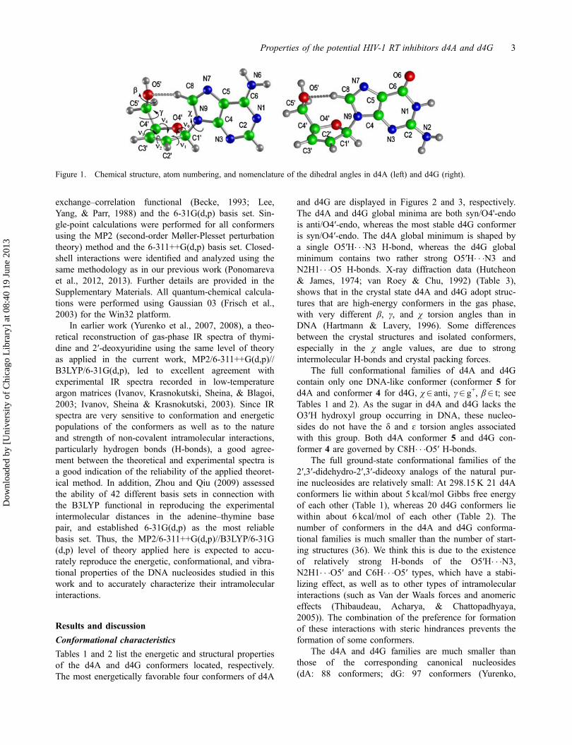
Computational methodology
The chemical structures of d4A and d4G are presentedin Figure 1, together with the atom numbering and defi-nition of the dihedral angles (Saenger, 1984). The sugarring pseudorotational concept by Altona and Sundaralin-gam (1972) was employed in this work. A conforma-tional analysis was performed by density functionaltheory (DFT) using the B3LYP nonlocal hybrid
2 A.G. Ponomareva et al.
Dow
nloa
ded
by [
Uni
vers
ity o
f C
hica
go L
ibra
ry]
at 0
8:40
19
June
201
3
exchange–correlation functional (Becke, 1993; Lee,Yang, & Parr, 1988) and the 6-31G(d,p) basis set. Sin-gle-point calculations were performed for all conformersusing the MP2 (second-order Møller-Plesset perturbationtheory) method and the 6-311++G(d,p) basis set. Closed-shell interactions were identified and analyzed using thesame methodology as in our previous work (Ponomarevaet al., 2012, 2013). Further details are provided in theSupplementary Materials. All quantum-chemical calcula-tions were performed using Gaussian 03 (Frisch et al.,2003) for the Win32 platform.
In earlier work (Yurenko et al., 2007, 2008), a theo-retical reconstruction of gas-phase IR spectra of thymi-dine and 2′-deoxyuridine using the same level of theoryas applied in the current work, MP2/6-311++G(d,p)//B3LYP/6-31G(d,p), led to excellent agreement withexperimental IR spectra recorded in low-temperatureargon matrices (Ivanov, Krasnokutski, Sheina, & Blagoi,2003; Ivanov, Sheina & Krasnokutski, 2003). Since IRspectra are very sensitive to conformation and energeticpopulations of the conformers as well as to the natureand strength of non-covalent intramolecular interactions,particularly hydrogen bonds (H-bonds), a good agree-ment between the theoretical and experimental spectra isa good indication of the reliability of the applied theoret-ical method. In addition, Zhou and Qiu (2009) assessedthe ability of 42 different basis sets in connection withthe B3LYP functional in reproducing the experimentalintermolecular distances in the adenine–thymine basepair, and established 6-31G(d,p) as the most reliablebasis set. Thus, the MP2/6-311++G(d,p)//B3LYP/6-31G(d,p) level of theory applied here is expected to accu-rately reproduce the energetic, conformational, and vibra-tional properties of the DNA nucleosides studied in thiswork and to accurately characterize their intramolecularinteractions.
Results and discussion
Conformational characteristics
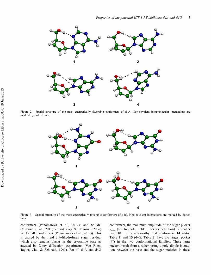
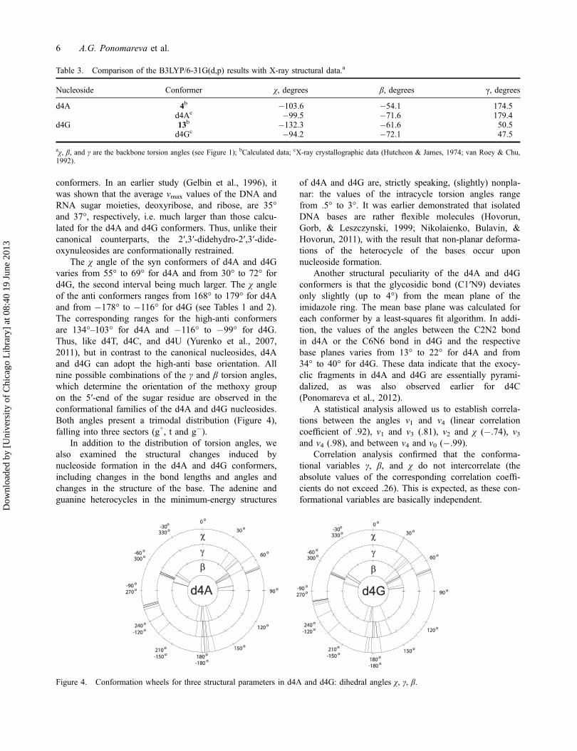
Tables 1 and 2 list the energetic and structural propertiesof the d4A and d4G conformers located, respectively.The most energetically favorable four conformers of d4A
and d4G are displayed in Figures 2 and 3, respectively.The d4A and d4G global minima are both syn/O4'-endois anti/O4′-endo, whereas the most stable d4G conformeris syn/O4′-endo. The d4A global minimum is shaped bya single O5′H� � �N3 H-bond, whereas the d4G globalminimum contains two rather strong O5′H� � �N3 andN2H1� � �O5 H-bonds. X-ray diffraction data (Hutcheon& James, 1974; van Roey & Chu, 1992) (Table 3),shows that in the crystal state d4A and d4G adopt struc-tures that are high-energy conformers in the gas phase,with very different β, γ, and χ torsion angles than inDNA (Hartmann & Lavery, 1996). Some differencesbetween the crystal structures and isolated conformers,especially in the χ angle values, are due to strongintermolecular H-bonds and crystal packing forces.
The full conformational families of d4A and d4Gcontain only one DNA-like conformer (conformer 5 ford4A and conformer 4 for d4G, χ2 anti, γ2 g+, β2 t; seeTables 1 and 2). As the sugar in d4A and d4G lacks theO3′H hydroxyl group occurring in DNA, these nucleo-sides do not have the δ and ɛ torsion angles associatedwith this group. Both d4A conformer 5 and d4G con-former 4 are governed by C8H� � �O5′ H-bonds.
The full ground-state conformational families of the2′,3′-didehydro-2′,3′-dideoxy analogs of the natural pur-ine nucleosides are relatively small: At 298.15K 21 d4Aconformers lie within about 5 kcal/mol Gibbs free energyof each other (Table 1), whereas 20 d4G conformers liewithin about 6 kcal/mol of each other (Table 2). Thenumber of conformers in the d4A and d4G conforma-tional families is much smaller than the number of start-ing structures (36). We think this is due to the existenceof relatively strong H-bonds of the O5′H� � �N3,N2H1� � �O5′ and С6H� � �O5′ types, which have a stabi-lizing effect, as well as to other types of intramolecularinteractions (such as Van der Waals forces and anomericeffects (Thibaudeau, Acharya, & Chattopadhyaya,2005)). The combination of the preference for formationof these interactions with steric hindrances prevents theformation of some conformers.
The d4A and d4G families are much smaller thanthose of the corresponding canonical nucleosides(dA: 88 conformers; dG: 97 conformers (Yurenko,
Figure 1. Chemical structure, atom numbering, and nomenclature of the dihedral angles in d4A (left) and d4G (right).
Properties of the potential HIV-1 RT inhibitors d4A and d4G 3
Dow
nloa
ded
by [
Uni
vers
ity o
f C
hica
go L
ibra
ry]
at 0
8:40
19
June
201
3

Zhurakivsky, & Hovorun, 2009)). The considerablereduction in the number of conformers on going fromthe canonical nucleosides to their 2′,3′-didehydro-2′,3′-
dideoxy analogs was also observed for dT, dU, and dC(88 dT vs. 19 d4T conformers (Ponomareva et al.,2013); 94 dU (Yurenko et al., 2008) vs. 20 d4U
Table 1. Energetic and structural properties of the d4A conformers calculated at the MP2/6-311++G(d,p)//B3LYP/6-31G(d,p) levelof theory.a
Conformer ΔG, kcal/mol μ, D νmax, degrees χ, degrees γ, degrees β, degrees τmax, degrees RC1′N9, Å
1 .00 3.34 7.7 55.3 35.4 54.5 .8 1.4682 1.77 2.54 3.3 �106.1 62.5 62.9 �.6 1.4573 2.14 3.43 1.5 68.8 175.5 �59.4 .9 1.4634 2.15 3.06 .5 �103.6 174.5 �54.1 .6 1.4585 2.49 4.72 1.7 �120.8 45.5 163.3 �.6 1.4596 2.78 2.08 2.0 167.9 49.6 56.2 �1.5 1.4787 3.03 3.60 2.8 �104.4 �66.1 �177.2 .6 1.4598 3.23 1.25 1.1 67.4 �69.5 168.7 .9 1.4659 3.45 4.16 2.4 �134.2 50.9 �60.3 �.7 1.46010 3.60 2.67 2.9 �104.6 �65.3 �74.7 .6 1.45811 3.63 2.58 2.3 175.5 �67.7 177.9 �.9 1.47912 3.99 2.78 1.2 66.8 �67.7 �73.5 .9 1.46313 4.10 2.24 1.4 68.8 �66.0 90.0 1.0 1.46514 4.17 2.96 9.1 178.5 178.6 �176.2 �.7 1.48015 4.24 2.87 1.7 176.7 �66.5 �74.2 �.8 1.47816 4.38 4.46 1.6 �105.3 �170.4 �167.2 .7 1.46117 4.65 3.44 2.4 �105.6 �67.9 80.4 .6 1.45818 4.71 3.40 8.5 �178.2 169.3 57.3 �.5 1.47719 4.83 .98 3.7 173.8 �67.6 81.5 �1.0 1.47920 5.14 4.10 4.2 �108.8 179.7 60.1 .7 1.45721 5.17 3.17 1.7 68.5 �179.4 62.8 1.1 1.464
aΔG= relative Gibbs free energy at 298.15K; μ= dipole moment; νmax =maximum degree of pucker: vmax ¼ v2cosP (Saenger, 1984), χ, γ, β are the
backbone torsion angles (see Figure 1); τmax =maximum torsion angle in the base; RC1′N9 is the glycosidic bond length. All torsion angles arein degrees. Structural characteristics were obtained at the B3LYP/6-31G(d,p) level of theory; relative electronic energies were calculated atthe MP2/6-311++G(d,p)//B3LYP/6-31G(d,p) level. The conformers are numbered in order of increasing Gibbs free energy. The puckeringparameters were calculated using an in-house SciLab routine.
Table 2. Energetic and structural properties of the d4G conformers calculated at the MP2/6-311++G(d,p)//B3LYP/6-31G(d,p) levelof theory.a
Conformer ΔG, kcal/mol μ, D νmax, degrees χ, degrees γ, degrees β, degrees τmax, degrees RC1'N9, Å
1 .00 5.65 8.5 53.5 39.1 44.9 1.0 1.4652 2.00 7.72 3.0 �103.2 59.6 64.4 1.8 1.4573 2.55 7.11 1.1 �100.5 174.9 �55.7 1.7 1.4584 3.00 6.72 1.0 �115.8 45.0 155.8 1.9 1.4595 3.11 5.45 1.0 �98.9 �66.2 �178.8 1.8 1.4606 3.19 6.57 2.7 69.4 174.5 �56.3 1.7 1.4597 3.72 7.82 .1 173.1 47.6 60.7 1.2 1.4798 4.03 7.57 1.5 �100.4 �65.8 �75.2 1.7 1.4589 4.31 7.20 2.7 71.7 �67.5 �176.1 2.0 1.46110 4.42 5.53 1.6 �100.1 �170.1 �163.3 1.9 1.46111 4.54 7.10 1.2 69.5 �65.4 �72.5 1.7 1.46112 4.55 6.42 .6 �100.0 �67.6 83.0 1.8 1.45913 4.78 8.75 2.3 �132.3 50.5 �61.6 2.1 1.46014 5.00 5.62 4.9 30.1 156.6 �62.0 2.7 1.47915 5.43 7.75 9.0 �177.8 178.3 �173.6 1.8 1.48016 5.53 7.94 .3 �172.3 �68.3 83.6 2.0 1.47517 5.61 6.71 2.4 69.0 �67.8 78.4 1.6 1.46118 5.66 8.18 1.2 �103.4 �179.0 61.8 1.9 1.45819 6.01 9.10 8.4 �174.6 168.9 56.8 1.9 1.47720 6.23 8.29 2.2 67.8 �179.3 60.4 1.6 1.460
aSee footnote, Table 1.
4 A.G. Ponomareva et al.
Dow
nloa
ded
by [
Uni
vers
ity o
f C
hica
go L
ibra
ry]
at 0
8:40
19
June
201
3
conformers (Ponomareva et al., 2012); and 88 dC(Yurenko et al., 2011; Zhurakivsky & Hovorun, 2006)vs. 19 d4C conformers (Ponomareva et al., 2012)). Thisis caused by the rigid 2,5-dihydrofuran sugar residue,which also remains planar in the crystalline state asattested by X-ray diffraction experiments (Van Roey,Taylor, Chu, & Schinazi, 1993). For all d4A and d4G
conformers, the maximum amplitude of the sugar puckerνmax (see footnote, Table 1 for its definition) is smallerthan 10°. It is noteworthy that conformers 14 (d4A,Table 1) and 15 (d4G, Table 2) have the largest pucker(9°) in the two conformational families. These largepuckers result from a rather strong dipole–dipole interac-tion between the base and the sugar moieties in these
Figure 2. Spatial structure of the most energetically favorable conformers of d4A. Non-covalent intramolecular interactions aremarked by dotted lines.
Figure 3. Spatial structure of the most energetically favorable conformers of d4G. Non-covalent interactions are marked by dottedlines.
Properties of the potential HIV-1 RT inhibitors d4A and d4G 5
Dow
nloa
ded
by [
Uni
vers
ity o
f C
hica
go L
ibra
ry]
at 0
8:40
19
June
201
3
conformers. In an earlier study (Gelbin et al., 1996), itwas shown that the average νmax values of the DNA andRNA sugar moieties, deoxyribose, and ribose, are 35°and 37°, respectively, i.e. much larger than those calcu-lated for the d4A and d4G conformers. Thus, unlike theircanonical counterparts, the 2′,3′-didehydro-2′,3′-dide-oxynuleosides are conformationally restrained.
The χ angle of the syn conformers of d4A and d4Gvaries from 55° to 69° for d4A and from 30° to 72° ford4G, the second interval being much larger. The χ angleof the anti conformers ranges from 168° to 179° for d4Aand from �178° to �116° for d4G (see Tables 1 and 2).The corresponding ranges for the high-anti conformersare 134°–103° for d4A and �116° to �99° for d4G.Thus, like d4T, d4C, and d4U (Yurenko et al., 2007,2011), but in contrast to the canonical nucleosides, d4Aand d4G can adopt the high-anti base orientation. Allnine possible combinations of the γ and β torsion angles,which determine the orientation of the methoxy groupon the 5′-end of the sugar residue are observed in theconformational families of the d4A and d4G nucleosides.Both angles present a trimodal distribution (Figure 4),falling into three sectors (g+, t and g�).
In addition to the distribution of torsion angles, wealso examined the structural changes induced bynucleoside formation in the d4A and d4G conformers,including changes in the bond lengths and angles andchanges in the structure of the base. The adenine andguanine heterocycles in the minimum-energy structures
of d4A and d4G are, strictly speaking, (slightly) nonpla-nar: the values of the intracycle torsion angles rangefrom .5° to 3°. It was earlier demonstrated that isolatedDNA bases are rather flexible molecules (Hovorun,Gorb, & Leszczynski, 1999; Nikolaienko, Bulavin, &Hovorun, 2011), with the result that non-planar deforma-tions of the heterocycle of the bases occur uponnucleoside formation.
Another structural peculiarity of the d4A and d4Gconformers is that the glycosidic bond (C1′N9) deviatesonly slightly (up to 4°) from the mean plane of theimidazole ring. The mean base plane was calculated foreach conformer by a least-squares fit algorithm. In addi-tion, the values of the angles between the C2N2 bondin d4A or the C6N6 bond in d4G and the respectivebase planes varies from 13° to 22° for d4A and from34° to 40° for d4G. These data indicate that the exocy-clic fragments in d4A and d4G are essentially pyrami-dalized, as was also observed earlier for d4C(Ponomareva et al., 2012).
A statistical analysis allowed us to establish correla-tions between the angles ν1 and ν4 (linear correlationcoefficient of .92), ν1 and ν3 (.81), ν2 and χ (�.74), ν3and ν4 (.98), and between ν4 and ν0 (�.99).
Correlation analysis confirmed that the conforma-tional variables γ, β, and χ do not intercorrelate (theabsolute values of the corresponding correlation coeffi-cients do not exceed .26). This is expected, as these con-formational variables are basically independent.
Figure 4. Conformation wheels for three structural parameters in d4A and d4G: dihedral angles χ, γ, β.
Table 3. Comparison of the B3LYP/6-31G(d,p) results with X-ray structural data.a
Nucleoside Conformer χ, degrees β, degrees γ, degrees
d4A 4b �103.6 �54.1 174.5d4Ac �99.5 �71.6 179.4
d4G 13b �132.3 �61.6 50.5d4Gc �94.2 �72.1 47.5
aχ, β, and γ are the backbone torsion angles (see Figure 1); bCalculated data; cX-ray crystallographic data (Hutcheon & James, 1974; van Roey & Chu,1992).
6 A.G. Ponomareva et al.
Dow
nloa
ded
by [
Uni
vers
ity o
f C
hica
go L
ibra
ry]
at 0
8:40
19
June
201
3
According to the Boltzmann distribution, the confor-mational equilibrium of d4A and d4G at 298.15K isshifted toward syn conformers, with syn:anti ratios of88:12% (d4A) and 94:6% (d4G). In this respect, d4Aand d4G differ drastically from stavudine (d4T), whichfavors the anti-base orientation in the free state (Pono-mareva et al., 2013). The distributions of the d4A andd4G sugar conformational subfamilies differ stronglyfrom those of the corresponding canonical nucleosides(Yurenko et al., 2007, 2011). The γ angle preferentiallyadopts the g+ conformation: γ2 g+ in 94% of d4A and98% of d4G conformers (whereas less than 1% of d4Aand d4G conformers contain the g� conformation for theγ angle). A similar distribution is observed for the βangle: β2 g+ in 93% d4A and 97% d4G conformers,whereas only �1% of d4A and d4G conformers adoptthe t conformation of the β angle.
The deoxyribose unit was shown to be the majorsource of the conformational flexibility of DNA (Foloppe& MacKerell, 1999; Poltev et al., 2010) and, thus, theoverall DNA structure is to a large extent determined bythe conformational properties of 2′-deoxyribonucleosides.Due to the tightness of the active sites of polymerases(Kim, Delaney, Essigmann, & Kool, 2005), thenucleoside triphosphate should possess a DNA-likeconformation to allow incorporation into DNA. Toinvestigate possible conformational effects on the biologi-cal activity of d4A and d4G, we optimized two structuresfor each nucleoside with the χ angle fixed at the “ideal”(Foloppe & MacKerell, 1999) A- and B-DNA forms,�158.9° and �101.9°, respectively. The results, shown inTable 4, indicate that, like d4T (Ponomareva et al., 2013),d4U and d4C (Ponomareva et al., 2012), d4A and d4Gcan adopt A as well as B conformations. This is animportant requirement to allow their incorporation intodouble-helical DNA. All four “ideal” DNA-like confor-mations of d4A and d4G are stabilized by a C8H� � �O5′H-bond, which plays an important role in maintaining themutual orientation of the base and sugar residues in DNA(Wahl & Sundaralingam, 1997). These results show thatthere are no energetic or conformational barriers for the
incorporation of d4A and d4G into double-helical DNA.This confirms the earlier suggestion that the biologicalactivity of the 2′,3′-didehydro-2′,3′-dideoxy analogs ofthe natural nucleosides is directly related to the lack of ahydroxyl group at the С3′-position of the sugar moiety(Hamamoto et al., 1987). As a result, they function ascompetitive RT inhibitors through termination of theDNA biosynthesis in the 5′-3′ direction. We are currentlyperforming calculations to investigate the interactions ofthe triphosphates of the 2′,3′-didehydro-2′,3′-dideoxynucleosides with HIV-1 RT, with the aim to provide amore detailed understanding of the biological activity ofd4A and d4G.
Specific intramolecular interactions.
In general, 21 specific intramolecular contacts involvinga hydrogen atom (hereafter called H-contacts), wereidentified in d4A and 23 in d4G by means of QTAIM(quantum theory of atoms in molecules) electron densitytopological analysis (Bader, 1990), see Tables SI and SII(Supplementary Materials). Most conformers (19 d4Aand 14 d4G conformers) are shaped by only oneH-contact, whereas one d4A and four d4G conformersare stabilized by two H-contacts.
The specific intramolecular contacts are sensitive tothe nucleoside conformation. In particular, theC8H� � �O5′, C2′H� � �N3, O5′H� � �C8 and C8H� � �H1/2C5′interactions only occur in conformers with an anti-ori-ented base, whereas O5′H� � �N3, N2H1� � �O5′, and C5′H1/2� � �N3 contacts can be formed in syn-conformers.Like in d4T, d4U, and d4C (Ponomareva et al., 2012;2013), the orientation of γ determines which of the twohydrogen atoms of the C5′H group (H1 or H2, seeFigure 1) is involved in H-bonding: If γ2 t, it is H1, andif γ2 g� it is H2 that participates in H-bonding. All spe-cific intramolecular contacts are formed exclusivelybetween the base and the sugar residue, in contrast tothe canonical 2′-deoxyribonucleosides, where intra-sugarH-bonds of the C2′H� � �O5′ type are also observed (Yur-enko et al., 2011). The N3 atom of the nucleobase is themost common hydrogen-bonding acceptor in d4A andd4G (11 interactions in d4A and 9 in d4G).
Table 4. Energetic and conformational characteristics of the DNA-like conformers of d4A and d4G (5 and 4, respectively) and thepartially optimized structures (with fixed χ angles) that correspond to the ВI- (χ =�158.9°) and АI-(χ=�101.9°) DNA forms.a
Nucleoside Conformer ΔG, kcal/mol μ, D νmax, degrees χ, degrees γ, degrees β, degrees τmax, degrees RC1′N9, Å
d4A 5 2.49 4.72 1.7 �120.9 45.4 163.3 .6 1.459A-DNA 3.91 4.83 4.1 �158.9b 50.3 172.9 2.0 1.468B-DNA 2.75 4.42 1.4 �101.9b 46.1 152.4 1.5 1.458
d4G 4 3.01 6.72 1.0 �115.8 45.0 155.8 1.9 1.459A-DNA 4.23 7.46 4.1 �158.9b 49.4 170.4 2.0 1.468B-DNA 3.22 6.74 1.5 �101.9b 45.1 180.0b 2.0 1.458
aSee footnote, Table 1.bThese values have been frozen during geometry optimization.
Properties of the potential HIV-1 RT inhibitors d4A and d4G 7
Dow
nloa
ded
by [
Uni
vers
ity o
f C
hica
go L
ibra
ry]
at 0
8:40
19
June
201
3
The specific intramolecular interactions in d4A andd4G can be divided into three categories: conventional(OH� � �N, NH� � �O) H-bonds, non-conventional(CH� � �O, OH� � �C, CH� � �N) H-bonds, and hydrogen–hydrogen contacts (CH� � �HC) (Bakhmutov, 2008; Mat-ta, 2006; Matta, Hernández-Trujillo, Tang, & Bader,2003). The conventional OH� � �N and NH� � �O H-bondsin d4A and d4G possess distinct spectral characteristics(Table 5), including a perceptible red shift of thestretching mode frequency ν (OH/NH), an increase inthe stretching mode intensity and an increase in the out-of-plane γ (OH/NH) vibrational frequency. Based onthese results, the energy of the conventional H-bondswas evaluated by means of Iogansen’s empiricalformula (Equation (3) in Supplementary Materials) torange from 3.08 to 6.48 kcal/mol.
The magnitude of the electron density ρ at the bondcritical points of the noncovalent interactions in d4A andd4G (Tables SI and SII, Supplementary Materials) are ingood agreement with the H-bonding criteria of (Koch &Popelier, 1995). All H-contacts, except the CH� � �HC andO5′H� � �C8 interactions, meet Bondi’s geometric require-ments for H-bonding (Bondi, 1964), in particular therequirement that the distance between the donor andacceptor atoms should be smaller than the sum of thevan der Waals atomic radii, ΣrvdW (2.72Å for H� � �O andH� � �N and 2.2Å for H� � �H). In general, there is a goodcorrelation between the ΣrvdW values and the H� � �B dis-tance (where B is the acceptor atom), with a correlationcoefficient of .988.
In this work, we used Grunenberg’s complianceconstant theory (Brandhorst & Grunenberg, 2008, 2010;Grunenberg, 2004) to evaluate the individual strengths ofthe intramolecular interactions. Compliance constants(Cij) are elements of the inverse force constant matrix.Unlike the elements of the regular force constant (Hes-sian) matrix, they depend only on the definition of thetwo internal coordinates pertaining to it and are indepen-dent of the definition of all other coordinates. Tables SIand SII, and Figure S1 (Supplementary Materials) clearlyshow that smaller values of Cij correspond to stronger
contacts, as quantified by the energies of the specificinteractions EHB calculated by the Espinosa-Molins-Lec-omte formula (Equation (1) in Supplementary Materials).The correlation is evidently not linear, as was assumedin our previous work (Ponomareva et al., 2012, 2013).The solid line in Figure S1 corresponds to a least-squarespower fit (Cij = 53.365�EHB
�1.2285; R=�.93). Some ofthe CH� � �HC contacts are characterized by very high Сij
values (> 50 Å/mDyn), indicating the relatively weaknature of these contacts.
The C8H� � �H2C5′/H1C5′ hydrogen–hydrogen con-tacts in d4A and d4G deserve special attention. These H-bonds were earlier detected in canonical nucleosides(Yurenko et al., 2007) and some amino acids (Matta &Bader, 2000) by means of Bader’s topological analysis(Bader, 1990). To find the H-bond donating (donor) andaccepting (acceptor) groups in these interactions (assum-ing these are genuine H-bonds (Matta, 2006; Mattaet al., 2003)), an integration (in natural coordinates) overBader atomic basins was performed for each hydrogenatom (Tables SV and SVI in Supplementary Materials).The hydrogen on the base moiety bears a partial positivecharge and has less negative atomic energy than thehydrogen of the sugar residue, which is negativelycharged and more energetically stable. (Note that thecharges are calculated with the QTAIM method.) Thus,the H-bond donating group is located on the sugar resi-due, whereas the acceptor is on the base unit. This is inagreement with the results for d4T (Ponomareva et al.,2013), d4U, and d4C (Ponomareva et al., 2012).
Conclusions
The conformational landscapes of two HIV-1 RTinhibitors, the nucleoside analogs 2′,3′-didehydro-2′,3′-dideoxyadenosine (d4A), and 2′,3′-didehydro-2′,3′-dideoxyguanosine (d4G) were explored using a compre-hensive set of modern computational techniques (DFTand MP2 calculations, Bader’s electron density topologi-cal analysis, Natural Bond Orbital (NBO) analysis, andcompliance constant theory).
Table 5. Vibrational and energetic characteristics of the intramolecular OH� � �N and NH� � �O H-bonds in d4A conformer 5 and d4Gconformers 1 and 14.
Nucleoside Conformer H-bond Δda, Å Δνb, cm�1 I/I0c Δγd, cm�1 ЕHB
e, kcal/mol
d4A 5 O5′H� � �N3 .019 �332 52.0 110 5.64d4G 1 O5′H� � �N3 .021 �425 52.7 148 6.48
N2H1� � �O5′ .004 �142 4.8 79 3.3314 O5′H� � �N3 .017 �245 14.5 98 4.72
N2H1� � �O5′ .004 �127 3.2 68 3.08
aDifference between the OH/NH bond length in the OH(NH)� � �O/N H-bond and in the nonbonded state.bDecrease in the OH/NH stretching mode frequency upon H-bonding.cI and I0: OH/NH stretching mode integral intensity upon H-bonding and in the nonbonded state, respectively.dIncrease in the OH/NH out-of-plane mode frequency upon H-bonding.eH-bond energy (in kcal/mol), calculated by Iogansen’s empirical formula (Equation [3] in Supplementary Materials).
8 A.G. Ponomareva et al.
Dow
nloa
ded
by [
Uni
vers
ity o
f C
hica
go L
ibra
ry]
at 0
8:40
19
June
201
3
We identified 21 d4A and 20 d4G conformers. Theconformers are shaped by a sophisticated network ofspecific interactions of various types (NH� � �O, OH� � �N,СH� � �O, OH� � �C, and CH� � �HC). The relative strengthsof these interactions can be efficiently predicted usingGrunenberg’s compliance constants theory (Brandhorst &Grunenberg, 2008, 2010; Grunenberg, 2004). NBO anal-ysis (Supplementary Materials) revealed the electronicnature of the noncovalent interactions. For the OH� � �O,NH� � �O, and CH� � �O H-bonds hyperconjugative interac-tions between the oxygen lone pairs and the antibondingorbital of the donor group are observed. The electrondensity of the CH� � �HC contacts migrates from the anti-bonding orbital, corresponding to the CH group of thesugar residue, to the bonding orbital relative to the samegroup in the nucleobase.
The results indicate that there are no energetic orconformational obstacles to the incorporation of d4A andd4G into double-helical DNA. This provides furthersupport for the suggestion that the biological activity of2′,3′-didehydro-2′,3′-dideoxy analogs is directly relatedto the lack of a hydroxyl group at the С3′-position of thesugar moiety (Hamamoto et al., 1987). As a result, thesenucleosides act as competitive RT inhibitors throughtermination of the DNA biosynthesis in the 5′-3′direction.
The biological relevance of the results presented inthis work lies in the considerable flexibility of the2′,3′-didehydro-2′,3′-didehydro-nucleosides; as biologicalfunction of these nucleosides is structure-dependent, aconformational analysis is the initial, but very important,step in elucidating their biological activity. A detailedknowledge of the full conformational families of thesenucleosides is essential for establishing the biologicallyrelevant conformations that are formed in the course ofbiomolecular recognition by different enzymes (transport-ers, kinases/5′-nucleotidases, polymerases). In addition,we have identified and characterized among all the con-formers the one that can be introduced in the active siteand potentially block the DNA biosynthesis. A study ofthe interactions of the triphosphates of these nucleosideswith HIV-1 RT could provide a more detailed understand-ing of the biological activity of d4A and d4G. Suchcalculations are currently being performed in our researchgroups.
Acknowledgements
The authors greatly appreciate useful discussion of the resultswith Prof. M. Ghomi and Dr B. Hernandez (Université Paris13). Y.P.Y. is deeply grateful to Gaussian Inc. for granting aG03 molecular modeling package for the Win32 platform. Thiswork was partly supported by the State Fund for FundamentalResearch of Ukraine within Ukrainian-Slovenian bilateralresearch project (No М/451-2011).
Supplementary material
The supplementary material for this paper is availableonline at http://dx.doi.10.1080/07391102.2013.789401.
References
Alber, F., & Carloni, P. (2000). Ab initio molecular dynamicsstudies on HIV-1 reverse transcriptase triphosphate bindingsite: Implications for nucleoside-analog drug resistance.Protein Science, 9, 2535–2546.
Altona, C., & Sundaralingam, M. (1972). Conformationalanalysis of the sugar ring in nucleosides and nucleotides.New description using the concept of pseudorotation.Journal of the American Chemical Society, 94, 8205–8212.
Bader, R. F. W. (1990). Atoms in molecules: A quantum theory.Oxford: Clarendon.
Bakhmutov, V. I. (2008). Dihydrogen bond: Principles,experiments and applications. Hoboken, NJ: John Wiley &Sons.
Balzarini, J., Kruining, J., Wedgwood, O., Pannecouque, C.,Aquaro, S., Perno, C. F., … McGuigan, C. (1997).Conversion of 2′,3′-dideoxyadenosine (ddA) and 2′,3′-dide-hydro-2′,3′-dideoxyadenosine (d4A) to their correspondingaryloxyphosphoramidate derivatives markedly potentiatestheir activity against human immunodeficiency virus andhepatitis B virus. FEBS Letters, 410, 324–328.
Becke, A. D. (1993). Density functional thermochemistry. 3.The role of exact exchange. Journal of Chemical Physics,98, 5648–5652.
Bondi, A. (1964). Van der Waals volumes and radii. Journal ofPhysical Chemistry, 68, 441–451.
Brandhorst, K., & Grunenberg, J. (2008). How strong is it?The interpretation of force and compliance constants asbond strength descriptors. Chemical Society Reviews, 37,1558–1567.
Brandhorst, K., & Grunenberg, J. (2010). Efficient computationof compliance matrices in redundant internal coordinatesfrom Cartesian Hessians for nonstationary points. Journalof Chemical Physics, 132, 184101–184107.
Chu, C. K., Schinazi, R. F., Arnold, B. H., Cannon, D. L.,Doboszewski, B., Bhadti, V. B., & Gu, Z. P. (1988). Com-parative activity of 2′,3′-saturated and unsaturated pyrimi-dine and purine nucleosides against humanimmunodeficiency virus type-1 in peripheral-blood mono-nuclear-cells. Biochemical Pharmacology, 37, 3543–3548.
el Kouni, M. H. (2002). Trends in the design of nucleosideanalogues as anti-HIV drugs. Current PharmaceuticalDesign, 8, 581–593.
Everaert, D. H., Peeters, O. M., Deranter, C. J., Blaton, N. M.,Vanaerschot, A., & Herdewijn, P. (1993). Conformational-analysis of substituent effects on the sugar puckering modeand the anti-hiv activity of 2′,3′-dideoxypyrimidine nucleo-sides. Antiviral Chemistry & Chemotherapy, 4, 289–299.
Foloppe, N., & MacKerell, A. D. (1999). Intrinsic conforma-tional properties of deoxyribonucleosides: Implicated rolefor cytosine in the equilibrium among the A, B, and Zforms of DNA. Biophysics Journal, 76, 3206–3218.
Frisch, M. J., Trucks, G. W., Schlegel, H. B., Scuseria, G. E.,Robb, M. A., Cheeseman, J. R., … Pople, J. A. (2003).Gaussian 03. Pittsburgh, PA: Gaussian.
Gelbin, A., Schneider, B., Clowney, L., Hsieh, S.-H., Olson,W. K., & Berman, H. M. (1996). Geometric parameters innucleic acids: Sugar and phosphate constituents. Journal ofthe American Chemical Society, 118, 519–529.
Properties of the potential HIV-1 RT inhibitors d4A and d4G 9
Dow
nloa
ded
by [
Uni
vers
ity o
f C
hica
go L
ibra
ry]
at 0
8:40
19
June
201
3
Grunenberg, J. (2004). Direct assessment of interresidue forcesin Watson-Crick base pairs using theoretical complianceconstants. Journal of the American Chemical Society, 126,16310–16311.
Hamamoto, Y., Nakashima, H., Matsui, T., Matsuda, A., Ueda,T., & Yamamoto, N. (1987). Inhibitory effect of 2′,3′-dide-hydro-2′,3′-dideoxynucleosides on infectivity, cytopathiceffects, and replication of human-immunodeficiency-virus.Antimicrobial Agents and Chemotherapy, 31, 907–910.
Hartmann, B., & Lavery, R. (1996). DNA structural forms.Quarterly Reviews of Biolphysics, 29, 309–368.
Herdewijn, P. (Ed.). (2008). Modified nucleosides in biochemis-try, biotechnology and medicine. Weinheim: Wiley.
Hitchcock, M. J. M. (1991). 2′ 3′ Didehydro-2′ 3′-dideoxyt-hymidine d4T an anti-HIV agent. Antiviral Chemistry andChemotherapy, 2, 125–132.
Hovorun, D. M., Gorb, L., & Leszczynski, J. (1999). From thenonplanarity of the amino group to the structural nonrig-idity of the molecule: A post-Hartree–Fock ab initio studyof 2-aminoimidazole. International Journal of QuantumChemistry, 75, 245–253.
Hutcheon, W. L., & James, M. N. G. (1974). Molecular geome-try of 2′,3′-dideoxy-2′3′-didehydroadenosine from an X-raycrystal-structure determination. Acta Crystallographica B,B30, 1777–1782.
Ivanov, A. Y., Krasnokutski, S. A., Sheina, G., & Blagoi, Y. P.(2003). Conformational structures and vibrational spectra ofisolated pyrimidine nucleosides: Fourier transform infraredmatrix isolation study of 2-deoxyuridine. SpectrochimicaActa A: Molecular Biomolecular Spectros, 59, 1959–1973.
Ivanov, A. Y., Sheina, G. G., & Krasnokutski, S. A. (2003).Molecular structures of thymidine isomers isolated in low-temperature inert matrices. Low Temperature Physics, 29,809–813.
Jeang, K.-T. (2000). HIN-I: Molecular biology and pathogene-sis. San Diego, CA: Academic press.
Kim, T. W., Delaney, J. C., Essigmann, J. M., & Kool, E. T.(2005). Probing the active site tightness of DNA polymer-ase in subangstrom increments. Proceedings of the Nationalacademy of Sciences of the United States of America, 102,15803–15808.
King, A. E., Ackley, M. A., Cass, C. E., Young, J. D., & Bald-win, S. A. (2006). Nucleoside transporters: From scaveng-ers to novel therapeutic targets. Trends in ParmacologicalSciences, 27, 416–425.
Koch, U., & Popelier, P. L. A. (1995). Characterization of C–H–O hydrogen-bonds on the basis of the charge density.Journal of Physical Chemistry, 99, 9747–9754.
Lee, C., Yang, W., & Parr, R. G. (1988). Physical Review B,37, 785–789.
Matta, C. F. (2006). Hydrogen–hydrogen bonding. The non-electrostatic limit of closed-shell interaction between twohydrogen atoms. A critical review. In S. Grabowski (Ed.),Hydrogen bonding – New insights (pp. 337–375). Dordr-echt (Netherlands): Springer.
Matta, C. F., & Bader, R. F. W. (2000). An atoms-in-moleculesstudy of the genetically-encoded amino acids: I. Effects ofconformation and of tautomerization on geometric, atomic,and bond properties. Proteins: Structure, Function andGenetics, 40, 310–329.
Matta, C. F., Hernández-Trujillo, J., Tang, T.-H., & Bader, R.F. W. (2003). Hydrogen–hydrogen bonding: A stabilizinginteraction in molecules and crystals. Chemistry A Euro-pean Journal, 9, 1940–1951.
Mitsuya, H., Yarchoan, R., Kageyama, S., & Broder, S. (1991).Targeted therapy of human immunodeficiency virus-relateddisease. Faseb Journal, 5, 2369–2381.
Nikolaienko, T. Y., Bulavin, L. A., & Hovorun, D. M. (2011).How flexible are DNA constituents? The quantum-mechani-cal study. Journal of Biomolecular Structure & Dynamics,29, 563–575.
Palafox, M. A., Iza, N., de la Fuente, M., & Navarro, R.(2009). Simulation of the first hydration shell of nucleo-sides D4T and thymidine: Structures obtained using MP2and DFT methods. Journal of Physical Chemistry B, 113,2458–2476.
Poltev, V. I., Anisimov, V. M., Danilov, V. I., van Mourik, T.,Deriabina, A., González, E., … Polteva, N. (2010). DFTstudy of polymorphism of the DNA double helix at thelevel of dinucleoside monophosphates. International Jour-nal of Quantum Chemistry, 110, 2548–2559.
Ponomareva, A. G., Yurenko, Y. P., Zhurakivsky, R. O., vanMourik, T., & Hovorun, D. M. (2012). Completeconformational space of the potential HIV-1 reversetranscriptase inhibitors d4U and d4C. A quantum chemicalstudy. Physical Chemistry Chemical Physics: PCCP, 14,6787–6795.
Ponomareva, A. G., Yurenko, Y. P., Zhurakivsky, R. O., vanMourik, T., & Hovorun, D. M. (2013). Conformationallandscape of the nucleoside reverse transcriptase inhibitord4T: A comprehensive quantum-chemical approach. Cur-rent Physical Chemistry, 3, 83–92.
Rao, S. N. (1998a). Conformational studies on nucleosides withfuranose ring modifications. 1. Nucleosides and Nucleo-tides, 17, 791–814.
Rao, S. N. (1998b). Molecular mechanics studies on the con-formations of 2′,3′-dideoxy-2′,3′-didehydroguanine nucleo-side, d4G. Biophysics Journal, 74, 3131–3139.
Ray, A. S., Yang, Z. J., Chu, C. K., & Anderson, K. S. (2002).Novel use of a guanosine prodrug approach to convert2′,3′-didehydro-2′,3′-dideoxyguanosine into a viable antivi-ral agent. Antimicrobial Agents and Chemotherapy, 46,887–891.
Russell, J. W., Whiterock, V. J., Marrero, D., & Klunk, L. J.(1989). Pharmacokinetics of a new anti-HIV agent: 2′,3′-dideoxy-2′,3′-didehydrothymidine (d4T). Nucleosides andNucleotides, 8, 845–848.
Saenger, W. (1984). Principles of nucleic acid structure. NewYork, NY: Springer-Verlag.
Sahlberg, C., & Zhou, X.-X. (2008). Development ofnon-nucleoside reverse transcriptase inhibitors for anti-HIVtherapy. Anti-Infective Agents in Medicinal Chemistry, 7,101–117.
Skowron, G., & Ogden, R. (2006). Reverse transcriptaseinhibitors in HIV/AIDS therapy. Totowa, NJ: Humana Press.p. 527.
Sluis-Cremer, N., Temiz, N. A., & Bahar, I. (2004).Conformational changes in HIV-1 reverse transcriptaseinduced by nonnucleoside reverse transcriptase inhibitorbinding. Current HIV Research, 2, 323–332.
Thibaudeau, C., Acharya, P., & Chattopadhyaya, J. (2005).Stereoelectronic effects in nuxleosides and nucleotides andtheir structural implications. Uppsala: Uppsala UniversityPress.
van Roey, P., & Chu, C. K. (1992). The crystal andmolecular-structure of the complex of 2′,3′-didehydro-2′,3′-dideoxyguanosine with pyridine. Nucleosides &Nucleotides, 11, 1229–1239.
10 A.G. Ponomareva et al.
Dow
nloa
ded
by [
Uni
vers
ity o
f C
hica
go L
ibra
ry]
at 0
8:40
19
June
201
3
Van Roey, P., Taylor, E. W., Chu, C. K., & Schinazi, R. F.(1993). Conformational analysis of 2′,3′-didehydro-2′,3′-dideoxypyrimidine nucleosides. Journal of the AmericanChemical Society, 115, 5365–5371.
Van Rompay, A. R., Johansson, M., & Karlsson, A. (2003).Substrate specificity and phosphorylation of antiviral andanticancer nucleoside analogues by human deoxyribonucle-oside kinases and ribonucleoside kinases. Pharmacology &Therapeutics, 100, 119–139.
Wahl, M. C., & Sundaralingam, M. (1997). C–H � � �O hydro-gen bonding in biology. Trends in Biochemical Sciences,22, 97–102.
Yurenko, Y. P., Zhurakivsky, R. O., Ghomi, M., Samijlenko, S.P., & Hovorun, D. M. (2007a). Comprehensive conforma-tional analysis of the nucleoside analogue 2′-β-deoxy-6-aza-cytidine by DFT and MP2 calculations. Journal of PhysicalChemistry B, 111, 6263–6271.
Yurenko, Y. P., Zhurakivsky, R. O., Ghomi, M., Samijlenko,S. P., & Hovorun, D. M. (2007b). How many conformersdetermine the thymidine low-temperature matrix infraredspectrum? DFT and MP2 quantum chemical study. Journalof Physical Chemistry B, 111, 9655–9663.
Yurenko, Y. P., Zhurakivsky, R. O., Ghomi, M., Samijlenko,S. P., & Hovorun, D. M. (2008). Ab initio comprehensiveconformational analysis of 2′-deoxyuridine, the biologically
significant DNA minor nucleoside, and reconstruction of itslow-temperature matrix infrared spectrum. Journal ofPhysical Chemistry B, 112, 1240–1250.
Yurenko, Y. P., Zhurakivsky, R. O., & Hovorun, D. M. (2009).Intermolecular C-H...O hydrogen bonds in biologically sig-nificant conformers of canonical 2-deoxyribonucleotides:Ab initio topological analysis of the electron density. Phys-ics Alive (in Ukrainian), 17, 44–53.
Yurenko, Y. P., Zhurakivsky, R. O., Samijlenko, S. P., Ghomi,M., & Hovorun, D. M. (2007). The whole ofintramolecular H-bonding in the isolated DNA nucleosidethymidine. AIM electron density topological study.Chemical Physics Letters, 447, 140–146.
Yurenko, Y. P., Zhurakivsky, R. O., Samijlenko, S. P., &Hovorun, D. M. (2011). Intramolecular CH� � �O hydrogenbonds in the AI and BI DNA-like conformers of canonicalnucleosides and their Watson-Crick pairs. Quantumchemical and AIM analysis. Journal of BiomolecularStructure & Dynamics, 29, 51–65.
Zhou, P.-P., & Qiu, W.-Y. (2009). Red-shifted hydrogen bondsand blue-shifted van der Waals contact in the standard Wat-son�Crick adenine�thymine base pair. Journal of PhysicalChemistry A, 113, 10306–10320.
Zhurakivsky, R. O., & Hovorun, D. M. (2006). Extensive con-formational analysis of canonical nucleoside 2′-deoxycyti-dine: A DFT study. Physics Alive (in Ukrainian), 14, 33–46.
Properties of the potential HIV-1 RT inhibitors d4A and d4G 11
Dow
nloa
ded
by [
Uni
vers
ity o
f C
hica
go L
ibra
ry]
at 0
8:40
19
June
201
3


![Synthesis, Biological Evaluation, and Binding Mode of Novel 1-[2-(Diarylmethoxy)ethyl]-2-methyl-5-nitroimidazoles Targeted at the HIV1 Reverse Transcriptase](https://static.fdokumen.com/doc/165x107/6314434585333559270ca074/synthesis-biological-evaluation-and-binding-mode-of-novel-1-2-diarylmethoxyethyl-2-methyl-5-nitroimidazoles.jpg)


![Synthesis, biological evaluation, and binding mode of novel 1-[2-(diarylmethoxy) ethyl]-2-methyl-5-nitroimidazoles targeted at the HIV-1 reverse transcriptase](https://static.fdokumen.com/doc/165x107/6335adc702a8c1a4ec01e00d/synthesis-biological-evaluation-and-binding-mode-of-novel-1-2-diarylmethoxy.jpg)