Temporal differences in food abundance promote coexistence between two congeneric passerines
Upload
independentCategory
view
1download
0
Accepted by D. Gordon: 5 Aug. 2014; published: 28 Aug. 2014
Licensed under a Creative Commons Attribution License http://creativecommons.org/licenses/by/3.0
ZOOTAXA
ISSN 1175-5326 (print edition)
ISSN 1175-5334 (online edition)Copyright © 2014 Magnolia Press
Zootaxa 3857 (2): 151–182
www.mapress.com/zootaxa/
Article
151
http://dx.doi.org/10.11646/zootaxa.3857.2.1
http://zoobank.org/urn:lsid:zoobank.org:pub:96CEC1DB-94B8-4E38-88E1-CBA15871C2AE
The identity of the invasive fouling bryozoan Watersipora subtorquata
(d’Orbigny) and some other congeneric species
LEANDRO M. VIEIRA1,2
, MARY SPENCER JONES3
& PAUL D. TAYLOR4
1
Centro de Biologia Marinha, Universidade de São Paulo, São Sebastião, São Paulo, Brazil. E-mail: [email protected]
2
Departamento de Zoologia, Centro de Ciências Biológicas, Universidade Federal de Pernambuco, Recife, Pernambuco, Brazil
3
Department of Life Sciences, Natural History Museum, London, UK. E-mail: [email protected]
4
Department of Earth Sciences, Natural History Museum, London, UK. E-mail: [email protected]
Abstract
Watersipora subtorquata (d’Orbigny, 1852) has been widely reported as a fouling species from tropical to temperate wa-
ters. The continued confusion over the correct name for this species led us to provide a redescription of d’Orbigny’s type
of Cellepora subtorquata, and to make comparisons with other species of Watersipora. We show that the majority of spec-
imens assigned to W. subovoidea (d’Orbigny, 1852) are morphologically distinct from the recently erected neotype of W.
subovoidea; these specimens are here reidentified as Watersipora subtorquata. Other specimens previously assigned to W.
subtorquata belong to W. subatra (Ortmann, 1890), described originally from Japan. Owing these inconsistences, we sug-
gest setting aside the neotype of Watersipora subovoidea, which is based on Busk’s Lepralia cucullata and is not from the
same locality as d’Orbigny’s type. Watersipora cucullata is redescribed and figured using Busk’s specimens; the species
is known from the Mediterranean, including the Adriatic. Three other species—Watersipora atrofusca (Busk, 1856), Wa-
tersipora aterrima (Ortmann, 1890) and Watersipora nigra (Canu & Bassler, 1930)—are also refigured. Watersipora ed-
mondsoni Soule & Soule, 1975 is synonymised with W. subtorquata (d’Orbigny). Two new species are described,
Watersipora mawatarii n. sp. from Japan and Watersipora souleorum n. sp. from the Azores, Cape Verde, Naples and In-
dian Ocean. A key is given to the Recent species of Watersipora.
Key words: Bryozoa, Cheilostomata, Watersiporidae, fouling species, invasive species, new species, taxonomy, type
specimens
Introduction
Watersipora subtorquata (d’Orbigny, 1852) and Watersipora subovoidea (d’Orbigny, 1852) have been widely
reported as fouling species in harbour areas, from tropical to temperate waters (Harmer 1957; Ryland 1974; Soule
& Soule 1975; Ryland et al. 2009; Mackie et al. 2012). Ryland et al. (2009) noted that these species are frequently
confused owing to the absence of a modern taxonomic account comparing W. subtorquata with W. subovoidea.
D’Orbigny (1842) first reported Escharina torquata (Lamouroux, 1825) from Rio de Janeiro, Brazil, later
renaming the species Cellepora subtorquata d’Orbigny, 1852 because of homonymy (d’Orbigny 1852). Marcus
(1937) described similarities between specimens from Santos and Rio de Janeiro that had been described by
d’Orbigny, but adopted the name Watersipora cucullata (Busk, 1854), commonly used by contemporary
taxonomists, for this Brazilian material. This name was still being used for specimens from São Paulo and Espírito
Santo, Brazil (Marcus 1938, 1955) until Vieira et al. (2008) followed Taylor & Gordon (2002) in using the name W.
subtorquata for the Brazilian specimens. Recently, Ramalho et al. (2011) compared the Rio de Janeiro specimens
with recent descriptions given by Ryland et al. (2009) and applied the name W. subovoidea to Brazilian material,
even though this species was originally introduced by d’Orbigny (1852) for material (Savigny 1817, pl. 8, fig. 1)
from Egypt. These authors suggested that the specimens of Marcus (1937, 1938, 1955) required revision.
Molecular studies investigating introduced populations of Watersipora (Mackie et al. 2006, 2012; Geller et al.
2008) have suggested that a common haplotype of W. subtorquata is found in southern Australia, New Zealand and

VIEIRA ET AL. 152 · Zootaxa 3857 (2) © 2014 Magnolia Press
California, whereas W. subovoidea is found in the Western Atlantic (Florida and Brazil) and Australia. Despite the
type locality of W. subtorquata being Rio de Janeiro, neither recent taxonomic nor molecular studies have reported
the occurrence of this species along the Brazilian coast (Mackie et al. 2006, 2012; Geller et al. 2008; Ryland et al.
2009; Ramalho et al. 2011). Ryland et al. (2009) designated a neotype for W. subovoidea to recognize its
conspecificity with W. cucullata, but no redescription of d’Orbigny’s type of Cellepora subtorquata was given.
Previously, Taylor & Gordon (2002) had reproduced d’Orbigny’s original plates (d’Orbigny 1842, pl. 4, figs 2 and
3), along with a scanning electron micrograph of the putative type specimen found in the d’Orbigny Collection, but
did not redescribe this important invasive fouling species.
The current paper provides a redescription of the type specimen of Watersipora subtorquata and makes
comparisons with a range of congeneric species based on new SEM studies. This taxonomic revision clarifies the
complex taxonomy of Watersipora and leads to the introduction of two new species. All Recent species of
Watersipora are included in a key that aims to simplify identification of the species belonging to this invasive
fouling bryozoan.
Material and methods
Specimens used in this study are deposited at the Musée Océanographique de Monaco, Monaco (MOM), Museu de
Zoologia da Universidade de São Paulo, Brazil (MZUSP), Natural History Museum, London (NHMUK), National
Museum of Natural History, Smithsonian Institution, Washington DC (USNM), and Santa Barbara Museum of
Natural History, Santa Barbara (SBMNH). D’Orbigny’s type of Watersipora subtorquata is deposited in the
palaeontological collections at the Muséum national d’Histoire naturelle, Paris (MNHN). The type specimens of
Watersipora aterrima (Ortmann, 1890) and Watersipora subatra (Ortmann, 1890) are deposited in the collection of
the Musée zoologique de la Ville de Strasbourg (MZS). Material was photographed using a Zeiss Discovery V20
stereomicroscope with AxioCam HRc. Selected specimens were imaged at the NHMUK using a LEO 1455-VP
scanning electron microscope (SEM) equipped with a low-vacuum chamber and back-scattered electron detector.
Measurements were made directly from digital SEM images using ImageJ analysing software (http://
rsbweb.nih.gov/ij/). Abbreviations used for measurements are as follows: ZL, zooid length; ZW, zooid width; ZA,
zooid area, calculated as a rectangle (ZL x ZW); OL, orifice length; OW, orifice width; OA, orifice area, calculated
as an ellipse (ð · OL/2 · OW/2); SinL, sinus length; SinW, sinus width; PorD, pore diameter.
Results and discussion
Identity of Watersipora subtorquata (d’Orbigny, 1852)
Critical to understanding the correct name of the common fouling species of Watersipora is establishing the
identity of W. subtorquata (d’Orbigny, 1852). Specimens of a bryozoan from Rio de Janeiro described by
d’Orbigny (1842) as Escharina torquata (Lamouroux, 1825) were later renamed by him (d’Orbigny 1852, p. 399)
as Cellepora subtorquata, presumably to avoid homonymy with another species he referred to as Cellepora
torquata (Quoy & Gaimard, 1827) (see d’Orbigny 1852, p. 403). Waters (1879) synonymised d’Orbigny’s
Cellepora subtorquata with Lepralia cucullata Busk, 1854 but inexplicably applied the name of the junior
synonym L. cucullata for his specimens from Naples. The first revision of d’Orbigny’s type was undertaken
subsequently by Waters (1905) who studied d’Orbigny’s material deposited at the MNHN. He noted the high
quality of d’Orbigny’s original figures but remarked that, in the original specimens, the projecting lateral wings on
each side of the sinus were more developed than those depicted by d’Orbigny (1842). Taylor & Gordon (2002)
reviewed the bryozoan work of Alcide d’Orbigny (1802–1857), publishing a scanning electron micrograph of
d’Orbigny’s type specimen (Taylor & Gordon 2002, fig. 1C) in which it is possible to recognize some of the
diagnostic features (orifice shape and frontal pseudopores) depicted in the original figures of this species. The type
specimen of d’Orbigny (MNHN, d’Orbigny Collection 13637) (Figs 1–5) comprises a large colony with some
intact opercula (Figs 1–2); the projecting proximolateral wings are well developed in most zooids (Fig. 4), as
observed by Waters (1905), but with increasing calcification can become obscured, shallow and inconspicuous in

Zootaxa 3857 (2) © 2014 Magnolia Press · 153THE IDENTITY OF WATERSIPORA SUBTORQUATA
frontal views of the orifice (Figs 3, 5). The subcircular orifice (Fig. 5) was well figured in d’Orbigny’s plates; the
orifice is slightly wider than long, with a U-shaped sinus demarcated by triangular condyles that project
distomedially.
Comparisons of d’Orbigny’s type specimen and the specimens identified by Marcus (1937, 1938) from Brazil
under the name Watersipora cucullata (NHMUK 1948.2.16.18, and uncatalogued specimens deposited at MZUSP)
indicate that they represent the same species, as has been suggested by Gordon (1989) and Ryland et al. (2009).
Marcus (1937, pl. 24, fig. 63A, B) showed the orifice to have well-developed proximolateral wings in Brazilian
colonies. In addition, the operculum in the Brazilian specimens is characterized by a parallel-sided dark band of
two proximal lucidae adjacent to the condyles (Marcus 1937, pl. 24, fig. 63A, B). The same operculum shape and
lucidae were observed in Rio de Janeiro specimens figured by Ramalho et al. (2009, fig. 3D) in material they
named Watersipora subovoidea. Ramalho et al. (2011) followed Ryland et al. (2009) in using the name W.
subovoidea for specimens with “triangular, tooth-like condyles, located distomedially [sic], and a strongly
pigmented operculum with a parallel-sided dark central band”. All characteristics described for W. subovoidea by
Ryland et al. (2009) suggest that their specimens actually belong to W. subtorquata, and restudy of the material
they analyzed (NHMUK 2007.12.14.2–8) confirms this supposition. However, neither figures nor descriptions of
Busk’s type specimen of Lepralia cucullata (NHMUK 1854.11.15.189), designated by Ryland et al. (2009: 54) as
the neotype of Watersipora subovoidea, were given by Ryland et al. (2009), a situation which we rectify below.
Identity of Watersipora cucullata (Busk, 1854)
Busk (1854) described Lepralia cucullata from the Aegean Sea and noted that, owing to the absence of ovicells, it
was not possible to assign his specimens to Savigny’s species (Savigny 1817, pl. 8, fig. 6), which had been named
Cellepora mangnevillana Lamouroux, 1816 by Audouin (1826). The species was characterized by a black
colouration and a granular surface (Busk 1854), and Waters (1879) described frontal pseudopores in Busk’s
specimens as well as his own colonies from Naples. Hincks (1886) used the name Schizoporella atrofusca Busk,
1856 for the specimens reported by Waters (1879) and included the form labiosa from the Arabian Sea, but gave no
additional accounts of Busk’s types of L. cucullata or S. atrofusca. Waters (1909) and Hastings (1927, 1930) also
included S. atrofusca under L. cucullata, but their description encompassed more than one species (see also Soule
& Soule 1975). Harmer (1957) synonymised three species—Cellepora subtorquata, Lepralia cucullata and
Schizoporella atrofusca—under Watersipora subovoidea (as Dakaria subovoidea), which contrasts with the
statement of Hastings (1930), who suggested the existence of more than one species under the name Watersipora
cucullata; nevertheless, Hastings did not introduce new names for specimens with distinct opercular shapes.
Mawatari (1952), however, introduced the variety watersi for specimens reported by Waters (1909) and Hastings
(1930), which may represent at least two distinct species (see below).
Following examination of the type specimens of Busk’s L. cucullata—i.e. Watersipora cucullata, NHMUK
1854.11.15.189, lectotype (Figs 6–9), chosen by Soule & Soule (1975), plus NHMUK 2012.6.30.1, paralectotype
(Figs 25–29)—and S. atrofusca—i.e. Watersipora atrofusca, NHMUK 1892.9.6.4 (Fig. 10)—we conclude that
these should be considered as distinct species, as was suggested by Soule & Soule (1975). Orifice size in the two
species differs, being larger in W. cucullata than in W. atrofusca. Two small latero-oral multiporous septula in the
frontal shield (equivalent to the ‘intrazoidal septula’ of Banta 1970, p. 39), one on each side of the orificial sinus,
are present in W. cucullata (Figures 8, 29) but are lacking in W. atrofusca. Watersipora subtorquata is distinguished
from Busk’s W. cucullata by the shape of the orifice, the shape and size of the condyles, and the size of the frontal
pseudopores. Ryland et al. (2009) considered the identity of Busk’s W. cucullata “clear”, following the descriptions
of Hastings (1930), but Hastings mentioned a distinct variety among her specimens, as noted in her description and
plates (Hastings 1930, p. 730, pls 102–104).
Ryland et al. (2009, p. 53) attempted to solve nomenclatural problems concerning W. subovoidea and to
stabilise d’Orbigny’s species name by designating Busk’s type of W. cucullata as the neotype of d’Orbigny’s W.
subovoidea, thereby making Watersipora subovoidea the senior synonym of Watersipora cucullata. Unfortunately,
the descriptions and figures of W. subovoidea in Ryland et al. (2009) are actually of W. subtorquata (see above).
Furthermore, the selection of a neotype for W. subovoidea from a site (Aegean Sea) distant from the type locality
(Egyptian Red Sea) is contentious. According to the ICZN (1999, Article 75.3.6) evidence should be provided that
VIEIRA ET AL. 154 · Zootaxa 3857 (2) © 2014 Magnolia Press
the neotype came as nearly as practicable from the original type locality, which was not provided in the Ryland et
al. (2009) paper. Thus, Cellepora subovoidea d’Orbigny, 1852 is here considered a nomen dubium.
Below we provide redescriptions of d’Orbigny’s species W. subtorquata and W. cucullata and reassign the
species erroneously identified as Watersipora subtorquata by Ryland et al. (2009) to a third species previously
described by Ortmann (1890) under the name Schizoporella aterrima var. subatra (=Watersipora subatra, see
below). We also describe two new species: Watersipora mawatarii n. sp. from Japan, and Watersipora souleorum
n. sp. for specimens from Cape Verde and Naples previously assigned to Watersipora cucullata.
Taxonomic account
Family Watersiporidae Vigneaux, 1949
Genus Watersipora Neviani, 1896
Watersipora Neviani, 1896: 120; Osburn 1952: 471; Gordon 1989: 40.
Dakaria Jullien in Jullien & Calvet, 1903: 90; Harmer 1926: 1921 (part).
Pachycleithonia Canu & Bassler, 1930: 25.
Type species. Lepralia cucullata Busk, 1854, by original designation.
Diagnosis. Colony encrusting, multiserial, uni- to multilamellar, or erect, foliaceous and bilamellar. Colony
coloured reddish to black in life owing to pigmented epitheca. Autozooids subrectangular to hexagonal, separated
by raised walls. Cryptocystidean frontal shield with numerous rounded pseudopores; latero-oral intrazooidal
septula sometimes present proximolateral to orifice; intrazooidal septula sometimes present at proximal corners of
frontal shield. Orifice subcircular to oval; poster sometimes with well-defined proximal sinus; condyles present.
Operculum reddish-brown to black in colour, often with central band demarcated by sclerites; lucidae often
present. Spines absent. Avicularia absent. Ooecia absent; embryos brooded internally in maternal zooid.
Multiporous mural pore plates in distolateral and transverse distal walls. Ancestrula schizoporelloid, single, smaller
than autozooids, often obscured in later astogeny.
Remarks. Watersipora was introduced monotypically for “Smittia cucullata Busk, 1854” (=Lepralia cucullata
Busk, 1854) by Neviani (1896) who stated that his fossil specimens were morphologically distinct from Lepralia
cucullata. As a consequence, Harmer (1957) suggested using Dakaria Jullien in Jullien & Calvet, 1903 (type
species: Dakaria chevreuxi Jullien in Jullien & Calvet, 1903, now Watersipora subtorquata; see below) rather than
Watersipora for Busk’s species. Despite Neviani’s misidentification of his fossil specimens as Smittia
(Watersipora) cucullata (Neviani 1896, p. 120; Gordon 1989, p. 40), Lepralia cucullata sensu Busk, 1854 is best
used as the type species of Watersipora for the purpose of stability (see ICZN 1999, Article 70.3).
Pachycleithonia Canu & Bassler, 1930 was introduced monotypically for Pachycleithonia nigra Canu &
Bassler, 1930, from the Galapagos. The genus has been characterized as having ovicells (Cook 1985), but the
ovicellate specimens are distinct from Canu & Bassler’s P. nigra, and Tilbrook (2006) subsequently reassigned
them to Nigropercula Tilbrook, 2006. Osburn (1952) synonymised Pachycleithonia with Watersipora, but included
Pachycleithonia nigra under the name Watersipora cucullata. We have examined the type material of
Pachycleithonia nigra (USNM 8495; Fig. 11) and additional material from the Galapagos (NHMUK 1975.5.12.1);
this species resembles W. cucullata in colony shape and in having a thick-rimmed orifice and triangular condyles.
However, whereas Pachycleithonia nigra has a sinusoid orifice with a convex proximal edge, W. cucullata has a
straight or slightly concave proximal edge, as found in the majority of species assigned to Watersipora, and paired
intrazooidal (frontal) septula, which are lacking in Pachycleithonia nigra. These two characters in Pachycleithonia
nigra also occur, however, in other Watersipora species, e.g. Watersipora subtorquata, which lacks paired
intrazooidal septula, and Watersipora arcuata Banta, 1969a, which has an orifice with a convex proximal edge.
Following Soule (1961), we recognize the combination Watersipora nigra (Canu & Bassler, 1930).
Two other genera have been assigned to the family Watersiporidae. Veleroa Osburn, 1952 is distinct in having
numerous communication pores covering the surface of the lateral and distal walls (see Osburn 1952: pl. 57, fig. 7),
and Uscia Banta, 1969b has dimorphic ovicellate zooids (see Banta 1969b, figs 2–4).

Zootaxa 3857 (2) © 2014 Magnolia Press · 155THE IDENTITY OF WATERSIPORA SUBTORQUATA
Watersipora subtorquata (d’Orbigny, 1852)
(Figs 1–5, 12–16, 18–24, 67, 70; Table 1)
Escharina torquata: d’Orbigny, 1842: pl. 4, fig. 3; 1847: 11 [Brazil].
Cellepora subtorquata d’Orbigny, 1852: 399 [Brazil]. ?Non Cellepora subovoidea d’Orbigny, 1852: 402 [Red Sea; nomen
dubium].
Schizoporella atrofusca: Hincks 1886: 269 (part; f. labiosa), pl. 10, fig. 5 (non fig. 4) [Arabian Sea]. Non Schizoporella
atrofusca Busk, 1856: 178 [Mexico].
Dakaria chevreuxi Jullien in Jullien & Calvet, 1903: 90, pl. 10, fig. 6 [Senegal].
Lepralia? cucullata: Waters 1909: 150 (part), pl. 15, fig. 1 [Suez]. Non Lepralia cucullata Busk, 1854: 81, pl. 96, figs 4–5
[Aegean Sea].
Watersipora cucullata: Hastings 1930: 729 (part), pl. 15, figs 102 [Suez].
Watersipora cucullata: Marcus 1937: 118, pl. 24, fig. 63A, B [Brazil].
Watersipora cucullata: Marcus 1938: 46 [Brazil].
Watersipora cucullata: Osburn 1952: 472 (part), pl. 56, fig. 4 [Colombia].
Dakaria subovoidea: Harmer 1957: 1022 (part).
Watersipora edmondsoni Soule & Soule, 1968: 215, pl. 2, fig. 3 [Hawaii].
Watersipora subovoidea: Ryland 1974: 345, fig. 3A [SE Australia].
Watersipora subtorquata: Soule & Soule 1975: 302, pl. 3, fig. 3 [Brazil]; 304, pl. 2, fig. 3 [Virgin Islands]; pl. 2, fig. 5
[Bermuda].
Watersipora subovoidea fide Harmer: Soule & Soule 1975: 302 (part), pl. 3, fig. 4 [Alexandria].
Watersipora subovoidea: Winston 1982: 139, fig. 66 [Florida].
Watersipora subtorquata: d’Hondt 1988: 199, figs. 6.1–2 [Israel].
Watersipora subtorquata: Seo 1999: 222 (?part), fig. 1 [Korea].
Watersipora subtorquata: Taylor & Gordon 2002: 4 (text), fig. 1A–C [Brazil].
Watersipora subtorquata: Florence et al. 2007: 39, fig. 14I, J [South Africa].
Watersipora subtorquata: Abdel Salam & Ramadan 2008: 9, fig. 3 [Alexandria].
Watersipora subovoidea: Ryland et al. 2009: 54, figs. 4C, D, G, H [Australia and Italy].
Watersipora subovoidea: Ramalho et al. 2011: 772, fig. 3 [Brazil].
Material examined. Holotype: MNHN, d’Orbigny Collection 13637, Rio de Janeiro, Brazil. Other material:
MZUSP 0257, Watersipora subtorquata, Araçá, São Sebastião, São Paulo, Brazil, 7 July 2009. NHMUK
1863.8.2.41, dry, Watersipora cucullata, Alexandria, Egypt, Station 34a, Eastern Harbour, O’Donoghue coll.
NHMUK 1879.4.25.23, dry slide, Schizoporella atrofusca, Naples, Italy, A.W. Waters coll. NHMUK 1884.4.21.2,
dry, Watersipora cucullata, South Chinese Seas. NHMUK 1885.1.26.6–7, dry, Watersipora subovoidea, Red Sea,
A. Carpenter Esq. NHMUK 1888.11.14.110, dry slide, Schizoporella sp., Port Phillip Heads, Australia, January
1887, K. Bracebridge Wilson coll. NHMUK 1888.11.14.111, dry slide, Schizoporella cucullata, Port Phillip Heads,
Australia, January 1887, J. Bracebride Wilson coll. NHMUK 1888.11.14.358, dry slide, Lepralia cucullata, Port
Phillip Heads, Australia, January 1887, J. Bracebride Wilson coll. NHMUK 1899.5.1.974 [one dry slide and one
balsam slide], Schizoporella atrofusca form. labiosa, Type, Arabian Sea, T. Hincks coll. NHMUK 1899.7.1.139,
dry slide, Lepralia cucullata, John Adams Bank, H.M.S. Herald, G. Busk coll. NHMUK 1899.7.1.5061, dry slide,
Phylactella sp., Pensian Gulf?, G. Busk coll. NHMUK 1899.7.1.5214, dry slide, Lepralia cucullata, Tangiers,
Morocco, 35 fm [62.18 m], HMS Shearwater, G. Busk coll. NHMUK 1913.6.4.11, dry slide, Leraplia cucullata,
Muscat, Oman, 8–10 fm [14.6–18.3 m], Major S.G. Knox. NHMUK 1926.9.6.164, dry slide, Lepralia cucullata,
Suez Canal, K13, 4 November 1924, A. Hastings. NHMUK 1926.9.6.165, dry slide, Lepralia cucullata, Suez
Canal, K13, 4 November 1924, A. Hastings. NHMUK 1926.9.6.166, balsam slide, Lepralia cucullata, Suez Canal,
K13, 4 November 1924, A. Hastings. NHMUK 1929.8.31.1, balsam slide, Watersipora cucullata, Shema Creek,
Malta, Mediterranean. NHMUK 1948.2.16.28, wet, Watersipora cucullata, Santos, Brazil, E. Marcus coll.
NHMUK 1968.1.16.77, dry, Watersipora cucullata, St James, South Africa, 15 February 1933, F78, O’Donoghue
coll. NHMUK 1963.8.2.41pt, dry, Watersipora subovoidea, J. Soule & D. Soule det., Alexandria, Egypt, Station
34a, Eastern Harbour, O’Donoghue coll. NHMUK 1973.1.10.3, dry, Accra, Ghana, No. 13B, 14 February 1949,
Bassindale coll. NHMUK 1973.1.10.4pt, dry slide, Watersipora cucullata, Pram Pram 0.25 m, Gold coast [Ghana],
6 February 1950, Bassindale coll. NHMUK 1980.1.12.3, dry slide, Watersipora subovoidea, Townsville harbour,
Queensland, Australia, D. Hall. NHMUK 1981.4.1.2, dry slide, Schizoporella cucullata, Hurghada, Red Sea,
Yellow 935. NHMUK 1986.8.14.3, dry slide, Watersipora subovoidea, Hutchinson Island, Florida, 14 November
1974, JWD coll. NHMUK 1996.9.4.1, dry, Watersipora subtorquata, ?Site 4, Punta Mochila, Venezuela, October

VIEIRA ET AL. 156 · Zootaxa 3857 (2) © 2014 Magnolia Press
1971, J.S. Ryland. NHMUK 1996.9.4.2, dry, Watersipora subtorquata, Site 1, West of Punto Arrecifes, West of
Caracas, Venezuela, 8 October 1971, J.S. Ryland. NHMUK 1997.3.12.7, dry, Watersipora subtorquata, Mochina
Bay, nr. Naiguatá, coast of Caracas, Venezuela, Loc. 2a, West shore of Gay, 11 October 1971, J.S. Ryland.
NHMUK 1997.3.12.3, dry, Watersipora subtorquata, Site 5, Playa Caribe, Juan Griego, Venezuela, 12 October
1971, J.S. Ryland. NHMUK 1998.8.5.2, dry, Watersipora subtorquata, Mughsay’, near Salalah, Southern Oman,
10 October 1983, J.D. Taylor coll. NHMUK 2012.6.30.3, dry slide A/B [part of NHMUK 1998.8.5.2], Watersipora
subtorquata, Mughsay’, near Salalah, Southern Oman, 10 October 1983, J.D. Taylor coll. NHMUK 1999.3.30.11,
dry slide, Watersipora sp., New Caledonia, Beach of Ile du Phare (Lighthouse), Noumea, A. Willey. NHMUK
2007.12.14.2, balsam slide, Watersipora subovoidea, J.S. Ryland det., Naples, Italy, 20 October 1960. NHMUK
2007.12.14.3, balsam slide, Watersipora subovoidea, J.S. Ryland det., Arrawarra, New South Wales, Australia,
January 1972. NHMUK 2007.12.14.4, balsam slide, Watersipora subovoidea, J.S. Ryland det., Arrawarra, New
South Wales, Australia, January 1972. NHMUK 2007.12.14.5, balsam slide, Watersipora subovoidea, J.S. Ryland
det., Arrawarra, New South Wales, Australia, January 1972, J.S. Ryland coll. NHMUK 2007.12.14.6, balsam slide,
Watersipora subovoidea, J.S. Ryland det., Genoa Harbour, Italy, 12 November 2007, M. Faimali coll. NHMUK
2007.12.14.7, balsam slide, Watersipora subovoidea, J.S. Ryland det., Genoa Harbour, Italy, 12 November 2007,
M. Faimali coll. NHMUK 2007.12.14.8, balsam slide, Watersipora subovoidea, J.S. Ryland det., Genoa Harbour,
Italy, 12 November 2007, M. Faimali coll. NHMUK 2012.6.30.15, dry, Watersipora subovoidea, Shaab Baraja,
Sudan, Red Sea, Large lump covered in soft coral, 23m, 15 September 1978, Dumont coll. NHMUK 2012.6.30.16,
dry, Watersipora subovoidea, Shaab Baraja, Sudan, Red Sea, 25º52’N, 37º24’W, coral rock, 20 m, 22 September
1978, Dumont coll. NHMUK 1926.9.6.167, balsam slide, Lepralia cucullata, Suez Canal, K2, 17 October 1924, A.
Hastings. NHMUK 2012.6.30.2, dry slide [part of NHMUK 1973.1.10.3], Watersipora subovoidea, Accra, Ghana,
No. 13B, 14 February 1949, Bassindale coll. NHMUK 2012.6.30.4, dry slide [part of 1970.6.1.23], Watersipora
subovoidea, Mikhmoret, Israel, 16 October 1967, G. Eitan.
Description. Colony encrusting, multiserial, primarily unilamellar on flat substrata, becoming multilamellar
on irregular substrata, sometimes erect, foliaceous and bilamellar; orange (Figs 20–21) to brownish-purple or black
in life, dried colonies becoming dark-orange to grey. Zooids subrectangular to hexagonal, about twice as long as
wide, separated by slightly raised lateral walls. Frontal shield flat to slightly convex, sometimes raised more
distally than proximally, with numerous round pseudopores about 25 µm in diameter; latero-oral and disto-oral
pseudopores sometimes smaller than frontal pseudopores. Latero-oral intrazooidal septula absent. Frontal shield
obscured by transparent cuticle in unbleached material. Orifice large, subcircular to oval, slightly wider than long,
with well-defined proximal sinus; orificial rim often with raised subtriangular projections on each side of sinus;
condyles triangular, projecting distomedially, tooth-like, sometimes inconspicuous and obscured by proximal
raised projections. Operculum with broad, parallel-sided dark central band demarcated by sclerites; two lucidae
proximally, adjacent to condyles. Avicularia absent. Ooecia absent; orange-red embryos brooded internally, filling
most of coelom of maternal zooid. Polypides bright red-orange, lophophore with 20–22 tentacles.
Remarks. We figure scanning electron micrographs of specimens from Mediterranean Israel (NHMUK
1970.6.1.23, Figs 12, 18), the Indian Ocean [Oman (NHMUK 1998.8.5.2, Fig. 13)] and the Atlantic [Ghana
(NHMUK 1973.1.10.3, Figs 14–16), as well as from Brazil (MZUSP 0257; Fig. 19)] to show their similarity with
the holotype of W. subtorquata (Figs 1–5). Ryland et al. (2009), followed by Ramalho et al. (2011), used the name
W. subovoidea in error for this species. Watersipora subtorquata is characterized by a suborbicular orifice with a
sinus demarcated by triangular condyles and an operculum with a parallel-sided dark band and two well-defined
lucidae adjacent to the condyles. Ryland et al. (2009) suggested that Lepralia cucullata of Waters (1909)
comprised two species. Based on the figured specimens we suggest that the Suez specimens belong to W.
subtorquata and the Naples specimens (Waters 1909, pl. 15, figs 2–4) to an undescribed species. We also found a
species from Naples in the NHMUK collection (NHMUK 1912.12.21.1019, Fig. 17) that differs from W.
subtorquata in the operculum and in having a wider orifice compared with the Brazilian specimens; this specimen
resembles Waters’s specimens from Naples, which are here reassigned to Watersipora souleorum n. sp. (see
below). Mawatari (1952) erected the ‘variety’ watersi for specimens of Lepralia cucullata reported by Waters
(1909), but this name is preoccupied by a fossil, Microporella watersi de Stefani, 1884, which is supposedly a
species of Watersipora (see Waters 1909). Japanese specimens assigned to W. cucullata var. watersi by Mawatari
(1952, fig. 1G) lack the triangular condyles and lucidae in the operculum and probably belong to Watersipora
subatra (see below).

Zootaxa 3857 (2) © 2014 Magnolia Press · 157THE IDENTITY OF WATERSIPORA SUBTORQUATA
TABLE 1. Measurements (in µm) for Watersipora subtorquata (d’Orbigny, 1852). The holotype specimen is in the first
column. A
= Holotype specimen of Watersipora edmondsoni Soule & Soule, 1969 (=W. subtorquata).
Ryland et al. (2009) noted that the rim of the orifice in W. subtorquata (as W. subovoidea) may be elevated on
either side of the sinus, a character easily recognizable in colonies on algae from Israel and Brazil (Figs 18–19).
Gordon (1989) showed a range of orificial morphologies in New Zealand specimens identified as W. subovoidea,
MNHN SBMNH NHMUK NHMUK NHMUK
13637 96310A
1970.6.1.23 1973.1.10.3 1998.8.5.2
Brazil (n=10) Hawaii (n=10) Israel (n=5) Ghana (n=10) Oman (n=10)
ZL
Min–Max 728–1029 670–1005 792–962 660–878 636–1184
Mean (SD) 863 (91) 818 (108) 870 (70) 775 (75) 864 (158)
ZW
Min–Max 226–398 319–426 342–406 295–436 242–396
Mean (SD) 329 (50) 356 (36) 372 (32) 354 (44) 299 (56)
ZL x ZW [x103
]
Min–Max 222–327 255–344 229–346 222–346 185–323
Mean (SD) 282 (42) 296 (23) 322 (21) 272 (41) 254 (40)
OL
Min–Max 161–180 168–195 178–191 162–187 181–198
Mean (SD) 171 (6) 182 (11) 186 (6) 174 (10) 189 (5)
OW
Min–Max 190–230 212–242 193–219 185–218 191–228
Mean (SD) 213 (15) 227 (8) 202 (12) 205 (9) 214 (12)
OA [x103
]
Min–Max 25.7–31.8 28.8–36.6 27.1–32.8 23.5–32.0 27.1–34.2
Mean (SD) 28.7 (2.6) 32.5 (2.5) 29.6 (2.6) 28.0 (2.5) 31.7 (2.2)
ZL / OL
Min–Max 4.5–5.8 3.5–5.8 4.1–5.2 3.7–5.4 3.4–6.1
Mean (SD) 5.0 (0.5) 4.5 (0.7) 4.7 (0.5) 4.4 (0.5) 4.6 (0.8)
OL / OW
Min–Max 0.75–0.93 0.72–0.89 0.87–0.95 0.79–0.93 0.80–0.99
Mean (SD) 0.81 (0.06) 0.80 (0.05) 0.92 (0.03) 0.85 (0.04) 0.88 (0.06)
ZA / OA
Min–Max 7.7–12.3 8.1–11.9 10.1–12.0 8.3–11.9 6.8–9.9
Mean (SD) 9.9 (1.3) 9.1 (1.2) 10.9 (0.7) 9.8 (1.1) 8.0 (0.9)
SinL
Min–Max 33–46 33–48 38–46 26–41 32–45
Mean (SD) 37.6 (3.7) 40.7 (5.1) 41.5 (3.4) 34.9 (5.9) 40 (4.7)
SinW
Min–Max 86–123 97–128 105–116 101–122 96–116
Mean (SD) 107 (12) 113 (9) 110 (6) 110 (7) 106 (8)
PorD
Min–Max 21–32 36–37 20–27 22–29 24–36
Mean (SD) 28 (3) 31 (4) 24 (3) 25 (3) 29 (4)

VIEIRA ET AL. 158 · Zootaxa 3857 (2) © 2014 Magnolia Press
which all seem to represent one species (D.P. Gordon pers. comm. to LMV, 2013). He described two latero-oral
intrazooidal septula in his specimens (Gordon 1989, p. 40), a character we also saw in specimens from New
Zealand deposited at the NHMUK (Fig. 47). Thus, the New Zealand specimens are here reassigned to Watersipora
subatra.
FIGURES 1–5. Watersipora subtorquata (d’Orbigny, 1852), MNHN, d’Orbigny Collection 13637, holotype, Rio de Janeiro,
Brazil. 1, general view of the colony; 2, autozooidal arrangement around the ancestrular area; 3, autozooids; 4, close-up of the
ancestrula, showing the orifice with projecting proximo-lateral wings; 5, close-up of orifice with wide sinus and condyles.

Zootaxa 3857 (2) © 2014 Magnolia Press · 159THE IDENTITY OF WATERSIPORA SUBTORQUATA
FIGURES 6–11. 6–9, Watersipora cucullata (Busk, 1854), NHMUK 1854.11.15.189, lectotype, Aegean Sea. 6, specimen with
two autozooids; 7, close-up of an orifice; 8, close-up of the lateral edge of the orifice, showing the condyle and the latero-oral
intrazooidal septulum; 9, close-up of the frontal shield. 10, Watersipora atrofusca (Busk, 1856), NHMUK 1892.9.6.4pt,
lectotype, Mazatlan, Mexico; autozooidal arrangement. 11, Watersipora nigra (Canu & Bassler, 1930), USNM 8495, holotype,
Galapagos; autozooidal arrangement.

VIEIRA ET AL. 160 · Zootaxa 3857 (2) © 2014 Magnolia Press
FIGURES 12–16. 12–15, Watersipora subtorquata (d’Orbigny, 1852); 12, colony from Israel (NHMUK 1970.6.1.23); 13,
colony from Oman (NHMUK 1998.8.5.2); 14–16, colony from Ghana (NHMUK 1973.1.10.3); 14, autozooidal arrangement;
15, close-up of autozooids; 16, close-up of an orifice showing a condyle and the absence of a latero-oral intrazooidal septulum.
17, Watersipora souleorum n. sp., NHMUK 1912.12.21.1019, Naples, Italy; specimen previously misidentified as W.
subtorquata; the zooids are wider than those of W. subtorquata.

Zootaxa 3857 (2) © 2014 Magnolia Press · 161THE IDENTITY OF WATERSIPORA SUBTORQUATA
FIGURES 18–22. Watersipora subtorquata (d’Orbigny, 1852); 18, 19, orifices with projecting proximolateral wings; 18,
colony from Israel (NHMUK 1970.6.1.23); 19, colony from São Sebastião, Brazil (MZUSP 0257); 20–22, SBMNH 96310,
AHF 149 (holotype of Watersipora edmondsoni Soule & Soule, 1969), Hawaii; 20, colony; 21, 22, close-up of zooids.
Hincks (1886) and Calvet (in Jullien & Calvet 1903) both independently introduced ‘labiosa’: as Schizoporella
atrofusca f. labiosa Hincks, 1886 (p. 269, pl. 10, fig. 5), from the Arabian Sea; and Schizoporella cucullata var.
labiosa Calvet in Jullien & Calvet, 1903 (p. 141, pl. 16, fig 7), from Cape Verde. The specimens identified as form
labiosa by Hincks (1886) are conspecific with other specimens examined from the Arabian Sea (Fig. 13) and
belong to W. subtorquata. The specimens previously identified as var. labiosa from the Azores by Calvet (in Jullien
& Calvet 1903) (MOM 420379) are distinct in having a deep sinus and an operculum with a narrow, biconcave
dark central band; these specimens are badly preserved, but they may be conspecific with W. souleorum n. sp. (see
below).
VIEIRA ET AL. 162 · Zootaxa 3857 (2) © 2014 Magnolia Press
FIGURES 23, 24. Living colonies of Watersipora subtorquata (d’Orbigny, 1852) from São Sebastião, Brazil.
Seo (1999, 2005) figured specimens of putative Watersipora subtorquata from Korea, with reddish-brown to
black colonies (Seo 2005: pls 154–156). The size of the orifice in her specimens (Seo 2005: pl. 156B) resembles W.
subtorquata but the condyles are sometimes minute and obscured by the proximal peristome.
Material reported as W. subtorquata by Ryland et al. (2009) has shallow, bar-shaped condyles and an
operculum with a proximal biconcave band, quite distinct from W. subtorquata and W. cucullata; these specimens
are here identified as Watersipora subatra (see below).
Examination of the holotype material of Watersipora edmondsoni Soule & Soule, 1968 (SBMNH 96310, AHF
149; Fig. 20–22) revealed it to be W. subtorquata. We believe, however, that the additional specimens examined by
Soule & Soule (1968, 1975) probably include one or more additional species from the same locality [e.g. the
specimen reported as W. edmondsoni (Soule & Soule 1975, pl. 1, fig. 7)]. Specimens recently reported as
Watersipora subovoidea sensu Harmer by Dick et al. (2006) belong to an undescribed species that may be
conspecific with W. edmondsoni sensu Soule & Soule, 1975 (non Soule & Soule, 1968).
Distribution. Atlantic (Brazil, Caribbean, Virgin Islands, Florida, Cape Verde, Senegal, Ghana and South
Africa), Mediterranean (Italy, Alexandria), Red Sea, Arabian Sea and Pacific (China Sea, Korea, Hawaii and
Australia).
Watersipora cucullata (Busk, 1854)
(Figs 6–9, 25–34, 65; Table 2)
?Cellepora subovoidea d’Orbigny, 1852: 402 [Red Sea; nomen dubium].
Lepralia cucullata Busk, 1854: 81, pl. 96, figs 4–5 [Aegean Sea].
Schizoporella atrofusca: Hincks 1886: 269 (part), pl. 10, fig. 4 (non fig. 5) [Adriatic]. Non Schizoporella atrofusca Busk, 1856:
178 [Mexico].
Dakaria subovoidea: Harmer 1957: 1022 (part) [Aegean Sea].
Watersipora cucullata: Soule & Soule,1975: 302, pl. 2, fig. 2; pl. 3, fig. 1; pl. 4, fig. 3 [Aegean Sea; Naples].
Watersipora subovoidea: Hayward & McKinney 2002: 63, fig. 29A–B [Adriatic]. Non Watersipora subovoidea: Ryland et al.
2009: 54, fig. 4C, D, G, H [=Watersipora subtorquata].
Material examined. Lectotype (chosen by Soule & Soule 1975): NHMUK 1854.11.15.189 (specimen mounted on
one dry slide and two balsam slides), Lepralia cucullata, 1854, G. Busk det., Aegean Sea, E. Forbes.
Paralectotypes: NHMUK 1899.7.1.1398, dry, same data as for lectotype; NHMUK 2012.6.30.1, dry slide, same
data as for lectotype. Other material: NHMUK 1899.5.1.456, Schizoporella atrofusca, dry slide, T. Hincks det.,
Adriatic, Pieper coll. NHMUK 1899.5.1.975, dry slide, Schizoporella atrofusca, T. Hincks det., Adriatic. NHMUK
1965.8.14.10, dry slide, Watersipora cucullata, Balearic Islands, Mediterranean, Cox coll.
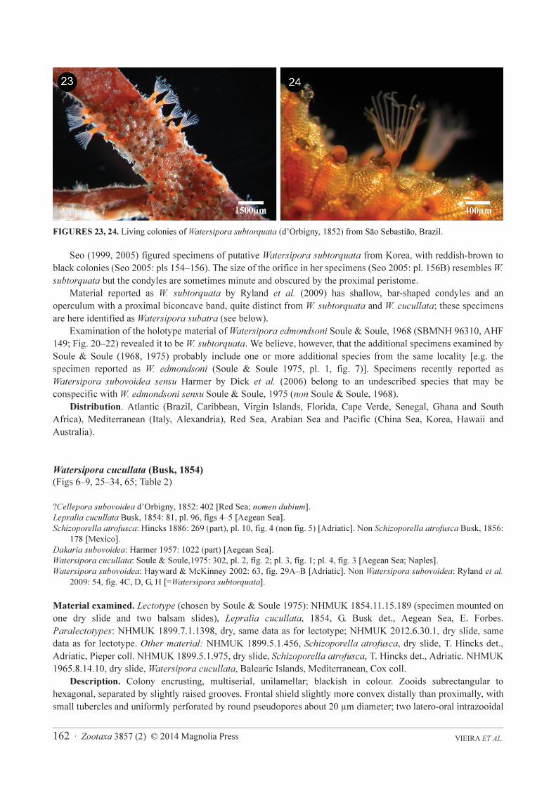
Description. Colony encrusting, multiserial, unilamellar; blackish in colour. Zooids subrectangular to
hexagonal, separated by slightly raised grooves. Frontal shield slightly more convex distally than proximally, with
small tubercles and uniformly perforated by round pseudopores about 20 µm diameter; two latero-oral intrazooidal
Zootaxa 3857 (2) © 2014 Magnolia Press · 163THE IDENTITY OF WATERSIPORA SUBTORQUATA
septula, small, near the lateral zooidal margin proximolateral to the orifice; each with 2–4 small pores; frontal
shield obscured by thick, black epitheca. Orifice large, slightly wider than long, with a well-defined, broad,
shallow, U-shaped sinus demarcated by triangular projections; orificial rim robust and thickened around whole
orifice, sometimes better developed proximally than distally; condyles upturned, large and conspicuous.
Operculum with broad, parallel-sided dark central band and slightly thinner lateral area. Avicularia absent. Ooecia
absent.
FIGURES 25–29. Watersipora cucullata (Busk, 1854), NHMUK 2012.6.30.1, paralectotype, Aegean Sea. 25, autozooidal
arrangement; 26, autozooid; 27, close-up of an orifice; 28, close-up of the frontal shield; 29, close-up of the lateral edge of the
orifice, showing a condyle and the latero-oral intrazooidal septulum.

VIEIRA ET AL. 164 · Zootaxa 3857 (2) © 2014 Magnolia Press
TABLE 2. Measurements (in µm) for Watersipora cucullata (Busk, 1854), W. atrofusca (Busk, 1856) and W. nigra
(Canu & Bassler, 1930). Type specimens are marked with asterisks.
Remarks. Watersipora subovoidea has been reported in subtropical and tropical waters, i.e. Brazil, Florida and
Australia (Mackie et al. 2006; Geller et al. 2008; Ryland et al. 2009), but the great majority of these records belong
to W. subtorquata (see above). D’Orbigny (1852) did not give any description or figures for Cellepora subovoidea,
W. atrofusca W. cucullata W. nigra
NHMUK
1892.9.6.4*
NHMUK
2012.6.30.1
NHMUK
1899.5.1.975
NHMUK
1975.5.12.1
Japan (n=6) Aegean Sea (n=6) Adriatic (n=10) Galapagos (n=10)
ZL
Min–Max 567–801 697–902 673–911 972–1102
Mean (SD) 646 (96) 789 (80) 805 (80) 1038 (47)
ZW
Min–Max 252–359 480–570 422–650 663–861
Mean (SD) 304 (47) 524 (30) 534 (68) 740 (76)
ZL x ZW [x103
]
Min–Max 162–214 362–457 284–518 673–926
Mean (SD) 194 (20) 413 (46) 431 (77) 769 (93)
OL
Min–Max 158–175 202–228 200–226 224–236
Mean (SD) 168 (6) 216 (9) 218 (8) 230 (5)
OW
Min–Max 160–185 227–245 223–258 317–333
Mean (SD) 175 (10) 233 (7) 240 (11) 325 (7)
OA [x103
]
Min–Max 21.0–25.4 35.9–41.5 35.0–44.6 58.3–59.5
Mean (SD) 23.2 (1.9) 39.2 (1.9) 41.1 (3.2) 58.7 (0.4)
ZL / OL
Min–Max 3.3–5.1 3.3–4.2 3.1–4.0 4.3–4.7
Mean (SD) 3.8 (0.7) 3.7 (0.3) 3.6 (0.3) 4.5 (0.1)
OL / OW
Min–Max 0.92–1.06 0.86–0.98 0.85–0.94 0.67–0.75
Mean (SD) 0.96 (0.05) 0.93 (0.05) 0.90 (0.03) 0.71 (0.03)
ZA / OA
Min–Max 7.6–9.6 9.1–11.9 7.0–13.5 11.47–15.56
Mean (SD) 8.4 (0.8) 10.5 (1.2) 10.3 (2.0) 13.1 (1.5)
SinL
Min–Max 39–52 40–56 39–50 51–58
Mean (SD) 44 (6) 48 (5) 44 (4) 55 (3)
SinW
Min–Max 112–131 109–130 100–134 213–233
Mean (SD) 122 (8) 115 (7) 122 (11) 219 (7)
PorD
Min–Max 17–23 20–27 21–29 27–43
Mean (SD) 20 (2) 24 (2) 25 (3) 30 (6)

Zootaxa 3857 (2) © 2014 Magnolia Press · 165THE IDENTITY OF WATERSIPORA SUBTORQUATA
and according to the ICZN Article 75.3.6 “evidence that the neotype came as nearly as practicable from the same
original type locality” should be provided, which is not apparent in the Ryland et al. (2009) paper. Thus, we suggest
setting aside the neotype selection of Cellepora subovoidea (NHMUK 1854.11.15.189) made by Ryland et al.
(2009).
FIGURES 30–34. Watersipora cucullata (Busk, 1854), NHMUK 1899.5.1.975, T. Hincks Collection, Adriatic. 30, general
view of the colony; 31, autozooid; 32, close-up of an orifice with operculum; 33, close-up of the frontal shield; 34, close-up of
the lateral edge of the orifice, showing the condyle and a latero-oral intrazooidal septulum.

VIEIRA ET AL. 166 · Zootaxa 3857 (2) © 2014 Magnolia Press
FIGURES 35–38. Watersipora aterrima (Ortmann, 1890), MZS BRY 003, syntype, Japan. 35, autozooidal arrangement; 36,
close-up of an orifice; 37, close-up of the lateral edge of the orifice, showing the latero-oral intrazooidal septulum; 38, close-up
of the frontal shield.
Watersipora cucullata is characterized by a suborbicular orifice with a shallow, wide sinus demarcated by
triangular projections (Figs 8, 27, 29, 31, 32, 34), large, conspicuous condyles (Figs 8, 27, 29, 31), and a small
latero-oral intrazooidal septula on each side of the zooid (Fig 8, 29, 34). Latero-oral intrazooidal septula are also
present in Watersipora aterrima and W. subatra, but these species are distinguished by the shape of the orifice and
condyles (see below). Watersipora cucullata differs from W. nigra in having smaller zooids and orificial area (see
Canu & Bassler, 1930, p. 26; Fig. 11; Table 2) and paired intrazooidal septula, near the lateral zooidal margin,
proximolateral to the orifice.
Watersipora atrofusca (Busk, 1856) (Fig. 10; Table 2), known from Mazatlan (Mexico), resembles W.
cucullata in colony shape but differs in having a smaller, almost circular orifice and in lacking intrazooidal septula.
Distribution. Mediterranean (including the Adriatic and Aegean seas).
Watersipora subatra (Ortmann, 1890) n. stat.
(Figs 39–53, 66, 69; Table 3)
Schizoporella aterrima var. subatra Ortmann, 1890: 49 [Japan].
?Watersipora cucullata var. watersi Mawatari, 1952: 12 (part), fig. 1G [Japan].
Dakaria subovoidea: Harmer 1957: p. 1022 (part), pl. 49, figs 11–12, 14 [Indonesia].
?Cellepora subovoidea d’Orbigny, 1852: 402 [Red Sea; nomen dubium].
Watersipora subtorquata: Ryland 1974: 345, fig. 3C [Low Island]. Non Cellepora subtorquata d’Orbigny, 1852: 399 [Brazil].

Zootaxa 3857 (2) © 2014 Magnolia Press · 167THE IDENTITY OF WATERSIPORA SUBTORQUATA
“Watersipora subovoidea” fide Harmer: Soule & Soule 1975: 308 (part), pl. 3, fig. 6 [Australia].
“Watersipora subovoidea” fide Harmer: Winston & Heimberg, 1986: 15, figs 35–37 [Komodo].
Watersipora subtorquata: Gordon 1989: 40, pl. 20, B–H [New Zealand].
Watersipora subtorquata: Gordon & Mawatari, 1992: 30 [New Zealand].
Watersipora subtorquata: Ryland et al. 2009: 55, figs 3, 4A, C, E, F [Australia, Bay of Biscay and English Channel].
Watersipora subtorquata: Kelso & Wyse-Jackson 2012: 2010, fig. 1 [Ireland].
Watersipora subtorquata: Kuhlenkamp & Kind 2013: 3, figs 1B, C, 2A–F [North Sea: Helgoland].
FIGURES 39–42. Watersipora subatra (Ortmann, 1890), MZS BRY 002, holotype, Sagami Bay, Japan. 39, autozooidal
arrangement; 40, close-up of an orifice; 41, close-up of the lateral edge of the orifice, showing the very small condyle; 42,
close-up of the frontal shield.
Material examined. Holotype: MZS 002, Ortmann Collection, Sagami Bay, Japan, 50–100 fm (15–30 m), 1882.
Other material: NHMUK 1922.10.8.3, dry slide, Schizoporella cucullata, sea level, Misaki, Japan, No. 1, Insole
coll. NHMUK 1929.12.31.1, dry slide, Watersipora atrofusca, Australia, MacGillivray coll. NHMUK
2005.5.26.19, dry, Watersipora subtorquata, scraped from hull of Seasprite, Leigh Wharf/Marina, North Island,
New Zealand, 6 October 2004, K. Tilbrook coll. NHMUK 2007.12.14.9, balsam slide, Watersipora subtorquata,
J.S. Ryland det., Low Islands, Great Barrier Reef, July 1974, J.S. Ryland coll. NHMUK 2007.12.14.10, balsam
slide, Watersipora subtorquata, J.S. Ryland det., Low Islands, Great Barrier Reef, July 1974, J.S. Ryland coll.
NHMUK 2007.12.14.12, balsam slide, Watersipora subtorquata, J.S. Ryland don., on shell, Bay of Arcachon,
August 2003, Hans de Blauwe coll. NHMUK 2007.12.14.14, balsam slide, Watersipora subtorquata, J.S. Ryland
don., St-Jacut-sur-la-Mer, April 2005, Hans de Blauwe coll. NHMUK 2007.12.14.15, balsam slide, Watersipora
subtorquata, J.S. Ryland don., St-Jacut-sur-la-Mer, April 2005, Hans de Blauwe coll. NHMUK 2007.12.14.16,
balsam slide, Watersipora subtorquata, J.S. Ryland don., Guersey, 2007, R. Lord coll. NHMUK 2007.12.14.17,
balsam slide, Watersipora subtorquata, J.S. Ryland don., Guersey, 2007, R. Lord coll. NHMUK 2007.12.14.18,

VIEIRA ET AL. 168 · Zootaxa 3857 (2) © 2014 Magnolia Press
balsam slide, Watersipora subtorquata, J.S. Ryland don., Guersey, 2007, R. Lord coll. NHMUK 2010.12.1.2, dry,
Watersipora sp., Point Loma, Marina, San Diego, California, USA, September 2010. P.D. Taylor & B. Okamura
coll. NHMUK 2010.6.30.7, dry and dry slide, Long Beach, Los Angeles, USA. NHMUK 2012.6.30.6, dry slide
[part of NHMUK 2005.5.26.19], Watersipora subtorquata, scraped from hull of Seasprite, hauled out Leigh
Wharf/Marina, North Island, New Zealand, 6 October 2004, K. Tilbrook coll.
FIGURES 43–47. Watersipora subatra (Ortmann, 1890), NHMUK 2005.5.26.19, New Zealand. 43, autozooidal arrangement;
44, autozooid; 45, close-up of an orifice; 46, close-up of the frontal shield; 47, close-up of the lateral edge of the orifice,
showing the condyle and the latero-oral intrazooidal septulum.

Zootaxa 3857 (2) © 2014 Magnolia Press · 169THE IDENTITY OF WATERSIPORA SUBTORQUATA
TABLE 3. Measurements (in µm) for Watersipora aterrima (Ortmann, 1890) and W. subatra (Ortmann, 1890). Type
specimens are marked with asterisks.
Description. Colonies encrusting, multiserial, uni- to multilamellar; sometimes erect, foliaceous and
bilamellar; colour in life variable, from orange to brownish-purple or greyish to black. Zooids subrectangular to
hexagonal, about twice as long as wide, separated by slightly raised lateral walls. Frontal shield slightly convex,
with numerous round pseudopores about 25 µm diameter; two large latero-oral intrazooidal septula near lateral
W. aterrima W. subatra
MZS BRY 003* MZS BRY 002* NHMUK 2005.5.26.19 NHMUK 2012.6.30.5
Japan (n=5) Japan (n=10) New Zealand (n=10) California (n=10)
ZL
Min–Max 1155–1393 815–1000 685–1430 921–1259
Mean (SD) 1260 (101) 919 (60) 909 (213) 1062 (111)
ZW
Min–Max 348–430 320–489 334–598 308–447
Mean (SD) 378 (32) 409 (62) 445 (76) 368 (38)
ZL x ZW [x103
]
Min–Max 447–508 299–437 286–633 304–504
Mean (SD) 474 (45) 374 (53) 400 (93) 392 (67)
OL
Min–Max 215–229 245–283 220–289 210–242
Mean (SD) 220 (6) 263 (14) 257 (19) 228 (10)
OW
Min–Max 230–249 263–348 280–329 255–271
Mean (SD) 238 (7) 309 (22) 303 (13) 261 (6)
OA [x103
]
Min–Max 39.5–44.7 52.8–77.3 48.3–72.3 43.7–50.9
Mean (SD) 41.3 (2.1) 63.9 (6.6) 61.4 (6.4) 46.9 (2.3)
LZ / OL
Min–Max 5.3–6.2 2.9–3.9 2.9–5.1 3.4–5.5
Mean (SD) 5.7 (0.4) 3.5 (0.3) 3.5 (0.6) 4.6 (0.6)
OL / OW
Min–Max 0.89–0.98 0.78–0.97 0.76–0.98 0.79–0.95
Mean (SD) 0.92 (0.03) 0.85 (0.06) 0.85 (0.05) 0.87 (0.04)
ZA / OA
Min–Max 10.6–12.8 5.0–7.1 5.3–8.7 6.8–10.1
Mean (SD) 11.5 (0.9) 5.8 (0.7) 6.5 (1.0) 8.3 (1.3)
SinL
Min–Max 38–48 61–74 52–71 47–65
Mean (SD) 43 (4) 67.3 (4.2) 61.1 (5.6) 55.4 (4.8)
SinW
Min–Max 85–101 125–174 152–178 121–140
Mean (SD) 94 (6) 145 (16) 164 (9) 130 (6)
PorD
Min–Max 16–20 22–38 23–32 18–27
Mean (SD) 17 (1) 29 (5) 27 (3) 22 (3)

VIEIRA ET AL. 170 · Zootaxa 3857 (2) © 2014 Magnolia Press
zooidal margin, proximolateral to orifice, each with 3–8 (often 5) small pores. Frontal shield obscured by
translucent cuticle. Orifice large, subcircular to oval, slightly wider than long, with broad, well-defined proximal,
U-shaped sinus; orificial rim thin, sometimes slightly raised; narrow bar-shaped condyles, sometimes
inconspicuous. Operculum with broad, biconcave proximal band, without lucidae. Avicularia absent. Ooecia
absent.
FIGURES 48–53. Watersipora subatra (Ortmann, 1890), NHMUK 2012.6.30.5, California. 48, autozooidal arrangement; 49,
autozooid; 50, close-up of an orifice; 51, close-up of the frontal shield; 2, close-up of the lateral edge of the orifice, showing the
condyle and the latero-oral intrazooidal septulum; 53, internal view of the orificial area; arrows show the latero-oral
intrazooidal septula.

Zootaxa 3857 (2) © 2014 Magnolia Press · 171THE IDENTITY OF WATERSIPORA SUBTORQUATA
Remarks. Ortmann (1890) described and figured Schizoporella aterrima from Japan, including the new
variety subatra without drawings or description. Ryland et al. (2009) noted that Schizoporella aterrima Ortmann,
1890 is unrecognizable and that the description given by Mawatari (1952) for Watersipora cucullata var. watersi,
another species reported from Japan, may include at least two species. One of us (MSJ) has examined Ortmann’s
specimens deposited at the Musée zoologique de la Ville de Strasbourg, concluding that they represent two species,
Watersipora aterrima (MZS 003; Figs 35–38; Table 3) and Watersipora subatra (MZS 002; Fig. 39–42; Table 3).
Watersipora aterrima is distinguished from W. subatra by having a smaller orifice, narrower sinus and smaller
frontal pseudopores.
Mawatari (1952) introduced variety watersi for specimens with distinctive opercula. His notes on this new
variety are based on specimens from Japan, and two others recorded by Waters (1909) from Naples and by
Hastings (1930) from Cape Verde. Unfortunately, Mawatari’s specimens from Japan assigned to W. cucullata var.
watersi could not be located, but part of his description and figures resembles W. subatra and may refer to this
species. We have assigned the Cape Verde and Naples specimens to a different species, Watersipora souleoroum n.
sp. (see below).
Comparisons between specimens from New Zealand (Figs 43–47), California (Fig 48–53) and Britain (some
slides at NHMUK; see Ryland et al. 2009, fig. 3, as W. subovoidea) with the type specimens of W. cucullata, W.
subatra and W. subtorquata, indicate that W. subatra was previously misidentified as W. subtorquata by some
authors (e.g. Ryland 1974; Gordon 1989; Gordon & Mawatari 1992; Mackie et al. 2006, 2012; Geller et al. 2008;
Ryland et al. 2009; Láruson et al. 2012; Cockrell & Sorte 2013; Needles & Wendt 2013; Sorte & White 2013;
Davis & Marshall 2014). Since Ryland et al. (2009) found the COI haplotype in colonies from Guernsey and
Brittany to be identical to the commonest haplotype identified in other areas, and the morphology of the genetically
identified specimens to be the same (as W. subtorquata; Mackie et al. 2006), we believe that W. subatra is the
commonest putatively invasive species of Watersipora in Britain, Australia, New Zealand and California, whereas
W. subtorquata is the predominant species in subtropical and tropical waters of the Atlantic and Mediterranean.
Colonies from California (Figs 48–53) often have a well-demarcated, U-shaped sinus and a smooth suborificial
region (without pseudopores), as has been figured in specimens from Brittany (Ryland et al. 2009, fig. 3A, D, as W.
subtorquata). All specimens assigned to W. subatra have an orifice with a U-shaped sinus and narrow, bar-shaped
condyles, opercula with a broad, biconcave band proximally, frontal shields with pseudopores 18–30 µm in
diameter, and two latero-oral intrazooidal septula.
Soule and Soule (1975) indicated that Harmer (1957) included at least two species under Dakaria subovoidea,
which may be a mixture of W. subtorquata and W. aterrima. According to the list of specimens published by
Harmer (1957), however, his description and remarks included at least four species: W. subtorquata, W. cucullata,
W. atrofusca and W. subatra (specimens figured from Siboga Stns 181 and 184, see Harmer 1957, pl. 49, figs
11–12, 14). Watersipora subatra is distinguished from W. subtorquata by the shapes of the orifice and condyles,
and the presence of latero-oral intrazooidal septula (Figs 47, 52, 53). Latero-oral intrazooidal septula are also found
in W. cucullata (Figs 8, 29, 34), W. aterrima (Fig. 37) and W. mawatarii n. sp. (Fig. 58; see below), but these
species have different shapes of orifice and condyles.
Despite the wide distribution of W. subatra in Pacific waters (see Ryland et al. 2009; Mackie et al. 2012; as W.
subtorquata), its occurrence in some localities in the northwest Pacific, as well as the occurrence of W. subtorquata,
are unconfirmed and require morphological and molecular investigation.
Distribution. NE Atlantic (Ireland and British Isles), Indo-West Pacific (Indonesia) and Pacific (New Zealand,
Australia and California).
Watersipora mawatarii n. sp.
(Figs 54–58; Table 4)
?Dakaria typica Okada & Mawatari, 1937: 438, pl. 11, fig. 6; text-fig. 2 [Japan].
?Dakaria subovoidea: Kubota & Mawatari, 1985: 203, fig. 4 [Japan].
Material examined. Holotype: NHMUK 2012.6.30.8, Oshoro Marine Station, near Otaru, Hokkaido, Japan,
intertidal, 16 April 1996, M.J.Weedon coll. Paratypes: NHMUK 2012.6.30.9–11, same data as holotype; NHMUK
2012.6.30.12–13, 17, Otaru chiko, Japan, on dead barnacles near shore, 12 December 1996, T. Kato coll. NHMUK

VIEIRA ET AL. 172 · Zootaxa 3857 (2) © 2014 Magnolia Press
2012.6.30.18, dry, Amakusa Marine Biological Laboratory, Kyushu, Japan, 40–50m, dredged, 22 October 1996, T.
Kato coll.
Etymology. Named for Japanese bryozoologist Prof. Shunsuke F. Mawatari.
FIGURES 54–58. Watersipora mawatarii n. sp., from Japan. 54–55, NHMUK 2012.6.30.8, holotype, Hokkaido; 54,
autozooidal arrangement; 55, distal of an autozooid. 56–58, NHMUK 2012.6.30.13, paratype, Otaru; 56, autozooids; 57, close-
up of an orifice and the two latero-oral intrazooidal septula; 58, close-up of the lateral edge of the orifice, showing a condyle
and a latero-oral intrazooidal septulum.

Zootaxa 3857 (2) © 2014 Magnolia Press · 173THE IDENTITY OF WATERSIPORA SUBTORQUATA
TABLE 4. Measurements (in µm) of Watersipora souleorum n. sp. and W. mawatarii n. sp. Holotype specimens are
marked with asterisks.
Description. Colonies encrusting, multiserial, uni- to multilamellar; sometimes erect, foliaceous and
bilamellar; colour greyish to black in dead colonies. Zooids elongate-elliptical to rectangular, widest below the
orificial area, about twice as long as wide, separated by slightly raised lateral walls; zooids arranged in quincuncial
W. souleorum n. sp. W. mawatarii n. sp.
NHMUK
2014.07.29.1*
NHMUK
1912.12.21.1019
NHMUK
2012.6.30.8*
NHMUK
2012.6.30.13
Cape Verde (n=10) Naples (n=7) Hokkaido (n=10) Otaru (n=3)
ZL
Min–Max 807–930 815–869 850–999 818–971
Mean (SD) 872 (39) 848 (29) 909 (53) -
ZW
Min–Max 316–493 339–496 408–510 395–433
Mean (SD) 402 (70) 459 (53) 463 (45) -
ZL x ZW [x103
]
Min–Max 282–424 325–431 370–461 354–383
Mean (SD) 350 (55) 391 (57) 420 (27) -
OL
Min–Max 228–250 237–247 200–230 187–220
Mean (SD) 239 (8) 240 (6) 211 (10) -
OW
Min–Max 226–260 234–251 238–265 239–270
Mean (SD) 244 (10) 242 (8) 252 (8) -
OA [x103
]
Min–Max 40.5–48.4 43.7–48.7 37.9–45.6 35.1–46.6
Mean (SD) 45.7 (2.3) 45.8 (2.5) 41.0 (2.6) -
ZL / OL
Min–Max 3.3–3.9 3.3–3.6 3.7–4.7 3.7–4.7
Mean (SD) 3.7 (0.2) 3.5 (0.1) 4.3 (0.3) -
OL / OW
Min–Max 0.91–1.05 0.98–1.01 0.77–0.92 0.78–0.82
Mean (SD) 0.98 (0.04) 0.99 (0.02) 0.84 (0.04) -
ZA/OA
Min–Max 6.3–9.1 7.2–9.5 8.7–11.4 7,6–9.6
Mean (SD) 7.6 (1.0) 8.5 (1.1) 10.0 (0.9) -
SinL
Min–Max 60–70 57–65 40–48 48–59
Mean (SD) 64.9 (3.3) 59.7 (4.6) 43.8 (2.7) -
SinW
Min–Max 110–130 133–148 117–137 126–150
Mean (SD) 117 (8) 138 (8) 126 (6) -
PorD
Min–Max 23–29 25–32 9–15 10–14
Mean (SD) 27 (2) 29 (3) 12 (2) -

VIEIRA ET AL. 174 · Zootaxa 3857 (2) © 2014 Magnolia Press
series. Frontal shield thick, granulated, slightly convex, with numerous small (10–15 µm diameter), round
pseudopores covering entire surface except for suborificial region; two latero-oral intrazooidal septula, near lateral
zooidal margin, proximolateral to orifice, each with 3–6 small pores. Frontal shield obscured by opaque, dark
cuticle. Orifice large, transversely elliptical, usually conspicuoualy wider than long, with well-defined proximal
broad sinus; orificial rim often thick and raised, but some zooids with thin, slightly raised rim; narrow bar-shaped
condyles occupying entire proximal edge of orifice, sometimes projecting medially as triangular projection.
Operculum black, mushroom-shaped; lucidae present. Avicularia absent. Ovicells absent.
Remarks. The overall zooidal morphology of specimens here figured and described resemble some Japanese
specimens—viz. Dakaria typica Okada & Mawatari, 1937 (=Watersipora typica) from Miyagi, and Dakaria
subovoidea from Hokkaido (Kubota & Mawatari 1985)—but they are distinguished by having fewer pseudopores
in the frontal shield than Watersipora mawatarii n. sp. The thinner circular area in the distal half of the operculum,
characteristic of W. typica, is also absent in W. mawatarii n. sp.
Watersipora mawatarii n. sp. resembles W. edmondsoni sensu Soule & Soule (1975) from Hawaii (the
holotype of Watersipora edmondsoni belongs to Watersipora subtorquata, see above), characterized by its long
zooids (about 0.80–1.20 mm long and 0.40–0.60 mm wide) and dark-brown mushroom-shaped opercular
pigmentation, with a curved lower portion that fits into the sinus area. Soule & Soule (1975) also reported frontal
shields with very small pseudopores like those of W. mawatarii n. sp. The shape of the zooids (widest below the
orificial area) and the absence of pseudopores in the suborificial region were also described for some specimens
from different Indo-West Pacific sites—Bali (Winston & Heimberg 1986; as Watersipora edmondsoni), Vanuatu
(Tilbrook et al. 2001; as Watersipora subovoidea sensu Harmer) and Hawaii (Dick et al. 2006; as Watersipora
subovoidea sensu Harmer)—which we believe belong to an undescribed species; these specimens differ from W.
mawatarii n. sp. in having a narrower sinus and smaller orifice: 0.189–230 mm long and 0.238–0.270 mm wide in
W. mawatarii n. sp. versus 0.162–0.216 mm long and 0.198–0.234 mm wide in species from Bali (Winston &
Heimberg 1986), and 0.15–0.18 mm long and 0.20–0.25 mm wide in specimens from Hawaii (Dick et al. 2006).
Watersipora mawatarii n. sp. resembles W. subatra in having a U-shaped sinus, bar-shaped condyles, and two
latero-oral intrazooidal septula. The two species differ, however, in the size of the pseudopores (smaller in W.
mawatarii n. sp.) and condyles (more conspicuous in W. mawatarii n. sp.) and in the absence of pseudopores in the
suborificial region (characteristic of W. mawatarii n. sp.).
Distribution. Japan.
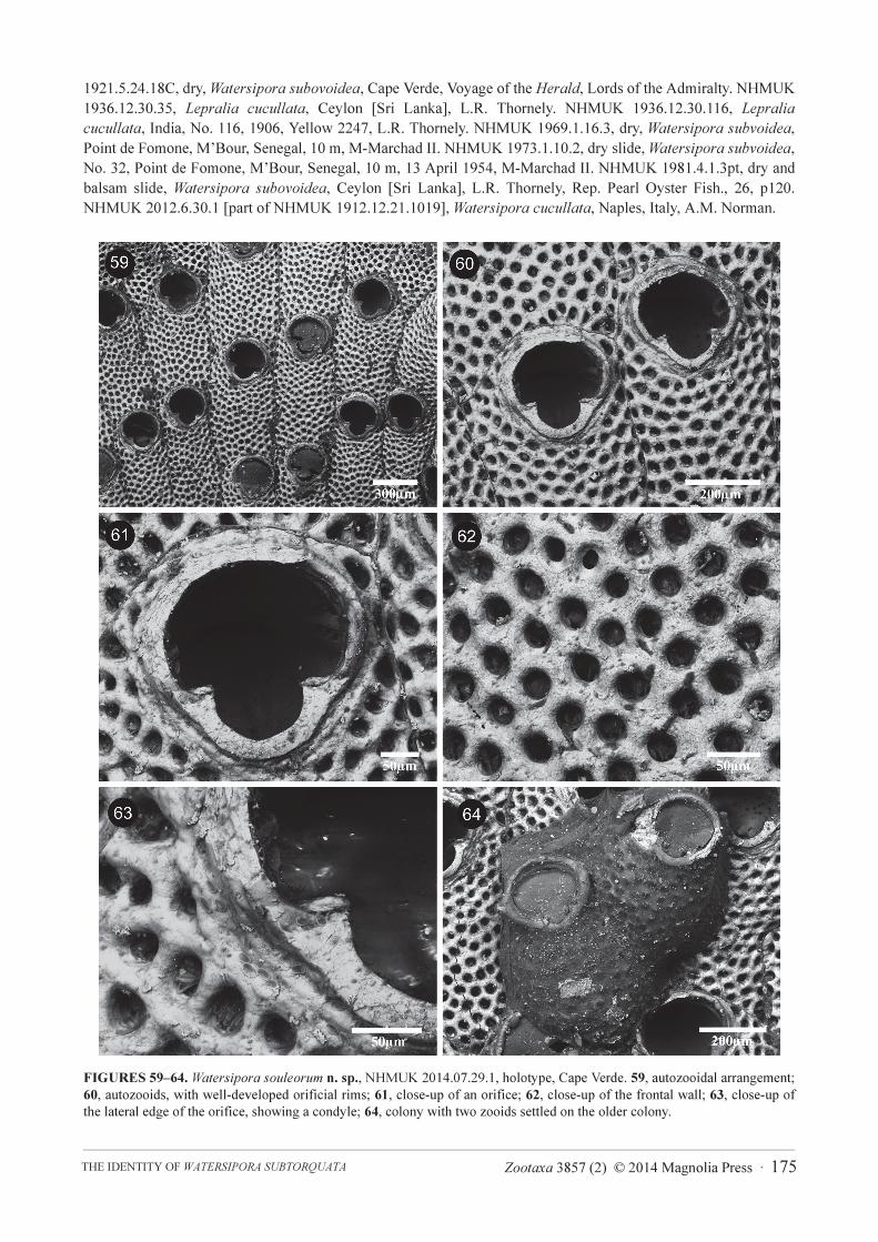
Watersipora souleorum n. sp.
(Figs 17, 59–64, 68, 71; Table 4)
?Cellepora subovoidea d’Orbigny, 1852: 402 [Red Sea; nomen dubium].
Lepralia cucullata: Thornely 1905: 120 [Sri Lanka]. Non Lepralia cucullata Busk, 1854: 81, pl. 96, figs 4–5 [Aegean Sea].
Schizoporella cucullata var. labiosa Calvet in Jullien & Calvet, 1903: 141, pl. 16, fig. 7a–c [Azores].
Lepralia? cucullata: Waters 1909: 150 (part), pl. 15, figs 2–5 [Naples and ?Cape Verde].
Watersipora cucullata: Hastings 1930: 729 (part), pl. 15, fig. 104 [Cape Verde].
Watersipora cucullata var. watersi Mawatari, 1952: 12 (part) [Cape Verde].
“Watersipora subovoidea” fide Harmer: Soule & Soule 1975: 308 (part), pl. 3, fig. 5 [Sri Lanka].
Material examined. Holotype: NHMUK 2014.07.29.1, dry slide, Porto Grande, St. Vicent, Cape Verde Islands, on
coral, 10 fm [18.3 m], Vallentin coll. Paratype: NHMUK 1935.3.6.367, same data as for holotype. Other material:
MOM 420379, Schizoporella cucullata var. labiosa, L. Calvet 1903, l’Hirondelle, Stn 236, Azores, 20 August
1888. NHMUK 1872.2.3.147, dry, Watersipora cucullata var. atrofusca, west coast of Spain, S. Kent coll., on
shells. NHMUK 1873.7.21.7, dry and dry slide, Watersipora cucullata, Ceylon [Sri Lanka], E.W.H. Holdsworth.
NHMUK 1882.10.18.3.4–41pt., Watersipora sp., Seychelles Island, on coral, 12 fm [22 m], Voyage of H.M.S.
Alert, pres. Lords of the Admiralty. NHMUK 1882.10.18.3.100–104., Schizoporella sp., Seychelles Island, coral,
12 fm [22 m], Voyage of H.M.S. Alert, pres. Lords of the Admiralty. NHMUK 1899.5.1.976 [3 slides], Watersipora
atrofusca, Trincomalee, Sri Lanka, T. Hincks coll. NHMUK 1899.7.1.1396, dry slide, Lepralia cucullata, Persian
Gulf, on pearl oyster, G. Busk coll. NHMUK 1899.7.1.1893, dry slide [part in balam slide], Lepralia cucullata,
Cape Verde Island, Mrs Gatty, G. Busk coll. NHMUK 1890.1.31.13, dry, Watersipora subovoidea, Madras
[Chennai], India. NHMUK 1912.12.21.1019, Watersipora cucullata, Naples, Italy, A.M. Norman. NHMUK
Zootaxa 3857 (2) © 2014 Magnolia Press · 175THE IDENTITY OF WATERSIPORA SUBTORQUATA
1921.5.24.18C, dry, Watersipora subovoidea, Cape Verde, Voyage of the Herald, Lords of the Admiralty. NHMUK
1936.12.30.35, Lepralia cucullata, Ceylon [Sri Lanka], L.R. Thornely. NHMUK 1936.12.30.116, Lepralia
cucullata, India, No. 116, 1906, Yellow 2247, L.R. Thornely. NHMUK 1969.1.16.3, dry, Watersipora subvoidea,
Point de Fomone, M’Bour, Senegal, 10 m, M-Marchad II. NHMUK 1973.1.10.2, dry slide, Watersipora subvoidea,
No. 32, Point de Fomone, M’Bour, Senegal, 10 m, 13 April 1954, M-Marchad II. NHMUK 1981.4.1.3pt, dry and
balsam slide, Watersipora subovoidea, Ceylon [Sri Lanka], L.R. Thornely, Rep. Pearl Oyster Fish., 26, p120.
NHMUK 2012.6.30.1 [part of NHMUK 1912.12.21.1019], Watersipora cucullata, Naples, Italy, A.M. Norman.
FIGURES 59–64. Watersipora souleorum n. sp., NHMUK 2014.07.29.1, holotype, Cape Verde. 59, autozooidal arrangement;
60, autozooids, with well-developed orificial rims; 61, close-up of an orifice; 62, close-up of the frontal wall; 63, close-up of
the lateral edge of the orifice, showing a condyle; 64, colony with two zooids settled on the older colony.

VIEIRA ET AL. 176 · Zootaxa 3857 (2) © 2014 Magnolia Press
FIGURES 65–68. Schematic drawings of opercula of Watersipora. 65, Watersipora cucullata (Busk, 1852), Aegean Sea
(redrawn from Hastings 1930, pl. 15, fig. 98); 66, Watersipora subatra (Ortmann, 1890), New Zealand; 67, W. subtorquata
(d’Orbigny, 1982), Brazil; 68, Watersipora souleorum n. sp., Cape Verde (redrawn from Hastings 1930, pl. 15, fig. 104).
FIGURES 69–71. Opercula of some species of Watersipora. 69, Watersipora subatra (Ortmann, 1890), NHMUK
2007.12.14.18, Guersey; 70, Watersipora subtorquata (d’Orbigny, 1982), NHMUK 1926.9.6.165A, Suez Canal; 71,
Watersipora souleorum n. sp., NHMUK 1899.7.1.1393, Cape Verde.
Description. Colonies encrusting, multiserial, uni- to multilamellar, sometimes erect, foliaceous and
bilamellar; colour of dead colonies black. Zooids subrectangular, about twice as long as wide, separated by slightly
raised lateral walls. Frontal shield slightly convex, with numerous (25–32 µm diameter), round pseudopores;
latero-oral intrazooidal septula absent. Frontal shield obscured by dark cuticle. Orifice large, oval, with well-
defined, broad, deeply U-shaped proximal sinus; slightly raised orificial rim sometimes present; bar-shaped
condyles. Operculum with a narrow, biconcave dark central band, with lucidae. Avicularia absent. Ovicells absent.
Etymology. Named for John D. Soule (1920–2001) and Dorothy F. Soule (1923–2005), in recognition of their
contributions to bryozoan taxonomy.
Remarks. The name labiosa was first introduced by Hincks (1886)—as Schizoporella atrofusca f.
labiosa—but was synonymised under W. subovoidea by Ryland et al. (2009) (=W. subtorquata). Calvet (in Jullien
& Calvet 1903) also introduced the name labiosa as Schizoporella cucullata var. labiosa—this name has never
been considered valid since the original description and it was synonymised with W. cucullata by Hastings
(1927)—for material from the Azores. Calvet’s specimens (MOM 420379) are distinct from W. subtorquata,
however, in having a deep sinus and an operculum with a biconcave dark central band. Hastings (1930, pl. 15, fig.
104, as W. cucullata) included an excellent drawing based on specimens from Cape Verde that corresponds with
Calvet’s variety. Waters (1909) used the name L. cucullata for species from Naples, Cape Verde and Suez, but the
descriptions encompass at least two species. His specimens from Naples (Waters 1909, figs 2–5) have a wider sinus

Zootaxa 3857 (2) © 2014 Magnolia Press · 177THE IDENTITY OF WATERSIPORA SUBTORQUATA
and distinctive operculum when compared to those from Suez (=W. subtorquata); specimens from Naples resemble
those reported by Calvet (in Jullien & Calvet 1903) and Hastings (1930) from the Azores and Cape Verde,
respectively. Mawatari (1952) erected var. watersi for material with this distinctive type of operculum, but the
name is preoccupied by the fossil Microporella watersi de Stefani, 1884, based on Lepralia cucullata of Manzoni
(1875) which, to judge by Manzoni’s figure (1875, pl. 4, fig. 47), is likely a species of Watersipora (see Waters
1909). We introduce the name Watersipora souleorum n. sp. for these specimens.
Watersipora souleorum n. sp. resembles W. subtorquata in lacking latero-oral intrazooidal septula. The new
species differs, however, in having a larger orifice than W. subtorquata, a deep U-shaped sinus, bar-shaped
condyles, and an operculum with a narrow dark central band.
Distribution. Atlantic (Azores, Cape Verde and Senegal), Mediterranean (Naples) and Indian Ocean (Sri
Lanka and India).
FIGURE 72. Distribution map (not exhaustive) of species of Watersipora.
Discussion
According to Bock (2010), nine species have been assigned to Watersipora Neviani, 1895: Watersipora arcuata
Banta, 1969a, Watersipora aterrima (Ortmann, 1890), Watersipora complanata (Norman, 1864), Watersipora
edmondsoni Soule & Soule, 1968, Watersipora grandis (Canu & Bassler, 1923), Watersipora platypora Seo, 1999,
W. subovoidea, W. subtorquata and Watersipora watersi Mawatari, 1952. Some other nominal species have been
synonymised under these names (Soule & Soule 1975; Abdel-Salam & Ramadan 2008; Ryland et al. 2009). Apart
from Watersipora grandis (Canu & Bassler, 1923), which is a fossil from the Neogene of the southeastern USA, all
of the species listed above are extant. Examination of type specimens has revealed that the holotype of W.
edmondsoni is synonymous with W. subtorquata, and that two species previously synonymised under W.
subtorquata and W. subovoidea belong to distinct species: Watersipora atrofusca (Busk, 1856) and Watersipora

VIEIRA ET AL. 178 · Zootaxa 3857 (2) © 2014 Magnolia Press
subatra (Ortmann, 1890). Thus, Watersipora contains at least 13 valid Recent species: W. aterrima, W. arcuata, W.
atrofusca, W. bidentata (Ortmann, 1890), W. complanata, W. cucullata, W. mawatarii n. sp., W. nigra n. comb., W.
platypora, W. souleorum n. sp. (=Watersipora watersi Mawatari, 1952, non Microporella watersi de Stefani, 1884),
W. subatra, W. subtorquata and W. typica. The taxonomic status of Microporella watersi de Stefani, 1884, a fossil
species supposedly of Watersipora (see Waters 1909), requires reinvestigation.
The lack in Watersipora of diagnostic structures such as avicularia and ooecia that are often used in
cheilostome taxonomy has led to the use of morphological and morphometric characters of the frontal shield,
orifice and operculum to identify species in the genus (Soule & Soule 1975; Seo 1999; Ryland et al. 2009). Six
species—W. cucullata, W. arcuata, W. atrofusca, W. bidentata, W. nigra and W. platypora—have an orifice with the
sinus almost square or rectangular in shape (see Okada & Mawatari 1937; Banta 1969a; Seo 1999), while W.
complanata is distinct in having an orifice with a straight proximal edge lacking a sinus (Hayward & McKinney
2002). The other six species—W. aterrima, W. mawatarii n. sp., W. souleorum n. sp., W. subatra, W. subtorquata
and W. typica—have an orifice with a concave to almost straight proximal edge (V- or U-shaped sinus). The main
distinguishing characteristics of these species are the morphometry of the zooids and orifices, shape of the orifice
and operculum (used for some specimens, e.g. Figs 65–71), and morphology of the frontal shield, including
pseudopores and the presence or absence of intrazooidal septula, which are difficult to observe except by using
SEM. Early astogenetic characters, including the morphology of the ancestrula, deserve future investigation to
evaluate their potential in discriminating species and subclades of Watersipora.
Ryland et al. (2009) suggested the need for more genetic studies on species of Watersipora because of cryptic
speciation in W. subatra (as W. subtorquata) and a Californian lineage 15% divergent in COI nucleotide sequence
from another widespread clade. Mackie et al. (2012) also showed two genetically shallow groups in W. subatra (as
W. subtorquata): clade A in Europe and Australasia, and clade B in California. Since two species of the
Watersipora subatra complex were considered not distinguishable using the zooid-area to orifice-area ratio
(Ryland et al. 2009; as W. subtorquata), we suggest additional morphological studies based on the new descriptions
presented here.
Resolution of the identity of Brazilian specimens, previously attributed to Watersipora subovoidea, as W.
subtorquata, allows us to falsify the assertion of Mackie et al. (2012) that W. subtorquata has been displaced from
its native locale of Brazil. The actual distribution of W. subtorquata and all congenerics are presented in Fig. 72.
Populations of Watersipora subtorquata from Florida, Brazil and Australian are about 1% divergent in COI
sequence, suggesting recent, widespread introductions of the species in tropical waters (Mackie et al. 2012, as W.
subovoidea). Genetic studies should be conducted, however, to investigate the degree of genetic divergence along
the Brazilian coast and to understand dispersal patterns in this important fouling species.
The continued confusion over the correct names for the two species commonly identified as Watersipora
subtorquata and Watersipora subovoidea is particularly relevant in view of their importance as invasive fouling
species. Their true identities are clarified by the new descriptions presented in this paper.
Key to Recent species of Watersipora
1 Orifice bell-shaped, without conspicuous sinus demarcated by condyles . . . . . . . . . . . . . . . . . . . . . . . . . . . . . . . . . complanata
Orifice almost circular to skull-shaped, sinus demarcated by condyles . . . . . . . . . . . . . . . . . . . . . . . . . . . . . . . . . . . . . . . . . . . 2
2 Sinus V- or U-shaped. . . . . . . . . . . . . . . . . . . . . . . . . . . . . . . . . . . . . . . . . . . . . . . . . . . . . . . . . . . . . . . . . . . . . . . . . . . . . . . . . . . 3
Sinus approximately square or rectangular . . . . . . . . . . . . . . . . . . . . . . . . . . . . . . . . . . . . . . . . . . . . . . . . . . . . . . . . . . . . . . . . . 8
3 Operculum with three small, thin circular areas . . . . . . . . . . . . . . . . . . . . . . . . . . . . . . . . . . . . . . . . . . . . . . . . . . . . . . . . . typica
Operculum with distinct central band . . . . . . . . . . . . . . . . . . . . . . . . . . . . . . . . . . . . . . . . . . . . . . . . . . . . . . . . . . . . . . . . . . . . . . 4
4 Latero-oral intrazooidal septula absent . . . . . . . . . . . . . . . . . . . . . . . . . . . . . . . . . . . . . . . . . . . . . . . . . . . . . . . . . . . . . . . . . . . 5
Latero-oral intrazooidal septula present . . . . . . . . . . . . . . . . . . . . . . . . . . . . . . . . . . . . . . . . . . . . . . . . . . . . . . . . . . . . . . . . . . . . 6
5 Operculum with narrow, biconcave dark central band; bar-shaped condyles; orifice as long as wide, 0.225–0.250 mm long and
0.225–0.260 mm wide . . . . . . . . . . . . . . . . . . . . . . . . . . . . . . . . . . . . . . . . . . . . . . . . . . . . . . . . . . . . . . . . . . . . . souleorum n. sp.
Operculum with a parallel-sided dark central band; tooth-like condyles, projecting distomedially; orifice wider than long,
0.160–0.200 mm long and 0.190–0.240 mm wide . . . . . . . . . . . . . . . . . . . . . . . . . . . . . . . . . . . . . . . . . . . . . . . . . . . subtorquata
6 Pseudopores smaller than 0.016 mm in diameter . . . . . . . . . . . . . . . . . . . . . . . . . . . . . . . . . . . . . . . . . . . . . . . . mawatarii n. sp.
Pseudopores larger than 0.016 mm in diameter . . . . . . . . . . . . . . . . . . . . . . . . . . . . . . . . . . . . . . . . . . . . . . . . . . . . . . . . . . . . . 7
7 Condyles inconspicuous; sinus 0.085–0.101 mm long and 0.038–0.048 mm wide . . . . . . . . . . . . . . . . . . . . . . . . . . . . aterrima
Condyles bar-shaped; sinus 0.120–0.178 mm long and 0.048–0.074 mm wide . . . . . . . . . . . . . . . . . . . . . . . . . . . . . . . subatra

Zootaxa 3857 (2) © 2014 Magnolia Press · 179THE IDENTITY OF WATERSIPORA SUBTORQUATA
8 Poster with straight proximal border . . . . . . . . . . . . . . . . . . . . . . . . . . . . . . . . . . . . . . . . . . . . . . . . . . . . . . . . . . . . . . . . . . . . . . 9
Poster with convex proximal border . . . . . . . . . . . . . . . . . . . . . . . . . . . . . . . . . . . . . . . . . . . . . . . . . . . . . . . . . . . . . . . . . . . . . 11
9 Condyles bar-shaped; orifice wider than 0.270 mm . . . . . . . . . . . . . . . . . . . . . . . . . . . . . . . . . . . . . . . . . . . . . . . . . . . . bidentata
Condyles triangular; orifice narrower than 0.250 mm . . . . . . . . . . . . . . . . . . . . . . . . . . . . . . . . . . . . . . . . . . . . . . . . . . . . . . . 10
10 Orifice up to 0.175 mm long and 0.185 mm wide; latero-oral intrazooidal septula absent . . . . . . . . . . . . . . . . . . . . . . atrofusca
Orifice more than 0.200 mm long and 0.220 mm wide; latero-oral intrazooidal septula present . . . . . . . . . . . . . . . . . cucullata
11 Sinus demarcated by triangular projections of the peristome and condyles . . . . . . . . . . . . . . . . . . . . . . . . . . . . . . . . . . . . .nigra
Sinus only demarcated by condyles . . . . . . . . . . . . . . . . . . . . . . . . . . . . . . . . . . . . . . . . . . . . . . . . . . . . . . . . . . . . . . . . . . . . . 12
12 Frontal wall with about 100 pseudopores; orifice more than 0.320 mm wide . . . . . . . . . . . . . . . . . . . . . . . . . . . . . . . . platypora
Frontal wall with about 150 pseudopores; orifice up to 0.225 mm wide . . . . . . . . . . . . . . . . . . . . . . . . . . . . . . . . . . . . . arcuata
Acknowledgements
We are grateful to Michelè Bruni (Musée Océanographique de Monaco, Monaco), Dennis P. Gordon (National
Institute of Water & Atmospheric Research, New Zealand), Joshua Mackie (Moss Landing Marine Laboratories,
USA), Andrew N. Ostrovsky (University of Vienna, Austria), JoAnn Sanner (National Museum of Natural History,
Smithsonian Institution, USA), Judith E. Winston (Virginia Museum of Natural History, USA), Marie Meister
(Musée zoologique de la Ville de Strasbourg) and Henry W. Chaney (Santa Barbara Museum of Natural History)
for supplying useful information and material on the species of Watersipora included in this study. SEMs images of
some type specimens were kindly provided by Daniel Geiger, Henry W. Chaney and JoAnn Sanner. John S. Ryland
and Matthew H. Dick provided suggestions for improving the manuscript. The Natural History Museum, London,
Musée Oceanographique de Monaco and the National Museum of Natural History, Smithsonian Institution
provided logistical support during visits by LMV who received funding from FAPESP (Proc. n. 2012/24285-1).
This is a contribution of NP-BioMar, USP.
References
Abdel-Salam, K.M. & Ramadan, S.E. (2008) Fouling Bryozoa from some Alexandria harbours, Egypt. (II) Encrusting species.
The Mediterranean Marine Science Journal, 9 (2), 5–20.
http://dx.doi.org/10.12681/mms.129
Audouin, J.V. (1826) Explication sommaire des planches de Polypes de l’Egypte et de la Syrie, publiées par Jules-César
Savigny. In: Panckoucke, C.L.F. (Org.), Description de l’Egypte ou receuil des observations et des recherches qui ont été
faites en Egyptes pendant l’Expedition de l’Armée française … Histoire naturelle. Tome 1(4). Imprimerie Impériale, Paris,
pp. 225–244.
Banta, W.C. (1969a) Watersipora arcuata, a new species in the subovoidea-cucullata-nigra complex (Bryozoa, Cheilostomata).
Bulletin of the Southern California Academy of Science, 68, 96–102.
Banta, W.C. (1969b) Uscia mexicana, new genus, new species, a watersiporid bryozoan with dimorphic autozooids. Bulletin of
the Southern California Academy of Science, 68, 20–35.
Banta, W.C. (1970) The body wall of cheilostome Bryozoa, III. The frontal wall of Watersipora arcuata Banta, with a revision
of the Cryptocystidea. Journal of Morphology, 131, 37–56.
http://dx.doi.org/10.1002/jmor.1051310104
Bock, P. (2010) Watersipora Neviani, 1896. In: Recent and Fossil Bryozoa. Mount Waverley (Vic): Philip Bock. Available
from: http://bryozoa.net/cheilostomata/watersiporidae/watersipora.html (accessed 20 June 2014)
Busk, G. (1854) Catalogue of marine Polyzoa in the collection of the British Museum, II. Cheilostomata (part). Trustees of the
British Museum (Natural History), London, 54 pp.
Busk, G. (1856) Zoophytology. Quarterly Journal of Microscopical Science, 4, 176–179, pls 7–8.
Canu, F. & Bassler, R.S. (1923) North American later Tertiary and Quaternary Bryozoa. United States National Museum
Bulletin, 125, 1–302.
Canu, F. & Bassler, R.S. (1930) The Bryozoa of the Galapagos Islands. Proceedings of the United States National Museum, 76,
1–78.
Cockrell, M.L. & Sorte, C.J.B. (2013) Predicting climate-induced changes in population dynamics of invasive species in a
marine epibenthic community. Journal of Experimental Marine Biology and Ecology, 440, 42–48.
http://dx.doi.org/10.1016/j.jembe.2012.11.008
Cook, P.L. (1985) Bryozoa from Ghana. Tervuren, België Koninklijk Museum voor Midden-Afrika. Zoologische Weteschappen,
238, 1–315.
Davis, K. & Marshall, D.J. (2014) Offspring size in a resident species affects community assembly. Journal of Animal Ecology,
83, 322–331.

VIEIRA ET AL. 180 · Zootaxa 3857 (2) © 2014 Magnolia Press
http://dx.doi.org/10.1111/1365-2656.12136
Dick, M.H., Tilbrook, J. & Mawatari, S.F. (2007) Diversity and taxonomy of rocky-intertidal Bryozoa on the Island of Hawaii,
USA. Journal of Natural History, 40, 2197–2258.
http://dx.doi.org/10.1080/00222930601062771
Florence, W.K., Hayward, P.J. & Gibbons, M.J. (2007) Taxonomy of shallow-water Bryozoa from the west coast of South
Africa. African Natural History, 3, 1–58.
Geller, J., Mackie, J.A., Schroeder, G. & Gerhinger, D. (2008) Distribution of highly invasive bryozoans belonging to a cryptic
species complex in the genus Watersipora determined by DNA sequences. Moss Landing Marine Laboratories, Moss
Landing, 40 pp.
Gordon, D.P. (1989) The marine fauna of New Zealand: Bryozoa: Gymnolaemata (Cheilostomida Ascophorina) from the
western South Island continental shelf and slope. New Zealand Oceanographic Institute Memoir, 97, 1–158.
Gordon, D.P. & Mawatari, S.H. (1992) Atlas of marine-fouling Bryozoa of New Zealand ports and harbours. Miscellaneous
Publications of the New Zealand Oceanographic Institute, 107, 1–52.
Harmer, S.F. (1957) The Polyzoa of the Siboga Expedition, Part 4. Cheilostomata Ascophora II. Siboga-Expeditie, 28d, xv,
641–1147, pls. 42–74.
Hastings A.B. (1927) Zoological results of the Cambridge expedition to the Suez Canal, 1924, 20. Report on the Polyzoa.
Transactions of the Zoological Society of London, 22, 331–353.
http://dx.doi.org/10.1111/j.1096-3642.1927.tb00381.x
Hastings, A.B. (1930) Cheilostomatous Polyzoa from the vicinity of the Panama Canal collected by Dr. A. Crossland on the
cruise of the S.Y. 'St. George'. Proceedings of the Zoological Society of London, 99, 697–740. [1929 on cover]
http://dx.doi.org/10.1111/j.1096-3642.1929.tb01453.x
Hayward, P.J. & McKinney, F.K. (2002) Northern Adriatic Bryozoa from the vicinity of Rovinj, Croatia. Bulletin of the
American Museum of Natural History, 270, 1–139.
http://dx.doi.org/10.1206/0003-0090(2002)270<0001:NABFTV>2.0.CO;2
Hincks, T. (1886) The Polyzoa of the Adriatic: a supplement to Prof. Heller’s ‘Die Bryozoen des adriatischen Meeres’, 1867.
Annals and Magazine of Natural History, Series 5, 17, 254–271.
http://dx.doi.org/10.1080/00222938609460142
Hondt, J. L. d’ (1988) Bryozoa from the coast of Israel. Italian Journal of Zoology, 55, 191–203.
http://dx.doi.org/10.1080/11250008809386617
ICZN (1999) International Code of Zoological Nomenclature. 4th
Edition. International Trust for Zoological Nomenclature,
London, xx + 365 pp.
Jullien, J. & Calvet, L. (1903) Bryozoaires provenant des campagnes de l’Hirondelle (1886–1888). Résultats des campagnes
scientifiques accomplies sur son yacht par Albert 1er, Prince Souverain de Monaco, 1903, 23, 1–188.
Kelso, A. & Wyse Jackson, P.N. (2012) Invasive bryozoans in Ireland: first record of Watersipora subtorquata (d’Orbigny,
1852) and and extension of the range of Tricellaria inopinata d’Hondt and Occhipinti Ambrogi, 1985. BioInvasions
Records, 1, 209–214.
http://dx.doi.org/10.3391/bir.2012.1.3.06
Kuhlenkamp, R. & Kind, B. (2013) Arrival of the invasive Watersipora subtorquata (Bryozoa) at Helgoland (Germany, North
Sea) on floating macroalgae (Himanthalia). Marine Biodiversity Records, 6, e73.
http://dx.doi.org/10.1017/ S1755267213000481
Kubota, K. & Mawatari, S.F. (1985) A systematic study of cheilostomatous bryozoans from Oshoro Bay, Hokkaido. 2.
Ascophora. Environmental Science, Hokkaido University, 8 (2), 195–208.
Lamouroux, J.V.F. (1816) Histoire des Polypiers coralligènes flexibles, vulgairement nommés Zoophytes. F. Poisson, Caen, 559
pp.
Lamouroux, J.V.F. (1824–26) Description des polypiers flexibles. In: Quoy, J.R.C. & Gaimard, J.P. (Eds.), Voyage autour du
Monde … exécuté sur l’Uranie et la Physicienne pendant les années 1817–1820. Vol. 3. Zoologie IV. Pilet Ainé, Paris, pp.
603–642.
Láruson, A.J., Craig, S.F., Messer, K.J. & Mackie, J.A. (2012) Rapid and reliable inference of mitochondrial phylogroups
among Watersipora species, and invasive group of ship-fouling species (Bryozoa, Cheilostomata). Conservation Genetics
Resources, 4, 617–619.
http://dx.doi.org/10.1007/s12686-012-9606-9
Mackie, J.A., Keough, M.J. & Christidis, L. (2006) Invasion patterns inferred from cytochrome oxidase I sequences in three
bryozoans, Bugula neritina, Watersipora subtorquata, and Watersipora arcuata. Marine Biology, 149, 285–295.
http://dx.doi.org/10.1007/s00227-005-0196-x
Mackie, J.A., Darling, J.A. & Geller, J.B. (2012) Ecology of cryptic invasions: latitudinal segregation among Watersipora
(Bryozoa) species. Scientific Reports, 2 (871), 1–10.
http://dx.doi.org/10.1038/srep00871
Manzoni, A. (1875) I briozoi del pliocene antico di Castrocaro. Gamberini & Parmeggiani, Bologna, 64 pp.
Marcus, E. (1937) Bryozoários Marinhos Brasileiros, 1. Boletim da Faculdade de Filosofia, Ciências e Letras da Universidade
de São Paulo, Zoologia, 1, 5–224.
Marcus, E. (1938) Bryozoários Marinhos Brasileiros, 2. Boletim da Faculdade de Filosofia, Ciências e Letras da Universidade

Zootaxa 3857 (2) © 2014 Magnolia Press · 181THE IDENTITY OF WATERSIPORA SUBTORQUATA
de São Paulo, Zoologia, 2, 1–196.
Marcus, E. (1955) Notas Sobre Briozoos Marinhos Brasileiros. Arquivos do Museu Nacional do Rio de Janeiro, 42, 273–341.
Mawatari, S. (1952) On Watersipora cucullata (Busk). I. Systematic study. Miscellaneous Reports of the Research Institute of
Natural Resources (Tokyo), 25, 14–17.
Needles, L.A. & Wendt, D.E. (2013) Big changes to a small bay: introduced species and long-term compositional shifts to the
fouling community of Morro Bay (CA). Biological Invasions, 15, 1231–1251.
http://dx.doi.org/10.1007/s10530-012-0362-2
Neviani, A. (1896) Briozoi fossili della Farnesina e Monte Mario presso Roma. Palaeontographia italica, 1, 77–140.
Norman, A.M. (1864) On undescribed British Hydrozoa, Actinozoa and Polyzoa. Annals and Magazine of Natural History,
Series 3, 13, 82–90.
http://dx.doi.org/10.1080/00222936408681578
Okada, Y. & Mawatari, S. (1937) On the collection of Bryozoa along the coast of Onagawa bay and its vicinity, the northern
part of Honshu, Japan. Science Reports of the Tôhoku Imperial University, Biology, 11, 433–445.
Orbigny, A. d’ (1841–1847) Zoophytes. Voyage dans L`Amerique Méridionale, 5(4), 7–28.
http://dx.doi.org/10.5962/bhl.title.8353
Orbigny, A. d’ (1851–1854) Paléontologie française. Description des Mollusques et Rayonnés fossiles. Terrains crétacés, V.
Bryozoaires. Victor Masson, Paris, pp. 1–188 (1851); pp. 185 bis–472 (1852); pp. 473–984 (1853); pp. 985–1192 (1854);
pls 600–800 (1899). [Dates as given by Sherborn]
Ortmann, A. (1890) Die Japanische Bryozoenfauna. Bericht über die von Herrn Dr. L. Döderlein im Jahre 1880–81 gemachten
Sammlungen. Archiv für Naturgeschichte, 54, 1–74.
Osburn, R.C. (1952) Bryozoa of the Pacific coast of America, part 2, Cheilostomata–Ascophora. Allan Hancock Pacific
Expeditions, 14, 271–611.
http://dx.doi.org/10.5962/bhl.title.6542
Quoy, J.R.C. & Gaimard, J.P. (1827) Observations zoologiques faites à bord de l’Astrolabe, en mai 1826, dans le détroit de
Gibraltar. Annales des Sciences Natureles, Series 1, 10, 1–239.
Ramalho, L.V., Muricy, G. & Taylor, P.D. (2011) Taxonomic revision of some lepraliomorph cheilostome bryozoans (Bryozoa:
Lepraliomorpha) from Rio de Janeiro State, Brazil. Journal of Natural History, 45, 757–798.
http://dx.doi.org/10.1080/00222933.2010.535917
Ryland, J.S. (1974) Bryozoa in the Great Barrier Reef province. In: Cameron, A.M. et al. (Eds.), Proceedings of the Second
International Reef Symposium. Vol. 2. The Great Barrier Reef Committee, Brisbane, pp. 341–348.
Ryland, J.S., de Blauwe, H., Lord, R. & Mackie, J.A.A. (2009) Recent discoveries of alien Watersipora (Bryozoa) in Western
Europe, with redescriptions of species. Zootaxa, 2093, 43–59.
Savigny, J.C. (1817) Description de l’Égypte, ou receuil des observations et des recherches qui ont été faites en Égypte pendant
l’Expedition de ‘Armée française ... Histoire naturelle. Vol. 2. Imprimerie Royale, Paris, 125 pls.
Seo, J.E. (1999) Taxonomic review of Korean Watersipora (Bryozoa, Gymnolaemata, Cheilostomata). Korean Journal of
Systematic Zoology, 15, 221–229.
Seo, J.E. (2005) Illustrated Encyclopedia of Fauna and Flora of Korea. Vol. 40. Bryozoa. Ministry of Education and Human
Resources Development, Daehan Printing and Publishing, Seoul, 596 pp.
Sherborn, C.D. (1899) On the dates of the “Paléontologie Française” of d’Orbigny. Geological Magazine, Series 4, 6, 223–225.
http://dx.doi.org/10.1017/s0016756800143304
Sorte, C.J.B. & White, J.W. (2013) Competitive and demographic leverage points of community shits under climate warming.
Proceedings of the Royal Society B, 280, 20130572.
http://dx.doi.org/10.1098/rspb.2013.0572
Soule, D.F. & Soule, J.D. (1968) Bryozoan fouling organisms from Oahu, Hawaii, with a new species of Watersipora. Bulletin
of the Southern California Academy of Science, 67, 203–218.
Soule, D.F. & Soule, J.D. (1975) Species groups in Watersiporidae. Bryozoa 1974. Documents des Laboratoires de Géologie de
la Faculté des Science de Lyon, h.s. 3, 2, 299–309.
Soule, J.D. (1961) Results of the Puritan-American Museum of Natural History Expedition to Western Mexico 13. Ascophoran
Cheilostomata (Bryozoa) of the Gulf of California. American Museum Novitates, 2053, 1–66.
Stefani, C. de (1884) Escursione scientifica nella Calabria (1877–78): Jejo, Montalto e Capo Vaticano. Memorie dela Regia
Accademia di Scienze Fisiche, Matematiche e Naturali, Series 3, 18, 1–292.
Taylor, P.D. & Gordon, D.P. (2002) Alcide d’Orbigny’s work on Recent and fossil bryozoans. Compte Rendu Palévol, 1,
533–547.
http://dx.doi.org/10.1016/S1631-0683(02)00067-2
Thornely, L.R. (1905) Report on the Polyzoa collected by Professor Herdmann, at Ceylon, in 1902. Report to the Government
of Ceylon on the Pearl Oyster Fisheries of the Gulf of Manaar, 4, 107–130.
Tilbrook, K.J., Hayward, P.J. & Gordon, D.P. (2001) Cheilostomatous Bryozoa from Vanuatu. Zoological Journal of the
Linnean Society, 131, 35–109.
http://dx.doi.org/10.1006/zjls.2000.0249
Tilbrook, K.J. (2006) Cheilostomatous Bryozoa from the Solomon Islands. Santa Barba Museum of Natural History
Monographs, 4 (Studies in Biodiversity Number 3), 1–386.

VIEIRA ET AL. 182 · Zootaxa 3857 (2) © 2014 Magnolia Press
Vieira, L.M., Migotto, A.E. & Winston, J.E. (2008) Synopsis and annotated checklist of Recent marine Bryozoa from Brazil.
Zootaxa, 1810, 1–39.
Vigneaux, M. (1949) Révision des Bryozoaires néogènes du Bassin d’Aquitaine et essai de classification. Mémoires de la
Société Géologique de France, New Series, 28, 1–153.
Waters, A.W. (1879) III.—On the Bryozoa (Polyzoa) of the Bay of Naples. Annals and Magazine of Natural History, ser. 5, 3,
28–43.
http://dx.doi.org/10.1080/00222937908682488
Waters, A.W. (1905) Notes on some Recent Bryozoa in d’Orbigny’s collection. Annals and Magazine of Natural History, Series
7, 15, 1–16.
Waters, A.W. (1909) Reports on the Marine Biology of the Sudanese Red Sea, from collections made by Cyril Crossland, M.A.,
B.Sc., F.Z.S.; together with collections made in the Red Sea by Dr. Hartmeyer. XII. The Bryozoa, Part 1, Cheilostomata.
Linnean Society of London Journal of Zoology, 31, 123–181.
http://dx.doi.org/10.1111/j.1096-3642.1909.tb00458.x
Winston, J.E. (1982) Marine bryozoans (Ectoprocta) of the Indian River area, Florida. Bulletin of the American Museum of
Natural History, 173, 99–176.
Winston, J.E. & Heimberg, B.F. (1986) Bryozoans from Bali, Lombok, and Komodo. American Museum Novitates, 2847,
1–49.
Copyright © 2022 FDOKUMEN