Stress monitoring using multimodal bio-sensing headset
7
Stress monitoring using multimodal bio-sensing headset Joong Hoon Lee 1 KU-KIST Graduate School of Converging Science and Technology, Korea University Seoul, Republic of Korea [email protected] Siyuan Ma Jacquelin Remaley Applied Sciences, Microsoft Redmond, WA, USA [email protected] [email protected] Hannes Gamper Ivan Tashev Microsoft Research Redmond, WA, USA [email protected] [email protected] James D. Holbery 1 Tactual Labs Redmond, WA, USA [email protected] Steven Dong Human Factors, Microsoft Redmond, WA, USA [email protected] Sang Ho Yoon Applied Sciences, Microsoft Redmond, WA, USA [email protected] 1 Work done while the author was at Microsoft. Permission to make digital or hard copies of part or all of this work for personal or classroom use is granted without fee provided that copies are not made or distributed for profit or commercial advantage and that copies bear this notice and the full citation on the first page. Copyrights for third-party components of this work must be honored. For all other uses, contact the owner/author(s). CHI ’20 Extended Abstracts, April 25–30, 2020, Honolulu, HI, USA. © 2020 Copyright is held by the author/owner(s). ACM ISBN 978-1-4503-6819-3/20/04. http://dx.doi.org/10.1145/3334480.3382891 Abstract Exposure to continuous stress can have a negative impact on a person’s mental and physical well-being. Stress mon- itoring and management, with the aim to analyze or miti- gate the effects of stress, are an active area of research. A promising approach for detecting stress is by measur- ing bio-signals such as an electroencephalogram (EEG) or an electrocardiogram (ECG). In this study, we introduce a wearable in- and over-ear device that measures EEG and ECG signals simultaneously. The device is composed of dry and soft sensing electrodes which are conformally in- tegrated on the surface of earbuds. We carried out a pilot study exposing test subjects to three standard stressors (stroop, memory search, and mental arithmetic) while mea- suring their EEG and ECG signals. Preliminary results in- dicate the feasibility of classifying various stress conditions using a convolutional neural network. Author Keywords Wearable; Biometric Sensing; Stress detection; machine learning; CNN. CCS Concepts •Human-centered computing → Ubiquitous and mobile computing; •Hardware → Sensor devices and platforms;
-
Upload
khangminh22 -
Category
Documents
-
view
5 -
download
0
Transcript of Stress monitoring using multimodal bio-sensing headset

Stress monitoring using multimodalbio-sensing headset
Joong Hoon Lee1
KU-KIST Graduate School ofConverging Science andTechnology, Korea UniversitySeoul, Republic of [email protected]
Siyuan MaJacquelin RemaleyApplied Sciences, MicrosoftRedmond, WA, [email protected]@gmail.com
Hannes GamperIvan TashevMicrosoft ResearchRedmond, WA, [email protected]@microsoft.com
James D. Holbery1
Tactual LabsRedmond, WA, [email protected]
Steven DongHuman Factors, MicrosoftRedmond, WA, [email protected]
Sang Ho YoonApplied Sciences, MicrosoftRedmond, WA, [email protected]
1Work done while the author was at Microsoft.
Permission to make digital or hard copies of part or all of this work for personal orclassroom use is granted without fee provided that copies are not made or distributedfor profit or commercial advantage and that copies bear this notice and the full citationon the first page. Copyrights for third-party components of this work must be honored.For all other uses, contact the owner/author(s).CHI ’20 Extended Abstracts, April 25–30, 2020, Honolulu, HI, USA.© 2020 Copyright is held by the author/owner(s).ACM ISBN 978-1-4503-6819-3/20/04.http://dx.doi.org/10.1145/3334480.3382891
AbstractExposure to continuous stress can have a negative impacton a person’s mental and physical well-being. Stress mon-itoring and management, with the aim to analyze or miti-gate the effects of stress, are an active area of research.A promising approach for detecting stress is by measur-ing bio-signals such as an electroencephalogram (EEG) oran electrocardiogram (ECG). In this study, we introduce awearable in- and over-ear device that measures EEG andECG signals simultaneously. The device is composed ofdry and soft sensing electrodes which are conformally in-tegrated on the surface of earbuds. We carried out a pilotstudy exposing test subjects to three standard stressors(stroop, memory search, and mental arithmetic) while mea-suring their EEG and ECG signals. Preliminary results in-dicate the feasibility of classifying various stress conditionsusing a convolutional neural network.
Author KeywordsWearable; Biometric Sensing; Stress detection; machinelearning; CNN.
CCS Concepts•Human-centered computing→ Ubiquitous and mobilecomputing; •Hardware→ Sensor devices and platforms;
IntroductionStress is defined as human body’s unspecific reaction toa perceived mental, emotional or physical distress [11].Chronic stress is one of the major factors involved in sev-eral medical diseases including depression, cardiovas-cular disease, stroke, and even cancers [3, 12, 17]. Eval-uating and managing stress in everyday life is importantbecause early detection of stress may help prevent se-vere health problems. Traditionally, stress has been mea-sured and evaluated by questionnaires, and chemical andphysiological methods [7, 10, 20]. Recently, physiologicalstress detection methods that combine various signals havebecome popular due to their ease of access and fast re-sponse, and as they enable continuous monitoring. TheElectroencephalogram (EEG) and the Electrocardiogram(ECG) have shown to be among the most promising physi-ological bio-signals for the detection of stress levels. [1, 3].Previously, researchers showed that simultaneously record-ing EEG and ECG signals in combination with machinelearning (ML) techniques has the potential to improve theaccuracy of stress assessment [6]. More recent works haveadopted a wearable form factor for recording biosignals [5].However, measuring both EEG and ECG signals in a sin-gle wearable device remains challenging due to limited andlocalized on-body signal collecting spots [8]. Moreover, cur-rent wearable biometric signal measuring devices are oftenimpractical due to their obtrusive designs [4, 14].
Figure 1: Proposed multimodalbiometric sensing wearable.(a) In-and over-ear biometricsensing device with EEG & ECGelectrodes and signal recordingsystem. (b) Detailed view of thein-ear EEG electrode (c) Detailedview of over-ear ECG electrode.(d) Magnified image of designedease-access connectorsstructures. (e) User wearsproposed prototype.
Here, we present a wearable in-and over-ear biometricsensing device that measures EEG and ECG signals si-multaneously and evaluate its performance for stress detec-tion. We propose a novel sensing electrode which is highlyconductive, dry and flexible(as shown in Figure 1). It wasdesigned with portability and comfortable long-term usage.It is worth noting that our design realized simultaneouslyrecording of ECG and EEG signal with a highly integrated
single-volume device for the first time. In this pilot study,biometric signals were recorded using from test participantssubjected to three standard stress inducing experiments. Aconvolutional neural network (CNN) was trained to classifystress conditions from the biometric signals. The experi-mental results suggest that the proposed bio-sensing head-set records signals directly applicable to stress detectionand monitoring.
Related WorkStress detection has been widely investigated in the areaof physiological signal measurement. Heart rate variabil-ity (HRV), Electro dermal activity (EDA), electromyogram(EMG), and body movement are the most widely usedphysiological signals for stress detection [21]. By combin-ing various biosignals, e.g., HRV and EDA, two-class stressclassification with an accuracy of around 90 percent wasshown previously [2, 5]. Using a 14-channel head-wornEEG system, it was reported 89 percent accuracy for clas-sifying four stress levels [19]. However, a standard EEGrecording device is not suitable for daily use due its weight,design, and lack of portability [4, 15]. We introduce an EEGand ECG sensing device in a headset form factor.
In- and Over-Ear Biometric Sensing Devicewe propose a wearable in- and over-ear biometric headsetdesigned as a sports earphone that offers reliable signalrecording (Figure 1(a)). The mechanical frame was de-signed and fabricated using a 3-D printer (Ultimaker Cura)with a combination of rigid (blue frame composed of ABS)and flexible materials (yellow frame composed of thermo-plastic polyurethane). A pair of in-ear EEG electrodes andover-ear ECG electrodes were integrated on each side ofthe frame. One common ground electrode was placed inthe left ear for both EEG and ECG signal measurement. Anintegrated recording circuit was embedded on the back of

the device and connected to the electrodes with a harness.The fabricated conductive and flexible dry electrodes wereintegrated as both EEG (Figure 1(b)) and ECG electrodes(Figure 1(c)). The electrodes and measurement circuit wereconnectedby snap button connectors allowing ease of elec-trode replacement when necessary (cf. Figure 1(d-e)).
Figure 2: Electrode and systemcharacteristics. (a) Electricalcharacteristics of dry-typeCNTs/PDMS electrodes fabricatedusing different CNT types andweight percent. (b) Skin-electrodecontact impedance comparisonbetween CNTs/PDMS electrodeand commercial Ag/AgCl electrode.(c) System overview.
Sensor Fabrication and SystemDue to the small signal amplitude of EEG and ECG mea-sured at the ear (on the order of few µV), the sensing elec-trodes should be highly conductive and the measurementsystem should offer high signal amplification. The advan-tages of composite dry electrodes for wearable systemsinclude low cost, ease of manufacturing, and good skin con-formability without dehydration. Conductive carbon nan-otubes (CNTs) and polydimethylsiloxane (PDMS) weremechanically mixed together to form CNTs/PDMS com-posite as a sensing material. Electrode sheet resistancechanges as a function of the average length of CNTs (20µm for short CNT, SCNT ; 100 µm for long CNT, LCNT ).Mass loading of CNTs in PDMS matrix (10 wt % of LCNT)directly influences electrode sheet resistance which wasoptimized by adding high wt % and long CNTs to PDMS(Figure 2(a)). The skin-electrode contact impedance at 30wt% LCNT of the optimized condition was equivalent to thatof commercial (Ag/AgCl) wet electrodes (Figure 2(b)).
To enable reliable recording of small-amplitude signals in afrequency range of 0.5–30 Hz (characteristic signals of in-terest), the EEG and ECG signals pass through an analogfront-end (INA 118, Texas Instruments Inc.), third-order But-terworth band-pass filter (fc = 0.5–30 Hz), 3000-times sig-nal amplifier (OP497, Analog Devices Inc.) and a 1 kHz A/Dconverter (AD974, Analog Devices Inc.) (cf. Figure 2(c)).The signals were finally transmitted to a microprocessor (32
Figure 3: (a) Power spectral density and (b) Spectrogram of EEGsignal, exhibiting alpha wave near 10 Hz; (c) ECG signal from dryCNTs/PDMS electrode at arm (blue) and ear (red), and referencewet Ag/AgCl electrode at ear (black); (d) ECG QRS peaks.
bit ARM Cortex-M4) and transferred by a Bluetooth module(Bluetooth Mate Silver, SparkFun Electronics).
EEG and ECG Signal MeasurementIn accordance with the standard EEG and ECG paradigm,alpha rhythm detection and QRS peak detection were con-ducted for feasibility validation. In the EEG measurement,the right in-ear source electrode, left in-ear reference elec-trode, and left in-ear ground electrode were used. Subjectswere instructed to close their eyes for 30 seconds (s) duringthe 60 s recording. The alpha peak was measured near 10Hz while eyes were closed, as shown in the power spec-tral density in Figure 3(a). The EEG spectrogram showedhighly detailed information including the time, frequency
and power of the EEG signal (Figure 3(b)). For the ECGdetection, signal collection was based on using electrodeswhich are under the right ear as source electrode, underthe left ear as reference electrode, and left in-ear as groundelectrode, respectively. In addition, to evaluate signal qualityinfluenced by electrode position , additional electrodes wereattached on arms according to the standard limb lead 1 po-sition [18]. The conventional Ag/AgCl wet electrodes wereattached at the same locations for comparison. The overallamplitude of the over-ear ECG (red line) was much lowerthan that of standard ECG (blue line) due to their closerdistance to the heart.However, the signal quality of the dryelectrodes was found to be similar to the commercial wetelectrodes (black line) and clear QRS peaks were detectedin over-ear ECG (Figure 3(c)).These results confirm that thedeveloped biometric device enables simultaneous measure-ment of EEG and ECG signals with high signal quality.
Experimental Evaluation
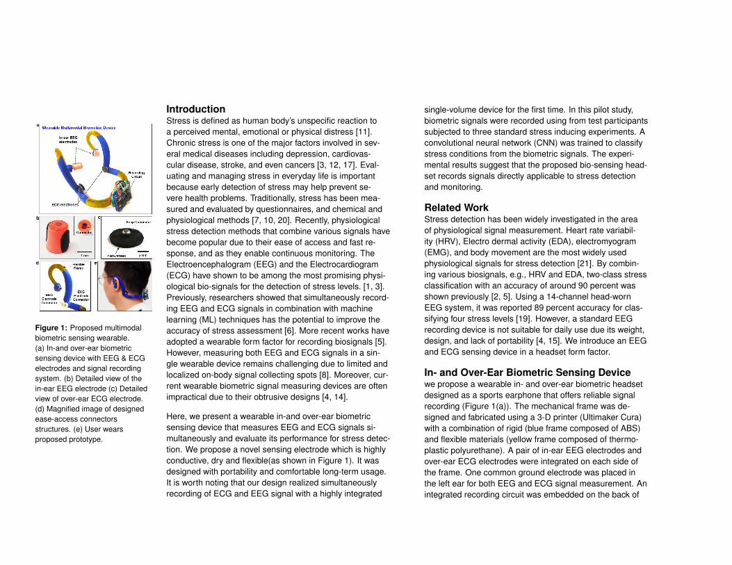
Figure 4: Questionnaire results for(a) Stroop, (b) Memory search, and(c) Mental arithmetic task.
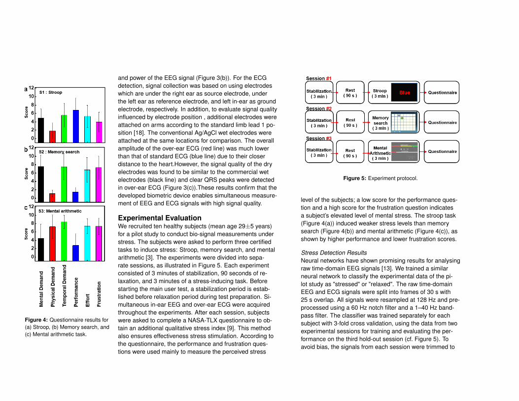
We recruited ten healthy subjects (mean age 29±5 years)for a pilot study to conduct bio-signal measurements understress. The subjects were asked to perform three certifiedtasks to induce stress: Stroop, memory search, and mentalarithmetic [3]. The experiments were divided into sepa-rate sessions, as illustrated in Figure 5. Each experimentconsisted of 3 minutes of stabilization, 90 seconds of re-laxation, and 3 minutes of a stress-inducing task. Beforestarting the main user test, a stabilization period is estab-lished before relaxation period during test preparation. Si-multaneous in-ear EEG and over-ear ECG were acquiredthroughout the experiments. After each session, subjectswere asked to complete a NASA-TLX questionnaire to ob-tain an additional qualitative stress index [9]. This methodalso ensures effectiveness stress stimulation. According tothe questionnaire, the performance and frustration ques-tions were used mainly to measure the perceived stress
Figure 5: Experiment protocol.
level of the subjects; a low score for the performance ques-tion and a high score for the frustration question indicatesa subject’s elevated level of mental stress. The stroop task(Figure 4(a)) induced weaker stress levels than memorysearch (Figure 4(b)) and mental arithmetic (Figure 4(c)), asshown by higher performance and lower frustration scores.
Stress Detection ResultsNeural networks have shown promising results for analysingraw time-domain EEG signals [13]. We trained a similarneural network to classify the experimental data of the pi-lot study as "stressed" or "relaxed". The raw time-domainEEG and ECG signals were split into frames of 30 s with25 s overlap. All signals were resampled at 128 Hz and pre-processed using a 60 Hz notch filter and a 1–40 Hz band-pass filter. The classifier was trained separately for eachsubject with 3-fold cross validation, using the data from twoexperimental sessions for training and evaluating the per-formance on the third hold-out session (cf. Figure 5). Toavoid bias, the signals from each session were trimmed to
3 7 6 9 2 11 10 5 4 8 1
Subject ID
0
50
100
[%]
a)a)
3 7 6 9 2 11 10 5 4 8 1
Subject ID
0
50
100
[%]
b)b)
3 7 6 9 2 11 10 5 4 8 1
Subject ID
0
50
100
[%]
c)c)
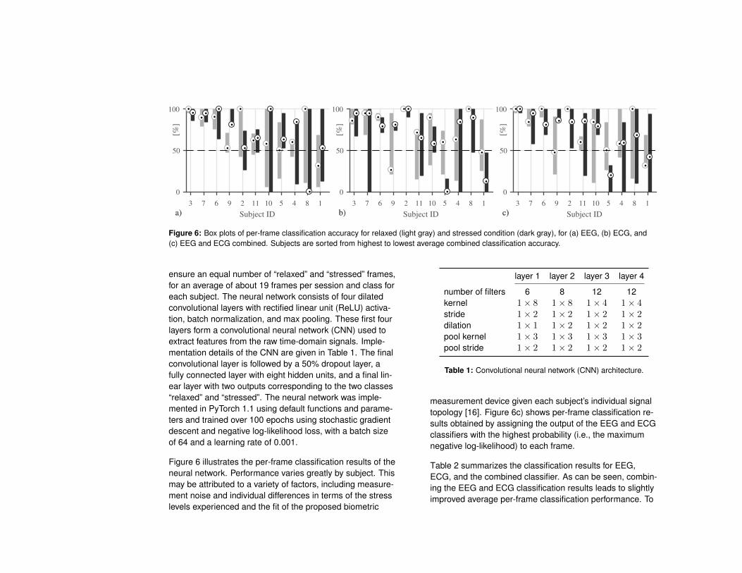
Figure 6: Box plots of per-frame classification accuracy for relaxed (light gray) and stressed condition (dark gray), for (a) EEG, (b) ECG, and(c) EEG and ECG combined. Subjects are sorted from highest to lowest average combined classification accuracy.
ensure an equal number of “relaxed” and “stressed” frames,for an average of about 19 frames per session and class foreach subject. The neural network consists of four dilatedconvolutional layers with rectified linear unit (ReLU) activa-tion, batch normalization, and max pooling. These first fourlayers form a convolutional neural network (CNN) used toextract features from the raw time-domain signals. Imple-mentation details of the CNN are given in Table 1. The finalconvolutional layer is followed by a 50% dropout layer, afully connected layer with eight hidden units, and a final lin-ear layer with two outputs corresponding to the two classes“relaxed” and “stressed”. The neural network was imple-mented in PyTorch 1.1 using default functions and parame-ters and trained over 100 epochs using stochastic gradientdescent and negative log-likelihood loss, with a batch sizeof 64 and a learning rate of 0.001.
Figure 6 illustrates the per-frame classification results of theneural network. Performance varies greatly by subject. Thismay be attributed to a variety of factors, including measure-ment noise and individual differences in terms of the stresslevels experienced and the fit of the proposed biometric
layer 1 layer 2 layer 3 layer 4
number of filters 6 8 12 12kernel 1× 8 1× 8 1× 4 1× 4stride 1× 2 1× 2 1× 2 1× 2dilation 1× 1 1× 2 1× 2 1× 2pool kernel 1× 3 1× 3 1× 3 1× 3pool stride 1× 2 1× 2 1× 2 1× 2
Table 1: Convolutional neural network (CNN) architecture.
measurement device given each subject’s individual signaltopology [16]. Figure 6c) shows per-frame classification re-sults obtained by assigning the output of the EEG and ECGclassifiers with the highest probability (i.e., the maximumnegative log-likelihood) to each frame.
Table 2 summarizes the classification results for EEG,ECG, and the combined classifier. As can be seen, combin-ing the EEG and ECG classification results leads to slightlyimproved average per-frame classification performance. To

per-frame per-session“relaxed” “stressed” “relaxed” “stressed”
EEG 67.1 69.0 72.7 78.8ECG 73.1 63.2 75.8 66.7combined 72.3 68.4 75.8 75.8
Table 2: Percentage of correct classifications, averaged over alltest subjects and experiment sessions.
arrive at a single classification per subject and experimentsession, a voting strategy is employed whereby for eachsession the most frequent classification output is chosen asthe per-session result. The average per-session classifica-tion accuracy exceeds 70% for all conditions except for theECG classifier and the “stressed” condition.
Conclusions and Future WorkWe introduce a wearable in and over-ear biometric sensingdevice to monitor stress conditions via multimodal physio-logical signals. The proposed device is able to detect stressby simultaneously recording EEG and ECG signals with ro-bust signal quality. The nano-materials flexible electrodesoffers robust bio-signal recording while providing user com-fort. A pilot study suggests that a neural network is capa-ble of classifying relaxed and stressed mental states of auser by analysing two minutes of EEG and ECG signals ob-tained with the proposed device, with an average accuracyof about 75%. We believe that the design reported in thisstudy has significant potential in active and social bio-signalmeasurement applications. The stress detection in this pa-per was conducted in laboratory conditions. However, weenvision that the proposed wearable biometric sensing de-vice can be used in various daily activities. Future workincludes field studies in a larger variety of stress-inducingconditions. Collecting data from a larger subject pool would
allow improving classification performance as well as train-ing and evaluating a between-subject classifier that doesnot require per-subject training.
REFERENCES[1] J. W. Ahn, Y. Ku, and H. C. Kim. 2019. A Novel
Wearable EEG and ECG Recording System for StressAssessment. Sensors 19, 9 (2019), 1991.
[2] A. O. Akmandor and N. K. Jha. 2017. Keep the stressaway with SoDA: Stress detection and alleviationsystem. IEEE Transactions on Multi-Scale ComputingSystems 3, 4 (2017), 269–282.
[3] F. Al-Shargie, M. Kiguchi, N. Badruddin, S. C. Dass,A. F. M. Hani, and T. B. Tang. 2016. Mental stressassessment using simultaneous measurement of EEGand fNIRS. Biomedical optics express 7, 10 (2016),3882–3898.
[4] M. G. Bleichner and S. Debener. 2017. Concealed,unobtrusive ear-centered EEG acquisition: cEEGridsfor transparent EEG. Frontiers in human neuroscience11 (2017), 163.
[5] Y. S. Can, N. Chalabianloo, D. Ekiz, and C. Ersoy.2019. Continuous Stress Detection Using WearableSensors in Real Life: Algorithmic ProgrammingContest Case Study. Sensors 19, 8 (2019), 1849.
[6] L.-l. Chen, Y. Zhao, P.-f. Ye, J. Zhang, and J.-z. Zou.2017. Detecting driving stress in physiological signalsbased on multimodal feature analysis and kernelclassifiers. Expert Systems with Applications 85(2017), 279–291.
[7] S. Cohen, R. C. Kessler, and L. U. Gordon. 1997.Measuring stress: A guide for health and socialscientists. Oxford University Press on Demand.
[8] V. Goverdovsky, W. von Rosenberg, T. Nakamura, D.Looney, D. J. Sharp, C. Papavassiliou, M. J. Morrell,and D. P. Mandic. 2017. Hearables: Multimodalphysiological in-ear sensing. Scientific reports 7, 1(2017), 6948.
[9] S. G. Hart and L. E. Staveland. 1988. Development ofNASA-TLX (Task Load Index): Results of empiricaland theoretical research. In Advances in psychology.Vol. 52. Elsevier, 139–183.
[10] D. H. Hellhammer, S. Wüst, and B. M. Kudielka. 2009.Salivary cortisol as a biomarker in stress research.Psychoneuroendocrinology 34, 2 (2009), 163–171.
[11] S. A. Hosseini, M. A. Khalilzadeh, and S. Changiz.2010. Emotional stress recognition system for affectivecomputing based on bio-signals. Biological Systems18, spec01 (2010), 101–114.
[12] M. Irie, S. Asami, S. Nagata, M. Miyata, and H. Kasai.2001. Relationships between perceived workload,stress and oxidative DNA damage. Int Arch OccupEnviron Health 74, 2 (2001), 153–157.
[13] V. J. Lawhern, A. J. Solon, N. R. Waytowich, S. M.Gordon, C. P. Hung, and B. J. Lance. 2018. EEGNet: acompact convolutional neural network for EEG-basedbrain–computer interfaces. Journal of neuralengineering 15, 5 (2018), 056013.
[14] J. H. Lee, J.-Y. Hwang, J. Zhu, H. R. Hwang, S. M.Lee, H. Cheng, S.-H. Lee, and S.-W. Hwang. 2018.Flexible conductive composite integrated with personalearphone for wireless, real-time monitoring ofelectrophysiological signs. ACS applied materials &interfaces 10, 25 (2018), 21184–21190.
[15] D. Looney, P. Kidmose, C. Park, M. Ungstrup, M. L.Rank, K. Rosenkranz, and D. P. Mandic. 2012. The
in-the-ear recording concept: User-centered andwearable brain monitoring. IEEE pulse 3, 6 (2012),32–42.
[16] L. Ma, J. W. Minett, T. Blu, and W. S. Wang. 2015.Resting State EEG-based biometrics for individualidentification using convolutional neural networks. In2015 37th Annual International Conference of theIEEE Engineering in Medicine and Biology Society(EMBC). 2848–2851. DOI:http://dx.doi.org/10.1109/EMBC.2015.7318985
[17] C. Schubert, M. Lambertz, R. Nelesen, W. Bardwell,J.-B. Choi, and J. Dimsdale. 2009. Effects of stress onheart rate complexity—a comparison betweenshort-term and chronic stress. Biological psychology80, 3 (2009), 325–332.
[18] F. Stauffer, M. Thielen, C. Sauter, S. Chardonnens, S.Bachmann, K. Tybrandt, C. Peters, C. Hierold, and J.Vörös. 2018. Skin conformal polymer electrodes forclinical ECG and EEG recordings. Advancedhealthcare materials 7, 7 (2018).
[19] V. Vanitha and P. Krishnan. 2016. Real time stressdetection system based on EEG signals. BiomedicalResearch (2016).
[20] G. K. Verma and U. S. Tiwary. 2014. Multimodal fusionframework: A multiresolution approach for emotionclassification and recognition from physiologicalsignals. NeuroImage 102 (2014), 162–172.
[21] J. Wijsman, B. Grundlehner, H. Liu, H. Hermens, andJ. Penders. 2011. Towards mental stress detectionusing wearable physiological sensors. In Proc. IEEEInt. Conf. Engineering in Medicine and BiologySociety. IEEE, 1798–1801.