That which does not kill me makes me stronger: adapting to chronic ER stress

Pharmacology & Therapeutics 134 (2012) 306–316
Contents lists available at SciVerse ScienceDirect
Pharmacology & Therapeutics
j ou rna l homepage: www.e lsev ie r .com/ locate /pharmthera
Associate editor: M. Panagiotidis
Stress management at the ER: Regulators of ER stress-induced apoptosis
Adrienne M. Gorman, Sandra J.M. Healy, Richard Jäger, Afshin Samali ⁎Apoptosis Research Centre, School of Natural Sciences, National University of Ireland, Galway, University Road., Galway, Ireland
Abbreviations: ASK1, apoptosis signal-regulating kscription factor; BAK, Bcl-2 homologous antagonist/killtein X; BCL-2, B-cell lymphoma 2; BH3, Bcl-2 homology1; Bim, Bcl-2 interacting mediator of cell death; C/EBPtein; CHOP, C/EBP homologous protein; cIAP, cellulareIF2α, eukaryotic initiation factor 2 α; ER, endoplasmicrest and DNA damage-inducible gene; GRP, glucoseshock protein; IAP, inhibitor of apoptosis protein; IRE1JNK, c-Jun N-terminal kinase; MEF, mouse embryonic fi
kinase or PKR-like kinase; RIDD, regulated IRE1-depenspliced X box-binding protein 1; TRAF2, tumor necrosis ffactor 2; TRB3, tribbles homolog 3; UPR, unfolded proteinprotein 1; XIAP, X chromosome-linked IAP.⁎ Corresponding author. Tel.: +353 91 492440; fax: +
E-mail address: [email protected] (A. Sam
0163-7258/$ – see front matter © 2012 Elsevier Inc. Alldoi:10.1016/j.pharmthera.2012.02.003
a b s t r a c t
a r t i c l e i n f oKeywords:
ApoptosisBcl-2 familyEndoplasmic reticulum (ER) stressUnfolded protein response (UPR)The endoplasmic reticulum (ER) is an elaborate cellular organelle essential for cell function and survival. Con-ditions that interfere with ER function lead to the accumulation and aggregation of unfolded proteins whichare detected by ER transmembrane receptors that initiate the unfolded protein response (UPR) to restore nor-mal ER function. If the ER stress is prolonged, or the adaptive response fails, apoptotic cell death ensues. Manystudies have focused on how this failure initiates apoptosis, particularly because ER stress-induced apoptosisis implicated in the pathophysiology of several neurodegenerative and cardiovascular diseases. In this reviewwe aim to shed light on the proteins that are not core components of the UPR signaling pathway but which caninfluence the course of the ER stress response by regulating the switch from the adaptive phase to apoptosis.
© 2012 Elsevier Inc. All rights reserved.
Contents
1. Introduction . . . . . . . . . . . . . . . . . . . . . . . . . . . . . . . . . . . . . . . . . . . . . 3062. Regulation of the unfolded protein response and initiation phase of ER stress-induced apoptosis . . . . . . 3073. Commitment phase of ER stress-induced apoptosis . . . . . . . . . . . . . . . . . . . . . . . . . . . 3074. The execution phase of ER stress-induced apoptosis . . . . . . . . . . . . . . . . . . . . . . . . . . . 3075. Novel regulation of UPR and ER stress-induced apoptosis . . . . . . . . . . . . . . . . . . . . . . . . 3086. Conclusion . . . . . . . . . . . . . . . . . . . . . . . . . . . . . . . . . . . . . . . . . . . . . . 308Acknowledgments . . . . . . . . . . . . . . . . . . . . . . . . . . . . . . . . . . . . . . . . . . . . . 308
311313314314314
References . . . . . . . . . . . . . . . . . . . . . . . . . . . . . . . . . . . . . . . . . . . . . . . . . 308314
1. Introduction
The endoplasmic reticulum (ER) is a continuous membranous net-work of sacs and tubes in the cell. It includes the regions known as thesmooth ER, rough ER and the outer nuclear envelope. It is also in closecontact with the mitochondria. It is the primary site for the synthesis
inase 1; ATF, activating tran-er; BAX, Bcl-2 associated pro-domain 3; BI-1, Bax inhibitor
, CAAT/enhancer-binding pro-inhibitor of apoptosis protein;reticulum; GADD, growth ar-regulated protein; HSP, heat, inositol-requiring enzyme 1;broblast; PERK, pancreatic ERdent decay of mRNA; XBP1s,actor (TNF) receptor-associatedresponse; XBP1, X box-binding
353 91 495504.ali).
rights reserved.
and folding of secreted, membrane-bound and some organelle-targeted proteins. Optimum protein folding within the ER lumen re-quires ATP, Ca2+ and an oxidizing environment to allow disulfidebond formation (Gaut & Hendershot, 1993). Conditions that perturbcellular energy levels, the redox state or Ca2+ concentration reducethe protein folding capacity of the ER. Such conditions, as well as mu-tations within proteins that impair their correct folding or otherwiseimpede their further processing or transport within the ER, result inthe accumulation and/or aggregation of unfolded proteins, a conditionreferred to as ER stress. To overcome the deleterious effects of ERstress, cells have evolved a series of adaptive and protective strategiescollectively termed the unfolded protein response (UPR). However, ifunresolved, ER stress is lethal to cells via what is known as ER stress-induced apoptosis. Intriguingly, both responses are downstream ofUPR and unsurprisingly, there are numerous pathophysiological con-ditions associated with ER stress, including ischemia, neurodegenera-tive diseases, cancer and diabetes (Healy et al., 2009; Doyle et al.,2011).
The UPR is a concerted and complex cellular response that is medi-ated through three ER transmembrane receptors: pancreatic ER kinase

307A.M. Gorman et al. / Pharmacology & Therapeutics 134 (2012) 306–316
or PKR-like ER kinase (PERK), activating transcription factor-6 (ATF6)and inositol-requiring enzyme 1 (IRE1, also known as ERN 1). In restingcells, all three ER stress receptors are maintained in an inactive statethrough association with the ER chaperone, 78 kDa glucose regulatedprotein (GRP78). Under conditions of ER stress, accumulating unfoldedproteins leads to GRP78 dissociation and activation of the three ERstress receptors triggering the UPR. The UPR is a pro-survival responseaimed at reducing the backlog of unfolded proteins and restoring nor-mal ER function (Schroder & Kaufman, 2005) (Fig. 1). However, if thestress cannot be resolved, this otherwise protective signaling switchesto a pro-apoptotic response. This review examines themolecularmech-anisms that promote an apoptotic response by influencing UPR-mediated signals during the three distinct phases of ER stress-inducedapoptosis: initiation, commitment and, execution (Szegezdi et al.,2006). We will discuss regulators of ER stress-induced apoptosis,where we define a regulator as any protein that is not required fortransducing the signal in the UPR pathway, but which can modulatethe activity of the pathway such that the likelihood of apoptosis occur-ring is altered.
2. Regulation of the unfolded protein responseand initiation phase of ER stress-induced apoptosis
2.1. PERK, ATF6 and IRE1 mediate the initiation phase of the UPR
2.1.1. PERKDissociation of GRP78 from PERK results in its dimerization,
autophosphorylation and activation. Active PERK phosphorylates the
Fig. 1. The UPR is mediated by three ER stress sensors. Binding of unfolded proteins to GRP7autophosphorylates. It phosphorylates eIF2α and thus general Cap-dependent translationsuch as ATF4 which activates CHOP transcription. One of the genes induced by CHOP iseIF2α. Activation of ATF6 allows its translocation to the Golgi where it undergoes cleavagedual kinase and endonuclease. One of its targets is XBP1 mRNA which undergoes splicing t
alpha subunit of the eukaryotic initiation factor 2 (eIF2α), inhibitinggeneral protein translation (Harding et al., 1999). Inhibition of proteintranslation aids cell survival by decreasing the load of nascent proteinsin the ER. In fact, PERK−/−mouse embryonic fibroblasts (MEFs), whenchallenged with ER stress-inducing agents, failed to block proteintranslation and exhibited increased cell death. Inhibition of translationwith cycloheximide reduced ER stress-induced cell death, confirmingthat blocking the buildup of unfolded nascent proteins is critical forcell survival (Harding et al., 2000b). This attenuation of translationis, however, not absolute; mRNAs carrying certain regulatory se-quences in their 5′ untranslated regions can bypass the phospho-eIF2α-mediated translational block and can sometimes be translatedat even higher rates (Schroder & Kaufman, 2005). The most studiedof such transcripts encodes ATF4, a member of the CCAAT/enhancer-binding protein (C/EBP) family of transcription factors. ATF4 transla-tion is upregulated upon eIF2α phosphorylation and promotes cellsurvival by inducing genes involved in amino acid metabolism,redox reactions, stress response and protein secretion (Harding etal., 2003). However, not all the genes induced by ATF4 are associatedwith cell survival. The transcription factor C/EBP homologous protein(CHOP), whose induction strongly depends on ATF4 (Harding et al.,2000a), is thought to promote apoptosis (Fig. 1). In conclusion, activa-tion of PERK is initially protective and critical for survival in the face ofmild ER stress. However, activation of PERK also leads to induction ofCHOP, which, as detailed later, is an important element in the switchfrom pro-survival to pro-death signaling.
A second target of PERK is the transcription factor, nuclear factor(erythroid-derived 2)-related factor 2 (NRF2) whose phosphorylation
8 within the ER lumen allows activation of PERK, ATF6 and IRE1. PERK dimerises andis inhibited. Cap-independent translation allows the translation of certain proteinsGADD34 which regulates protein phosphatase 1 (PP1), which can dephosphorylateby S1P and S2P proteases. Cleaved ATF6 activates XBP1 transcription. Active IRE1 is ao produce an active transcription factor, XBP1s. One of the targets of XBP1s is p58IPK.

308 A.M. Gorman et al. / Pharmacology & Therapeutics 134 (2012) 306–316
liberates it from its inhibitor KEAP1, allowing for induction of targetgenes that are mainly involved in oxidative stress signaling(Cullinan & Diehl, 2004).
2.1.2. ATF6Dissociation of GRP78 from ATF6 allows its translocation to the
Golgi where it is cleaved to its active form by Site-1 and Site-2 prote-ases (S1P and S2P). Active ATF6 then translocates to the nucleus to in-duce expression of geneswith an ER stress response element (ERSE) intheir promoter (Schroder & Kaufman, 2005). The targets of ATF6include ER chaperone proteins such as GRP78, GRP94, protein disul-phide isomerase, and the transcription factors CHOP and X box-binding protein-1 (XBP1) (Yoshida et al., 2000). XBP1 is importantin IRE1 signaling and thus links ATF6with pro-survival signaling emit-ted through IRE1, as discussed below in more detail (Fig. 1). AlthoughATF6 has long been thought to transduce purely pro-survival signalsand counteract ER stress, overexpression of ATF6 can induce CHOPmRNA expression as well, whereas overexpression of a dominant neg-ative ATF6 mutant blocks CHOP induction by ER stress (Yoshida et al.,2000). Recently, ATF6 has been linked to ER stress-induced apoptosisin a myoblast cell line where it was shown to induce apoptosis by in-directly downregulating expression of anti-apoptotic protein MCL-1(Morishima et al., 2011).
2.1.3. IRE1αIRE1α (hereafter referred to as IRE1) is a dual-activity enzyme. In
its cytoplasmic part it possesses a serine–threonine kinase domainand a C-terminal endoribonuclease domain. Once activated, the endo-nuclease activity induces the rapid turnover of mRNAs encodingmembrane and secreted proteins, through a pathway referred to asregulated IRE1-dependent decay (RIDD) (Hollien et al., 2009). Howev-er, a more selective function of the endonuclease is removal of a 26-nucleotide intron from the XBP1 mRNA transcript, which is inducedby ATF6. The frame-shift splice variant thus generated (XBP1s) codesfor a stable, active transcription factor (Yoshida et al., 2001). XBP1shas diverse targets including ER chaperones and the HSP40 familymember, p58IPK (Lee et al., 2003) (Fig. 1). p58IPK was initially thoughtto inhibit PERK by interacting with the cytoplasmic kinase domain,thereby providing a negative feedback loop that relieves the PERK-mediated translational block (Yan et al., 2002). Recent data, however,suggest it is located inside the ER lumen where it functions as a co-chaperone of GRP78 (Rutkowski et al., 2007). In contrast to its RNaseactivity, IRE1 kinase function can induce pro-death signaling throughbinding to TRAF2 which leads to JNK activation (Urano et al., 2000).
Recently it was shown that experimental prolongation of IRE1 sig-naling (as determined by XBP1 splicing), independent of ER stress canpromote cell survival (Lin et al., 2007; Lin et al., 2009). During ERstress, IRE1 is switched off earlier than PERK, therefore the cytoprotec-tive function of IRE1 is no longer present while PERK signaling is stillenduring (Lin et al., 2007). Mechanisms to modulate the duration ofIRE1 signaling could therefore influence cell fate in terms of deathand survival. The activity of IRE1 is influenced by several interactingproteins (see below) with which it forms a protein complex referredto as the UPRosome (Hetz & Glimcher, 2009). It is noteworthy thatof all of the three ER stress sensors, IRE1 is the most highly regulated,which may reflect a key role in controlling the switch between adap-tive responses and initiation of the apoptosis programme.
2.2. HSP family regulation of the three mediators
Heat shock proteins (HSPs), also called stress proteins, are highlyconserved proteins whose expression is induced by different kindsof cellular stresses (Concannon et al., 2003; Fulda et al., 2010). HSPshave strong cytoprotective effects and behave as molecular chaper-ones for other cellular proteins. Inappropriate activation of signalingpathways occurs during acute or chronic stress as a result of protein
misfolding, protein aggregation or disruption of regulatory com-plexes. The action of chaperones, through their roles in protein ho-meostasis, is thought to restore the balance. Mammalian HSPs havebeen classified into two groups according to their size: highmolecularweight HSPs and small molecular weight HSPs. The first group in-cludes three major families: HSP90, HSP70 and HSP60. Some ofthese are expressed constitutively whereas expression of others is in-duced by stressful conditions. These proteins can be targeted to dif-ferent sub-cellular compartments. High molecular weight HSPs areATP-dependent chaperones and require co-chaperones to modulatetheir conformation and ATP binding. In contrast, small HSPs, such asHSP27, are ATP-independent chaperones (Parcellier et al., 2003).Among the different HSPs, HSP27 and HSP70 are the most strongly in-duced after stresses such as anticancer drugs, oxidative stress or irra-diation. While expressed at very low levels in non-transformed cells,both HSP27 and HSP70 are abundantly expressed in cancer cells, andtherefore have been suggested to be important prognostic factors inmalignant diseases (Garrido et al., 2006). Also within the ER lumen,homologues of HSPs are present that aid in protein folding, e.g.,GRP78. HSPs can regulate ER stress signaling from both the cytoplas-mic and the ER-luminal side. Conceivably, interactions of ER stressmediators with cytoplasmic HSPs would integrate general cellularstress pathways and UPR signaling outputs. Regulation of UPR byHSP family proteins is described in the sections below.
2.2.1. GRP78 regulation of the three mediatorsThe intraluminal ER chaperone GRP78 (also termed BiP) is a mem-
ber of the HSP70 family of heat shock proteins and is the main regu-lator of the ER stress mediators PERK, ATF6, and IRE1 which are keptinactive by binding of GRP78 to their luminal domains (reviewed in(Kimata & Kohno, 2011)). Classically it is thought that accumulatingunfolded proteins in the ER lumen compete for binding of GRP78 tothe ER stress mediators, leading to their activation when GRP78 dis-sociates. This model proposes that unfolded proteins behave as clientproteins of GRP78, involving rapid cycles of substrate binding and re-lease in an ATP-dependent manner. There is, however, evidence thatGRP78 might remain stably bound to substrate proteins until thesechange their conformation. In addition, it is not entirely clear whetherGRP78 dissociation from the luminal domains of the three ER stressmediators is fully explained by this competition mechanism, and ithas been suggested that co-chaperones might stimulate the releaseof GRP78 (Shen et al., 2005). In this regard it is of note that p58IPK
is reported to be localized inside the ER lumen where it acts as co-chaperone of GRP78 (Rutkowski et al., 2007). Together with the ear-lier report showing p58IPK interacts with the kinase domain of PERK(Yan et al., 2002), this suggests that p58IPK is present in both the cy-toplasm and the ER lumen.
How GRP78 dissociation activates the mediators is also not fullyunderstood. The ER-luminal domains interacting with GRP78 arehighly conserved between IRE1 and PERK but not so with ATF6. Inyeast, IRE1 is activated in a receptor-like manner by direct binding ofunfolded proteins to the luminal domain rather than by dissociationof GRP78 (Gardner & Walter, 2011). Activation of mammalian IRE1,as well as of PERK, however seems to be triggered by dissociation ofGRP78 as the main mechanism (Kimata & Kohno, 2011).
Expression of GRP78 is upregulated by ER stress through transcrip-tion factors ATF6 (Yoshida et al., 1998) and ATF4 (Luo et al., 2003). Theresulting increase in GRP78 protein therefore not only aids folding ofproteins inside the ER, but may inactivate PERK, ATF6 and IRE1 bybinding to their luminal domains. Thus, GRP78 is component of a feed-backmechanism that ensures protein refolding and inactivation of theUPR. As suggested by Rutlowski and Kaufman (Rutkowski & Kaufman,2004), it is conceivable that differential affinity of the three UPRmedi-ators for GRP78might differentially regulate the kinetics of their inac-tivation. However, there is limited evidence to support such a model.During UPR-induced B-cell differentiation, there is activation of the

309A.M. Gorman et al. / Pharmacology & Therapeutics 134 (2012) 306–316
IRE1 pathway (Reimold et al., 2001) in the absence of CHOP induction(which is dependent on ATF6 and PERK pathways) (Gass et al., 2002).However, it has recently been reported that this limited ER stress re-sponse may be the result of suppression of the PERK signal ratherthan selective activation of IRE1 (Ma et al., 2010).
2.2.2. Regulation of IRE1 and PERK by cytoplasmic HSPs
2.2.2.1. HSP90 regulation of PERK and IRE1. HSP90 has been shown tophysically interact with the cytoplasmic part of IRE1 and PERK(Marcu et al., 2002). Disruption of this interaction by HSP90 inhibitorsleads to a reduced half-life of IRE1 and PERK protein, resulting in lessprotein, suggesting stabilization of both proteins by HSP90 in the nor-mal situation. In this study the influence of HSP90 on IRE1 activity andsignaling outputs was not addressed, however, it was noted that inhi-bition of Hsp90 did not impair translational attenuation during ERstress, suggesting that even reduced amounts of PERK were sufficientto block translation.
2.2.2.2. Hsp70 regulation of IRE1. HSP72 is a stress-induciblemember ofthe HSP70 family whose chaperoning activity is dependent on ATP(Young, 2010). It also requires co-chaperones for efficient activity.When overexpressed in cells HSP72was found to be protective againstER stress-induced cell death (Gupta et al., 2010b). Remarkably thiscytoprotection was not solely attributable to general effects on the ap-optotic machinery but was dependent on production of spliced XBP1.In fact, HSP72 overexpression leads to enhanced and prolonged XBP1splicing during ER stress, suggesting stimulation of IRE1 activity. Co-
Fig. 2. Regulators of IRE1 signaling. Under resting conditions GRP78 in the ER lumen keeps IREphosphorylation, probably utilizing RACK as a scaffold protein. BI-1 and JAB1 also interactstress, GRP78 dissociates allowing IRE1 to dimerize. It undergoes autophosphorylation in tsignaling. AIP stimulates IRE1 dimerization. HSP72 interaction with the cytosolic region endegraded by the proteasome. JAB1 interaction with IRE1 is enhanced. With prolonged ERBAX/BAK effect on IRE1 signaling. TRAF2 is recruited to the kinase domain, either directly o
immunoprecipitation experiments revealed a physical interaction be-tween the cytoplasmic part of IRE1 and HSP72 that was dependent onthe ATPase domain of HSP72 (Gupta et al., 2010a) and involved the co-chaperone HSP40 (unpublished observations) (Fig. 2). Thus, as astress-inducible protein HSP72 might serve as a hub connectingother stress pathways with the UPR. It is noteworthy that HSP72 isnormally not expressed in unstressed cells but is expressed at highlevels in cancer cells. This implies that HSP72 is not an essential com-ponent of the UPRosome. However, in cancer cells that express highlevels of Hsp72, it forms a complex with IRE1, and by stimulatingXBP1 splicing activity, HSP72 contributes to enhanced pro-survivalsignaling.
2.2.2.3. p58IPK — modulator of PERK or co-chaperone of Grp78. Thestress-inducible protein p58IPK is a HSP40 family member, initiallyidentified as an inhibitor of the PERK-related eIF2α kinase PKR, possi-bly modulating HSP70 activity to refold and inhibit the kinase(Melville et al., 1999). Later p58IPK was found localized at the ER andby co-immunoprecipitation experiments shown to physically interactwith the cytoplasmic PERK kinase domain, inhibiting PERK activity(Yan et al., 2002). Consistent with this finding, p58IPK expressionleads to a decrease in eIF2α phosphorylation (Yan et al., 2002; vanHuizen et al., 2003). Since p58IPK expression is regulated by XBP1s, itis part of a negative feedback regulation limiting PERK activity duringER stress. In a later study, however, p58IPK was suggested to be partof a protein complex with HSP70 that mediates degradation of pro-teins during their co-translational import into the ER lumen, therebylowering protein load of the stressed ER (Oyadomari et al., 2006).
1 in an inactive state. On the cytosolic side, protein phosphatase 2A (PP2A) prevents itswith IRE1 under resting conditions. HSP90 interacts with IRE1, stabilizing it. Upon ERhe kinase domain. Binding of BAX/BAK enhance IRE1-mediated XBP1 splicing and JNKhances XBP1 splicing activity. BI-1 becomes ubiquitinated by the E3 ligase BAR and isstress BAR becomes downregulated, thus allowing stabilization of BI-1 which inhibitsr perhaps via JIK. TRAF2 recruits ASK1 which leads to JNK activation.

310 A.M. Gorman et al. / Pharmacology & Therapeutics 134 (2012) 306–316
Importantly, both proposed functions of p58IPK were recently chal-lenged by a study which demonstrated localization of p58IPK withinthe ER lumen where it acts as a co-chaperone of GRP78 (Rutkowskiet al., 2007). While this study could not confirm the aforementionedeffects on protein import into the ER, it remains possible that p58IPK
indirectly, via GRP78 binding, affects PERK activity.
2.3. Bcl-2 family regulation of IRE1
Members of the BCL-2 family of proteins are major regulators ofapoptotic cell death (see (Chipuk et al., 2010; Szegezdi et al., 2009) forreview). They are characterized by up to four conserved BCL-2 homolo-gy (BH) domains which are involved in their protein–protein interac-tions. Multi-domain family members can be subdivided according totheir pro-apoptotic (BAX and BAK) and anti-apoptotic effects (such asBCL-2, BCL-XL, MCL-1). They are located at intracellular membranes,and their best studied mechanism of apoptosis regulation is by control-ling the release of cytotoxic molecules from mitochondria. The activityof multi-domain BCL-2 family members is modulated by interactionwith BH3-only proteins (such as BIM, BIK, BAD) that contain only thethird BH domain and are generally pro-apoptotic. The first reports link-ing ER stress-induced cell death to the BCL-2 family of proteins showedthat overexpression of BCL-2, or deficiency of BAX and BAK, conferredprotection against lethal ER stress (Distelhorst & McCormick, 1996;Wei et al., 2001). These studies assumed that ER stress-inducedapoptosis proceeds via the intrinsic pathway, which is controlled bymitochondrially-localized BCL-2 proteins. However, from early on itwas noticed thatmembers of the BCL-2 family also reside and are activeat the ER membrane (Krajewski et al., 1993; Zong et al., 2003) and thatartificial targeting of BCL-2 to the ER membrane provided protectionagainst ER stress (Hacki et al., 2000). BCL-2 family proteins whereshown to influence ER Ca2+ homeostasis and ER-mitochondrial
Fig. 3. Pro-apoptotic signaling by IRE1. JNK is activated downstream of TRAF2–ASK1 interacmotor complex, which stabilizes it. This allows it to draw anti-apoptotic BCL-2 family memouter membrane permeabilization and cytochrome c release. JNK can also phosphorylate Binteract with IRE1 at the ER membrane. This interaction can be blocked by BI-1. The endowhich leads to a decrease in the production of ER chaperone proteins.
crosstalk and might by this means indirectly affect ER stress-inducedcell death (Szegezdi et al., 2009). However, they can also interact direct-ly with IRE1, modulating its functioning during ER stress, and thus reg-ulate ER stress-induced cell death at the level of IRE1 signaling (Hetz etal., 2006) (Fig. 3).
BAX and BAK were shown to form a complex with IRE1 in an ERstress dependent manner, and BAX/BAK-deficient cells displayed re-duced levels of spliced XBP1 and phosphorylated IRE1 during ER stress(Hetz et al., 2006). When the BAX/BAK-deficient cells were reconsti-tuted with a BAK mutant that was fused to an ER-localization motif(BAK-cb5), levels of XBP1s and of JNK phosphorylationwere increased(Hetz et al., 2006). Therefore, the interaction with BAX and BAKappears to be essential for activation of IRE1 and might represent apossible switching mechanism towards pro-apoptotic signaling byIRE1. This was suggested by overexpression of BAK-cb5 in BAX/BAK-deficient cells. In these cells, co-expression of ER-targeted BH3-onlyproteins Bim or Puma resulted in IRE1-dependent JNK activation inthe absence of XBP1 splicing (Klee et al., 2009). As JNK activationcould be abolished by interfering with either TRAF2 or IRE1, these ex-periments reveal a specific activation of pro-apoptotic signaling ofIRE1 by pro-apoptotic BH3-only proteins acting at the ER membrane.The association of IRE1 with Bax and BAK is influenced by BAX Inhib-itor 1 (BI-1), a transmembrane protein localized at the ER and nuclearenvelope, that had initially been identified in a genetic screen for sup-pressors of BAX-induced cell death (Robinson et al., 2011). BI-1 formsa complex with the cytoplasmic part of IRE1 (Fig. 3). BI-1 deficiency ofcells resulted in higher IRE1 activity and enhanced XBP1 splicing,while BI-1 overexpression disrupted the interaction between IRE1and Bax or BAK (Lisbona et al., 2009). BI-1 itself is regulated by thebifunctional apoptosis regulator (BAR), an ER-localized RING-type E3ligase, that mediates the proteasomal degradation of BI-1 and thusremoves the block of IRE1 activation provided by BI-1 (Rong et al.,
tion with IRE1. Phosphorylation by JNK of BIM causes it to dissociate from the dyneinbers such as BCL-2 away from BAX/BAK which are thus free to mediate mitochondrialCL-2, which inhibits its ability to regulate ER Ca2+ homeostasis. BAX and BAK can alsonuclease activity of IRE1 mediates Regulated IRE1-dependent decay of mRNA (RIDD),

311A.M. Gorman et al. / Pharmacology & Therapeutics 134 (2012) 306–316
2011). However, BAR becomes downregulated during ER stress, andthe resulting increase in BI-1 might thus contribute to attenuation ofIRE1 signaling during prolonged ER stress (Fig. 3).
2.4. Regulation of pro-apoptotic functions of IRE1
If IRE1 has both pro- and anti-apoptotic functions, how are thesetwo opposing functions separated? As mentioned above, IRE1 is partof a large protein complex termed the UPRosome (Fig. 3). Most, butnot all, of the components have been shown to modulate both IRE1'skinase and RNase function, in parallel. For example, in pancreaticbeta cells glucose was shown to stimulate assembly of a complex be-tween IRE1, the scaffolding protein RACK and protein phosphatase2A (PP2A), leading to dephosphorylation and inactivation of IRE1.Under ER stress PP2A dissociates, concomitant with IRE1 activation(Qiu et al., 2010). IRE1 was also found to associate with ASK1 interact-ing protein (AIP) which stimulates IRE1 dimerization, enhancing JNKactivation and XBP1 splicing (Luo et al., 2008). In contrast, the specificactivation of the IRE1/TRAF2-mediated JNK activation by BH3-onlyproteins described above offers one example of how the two signalingoutputs of IRE1 can be separated by specific interactions within theUPRosome. Another example of this separation is possibly providedby JNK inhibitory kinase (JIK) and Jun activation domain-bindingprotein-1 (JAB1) which have been identified as IRE1-interacting pro-teins in yeast two-hybrid screens. The ability of JIK to bind both IRE1and TRAF2 was also shown, pointing to a potential role for JIK in regu-lating the recruitment of TRAF2 and thus activation of the JNK path-way. The other IRE1-binding partner, Jun activation domain-bindingprotein 1 (JAB1), was shown to bind to IRE1 in resting conditions.Mild ER stress enhanced this interaction, whereas strong ER stressdiminished it. Thus, JAB1 might regulate the choice between theUPR-induced survival and apoptosis by association with or dissocia-tion from IRE1. Whether JIK or JAB1 provide the switch between thepro-survival and pro-apoptotic IRE1 signaling remains to be furtherinvestigated.
Another mechanism of pro-apoptotic signaling by IRE1 is theRNase domain-mediated decay of mRNAs (RIDD, see Section 2.1) dur-ing prolonged ER stress (Hollien et al., 2009). As this affects mRNAsencoding ER resident proteins such as chaperones, as well as trans-membrane or secreted proteins such as growth factors and theirreceptors, that are crucial for cell viability, RIDD is considered pro-apoptotic (Han et al., 2009). At present it is completely unknownwhether regulators of IRE1 within the UPRosome might switch speci-ficity of the IRE1 RNase domain from XBP1 splicing to mRNA decayand how theymight do so. However, there is evidence that XBP1 splic-ing and RIDD can be induced separately. Certain kinase inhibitors aswell as specific IRE1-derived peptides are reported to selectively stim-ulate XBP1 splicing in absence of RIDD (Han et al., 2009; Bouchecareilhet al., 2011). Since this was paralleled by the absence of IRE1 kinaseand JNK activation, it appears that the pro-apoptotic functions ofIRE1, RIDD and TRAF2-JNK signaling are coordinately activated, possi-bly involving a specific conformational or oligomeric status of IRE1.
On the basis of these studies, IRE1 seems to be important for theinitiation of pro-apoptotic signals. Interestingly, IRE1 is thought to bethe last arm of the UPR to be activated, with PERK being the first, close-ly followed by ATF6. It is possible that the PERK- and ATF6-mediatedpathways attempt to resolve the stress prior to activation of IRE1.Once activated, IRE1 initially aids the UPR by splicing XBP1, but thisactivity is transient and its termination, either alone or in combinationwith pro-apoptotic effects of continued RIDD or JNK signaling, and/orsustained PERK signaling, ultimately triggers apoptosis.
3. Commitment phase of ER stress-induced apoptosis
Signaling through PERK, ATF6 and IRE1 can trigger pro-apoptoticsignals during prolonged ER stress. They do so indirectly through
the activation of downstream molecules such as CHOP or JNK,which regulate the expression and activity of various pro- and anti-apoptotic proteins such as BCL-2 family members and further pushthe cell down the path of apoptosis. The commitment phase of ERstress-induced apoptosis focuses on how CHOP and JNK relay thepro-apoptotic signal to the final execution phase.
3.1. CHOP
One of the characteristic features of ER stress is increased expres-sion of CHOP which is also known as growth arrest- and DNAdamage-inducible gene 153 (GADD153). It is a member of the C/EBPfamily of transcription factors. Although CHOP was originally identi-fied as part of the DNA damage response pathway, its induction isprobably most sensitive to ER stress conditions where it plays a keyrole in ER stress-induced apoptosis, through mechanisms that arenot entirely delineated.
The PERK-eIF2α-ATF4 arm of the UPR is required to induce CHOPprotein expression (Ma et al., 2002). In addition to being controlled atthe level of transcription and translation, CHOP is also regulatedthrough post-translational phosphorylation on serine residues 78and 81 by p38 MAPK, which increases CHOP activity (Wang & Ron,1996). In fact, CHOP-mediated myocardiocyte apoptosis is reducedin transgenic mice expressing a dominant negative version of p38α(Sari et al., 2011). Notably, p38 is a substrate of apoptosis signal-regulating kinase (ASK1), which is recruited to the IRE1-TRAF2 com-plex upon ER stress. Thus, during prolonged stress, the PERK and theIRE1 pathways might converge on CHOP, with IRE1-mediated ASK1activation possibly potentiating CHOP activity (Ron & Walter, 2007).CHOP activity is also regulated through its dimerization with otherbasic leucine zipper proteins such as C/EBPα and activating transcrip-tion factors (ATFs) (Fawcett et al., 1996; Puthalakath et al., 2007).
The role of CHOP in ER stress-induced apoptosis has been illustratedusing CHOP−/− mice. While born at the expected frequency and withnormal phenotypic appearance, these mice display resistance to ERstress-induced apoptosis in a number of disease models such as diabe-tes, Parkinson's disease, atherosclerosis and cardiac disease (Zinszneret al., 1998; Oyadomari et al., 2002; Silva et al., 2005; Song et al.,2008; Thorp et al., 2009; Fu et al., 2010). Nonetheless, MEFs derivedfrom CHOP−/− mice show that CHOP deficiency provides only partialresistance to ER stress-induced apoptosis (Zinszner et al., 1998).
It is becoming apparent that CHOP mediates cell death primarilythrough two mechanisms. Firstly, CHOP alters the transcription ofgenes involved in apoptosis and oxidative stress (including BCL-2family members, endoplasmic reticulum oxidoreductin 1 (ERO1α),Tribbles-related protein 3 (TRB3) and death receptor 5 (DR5)), andsecondly, through a feedback loop, CHOP relieves the inhibition ofprotein translation imposed by PERK signaling (via induction ofGADD34 expression). CHOP transcriptional activity has been reportedto shift the balance between pro- and anti-apoptotic BCL-2 familymembers in favor of pro-apoptotic members. CHOP-induced celldeath has been associated with downregulation of BCL-2 levels(McCullough et al., 2001). This would relieve constraints on BAXallowing it to translocate to the mitochondria and cause cytochromec release resulting in apoptosis. However, in a number of reports nochange in BCL-2 expression is detected despite robust induction inCHOP, suggesting that other BCL-2 family members may be more crit-ical for the initiation of ER stress-induced apoptosis in certain celltypes (Puthalakath et al., 2007; Szegezdi et al., 2008). In this regard,BIM has also been identified as a target of CHOP that is required forER stress-induced apoptosis (Puthalakath et al., 2007; Szegezdi et al.,2008). ER stress-induced upregulation of bim mRNA is mediated byCHOP-C/EBPα heterodimers (Puthalakath et al., 2007) and althoughthe conventional bim promoter does not contain a CHOP binding site,there is a CHOP-C/EBPα heterodimer binding site (TGCAAT) withinthe first intron of mouse and human bim genes. ERO1α, induced by

312 A.M. Gorman et al. / Pharmacology & Therapeutics 134 (2012) 306–316
CHOP, causes oxidation of the ER lumen (Marciniak et al., 2004), whichpromotes disulphide bond formation to help protein folding. However,during ER stress-induced apoptosis a hyperoxidizing environment maybe produced leading to cell death. It has also been observed that ERO1αcan activate the inositol-1,4,5-trisphosphate receptor 1 (IP3R1) whichstimulates ER calcium release (Li et al., 2009). This calcium release canlead to mitochondrial membrane permeability transition and apoptosis(Orrenius et al., 2003). TRB3 is another target of CHOP, with CHOP andATF4 cooperating to activate the TRB3 promoter (Ohoka et al., 2005).TRB3 is an intracellular pseudokinase that modulates the activity ofseveral signal transduction cascades (Hegedus et al., 2007). Knockdownof TRB3 expression decreased tunicamycin-induced death in 293 andHeLa cells, with overexpression increasing tunicamycin-induced celldeath in these cells (Ohoka et al., 2005). It has been suggested thatTRB3 promotes apoptosis by binding to the pro-survival serine/threo-nine kinase AKT, thereby preventing its phosphorylation and reducingits kinase activity (Du et al., 2003; Zou et al., 2009). InMCF-7 breast can-cer cells, ER stress caused a transient activation of AKT, and blocking ofAKT activity during this response sensitized the cells to ER stress-induced apoptosis (Hu et al., 2004). TRB3 is regulated in a number ofways. In a negative feedback loop TRB3 binds to CHOP and repressesCHOP/ATF4 transactivation, thus downregulating its own induction(Ohoka et al., 2005). Furthermore, it is an unstable protein and is de-graded by the ubiquitin–proteasome system (Ohoka et al., 2010). To-gether, these would allow the cell to return to normal functionfollowing transient ER stress. There has also been a suggestion thatTRB3 inhibition of the AKT/mammalian target of rapamycin complex1 (mTORC1) axis promotes autophagy, thus providing a link betweenthese two pathways (Salazar et al., 2009).
GADD34, a growth arrest and DNA damage gene and another impor-tant target of CHOP that is translated during ER stress, mediates the re-lief of inhibition of protein translation. GADD34 binds to the α-isoformof protein phosphatase-1 (PP1α), and facilitates PP1-mediated dephos-phorylation of eIF2α, thus creating a feedback loop that relieves thetranslation repression imposed by PERK-dependent phosphorylationof eIF2α (Novoa et al., 2001). Phosphorylation of eIF2α and the conse-quent translational repression are linked to prolonged survival of thecells. Therefore, GADD34 dephosphorylation of eIF2αwill have the op-posite effect, promoting cell death. In a number of studies, expression ofGADD34 has been linked to increased sensitivity to apoptosis (Chen etal., 2000). For example, GADD34 can sensitize cells to induction of apo-ptosis by ionizing radiation (Adler et al., 1999) and oxidative stress (Luet al., 2004). Notably, GADD34 protein has a short half-life, a factorwhich may be important in the switch between survival and death inmammalian cells and indeed in ensuring the survival of cells in theface of subsequent bouts of stress. The mechanism by which GADD34promotes apoptosis is unknown, although it is likely that its effect oneIF2α plays an important role. For example, restoration of protein syn-thesis by GADD34 may allow the synthesis of pro-apoptotic proteinssuch as pro-apoptotic BCL-2 family members. Indeed, compared withwild type mice, mice deficient in GADD34-directed eIF2α dephosphor-ylation display milder renal toxicity by tunicamycin, a drug whichcauses ER stress through the inhibition of protein glycosylation(Marciniak et al., 2004). Moreover, chemical inhibition of eIF2α phos-phatases (with salubrinal) has been reported to reduce ER stress-induced apoptosis suggesting that maintaining eIF2α phosphorylationand thus repressing translation has an overall pro-survival effect(Boyce et al., 2005). This is supported by the observation that completeloss of PERK-mediated eIF2α phosphorylation sensitizes cells to ERstress-induced death (Harding et al., 2000b; Scheuner et al., 2001;Harding et al., 2003).
3.2. JNK regulation of Bcl-2 family
ER stress-induced apoptosis proceeds via the intrinsic pathway ofapoptosis which is mediated by BCL-2 family proteins that control
release of cytotoxic molecules from mitochondria (Boya et al., 2002;Hacki et al., 2000). Nonetheless, how expression and activity of BCL-2 family proteins are regulated by ER stress is less well understood.To date, UPR-mediated activation of CHOP and JNK has been shownto be involved. As described above, CHOP is known to shift the balanceof pro- and anti-apoptotic BCL-2 family members in favor of the for-mer. Overexpression of CHOP has been shown to induce apoptosis,which is associated with the activation and mitochondrial transloca-tion of BAX. In this model, overexpression of BCL-2 can block CHOP-induced apoptosis (Matsumoto et al., 1996; McCullough et al., 2001).As discussed in the section on the initial phase, JNK is activated bythe IRE1-TRAF2 branch of the UPR. JNK is known to phosphorylate dif-ferent members of the BCL-2 family, altering their activity and stabil-ity. First of all, JNK is able to phosphorylate BCL-2 localized to the ER.This has a knock-on pro-apoptotic effect, as phosphorylated BCL-2 isunable to sequester and inhibit pro-apoptotic BH3-only proteins andcannot control ER Ca2+ fluxes (Bassik et al., 2004; Fig. 3). JNK canalso target the BH3-only proteins, whose induction and/or post-translational modification has a central role in setting the apoptoticcascade in motion. Of the BH3-only subfamily, p53-upregulatedmodulator of apoptosis (PUMA), NOXA and BIM have been reportedto have a role in ER stress-induced apoptosis. BIM exists in several iso-forms, including a short version (BIMS) and two longer forms: BIML
and BIMEL, with the latter two being constitutively expressed. In un-stressed cells the pro-apoptotic effects of BIML and BIMEL are restrictedby their binding to the dyneinmotor complex. Phosphorylation by JNKreleases Bim from this inhibitory association allowing it to exert itspro-apoptotic effects (Lei & Davis, 2003; Fig. 3). BIM translocatesfrom the cytoskeleton to the ER in C2C12 cells exposed to tunicamycin(Morishima et al., 2004), whereas others have detected strong induc-tion of BIM in a range of different cell types treated with thapsigargin(Puthalakath et al., 2007; Szegezdi et al., 2008). Together, these datasuggest that JNK activated by ER stress targets BCL-2 proteins, whichwould permit the activation of BAX and BAK leading to the executionof apoptosis. Interestingly, a reverse interaction between JNK andBAX/BAK was recently reported (Hetz et al., 2006). In BAX/BAK−/−
mice, tunicamycin failed to induce XBP1s and JNK phosphorylation.Moreover, BAX and BAK were found to interact directly with IRE1upon ER stress. Reconstitution of BAK expression in the BAX/BAK−/−
MEF cells restored tunicamycin-induced JNK phosphorylation, sug-gesting a direct, although unexpected, connection between the UPRand the apoptotic machinery.
Another two BH3-only proteins have been shown to be regulatedby ER stress. Microarray analysis of tunicamycin-treated SH-SY5Yneuroblastoma cells reported an upregulation of PUMA (Reimertz etal., 2003). Likewise, another study examining the expression of BH3-only proteins in response to either thapsigargin or tunicamycinshowed upregulation of both PUMA and NOXA in a p53-dependentmanner (Li et al., 2006). However, we have no knowledge of howp53 is activated during the UPR. Nevertheless, the induction or the ac-tivation of BH3-only proteins during ER stress activates BAX and BAKleading to mitochondrial membrane permeabilization, caspase activa-tion and cell death (Fig. 3).
3.3. Cross-talk between mitochondrialand ER stress signaling at the commitment phase
Although the upstream initiation of apoptosis due to ER stress is dis-tinct from that invoked by other apoptosis initiation pathways it is no-table that the commitment phase of ER stress-induced apoptosis islargely dependent on mitochondria. The weight of evidence indicatesthat, at least inmost cell types, ER stress-induced apoptosis leads to cas-pase activation through the release of cytochrome c frommitochondria.Indeed as noted earlier calcium-mediated mitochondrial permeabilitytransition can occur as a result of IP3R1 regulation by the CHOP targetERO1α (Li et al., 2009). Also at the intersection of the two pathways
313A.M. Gorman et al. / Pharmacology & Therapeutics 134 (2012) 306–316
are BCL-2 family members which control the release of pro-apoptoticfactors from mitochondria.
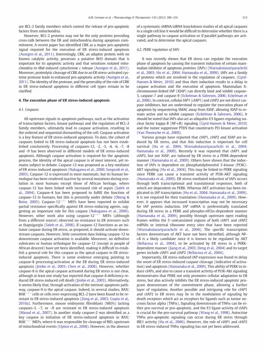
However, BCL-2 proteins may not be the only proteins providingcross-talk between the ER and mitochondria during apoptosis com-mitment. A recent paper has identified CRK as a major pro-apoptoticsignal required for the execution of ER stress-induced apoptosis(Austgen et al., 2011). Interestingly, CRK, an adaptor protein with noknown catalytic activity, possesses a putative BH3 domain that isimportant for its apoptotic activity and that sensitizes isolated mito-chondria to tBid-induced cytochrome c release (Austgen et al., 2011).Moreover, proteolytic cleavage of CRK due to an ER stress-activated cys-teine protease leads to enhanced pro-apoptotic activity (Austgen et al.,2011). The identity of the protease, and the generality of the role of CRKin ER stress-induced apoptosis in different cell types remain to beclarified.
4. The execution phase of ER stress-induced apoptosis
4.1. Caspases
All upstream signals in apoptosis pathways, such as the activationof transcription factors, kinase pathways and the regulation of BCL-2family members, ultimately lead to caspase activation, resulting inthe ordered and sequential dismantling of the cell. Caspase activationis a key feature of ER stress-induced apoptosis. To date, the cohort ofcaspases linked to ER stress-induced apoptosis has not been estab-lished conclusively. Processing of caspases-12, -2, -3, -4, -6, -7, -8and -9 has been observed in different models of ER stress-inducedapoptosis. Although caspase activation is required for the apoptoticprocess, the identity of the apical caspase is of most interest, yet re-mains subject to debate. Caspase-12 was proposed as a key mediatorof ER stress-induced apoptosis (Nakagawa et al., 2000; Szegezdi et al.,2003). Caspase-12 is expressed in most mammals; but its human ho-mologue has been rendered inactive by several mutations during evo-lution in most humans except those of African heritage, wherecaspase-12 has been linked with increased risk of sepsis (Saleh etal., 2004). Caspase-4 has been proposed to fulfill the function ofcaspase-12 in humans, but this is presently under debate (Obeng &Boise, 2005). Caspase-12−/− MEFs have been reported to exhibitpartial resistance specifically against ER stress-inducing agents, sug-gesting an important role for this caspase (Nakagawa et al., 2000).However, other work also using caspase-12−/− MEFs (althoughfrom a different source) observed no resistance to ER stressors suchas thapsigargin (Saleh et al., 2006). If caspase-12 functions as an ini-tiator caspase during ER stress, as proposed, it should activate down-stream caspases. However, little consistent data linking caspase-12 todownstream caspase activation is available. Moreover, no definitivesubstrates or human orthologue for caspase-12 (except in people ofAfrican descent) have yet been identified, making it difficult to estab-lish a general role for this caspase as the apical caspase in ER stress-induced apoptosis. There is some evidence emerging pointing tocaspase-8 processing/activation at the ER during ER stress-inducedapoptosis (Jimbo et al., 2003; Chen et al., 2008). However, whethercaspase-8 is the apical caspase activated during ER stress is not clear,although at least one study has reported that caspase-8 deficiency re-duced ER stress-induced cell death (Jimbo et al., 2003). Alternatively,it seems likely that, through activation of the intrinsic apoptosis path-way, caspase-9 is the apical caspase. Indeed, in several studies, BAX/BAK−/− cells or cells over-expressing BCL-2 have been found to be re-sistant to ER stress-induced apoptosis (Zong et al., 2003; Gupta et al.,2010a). Furthermore, mouse embryonic fibroblasts (MEFs) lackingcaspases-3, –7, or –9 are resistant to ER stress-induced apoptosis(Masud et al., 2007). In another study caspase-2 was identified as akey caspase in initiation of ER stress-induced apoptosis in BAX/BAK−/− MEFs, where it was responsible for cleavage of BID, upstreamof mitochondrial events (Upton et al., 2008). However, in the absence
of a systematic shRNA/siRNA knockdown studies of all apical caspasesin a single cell line it would be difficult to determinewhether there is asingle pathway to caspase activation or if parallel pathways are acti-vated, and to determine the apical caspases.
4.2. PERK regulation of IAPs
It was recently shown that ER stress can regulate the executionphase of apoptosis by causing the transient induction of certain mam-malian inhibitor of apoptosis proteins (IAPs) (Warnakulasuriyarachchiet al., 2003; Hu et al., 2004; Hamanaka et al., 2009). IAPs are a familyof proteins which are involved in the regulation of caspases, (Gyrd-Hansen & Meier, 2010) and thus their induction results in a delay incaspase activation and the execution of apoptosis. Mammalian X-chromosome-linked IAP (XIAP) can directly bind and inhibit caspase-3, caspase-7 and caspase-9 (Eckelman & Salvesen, 2006; Eckelman etal., 2006). In contrast, cellular IAP1 (cIAP1) and cIAP2 are not direct cas-pase inhibitors, but are understood to regulate the execution phase ofapoptosis by sequestering SMAC away from XIAP, allowing XIAP to re-main active and to inhibit caspases (Eckelman & Salvesen, 2006). Itshould be noted that IAPs also act as ubiquitin-E3 ligases regulating nu-clear factor kappa B (NF-κB) signaling (Gyrd-Hansen & Meier, 2010)and the tumor suppressor PTEN that counteracts PI3 kinase activation(Van Themsche et al., 2009).
Various groups have reported that cIAP1, cIAP2 and XIAP are in-duced by ER stress, and that this induction is important for cellsurvival (Hu et al., 2004; Warnakulasuriyarachchi et al., 2004;Hamanaka et al., 2009). Recently it was reported that cIAP1 andcIAP2, but not XIAP, are induced by ER stress in a PERK-dependentmanner (Hamanaka et al., 2009). Others have shown that the induc-tion of IAPs is dependent on phosphatidylinositol 3-kinase (PI3K)-AKT signaling (Hu et al., 2004). This may be linked to PERK signalingsince PERK can cause a transient activity of PI3K-AKT signaling(Hamanaka et al., 2009). ER stress-mediated induction of IAPs occursthrough both transcriptional and translational responses, both ofwhich are dependent on PERK. Whereas AKT activation has been im-plicated in IAP transcription (Hu et al., 2004; Hamanaka et al., 2009),it is not required for their translation (Hamanaka et al., 2009). How-ever, it appears that increased transcription may not be necessaryfor IAP protein induction. IAP mRNA is preferentially translatedduring ER stress in a PERK and phospho-eIF2α-dependent manner(Hamanaka et al., 2009), possibly through upstream open readingframes within the 5′-untranslated regions of both cIAP1 and cIAP2mRNAs or internal ribosome entry sites that have been reported(Warnakulasuriyarachchi et al., 2004). The specific transcriptionfactors downstream of AKT have not been identified, although NF-κB is a likely candidate since it is known to be regulated by AKT(Bellacosa et al., 2004), to be activated by ER stress in a PERK-dependent manner (Jiang et al., 2003; Deng et al., 2004) and its targetgenes include cIAP1 and cIAP2 (Bellacosa et al., 2004).
Importantly, ER stress-induced IAP expression was found to delaythe onset of ER stress-induced caspase cleavage (indicative of activa-tion) and apoptosis (Hamanaka et al., 2009). This ability of PERK to in-duce cIAPs, and also to cause a transient activity of PI3K-Akt signalingdemonstrates that PERK not only promotes cellular adaptation to ERstress, but also actively inhibits the ER stress-induced apoptotic pro-gram downstream of the commitment phase, allowing a furtherlayer of regulation. Another possible and intriguing role for cIAP1and cIAP2 in ER stress may lie in the modulation of signaling bydeath receptors which act as receptors for ligands such as tumor ne-crosis factor alpha (TNFα). Signaling downstream of TNFα can be ei-ther pro-survival or pro-apoptotic, and the E3 ligase activity of cIAPsis crucial for the pro-survival pathway (Wang et al., 1998). AutocrineTNFα pro-apoptotic signaling can occur during ER stress throughIRE1 activity (Hu et al., 2006). However, the role of cIAP1 and cIAP2in ER stress-induced TNFα signaling has not yet been addressed.

314 A.M. Gorman et al. / Pharmacology & Therapeutics 134 (2012) 306–316
5. Novel regulation of UPR and ER stress-induced apoptosis
Thus far we have described the more well known mechanisms bywhich the UPR machinery is regulated. However, there are severalother mechanisms that are emerging that may prove significant anddevelop into attractive targets in the future. For example, regulationof the adaptor protein CRKmay prove to be an important factor in con-trolling ER stress-induced apoptosis (this has already been describedin Section 3.3).
There are a number of recent reports describing roles of micro-RNAs in ER stress. Both CHOP and XBP1s have been shown to controlexpression of certain miRNAs (Bartoszewski et al., 2011; Behrman etal., 2011). In human hepatocellular carcinoma (HCC) cells, ER stresscaused downregulation of the microRNAmiR-221/222 which contrib-uted to protection against ER stress-induced apoptosis (Dai et al.,2010). Another microRNA, miR-122, was shown to negatively regu-late the UPR in HCC cells and downregulation of miR-122 enhancedchemoresistance (Yang et al., 2011). Thus, miRNAs are possible regu-lators of the switch from survival to death in cells under ER stress.
An intriguing novel link has been established between innate im-munity and UPR signaling by uncovering activation of IRE1 and subse-quent XBP1 splicing through Toll-like receptors (TLRs) (Martinon etal., 2010). In these experiments, IRE1 activation by TLRs contributedto cytokine production and was important for the host defense ofmice against an intracellular pathogen. However, the mechanisms ofregulation of IRE1 by TLRs have not yet been elucidated.
Enzymes mediating post-translational modification of XBP1s areemerging as another group of ER stress regulators and include acetyltransferases and deacetylating enzymes, as well as E3-ligases mediat-ing modification by small ubiquitin-like modifiers (SUMO). Whereasacetylation (mediated by p300)was shown to enhance transcriptionalactivity of XBP1s, deacetylation (by sirtuin 1) had the opposite effects(Wang et al., 2011). Furthermore, SUMOylation by PIAS [protein in-hibitor of activated STAT (signal transducer and activator of transcrip-tion)]was shown to decrease transcriptional activity of XBP1s (Chen &Qi, 2010).
There have been some interesting new studies regarding GRP78and cancer. High levels of cell surface GRP78 have been detected on avariety of cancer cells but not on normal cells (Vollmers & Brandlein,2009; Zhang et al., 2010; Ni et al., 2011). Cell surface GRP78 formscomplexes with a growing number of extracellular ligands andmembrane-anchored proteins, influencing regulation of pro-survivalor pro-apoptotic signaling pathways, such as PI3K/Akt signalling (Niet al., 2011). Thus, cell surface GRP78 could regulate ER stress-inducedapoptosis.
6. Conclusion
ER stress conditions have been observed in numerous diseases in-cluding Alzheimer's disease, Creutzfeldt–Jakob disease, Huntington'sdisease, Parkinson's disease, diabetes, cardiovascular diseases, andcancer indicating that ER stress-induced apoptosis is an important fac-tor in pathophysiological conditions. To be able to intervene in suchconditions, a firm understanding of the mechanisms mediating ERstress-induced apoptosis is essential. Currently, it appears that manycandidate proteins are involved in orchestrating the switch from theprotective UPR signaling to pro-apoptotic signaling. Some of theseproteins, like P58IPK, GADD34 and TRB3, are involved in shuttingdown the PERK-mediated pathway. Ending the protective UPR can un-derstandably be a central element of the switch from adaptation tosuicide. These proteins, however, seem to affect sensitivity to ER stressin opposingways. Besides these proteins with negative feedback func-tions, two additional, quite potent, pro-apoptotic molecules are acti-vated during the UPR: CHOP and JNK. Both overexpression andknockdown experiments have confirmed the pro-apoptotic role ofthese proteins in ER stress. Bothmolecules can target the BCL-2 family
and are able to set the death machinery in motion. Furthermore, acti-vation of ASK1/JNK might be regulated by stress-sensitive adaptorproteins like JAB1, offering another possible mechanism to switchfrom an adaptive response to cell suicide.
It should be remembered that in studying ER stress responses avariety of different inducers are used, the most common being thap-sigargin (inhibitor of the sarco/endoplasmic reticulum Ca2+ pump),tunicamycin (inhibitor of N-linked glycosylation) and brefeldin A (in-hibitor of ER-Golgi transport). Since these have very different targetsit is possible there are differences in the precise set of responses in-duced and therefore caution should be exercised when comparingdata from different systems. For example, Sep15, a thioredoxin-likeselenoprotein is differentially regulated depending on the underlyingmechanism by which ER stress is induced (Labunskyy et al., 2009).Tunicamycin and brefeldin A, which both rely on protein synthesisto exert their ER stress effects led to increased expression of Sep15,while thapsigargin and dithiothreitol, which induce protein foldingindependently of protein synthesis, stimulated rapid degradation ofSep15 (Labunskyy et al., 2009). Finally, it is worth noting that someof the regulators of ER stress-induced apoptosis are specific to ERstress, while others are common to other stress. They thereforecould serve as useful targets for therapeutic intervention where ERstress plays a key role. Continued research in this field is necessaryin order to tease out the complexities of this cell-death pathway.
Acknowledgments
Our research is supported by Science Foundation Ireland (09/RFP/BIC2371; 09/RFP/BMT2153), the Health Research Board (HRA/2009/59) and Breast Cancer Campaign (2008NovPhD21; 2010NovPR13).
References
Adler, H. T., Chinery, R., Wu, D. Y., Kussick, S. J., Payne, J. M., Fornace, A. J., Jr., et al.(1999). Leukemic HRX fusion proteins inhibit GADD34-induced apoptosis and as-sociate with the GADD34 and hSNF5/INI1 proteins. Mol Cell Biol 19, 7050–7060.
Austgen, K., Johnson, E. T., Park, T. J., Curran, T., & Oakes, S. A. (2011). The adaptor pro-tein CRK is a pro-apoptotic transducer of endoplasmic reticulum stress. Nat CellBiol 14, 87–92.
Bartoszewski, R., Brewer, J. W., Rab, A., Crossman, D. K., Bartoszewska, S., Kapoor, N.,et al. (2011). The unfolded protein response (UPR)-activated transcription factorX-box-binding protein 1 (XBP1) induces microRNA-346 expression that targetsthe human antigen peptide transporter 1 (TAP1) mRNA and governs immune reg-ulatory genes. J Biol Chem 286, 41862–41870.
Bassik, M. C., Scorrano, L., Oakes, S. A., Pozzan, T., & Korsmeyer, S. J. (2004). Phosphor-ylation of BCL-2 regulates ER Ca2+ homeostasis and apoptosis. Embo J 23(5),1207–1216.
Behrman, S., Acosta-Alvear, D., & Walter, P. (2011). A CHOP-regulated microRNA con-trols rhodopsin expression. J Cell Biol 192, 919–927.
Bellacosa, A., Testa, J. R., Moore, R., & Larue, L. (2004). A portrait of AKT kinases: Humancancer and animal models depict a family with strong individualities. Cancer BiolTher 3, 268–275.
Bouchecareilh, M., Higa, A., Fribourg, S., Moenner, M., & Chevet, E. (2011). Peptides de-rived from the bifunctional kinase/RNase enzyme IRE1alpha modulate IRE1alphaactivity and protect cells from endoplasmic reticulum stress. FASEB J 25, 3115–3129.
Boya, P., Cohen, I., Zamzami, N., Vieira, H. L., & Kroemer, G. (2002). Endoplasmic retic-ulum stress-imdiced cell death requires mitochondrial membrane perme-abilization. Cell Death Differ 9(4), 465–467.
Boyce, M., Bryant, K. F., Jousse, C., Long, K., Harding, H. P., Scheuner, D., et al. (2005). Aselective inhibitor of eIF2alpha dephosphorylation protects cells from ER stress.Science 307, 935–939.
Chen, A., Wu, K., Fuchs, S. Y., Tan, P., Gomez, C., & Pan, Z. Q. (2000). The conserved RING-H2 finger of ROC1 is required for ubiquitin ligation. J Biol Chem 275, 15432–15439.
Chen, H., & Qi, L. (2010). SUMO modification regulates the transcriptional activity ofXBP1. Biochem J 429, 95–102.
Chen, L. H., Jiang, C. C., Watts, R., Thorne, R. F., Kiejda, K. A., Zhang, X. D., et al. (2008).Inhibition of endoplasmic reticulum stress-induced apoptosis of melanoma cellsby the ARC protein. Cancer Res 68, 834–842.
Chipuk, J. E., Moldoveanu, T., Llambi, F., Parsons, M. J., & Green, D. R. (2010). The BCL-2family reunion. Mol Cell 37, 299–310.
Concannon, C. G., Gorman, A. M., & Samali, A. (2003). On the role of Hsp27 in regulatingapoptosis. Apoptosis 8, 61–70.
Cullinan, S. B., & Diehl, J. A. (2004). PERK-dependent activation of Nrf2 contributes toredox homeostasis and cell survival following endoplasmic reticulum stress. JBiol Chem 279, 20108–20117.

315A.M. Gorman et al. / Pharmacology & Therapeutics 134 (2012) 306–316
Dai, R., Li, J., Liu, Y., Yan, D., Chen, S., Duan, C., et al. (2010). miR-221/222 suppressionprotects against endoplasmic reticulum stress-induced apoptosis via p27(Kip1)-and MEK/ERK-mediated cell cycle regulation. Biol Chem 391, 791–801.
Deng, J., Lu, P. D., Zhang, Y., Scheuner, D., Kaufman, R. J., Sonenberg, N., et al. (2004).Translational repression mediates activation of nuclear factor kappa B by phos-phorylated translation initiation factor 2. Mol Cell Biol 24, 10161–10168.
Distelhorst, C. W., & McCormick, T. S. (1996). Bcl-2 acts subsequent to and independentof Ca2+ fluxes to inhibit apoptosis in thapsigargin- and glucocorticoid-treatedmouse lymphoma cells. Cell Calcium 19, 473–483.
Doyle, K. M., Kennedy, D., Gorman, A. M., Gupta, S., Healy, S. J., & Samali, A. (2011). Un-folded proteins and endoplasmic reticulum stress in neurodegenerative disorders.J Cell Mol Med 15, 2025–2039.
Du, K., Herzig, S., Kulkarni, R. N., & Montminy, M. (2003). TRB3: a tribbles homolog thatinhibits Akt/PKB activation by insulin in liver. Science 300, 1574–1577.
Eckelman, B. P., & Salvesen, G. S. (2006). The human anti-apoptotic proteins cIAP1 andcIAP2 bind but do not inhibit caspases. J Biol Chem 281, 3254–3260.
Eckelman, B. P., Salvesen, G. S., & Scott, F. L. (2006). Human inhibitor of apoptosis pro-teins: Why XIAP is the black sheep of the family. EMBO Rep 7, 988–994.
Fawcett, T. W., Eastman, H. B., Martindale, J. L., & Holbrook, N. J. (1996). Physical andfunctional association between GADD153 and CCAAT/enhancer-binding proteinbeta during cellular stress. J Biol Chem 271, 14285–14289.
Fu, H. Y., Okada, K., Liao, Y., Tsukamoto, O., Isomura, T., Asai, M., et al. (2010). Ablation ofC/EBP homologous protein attenuates endoplasmic reticulum-mediated apoptosisand cardiac dysfunction induced by pressure overload. Circulation 122, 361–369.
Fulda, S., Gorman, A. M., Hori, O., & Samali, A. (2010). Cellular stress responses: Cellsurvival and cell death. Int J Cell Biol 2010, 214074.
Gardner, B. M., & Walter, P. (2011). Unfolded proteins are Ire1-activating ligands thatdirectly induce the unfolded protein response. Science 333, 1891–1894.
Garrido, C., Brunet, M., Didelot, C., Zermati, Y., Schmitt, E., & Kroemer, G. (2006). Heatshock proteins 27 and 70: Anti-apoptotic proteins with tumorigenic properties.Cell Cycle 5, 2592–2601.
Gass, J. N., Gifford, N. M., & Brewer, J. W. (2002). Activation of an unfolded protein re-sponse during differentiation of antibody-secreting B cells. J Biol Chem 277,49047–49054.
Gaut, J. R., & Hendershot, L. M. (1993). The modification and assembly of proteins in theendoplasmic reticulum. Curr Opin Cell Biol 5, 589–595.
Gupta, S., Cuffe, L., Szegezdi, E., Logue, S. E., Neary, C., Healy, S., et al. (2010). Mecha-nisms of ER stress-mediated mitochondrial membrane permeabilization. Int J CellBiol 2010, 170215.
Gupta, S., Deepti, A., Deegan, S., Lisbona, F., Hetz, C., & Samali, A. (2010). HSP72 protectscells from ER stress-induced apoptosis via enhancement of IRE1alpha-XBP1 signal-ing through a physical interaction. PLoS Biol 8, e1000410.
Gyrd-Hansen, M., & Meier, P. (2010). IAPs: from caspase inhibitors to modulators ofNF-kappaB, inflammation and cancer. Nat Rev Cancer 10, 561–574.
Hacki, J., Egger, L., Monney, L., Conus, S., Rosse, T., Fellay, I., et al. (2000). Apoptoticcrosstalk between the endoplasmic reticulum and mitochondria controlled byBcl-2. Oncogene 19, 2286–2295.
Hamanaka, R. B., Bobrovnikova-Marjon, E., Ji, X., Liebhaber, S. A., & Diehl, J. A. (2009).PERK-dependent regulation of IAP translation during ER stress. Oncogene 28,910–920.
Han, D., Lerner, A. G., Vande Walle, L., Upton, J. P., Xu, W., Hagen, A., et al. (2009).IRE1alpha kinase activation modes control alternate endoribonuclease outputs todetermine divergent cell fates. Cell 138, 562–575.
Harding, H. P., Zhang, Y., & Ron, D. (1999). Protein translation and folding are coupledby an endoplasmic-reticulum-resident kinase. Nature 397, 271–274.
Harding, H. P., Novoa, I., Zhang, Y., Zeng, H., Wek, R., Schapira, M., et al. (2000). Regu-lated translation initiation controls stress-induced gene expression in mammaliancells. Mol Cell 6, 1099–1108.
Harding, H. P., Zhang, Y., Bertolotti, A., Zeng, H., & Ron, D. (2000). Perk is essential fortranslational regulation and cell survival during the unfolded protein response.Mol Cell 5, 897–904.
Harding, H. P., Zhang, Y., Zeng, H., Novoa, I., Lu, P. D., Calfon, M., et al. (2003). An inte-grated stress response regulates amino acid metabolism and resistance to oxida-tive stress. Mol Cell 11, 619–633.
Healy, S. J., Gorman, A. M., Mousavi-Shafaei, P., Gupta, S., & Samali, A. (2009). Targetingthe endoplasmic reticulum-stress response as an anticancer strategy. Eur J Pharma-col 625, 234–246.
Hegedus, Z., Czibula, A., & Kiss-Toth, E. (2007). Tribbles: a family of kinase-like proteinswith potent signalling regulatory function. Cell Signal 19, 238–250.
Hetz, C., Bernasconi, P., Fisher, J., Lee, A. H., Bassik, M. C., Antonsson, B., et al. (2006).Proapoptotic BAX and BAK modulate the unfolded protein response by a direct in-teraction with IRE1alpha. Science 312, 572–576.
Hetz, C., & Glimcher, L. H. (2009). Fine-tuning of the unfolded protein response: As-sembling the IRE1alpha interactome. Mol Cell 35, 551–561.
Hollien, J., Lin, J. H., Li, H., Stevens, N., Walter, P., & Weissman, J. S. (2009). RegulatedIre1-dependent decay of messenger RNAs in mammalian cells. J Cell Biol 186,323–331.
Hu, P., Han, Z., Couvillon, A. D., & Exton, J. H. (2004). Critical role of endogenousAkt/IAPs and MEK1/ERK pathways in counteracting endoplasmic reticulumstress-induced cell death. J Biol Chem 279, 49420–49429.
Hu, P., Han, Z., Couvillon, A. D., Kaufman, R. J., & Exton, J. H. (2006). Autocrine tumornecrosis factor alpha links endoplasmic reticulum stress to the membrane deathreceptor pathway through IRE1alpha-mediated NF-kappaB activation and down-regulation of TRAF2 expression. Mol Cell Biol 26(8), 3071–3084.
Jiang, H. Y., Wek, S. A., McGrath, B. C., Scheuner, D., Kaufman, R. J., Cavener, D. R., et al.(2003). Phosphorylation of the alpha subunit of eukaryotic initiation factor 2 is
required for activation of NF-kappaB in response to diverse cellular stresses. MolCell Biol 23, 5651–5663.
Jimbo, A., Fujita, E., Kouroku, Y., Ohnishi, J., Inohara, N., Kuida, K., et al. (2003). ER stressinduces caspase-8 activation, stimulating cytochrome c release and caspase-9 acti-vation. Exp Cell Res 283, 156–166.
Kimata, Y., & Kohno, K. (2011). Endoplasmic reticulum stress-sensing mechanisms inyeast and mammalian cells. Curr Opin Cell Biol 23, 135–142.
Klee, M., Pallauf, K., Alcala, S., Fleischer, A., & Pimentel-Muinos, F. X. (2009). Mitochon-drial apoptosis induced by BH3-only molecules in the exclusive presence of endo-plasmic reticular Bak. EMBO J 28, 1757–1768.
Krajewski, S., Tanaka, S., Takayama, S., Schibler, M. J., Fenton, W., & Reed, J. C. (1993).Investigation of the subcellular distribution of the bcl-2 oncoprotein: Residencein the nuclear envelope, endoplasmic reticulum, and outer mitochondrial mem-branes. Cancer Res 53, 4701–4714.
Labunskyy, V. M., Yoo, M. H., Hatfield, D. L., & Gladyshev, V. N. (2009). Sep15, athioredoxin-like selenoprotein, is involved in the unfolded protein response anddifferentially regulated by adaptive and acute ER stresses. Biochemistry 48,8458–8465.
Lee, A. H., Iwakoshi, N. N., & Glimcher, L. H. (2003). XBP-1 regulates a subset of endo-plasmic reticulum resident chaperone genes in the unfolded protein response.Mol Cell Biol 23, 7448–7459.
Lei, K., & Davis, R. J. (2003). JNK phosphorylation of Bim-related members of the Bcl2family induces Bax-dependent apoptosis. Proc Natl Acad Sci USA 100(5),2432–2437.
Li, G., Mongillo, M., Chin, K. T., Harding, H., Ron, D., Marks, A. R., et al. (2009). Role ofERO1-alpha-mediated stimulation of inositol 1,4,5-triphosphate receptor activityin endoplasmic reticulum stress-induced apoptosis. J Cell Biol 186, 783–792.
Li, J., Lee, B., & Lee, A. S. (2006). Endoplasmic reticulum stress-induced apoptosis: Mul-tiple pathways and activation of p53-up-regulated modulator of apoptosis (PUMA)and NOXA by p53. J Biol Chem 281, 7260–7270.
Lin, J. H., Li, H., Yasumura, D., Cohen, H. R., Zhang, C., Panning, B., et al. (2007). IRE1 sig-naling affects cell fate during the unfolded protein response. Science 318, 944–949.
Lin, J. H., Li, H., Zhang, Y., Ron, D., & Walter, P. (2009). Divergent effects of PERK andIRE1 signaling on cell viability. PLoS One 4, e4170.
Lisbona, F., Rojas-Rivera, D., Thielen, P., Zamorano, S., Todd, D., Martinon, F., et al.(2009). BAX inhibitor-1 is a negative regulator of the ER stress sensor IRE1alpha.Mol Cell 33, 679–691.
Lu, P. D., Jousse, C., Marciniak, S. J., Zhang, Y., Novoa, I., Scheuner, D., et al. (2004). Cyto-protection by pre-emptive conditional phosphorylation of translation initiationfactor 2. EMBO J 23, 169–179.
Luo, D., He, Y., Zhang, H., Yu, L., Chen, H., Xu, Z., et al. (2008). AIP1 is critical in transduc-ing IRE1-mediated endoplasmic reticulum stress response. J Biol Chem 283,11905–11912.
Luo, S., Baumeister, P., Yang, S., Abcouwer, S. F., & Lee, A. S. (2003). Induction ofGrp78/BiP by translational block: Activation of the Grp78 promoter by ATF4through and upstream ATF/CRE site independent of the endoplasmic reticulumstress elements. J Biol Chem 278, 37375–37385.
Ma, Y., Brewer, J. W., Diehl, J. A., & Hendershot, L. M. (2002). Two distinct stress signal-ing pathways converge upon the CHOP promoter during the mammalian unfoldedprotein response. J Mol Biol 318, 1351–1365.
Ma, Y., Shimizu, Y., Mann, M. J., Jin, Y., & Hendershot, L. M. (2010). Plasma cell differen-tiation initiates a limited ER stress response by specifically suppressing the PERK-dependent branch of the unfolded protein response. Cell Stress Chaperones 15,281–293.
Marciniak, S. J., Yun, C. Y., Oyadomari, S., Novoa, I., Zhang, Y., Jungreis, R., et al. (2004).CHOP induces death by promoting protein synthesis and oxidation in the stressedendoplasmic reticulum. Genes Dev 18, 3066–3077.
Marcu, M. G., Doyle, M., Bertolotti, A., Ron, D., Hendershot, L., & Neckers, L. (2002). Heatshock protein 90 modulates the unfolded protein response by stabilizing IRE1al-pha. Mol Cell Biol 22, 8506–8513.
Martinon, F., Chen, X., Lee, A. H., & Glimcher, L. H. (2010). TLR activation of the tran-scription factor XBP1 regulates innate immune responses in macrophages. NatImmunol 11, 411–418.
Masud, A., Mohapatra, A., Lakhani, S. A., Ferrandino, A., Hakem, R., & Flavell, R. A.(2007). Endoplasmic reticulum stress-induced death of mouse embryonic fibro-blasts requires the intrinsic pathway of apoptosis. J Biol Chem 282, 14132–14139.
Matsumoto, M., Minami, M., Takeda, K., Sakao, Y., & Akira, S. (1996). Ectopic expressionof CHOP (GADD153) induces apoptosis in M1myeloblastic leukemia cells. FEBS Lett395, 143–147.
McCullough, K. D., Martindale, J. L., Klotz, L. O., Aw, T. Y., & Holbrook, N. J. (2001).Gadd153 sensitizes cells to endoplasmic reticulum stress by down-regulatingBcl2 and perturbing the cellular redox state. Mol Cell Biol 21, 1249–1259.
Melville, M. W., Tan, S. L., Wambach, M., Song, J., Morimoto, R. I., & Katze, M. G. (1999).The cellular inhibitor of the PKR protein kinase, P58(IPK), is an influenza virus-activated co-chaperone that modulates heat shock protein 70 activity. J Biol Chem274, 3797–3803.
Morishima, N., Nakanishi, K., Tsuchiya, K., Shibata, T., & Seiwa, E. (2004). Translocation ofBim to the endoplasmic reticulum (ER) mediates ER stress signaling for activation ofcaspase-12 during ER stress-induced apoptosis. J Biol Chem 279, 50375–50381.
Morishima, N., Nakanishi, K., & Nakano, A. (2011). Activating transcription factor-6(ATF6) mediates apoptosis with reduction of myeloid cell leukemia sequence 1(Mcl-1) protein via induction of WW domain binding protein 1. J Biol Chem 286,35227–35235.
Nakagawa, T., Zhu, H., Morishima, N., Li, E., Xu, J., Yankner, B. A., et al. (2000). Caspase-12 mediates endoplasmic-reticulum-specific apoptosis and cytotoxicity byamyloid-beta. Nature 403, 98–103.

316 A.M. Gorman et al. / Pharmacology & Therapeutics 134 (2012) 306–316
Ni, M., Zhang, Y., & Lee, A. S. (2011). Beyond the endoplasmic reticulum: AtypicalGRP78 in cell viability, signalling and therapeutic targeting. Biochem J 434,181–188.
Novoa, I., Zeng, H., Harding, H. P., & Ron, D. (2001). Feedback inhibition of the unfoldedprotein response by GADD34-mediated dephosphorylation of eIF2alpha. J Cell Biol153, 1011–1022.
Obeng, E. A., & Boise, L. H. (2005). Caspase-12 and caspase-4 are not required forcaspase-dependent endoplasmic reticulum stress-induced apoptosis. J Biol Chem280, 29578–29587.
Ohoka, N., Yoshii, S., Hattori, T., Onozaki, K., & Hayashi, H. (2005). TRB3, a novel ERstress-inducible gene, is induced via ATF4-CHOP pathway and is involved in celldeath. EMBO J 24, 1243–1255.
Ohoka, N., Sakai, S., Onozaki, K., Nakanishi, M., & Hayashi, H. (2010). Anaphase-promoting complex/cyclosome-cdh1 mediates the ubiquitination and degradationof TRB3. Biochem Biophys Res Commun 392, 289–294.
Orrenius, S., Zhivotovsky, B., & Nicotera, P. (2003). Regulation of cell death: thecalcium-apoptosis link. Nat Rev Mol Cell Biol 4, 552–565.
Oyadomari, S., Koizumi, A., Takeda, K., Gotoh, T., Akira, S., Araki, E., et al. (2002). Tar-geted disruption of the Chop gene delays endoplasmic reticulum stress-mediateddiabetes. J Clin Invest 109, 525–532.
Oyadomari, S., Yun, C., Fisher, E. A., Kreglinger, N., Kreibich, G., Oyadomari, M., et al.(2006). Cotranslocational degradation protects the stressed endoplasmic reticu-lum from protein overload. Cell 126, 727–739.
Parcellier, A., Gurbuxani, S., Schmitt, E., Solary, E., & Garrido, C. (2003). Heat shock pro-teins, cellular chaperones that modulate mitochondrial cell death pathways. Bio-chem Biophys Res Commun 304, 505–512.
Puthalakath, H., O'Reilly, L. A., Gunn, P., Lee, L., Kelly, P. N., Huntington, N. D., et al. (2007).ER stress triggers apoptosis by activating BH3-only protein Bim. Cell 129, 1337–1349.
Qiu, Y., Mao, T., Zhang, Y., Shao, M., You, J., & Ding, Q. (2010). A crucial role for RACK1 inthe regulation of glucose-stimulated IRE1alpha activation in pancreatic beta cells.Sci Signal 3, ra7.
Reimertz, C., Kogel, D., Rami, A., Chittenden, T., & Prehn, J. H. (2003). Gene expressionduring ER stress-induced apoptosis in neurons: induction of the BH3-only proteinBbc3/PUMA and activation of the mitochondrial apoptosis pathway. J Cell Biol 162,587–597.
Reimold, A. M., Iwakoshi, N. N., Manis, J., Vallabhajosyula, P., Szomolanyi-Tsuda, E.,Gravallese, E. M., et al. (2001). Plasma cell differentiation requires the transcriptionfactor XBP-1. Nature 412, 300–307.
Robinson, K. S., Clements, A., Williams, A. C., Berger, C. N., & Frankel, G. (2011). Bax in-hibitor 1 in apoptosis and disease. Oncogene 30, 2391–2400.
Ron, D., & Walter, P. (2007). Signal integration in the endoplasmic reticulum unfoldedprotein response. Nat Rev Mol Cell Biol 8, 519–529.
Rong, J., Chen, L., Toth, J. I., Tcherpakov, M., Petroski, M. D., & Reed, J. C. (2011). Bifunc-tional apoptosis regulator (BAR), an endoplasmic reticulum (ER)-associated E3ubiquitin ligase, modulates BI-1 protein stability and function in ER stress. J BiolChem 286, 1453–1463.
Rutkowski, D. T., & Kaufman, R. J. (2004). A trip to the ER: coping with stress. Trends CellBiol 14, 20–28.
Rutkowski, D. T., Kang, S. W., Goodman, A. G., Garrison, J. L., Taunton, J., Katze, M. G.,et al. (2007). The role of p58IPK in protecting the stressed endoplasmic reticulum.Mol Biol Cell 18, 3681–3691.
Salazar, M., Carracedo, A., Salanueva, I. J., Hernandez-Tiedra, S., Lorente, M., Egia, A.,et al. (2009). Cannabinoid action induces autophagy-mediated cell death throughstimulation of ER stress in human glioma cells. J Clin Invest 119, 1359–1372.
Saleh, M., Vaillancourt, J. P., Graham, R. K., Huyck, M., Srinivasula, S. M., Alnemri, E. S.,et al. (2004). Differential modulation of endotoxin responsiveness by humancaspase-12 polymorphisms. Nature 429, 75–79.
Saleh, M., Mathison, J. C., Wolinski, M. K., Bensinger, S. J., Fitzgerald, P., Droin, N., et al.(2006). Enhanced bacterial clearance and sepsis resistance in caspase-12-deficientmice. Nature 440, 1064–1068.
Sari, F. R., Widyantoro, B., Thandavarayan, R. A., Harima, M., Lakshmanan, A. P., &Zhang, S. (2011). Attenuation of CHOP-mediated myocardial apoptosis inpressure-overloaded dominant negative p38alpha mitogen-activated protein ki-nase mice. Cell Physiol Biochem 27, 487–496.
Scheuner, D., Song, B., McEwen, E., Liu, C., Laybutt, R., Gillespie, P., et al. (2001). Trans-lational control is required for the unfolded protein response and in vivo glucosehomeostasis. Mol Cell 7, 1165–1176.
Schroder, M., & Kaufman, R. J. (2005). The mammalian unfolded protein response. AnnuRev Biochem 74, 739–789.
Shen, J., Snapp, E. L., Lippincott-Schwartz, J., & Prywes, R. (2005). Stable binding of ATF6to BiP in the endoplasmic reticulum stress response. Mol Cell Biol 25, 921–932.
Silva, R. M., Ries, V., Oo, T. F., Yarygina, O., Jackson-Lewis, V., Ryu, E. J., et al. (2005).CHOP/GADD153 is a mediator of apoptotic death in Substantia nigra dopamineneurons in an in vivo neurotoxin model of parkinsonism. J Neurochem 95, 974–986.
Song, B., Scheuner, D., Ron, D., Pennathur, S., & Kaufman, R. J. (2008). Chop deletion re-duces oxidative stress, improves beta cell function, and promotes cell survival inmultiple mouse models of diabetes. J Clin Invest 118, 3378–3389.
Szegezdi, E., Fitzgerald, U., & Samali, A. (2003). Caspase-12 and ER-stress-mediated ap-optosis: the story so far. Ann N Y Acad Sci 1010, 186–194.
Szegezdi, E., Logue, S. E., Gorman, A. M., & Samali, A. (2006). Mediators of endoplasmicreticulum stress-induced apoptosis. EMBO Rep 7, 880–885.
Szegezdi, E., Herbert, K. R., Kavanagh, E. T., Samali, A., & Gorman, A. M. (2008). Nervegrowth factor blocks thapsigargin-induced apoptosis at the level of the mitochon-drion via regulation of Bim. J Cell Mol Med 12, 2482–2496.
Szegezdi, E., Macdonald, D. C., Ni Chonghaile, T., Gupta, S., & Samali, A. (2009). Bcl-2family on guard at the ER. Am J Physiol 296, C941–C953.
Thorp, E., Li, G., Seimon, T. A., Kuriakose, G., Ron, D., & Tabas, I. (2009). Reducedapoptosis and plaque necrosis in advanced atherosclerotic lesions of Apoe−/−and Ldlr−/− mice lacking CHOP. Cell Metab 9, 474–481.
Upton, J. P., Austgen, K., Nishino, M., Coakley, K. M., Hagen, A., Han, D., et al. (2008).Caspase-2 cleavage of BID is a critical apoptotic signal downstream of endoplasmicreticulum stress. Mol Cell Biol 28, 3943–3951.
Urano, F., Wang, X., Bertolotti, A., Zhang, Y., Chung, P., Harding, H. P., et al. (2000). Cou-pling of stress in the ER to activation of JNK protein kinases by transmembraneprotein kinase IRE1. Science 287, 664–666.
van Huizen, R., Martindale, J. L., Gorospe, M., & Holbrook, N. J. (2003). P58IPK, a novelendoplasmic reticulum stress-inducible protein and potential negative regulator ofeIF2alpha signaling. J Biol Chem 278, 15558–15564.
Van Themsche, C., Leblanc, V., Parent, S., & Asselin, E. (2009). X-linked inhibitor of ap-optosis protein (XIAP) regulates PTEN ubiquitination, content, and compartmen-talization. J Biol Chem 284, 20462–20466.
Vollmers, H. P., & Brandlein, S. (2009). Natural antibodies and cancer. N Biotechnol 25,294–298.
Wang, C. Y., Mayo, M. W., Korenluk, R. G., Goeddel, D. V. & Baldwin Jr., A.S. (1998). NF-kappaB antiapoptosis: induction of TRAF1 and TRAF2 and c-IAP1 and c-IAP2 tosuppress caspase-8 activation. Science 281(5383), 1680–1683.
Wang, F. M., Chen, Y. J., & Ouyang, H. J. (2011). Regulation of unfolded protein responsemodulator XBP1s by acetylation and deacetylation. Biochem J 433, 245–252.
Wang, X. Z., & Ron, D. (1996). Stress-induced phosphorylation and activation of thetranscription factor CHOP (GADD153) by p38 MAP kinase. Science 272, 1347–1349.
Warnakulasuriyarachchi, D., Ungureanu, N. H., & Holcik, M. (2003). The translation ofan antiapoptotic protein HIAP2 is regulated by an upstream open reading frame.Cell Death Differ 10, 899–904.
Warnakulasuriyarachchi, D., Cerquozzi, S., Cheung, H. H., & Holcik, M. (2004). Transla-tional induction of the inhibitor of apoptosis protein HIAP2 during endoplasmic re-ticulum stress attenuates cell death and is mediated via an inducible internalribosome entry site element. J Biol Chem 279, 17148–17157.
Wei, M. C., Zong, W. X., Cheng, E. H., Lindsten, T., Panoutsakopoulou, V., Ross, A. J., et al.(2001). Proapoptotic BAX and BAK: a requisite gateway to mitochondrial dysfunc-tion and death. Science 292, 727–730.
Yan, W., Frank, C. L., Korth, M. J., Sopher, B. L., Novoa, I., Ron, D., et al. (2002). Control ofPERK eIF2alpha kinase activity by the endoplasmic reticulum stress-induced mo-lecular chaperone P58IPK. Proc Natl Acad Sci U S A 99, 15920–15925.
Yang, F., Zhang, L., Wang, F., Wang, Y., Huo, X. S., Yin, Y. X., et al. (2011). Modulation ofthe unfolded protein response is the core of microRNA-122-involved sensitivity tochemotherapy in hepatocellular carcinoma. Neoplasia 13, 590–600.
Yoshida, H., Haze, K., Yanagi, H., Yura, T., & Mori, K. (1998). Identification of the cis-acting endoplasmic reticulum stress response element responsible for transcrip-tional induction of mammalian glucose-regulated proteins. Involvement of basicleucine zipper transcription factors. J Biol Chem 273, 33741–33749.
Yoshida, H., Okada, T., Haze, K., Yanagi, H., Yura, T., Negishi, M., et al. (2000). ATF6 ac-tivated by proteolysis binds in the presence of NF-Y (CBF) directly to the cis-actingelement responsible for the mammalian unfolded protein response. Mol Cell Biol20, 6755–6767.
Yoshida, H., Matsui, T., Yamamoto, A., Okada, T., & Mori, K. (2001). XBP1 mRNA is in-duced by ATF6 and spliced by IRE1 in response to ER stress to produce a highly ac-tive transcription factor. Cell 107, 881–891.
Young, J. C. (2010). Mechanisms of the Hsp70 chaperone system. Biochem Cell Biol 88,291–300.
Zhang, Y., Liu, R., Ni, M., Gill, P., & Lee, A. S. (2010). Cell surface relocalization of the en-doplasmic reticulum chaperone and unfolded protein response regulatorGRP78/BiP. J Biol Chem 285, 15065–15075.
Zinszner, H., Kuroda, M., Wang, X., Batchvarova, N., Lightfoot, R. T., Remotti, H., et al.(1998). CHOP is implicated in programmed cell death in response to impairedfunction of the endoplasmic reticulum. Genes Dev 12, 982–995.
Zong, W. X., Li, C., Hatzivassiliou, G., Lindsten, T., Yu, Q. C., Yuan, J., et al. (2003). Baxand Bak can localize to the endoplasmic reticulum to initiate apoptosis. J CellBiol 162, 59–69.
Zou, C. G., Cao, X. Z., Zhao, Y. S., Gao, S. Y., Li, S. D., Liu, X. Y., et al. (2009). The molecularmechanism of endoplasmic reticulum stress-induced apoptosis in PC-12 neuronalcells: the protective effect of insulin-like growth factor I. Endocrinology 150, 277–285.
Copyright © 2022 FDOKUMEN