That which does not kill me makes me stronger: adapting to chronic ER stress
8
That which does not kill me makes me stronger: adapting to chronic ER stress D. Thomas Rutkowski 1 and Randal J. Kaufman 1, 2 1 Howard Hughes Medical Institute, University of Michigan Medical Center, Ann Arbor, MI 48109, USA 2 Departments of Biological Chemistry and Internal Medicine, University of Michigan Medical Center, Ann Arbor, MI 48109, USA Cells respond to the accumulation of unfolded proteins by activating signal transduction cascades that improve protein folding. One example of such a cascade is the unfolded protein response (UPR), which senses protein folding stress in the endoplasmic reticulum (ER) and leads to improvement in the protein folding and proces- sing capacity of the organelle. A central paradox of the UPR, and indeed of all such stress pathways, is that the response is designed to facilitate both adaptation to stress and apoptosis, depending upon the nature and severity of the stressor. Understanding how the UPR can allow for adaptation, instead of apoptosis, is of tremen- dous physiological importance. Recent advances have improved our understanding of ER stress and the vertebrate UPR, which suggest possible mechanisms by which cells adapt to chronic stress. The two-faced nature of ER stress The term ‘endoplasmic reticulum stress’ (see Glossary) defines any perturbation that compromises the protein folding functionality of the ER. In addition to being the site of synthesis of all secretory proteins and resident proteins of the endomembrane system, the ER also partici- pates in lipid metabolism, oligosaccharide synthesis, calcium storage, and in certain cell types, drug detoxifica- tion. Accordingly, any number of pathological or pharma- cological insults can produce ER stress either directly or indirectly, and this perturbation can be acute or chronic in nature [1,2]. As examples of acute stress, insults such as hypoxia and/or ischemia, calcium depletion and glucose deprivation can all lead to rapid activation of the unfolded protein response (UPR). In principle, the cell needs only to tolerate such stresses for their relatively brief durations (on the order of minutes to hours) and clear the ER of whatever unfolded proteins have accumulated in that time, which demands a UPR that is relatively fast acting and also readily deactivated. By contrast, chronic stresses require quasi-permanent changes in cellular function in general and in ER function in particular. Chronic stresses that activate the UPR can encompass: genetic mutation of either ER chaperones or chaperone substrates that lead to persistent misfolding problems; viral infection that can co-opt the secretory pathway for production of viral proteins; various neurode- generative disorders of protein aggregation; or even the normal differentiation and maintenance of cells whose primary functions include the production and secretion of proteins, such as immune cells, endocrine and paracrine cells, and hepatocytes [3]. A commonality among these chronic ER stressors is that they demand a mechanism whereby ER stress can be persistently tolerated on a time- scale ranging from days to years (Box 1). In these contexts, even if cell death occurs to a small extent, the majority of cells must ultimately survive and adapt to the stressful stimulus. Much of our understanding of the UPR, including its activation, perpetuation and regulation, comes from stu- dies that utilize severe pharmacological perturbation of cultured cells. However, these studies almost certainly fail to recapitulate the UPR as it is elicited under the con- ditions of milder, but more persistent ER stress that could be encountered physiologically. Thus, one of the funda- mental questions of the UPR is how can the response allow cells to escape death and adapt to stress? We will therefore consider recent insights into how the response is regulated, which might provide clues as to how stress signals from the ER are integrated into a life-or-death response. The four As of the UPR: activation, acute response, adaptive response and apoptosis The mechanisms of UPR activation, the signaling pathways initiated by it, the changes in gene expression that result and the subsequent induction of apoptotic cas- cades have been exhaustively reviewed elsewhere, and are summarized in Figure 1. Here, we will provide a brief summary of these events as a way of introducing what might represent control points in cellular adaptive decisions. Review TRENDS in Biochemical Sciences Vol.32 No.10 Glossary Acute stress: Stresses that are transient in nature (on the order of minutes to hours) and thus require only that cells tolerate a brief period of perturbation, and then recover by restoring homeostasis within the ER. Adaptation: The ability to maintain cellular function and avoid apoptosis during chronic stress. Chronic stress: Stresses that are persistent in nature (on the order of days to years). These can encompass either pathological or developmental processes, but in either case they require long term adjustments to cellular function. ER stress: Any perturbation that compromises the protein folding functionality of the ER. Preconditioning: The ability of a brief exposure to stress to protect cells from a subsequent stress, both in terms of cell survival and activation of stress response pathways. Protein processing: Refers collectively to the ability of the ER to fold, modify, traffic, and if necessary degrade newly imported proteins. Corresponding author: Kaufman, R.J. ([email protected]). www.sciencedirect.com 0968-0004/$ – see front matter ß 2007 Elsevier Ltd. All rights reserved. doi:10.1016/j.tibs.2007.09.003
-
Upload
univ-paris8 -
Category
Documents
-
view
2 -
download
0
Transcript of That which does not kill me makes me stronger: adapting to chronic ER stress
That which does not kill me makes mestronger: adapting to chronic ER stressD. Thomas Rutkowski1 and Randal J. Kaufman1,2
1 Howard Hughes Medical Institute, University of Michigan Medical Center, Ann Arbor, MI 48109, USA2 Departments of Biological Chemistry and Internal Medicine, University of Michigan Medical Center, Ann Arbor, MI 48109, USA
Review TRENDS in Biochemical Sciences Vol.32 No.10
Cells respond to the accumulation of unfolded proteinsby activating signal transduction cascades that improveprotein folding. One example of such a cascade is theunfolded protein response (UPR), which senses proteinfolding stress in the endoplasmic reticulum (ER) andleads to improvement in the protein folding and proces-sing capacity of the organelle. A central paradox of theUPR, and indeed of all such stress pathways, is that theresponse is designed to facilitate both adaptation tostress and apoptosis, depending upon the nature andseverity of the stressor. Understanding how the UPR canallow for adaptation, instead of apoptosis, is of tremen-dous physiological importance. Recent advances haveimproved our understanding of ER stress and thevertebrate UPR, which suggest possible mechanismsby which cells adapt to chronic stress.
Glossary
Acute stress: Stresses that are transient in nature (on the order of minutes to
hours) and thus require only that cells tolerate a brief period of perturbation,
and then recover by restoring homeostasis within the ER.
Adaptation: The ability to maintain cellular function and avoid apoptosis
during chronic stress.
Chronic stress: Stresses that are persistent in nature (on the order of days to
years). These can encompass either pathological or developmental processes,
but in either case they require long term adjustments to cellular function.
ER stress: Any perturbation that compromises the protein folding functionality
of the ER.
Preconditioning: The ability of a brief exposure to stress to protect cells from a
subsequent stress, both in terms of cell survival and activation of stress
response pathways.
The two-faced nature of ER stressThe term ‘endoplasmic reticulum stress’ (see Glossary)defines any perturbation that compromises the proteinfolding functionality of the ER. In addition to being thesite of synthesis of all secretory proteins and residentproteins of the endomembrane system, the ER also partici-pates in lipid metabolism, oligosaccharide synthesis,calcium storage, and in certain cell types, drug detoxifica-tion. Accordingly, any number of pathological or pharma-cological insults can produce ER stress either directly orindirectly, and this perturbation can be acute or chronic innature [1,2]. As examples of acute stress, insults such ashypoxia and/or ischemia, calcium depletion and glucosedeprivation can all lead to rapid activation of the unfoldedprotein response (UPR). In principle, the cell needs only totolerate such stresses for their relatively brief durations(on the order of minutes to hours) and clear the ER ofwhatever unfolded proteins have accumulated in thattime, which demands a UPR that is relatively fast actingand also readily deactivated.
By contrast, chronic stresses require quasi-permanentchanges in cellular function in general and in ER functionin particular. Chronic stresses that activate the UPR canencompass: genetic mutation of either ER chaperones orchaperone substrates that lead to persistent misfoldingproblems; viral infection that can co-opt the secretorypathway for production of viral proteins; various neurode-generative disorders of protein aggregation; or even the
Corresponding author: Kaufman, R.J. ([email protected]).
www.sciencedirect.com 0968-0004/$ – see front matter � 2007 Elsevier Ltd. All rights reserve
normal differentiation and maintenance of cells whoseprimary functions include the production and secretionof proteins, such as immune cells, endocrine and paracrinecells, and hepatocytes [3]. A commonality among thesechronic ER stressors is that they demand a mechanismwhereby ER stress can be persistently tolerated on a time-scale ranging from days to years (Box 1). In these contexts,even if cell death occurs to a small extent, the majority ofcells must ultimately survive and adapt to the stressfulstimulus.
Much of our understanding of the UPR, including itsactivation, perpetuation and regulation, comes from stu-dies that utilize severe pharmacological perturbation ofcultured cells. However, these studies almost certainly failto recapitulate the UPR as it is elicited under the con-ditions of milder, but more persistent ER stress that couldbe encountered physiologically. Thus, one of the funda-mental questions of the UPR is how can the response allowcells to escape death and adapt to stress? We will thereforeconsider recent insights into how the response is regulated,whichmight provide clues as to how stress signals from theER are integrated into a life-or-death response.
The four As of the UPR: activation, acute response,adaptive response and apoptosisThe mechanisms of UPR activation, the signalingpathways initiated by it, the changes in gene expressionthat result and the subsequent induction of apoptotic cas-cades have been exhaustively reviewed elsewhere, and aresummarized in Figure 1. Here, we will provide a briefsummary of theseevents asaway of introducingwhatmightrepresent control points in cellular adaptive decisions.
Protein processing: Refers collectively to the ability of the ER to fold, modify,
traffic, and if necessary degrade newly imported proteins.
d. doi:10.1016/j.tibs.2007.09.003
Box 1. Examples of chronic stress
Genetic
Insulin-producing b cells of the pancreas seem particularly sensitive
to disruption of ER function. Diabetic phenotypes can be induced by
loss of PERK–eIF2a signaling [14], genetic compromise of ER quality
control [59], and dietary stress in a UPR-sensitized background [60].
In the Akita mouse model of diabetes, heterozygosity for a point
mutation in the mouse Ins2 gene (which is one of two insulin-
encoding genes in the mouse genome) prevents proper insulin
oxidative folding, causes ER stress, and results in progressive
hyperglycemia [61]. More tellingly, loss of the proadaptive BiP
cochaperone p58IPK (58-kDa inhibitor of the RNA-activated human
and mouse protein kinase), which is upregulated by ER stress,
exacerbates the Akita phenotype [62,63]. This outcome underscores
the importance of improved ER protein folding in protecting against
chronic stress on the b cells.
Environmental
Elevated plasma levels of homocysteine (Hcy) are a major risk factor
for atherosclerosis, diabetes, neurodegenerative disorders, and
other chronic pathologies. Various factors related to gender, age,
genetic composition, and diet can contribute to hyperhomocystei-
nemia, which is particularly toxic to the liver [64]. Hcy can directly
induce ER stress in cultured hepatocytes [65]. Interestingly, chronic
alcohol consumption also both increases plasma Hcy and leads to
ER stress [66], which raises the possibility that alcoholic liver injury
might be at least partially a consequence of a gradual overwhelming
of the UPR protective capacity in the liver.
Pathogenic
Many viruses have been shown to activate the UPR upon infection.
One of the most notable of these is hepatitis C virus (HCV), which
can result in latent infection, or progress to chronic hepatitis,
cirrhosis, or hepatocellular carcinoma [67]. How persistent viral
infection leads to these alternate fates is poorly understood.
Although this process is certainly affected by UPR independent
mechanisms such as antiviral responses and autoimmunity, one
possible contributing factor would be if HCV and other viruses can
hijack the UPR to elicit survival and adaptation rather than
apoptosis, which would tend to favor the development of hepato-
cellular carcinoma.
Developmental
The best studied developmental role of the UPR is in the
differentiation of B lymphocytes into antibody-secreting plasma
cells. Stimulation of B cell differentiation results in massive ER
expansion and upregulation of ER chaperones, which is at least
partially a consequence of UPR activation [68]. Beyond merely
accommodating the increase in secretory protein production in
these and other secretory cells, the UPR must also maintain this
capacity for the life of the cell. Therefore, there must exist
mechanisms by which the upregulation of ER chaperones can be
maintained in the differentiated B cell while apoptotic cascades are
not executed.
470 Review TRENDS in Biochemical Sciences Vol.32 No.10
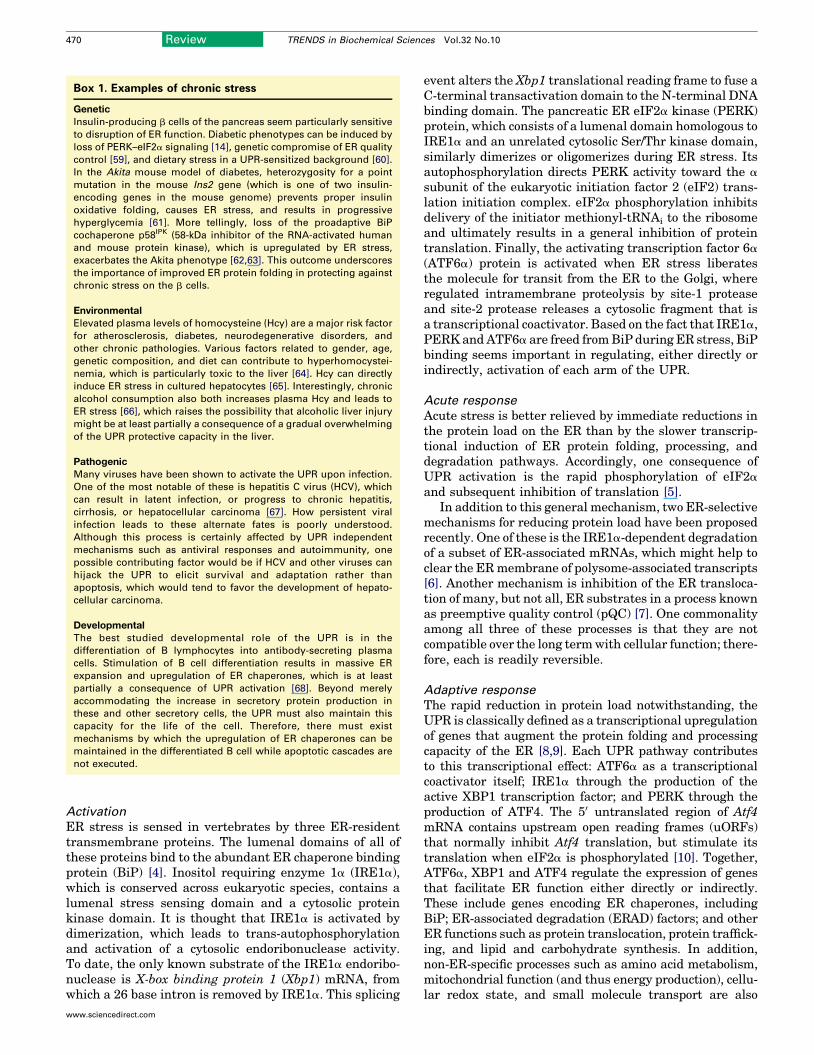
Activation
ER stress is sensed in vertebrates by three ER-residenttransmembrane proteins. The lumenal domains of all ofthese proteins bind to the abundant ER chaperone bindingprotein (BiP) [4]. Inositol requiring enzyme 1a (IRE1a),which is conserved across eukaryotic species, contains alumenal stress sensing domain and a cytosolic proteinkinase domain. It is thought that IRE1a is activated bydimerization, which leads to trans-autophosphorylationand activation of a cytosolic endoribonuclease activity.To date, the only known substrate of the IRE1a endoribo-nuclease is X-box binding protein 1 (Xbp1) mRNA, fromwhich a 26 base intron is removed by IRE1a. This splicing
www.sciencedirect.com
event alters the Xbp1 translational reading frame to fuse aC-terminal transactivation domain to the N-terminal DNAbinding domain. The pancreatic ER eIF2a kinase (PERK)protein, which consists of a lumenal domain homologous toIRE1a and an unrelated cytosolic Ser/Thr kinase domain,similarly dimerizes or oligomerizes during ER stress. Itsautophosphorylation directs PERK activity toward the a
subunit of the eukaryotic initiation factor 2 (eIF2) trans-lation initiation complex. eIF2a phosphorylation inhibitsdelivery of the initiator methionyl-tRNAi to the ribosomeand ultimately results in a general inhibition of proteintranslation. Finally, the activating transcription factor 6a
(ATF6a) protein is activated when ER stress liberatesthe molecule for transit from the ER to the Golgi, whereregulated intramembrane proteolysis by site-1 proteaseand site-2 protease releases a cytosolic fragment that isa transcriptional coactivator. Based on the fact that IRE1a,PERKandATF6a are freed fromBiP during ER stress, BiPbinding seems important in regulating, either directly orindirectly, activation of each arm of the UPR.
Acute response
Acute stress is better relieved by immediate reductions inthe protein load on the ER than by the slower transcrip-tional induction of ER protein folding, processing, anddegradation pathways. Accordingly, one consequence ofUPR activation is the rapid phosphorylation of eIF2a
and subsequent inhibition of translation [5].In addition to this general mechanism, two ER-selective
mechanisms for reducing protein load have been proposedrecently. One of these is the IRE1a-dependent degradationof a subset of ER-associated mRNAs, which might help toclear the ERmembrane of polysome-associated transcripts[6]. Another mechanism is inhibition of the ER transloca-tion of many, but not all, ER substrates in a process knownas preemptive quality control (pQC) [7]. One commonalityamong all three of these processes is that they are notcompatible over the long termwith cellular function; there-fore, each is readily reversible.
Adaptive response
The rapid reduction in protein load notwithstanding, theUPR is classically defined as a transcriptional upregulationof genes that augment the protein folding and processingcapacity of the ER [8,9]. Each UPR pathway contributesto this transcriptional effect: ATF6a as a transcriptionalcoactivator itself; IRE1a through the production of theactive XBP1 transcription factor; and PERK through theproduction of ATF4. The 50 untranslated region of Atf4mRNA contains upstream open reading frames (uORFs)that normally inhibit Atf4 translation, but stimulate itstranslation when eIF2a is phosphorylated [10]. Together,ATF6a, XBP1 and ATF4 regulate the expression of genesthat facilitate ER function either directly or indirectly.These include genes encoding ER chaperones, includingBiP; ER-associated degradation (ERAD) factors; and otherER functions such as protein translocation, protein traffick-ing, and lipid and carbohydrate synthesis. In addition,non-ER-specific processes such as amino acid metabolism,mitochondrial function (and thus energy production), cellu-lar redox state, and small molecule transport are also
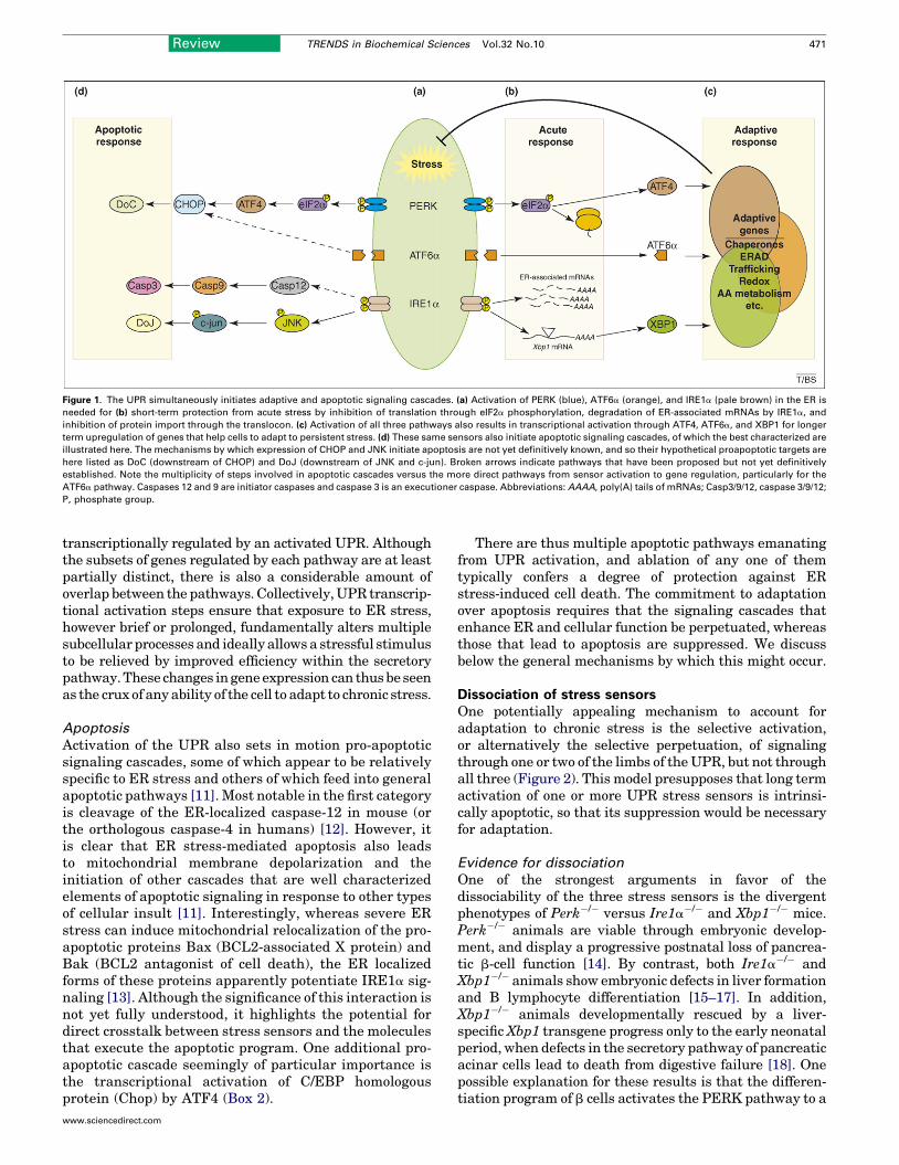
Figure 1. The UPR simultaneously initiates adaptive and apoptotic signaling cascades. (a) Activation of PERK (blue), ATF6a (orange), and IRE1a (pale brown) in the ER is
needed for (b) short-term protection from acute stress by inhibition of translation through eIF2a phosphorylation, degradation of ER-associated mRNAs by IRE1a, and
inhibition of protein import through the translocon. (c) Activation of all three pathways also results in transcriptional activation through ATF4, ATF6a, and XBP1 for longer
term upregulation of genes that help cells to adapt to persistent stress. (d) These same sensors also initiate apoptotic signaling cascades, of which the best characterized are
illustrated here. The mechanisms by which expression of CHOP and JNK initiate apoptosis are not yet definitively known, and so their hypothetical proapoptotic targets are
here listed as DoC (downstream of CHOP) and DoJ (downstream of JNK and c-jun). Broken arrows indicate pathways that have been proposed but not yet definitively
established. Note the multiplicity of steps involved in apoptotic cascades versus the more direct pathways from sensor activation to gene regulation, particularly for the
ATF6a pathway. Caspases 12 and 9 are initiator caspases and caspase 3 is an executioner caspase. Abbreviations: AAAA, poly(A) tails of mRNAs; Casp3/9/12, caspase 3/9/12;
P, phosphate group.
Review TRENDS in Biochemical Sciences Vol.32 No.10 471
transcriptionally regulated by an activated UPR. Althoughthe subsets of genes regulated by each pathway are at leastpartially distinct, there is also a considerable amount ofoverlap between the pathways. Collectively,UPR transcrip-tional activation steps ensure that exposure to ER stress,however brief or prolonged, fundamentally alters multiplesubcellular processes and ideally allows a stressful stimulusto be relieved by improved efficiency within the secretorypathway.These changes in geneexpression can thusbe seenas the crux of any ability of the cell to adapt to chronic stress.
Apoptosis
Activation of the UPR also sets in motion pro-apoptoticsignaling cascades, some of which appear to be relativelyspecific to ER stress and others of which feed into generalapoptotic pathways [11]. Most notable in the first categoryis cleavage of the ER-localized caspase-12 in mouse (orthe orthologous caspase-4 in humans) [12]. However, itis clear that ER stress-mediated apoptosis also leadsto mitochondrial membrane depolarization and theinitiation of other cascades that are well characterizedelements of apoptotic signaling in response to other typesof cellular insult [11]. Interestingly, whereas severe ERstress can induce mitochondrial relocalization of the pro-apoptotic proteins Bax (BCL2-associated X protein) andBak (BCL2 antagonist of cell death), the ER localizedforms of these proteins apparently potentiate IRE1a sig-naling [13]. Although the significance of this interaction isnot yet fully understood, it highlights the potential fordirect crosstalk between stress sensors and the moleculesthat execute the apoptotic program. One additional pro-apoptotic cascade seemingly of particular importance isthe transcriptional activation of C/EBP homologousprotein (Chop) by ATF4 (Box 2).
www.sciencedirect.com
There are thus multiple apoptotic pathways emanatingfrom UPR activation, and ablation of any one of themtypically confers a degree of protection against ERstress-induced cell death. The commitment to adaptationover apoptosis requires that the signaling cascades thatenhance ER and cellular function be perpetuated, whereasthose that lead to apoptosis are suppressed. We discussbelow the general mechanisms by which this might occur.
Dissociation of stress sensorsOne potentially appealing mechanism to account foradaptation to chronic stress is the selective activation,or alternatively the selective perpetuation, of signalingthrough one or two of the limbs of theUPR, but not throughall three (Figure 2). This model presupposes that long termactivation of one or more UPR stress sensors is intrinsi-cally apoptotic, so that its suppression would be necessaryfor adaptation.
Evidence for dissociation
One of the strongest arguments in favor of thedissociability of the three stress sensors is the divergentphenotypes of Perk�/� versus Ire1a�/� and Xbp1�/� mice.Perk�/� animals are viable through embryonic develop-ment, and display a progressive postnatal loss of pancrea-tic b-cell function [14]. By contrast, both Ire1a�/� andXbp1�/� animals show embryonic defects in liver formationand B lymphocyte differentiation [15–17]. In addition,Xbp1�/� animals developmentally rescued by a liver-specific Xbp1 transgene progress only to the early neonatalperiod, when defects in the secretory pathway of pancreaticacinar cells lead to death from digestive failure [18]. Onepossible explanation for these results is that the differen-tiation program of b cells activates the PERK pathway to a
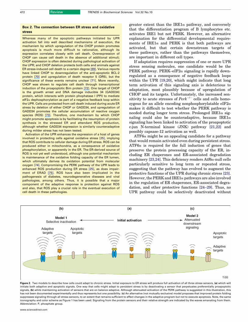
Figure 2. Two models to describe how cells could adapt to chronic stress. Initial exposu
initiate both adaptive and apoptotic signals. One way that cells might adapt to persist
signals, (b) while maintaining activation of sensors that are on balance adaptive. Althou
has not been documented experimentally and thus represents but one possibility. (c) An
suppresses signaling through all stress sensors, to an extent that remains sufficient to e
iconography and color scheme as Figure 1 has been used. Signaling from the protein se
Abbreviation: P, phosphate group.
Box 2. The connection between ER stress and oxidative
stress
Whereas many of the apoptotic pathways initiated by UPR
activation fall into well described mechanisms of execution, the
mechanism by which upregulation of the CHOP protein promotes
apoptosis is much more difficult to rationalize, although its
expression correlates closely with cell death. Overexpression of
CHOP can cause cell death in the absence of additional stimuli;
CHOP expression is often detected during pathological activation of
the UPR; and CHOP deletion protects both cells and animals against
ER stress-induced cell death to a certain degree [69]. Various reports
have linked CHOP to downregulation of the anti-apoptotic BCL-2
protein [70] and upregulation of death receptor 5 (DR5), but the
significance of these events remains unclear [71]. More recently,
CHOP was shown to regulate cell death through transcriptional
induction of the proapoptotic Bim protein [72]. One target of CHOP
is the growth arrest and DNA damage inducible 34 (GADD34)
protein, which interacts with protein phophatase I (PPI) to mediate
eIF2a dephosphorylation as part of a negative feedback loop within
the UPR. Cells are protected from cell death induced during acute ER
stress by deletion of either CHOP or GADD34, and upregulation of
GADD34 promotes the cellular accumulation of reactive oxygen
species (ROS) [73]. Therefore, one mechanism by which CHOP
might promote apoptosis is by facilitating the resumption of protein
synthesis in the stressed ER and attendant ROS production,
although whether GADD34 expression is similarly counteradaptive
during milder stress has not been tested.
Activation of the UPR enhances the expression of a host of genes
involved in protecting cells against oxidative stress [25], implying
that ROS contribute to cellular damage during ER stress. ROS can be
produced either in mitochondria, as a consequence of oxidative
phosphorylation, or apparently in the ER. The ER-derived source of
ROS is not yet well understood, although one potential mechanism
is maintenance of the oxidative folding capacity of the ER lumen,
which ultimately derives its oxidation potential from molecular
oxygen [74]. Compromising the PERK pathway of the UPR leads to
enhanced ROS production during ER stress [25], as does impair-
ment of ERAD [75]. ROS have also been implicated in the
pathogenesis of diabetes, neurodegenerative diseases and viral
pathologies, among others. Thus, it is possible that a major
component of the adaptive response is protection against ROS
and also, that ROS play a crucial role in the eventual execution of
cell death in these pathologies.
472 Review TRENDS in Biochemical Sciences Vol.32 No.10
www.sciencedirect.com
greater extent than the IRE1a pathway, and converselythat the differentiation program of B lymphocytes etc.activates IRE1 but not PERK. However, an alternativeexplanation for the differential developmental require-ments of IRE1a and PERK is that both pathways areactivated, but that certain downstream targets ofthese pathways, rather than the pathways themselves,are important in different cell types.
If adaptation requires suppression of one or more UPRstress sensing molecules, one candidate would be thePERK pathway. PERK–eIF2a signaling is rapidly down-regulated as a consequence of negative feedback loopswithin the UPR [19,20], which might indicate that longterm activation of this signaling axis is deleterious toadaptation, most plausibly because of upregulation ofCHOP and its targets. Unfortunately, the increased sen-sitivity to acute stresses of Perk�/� cells and cells homo-zygous for an allele encoding nonphosphorylatable eIF2a
makes it difficult to test whether the PERK pathway isneeded during longer term stress. Prolonged IRE1a sig-naling could also be counteradaptive, because IRE1a
signaling has been linked to activation of the proapoptoticc-jun N-terminal kinase (JNK) pathway [21,22] andpossibly capsase-12 activation as well.
ATF6a might be an appealing candidate for a pathwaythat would remain activated even during persistent stress.ATF6a is required for the full induction of genes thatpreserve the protein processing capacity of the ER, in-cluding ER chaperones and ER-associated degradationmachinery [23,24]. This deficiency renders Atf6a-null cellsparticularly sensitive to long term or repeated stress,suggesting that the pathway has evolved to augment theprotective functions of the UPR during chronic stress [23].However, the PERKand IRE1a pathways are also involvedin the regulation of ER chaperones, ER-associated degra-dation, and other protective functions [24–29]. Thus, noUPR pathway could be selectively deactivated without
re to ER stress will produce full activation of all three stress sensors, (a) which will
ent stress is by deactivating a sensor that perpetuates preferentially proapoptotic
gh attenuated activation of the PERK pathway is suggested in this illustration, this
alternative (not mutually exclusive) model proposes that improved protein folding
ffect changes in the adaptive program but not to execute apoptosis. Note, the same
nsors and their relative strength are indicated by the waves emanating from them.

Review TRENDS in Biochemical Sciences Vol.32 No.10 473
sacrificing at least some of the adaptive capacity of the cellas well.
Possible mechanisms of dissociation
Rather than complete and selective inactivation of a singleUPR pathway during chronic stress, long term UPR acti-vationmight allow the sensors to be partially suppressed togreater or lesser extents. In principle, any divergence inthe mechanism of activation of the three sensors couldaccount for partial dissociability. For example, duringactivation ATF6a appears to undergo changes in bothits disulfide bonded state and its glycosylation state[30,31]. If these events are required for its activation, thenboth the nature of the stressor and the alterations in ERfunction that occur as a consequence of UPR activationcould influence the extent to which ATF6a specifically isactivated. Likewise, IRE1a can be activated by autophos-phorylation in a way that brings about regulation ofdownstream genes but does not result in splicing ofXbp1 mRNA, implying that IRE1a exists in more statesthan simply ’on’ and ’off’ [32,33]. Perhaps ER stress sensorscan exist in alternate states subject to differential post-translational modification, each of which signals a uniquecascade, with the preponderance of these states dependentupon the nature and persistence of the stressor.
The possibility of at least partial dissociation of thestress sensors becomes more plausible if they are not allactivated by the same simple mechanism of titration fromBiP. Different stressors appear to activate the three path-ways with differing kinetics [34], which suggests somedivergence in the mechanisms of their activation. Thecrystal structure of yeast Ire1p led to the proposal thatIre1p binds to unfolded proteins directly, and that BiPbinding serves as, at most, a modulator of Ire1p activity[35]. However, the crystal structure of mammalian IRE1a
challenges this model [33]. Biochemical characterization ofthe activation and stress responsiveness of various dimer-ization-defective mutants of both IRE1a and PERKsuggests a similar mechanism of activation [33], and thispossibility is supported by the observation that the lume-nal domains of the two proteins are functionally inter-changeable [36,37]. Ultimately, the determination ofwhether the UPR can exist in a selectively activated statewill require more sensitive assays for directly monitoringsensor activation.
It is possible that additional limbs of the UPR alter thenature of UPR signaling and regulate cell fate decisionsduring chronic stress. ATF6a is but one of a rapidly emer-ging family of ER-localized transmembrane proteinscleaved by regulated intramembrane proteolysis duringER stress. ATF6b is loosely homologous to ATF6a, andlike ATF6a, is ubiquitously expressed. It is activated underthe same general conditions as is ATF6a, and can inprinciple bind to at least some of the same promoterelements [38]. It has been proposed that ATF6b acts asa transcriptional repressor of ATF6a-dependent genes[39]. It is conceivable that as cells adapt to stress, ATF6b
activity predominates over ATF6a activity, with potentialconsequences for the patterns of gene expression thatthen result. The cyclic-AMP-responsive-element-bindingprotein H (CREBH) protein has been identified as a liver
www.sciencedirect.com
specific, ER stress-activated transmembrane transcriptionfactor. In vivo knockdown of CREBH does not compromiseupregulation of ER stress dependent genes, but doesimpair the systemic inflammatory response initiated bythe liver in response to challenge [40], which illustratesthat the UPR has a role not just in cellular protection, butin organismal protection as well. Proteins such as CREBH,ATF6b, and others could interact with the core UPRmachinery to modulate its output. The potential for suchmodulation can be seen in the ability of XBP1 to regulatethe expression of different sets of genes, through differentcis-acting sequence motifs, in a cell-type specific manner[41]. Ultimately, the roles of these proteins in the adaptiveresponses of specific tissues awaits a more detailed charac-terization of the conditions under which they are activatedand of the nature of their targets.
Divergence in downstream responsesCells can survive and proliferate despite persistentexposure to pharmacological stress, if the stress is suffi-cientlymild, despite activation of all three arms of theUPR[42]. Their adaptation is marked by persistent expressionof ER chaperones such as BiP, but not of CHOP and at leastone of CHOP’s targets. These observations raise the possib-ility that cells might adapt to persistent stress not byselectively regulating activation of the three limbs of theUPR, but by the manner in which the expression of down-stream effector molecules of the UPR is controlled duringexposure to tolerable stress (Figure 2).
Adaptation by improved protein processing
Two stages would be required for cells to adapt by amechanism that alters the downstream signaling outputof the UPR: (i) the capacity of the ER to process clientproteins would have to be improved to an extent sufficientto mitigate the perturbing effects of the stressor andattenuate UPR signaling; and (ii) a new equilibrium wouldhave to be established in which signaling through the UPRcould maintain improved ER protein processing capacity,but not execute apoptotic cascades.
Functionality of ER protein processing can be improvedeither by the removal of misfolded proteins or by enhancedfolding and transport of nascent proteins such that theyavoid misfolding in the first place. The classical method forthe removal of misfolded proteins is ERAD, which isstimulated as a consequence of UPR activation, and canlikely be attributed to enhanced expression of ERAD-facil-itating proteins such as EDEM and Derlin-2 among others[27,29,43]. In addition, there apparently exist mechan-isms, at least in lower eukaryotes and possibly invertebrates as well, for segregating large quantities ofmisfolded or unfolded proteins for autophagic destruction[44]. However, although suchmechanisms no doubt help toalleviate the protein misfolding load during stress, it is notclear whether they actually improve the protein processingcapacity of the ER, or simplymitigatemore severe damage.
Unfortunately, the more direct demonstration that theER of the UPR-activated cell moves proteins more effi-ciently forward through the secretory pathway has beenlacking, in part because the conditions typically used toinduce ER stress are sufficiently severe that even a
474 Review TRENDS in Biochemical Sciences Vol.32 No.10
maximally activated UPR is unlikely to result in a netgain in ER functionality. However, indirect support forthis idea comes from studies in which cells that have beenbriefly UPR-activated (either by transient exposure to astressor, or by artificial activation of individual UPRpathways) become resistant to challenge with a secondstressor, both in terms of cell survival and UPR activationstatus. This phenomenon, termed ’preconditioning’, isaccompanied by upregulation of UPR-dependent genes,including ER chaperones [45,46]. Therefore, precondi-tioning likely protects cells by improving the ER proteinprocessing capacity.
Further support for the importance of improved proteinprocessing in adaptation to physiological stress comes fromstudies that explore the peripheral insulin resistance thataccompanies obesity. This resistance could be attenuatedby treatment of obese mice with chemical chaperones thatimproved protein folding in the periphery [47]. In addition,heterozygosity for XBP1 in mice sensitized these animalsto obesity-induced insulin resistance [48]. Thus, the role ofXBP1 in the protection of insulin signaling in the peripherymight be to improve ER protein folding, although this hasnot yet been demonstrated rigorously.
Suppression of downstream signaling
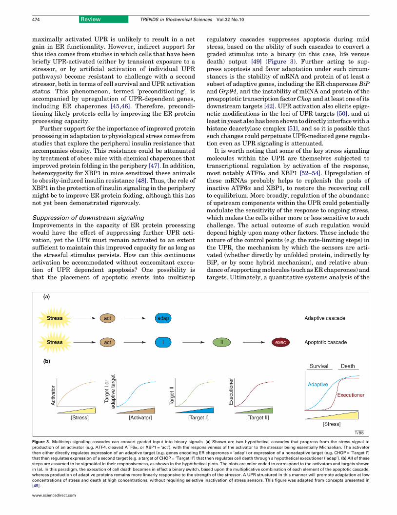
Improvements in the capacity of ER protein processingwould have the effect of suppressing further UPR acti-vation, yet the UPR must remain activated to an extentsufficient to maintain this improved capacity for as long asthe stressful stimulus persists. How can this continuousactivation be accommodated without concomitant execu-tion of UPR dependent apoptosis? One possibility isthat the placement of apoptotic events into multistep
Figure 3. Multistep signaling cascades can convert graded input into binary signals.
production of an activator (e.g. ATF4, cleaved ATF6a, or XBP1 = ‘act’), with the respons
then either directly regulates expression of an adaptive target (e.g. genes encoding ER
that then regulates expression of a second target (e.g. a target of CHOP = ‘Target II’) that
steps are assumed to be sigmoidal in their responsiveness, as shown in the hypothetica
in (a). In this paradigm, the execution of cell death becomes in effect a binary switch, ba
whereas production of adaptive proteins remains more linearly responsive to the streng
concentrations of stress and death at high concentrations, without requiring selective i
[49].
www.sciencedirect.com
regulatory cascades suppresses apoptosis during mildstress, based on the ability of such cascades to convert agraded stimulus into a binary (in this case, life versusdeath) output [49] (Figure 3). Further acting to sup-press apoptosis and favor adaptation under such circum-stances is the stability of mRNA and protein of at least asubset of adaptive genes, including the ER chaperones BiPand Grp94, and the instability of mRNA and protein of theproapoptotic transcription factorChop and at least one of itsdownstream targets [42]. UPR activation also elicits epige-netic modifications in the loci of UPR targets [50], and atleast inyeastalsohasbeenshowntodirectly interfacewithahistone deacetylase complex [51], and so it is possible thatsuch changes could perpetuate UPR-mediated gene regula-tion even as UPR signaling is attenuated.
It is worth noting that some of the key stress signalingmolecules within the UPR are themselves subjected totranscriptional regulation by activation of the response,most notably ATF6a and XBP1 [52–54]. Upregulation ofthese mRNAs probably helps to replenish the pools ofinactive ATF6a and XBP1, to restore the recovering cellto equilibrium. More broadly, regulation of the abundanceof upstream components within the UPR could potentiallymodulate the sensitivity of the response to ongoing stress,which makes the cells either more or less sensitive to suchchallenge. The actual outcome of such regulation woulddepend highly upon many other factors. These include thenature of the control points (e.g. the rate-limiting steps) inthe UPR, the mechanism by which the sensors are acti-vated (whether directly by unfolded protein, indirectly byBiP, or by some hybrid mechanism), and relative abun-dance of supportingmolecules (such as ER chaperones) andtargets. Ultimately, a quantitative systems analysis of the
(a) Shown are two hypothetical cascades that progress from the stress signal to
iveness of the activator to the stressor being essentially Michaelian. The activator
chaperones = ‘adap’) or expression of a nonadaptive target (e.g. CHOP = ‘Target I’)
then regulates cell death through a hypothetical executioner (‘adap’). (b) All of these
l plots. The plots are color coded to correspond to the activators and targets shown
sed upon the multiplicative combination of each element of the apoptotic cascade,
th of the stressor. A UPR structured in this manner will promote adaptation at low
nactivation of stress sensors. This figure was adapted from concepts presented in

Review TRENDS in Biochemical Sciences Vol.32 No.10 475
UPR will be necessary to understand how its output canbe modulated by the abundance and activities of itsconstitutents.
The UPR is known to interact with other cellularsignaling cascades that couldmodulate its output, includingmitogen-acitvated protein (MAP) kinase cascades [55] andnuclear factor kappa B (NF-kB) signaling [56,57]. Unfortu-nately, the paucity of studies that explore the consequencesof long term UPR activation makes it unclear how thesepathways might influence the outcome of UPR signaling.However, the apparent interface between the UPR andother cell signaling pathways is underscored by the recentdiscovery that chondrocytes, which are involved in bonedifferentiation and maturation, can adapt to the stress ofproduction of misfolded collagen by initiating a program ofreverse differentiation [58]. This process appears to tempor-arily interrupt endochondral bone formationwhile chondro-cytes upregulate the UPR and suppress expression of themutant collagen. Itwill be interesting to determinewhetherthis ability to closely tie differentiation status toER load is ageneral feature of the development of professional secretorycells, or is specific to chondroyctes.
Future directionsThe fact that diseases such as diabetes and neurode-generative disorders manifest over the course of decadesargues that only very slight adjustments to the balancebetween adaptation and apoptosis would be necessary tohave a significant beneficial impact on health. However,before such manipulation becomes feasible, much moreneeds to be understood about how the UPR responds tothe types of stresses that underlie such pathologies, com-pared with the classical UPR that has been defined withina relatively artificial experimental context. More sensitivemeasures to detect subtle variations in the status of theUPR sensors and their downstream effects will be neededto identify themechanisms of activation in response to verymild ER perturbations of different sorts. Understandingthe interface between the UPR and other cellular signalingcascades will help to identify additional mechanismswhereby cells adapt. Tissue specific deletion of stress sen-sing molecules and their downstream effectors in themouse, in combination with genetic or environmentalchallenge, will reveal critical control points in the cellulardecision to adapt to both pathological and developmentalstresses. Ultimately, the regulation of cellular adaptivedecisions during ER stress will shed light on how cellsadapt to the variety of chronic stresses that exist in diversedisease states.
AcknowledgementsWe thank D. Raden for insightful discussions. We would like to apologizeto those colleagues whose work could only be cited by reference to reviewsowing to space considerations. R.J.K. is supported by the Howard HughesMedical Institute and NIH grants R01 DK042934, R01 HL052173, andP01 HL057345.
References1 Kaufman, R.J. (2002) Orchestrating the unfolded protein response in
health and disease. J. Clin. Invest. 110, 1389–13982 Marciniak, S.J. and Ron, D. (2006) Endoplasmic reticulum stress
signaling in disease. Physiol. Rev. 86, 1133–1149
www.sciencedirect.com
3 van Anken, E. and Braakman, I. (2005) Endoplasmic reticulum stressand themaking of a professional secretory cell.Crit. Rev. Biochem.Mol.Biol. 40, 269–283
4 Hendershot, L.M. (2004) The ER chaperone BiP is amaster regulator ofER function. Mt. Sinai J. Med. 71, 289–297
5 Harding, H.P. et al. (2001) Translational regulation in the cellularresponse to biosynthetic load on the endoplasmic reticulum. ColdSpring Harb. Symp. Quant. Biol. 66, 499–508
6 Hollien, J. and Weissman, J.S. (2006) Decay of endoplasmic reticulum-localized mRNAs during the unfolded protein response. Science 313,104–107
7 Kang, S.W. et al. (2006) Substrate-specific translocational attenuationduring ER stress defines a pre-emptive quality control pathway. Cell127, 999–1013
8 Ma, Y. and Hendershot, L.M. (2004) ER chaperone functions duringnormal and stress conditions. J. Chem. Neuroanat. 28, 51–65
9 Schroder, M. and Kaufman, R.J. (2005) The mammalian unfoldedprotein response. Annu. Rev. Biochem. 74, 739–789
10 Lu, P.D. et al. (2004) Translation reinitiation at alternative openreading frames regulates gene expression in an integrated stressresponse. J. Cell Biol. 167, 27–33
11 Xu, C. et al. (2005) Endoplasmic reticulum stress: cell life and deathdecisions. J. Clin. Invest. 115, 2656–2664
12 Nakagawa, T. et al. (2000) Caspase-12 mediates endoplasmic-reticulum-specific apoptosis and cytotoxicity by amyloid-beta. Nature403, 98–103
13 Hetz, C. et al. (2006) Proapoptotic BAX andBAKmodulate the unfoldedprotein response by a direct interaction with IRE1alpha. Science 312,572–576
14 Harding, H.P. et al. (2001) Diabetes mellitus and exocrine pancreaticdysfunction in Perk�/� mice reveals a role for translational control insecretory cell survival. Mol. Cell 7, 1153–1163
15 Reimold, A.M. et al. (2000) An essential role in liver development fortranscription factor XBP-1. Genes Dev. 14, 152–157
16 Reimold, A.M. et al. (2001) Plasma cell differentiation requires thetranscription factor XBP-1. Nature 412, 300–307
17 Zhang, K. et al. (2005) The unfolded protein response sensor IRE1alphais required at 2 distinct steps in B cell lymphopoiesis. J. Clin. Invest.115, 268–281
18 Lee, A.H. et al. (2005) XBP-1 is required for biogenesis ofcellular secretory machinery of exocrine glands. EMBO J. 24, 4368–4380
19 Jousse, C. et al. (2003) Inhibition of a constitutive translation initiationfactor 2alpha phosphatase, CReP, promotes survival of stressed cells.J. Cell Biol. 163, 767–775
20 Novoa, I. et al. (2001) Feedback inhibition of the unfolded proteinresponse by GADD34-mediated dephosphorylation of eIF2alpha.J. Cell Biol. 153, 1011–1022
21 Nishitoh, H. et al. (2002) ASK1 is essential for endoplasmic reticulumstress-induced neuronal cell death triggered by expandedpolyglutamine repeats. Genes Dev. 16, 1345–1355
22 Urano, F. et al. (2000) Coupling of stress in the ER to activation of JNKprotein kinases by transmembrane protein kinase IRE1. Science 287,664–666
23 Wu, J. et al. ATF6 alpha optimizes long-term endoplasmic reticulumfunction to protect cells from chronic stress. Dev. Cell 10.1016/j.devcel.2007.07.005 www.developmentalcell.com
24 Yamamoto, K. et al. Transcriptional induction of mammalian ERquality control proteins is mediated by single or combined action ofATF6a and XBP1. Dev. Cell 10.1016/j.devcell.2007.07.018 www.developmentalcell.com
25 Harding, H.P. et al. (2003) An integrated stress response regulatesamino acid metabolism and resistance to oxidative stress.Mol. Cell 11,619–633
26 Novoa, I. et al. (2003) Stress-induced gene expression requiresprogrammed recovery from translational repression. EMBO J. 22,1180–1187
27 Lee, A.H. et al. (2003) XBP-1 regulates a subset of endoplasmicreticulum resident chaperone genes in the unfolded proteinresponse. Mol. Cell. Biol. 23, 7448–7459
28 Sriburi, R. et al. (2004) XBP1: a link between the unfolded proteinresponse, lipid biosynthesis, and biogenesis of the endoplasmicreticulum. J. Cell Biol. 167, 35–41
476 Review TRENDS in Biochemical Sciences Vol.32 No.10
29 Yoshida, H. et al. (2003) A time-dependent phase shift in themammalian unfolded protein response. Dev. Cell 4, 265–271
30 Nadanaka, S. et al. (2006) Reduction of disulfide bridges in the lumenaldomain of ATF6 in response to glucose starvation. Cell Struct. Funct.31, 127–134
31 Hong, M. et al. (2004) Underglycosylation of ATF6 as a novel sensingmechanism for activation of the unfolded protein response. J. Biol.Chem. 279, 11354–11363
32 Lipson, K.L. et al. (2006) Regulation of insulin biosynthesis inpancreatic beta cells by an endoplasmic reticulum-resident proteinkinase IRE1. Cell Metab. 4, 245–254
33 Zhou, J. et al. (2006) The crystal structure of human IRE1 luminaldomain reveals a conserved dimerization interface required foractivation of the unfolded protein response. Proc. Natl. Acad. Sci.U. S. A. 103, 14343–14348
34 DuRose, J.B. et al. (2006) Intrinsic capacities of molecular sensors ofthe unfolded protein response to sense alternate forms of endoplasmicreticulum stress. Mol. Biol. Cell 17, 3095–3107
35 Credle, J.J. et al. (2005) On the mechanism of sensing unfolded proteinin the endoplasmic reticulum. Proc. Natl. Acad. Sci. U. S. A. 102,18773–18784
36 Bertolotti, A. et al. (2000) Dynamic interaction of BiP and ER stresstransducers in the unfolded-protein response. Nat. Cell Biol. 2, 326–332
37 Liu, C.Y. et al. (2000) Ligand-independent dimerization activates thestress response kinases IRE1 and PERK in the lumen of theendoplasmic reticulum. J. Biol. Chem. 275, 24881–24885
38 Yoshida, H. et al. (2001) Endoplasmic reticulum stress-inducedformation of transcription factor complex ERSF including NF-Y(CBF) and activating transcription factors 6alpha and 6beta thatactivates the mammalian unfolded protein response. Mol. Cell. Biol.21, 1239–1248
39 Thuerauf, D.J. et al. (2004) Opposing roles for ATF6alpha andATF6beta in endoplasmic reticulum stress response gene induction.J. Biol. Chem. 279, 21078–21084
40 Zhang, K. et al. (2006) Endoplasmic reticulum stress activates cleavageof CREBH to induce a systemic inflammatory response. Cell 124, 587–599
41 Acosta-Alvear, D. et al. (2007) XBP1 controls diverse cell type- andcondition-specific transcriptional regulatory networks. Mol. Cell 27,53–66
42 Rutkowski, D.T. et al. (2006) Adaptation to ER stress is mediated bydifferential stabilities of pro-survival and pro-apoptotic mRNAs andproteins. PLoS Biol. 4, e374
43 Oda, Y. et al. (2006) Derlin-2 and Derlin-3 are regulated by themammalian unfolded protein response and are required for ER-associated degradation. J. Cell Biol. 172, 383–393
44 Bernales, S. et al. (2006) Autophagy counterbalances endoplasmicreticulum expansion during the unfolded protein response. PLoSBiol. 4, e423
45 Hung, C.C. et al. (2003) Protection of renal epithelial cellsagainst oxidative injury by endoplasmic reticulum stress precon-ditioning is mediated by ERK1/2 activation. J. Biol. Chem. 278,29317–29326
46 Lu, P.D. et al. (2004) Cytoprotection by pre-emptive conditionalphosphorylation of translation initiation factor 2. EMBO J. 23, 169–179
47 Ozcan, U. et al. (2006) Chemical chaperones reduce ER stress andrestore glucose homeostasis in a mouse model of type 2 diabetes.Science 313, 1137–1140
48 Ozcan, U. et al. (2004) Endoplasmic reticulum stress links obesity,insulin action, and type 2 diabetes. Science 306, 457–461
49 Ferrell, J.E., Jr (1999) Building a cellular switch: more lessons from agood egg. Bioessays 21, 866–870
50 Donati, G. et al. (2006) Dynamic recruitment of transcription factorsand epigenetic changes on the ER stress response gene promoters.Nucleic Acids Res. 34, 3116–3127
www.sciencedirect.com
51 Schroder, M. et al. (2004) The unfolded protein response repressesdifferentiation through the RPD3-SIN3 histone deacetylase. EMBO J.23, 2281–2292
52 Lee, K. et al. (2002) IRE1-mediated unconventionalmRNA splicing andS2P-mediated ATF6 cleavage merge to regulate XBP1 in signaling theunfolded protein response. Genes Dev. 16, 452–466
53 Yoshida, H. et al. (2001) XBP1 mRNA is induced by ATF6 and splicedby IRE1 in response to ER stress to produce a highly activetranscription factor. Cell 107, 881–891
54 Namba, T. et al. (2007) Transcriptional activation of ATF6 byendoplasmic reticulum stressors. Biochem. Biophys. Res. Commun.355, 543–548
55 Sekine, Y. et al. (2006) The ASK1-MAP kinase signaling in ER stressand neurodegenerative diseases. Curr. Mol. Med. 6, 87–97
56 Deng, J. et al. (2004) Translational repression mediates activation ofnuclear factor kappa B by phosphorylated translation initiation factor2. Mol. Cell. Biol. 24, 10161–10168
57 Pahl, H.L. et al. (1996) Activation of transcription factor NF-kappaB bythe adenovirus E3/19K protein requires its ER retention. J. Cell Biol.132, 511–522
58 Tsang, K.Y. et al. (2007) Surviving endoplasmic reticulum stress iscoupled to altered chondrocyte differentiation and function. PLoS Biol.5, e44
59 Ladiges, W.C. et al. (2005) Pancreatic {beta}-cell failure and diabetes inmice with a deletion mutation of the endoplasmic reticulum molecularchaperone gene P58IPK. Diabetes 54, 1074–1081
60 Scheuner, D. et al. (2005) Control of mRNA translation preservesendoplasmic reticulum function in beta cells and maintains glucosehomeostasis. Nat. Med. 11, 757–764
61 Oyadomari, S. et al. (2002) Targeted disruption of the Chop gene delaysendoplasmic reticulum stress-mediated diabetes. J. Clin. Invest. 109,525–532
62 Oyadomari, S. et al. (2006) Cotranslocational degradation protectsthe stressed endoplasmic reticulum from protein overload. Cell 126,727–739
63 Rutkowski, D.T. et al. (2007) The role of p58IPK in protecting thestressed endoplasmic reticulum. Mol. Biol. Cell 18, 3681–3691
64 Kaplowitz, N. and Ji, C. (2006) Unfolding newmechanisms of alcoholicliver disease in the endoplasmic reticulum. J. Gastroenterol. Hepat. 21(Suppl 3), S7–S9
65 Werstuck, G.H. et al. (2001) Homocysteine-induced endoplasmicreticulum stress causes dysregulation of the cholesterol andtriglyceride biosynthetic pathways. J. Clin. Invest. 107, 1263–1273
66 Ji, C. and Kaplowitz, N. (2003) Betaine decreaseshyperhomocysteinemia, endoplasmic reticulum stress, and liver injuryin alcohol-fed mice. Gastroenterology 124, 1488–1499
67 Levrero, M. (2006) Viral hepatitis and liver cancer: the case of hepatitisC. Oncogene 25, 3834–3847
68 Gass, J.N. et al. (2004) Stressed-out B cells? Plasma-cell differentiationand the unfolded protein response. Trends Immunol. 25, 17–24
69 Oyadomari, S. and Mori, M. (2004) Roles of CHOP/GADD153 inendoplasmic reticulum stress. Cell Death Differ. 11, 381–389
70 McCullough, K.D. et al. (2001) Gadd153 sensitizes cells to endoplasmicreticulum stress by down-regulating Bcl2 and perturbing the cellularredox state. Mol. Cell. Biol. 21, 1249–1259
71 Yamaguchi, H. and Wang, H.G. (2004) CHOP is involved inendoplasmic reticulum stress-induced apoptosis by enhancing DR5expression in human carcinoma cells. J. Biol. Chem. 279, 45495–45502
72 Puthalakath, H. et al. (2007) ER stress triggers apoptosis by activatingBH3-only protein Bim. Cell 129, 1337–1349
73 Marciniak, S.J. et al. (2004) CHOP induces death by promoting proteinsynthesis and oxidation in the stressed endoplasmic reticulum. GenesDev. 18, 3066–3077
74 Tu, B.P. and Weissman, J.S. (2004) Oxidative protein folding ineukaryotes: mechanisms and consequences. J. Cell Biol. 164, 341–346
75 Haynes, C.M. et al. (2004) Degradation of misfolded proteins preventsER-derived oxidative stress and cell death. Mol. Cell 15, 767–776