Stimulation of Inositol 1,4,5-Trisphosphate (IP3) Receptor Subtypes by Analogues of IP3
12
Stimulation of Inositol 1,4,5-Trisphosphate (IP 3 ) Receptor Subtypes by Adenophostin A and Its Analogues Huma Saleem 1 , Stephen C. Tovey 1 , Andrew M. Riley 2 , Barry V. L. Potter 2 , Colin W. Taylor 1 * 1 Department of Pharmacology, Cambridge, United Kingdom, 2 Wolfson Laboratory of Medicinal Chemistry, Department of Pharmacy and Pharmacology, University of Bath, Bath, United Kingdom Abstract Inositol 1,4,5-trisphosphate receptors (IP 3 R) are intracellular Ca 2+ channels. Most animal cells express mixtures of the three IP 3 R subtypes encoded by vertebrate genomes. Adenophostin A (AdA) is the most potent naturally occurring agonist of IP 3 R and it shares with IP 3 the essential features of all IP 3 R agonists, namely structures equivalent to the 4,5-bisphosphate and 6- hydroxyl of IP 3 . The two essential phosphate groups contribute to closure of the clam-like IP 3 -binding core (IBC), and thereby IP 3 R activation, by binding to each of its sides (the a- and b-domains). Regulation of the three subtypes of IP 3 R by AdA and its analogues has not been examined in cells expressing defined homogenous populations of IP 3 R. We measured Ca 2+ release evoked by synthetic adenophostin A (AdA) and its analogues in permeabilized DT40 cells devoid of native IP 3 R and stably expressing single subtypes of mammalian IP 3 R. The determinants of high-affinity binding of AdA and its analogues were indistinguishable for each IP 3 R subtype. The results are consistent with a cation-p interaction between the adenine of AdA and a conserved arginine within the IBC a-domain contributing to closure of the IBC. The two complementary contacts between AdA and the a-domain (cation-p interaction and 30-phosphate) allow activation of IP 3 R by an analogue of AdA (30-dephospho-AdA) that lacks a phosphate group equivalent to the essential 5-phosphate of IP 3 . These data provide the first structure-activity analyses of key AdA analogues using homogenous populations of all mammalian IP 3 R subtypes. They demonstrate that differences in the Ca 2+ signals evoked by AdA analogues are unlikely to be due to selective regulation of IP 3 R subtypes. Citation: Saleem H, Tovey SC, Riley AM, Potter BVL, Taylor CW (2013) Stimulation of Inositol 1,4,5-Trisphosphate (IP 3 ) Receptor Subtypes by Adenophostin A and Its Analogues. PLoS ONE 8(2): e58027. doi:10.1371/journal.pone.0058027 Editor: Alexander G. Obukhov, Indiana University School of Medicine, United States of America Received November 21, 2012; Accepted January 30, 2013; Published February 28, 2013 Copyright: ß 2013 Saleem et al. This is an open-access article distributed under the terms of the Creative Commons Attribution License, which permits unrestricted use, distribution, and reproduction in any medium, provided the original author and source are credited. Funding: This work was supported by grants from the Wellcome Trust to CWT [085295], and BVLP and AMR [082837]. HS is supported by a research studentship from the Jameel Family Trust. The funders had no role in study design, data collection and analysis, decision to publish, or preparation of the manuscript. Competing Interests: The authors have declared that no competing interests exist. * E-mail: [email protected] Introduction Inositol 1,4,5-trisphosphate receptors (IP 3 R) are intracellular Ca 2+ channels that are expressed in almost all animal cells. They allow release of Ca 2+ from intracellular stores in response to the many stimuli that activate phospholipase C [1,2]. The genomes of vertebrates encode three closely related IP 3 R subtypes (IP 3 R1-3), and most cells from vertebrates express functional IP 3 R that are homo- or hetero-tetrameric assemblies of these IP 3 R subtypes and their splice variants [3]. The physiological significance of this IP 3 R diversity is poorly understood, and nor are there ligands that usefully discriminate between IP 3 R subtypes. It is, however, clear that activation of IP 3 R is initiated by binding of IP 3 to the conserved IP 3 -binding core (IBC, residues 224-604 of IP 3 R1) of each IP 3 R subunit [4]. Mixed populations of IP 3 R in native cells make it difficult to define unambiguously the functional properties of each IP 3 R subtype. Stable heterologous expression of mamma- lian IP 3 R in the only vertebrate cell line engineered to lack all endogenous IP 3 R (DT40 KO cells) [5] provides an effective means of addressing this difficulty [6]. We previously used DT40 cells expressing homogeneous populations of each mammalian IP 3 R subtype to define structure-activity relationships for key endoge- nous and synthetic inositol phosphates [7]. Here, we extend the approach to examine the interactions of each IP 3 R subtype with adenophostin A (1, AdA) and its most important analogues [8] (Figure 1A). AdA, originally isolated from Penicillium brevicompactum [9,10] and later synthesized [11], is a potent agonist of IP 3 R. It is also resistant to degradation by the enzymes that degrade IP 3 via phosphorylation or dephosphorylation [10]. Although AdA is based on a glucose ring, rather than the inositol ring of IP 3 , its structure retains the key functional groups of IP 3 that are known to be essential for IP 3 activity at IP 3 R [12] (Figure 1A). Considerable evidence supports the original suggestion [10] that the essential 4,5-bisphosphate and 6-hydroxyl of IP 3 are effectively mimicked by the 40,30-bisphosphate and 20-hydroxyl of AdA (red highlights in Figure 1A). The interactions that allow AdA to bind to IP 3 R with about 10-fold greater affinity than IP 3 have been more difficult to resolve. One view was that the 29-phosphate of AdA is equivalent to the 1-phosphate of IP 3 and, like the latter [13] (blue in Figure 1A), contributes to high-affinity binding to the IBC. The suggestion was that the 29-phosphate of AdA forms a stronger interaction with the IBC than does the 1-phosphate of IP 3 . Our recent analyses have challenged this idea and instead suggest that a cation-p interaction between the adenine ring of AdA and a guanidinium side chain of an arginine residue within the a-domain of the IBC (R504 in IP 3 R1) may be a more important determinant of the increased affinity of AdA for IP 3 R [12]. PLOS ONE | www.plosone.org 1 February 2013 | Volume 8 | Issue 2 | e58027
-
Upload
independent -
Category
Documents
-
view
3 -
download
0
Transcript of Stimulation of Inositol 1,4,5-Trisphosphate (IP3) Receptor Subtypes by Analogues of IP3
Stimulation of Inositol 1,4,5-Trisphosphate (IP3) ReceptorSubtypes by Adenophostin A and Its AnaloguesHuma Saleem1, Stephen C. Tovey1, Andrew M. Riley2, Barry V. L. Potter2, Colin W. Taylor1*
1 Department of Pharmacology, Cambridge, United Kingdom, 2 Wolfson Laboratory of Medicinal Chemistry, Department of Pharmacy and Pharmacology, University of
Bath, Bath, United Kingdom
Abstract
Inositol 1,4,5-trisphosphate receptors (IP3R) are intracellular Ca2+ channels. Most animal cells express mixtures of the threeIP3R subtypes encoded by vertebrate genomes. Adenophostin A (AdA) is the most potent naturally occurring agonist of IP3Rand it shares with IP3 the essential features of all IP3R agonists, namely structures equivalent to the 4,5-bisphosphate and 6-hydroxyl of IP3. The two essential phosphate groups contribute to closure of the clam-like IP3-binding core (IBC), andthereby IP3R activation, by binding to each of its sides (the a- and b-domains). Regulation of the three subtypes of IP3R byAdA and its analogues has not been examined in cells expressing defined homogenous populations of IP3R. We measuredCa2+ release evoked by synthetic adenophostin A (AdA) and its analogues in permeabilized DT40 cells devoid of native IP3Rand stably expressing single subtypes of mammalian IP3R. The determinants of high-affinity binding of AdA and itsanalogues were indistinguishable for each IP3R subtype. The results are consistent with a cation-p interaction between theadenine of AdA and a conserved arginine within the IBC a-domain contributing to closure of the IBC. The twocomplementary contacts between AdA and the a-domain (cation-p interaction and 30-phosphate) allow activation of IP3Rby an analogue of AdA (30-dephospho-AdA) that lacks a phosphate group equivalent to the essential 5-phosphate of IP3.These data provide the first structure-activity analyses of key AdA analogues using homogenous populations of allmammalian IP3R subtypes. They demonstrate that differences in the Ca2+ signals evoked by AdA analogues are unlikely tobe due to selective regulation of IP3R subtypes.
Citation: Saleem H, Tovey SC, Riley AM, Potter BVL, Taylor CW (2013) Stimulation of Inositol 1,4,5-Trisphosphate (IP3) Receptor Subtypes by Adenophostin A andIts Analogues. PLoS ONE 8(2): e58027. doi:10.1371/journal.pone.0058027
Editor: Alexander G. Obukhov, Indiana University School of Medicine, United States of America
Received November 21, 2012; Accepted January 30, 2013; Published February 28, 2013
Copyright: � 2013 Saleem et al. This is an open-access article distributed under the terms of the Creative Commons Attribution License, which permitsunrestricted use, distribution, and reproduction in any medium, provided the original author and source are credited.
Funding: This work was supported by grants from the Wellcome Trust to CWT [085295], and BVLP and AMR [082837]. HS is supported by a research studentshipfrom the Jameel Family Trust. The funders had no role in study design, data collection and analysis, decision to publish, or preparation of the manuscript.
Competing Interests: The authors have declared that no competing interests exist.
* E-mail: [email protected]
Introduction
Inositol 1,4,5-trisphosphate receptors (IP3R) are intracellular
Ca2+ channels that are expressed in almost all animal cells. They
allow release of Ca2+ from intracellular stores in response to the
many stimuli that activate phospholipase C [1,2]. The genomes of
vertebrates encode three closely related IP3R subtypes (IP3R1-3),
and most cells from vertebrates express functional IP3R that are
homo- or hetero-tetrameric assemblies of these IP3R subtypes and
their splice variants [3]. The physiological significance of this IP3R
diversity is poorly understood, and nor are there ligands that
usefully discriminate between IP3R subtypes. It is, however, clear
that activation of IP3R is initiated by binding of IP3 to the
conserved IP3-binding core (IBC, residues 224-604 of IP3R1) of
each IP3R subunit [4]. Mixed populations of IP3R in native cells
make it difficult to define unambiguously the functional properties
of each IP3R subtype. Stable heterologous expression of mamma-
lian IP3R in the only vertebrate cell line engineered to lack all
endogenous IP3R (DT40 KO cells) [5] provides an effective means
of addressing this difficulty [6]. We previously used DT40 cells
expressing homogeneous populations of each mammalian IP3R
subtype to define structure-activity relationships for key endoge-
nous and synthetic inositol phosphates [7]. Here, we extend the
approach to examine the interactions of each IP3R subtype with
adenophostin A (1, AdA) and its most important analogues [8]
(Figure 1A).
AdA, originally isolated from Penicillium brevicompactum [9,10]
and later synthesized [11], is a potent agonist of IP3R. It is also
resistant to degradation by the enzymes that degrade IP3 via
phosphorylation or dephosphorylation [10]. Although AdA is
based on a glucose ring, rather than the inositol ring of IP3, its
structure retains the key functional groups of IP3 that are known to
be essential for IP3 activity at IP3R [12] (Figure 1A). Considerable
evidence supports the original suggestion [10] that the essential
4,5-bisphosphate and 6-hydroxyl of IP3 are effectively mimicked
by the 40,30-bisphosphate and 20-hydroxyl of AdA (red highlights
in Figure 1A). The interactions that allow AdA to bind to IP3R
with about 10-fold greater affinity than IP3 have been more
difficult to resolve. One view was that the 29-phosphate of AdA is
equivalent to the 1-phosphate of IP3 and, like the latter [13] (blue
in Figure 1A), contributes to high-affinity binding to the IBC. The
suggestion was that the 29-phosphate of AdA forms a stronger
interaction with the IBC than does the 1-phosphate of IP3. Our
recent analyses have challenged this idea and instead suggest that a
cation-p interaction between the adenine ring of AdA and a
guanidinium side chain of an arginine residue within the a-domain
of the IBC (R504 in IP3R1) may be a more important determinant
of the increased affinity of AdA for IP3R [12].
PLOS ONE | www.plosone.org 1 February 2013 | Volume 8 | Issue 2 | e58027
IP3 Receptors and Adenophostin Analogues
PLOS ONE | www.plosone.org 2 February 2013 | Volume 8 | Issue 2 | e58027
The high-affinity and metabolic stability of AdA have generated
considerable interest in both the synthesis of AdA analogues and
their application to analyses of IP3R activation and associated
changes in cytosolic Ca2+ signalling [12]. There has, however,
been no systematic analysis of the activities of AdA or its analogues
with defined populations of homogenous IP3R subtypes. The need
for such analyses is particularly important in attempting to explain
results in which Ca2+ signals evoked by IP3 differ from those
evoked by AdA [14,15,16,17,18,19,20,21], or where different
analogues of AdA evoke different cellular responses [reviewed in
12,22]. Here we use DT40 cells in which all endogenous IP3R
have been genetically inactivated [5] to stably express homogenous
populations of mammalian IP3R subtypes and thereby define
structure-activity relationships for AdA and its key analogues for
each IP3R subtype.
Materials and Methods
MaterialsSources of most reagents were provided in a previous
publication [7]. The structures of the ligands used and their
abbreviations are shown in Figure 1A. IP3 was from Alexis
Biochemicals (Nottingham, UK). AdA [23], imidophostin [24],
ribophostin [25], furanophostin [26], manno-AdA and xylo-AdA
[27], 30-dephospho AdA and 40-dephospho AdA [28], and 29-
dephospho AdA were synthesized, purified and characterized as
previously described.
Measurement Ca2+ Release by IP3 ReceptorsFrom quantitative analyses of western blots using antisera that
selectively recognise each IP3R subtype or react equally with all
three subtypes, we established that in the DT40 cells used, levels of
IP3R expression (relative to IP3R3) were IP3R1 (7168%, n = 3),
IP3R2 (4865%) and IP3R3 (100%) [7]. It is impracticable to
achieve identical levels of IP3R expression for each cell line, and
differences (albeit modest in our cell lines) may affect both the size
of the IP3-sensitive Ca2+ pool and its sensitivity to IP3 [29]. The
different levels of IP3R expression do not compromise the analyses
reported here, which are entirely concerned with relative potencies
of AdA analogues for each IP3R subtype (see below).
A comprehensive description of the methods used to measure
free [Ca2+] within the endoplasmic reticulum of permeabilized
DT40 cells was provided in preceding publications [7,30]. Briefly,
the endoplasmic reticulum of DT40 cells stably expressing each of
the three mammalian IP3R subtypes was loaded with a low-affinity
Ca2+ indicator (Mag fluo-4) [30]. After permeabilization of the
plasma membrane with saponin (10 mg/mL, ,4 min, 37uC), the
permeabilized cells in cytosol-like medium (CLM) were distributed
into 96-well plates at 20uC. Addition of MgATP (1.5 mM) then
allowed active Ca2+ accumulation, which was monitored at
intervals of ,1 s using a FlexStation 3 fluorescence plate-reader
(MDS Analytical Devices). CLM had the following composition:
140 mM KCl, 20 mM NaCl, 1 mM EGTA, 20 mM Pipes, pH 7,
free [Ca2+] ,220 nM (after addition of MgATP), and carbonyl
cyanide 4-trifluoromethoxy-phenyl hydrazone (FCCP, 10 mM) to
inhibit mitochondrial Ca2+ uptake. After 150 s, when the stores
had loaded to steady-state with Ca2+, IP3, AdA or its analogues
was added with thapsigargin (1 mM) to prevent further Ca2+
uptake, and after a further 30 s, the response was recorded.
Agonist-evoked Ca2+ release was expressed as a fraction of that
released by ionomycin (1 mM) [30]. All experiments were
performed at 20uC.
Statistical AnalysisConcentration-effect relationships were fitted to Hill equations
using GraphPad Prism (version 5.0) from which Hill coefficients
(h), the fraction of the intracellular Ca2+ stores released by
maximally effective concentrations of agonist, and pEC50 values (-
log EC50) were calculated. For convenience some results are
presented as EC50 values, but all statistical comparisons use pEC50
values. Within each experiment, the pEC50 for AdA was
determined to allow paired comparisons with values obtained for
each AdA analogue. These are reported as DpEC50, where:
DpEC50~pECAdA50 {pEC
analogue50
We note that Table 1 reports pooled results from experiments
collected over a considerable period, whereas DpEC50 values, like
those shown in Table 2, compare only paired values. The latter
provide the most robust means of comparing agonist potencies.
Results are expressed as means 6 SEM from n independent
experiments, with each experiment performed in triplicate.
Statistical comparisons used Student’s t-test or ANOVA
followed by Bonferroni’s post hoc test, as appropriate, with
P,0.05 considered significant. Because not all comparisons of
the relative potencies of AdA and IP3 were paired, the SEM of this
DpEC50 value was calculated from:
SEM~
ffiffiffiffiffiffiffiffiffiffiffiffiffiffiffiffi1
n1
z1
n2
sp
s
where, sp is the estimate of the population variance:
sp~
ffiffiffiffiffiffiffiffiffiffiffiffiffiffiffiffiffiffiffiffiffiffiffiffiffiffiffiffiffiffiffiffiffiffiffiffiffiffiffiffiffiffiffin1{1ð Þs2
1z n2{1ð Þs22
n1{n2{2
s
where, s1 and s2 are the sample standard deviations, and n1 and n2
are the sample sizes [31].
Results
Quantal Ca2+ Release Evoked by AdA and IP3
The kinetics of IP3-evoked Ca2+ release from intracellular stores
are unexpectedly complex. It is widely observed that under
conditions where Ca2+ uptake into the endoplasmic reticulum
(ER) is inhibited, submaximally effective concentrations of IP3
rapidly release only a fraction of the IP3-sensitive Ca2+ stores [32].
Thereafter, there is either no, or a massively reduced, effect of IP3
on the rate of Ca2+ release. The mechanisms underlying this
pattern of response, known as quantal Ca2+ release [33], remain
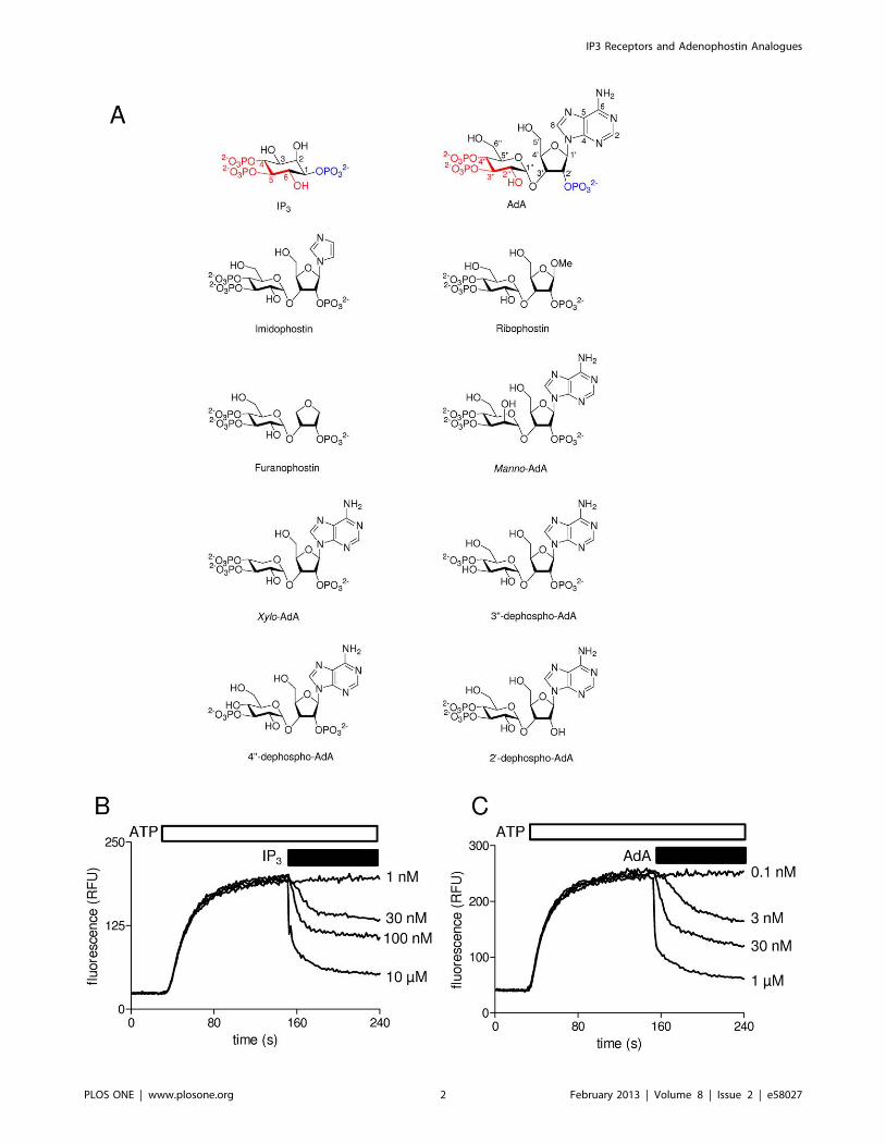
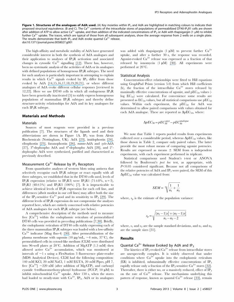
Figure 1. Structures of the analogues of AdA used. (A) Key moieties within IP3 and AdA are highlighted in matching colours to indicate theirproposed structural equivalence. (B and C). The Ca2+ contents of the intracellular stores of populations of permeabilized DT40-IP3R1 cells are shownafter addition of ATP to allow active Ca2+ uptake, and then addition of the indicated concentrations of IP3 or AdA with thapsigargin (1 mM) to inhibitfurther Ca2+ uptake. The traces, which are typical of those from all subsequent analyses, show the average response from 2 wells on a single plate.The results demonstrate that both IP3 and AdA evoke quantal Ca2+ release.doi:10.1371/journal.pone.0058027.g001
IP3 Receptors and Adenophostin Analogues
PLOS ONE | www.plosone.org 3 February 2013 | Volume 8 | Issue 2 | e58027

Ta
ble
1.
Effe
cts
of
Ad
Aan
alo
gu
es
on
Ca2
+re
leas
eb
ysu
bty
pe
so
fIP
3re
cep
tor.
IP3
R1
IP3
R2
IP3
R3
EC
50
pE
C5
0h
Ca
2+ re
lea
sen
EC
50
pE
C5
0h
Ca
2+ re
lea
sen
EC
50
pE
C5
0h
Ca
2+ re
lea
sen
(1,4
,5)I
P3
87
7.0
66
0.0
50
.996
0.0
57
56
13
11
45
6.8
46
0.0
61
.296
0.0
96
16
23
44
17
6.3
86
0.0
51
.266
0.0
76
46
23
0
Ad
A8
.38
.086
0.0
91
.176
0.0
97
26
31
01
8.2
7.7
46
0.0
61
.796
0.2
15
66
21
33
37
.486
0.0
91
.136
0.0
76
16
21
4
Imid
op
ho
stin
37
7.4
36
0.2
81
.176
0.2
17
86
53
68
7.1
76
0.1
41
.846
0.5
05
96
33
16
66
.786
0.1
61
.736
0.3
96
76
73
Rib
op
ho
stin
40
7.4
06
0.2
91
.346
0.1
67
76
43
10
26
.996
0.1
11
.606
0.5
06
16
23
29
56
.536
0.2
11
.426
0.0
86
86
43
Fura
no
ph
ost
in5
17
.296
0.2
50
.906
0.1
07
96
63
76
7.1
26
0.0
11
.736
0.2
06
06
33
45
76
.346
0.1
81
.276
0.2
17
16
33
Ma
nn
o-A
dA
34
7.4
76
0.1
91
.336
0.3
07
56
73
69
7.1
66
0.0
71
.336
0.2
25
76
33
24
56
.616
0.2
31
.236
0.1
56
96
43
Xyl
o-A
dA
5.9
8.2
36
0.1
71
.276
0.2
77
36
73
7.9
8.1
06
0.1
01
.526
0.4
05
26
63
29
7.5
46
0.1
21
.586
0.2
96
46
93
29-
de
ph
osp
ho
-Ad
A2
75
6.5
66
0.1
31
.316
0.1
56
66
73
57
56
.246
0.1
00
.856
0.0
76
36
23
69
26
.166
0.0
31
.56
0.2
25
56
74
30-
de
ph
osp
ho
-Ad
Ac
ND
ND
ND
156
6b
7N
DN
DN
D66
2b
6N
DN
DN
D76
7b
5
40-
de
ph
osp
ho
-Ad
AIn
acti
vea
Inac
tive
aIn
acti
vea
ND
6In
acti
vea
Inac
tive
aIn
acti
vea
ND
6In
acti
vea
Inac
tive
aIn
acti
vea
ND
5
Th
eEC
50
(nM
),p
EC5
0(/
M),
Hill
coe
ffic
ien
t(h
)an
dfr
acti
on
(%)
of
the
intr
ace
llula
rC
a2+
sto
res
rele
ase
db
ya
max
imal
lye
ffe
ctiv
eco
nce
ntr
atio
no
fe
ach
anal
og
ue
are
sho
wn
for
eac
hIP
3R
sub
typ
e.A
llre
sult
s(e
xce
pt
EC5
0)
are
sho
wn
asm
ean
s6
SEM
fro
mn
ind
ep
en
de
nt
exp
eri
me
nts
.aIn
acti
veat
30
0mM
.b
Ca2
+re
leas
ee
voke
db
y3
00
mM3
0-d
ep
ho
sph
oA
dA
.cR
efe
rto
Tab
le2
for
rela
tive
po
ten
cie
so
f3
0-d
ep
ho
sph
oA
dA
.N
D,
no
td
ete
rmin
ed
.d
oi:1
0.1
37
1/j
ou
rnal
.po
ne
.00
58
02
7.t
00
1
IP3 Receptors and Adenophostin Analogues
PLOS ONE | www.plosone.org 4 February 2013 | Volume 8 | Issue 2 | e58027

unclear. It may require desensitization of IP3R as the Ca2+ content
of the ER declines [34] or heterogeneity among IP3-senstive Ca2+
stores [35]. The results shown in Figures 1B and C confirm that
the Ca2+ release evoked by submaximal concentrations of either
IP3 or AdA from permeabilized DT40-IP3R1 cells is quantal.
These observations provide the justification for all subsequent
experiments in which the concentration-dependent effects of IP3
or AdA were measured 30 s after their addition (see Methods).
AdA is a Potent Agonist of All Three IP3 ReceptorSubtypes
The results shown in Figure 2 and Tables 1 and 2 demonstrate
that AdA is ,10-times more potent than IP3 at each IP3R subtype,
and for each subtype, maximally effective concentrations of IP3
and AdA release the same fraction of the intracellular Ca2+ stores.
This is consistent with many analyses of IP3 and AdA in a variety
of cell types using both functional and binding assays, in which
AdA behaves as a full agonist with ,10-fold greater affinity than
IP3 [reviewed in 8]. Our results do, however, provide the first
direct demonstration that AdA interacts similarly with all three
IP3R subtypes. Subsequent experiments examine the interactions
between key analogues of IP3 and AdA with each IP3R subtype.
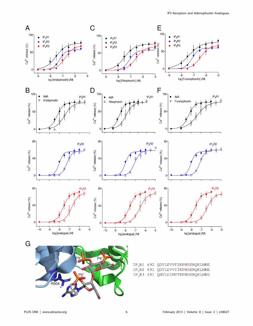
Trimming the Adenosine Moiety of AdA Reduces itsPotency at All IP3 Receptor Subtypes
Systematic trimming of the adenosine moiety of AdA succes-
sively produces imidophostin (which lacks the pyrimidine ring of
AdA), ribophostin (in which a methoxy group replaces the adenine
moiety of AdA) and furanophostin (in which only the furanoid ring
remains) (Figure 1A). Maximally effective concentrations of each
of these analogues released the same fraction of the intracellular
Ca2+ stores as AdA in cells expressing each of the three IP3R
subtypes, and each analogue was ,5-10-fold less potent than AdA
(Figure 3, Tables 1 and 2). These results are consistent with
previous analyses of IP3R in hepatocytes, which express predom-
inantly IP3R2 [24,36], with analyses of binding of ribophostin and
furanophostin to an N-terminal fragment of IP3R1 [12], and with
evidence from other analogues that trimming the adenosine
Figure 2. AdA is a potent agonist of all three IP3 receptorsubtypes. (A) Concentration-dependent effects of AdA on Ca2+ releasefrom the intracellular stores of cells expressing IP3R1, IP3R2 or IP3R3. Allresults are expressed as percentages of the Ca2+ release evoked byionomycin. The same colour codes are used in all subsequent figures.(B) Comparison, for each IP3R subtype, of the Ca2+ release evoked by IP3
and AdA. Results are means 6 SEM from the number of independentexperiments given in Table 1. Here, and in many subsequent figures,some error bars are smaller than the symbols.doi:10.1371/journal.pone.0058027.g002
Table 2. Relative potencies of AdA analogues at different IP3
receptor subtypes.
IP3R1 IP3R2 IP3R3
IP3 1.0260.02 0.960.30 1.160.30
Imidophostin 0.7860.15 0.7860.08 0.8160.04
Ribophostin 0.8260.18 0.9660.20 1.0660.07
Furanophostin 0.9260.13 0.8360.14 1.2560.05
Manno-AdA 0.7460.08 0.7960.18 0.9860.08
Xylo-AdA 20.0160.07 20.360.27 0.0560.08
29-dephospho-AdA 1.2460.33 1.6060.18 1.6860.16
30-dephospho-AdAa
4.0360.09 4.4760.30 4.1360.14
From paired comparisons with AdA, the potency (DpEC50) of the analoguesrelative to AdA is shown for each IP3R subtype. Results are means 6 SEM, with nprovided in Table 1. ND, not determined. aBecause the very low affinity of 30-dephospho AdA for IP3R made it impracticable to stimulate cells with amaximally effective concentration, ‘DpEC50’ for 30-dephospho AdA wasestimated by comparing concentrations of it and AdA that evoked the samesub-maximal Ca2+ release.doi:10.1371/journal.pone.0058027.t002
IP3 Receptors and Adenophostin Analogues
PLOS ONE | www.plosone.org 5 February 2013 | Volume 8 | Issue 2 | e58027
IP3 Receptors and Adenophostin Analogues
PLOS ONE | www.plosone.org 6 February 2013 | Volume 8 | Issue 2 | e58027
moiety decreases affinity for cerebellar IP3R, which are largely
IP3R1 [37].
These results are consistent with our earlier conclusion that the
10-fold greater affinity of AdA relative to IP3 requires the adenine
moiety of AdA positioned to allow it to form a cation-p interaction
with Arg-504 in the a-domain of the IBC of IP3R1, a residue that
is conserved in all IP3R subtypes [8,12] (Figure 3G). We suggest
that this interaction of AdA with IP3R is likely to be similar for all
IP3R subtypes.
Hydroxyl Moieties that are Important for IP3 Binding areLess Important for Binding of AdA
The 50-CH2OH and 20-OH substituents of the glucose ring of
AdA are thought to mimic the 3-OH and 6-OH of IP3,
respectively (Figure 1A). A structure equivalent to the 6-OH of
IP3 is an essential feature of all inositol phosphate analogues that
bind to IP3R [13,38,39] and inversion of its orientation from
equatorial to axial reduces affinity by more than 100-fold at all
IP3R subtypes [40]. It is therefore surprising, but consistent with
previous analyses of native hepatic IP3R [36], that manno-AdA,
which differs from AdA only in the orientation of its 20-OH,
should be only 5- to 10-fold less potent than AdA at each IP3R
subtype (Figures 4A and B, Tables 1 and 2). Why, when the 6-OH
of IP3 and 20-OH of AdA seem to be analogous in the ligand
structures, should these moieties make such different contributions
to the interactions of IP3 and AdA with IP3R?
The 6-OH of IP3 interacts, through a water molecule, with a
lysine residue (K569) in the IBC [41] and, by interacting with the
adjacent 1-phosphate, it has also been proposed to influence the
behaviour of the 4,5-bisphosphate moiety of IP3 [42]. The latter
interaction is unlikely to contribute to AdA binding because the
structures equivalent to the 6-OH (20-OH of AdA) and the 1-
phosphate of IP3 (29-phosphate of AdA) are in different rings in
AdA (Figure 1A). We suggest that the lesser importance in AdA of
a structure equivalent to the essential 6-OH of IP3 comes from this
hydroxyl mediating a relatively minor interaction with K569 in
AdA, whereas for IP3 it contributes also to appropriately orienting
the critical 4,5-bisphosphate moiety.
The 3-OH group, although less important than the 6-OH, is
another feature of IP3 that contributes to high-affinity binding
[43]. Our recent analyses of the functional effects of 3-deoxy-IP3
established that it was ,40-fold less potent than IP3 at all three
IP3R subtypes [7]. This is consistent with earlier work showing
that 3-deoxy-IP3 and analogues with other modifications of the 3-
position have reduced affinity for the three IP3R subtypes [40].
However, the equivalent modification of AdA, removal of its 50-
CH2OH to give xylo-AdA (Figure 1A), had no significant effect on
its potency at any IP3R subtype (Figures 4C and D, Tables 1 and
2). This is consistent with a previous functional analysis of hepatic
IP3R, where xylo-AdA was only marginally less potent than AdA
(DpEC50 ,0.28) [36]. Our results suggest that despite the
apparent structural similarity between the 3-OH of IP3 and the
50-CH2OH of AdA (Figure 1A), the two hydroxyl groups do not
contribute similarly to ligand binding. Previous analyses of IP3
analogues suggested that replacing the 3-OH with the larger
CH2OH moiety caused the affinity to decrease by no more than 7-
fold [40]. A partial explanation for the lack of effect of removing
the 50-CH2OH of AdA may therefore be that this moiety is less
readily accommodated than a hydroxyl group in the IBC. This
would suggest that an analogue of AdA in which the 50-CH2OH is
replaced by 50-OH might bind with increased affinity. We are
unaware of such an analogue having been synthesized. The larger
substituent at the 50-position of AdA is, however, unlikely to
provide the sole explanation for it making no discernible
contribution to binding.
The 29-phosphate of AdA is not a Super-optimal Mimic ofthe 1-phosphate of IP3
It has been suggested that the 29-phosphate of AdA interacts
with the IBC in a manner that allows it to behave as a super-
optimal mimic of the 1-phosphate of IP3 [44,45]. However, our
recent study combining structure-activity analyses with mutagen-
esis of the binding site suggest that the 1-phosphate of IP3 is more
important for binding than is the 29-phosphate of AdA [12].
Removal of the 1-phosphate from IP3 (to give (4,5)IP2) caused its
potency and affinity for IP3R1 to decrease by ,100-fold [12],
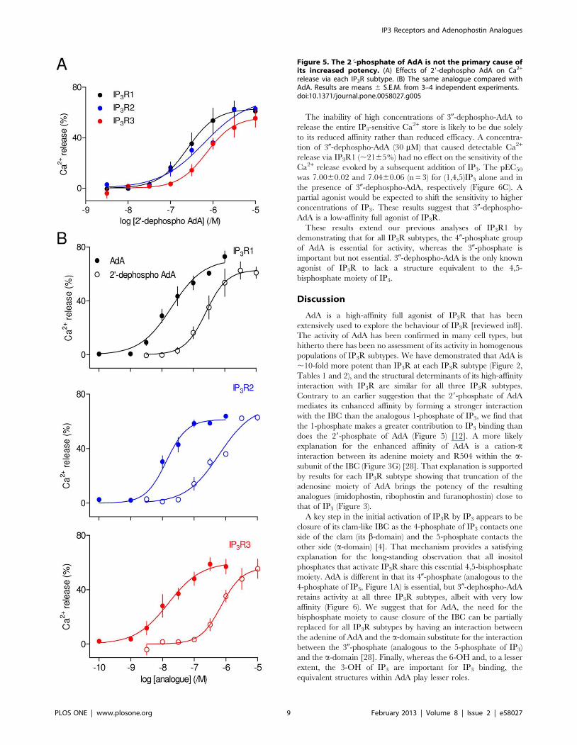
whereas removal of the 29-phosphate from AdA (29-dephospho
AdA) causes a decrease in potency of ,17-fold in IP3R1 (Figure 5)
and ,40-fold decreases in potency were obtained with 29-
dephospho AdA and IP3R2 and IP3R3 (Figure 5, Table 1 and
2). These results establish that for all three IP3R subtypes, the
enhanced affinity of AdA is not due to its 29-phosphate interacting
more effectively than the 1-phosphate of IP3 with the IBC.
A Bisphosphate Moiety is not Essential for Activation ofIP3 Receptors by AdA
All known active analogues of IP3 have structures equivalent to
its 4,5-bisphosphate moiety [13]. Structures of the IBC with and
without IP3 bound provide a rationale for this requirement by
revealing that these two phosphate groups contact opposite sides
(the a- and b-domains) of the clam-like IBC, closure of which
initiates IP3R activation [4,41]. Substantial evidence suggests that
the 40,30-bisphosphate moiety of AdA mimics the critical 4,5-
bisphosphate of IP3 [8] (Figure 1A).
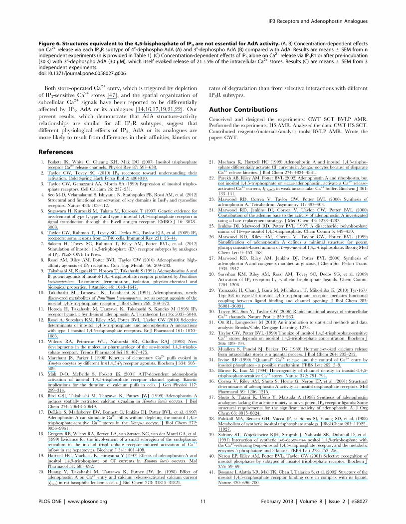
40-dephospho-AdA at concentrations up to 300 mM failed to
evoke Ca2+ release via any IP3R subtype (Figure 6A). This is
consistent with previous analyses by both functional and binding
assays of IP3R1 [28,46]. 30-dephospho-AdA did, however, cause
detectable Ca2+ release albeit with much reduced potency
(Figure 6B). The synthetic route used to prepare 30-dephospho-
AdA makes it extremely unlikely that the activity could be due to
minor contamination with AdA or related structures with a vicinal
bisphosphate moiety. Maximal attainable concentrations of 30-
dephospho-AdA (300 mM) failed to release the entire IP3-sensitive
Ca2+ store, but comparison of the concentrations required to
achieve the same submaximal Ca2+ release suggests that 30-
dephospho-AdA is ,10,000-fold less potent than AdA at all three
IP3R subtypes. With such a massive reduction in potency the lesser
sensitivity of DT40-IP3R3 cells to AdA means that even the
highest practicable concentration of 30-dephospho-AdA (300 mM)
is close to the threshold for detecting Ca2+ release (Figure 6B).
Figure 3. Trimming the adenosine moiety of AdA reduces potency. (A–F) Effects of imidophostin (A), ribophostin (C) and furanophostin (E)on Ca2+ release via each of the three IP3R subtypes, and the same analogues compared with AdA (B, D and F). Results are means 6 S.E.M. from 3independent experiments. (G) A cation-p interaction between the adenine of AdA and R504 within the a-domain of the IBC is proposed to stabilizeAdA binding (left). Closure of the clam-like IBC is proposed to be mediated by interactions between the 30-phosphate of AdA and the a-domain ofthe IBC (blue ribbon), and between the 40-phosphate and the b-domain of the IBC (green ribbon). In 30-dephospho AdA, a cation-p interactionbetween AdA and the IBC a-domain is proposed to be sufficient to allow some effective closure of the clam. R504 is conserved in all threemammalian IP3R subtypes (right).doi:10.1371/journal.pone.0058027.g003
IP3 Receptors and Adenophostin Analogues
PLOS ONE | www.plosone.org 7 February 2013 | Volume 8 | Issue 2 | e58027
Figure 4. Hydroxyl groups within the glucose ring of AdA are unimportant. (A–D) Effects of manno-AdA (A) and xylo-AdA (C) on Ca2+
release via each IP3R subtype, and the same analogues compared with AdA (B and D). Results are means 6 S.E.M. from 3 independent experiments.doi:10.1371/journal.pone.0058027.g004
IP3 Receptors and Adenophostin Analogues
PLOS ONE | www.plosone.org 8 February 2013 | Volume 8 | Issue 2 | e58027
The inability of high concentrations of 30-dephospho-AdA to
release the entire IP3-sensitive Ca2+ store is likely to be due solely
to its reduced affinity rather than reduced efficacy. A concentra-
tion of 30-dephospho-AdA (30 mM) that caused detectable Ca2+
release via IP3R1 (,2165%) had no effect on the sensitivity of the
Ca2+ release evoked by a subsequent addition of IP3. The pEC50
was 7.0060.02 and 7.0460.06 (n = 3) for (1,4,5)IP3 alone and in
the presence of 30-dephospho-AdA, respectively (Figure 6C). A
partial agonist would be expected to shift the sensitivity to higher
concentrations of IP3. These results suggest that 30-dephospho-
AdA is a low-affinity full agonist of IP3R.
These results extend our previous analyses of IP3R1 by
demonstrating that for all IP3R subtypes, the 40-phosphate group
of AdA is essential for activity, whereas the 30-phosphate is
important but not essential. 30-dephospho-AdA is the only known
agonist of IP3R to lack a structure equivalent to the 4,5-
bisphosphate moiety of IP3.
Discussion
AdA is a high-affinity full agonist of IP3R that has been
extensively used to explore the behaviour of IP3R [reviewed in8].
The activity of AdA has been confirmed in many cell types, but
hitherto there has been no assessment of its activity in homogenous
populations of IP3R subtypes. We have demonstrated that AdA is
,10-fold more potent than IP3R at each IP3R subtype (Figure 2,
Tables 1 and 2), and the structural determinants of its high-affinity
interaction with IP3R are similar for all three IP3R subtypes.
Contrary to an earlier suggestion that the 29-phosphate of AdA
mediates its enhanced affinity by forming a stronger interaction
with the IBC than the analogous 1-phosphate of IP3, we find that
the 1-phosphate makes a greater contribution to IP3 binding than
does the 29-phosphate of AdA (Figure 5) [12]. A more likely
explanation for the enhanced affinity of AdA is a cation-pinteraction between its adenine moiety and R504 within the a-
subunit of the IBC (Figure 3G) [28]. That explanation is supported
by results for each IP3R subtype showing that truncation of the
adenosine moiety of AdA brings the potency of the resulting
analogues (imidophostin, ribophostin and furanophostin) close to
that of IP3 (Figure 3).
A key step in the initial activation of IP3R by IP3 appears to be
closure of its clam-like IBC as the 4-phosphate of IP3 contacts one
side of the clam (its b-domain) and the 5-phosphate contacts the
other side (a-domain) [4]. That mechanism provides a satisfying
explanation for the long-standing observation that all inositol
phosphates that activate IP3R share this essential 4,5-bisphosphate
moiety. AdA is different in that its 40-phosphate (analogous to the
4-phosphate of IP3, Figure 1A) is essential, but 30-dephospho-AdA
retains activity at all three IP3R subtypes, albeit with very low
affinity (Figure 6). We suggest that for AdA, the need for the
bisphosphate moiety to cause closure of the IBC can be partially
replaced for all IP3R subtypes by having an interaction between
the adenine of AdA and the a-domain substitute for the interaction
between the 30-phosphate (analogous to the 5-phosphate of IP3)
and the a-domain [28]. Finally, whereas the 6-OH and, to a lesser
extent, the 3-OH of IP3 are important for IP3 binding, the
equivalent structures within AdA play lesser roles.
Figure 5. The 29-phosphate of AdA is not the primary cause ofits increased potency. (A) Effects of 29-dephospho AdA on Ca2+
release via each IP3R subtype. (B) The same analogue compared withAdA. Results are means 6 S.E.M. from 3–4 independent experiments.doi:10.1371/journal.pone.0058027.g005
IP3 Receptors and Adenophostin Analogues
PLOS ONE | www.plosone.org 9 February 2013 | Volume 8 | Issue 2 | e58027

IP3 Receptors and Adenophostin Analogues
PLOS ONE | www.plosone.org 10 February 2013 | Volume 8 | Issue 2 | e58027
Both store-operated Ca2+ entry, which is triggered by depletion
of IP3-sensitive Ca2+ stores [47], and the spatial organization of
subcellular Ca2+ signals have been reported to be differentially
affected by IP3, AdA or its analogues [14,16,17,19,21,22]. Our
present results, which demonstrate that AdA structure-activity
relationships are similar for all IP3R subtypes, suggest that
different physiological effects of IP3, AdA or its analogues are
more likely to result from differences in their affinities, kinetics or
rates of degradation than from selective interactions with different
IP3R subtypes.
Author Contributions
Conceived and designed the experiments: CWT SCT BVLP AMR.
Performed the experiments: HS AMR. Analyzed the data: CWT HS SCT.
Contributed reagents/materials/analysis tools: BVLP AMR. Wrote the
paper: CWT.
References
1. Foskett JK, White C, Cheung KH, Mak DO (2007) Inositol trisphosphate
receptor Ca2+ release channels. Physiol Rev 87: 593–658.
2. Taylor CW, Tovey SC (2010) IP3 receptors: toward understanding their
activation. Cold Spring Harb Persp Biol 2: a004010.
3. Taylor CW, Genazzani AA, Morris SA (1999) Expression of inositol trispho-
sphate receptors. Cell Calcium 26: 237–251.
4. Seo M-D, Velamakanni S, Ishiyama N, Stathopulos PB, Rossi AM, et al. (2012)
Structural and functional conservation of key domains in InsP3 and ryanodine
receptors. Nature 483: 108–112.
5. Sugawara H, Kurosaki M, Takata M, Kurosaki T (1997) Genetic evidence for
involvement of type 1, type 2 and type 3 inositol 1,4,5-trisphosphate receptors in
signal transduction through the B-cell antigen receptor. EMBO J 16: 3078–
3088.
6. Taylor CW, Rahman T, Tovey SC, Dedos SG, Taylor EJA, et al. (2009) IP3
receptors: some lessons from DT40 cells. Immunol Rev 231: 23–44.
7. Saleem H, Tovey SC, Rahman T, Riley AM, Potter BVL, et al. (2012)
Stimulation of inositol 1,4,5-trisphosphate (IP3) receptor subtypes by analogues
of IP3. PLoS ONE In Press.
8. Rossi AM, Riley AM, Potter BVL, Taylor CW (2010) Adenophostins: high-
affinity agonists of IP3 receptors. Curr Top Membr 66: 209–233.
9. Takahashi M, Kagasaki T, Hosoya T, Takahashi S (1994) Adenophostins A and
B: potent agonists of inositol-1,4,5-trisphosphate receptor produced by Penicillium
brevicompactum. Taxonomy, fermentation, isolation, physico-chemical and
biological properties. J Antibiot 46: 1643–1647.
10. Takahashi M, Tanzawa K, Takahashi S (1994) Adenophostins, newly
discovered metabolites of Penicillium brevicompactum, act as potent agonists of the
inositol 1,4,5-trisphosphate receptor. J Biol Chem 269: 369–372.
11. Hotoda H, Takahashi M, Tanzawa K, Takahashi S, Kaneko M (1995) IP3
receptor ligand.1: Synthesis of adenophostin A. Tetrahedon Lett 36: 5037–5040.
12. Rossi A, Sureshan KM, Riley AM, Potter BVL, Taylor CW (2010) Selective
determinants of inositol 1,4,5-trisphosphate and adenophostin A interactions
with type 1 inositol 1,4,5-trisphosphate receptors. Br J Pharmacol 161: 1070–
1085.
13. Wilcox RA, Primrose WU, Nahorski SR, Challiss RAJ (1998) New
developments in the molecular pharmacology of the myo-inositol 1,4,5-trispho-
sphate receptor. Trends Pharmacol Sci 19: 467–475.
14. Marchant JS, Parker I (1998) Kinetics of elementary Ca2+ puffs evoked in
Xenopus oocytes by different Ins(1,4,5)P3 receptor agonists. Biochem J 334: 505–
509.
15. Mak D-O, McBride S, Foskett JK (2001) ATP-dependent adenophostin
activation of inositol 1,4,5-trisphosphate receptor channel gating. Kinetic
implications for the duration of calcium puffs in cells. J Gen Physiol 117:
299–314.
16. Bird GSJ, Takahashi M, Tanzawa K, Putney JWJ (1999) Adenophostin A
induces spatially restricted calcium signaling in Xenopus laevis occytes. J Biol
Chem 274: 20643–20649.
17. DeLisle S, Marksberry EW, Bonnett C, Jenkins DJ, Potter BVL, et al. (1997)
Adenophostin A can stimulate Ca2+ influx without depleting the inositol 1,4,5-
trisphosphate-sensitive Ca2+ stores in the Xenopus oocyte. J Biol Chem 272:
9956–9961.
18. Gregory RB, Wilcox RA, Berven LA, van Straten NC, van der Marel GA, et al.
(1999) Evidence for the involvement of a small subregion of the endoplasmic
reticulum in the inositol trisphosphate receptor-induced activation of Ca2+
inflow in rat hepatocytes. Biochem J 341: 401–408.
19. Hartzell HC, Machaca K, Hirayama Y (1997) Effects of adenophostin-A and
inositol 1,4,5-trisphosphate on Cl- currents in Xenopus laevis oocytes. Mol
Pharmacol 51: 683–692.
20. Huang Y, Takahashi M, Tanzawa K, Putney JW, Jr. (1998) Effect of
adenophostin A on Ca2+ entry and calcium release-activated calcium current
(Icrac) in rat basophilic leukemia cells. J Biol Chem 273: 31815–31821.
21. Machaca K, Hartzell HC (1999) Adenophostin A and inositol 1,4,5-trispho-
sphate differentially activate Cl- currents in Xenopus oocytes because of disparateCa2+ release kinetics. J Biol Chem 274: 4824–4831.
22. Parekh AB, Riley AM, Potter BVL (2002) Adenophostin A and ribophostin, butnot inositol 1,4,5-trisphosphate or manno-adenophostin, activate a Ca2+ release-
activated Ca2+ current, ICRAC, in weak intracellular Ca2+ buffer. Biochem J 361:133–141.
23. Marwood RD, Correa V, Taylor CW, Potter BVL (2000) Synthesis ofadenophostin A. Tetrahedron: Asymmetry 11: 397–403.
24. Marwood RD, Jenkins DJ, Correa V, Taylor CW, Potter BVL (2000)Contribution of the adenine base to the activity of adenophostin A investigated
using a base replacement strategy. J Med Chem 43: 4278–4287.
25. Jenkins DJ, Marwood RD, Potter BVL (1997) A disaccharide polyphosphate
mimic of 1D-myo-inositol 1,4,5-trisphosphate. Chem Comm 5: 449–450.
26. Marwood RD, Riley AM, Correa V, Taylor CW, Potter BVL (1999)
Simplification of adenophostin A defines a minimal structure for potentglucopyranoside-based mimics of D-myo-inositol 1,4,5-trisphosphate. Bioorg Med
Chem Lett 9: 453–458.
27. Marwood RD, Riley AM, Jenkins DJ, Potter BVL (2000) Synthesis of
adenophostin A and congeners modified at glucose. J Chem Soc Perkin Trans:1935–1947.
28. Sureshan KM, Riley AM, Rossi AM, Tovey SC, Dedos SG, et al. (2009)
Activation of IP3 receptors by synthetic bisphosphate ligands. Chem Comm:
1204–1206.
29. Yamazaki H, Chan J, Ikura M, Michikawa T, Mikoshiba K (2010) Tyr-167/
Trp-168 in type1/3 inositol 1,4,5-trisphosphate receptor mediates functionalcoupling between ligand binding and channel opening. J Biol Chem 285:
36081–36091.
30. Tovey SC, Sun Y, Taylor CW (2006) Rapid functional assays of intracellular
Ca2+ channels. Nature Prot 1: 259–263.
31. Ott RL, Longnecker M (2010) An introduction to statistical methods and dataanalysis: Brooks/Cole, Cengage Learning. 1273.
32. Taylor CW, Potter BVL (1990) The size of inositol 1,4,5-trisphosphate-sensitiveCa2+ stores depends on inositol 1,4,5-trisphosphate concentration. Biochem J
266: 189–194.
33. Muallem S, Pandol SJ, Beeker TG (1989) Hormone-evoked calcium release
from intracellular stores is a quantal process. J Biol Chem 264: 205–212.
34. Irvine RF (1990) ‘‘Quantal’’ Ca2+ release and the control of Ca2+ entry by
inositol phosphates - a possible mechanism. FEBS Lett 262: 5–9.
35. Hirose K, Iino M (1994) Heterogeneity of channel density in inositol-1,4,5-
trisphosphate-sensitive Ca2+ stores. Nature 372: 791–794.
36. Correa V, Riley AM, Shuto S, Horne G, Nerou EP, et al. (2001) Structuraldeterminants of adenophostin A activity at inositol trisphosphate receptors. Mol
Pharmacol 59: 1206–1215.
37. Shuto S, Tatani K, Ueno Y, Matsuda A (1998) Synthesis of adenophostin
analogues lacking the adenine moiety as novel potent IP3 receptor ligands: Some
structural requirements for the significant activity of adenophostin A. J OrgChem 63: 8815–8824.
38. Polokoff MA, Bencen GH, Vacca JP, se Solms SJ, Young SD, et al. (1988)
Metabolism of synthetic inositol trisphosphate analogs. J Biol Chem 263: 11922–
11927.
39. Safrany ST, Wojcikiewicz RJH, Strupish J, Nahorski SR, Dubreuil D, et al.(1991) Interaction of synthetic D-6-deoxy-myo-inositol 1,4,5-trisphosphate with
the Ca2+-releasing D-myo-inositol 1,4,5-trisphosphate receptor, and the metabolic
enzymes 5-phosphatase and 3-kinase. FEBS Lett 278: 252–256.
40. Nerou EP, Riley AM, Potter BVL, Taylor CW (2001) Selective recognition ofinositol phosphates by subtypes of inositol trisphosphate receptor. Biochem J
355: 59–69.
41. Bosanac I, Alattia J-R, Mal TK, Chan J, Talarico S, et al. (2002) Structure of the
inositol 1,4,5-trisphosphate receptor binding core in complex with its ligand.
Nature 420: 696–700.
Figure 6. Structures equivalent to the 4,5-bisphosphate of IP3 are not essential for AdA activity. (A, B) Concentration-dependent effectson Ca2+ release via each IP3R subtype of 40-dephospho AdA (A) and 30-dephospho AdA (B) compared with AdA. Results are means 6 SEM from nindependent experiments (n is provided in Table 1). (C) Concentration-dependent effects of IP3 alone on Ca2+ release via IP3R1 or after pre-incubation(30 s) with 30-dephospho AdA (30 mM), which itself evoked release of 2165% of the intracellular Ca2+ stores. Results (C) are means 6 SEM from 3independent experiments.doi:10.1371/journal.pone.0058027.g006
IP3 Receptors and Adenophostin Analogues
PLOS ONE | www.plosone.org 11 February 2013 | Volume 8 | Issue 2 | e58027

42. Felemez M, Ballereau S, Schlewer G, Spiess B (2000) Inframolecular
protonation process of 6-modified myo-inositol 1,4,5-tris(phosphates): substitutioneffects on the cooperativity between the phosphate groups. New J Chem 24:
631–638.
43. Wilcox RA, Challiss RAJ, Traynor JR, Fauq AH, Ognayanov VI, et al. (1994)Molecular recognition at the myo-inositol 1,4,5-trisphosphate receptor. 3-position
substituted myo-inositol 1,4,5-trisphosphate analogues reveal the binding andCa2+ release requirements for high affinity interaction with the myo-inositol 1,4,5-
trisphosphate receptor. J Biol Chem 269: 26815–26821.
44. Takahashi S, Takeshi K, Takahashi M (1994) Adenophostins A and B: potentagonists of inositol-1,4,5-trisphosphate receptors produced by Penicillium
brevicompactum. Structure elucidation. J Antibiot 47: 95–100.
45. Hotoda H, Murayama K, Miyamoto S, Iwata Y, Takahashi M, et al. (1999)
Molecular recognition of adenophostin, a very potent Ca2+ inducer, at the D-
myo-inositol 1,4,5-trisphosphate receptor. Biochemistry 38: 9234–9241.
46. Sureshan KM, Riley AM, Thomas MP, Tovey SC, Taylor CW, et al. (2012)
Contribution of phosphates and adenine to the potency of adenophostins at the
IP3 receptor: synthesis of all possible bisphosphates of adenophostin A. J Med
Chem 55: 1706–1720.
47. Putney JW (2009) Capacitative calcium entry: from concept to molecules.
Immunol Rev 231: 10–22.
IP3 Receptors and Adenophostin Analogues
PLOS ONE | www.plosone.org 12 February 2013 | Volume 8 | Issue 2 | e58027