Sticks and Stones : the nature, prevalence and significance of trauma in the child.
34
1 Sticks and stones: exploring the nature and significance of child trauma in the past MARY E. LEWIS Department of Archaeology, University of Reading, Whiteknights, Reading, Berkshire, RG6 6AB. United Kingdom. 1. Introduction The identification of trauma in non-adult skeletons (aged <17 years) is limited compared to the rates recorded in adult samples. One of the reasons for this is that fractures behave differently in children. It is not that children did not suffer injury in the past, but the nature of immature bone and rapid repair can mask the subtle changes, meaning rates of non-adult trauma are almost certainly an underestimate. The most common forms of injury in the child today are due to motor vehicle accidents, falls (5-10 year olds), intentional abuse (infants), and recreational sports injuries (adolescents) (Wilber and Thompson 1998). In the past, play, occupation, warfare and physical abuse all exposed children to trauma. Although the causes behind skeletal injuries may have changed over time, the nature of paediatric bone and its reaction to trauma has not. An examination of the type and distribution of trauma in children from past societies may help us to identify the nature of activity in which these children were engaged. Some cases of physical injury in the child, documented in the past will be invisible to the osteologist; these include drowning, burns, ingestion of a foreign object or choking. Glencross (2011) emphasizes the importance of examining trauma patterns from a life course perspective, matching social age with cultural agency which dictates when certain activities take place. The biological and social development of the child will also influence the types of trauma to which they will be exposed. Between the ages of 0-2 years, children are almost entirely dependent on adults for their well-being and can suffer from abuse and neglect; as they develop physically and become more independent, the child’s social and cultural involvement increases. Therefore, today, long bone fractures in children before the age of 2 years are suspicious of abuse, whereas in children aged 2-3 years fractures may occur as part of the process of learning to walk and climb (Brown and Fisher 2004). Trauma is also culturally defined and types of injury may vary considerably across populations due to socio-economic conditions, the levels of urbanisation, subsistence strategies, technological advances, cultural practice and other population characteristics
Transcript of Sticks and Stones : the nature, prevalence and significance of trauma in the child.

1
Sticks and stones: exploring the nature and significance of child
trauma in the past
MARY E. LEWIS
Department of Archaeology, University of Reading, Whiteknights, Reading, Berkshire, RG6
6AB. United Kingdom.
1. Introduction
The identification of trauma in non-adult skeletons (aged <17 years) is limited compared to
the rates recorded in adult samples. One of the reasons for this is that fractures behave
differently in children. It is not that children did not suffer injury in the past, but the nature of
immature bone and rapid repair can mask the subtle changes, meaning rates of non-adult
trauma are almost certainly an underestimate. The most common forms of injury in the child
today are due to motor vehicle accidents, falls (5-10 year olds), intentional abuse (infants),
and recreational sports injuries (adolescents) (Wilber and Thompson 1998). In the past, play,
occupation, warfare and physical abuse all exposed children to trauma. Although the causes
behind skeletal injuries may have changed over time, the nature of paediatric bone and its
reaction to trauma has not. An examination of the type and distribution of trauma in children
from past societies may help us to identify the nature of activity in which these children were
engaged. Some cases of physical injury in the child, documented in the past will be invisible
to the osteologist; these include drowning, burns, ingestion of a foreign object or choking.
Glencross (2011) emphasizes the importance of examining trauma patterns from a life
course perspective, matching social age with cultural agency which dictates when certain
activities take place. The biological and social development of the child will also influence the
types of trauma to which they will be exposed. Between the ages of 0-2 years, children are
almost entirely dependent on adults for their well-being and can suffer from abuse and
neglect; as they develop physically and become more independent, the child’s social and
cultural involvement increases. Therefore, today, long bone fractures in children before the
age of 2 years are suspicious of abuse, whereas in children aged 2-3 years fractures may
occur as part of the process of learning to walk and climb (Brown and Fisher 2004).
Trauma is also culturally defined and types of injury may vary considerably across
populations due to socio-economic conditions, the levels of urbanisation, subsistence
strategies, technological advances, cultural practice and other population characteristics
2
(Cheng and Shen 1993). This can render broad comparisons across sites and time periods
meaningless. Most of the information we can gather about the incidence, type and pattern of
fractures are derived from modern clinical literature and tends to be the result of child
recreational activity that did not exist in the past, so male to female ratios and age at which
fractures occurred may not be relevant. It is a necessity that we glean what we can from
reported cases of child trauma in the archaeological literature, whilst being aware that these
may only represent a snapshot of the factors that surrounded the trauma, and in many cases
are isolated events. Given the paucity of data for child trauma in the past, we need to be
cautious about how we interpret the lack of evidence. For example, Glencross (2011) takes
the absence of fractures in the infants from the Indian Knoll in Kentucky as highlighting the
importance of family responsibility and social care of the child. This may certainly be the
case, but in order to make such statements, we need first to be sure that we are adequately
equipped to identify trauma in such tiny remains. We should also be aware that over-
reliance on the most obvious cases of child fracture, due to our inability to identify milder
forms, may misrepresent those most at risk from trauma, or highlight more extreme
accidents over every day injuries (Glencross and Stuart-Macadam 2001). This chapter will
first outline the mechanics behind the child trauma before reviewing what can be gleaned
from data compiled from a survey of published and unpublished injuries from across the
world. By taking a more holistic view of the data an attempt will be made to explore what the
types and rates of child trauma reveal about how they were raised, the dangers to which
they were exposed, when they were put to work, and if, or at what age they became
engaged in warfare.
2. Principles of paediatric trauma
Differences in the pattern and nature of pediatric trauma arise from factors associated with
the child’s size, anatomy and continuous growth. For example, a small child hit by a moving
vehicle (car, cart) is in much greater danger of serious injury than an older child or adult.
Firstly, they are lighter and more likely to become a projectile, sustaining further injury when
they hit the ground (Wilber and Thompson 1998). The ribs of a young child do not cover the
liver, spleen or intestines and the bladder is distended. When an adult falls, they tend to land
on their feet, causing fractures of the lower extremities, but a disproportionately large and
heavy cranium means that a child is more likely to land head first (Wilber and Thompson
1998). In toddlers, their upper limbs are too short to protect their heads on impact resulting in
a higher prevalence of cranial fractures. In older children, landing onto an outstretched hand
accounts for the larger number of radial fractures as the result of falls (Johnston and Foster
2001). Injury to a child from a fall or collision is much more likely to cause fatal soft tissue

3
injuries and peri-mortem fractures that are much more difficult to identify. Children’s bones
are highly cartilaginous and more plastic than the relatively brittle bones of an adult (Resnick
and Kransdorf 2005), meaning that greater force is needed to produce a complete fracture.
When fractures do occur they usually heal quickly without deformity, making them difficult to
detect (Currey and Butler 1975). The periosteum in a child is also thicker, stronger and more
biologically active than an adult’s due to the need for constant remodelling during growth
(Wilber and Thompson 1998). Although the periosteum is firmly attached to the ends of the
bone (metaphyses) through a dense network of fibres (zone of Ranvier), it is more loosely
attached to the shaft (diaphysis). This has an influence on the nature of pediatric trauma, as
the periosteum is less likely to rupture during a fracture but instead separates from the bone
more easily, remaining intact on the compressed side of the break. This lessens the extent of
deformity and allows tissue continuity that bridges the fracture gap and provides stability for
healing (Johnston and Foster 2001:29).
The most common forms of fracture in children are:
a) greenstick fractures, a partial fracture with bowing of the compressed side and
fracture of the tensile side,
b) plastic deformation, causing unusual bowing without fracture,
c) torus or buckle fractures, resulting in a bulge of the metaphyses as it fails under
compression, and
d) chondral and osteochondral fractures of the growth plate
A ‘greenstick’ fracture describes a partial fracture that penetrates the cortex ceasing within
the medullary cavity. These are the most common form of fracture seen in children, and
result from the lower elastic but higher plastic threshold of pediatric bone, causing it to break
more easily and with less force than mature bone. The porous nature of the cortex means
that some of the force is deflected from the surface of the bone, increasing the amount of
pressure needed to cause a fracture (Currey and Butler 1975). However, this load is still not
enough to force the fracture through the entire shaft and instead the force is dissipated
through transverse cleavage cracks, limiting its progression through the bone (Currey and
Butler 1975). The loose attachment of the periosteum to the cortex will often result in a
widespread haematoma and large callus formation along the shaft, with limited evidence of a
fracture line (Wilber and Thompson 1998). In modern cases, greenstick fractures are
common at the proximal metaphysis or diaphysis of the tibia, or the middle third of the radius
and ulna (Resnick and Kransdorf 2005). The porous nature of the metaphyseal ends,
together with a thinner cortical layer, makes this area of the bone particularly susceptible to
trauma (Currey and Butler 1975). Greenstick fractures are rarely recorded in non-adult

4
archaeological remains and this is due to their lack of deformation and ability to heal quickly.
One possible sign of an underlying greenstick fracture is the presence of a sheath of sub-
periosteal new bone (figure 1) but as in clinical medicine, these large deposits are more
likely to be interpreted as infection, vitamin C deficiency (scurvy) or bone tumours (Adams
and Hamblen 1991, John, Moorthy, and Swischuk 1997).
Described as the ‘greenest of greenstick fractures’ (Stuart-Macadam, Glencross, and Kricun
1998:260), plastic deformation or traumatic bowing of the long bones in children can occur
without the subsequent formation of sub-periosteal new bone (Borden 1974, John, Moorthy,
and Swischuk 1997). Acute plastic deformation results from excessive vertical compression
force along the shaft of the bone causing it to react in an elastic manner, bowing under the
force. If the force is removed, the bone returns to normal, but if it persists the bone will either
remain bowed (plastic deformation) with numerous micro-fractures occurring along the
convex aspect, or the bone will suffer partial or complete fracture (Borden 1974). Resnick
(1995) reports that the magnitude of force required to produce plastic deformation in children
can be as much as 100-150% of the child’s own body weight. Plastic deformation usually
occurs in one of the paired bones (i.e. radius and ulna or tibia and fibula) with lateral and
antero-posterior bending of the bone, but is most common in the ulna (Resnick and
Kransdorf 2005, Stuart-Macadam, Glencross, and Kricun 1998). These injuries cause many
clinical challenges, for example, in the leg, plastic deformation of the fibula occurs when the
force penetrating the tibia is absorbed by the interosseous membrane. This permanent
bowing deformity often limits attempts to reduce full tibial fractures (John, Moorthy, and
Swischuk 1997), producing a long term angular deformity. Permanent deformation of a
paired bone may also prevent relocation of a dislocation (Resnick and Kransdorf 2005: 809).
In young children, rapid remodelling will correct the deformity, but in children who sustain
injuries after ten years of age the deformity may persist, causing a reduction in pronation and
supination of the forearm and angular deformity (Borden 1974). Archaeological cases of
plastic deformation are rare and require comparison with the unaffected side and
consideration of numerous differential diagnoses. These include vitamin D deficiency
(rickets), neonatal bowing, osteogenesis imperfecta and post-mortem deformation (Stuart-
Macadam, Glencross, and Kricun 1998).
Torus (buckle) fractures are caused when there is an insufficient impaction or compression
force to cause a complete fracture, but instead the cortex ‘buckles’ (Resnick and Kransdorf
2005). These types of trauma usually occur at long bone metaphyses and might be identified
5
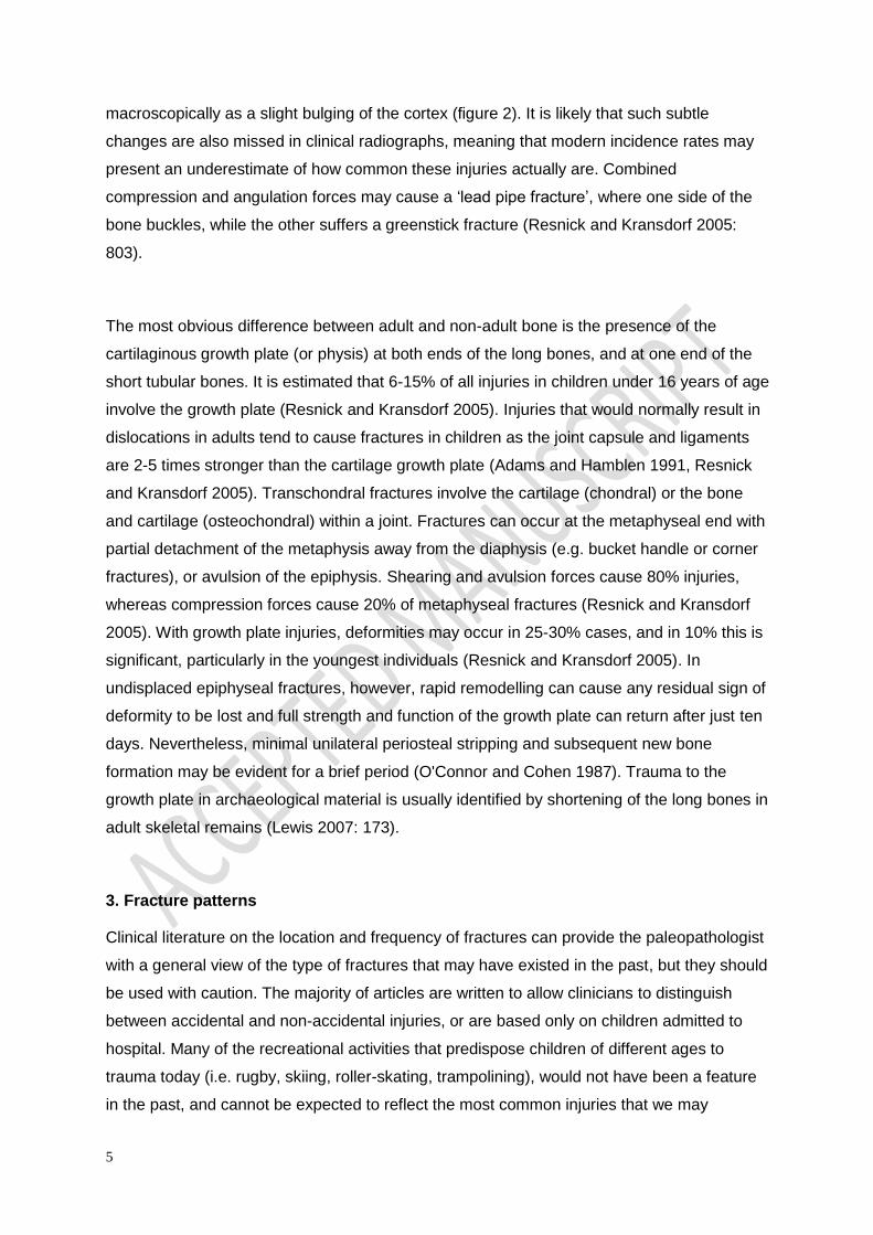
macroscopically as a slight bulging of the cortex (figure 2). It is likely that such subtle
changes are also missed in clinical radiographs, meaning that modern incidence rates may
present an underestimate of how common these injuries actually are. Combined
compression and angulation forces may cause a ‘lead pipe fracture’, where one side of the
bone buckles, while the other suffers a greenstick fracture (Resnick and Kransdorf 2005:
803).
The most obvious difference between adult and non-adult bone is the presence of the
cartilaginous growth plate (or physis) at both ends of the long bones, and at one end of the
short tubular bones. It is estimated that 6-15% of all injuries in children under 16 years of age
involve the growth plate (Resnick and Kransdorf 2005). Injuries that would normally result in
dislocations in adults tend to cause fractures in children as the joint capsule and ligaments
are 2-5 times stronger than the cartilage growth plate (Adams and Hamblen 1991, Resnick
and Kransdorf 2005). Transchondral fractures involve the cartilage (chondral) or the bone
and cartilage (osteochondral) within a joint. Fractures can occur at the metaphyseal end with
partial detachment of the metaphysis away from the diaphysis (e.g. bucket handle or corner
fractures), or avulsion of the epiphysis. Shearing and avulsion forces cause 80% injuries,
whereas compression forces cause 20% of metaphyseal fractures (Resnick and Kransdorf
2005). With growth plate injuries, deformities may occur in 25-30% cases, and in 10% this is
significant, particularly in the youngest individuals (Resnick and Kransdorf 2005). In
undisplaced epiphyseal fractures, however, rapid remodelling can cause any residual sign of
deformity to be lost and full strength and function of the growth plate can return after just ten
days. Nevertheless, minimal unilateral periosteal stripping and subsequent new bone
formation may be evident for a brief period (O'Connor and Cohen 1987). Trauma to the
growth plate in archaeological material is usually identified by shortening of the long bones in
adult skeletal remains (Lewis 2007: 173).
3. Fracture patterns
Clinical literature on the location and frequency of fractures can provide the paleopathologist
with a general view of the type of fractures that may have existed in the past, but they should
be used with caution. The majority of articles are written to allow clinicians to distinguish
between accidental and non-accidental injuries, or are based only on children admitted to
hospital. Many of the recreational activities that predispose children of different ages to
trauma today (i.e. rugby, skiing, roller-skating, trampolining), would not have been a feature
in the past, and cannot be expected to reflect the most common injuries that we may

6
observe in archaeological samples. However, the physical development of a child will
determine some characteristics of trauma prevalence. It is not the aim of this chapter to
discuss trauma associated with physical abuse in any great detail (instead see Lewis 2007),
but rather this will be referred to in general.
3.1 Cranial fractures
Adults and adolescents have rigid, unyielding crania that differ significantly in their pattern of
injuries compared to those of infants and younger children, whose crania are elastic in
nature and consist of flat bones, loosely joined by sutures and fontanelles until around 4
years of age (Pudenz, Todd, and Shelden 1961). As infant crania are soft, they may depress
inwards but not fracture as the result of trauma, known as a ‘ping-pong’ injury, or the sutures
may separate (diastatic fracture). In both cases, because the dura mater is so firmly attached
to the inner surface of the crania in a child, it is often torn directly beneath the fracture line
making them more susceptible to subdural hematomas. Linear fractures of the cranial vault
usually heal quickly, whereas fractures to the much firmer cranial base often go unnoticed in
clinical cases (Pudenz, Todd, and Shelden 1961). Today, the presence of a cranial fracture
in children under 2 years is considered highly indicative of abuse (Hobbs 1984) and Meservy
and colleagues (1987) argued that multiple cranial fractures, fractures that crossed sutures
and bilateral cranial fractures occurred more commonly in victims of abuse, although it is
increasingly being recognised that complex cranial fractures can also occur accidentally
(Wood et al. 2009). Archaeologically, the problem lies with distinguishing linear or
depression fractures, suture separation or bone displacement, from breaks and warping
caused post-mortem (Crist et al. 1997). As young crania are very thin and fragile, they are
often fragmentary, preventing detailed observations of pathology. In the past, the use of a
crochet to extract a child from the womb (Eccles 1982) may have resulted in peri-mortem cut
marks to the orbits and palatine surface of the maxilla, or from the mid-16th century, forceps
may have caused crush fractures to the frontal, parietal or occipital bone (Lewis 2007;
Rushton 1991). No such injuries have ever been identified archaeologically.
3.2 Long bone fractures
The prevalence of fractures to the long bones of children can be divided into several age
categories that reflect their growing level of mobility, independence and choice of activity. In
young children, this is best illustrated by the study of Agran and colleagues (2003) who
7
examined causes of injury in a cross-section of 23,173 children aged between 0-3 years in
California. In children aged 0-2 months, a fall from a height was the leading cause of injury
(e.g. being dropped), at 3-5 months battering resulted in the greatest number of fractures,
whereas between 6-8 months children tended to fall from furniture, at 9-11 months they
choked on foreign objects, they sustained more burns at 12-17 months, and from 2 to 3.5
years most of the injuries were due to mobility; for example falls from furniture, stairs and
buildings. The incidence of drowning also increased between the ages of 1 and 2.5 years.
Cheng and Shen (1993) examined the pattern of fractures in children from different age
categories, this time in 3350 children from Hong Kong. In their sample, 65% of the fractures
were of the forearm and elbow (distal radius, and supracondyle of the humerus) followed by
tibial fractures. Boys sustained more fractures than girls and this divide increased with age,
and was especially evident during adolescence, with female fractures dropping slightly and
males increasing dramatically. Hand fractures increased in the 12-16 year age group as did
fractures of the tibia and ankle.
3.3 Toddlers’ fractures
Today, the most common single fractured bones in children are of the radius and humerus
(Wilkins and Aroojis 2001:12). In many studies, long bone shaft fractures are considered
indicative of abuse in a child younger than 18 months, who is not yet mobile (Brown and
Fisher 2004, Coffey et al. 2005, Strait, Siegal, and Shapiro 1995). For example, Coffey and
colleagues (2005) reported that 75% of children in their study with lower extremity injuries
were victims of physical abuse. However, the pattern for shaft and distal humerus fractures
is less clear cut, with Strait, Siegal, and Shapiro (1995) finding fractures as the result of
abuse in only 36% of children under 15 months, and Shaw and Bohrer (1979) advise caution
when diagnosing abuse based on age or fracture patterning. After 2 years of age, children
are learning to walk and climb and are more likely to sustain fractures accidentally. A typical
fracture of this period commonly occurs at the distal tibia and is known as the ‘toddler’s
fracture’. Children generally present with a ‘spontaneous’ limp and pain without any obvious
traumatic event. A classic toddler’s fracture is a subtle hairline non-displaced oblique or
spiral fracture caused by twisting or rotational force of the foot, often missed on the initial
radiograph (Heinrich 2001, John, Moorthy, and Swischuk 1997). Today, these are often
caused by a child tripping or catching their foot in the bars of their playpen (John, Moorthy,
and Swischuk 1997). They tend to occur at the distal end of the tibia, but may also be seen
on the fibula, femur and first metatarsal (Resnick and Kransdorf 2005).
8
Fractures of the foot, typically buckle fractures of the first metatarsal, are caused when the
child falls or jumps from a height. Today, these are known as the ‘bunk-bed fractures’ (John,
Moorthy, and Swischuk 1997). A fall from height may cause multiple fractures at the base of
the metatarsals as the result of vertical loading and compression forces. Compression
fractures of the cuboid may occur when it is forced between the calcaneus and metatarsals
after a fall (John, Moorthy, and Swischuk 1997). Again, the only sign for the paleopathologist
may be new bone formation overlying the site. Although fractures of the talus and calcaneus
have been noted in toddlers, they are rare and extremely subtle (John, Moorthy, and
Swischuk 1997). Owen et al. (1995) describe fracture of the first metatarsal as being the
most common in under 5 year olds (73% in their sample), and suggest susceptibility to
compression forces is the result of their lack of a foot arch.
3.4 Birth trauma
Birth injury is defined as any condition that adversely affects the fetus during delivery
(Gresham 1975). Trauma may result from compression and traction forces during the birth
process, abnormal intra-uterine position, difficult prolonged labour, large fetal size, and
caesarean sections. Significant trauma results in around 2% of neonatal deaths and
stillbirths in the United States, with an average of 6-8 injuries per 1000 live births (Gresham
1975). Although any bone may be injured during birth, the most typical fractures occur to the
clavicle, humerus, proximal femur and cranium (Brill and Winchester 1987, Caffey 1978,
Resnick and Goergen 2002). The use of forceps may result in linear fractures to the parietals
and occipital bones, resulting in a haematoma (Sorantin, Brader, and Thimary 2006). Spinal
injuries causing death, transient or permanent paralysis occur most often in breech births
(Gresham 1975), whereas neurological damage to the brachial plexus during childbirth can
cause paralysis and atrophy of the arm (Erb’s Palsy). Caffey (1978) suggests that a callus
may be visible between 8-9 days after birth, but for paleopathologists, diaphyseal new bone
formation is difficult to differentiate from new bone laid down during the normal growth
process (Lewis 2007).
4. Healing
Rapid healing in the child often causes fractures to go unnoticed, as fracture lines, callus
formation and deformity are remodelled to retain the normal dimensions of the growing bone
(Adams and Hamblen 1991). Malunion, where the fragments are healed the wrong position
may be temporary in a child, and disappear during growth (Resnick and Kransdorf 2005:
794). Although angular deformities are remodelled in children under 10 years, after that age
they may contribute to a shortened limb (Stephens, Hsu, and Leong 1989). An unusual
complication of shaft fractures of children is overgrowth of the affected bone. This feature is

9
thought to result from increased vascularity of the bone associated with healing at the site of
fracture, and subsequent stimulation of the growth plate (Stephens, Hsu, and Leong 1989,
Stilli et al. 2008). Overgrowth is greater in femoral fractures (Stilli et al. 2008), and may result
in a discrepancy in the length of the affected and unaffected side by up to as much as 4 cm
(Clement and Colton 1986). In fractures that occur between 5-13 years of age, overgrowth
may continue for up to four years after fracture, and in 9% of cases continues up to skeletal
maturity (Stephens, Hsu, and Leong 1989, Stilli et al. 2008). No overgrowth is seen in
fractures that occur after the age of 13 years (Stephens, Hsu, and Leong 1989). Stilli and
colleagues (2008) suggest that overgrowth may be greater in fractures where there is
considerable separation of the periosteum from the cortex.
The time it takes for healing to occur and the callus to be removed depends on the type and
severity of the fracture, the nutritional status of the individual, their age, alignment of
fractured ends, the presence of infection or secondary pathological conditions, and the type
of bone fractured (Roberts 2000). Caffey (1978) noted that callus ossification takes place
within two weeks for infants, and three weeks for older children, and that some bones heal
without a callus ever forming (e.g. terminal phalanges, humerus tuberosity, tibial malleous).
Several authors have produced references for time-since-trauma based on radiographic
estimates, but they vary widely and this is not considered an exact science (see Chapman
1992, Islam et al. 2000, Klotzbach et al. 2003, O'Connor and Cohen 1987). In general,
although too variable to accurately predict, it is considered that in adults, cortical bone takes
3-5 months to heal depending on the size of the bone (Adams and Hamblen 1991), and
cancellous bone around six weeks (Ortner 2003). The final stage, where the callus is finally
removed, can take many years (Roberts and Manchester 2005). The period of fracture
healing in children, and especially infants, is greatly reduced. Salter (1980) illustrates the
rapidity of the healing process in a child, related to age. If a femoral fracture occurs at birth,
complete healing can take place within three weeks, in a child of eight years this process
could take eight weeks, 12 weeks in a 12-year-old child, and 20 weeks in a 20-year-old
individual. In the abused child, constant trauma to an area will prevent bony callus formation,
and may result in layers of periosteal new bone, with neglect and malnutrition further
delaying the healing process (O'Connor and Cohen 1987).
5 Child trauma in the archaeological record
The study of trauma in past skeletal populations provides information on occupation,
personal relationships, mortuary behaviour, accidents, subsistence and trauma treatment.
As children were involved in many aspects of life within a community, and preformed many
subsistence and occupational activities, evidence for trauma in their remains helps to
10
unravel many questions such as the age of apprenticeships or occupational activity, child
abuse, involvement in warfare, parental care, the home environment and, in the case of peri-
mortem cuts during autopsy, the development of paediatrics. Despite the wealth of evidence
for trauma in adult individuals in the archaeological record, the evidence for trauma in non-
adults is very limited and is probably due to both the difficulties in identifying these lesions in
the child and, until recently, our failure to even examine non-adults for such pathology. For
example, the identification of trauma in children in British samples was considered as rare as
not to warrant tabulation in the data collated by Roberts and Cox (2003) for the later
medieval and post-medieval periods. Lovejoy and Heiple (1981) argued that the low rate of
non-adult fractures in their sample of Amerindians from the Indian Knoll, was due to the fact
that children who sustained fractures during their growing years survived into adulthood
without obvious deformity. In addition, they argued that adults had a longer time to sustain
fractures, demonstrated by the increase in the prevalence of fractures in the older age
categories. Studies of accidental deaths from medieval coroner’s inquests (Towner and
Towner 2000) and miracle texts (Gordon 1991) also indicate that fractures sustained in
childhood were rare. In the medieval period at least, the greatest cause of death for children
under the age of 5-years was drowning, although this was followed by falls and collisions
with road traffic (horse and cart). In the miracle texts, Gordon (1991) only found four (2.9%)
cases of child fractures in the 134 records she examined.
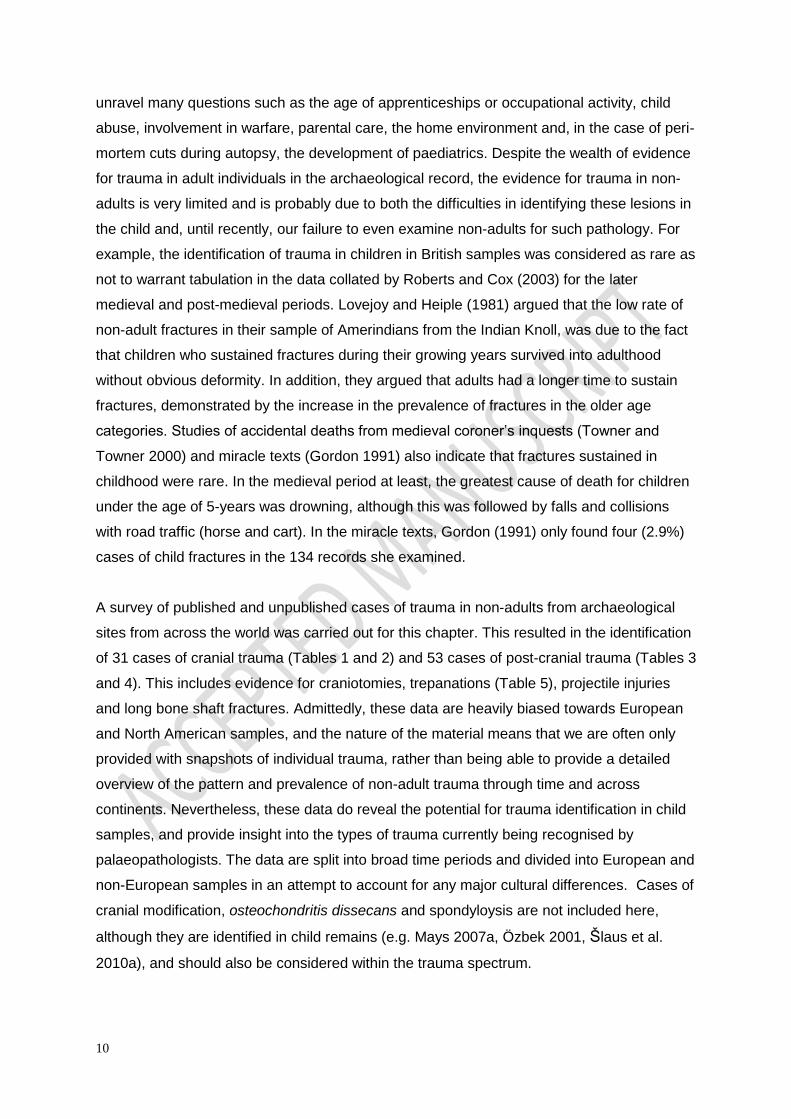
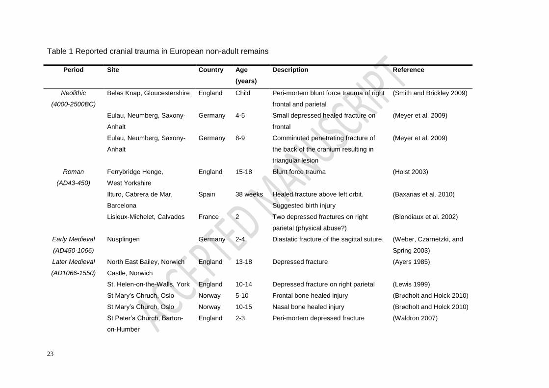
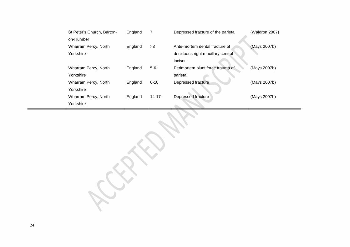
A survey of published and unpublished cases of trauma in non-adults from archaeological
sites from across the world was carried out for this chapter. This resulted in the identification
of 31 cases of cranial trauma (Tables 1 and 2) and 53 cases of post-cranial trauma (Tables 3
and 4). This includes evidence for craniotomies, trepanations (Table 5), projectile injuries
and long bone shaft fractures. Admittedly, these data are heavily biased towards European
and North American samples, and the nature of the material means that we are often only
provided with snapshots of individual trauma, rather than being able to provide a detailed
overview of the pattern and prevalence of non-adult trauma through time and across
continents. Nevertheless, these data do reveal the potential for trauma identification in child
samples, and provide insight into the types of trauma currently being recognised by
palaeopathologists. The data are split into broad time periods and divided into European and
non-European samples in an attempt to account for any major cultural differences. Cases of
cranial modification, osteochondritis dissecans and spondyloysis are not included here,
although they are identified in child remains (e.g. Mays 2007a, Özbek 2001, Šlaus et al.
2010a), and should also be considered within the trauma spectrum.
11
For non-surgical cranial trauma, 17 cases are reported from Europe and 15 from non-
European sites. The majority are depressed fractures and 59% (n=19) are in children aged
between the ages of 3 to10 years. That children were involved in warfare and massacres
comes from the inclusion of infants and children from mass grave sites in Neolithic Germany
(Whittle 1996:170), the Palaeolithic Sudan, and Mesolithic Bavaria (Thorpe 2003). In 2003,
Dawson and colleagues reported three perimortem depressed fractures and chipped teeth in
the skull of a 13-14 year old child (reportedly male) from a pit in Chalcolithic Israel (4500-
3200 BC). The authors argue the child received the blows during face-to-face combat,
suggesting that in their teens male children in this society became warriors. Two cases of
head trauma in children from Neolithic Eulau in Germany suggests they were caught up in a
raid (Meyer et al. 2009) with lethal consequences. In South Africa, the discovery of three
badly wounded crania of children in the same grave at the Modder River (dated to around
2600 BP) suggests they encountered a violent death with a penetrating weapon. The
reasons behind such injuries were not known, but isotopic evidence suggests they may have
been members of the same family, or at least sharing a very similar diet (Pfeiffer and Van
der Merwe 2004). A quartz projectile was found embedded in the spine of an adolescent
from the Neolithic site of Maderas Enco, Chile (Standen and Arriaza 2000), and a possible
case in a 9-12 years old from Mouse Creek, Tennessee (Smith 2003). A blade injury was
also identified on the mandible of a 4-6-year-old from Romano-British Lankhills, Winchester.
Brødholt and Holck (2010) describe an adolescent with a penetrating leg injury and two
cranial injuries in children from medieval Oslo, thought to be associated with the Civil War of
the 12-13th centuries. Stab wounds to the back of the scapula of two children, and the
humerus of another at Čepin, Croatia, suggest children were victims of Turkish raid in
AD1441, designed to spread panic throughout the region. The youngest child was under 4-
years-old (Šlaus et al. 2010b). Notably, these were the only cases of peri-mortem trauma
found in a review of 659 non-adults from the Balkans (Šlaus et al. 2010b).
In 1923, a study by MacCurdy of trepanations in Peruvian crania revealed no evidence in
non-adults, despite a prevalence of trepanations in 17% (47 of 273) of adult crania. Today,
five possible cases of trepanation have been reported, two from sites in Peru (Ortner 2003,
Shbat and Smrčka 2009). In three cases these are thought to be associated with
pathological conditions. A 5-6-year-old child from Fidenae in Rome is reported to have also
had hydrocephalus, and in Ancon, Peru, a child with a possible aural defect also had
evidence of surgical exploration. An unusual circular lesion was related to the trepanations
on the skull of a Neolithic 4-year-old from the Czech Republic (Shbat and Smrčka 2009).
Evidence for autopsy procedures from transverse cuts to the rib cage and craniotomies are
evident in four post-medieval sites in England (McKinley 2008, Miles et al. 2008, Miles and
12
Powers, 2007; Molleson and Cox 1993) and correspond to a rise in paediatrics during this
time (Lomax 1996). Two possible amputations, one believed to have occurred as the result
of a peri-mortem accident (Miles and Powers, 2007; Redfern 2007), have also been reported
in children from this period.
Of the 53 postcranial fractures identified from the literature, the vast majority (n=41) came
from European samples, with 11 from the Americas and two each from Africa and Asia. The
most common bones affected were the humerus (n=10), followed by the tibia (n=8), clavicle
(n=7) femur (n=6) and ribs (n=5). This pattern is similar to that seen in modern cases, with
the exception of fractures to the radius. Only three cases were reported in the archaeological
literature. The age distribution of lesions roughly followed that for the cranial lesions, with
fractures in children between the ages of 2-6 years, 6-10 years and 14-17 years most often
reported. Greenstick fractures have been suggested in five cases (Brothwell and Powers
2000, Ghalib 1999, Mays 2007b, McKinley 2008, Walker, Henderson, and Bland 2009), and
there are three cases of plastic deformation (Clough and Boyle 2010, Stuart-Macadam,
Glencross, and Kricun 1998, Wheeler et al. 2007). Trauma to the growth plates have been
recorded at later medieval Chichester, Sussex (Ortner 2003) and Pecos Pueblo in New
Mexico (Ortner 2003), post-medieval Christ Church Spitalfields in London (Lewis 2002),
early medieval Raunds Furnells in Northamptonshire (Lewis 2002) and medieval La
Madeleine, France (Kacki et al. 2011). Four postcranial fractures were found in children
living on the steep terraces of the Bronze Age Iberian Peninsula (Jimenez-Brobeil, Oumaoui,
and Du Souich 2007). The authors suggest that hazards due to playing and carrying out
chores in such a rugged environment explains the relatively high prevalence of fractures in
the children (3.2%).
Several authors have identified possible cases of birth trauma. In adults, Roberts (1989)
reported a case of Erb’s palsy in a young female from Romano-British Kingsholm, and Molto
(2000) identified humerus varus deformity in a male and a female from a Roman cemetery in
Dakhleh, Egypt which he interprets as evidence of birth trauma to the proximal epiphysis. In
non-adults, a healed fractured clavicle was evident in a 4-month-old child from Christ Church
Spitalfields, London, that may have occurred during birth (Lewis 2002), and Baxarias et al.
(2010) identified a healed linear fracture above the left orbit of a 38-week-old infant,
suggesting birth trauma caused the injury. Holst (2004) also identified disuse atrophy in the
right femur of a 2-4 month old child which has been suggested as a case of birth trauma.
Perhaps the most famous case of trauma to a perinate comes from Romano-British
Poundbury Camp, Dorchester, where extensive peri-mortem cut-marks to a neonate suggest
an embryotomy (Farwell and Molleson 1993).
13
The paucity of evidence for physical child abuse in past populations has been explained by
clandestine burials of the victims (Waldron 2000), that swaddling caused a different pattern
of injuries (De Mause 1974, Knight 1986), or that social circumstances meant that older
children, rather than infants, were the most common victims of abuse in the past (Walker
1997). Blondiaux and colleagues (2002) were the first to suggest physical abuse in a 2-year
old-child from Roman Lisieux-Michelet, France, who had sustained several head injuries and
rib fractures in addition to suffering rickets. Since then, three other possible cases have been
identified. Wheeler et al. (2007) reported on a 2 to 3-year-old child with multiple trauma and
widespread periosteal reactions from Kellis 2 in the Dakhleh Oasis. The child had fractures
of the humeri, clavicles, ribs and plastic deformation to the right ilium. Given the pattern of
trauma with various stages of healing, the authors suggest child abuse is the most probable
cause. Also from the Roman period (Lewis 2010) identified a tibial bucket-handle fracture in
an infant from Romano-British Poundbury Camp. While the tibial injury may be a toddler’s
fracture, at 18 months, this injury would be suspected as non-accidental today. Finally, a 1 to
2-year-old child from St Oswald’s Priory in Gloucester England, had a partially healed mid-
shaft fracture of the right humerus (figure 3). While an uncommon accidental injury in
children of this age today, it should also be noted that this child is also the only one in the
non-adult sample (n=144) with rickets. This underlying pathology may have weakened the
bone resulting in a fracture, but the presence of this disease may also signal on-going
neglect.
The most frequently recognised evidence we have for childhood fractures in the past is the
presence of shortened limbs in adults as a result of trauma and premature fusion of the
growth plate (Lewis 2000). How much shortening occurs depends on the age of the
individual at the time of the trauma, the epiphysis affected and the amount of longitudinal
growth still to occur. For example, in the tibia, 43% of longitudinal growth from the centre of
ossification is carried out by the distal growth plate, whereas 57% occurs at the proximal end
of the bone (Maresh 1955). Hence, a fracture and fusion of the proximal tibial epiphysis
would cause a greater degree of shortening (growth dysplasia) than similar trauma to the
distal end. In theory, detailed knowledge of proportional bone growth could provide evidence
of the age at which the fracture occurred. These data would, for example, allow us to
examine male: female ratios of trauma patterning in the past. Attempts to improve our
recognition and recording of such trauma has been carried out by Stuart-Macadam,
Glencross, and Kricun (1998), and Glencross and Stuart-Macadam (2000, 2001), who have
identified healed fractures of the humeral epicondyle, supra-condylar buckling and plastic
deformation in adults. Accurate identification and recording of these types of injuries would

14
greatly increase what we know about child trauma in the past. As an illustration, at the Indian
Knoll site, Glencross (2011) only recorded six (0.27%) fractures in 2200 complete children’s
bones, compared to 45% (n=35) of fractures in the adults believed to have occurred during
childhood. There is still a great deal of work to be carried out in this area.
6. Conclusions
The study of skeletal trauma in the remains of children from past societies is limited, with
only a handful of fractures being reported in the literature. The main reason for this deficit is
the plastic nature of paediatric bone, which results in incomplete, greenstick fractures that
heal rapidly without deformity, and the subtlety of other signatures of trauma such as
premature epiphyseal fusion and periosteal lesions. The latter are often described as
resulting from infection, and when widespread, assumed to be indicative of scurvy or
congenital syphilis. It is clear that, as today, children suffered from epiphyseal fractures,
traumatic shortening of the limb, and neurological injuries. The age at which these fractures
occurred, their type and frequency can provide information on birthing practices,
environmental hazards, the age at which children began work and were exposed to trauma.
Evidence of physical abuse is hindered by the often subtle and non-specific lesions that are
left behind, and problems of differential diagnosis. A survey of archaeological cases of child
trauma reveals that plastic deformation and greenstick fractures can be identified through
the use of x-rays and comparisons with unaffected bones. Cranial and weapon injuries
indicate that young children were often victims of raids and by the time they were teenagers,
were probably participating in warfare. There is increasing evidence for physical abuse in
archaeological material, although interpretations of the lesions remain challenging, and
diagnosis made on modern patterns of injury should be treated with caution. We still know
very little about the effect of occupation on the skeletons of children, who in some societies
may have begun work as apprentices by the age of 7 years (Lewis 2002), and children
would certainly have been involved with gathering of foodstuffs and household chores from a
much earlier age. Further work on identifying childhood trauma in adult populations may help
us build a picture of the age at which certain types of epiphyseal injury were sustained, and
differences between males and females. We should consider trauma as a possible aetiology
in the bones of infants and young children that display sheaths of periosteal new bone
formation, and utilize multiple view and comparative radiographs and CT scans to help us
visualize subtle fracture lines. However, it is the infants that cause the greatest challenges
for trauma diagnosis. Infant remains are often very fragile, fragmentary and are rarely
recovered in their entirety. Finally, we need to place our analysis of trauma within a life
15
course structure, taking into account the child’s physical development, and the cultural
context of the remains.
16
Acknowledgements
I thank all the people who sent me information about trauma in their child remains before
publication. In particular Malin Holst (York Osteoarchaeology Ltd.) and Natasha Powers
(MOLA).
Figure Captions
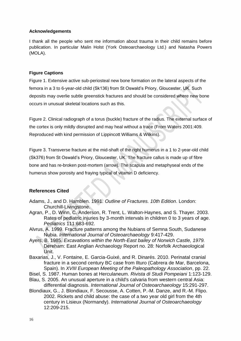
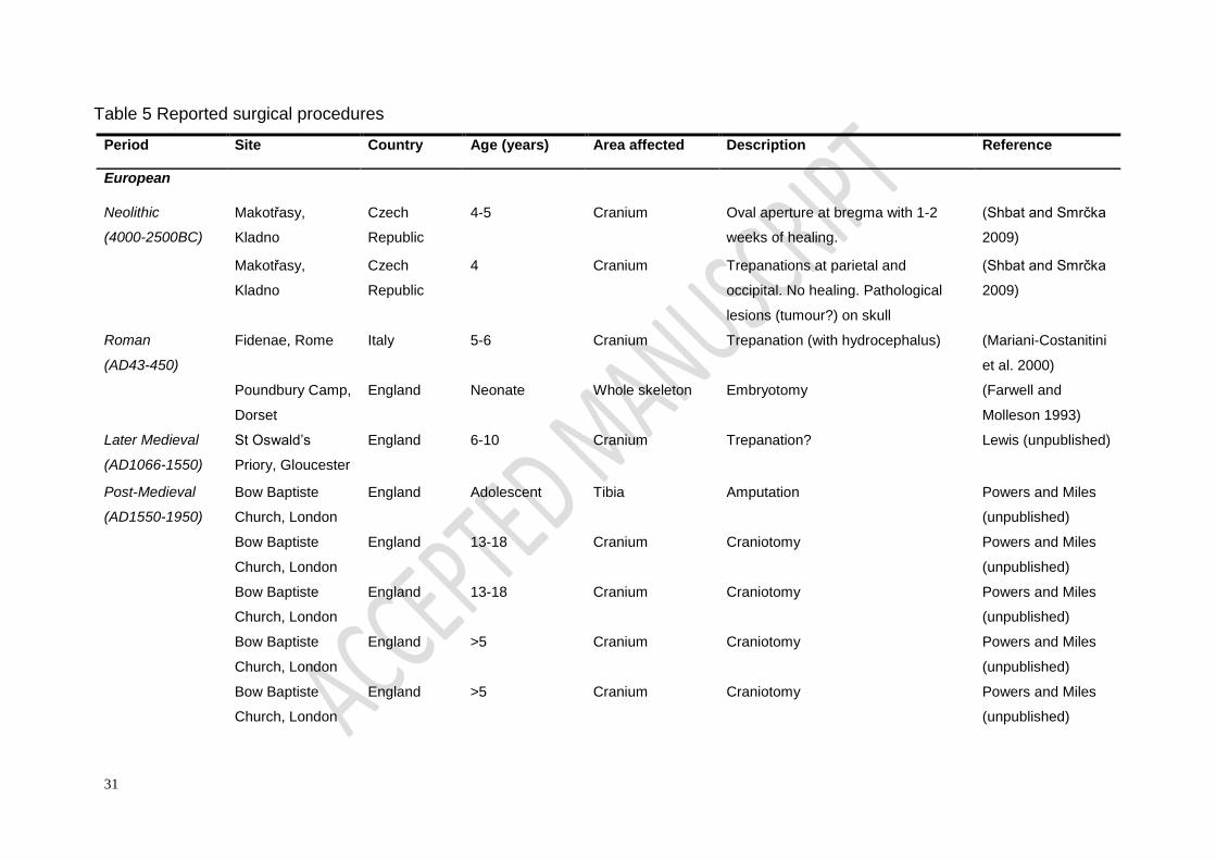
Figure 1. Extensive active sub-periosteal new bone formation on the lateral aspects of the
femora in a 3 to 6-year-old child (Sk136) from St Oswald’s Priory, Gloucester, UK. Such
deposits may overlie subtle greenstick fractures and should be considered where new bone
occurs in unusual skeletal locations such as this.
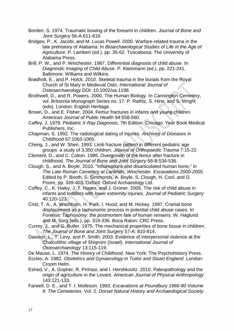
Figure 2. Clinical radiograph of a torus (buckle) fracture of the radius. The external surface of
the cortex is only mildly disrupted and may heal without a trace (From Waters 2001:409.
Reproduced with kind permission of Lippincott Williams & Wilkins).
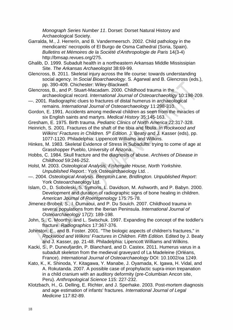
Figure 3. Transverse fracture at the mid-shaft of the right humerus in a 1 to 2-year-old child
(Sk376) from St Oswald’s Priory, Gloucester, UK. The fracture callus is made up of fibre
bone and has re-broken post-mortem (arrow). The scapula and metaphyseal ends of the
humerus show porosity and fraying typical of vitamin D deficiency.
References Cited
Adams, J., and D. Hamblen. 1991. Outline of Fractures. 10th Edition. London: Churchill-Livingstone.
Agran, P., D. Winn, C. Anderson, R. Trent, L. Walton-Haynes, and S. Thayer. 2003. Rates of pediatric injuries by 3-month intervals in children 0 to 3 years of age. Pediatrics 111:683-692.
Alvrus, A. 1999. Fracture patterns among the Nubians of Semna South, Sudanese Nubia. International Journal of Osteoarchaeology 9:417-429.
Ayers, B. 1985. Excavations within the North-East bailey of Norwich Castle, 1979. Dereham: East Anglian Archaeology Report no. 28: Norfolk Archaeological Unit.
Baxarias, J., V. Fontaine, E. Garcia-Guixé, and R. Dinarés. 2010. Perinatal cranial fracture in a second century BC case from Ilturo (Cabrera de Mar, Barcelona, Spain). In XVIII European Meeting of the Paleopathology Association, pp. 22.
Bisel, S. 1987. Human bones at Herculaneum. Rivista di Studi Pompeiani 1:123-129. Blau, S. 2005. An unusual aperture in a child's calvaria from western central Asia:
differential diagnosis. International Journal of Osteoarchaeology 15:291-297. Blondiaux, G., J. Blondiaux, F. Secousse, A. Cotten, P.-M. Danze, and R.-M. Flipo.
2002. Rickets and child abuse: the case of a two year old girl from the 4th century in Lisieux (Normandy). International Journal of Osteoarchaeology 12:209-215.
17
Borden, S. 1974. Traumatic bowing of the forearm in children. Journal of Bone and Joint Surgery 56-A:611-616.
Bridges, P., K. Jacobi, and M. Lucas Powell. 2000. Warfare-related trauma in the late prehistory of Alabama. In Bioarchaeological Studies of Life in the Age of Agriculture. P. Lambert (ed.), pp. 35-62. Tuscaloosa: The University of Alabama Press.
Brill, P. W., and P. Winchester. 1987. Differential diagnosis of child abuse. In Diagnostic Imaging of Child Abuse. P. Kleinmann (ed.), pp. 221-241. Baltimore: Williams and Wilkins.
Brødholt, E., and P. Holck. 2010. Skeletal trauma in the burials from the Royal Church of St Mary in Medieval Oslo. International Journal of Osteoarchaeology DOI: 10.1002/oa.1198.
Brothwell, D., and R. Powers. 2000. The Human Biology. In Cannington Cemetery, vol. Britannia Monograph Series no. 17. P. Rathtz, S. Hirst, and S. Wright (eds). London: English Heritage.
Brown, D., and E. Fisher. 2004. Femur fractures in infants and young children. American Journal of Public Health 94:558-560.
Caffey, J. 1978. Pediatric X-Ray Diagnosis, 7th Edition. Chicago: Year Book Medical Publishers, Inc.
Chapman, S. 1992. The radiological dating of injuries. Archives of Diseases in Childhood 67:1063-1065.
Cheng, J., and W. Shen. 1993. Limb fracture pattern in different pediatric age groups: a study of 3,350 children. Journal of Orthopaedic Trauma 7:15-22.
Clement, D., and C. Colton. 1986. Overgrowth of the femur after fracture in childhood. The Journal of Bone and Joint Surgery 68-B:534-536.
Clough, S., and A. Boyle. 2010. "Inhumations and disarticulated human bone," in The Late Roman Cemetery at Lankhills, Winchester. Excavations 2000-2005. Edited by P. Booth, S. Simmonds, A. Boyle, S. Clough, H. Cool, and D. Poore, pp. 339-403. Oxford: Oxford Archaeology Ltd.
Coffey, C., K. Haley, J. T. Hayes, and J. Groner. 2005. The risk of child abuse in infants and toddlers with lower extremity injuries. Journal of Pediatric Surgery 40:120-123.
Crist, T. A., A. Washburn, H. Park, I. Hood, and M. Hickey. 1997. Cranial bone displacement as a taphonomic process in potential child abuse cases. In Forensic Taphonomy: the postmortem fate of human remains. W. Haglund and M. Sorg (eds.), pp. 319-336. Boca Raton: CRC Press.
Currey, J., and G. Butler. 1975. The mechanical properties of bone tissue in children. The Journal of Bone and Joint Surgery 57-A: 810-814.
Dawson, L., T. Levy, and P. Smith. 2003. Evidence of interpersonal violence at the Chalcolithic village of Shiqmim (Israel). International Journal of Osteoarchaeology 13:115-119.
De Mause, L. 1974. The History of Childhood. New York: The Psychohistory Press. Eccles, A. 1982. Obstetrics and Gynaecology in Tudor and Stuart England. London:
Croom Helm. Eshed, V., A. Gopher, R. Pinhasi, and I. Hershkovitz. 2010. Paleopathology and the
origin of agriculture in the Levant. American Journal of Physical Anthropology 143:121-133.
Farwell, D. E., and T. I. Molleson. 1993. Excavations at Poundbury 1966-80 Volume II: The Cemeteries. Vol. 2. Dorset Natural History and Archaeological Society
18
Monograph Series Number 11. Dorset: Dorset Natural History and Archaeological Society.
Garralda, M., J. Herrerín, and B. Vandermeersch. 2002. Child pathology in the mendicants' necropolis of El Burgo de Osma Cathedral (Soria, Spain). Bulletins et Mémoires de la Société d'Anthropologie de Paris 14(3-4) http://bmsap.revues.org/275.
Ghalib, D. 1999. Subadult health in a northeastern Arkansas Middle Mississipian Site. The Arkansas Archaeologist 38:69-99.
Glencross, B. 2011. Skeletal injury across the life course: towards understanding social agency. In Social Bioarchaeology. S. Agarwal and B. Glencross (eds.), pp. 390-409. Chichester: Wiley-Blackwell.
Glencross, B., and P. Stuart-Macadam. 2000. Childhood trauma in the archaeological record. International Journal of Osteoarchaeology 10:198-209.
—. 2001. Radiographic clues to fractures of distal humerus in archaeological remains. International Journal of Osteoarchaeology 11:298-310.
Gordon, E. 1991. Accidents among medieval children as seen from the miracles of six English saints and martyrs. Medical History 35:145-163.
Gresham, E. 1975. Birth trauma. Pediatric Clinics of North America 22:317-328. Heinrich, S. 2001. Fractures of the shaft of the tibia and fibula. In Rockwood and
Wilkins' Fractures in Children. 5th Edition. J. Beaty and J. Kasser (eds), pp. 1077-1120. Philadelphia: Lippencott Williams and Wilkins.
Hinkes, M. 1983. Skeletal Evidence of Stress in Subadults: trying to come of age at Grasshopper Pueblo, University of Arizona.
Hobbs, C. 1984. Skull fracture and the diagnosis of abuse. Archives of Disease in Childhood 59:246-252.
Holst, M. 2003. Osteological Analysis. Fishergate House, North Yorkshire. Unpublished Report.: York Osteoarchaeology Ltd. .
—. 2004. Osteological Analysis. Bempton Lane, Bridlington. Unpublished Report: York Osteoarchaeology Ltd.
Islam, O., D. Soboleski, S. Symons, L. Davidson, M. Ashworth, and P. Babyn. 2000. Development and duration of radiographic signs of bone healing in children. American Journal of Roentgenology 175:75-78.
Jimenez-Brobeil, S., I. Oumaoui, and P. Du Souich. 2007. Childhood trauma in several populations from the Iberian Peninsula. International Journal of Osteoarchaeology 17(2): 189-198.
John, S., C. Moorthy, and L. Swischuk. 1997. Expanding the concept of the toddler's fracture. Radiographics 17:367-376.
Johnston, E., and B. Foster. 2001. "The biologic aspects of children's fractures," in Rockwood and Wilkins' Fractures in Children. Fifth Edition. Edited by J. Beaty and J. Kasser, pp. 21-48. Philadelphia: Lipencott Williams and Wilkins.
Kacki, S., P. Duneufjardin, P. Blanchard, and D. Castex. 2011. Humerus varus in a subadult skeleton from the medieval graveyard of La Madeleine (Orléans, France). International Journal of Osteoarchaeology DOI: 10.1002/oa 1249.
Kato, K., K. Shinoda, Y. Kitagawa, Y. Manabe, J. Oyamada, K. Igawa, H. Vidal, and A. Rokutanda. 2007. A possible case of prophylactic supra-inion trepanation in a child cranium with an auditory deformity (pre-Columbian Ancon site, Peru). Anthropological Science 115: 227-232.
Klotzbach, H., G. Delling, E. Richter, and J. Sperhake. 2003. Post-mortem diagnosis and age estimation of infants' fractures. International Journal of Legal Medicine 117:82-89.
19
Knight, B. 1986. The history of child abuse. Forensic Science International 30:135-141.
Kōhara, Y., T. Nakamuru, T. Nishizawa, and M. Susuki. 1971. Human infant remains of the earliest Jomon Period, suffering death by a falling rock. Journal of the Anthropological Society of Nippon 79:55-60.
Lewis, M. 2002. The impact of industrialisation: comparative study of child health in four sites from medieval and post-medieval England (850-1859 AD). American Journal of Physical Anthropology 119:211-223.
—. 2007. The Bioarchaeology of Children. Perspectives from Biological and Forensic Anthropology. Cambridge: Cambridge University Press.
—. 2010. Life and death in a civitas capital: metabolic disease and trauma in the children from late Roman Dorchester. Dorset. American Journal of Physical Anthropology 142:405-416.
Lewis, M. E. 1999. The Impact of Urbanisation and Industrialisation in Medieval and Post-medieval Britain. An assessment of the morbidity and mortality of non-adult skeletons from the cemeteries of two urban and two rural sites in England (AD 850-1859). University of Bradford.
—. 2000. Non-adult palaeopathology: current status and future potential. In Human Osteology in Archaeology and Forensic Science. M. Cox and S. Mays (eds), pp. 39-57. London: Greenwich Medical Media Ltd.
Lomax, E. M. R. 1996. Small and Special: The Development of Hospitals for Children in Victorian Britain. Medical History, Supplement No.16. London: Wellcome Institute for the History of Medicine.
Lovejoy, C.O., and Heiple, K. 1981. The analysis of fractures in skeletal populations with an example from the Libben site, Ottowa County, Ohio. American Journal of Physical Anthropology 55:529-541.
MacCurdy, G. 1923. Human skeletal remains from the highlands of Peru. American Journal of Physical Anthropology 6:217-352.
Maresh, M. M. 1955. Linear growth of long bones of extremities from infancy through adolescence. American Journal of Diseases in Children 89:725-742.
Mariani-Costanitini, R., P. Catalano, d. Gennaro, d. Tota, and L. Angeletti. 2000. New light on cranial surgery in ancient Rome. The Lancet 355:305-307.
Mays, S. 2007a. Spondylolysis in non-adult skeletons excavated from a medieval rural archaeological site in England. International Journal of Osteoarchaeology on line DOI: 10.1002/oa.878.
.—. 2007b. The human remains. In Wharram. A Study of a Settlement on the Yorkshire Wolds, XI: The Churchyard. York University Archaeological Publications 13. S. Mays, C. Harding, and C. Heighway (eds.), pp. 77-192. London: English Heritage.
McKinley, J. 2008. The 18th century Baptist Church and Burial Ground at West Butts Street, Poole, Dorset. Sailsbury: Wessex Archaeology Ltd.
Meservy, C., R. Towbin, R. McLaurin, P. Myers, and W. Ball. 1987. Radiographic characteristics of skull fractures resulting from child abuse. American Journal of Roentgenology 149:173-175.
Meyer, C., G. Brandt, W. Haak, R. Ganslmeier, H. Meler, and K. Alt. 2009. The Eulau eulogy: bioarchaeological interpretation of lethal violence in Corded Ware multiple burials from Saxony-Anhalt, Germany. Journal of Anthropological Archaeology 28:412-423.
20
Miles, A. N., and Powers, N. 2007. Bow Baptist Church Burial Ground, 2–25 Payne Road, London, E3, London Borough of Tower Hamlets. A post-excavation assessment and updated project design, unpublished MOLA report
Miles, A., N. Powers, R. Wroe-Brown, and D. Walker. 2008. St Marylebone Church and Burial Ground in the 18th-19th Centuries. Excavations at St Marylebone School, 1992 and 2004-6. MOLAS Monograph 46. London: Museum of London Archaeology Service.
Molleson, T., and M. Cox. 1993. The Spitalfields Project, Volume II - The Middling Sort. Vol. 2. York: Council for British Archaeology Research Report. 86.
Molto, J. 2000. Humerus varus deformity in Roman period burials from Kellis 2, Dakhleh, Egypt. American Journal of Physical Anthropology 113:103-109.
Nystrom, K. 2007. Trepanation in the Chachapoya region of Northern Peru. International Journal of Osteoarchaeology.
O'Connor, J., and J. Cohen. 1987. "Dating fractures," in Diagnostic Imaging of Child Abuse, Second Edition, vol. Three. Edited by P. Kleinman, pp. 103-113. Baltimore: Williams and Wilkins.
Ortner, D. 2003. Identification of Pathological Conditions in Human Skeletal Remains, 2nd Edition. New York: Academic Press.
Ortner, D. J. 2002. Research slide collection of Donald J. Ortner, Department of Anthropology, Smithsonian Institution, Washington, DC, and digitized and made available through funds supporting NSF grant SES-0138129 by Richard H. Steckel, Clark Spencer Larsen, Paul W. Sciulli and Phillip L. Walker, A History of Health in Europe from the Late Paleolithic Era to the Present. (Mimeo, Columbus, Ohio, 2002).
Ottini, L., G. Di Tota, R. Mariani-Costantini, L. Angeletti, M. La Verghetta, L. Capasso, A. Di Fabrizio, and R. D-Anastasio. 2001. Evidence of a forearm fracture in a young victim of the AD79 Vesuvius eruption. Journal of Palaeopathology 13:23-26.
Owen, R., F. Hickey, and D. Finlay. 1995. A study of metatarsal fractures in children. Injury 26:537-538.
Özbek, M. 2001. Cranial deformation in a subadult sample from Degirmentepe (Chalcolithic, Turkey). American Journal of Physical Anthropology 115:238-244.
Pfeiffer, S., and N. Van der Merwe. 2004. Cranial injuries in Stone Age children from the Modder River Mouth, Western Cape province, South Africa. South African Archaeological Bulletin 59:59-65.
Pudenz, R., E. Todd, and C. Shelden. 1961. Head injuries in infants and young children. California Medicine 94:6-71.
Redfern, R. 2007. An investigation of a possible perimortem limb amputation in a post-medieval subadult from the Isle of Wight, England. Paleopathology Newsletter 140:6-10.
Resnick, D. Editor. 1995. Diagnosis of Bone and Joint Disorders, 3rd edition. Philadelphia: W.B. Saunders Company.
Resnick, D., and T. Goergen. 2002. Physical injury: concepts and terminology. In Diagnosis of Bone and Joint Disorders. D. Resnick (ed.), pp. 2627-2789. Philadelphia: WB Saunders and Company.
Resnick, D., and M. Kransdorf. 2005. Bone and Joint Imaging. Third Edition. Philadelphia: Elsevier Saunders.
Roberts, C. 1989. The human remains from 76 Kingsholm, Gloucester. University of Bradford, Bradford, UK.

21
—. 2000. Trauma in biocultural perspective: past present and future work in Britain. In Human Osteology in Archaeology and Forensic Science. M. Cox and S. Mays (eds.), pp. 337-356. London: Greenwich Medical Media Ltd.
Roberts, C., and M. Cox. 2003. Health and Disease in Britain. Gloucestershire: Sutton Publishing Ltd.
Roberts, C., and K. Manchester. 2005. The Archaeology of Disease: 3rd edition. Gloucester: Sutton Publishing.
Rushton, J. 1991. The secret 'iron tongs' of midwifery. The Historian 30:12-15. Salter, R. 1980. Special features of fractures and dislocation in children. In Fracture
Treatment and Healing. R. Heppenstall (ed.), pp. 190. Philadelphia: WB Saunders.
Shaw, H., and S. Bohrer. 1979. The incidence of cone epiphyses and ivory epiphyses of the hand in Nigerian children. American Journal of Physical Anthropology 51:155-162.
Shbat, A., and V. Smrčka. 2009. Children's cranial lesions from Neolithic. Prague Medical Journal 110:114-119.
Šlaus, M., T. Cicvara-Pećina, I. Lucijanić, M. Pećina, and D. Stilinović. 2010a. Osteochonditis dissecans of the knee in a subadult from a medieval (ninth century AD) site in Croatia. Acta Clin Croatia 49:189-195.
Šlaus, M., M. Novak, V. Vyroubal, and Ž. Bedić. 2010b. The harsh life on the 15th century Croatia-Ottoman Empire border: analysing and identifying the reasons for the massacre in Čepin. American Journal of Physical Anthropology 141:358-372.
Smith, M. 2003. Beyond palisades: the nature and frequency of late prehistoric deliberate trauma in the Chickamauga Reservoir of East Tennessee. American Journal of Physical Anthropology 121:303-318.
Smith, M., and M. Brickely. 2009. People of the Long Barrows. Life, Death and Burial in the Earlier Neolithic. Gloucestershire: The History Press.
Sorantin, E., P. Brader, and F. Thimary. 2006. Neonatal trauma. European Journal of Radiology 60:199-207.
Standen, V., and B. Arriaza. 2000. Trauma in the Preceramic coastal populations of Northern Chile: violence or occupational hazards? American Journal of Physical Anthropology 112:239-249.
Stephens, M., L. Hsu, and J. Leong. 1989. Leg length discrepancy after femoral shaft fractures in children. The Journal of Bone and Joint Surgery 71-B:615-618.
Stilli, S., M. Magnani, M. Lampasi, D. Antonioli, C. Bettuzzi, and O. Donzelli. 2008. Remodelling and overgrowth after conservative treatment for femoral and tibial shaft fractures in children. La Chirugia Degli Organi Movimento 91:13-19.
Strait, R., R. Siegal, and R. Shapiro. 1995. Humeral fractures without obvious etiologies in children less than 3 years of age: when is it abuse? Pediatrics 97:667-671.
Stuart-Macadam, P., B. Glencross, and M. Kricun. 1998. Traumatic bowing deformities in tubular bones. International Journal of Osteoarchaeology 8:252-262.
Thorpe, I. 2003. Anthropology, archaeology, and the origin of warfare. World Archaeology 35:145-165.
22
Towner, E., and Towner, J. 2000. Developing the history of unintentional injury: the use of coroners' records in early modern England. Injury Prevention 6:102-105.
Tung, T., and K. Knudson. 2010. Childhood lost: abductions, sacrifice, and trophy heads of children in the Wari Empire of the ancient Andes. Latin American Antiquity 21:44-66.
Waldron, T. 2000. Hidden or overlooked? Where are the disadvantaged in the skeletal record? In Madness, disability and social exclusion, vol. 40, One World Archaeology. J. Hurbert (ed.), pp. 29-45. London: Routledge.
—. 2007. St Peter's, Barton-upon-Humber, Lincolnshire, Vol 2. The Human Remains. Oxford: Oxbow Books.
Walker, D., J. D. Henderson, and P. Bland. 2009. Advances in the use of radiography in osteological analysis. In the Proceedings of the 10th Annual Meeting of the British Association for Biological Anthropology and Osteoarchaeology. Oxford University: Oxford.
Walker, P. L. 1997. Skeletal evidence for child abuse: A physical anthropological perspective. Journal of Forensic Sciences 42:196-207.
Waters, P. 2001. Distal radius and ulna fractures. In Rockwood and Wilkins' Fractures in Children. 5th edition. J. Beaty and J. Kasser (eds.), pp. 381-442. Philadelphia: Lipencott Williams and Wilkins.
Weber, J., A. Czarnetzki, and A. Spring. 2003. Acquired sagittal suture diastasis in an infant skull from the early medieval period - a sign of raised intercranial pressure. Acta Neurochir 145:233-234.
Wheeler, S., P. Beauchesne, L. Wiliams, and J. Molto. 2007. Fractured childhood: a case of probable child abuse from the Kellis 2 cemetery, Dakhleh Oasis, Egypt. Paper presented at the 72nd Annual Meeting of the Society of American Archaeologists. Austin, Texas.
Whittle, A. 1996. Europe in the Neolithic: the creation of new worlds. Cambridge: Cambridge University Press.
Wilber, J., and G. Thompson. 1998. The multiply injured child. In Skeletal Trauma in Children, 2nd edition. N. Green and M. Swiontkowski (eds.), pp. 71-102. Philadelphia: W.B. Saunders Company.
Wilczak, C., R. Watkins, C. Null, and M. Blakey. 2004. Skeletal Indicators of work: musculoskeletal, arthritic and traumatic effects. In Skeletal Biology Final Report. New York: African Burial Ground Project Office of Public Education and Interpretation.
Wilkins, K., and A. Aroojis. 2001. "The present status of children's fractures," in Rockwood and Wilkins' Fractures in Children. 5th edition. J. Beaty and J. Kasser (eds.), pp. 3-20. Philadelphia: Lippencott Williams and Wilkins.
Wood, J., C. Christian, C. Adams, and D. Rubin. 2009. Skeletal surveys in infants with isolated skull fractures. Pediatrics 123:247-252.
23
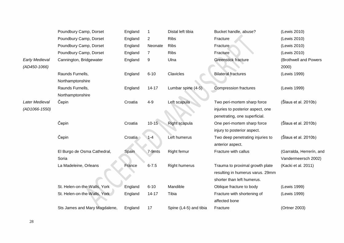
Table 1 Reported cranial trauma in European non-adult remains
Period Site Country Age
(years)
Description Reference
Neolithic
(4000-2500BC)
Belas Knap, Gloucestershire England Child Peri-mortem blunt force trauma of right
frontal and parietal
(Smith and Brickley 2009)
Eulau, Neumberg, Saxony-
Anhalt
Germany 4-5 Small depressed healed fracture on
frontal
(Meyer et al. 2009)
Eulau, Neumberg, Saxony-
Anhalt
Germany 8-9 Comminuted penetrating fracture of
the back of the cranium resulting in
triangular lesion
(Meyer et al. 2009)
Roman
(AD43-450)
Ferrybridge Henge,
West Yorkshire
England 15-18 Blunt force trauma (Holst 2003)
Ilturo, Cabrera de Mar,
Barcelona
Spain 38 weeks Healed fracture above left orbit.
Suggested birth injury
(Baxarias et al. 2010)
Lisieux-Michelet, Calvados France 2 Two depressed fractures on right
parietal (physical abuse?)
(Blondiaux et al. 2002)
Early Medieval
(AD450-1066)
Nusplingen Germany 2-4 Diastatic fracture of the sagittal suture. (Weber, Czarnetzki, and
Spring 2003)
Later Medieval
(AD1066-1550)
North East Bailey, Norwich
Castle, Norwich
England 13-18 Depressed fracture (Ayers 1985)
St. Helen-on-the-Walls, York England 10-14 Depressed fracture on right parietal (Lewis 1999)
St Mary’s Chruch, Oslo Norway 5-10 Frontal bone healed injury (Brødholt and Holck 2010)
St Mary’s Church, Oslo Norway 10-15 Nasal bone healed injury (Brødholt and Holck 2010)
St Peter’s Church, Barton-
on-Humber
England 2-3 Peri-mortem depressed fracture (Waldron 2007)
24
St Peter’s Church, Barton-
on-Humber
England 7 Depressed fracture of the parietal (Waldron 2007)
Wharram Percy, North
Yorkshire
England >3 Ante-mortem dental fracture of
deciduous right maxillary central
incisor
(Mays 2007b)
Wharram Percy, North
Yorkshire
England 5-6 Perimortem blunt force trauma of
parietal
(Mays 2007b)
Wharram Percy, North
Yorkshire
England 6-10 Depressed fracture (Mays 2007b)
Wharram Percy, North
Yorkshire
England 14-17 Depressed fracture (Mays 2007b)
25
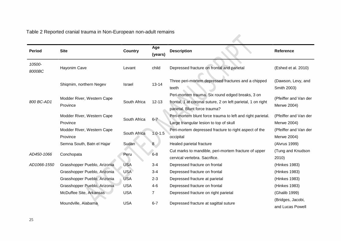
Table 2 Reported cranial trauma in Non-European non-adult remains
Period Site Country Age
(years) Description Reference
10500-
8000BC Hayonim Cave Levant child Depressed fracture on frontal and parietal (Eshed et al. 2010)
Shiqmim, northern Negev Israel 13-14
Three peri-mortem depressed fractures and a chipped
teeth
(Dawson, Levy, and
Smith 2003)
800 BC-AD1 Modder River, Western Cape
Province South Africa 12-13
Peri-mortem trauma. Six round edged breaks, 3 on
frontal, 1 at coronal suture, 2 on left parietal, 1 on right
parietal. Blunt force trauma?
(Pfeiffer and Van der
Merwe 2004)
Modder River, Western Cape
Province South Africa 6-7
Peri-mortem blunt force trauma to left and right parietal.
Large triangular lesion to top of skull
(Pfeiffer and Van der
Merwe 2004)
Modder River, Western Cape
Province South Africa 1.0-1.5
Peri-mortem depressed fracture to right aspect of the
occipital
(Pfeiffer and Van der
Merwe 2004)
Semna South, Batn el Hajar Sudan 8 Healed parietal fracture (Alvrus 1999)
AD450-1066 Conchopata Peru 6-8 Cut marks to mandible, peri-mortem fracture of upper
cervical vertebra. Sacrifice.
(Tung and Knudson
2010)
AD1066-1550 Grasshopper Pueblo, Arizonia USA 3-4 Depressed fracture on frontal (Hinkes 1983)
Grasshopper Pueblo, Arizonia USA 3-4 Depressed fracture on frontal (Hinkes 1983)
Grasshopper Pueblo, Arizonia USA 2-3 Depressed fracture at parietal (Hinkes 1983)
Grasshopper Pueblo, Arizonia USA 4-6 Depressed fracture on frontal (Hinkes 1983)
McDuffee Site, Arkansas USA 7 Depressed fracture on right parietal (Ghalib 1999)
Moundville, Alabama USA 6-7 Depressed fracture at sagittal suture (Bridges, Jacobi,
and Lucas Powell
26
2000)
Moundville, Alabama USA 17 Fractured mandibular ramus
(Bridges, Jacobi,
and Lucas Powell
2000)
Ocoee, Tennessee USA 9-11 Blunt force trauma (Smith 2003)
AD1550-1950 New York African Burial Ground USA 13-15 Fracture? (Wilczak et al. 2004)

27
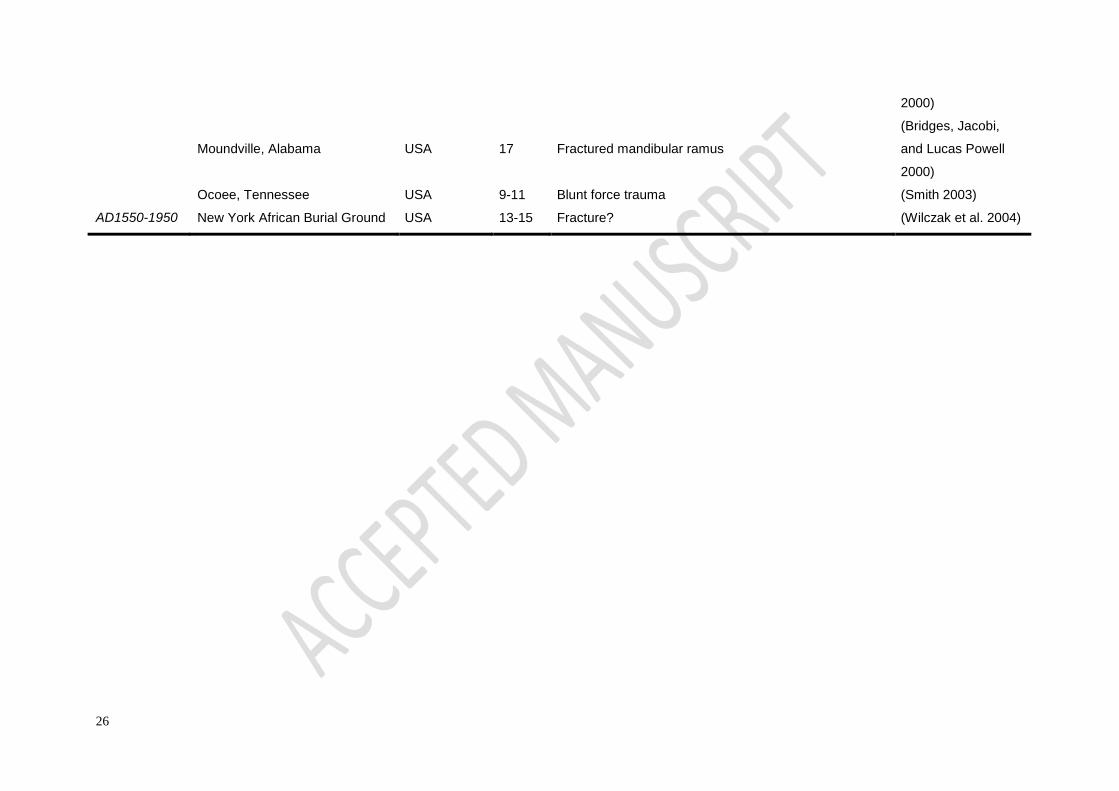
Table 3 Postcranial trauma in European samples
Period Site Country Age
(years)
Area affected Description Reference
Bronze Age
(2600-800BC)
Castellón Alto, Galera Spain Left clavicle Fracture at lateral aspect (Jimenez-Brobeil,
Oumaoui, and Du
Souich 2007)
Castellón Alto, Galera Spain 6-7 Left femur Fracture at midshaft with atrophy (Jimenez-Brobeil,
Oumaoui, and Du
Souich 2007)
Castellón Alto, Galera Spain 7-8 Right humerus Fracture at the distal aspect with
anterior displacement
(Jimenez-Brobeil,
Oumaoui, and Du
Souich 2007)
Roman
(AD43-450)
Bempton Lane, Bridlington England 2-4 mts Right femur Disuse atrophy, breech birth
trauma?
(Holst 2004)
Ferrybridge Henge, West
Yorkshire
England 15-17 Tibia Hematoma (Holst 2003)
Herculaneum Italy 7-8 Right radius and ulna Oblique fractures at distal aspects
of the shafts. Good alignment and
partial healing suggest a
successful reduction shortly
before death
(Bisel 1987) and (Ottini
et al. 2001)
Lankhills, Winchester England 4-6 Mandible Blade injury (Clough and Boyle
2010)
Lankhills, Winchester England 13-17 Sacrum, tibia Fractures (plastic deformation?) (Clough and Boyle
2010)
28
Poundbury Camp, Dorset England 1 Distal left tibia Bucket handle, abuse? (Lewis 2010)
Poundbury Camp, Dorset England 2 Ribs Fracture (Lewis 2010)
Poundbury Camp, Dorset England Neonate Ribs Fracture (Lewis 2010)
Poundbury Camp, Dorset England 7 Ribs Fracture (Lewis 2010)
Early Medieval
(AD450-1066)
Cannington, Bridgewater England 9 Ulna Greenstick fracture (Brothwell and Powers
2000)
Raunds Furnells,
Northamptonshire
England 6-10 Clavicles Bilateral fractures (Lewis 1999)
Raunds Furnells,
Northamptonshire
England 14-17 Lumbar spine (4-5) Compression fractures (Lewis 1999)
Later Medieval
(AD1066-1550)
Čepin Croatia 4-9 Left scapula Two peri-mortem sharp force
injuries to posterior aspect, one
penetrating, one superficial.
(Šlaus et al. 2010b)
Čepin Croatia 10-15 Right scapula One peri-mortem sharp force
injury to posterior aspect.
(Šlaus et al. 2010b)
Čepin Croatia 1-4 Left humerus Two deep penetrating injuries to
anterior aspect.
(Šlaus et al. 2010b)
El Burgo de Osma Cathedral,
Soria
Spain 7-9mts Right femur Fracture with callus (Garralda, Herrerín, and
Vandermeersch 2002)
La Madeleine, Orleans France 6-7.5 Right humerus Trauma to proximal growth plate
resulting in humerus varus. 29mm
shorter than left humerus.
(Kacki et al. 2011)
St. Helen-on-the-Walls, York England 6-10 Mandible Oblique fracture to body (Lewis 1999)
St. Helen-on-the-Walls, York England 14-17 Tibia Fracture with shortening of
affected bone
(Lewis 1999)
Sts James and Mary Magdalene, England 17 Spine (L4-5) and tibia Fracture (Ortner 2003)

29
Chichester, Sussex
Sts James and Mary Magdalene,
Chichester, Sussex
England 15-17 Sacrum (S3) Fracture (Ortner 2002)
St Mary’s Church, Oslo Norway 15-20 Tibia Penetrating injury, peri-mortem (Brødholt and Holck
2010)
St Oswald’s Priory, Gloucester England 1-2 Humerus Fracture with callus, abuse Lewis (unpublished)
St Peter’s Church, Barton-on-
Humber
England 17 Humerus, radius, ulna Dislocated elbow (Waldron 2007)
St Peter’s Church, Barton-on-
Humber
England 10 Tibia Partially healed sharp injury (Waldron 2007)
St Peter’s Church, Barton-on-
Humber
England 12-13 Femur Slipped femoral epiphysis (Waldron 2007)
Wharram Percy, North Yorkshire England 14-15 Right clavicle Fracture (Mays 2007b)
Wharram Percy, North Yorkshire England 1.5 Humerus Greenstick fracture (Mays 2007b)
Wharram Percy, North Yorkshire England >3 Dentition Ante-mortem tooth fracture (Mays 2007b)
Wharram Percy, North Yorkshire England 2-6 Left leg Disuse atrophy (Mays 2007b)
Wharram Percy, North Yorkshire England 13-15 Femur Slipped femoral epiphysis (Mays 2007b)
Post-Medieval
(AD1550-1950)
Christ Church Spitalfields, London England 14-17 Rib Fracture (Lewis 1999)
Christ Church Spitalfields, London England 6mts Ribs Fracture (Lewis 1999)
Christ Church Spitalfields, London England 6mts Clavicle Fracture, birth trauma? (Lewis 1999)
Christ Church Spitalfields, London England 10-14 Clavicle and humerus Fracture with shortening (Lewis 1999)
St Mary and St Michael,
Whitechapel, London
England Child Fibula Greenstick fracture visible on x-
ray.
(Walker, Henderson,
and Bland 2009)
West Butts Street, Poole, Dorset England 6-7 Foot Fracture (McKinley 2008)
West Butts Street, Poole, Dorset England 4-5 Left tibia Greenstick fracture (McKinley 2008)
30
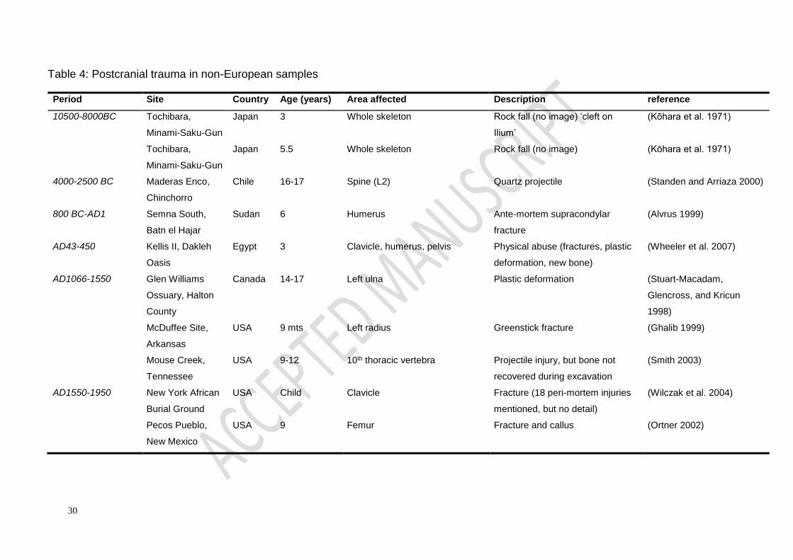
Table 4: Postcranial trauma in non-European samples
Period Site Country Age (years) Area affected Description reference
10500-8000BC Tochibara,
Minami-Saku-Gun
Japan 3 Whole skeleton Rock fall (no image) ‘cleft on
Ilium’
(Kōhara et al. 1971)
Tochibara,
Minami-Saku-Gun
Japan 5.5 Whole skeleton Rock fall (no image) (Kōhara et al. 1971)
4000-2500 BC Maderas Enco,
Chinchorro
Chile 16-17 Spine (L2) Quartz projectile (Standen and Arriaza 2000)
800 BC-AD1 Semna South,
Batn el Hajar
Sudan 6 Humerus Ante-mortem supracondylar
fracture
(Alvrus 1999)
AD43-450 Kellis II, Dakleh
Oasis
Egypt 3 Clavicle, humerus, pelvis Physical abuse (fractures, plastic
deformation, new bone)
(Wheeler et al. 2007)
AD1066-1550 Glen Williams
Ossuary, Halton
County
Canada 14-17 Left ulna Plastic deformation (Stuart-Macadam,
Glencross, and Kricun
1998)
McDuffee Site,
Arkansas
USA 9 mts Left radius Greenstick fracture (Ghalib 1999)
Mouse Creek,
Tennessee
USA 9-12 10th thoracic vertebra Projectile injury, but bone not
recovered during excavation
(Smith 2003)
AD1550-1950 New York African
Burial Ground
USA Child Clavicle Fracture (18 peri-mortem injuries
mentioned, but no detail)
(Wilczak et al. 2004)
Pecos Pueblo,
New Mexico
USA 9 Femur Fracture and callus (Ortner 2002)
31
Table 5 Reported surgical procedures
Period Site Country Age (years) Area affected Description Reference
European
Neolithic
(4000-2500BC)
Makotřasy,
Kladno
Czech
Republic
4-5 Cranium Oval aperture at bregma with 1-2
weeks of healing.
(Shbat and Smrčka
2009)
Makotřasy,
Kladno
Czech
Republic
4 Cranium Trepanations at parietal and
occipital. No healing. Pathological
lesions (tumour?) on skull
(Shbat and Smrčka
2009)
Roman
(AD43-450)
Fidenae, Rome Italy 5-6 Cranium Trepanation (with hydrocephalus) (Mariani-Costanitini
et al. 2000)
Poundbury Camp,
Dorset
England Neonate Whole skeleton Embryotomy (Farwell and
Molleson 1993)
Later Medieval
(AD1066-1550)
St Oswald’s
Priory, Gloucester
England 6-10 Cranium Trepanation? Lewis (unpublished)
Post-Medieval
(AD1550-1950)
Bow Baptiste
Church, London
England Adolescent Tibia Amputation Powers and Miles
(unpublished)
Bow Baptiste
Church, London
England 13-18 Cranium Craniotomy Powers and Miles
(unpublished)
Bow Baptiste
Church, London
England 13-18 Cranium Craniotomy Powers and Miles
(unpublished)
Bow Baptiste
Church, London
England >5 Cranium Craniotomy Powers and Miles
(unpublished)
Bow Baptiste
Church, London
England >5 Cranium Craniotomy Powers and Miles
(unpublished)
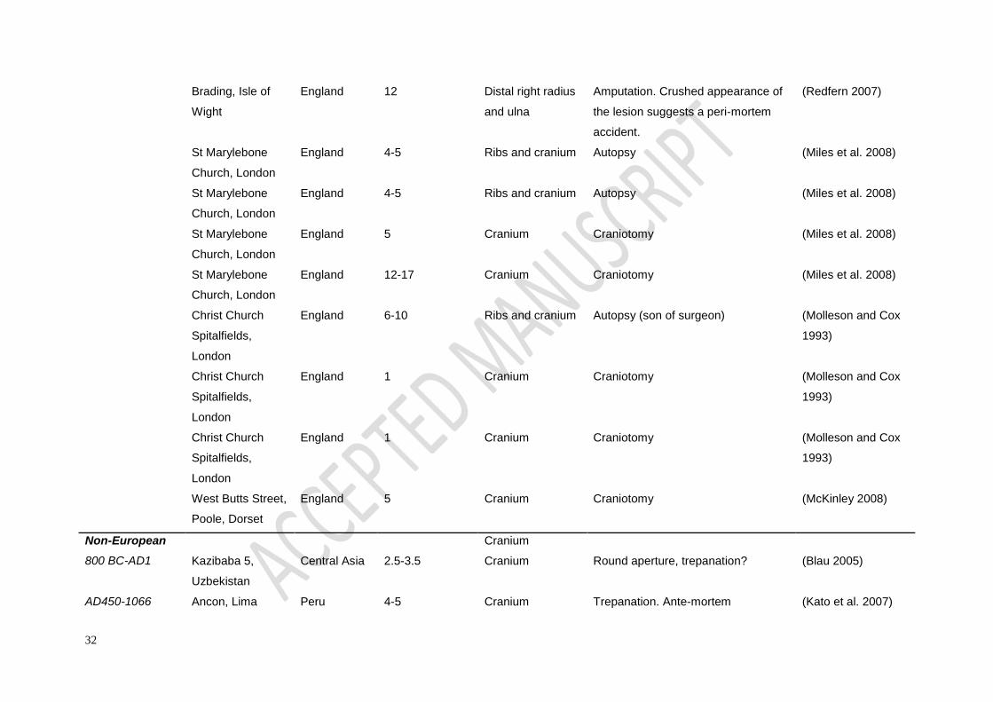
32
Brading, Isle of
Wight
England 12 Distal right radius
and ulna
Amputation. Crushed appearance of
the lesion suggests a peri-mortem
accident.
(Redfern 2007)
St Marylebone
Church, London
England 4-5 Ribs and cranium Autopsy (Miles et al. 2008)
St Marylebone
Church, London
England 4-5 Ribs and cranium Autopsy (Miles et al. 2008)
St Marylebone
Church, London
England 5 Cranium Craniotomy (Miles et al. 2008)
St Marylebone
Church, London
England 12-17 Cranium Craniotomy (Miles et al. 2008)
Christ Church
Spitalfields,
London
England 6-10 Ribs and cranium Autopsy (son of surgeon) (Molleson and Cox
1993)
Christ Church
Spitalfields,
London
England 1 Cranium Craniotomy (Molleson and Cox
1993)
Christ Church
Spitalfields,
London
England 1 Cranium Craniotomy (Molleson and Cox
1993)
West Butts Street,
Poole, Dorset
England 5 Cranium Craniotomy (McKinley 2008)
Non-European Cranium
800 BC-AD1 Kazibaba 5,
Uzbekistan
Central Asia 2.5-3.5 Cranium Round aperture, trepanation? (Blau 2005)
AD450-1066 Ancon, Lima Peru 4-5 Cranium Trepanation. Ante-mortem (Kato et al. 2007)
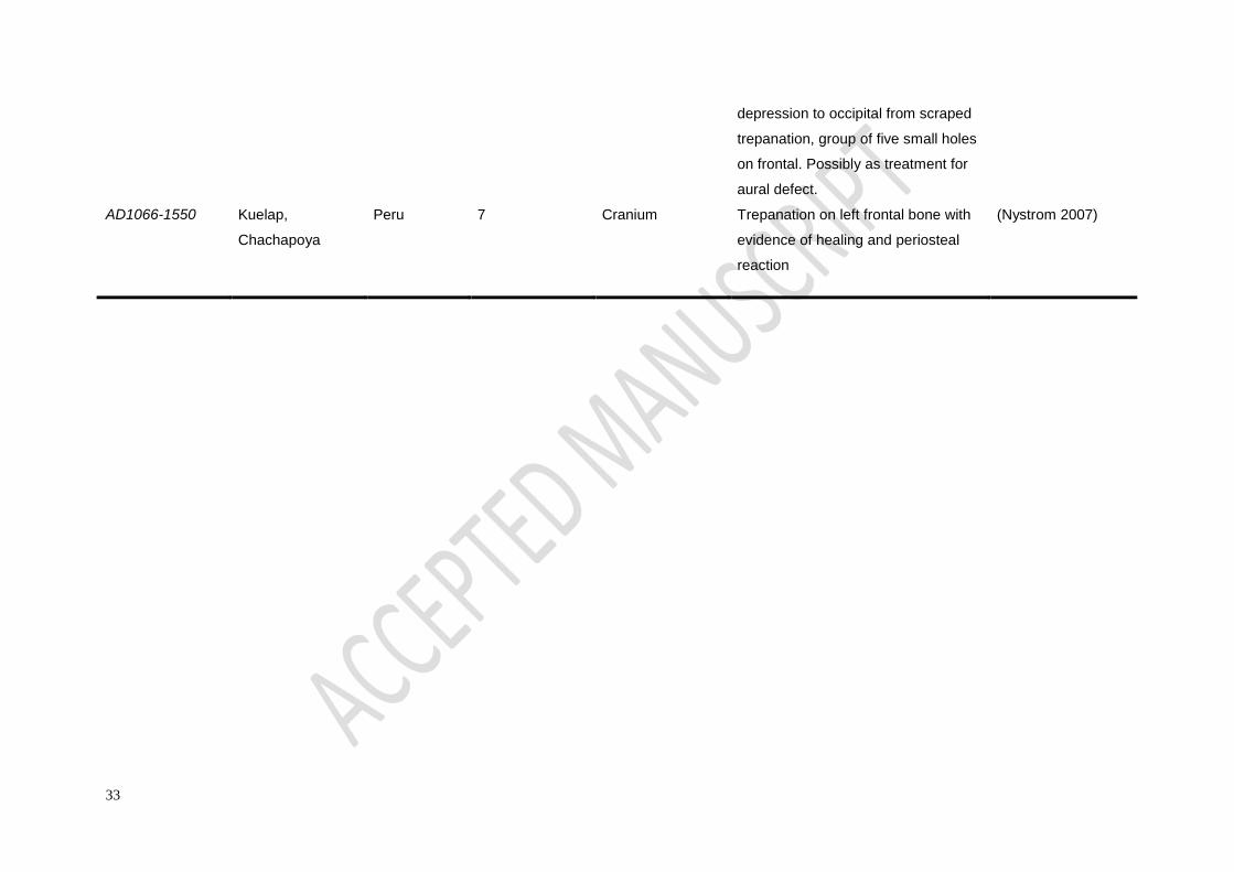
33
depression to occipital from scraped
trepanation, group of five small holes
on frontal. Possibly as treatment for
aural defect.
AD1066-1550 Kuelap,
Chachapoya
Peru 7 Cranium Trepanation on left frontal bone with
evidence of healing and periosteal
reaction
(Nystrom 2007)
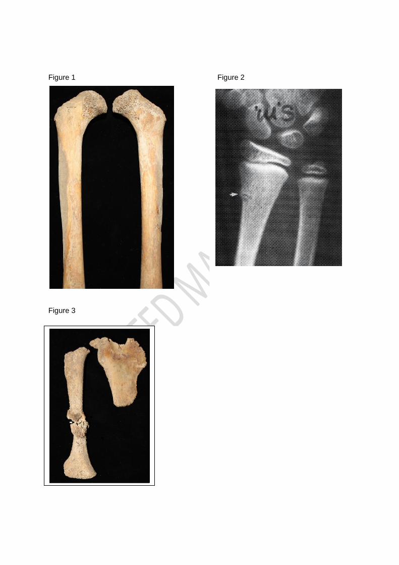
34
Figure 1 Figure 2
Figure 3