Steady-state flux and lag time in the stratum corneum lipid pathway: Results from finite element...
13
Steady-State Flux and Lag Time in the Stratum Corneum Lipid Pathway: Results from Finite Element Models H. FREDERICK FRASCH, ANA M. BARBERO Health Effects Laboratory Division, National Institute for Occupational Safety and Health, 1095 Willowdale Road, Morgantown, West Virginia 26505 Received 14 February 2003; revised 29 April 2003; accepted 1 May 2003 ABSTRACT: Finite element model (FEM) solutions of the diffusion through two- dimensional representations of the stratum corneum (SC) lipid pathway are presented. Both simplified, regular ‘‘brick and mortar’’ models and a more complex, irregular model are analyzed. It is assumed that diffusion occurs only within the SC lipids and the lipids are isotropic. The steady-state flux and lag time are solved and compared with the corresponding values for a homogeneous membrane of the same thickness consisting of lipid material. Results confirm that the heterogeneous SC model behaves like a homogeneous membrane, meaning that FEM diffusion simulations are well approxi- mated by an appropriate solution of the diffusion equation for a homogeneous membrane. Additionally, both steady-state flux and lag time (relative to these values in a homo- geneous membrane) can be predicted from algebraic equations based on simple dimensionless descriptors of SC geometry. However, values for diffusivity derived from homogeneous membrane approximations to the FEM solutions (effective diffusivity, D*) are not equal to the intrinsic diffusivity of the chemical in lipid. Furthermore, the pathlength derived from homogeneous membrane approximations to FEM solutions (effective pathlength, l*) is not equal to the lipid pathlength and is not dependent on SC tortuosity. Whereas l* is not a function of corneocyte overlap, D* is. These model results suggest that diffusion properties of the SC lipid pathway can be correlated to SC geometry, but intrinsic diffusion coefficients and SC tortuosity cannot be derived from common diffusion cell experiments. Use of the model equations to predict permeability and lag time of lipophilic solutes is described. ß 2003 Wiley-Liss, Inc. and the American Pharmacists Association J Pharm Sci 92:2196– 2207, 2003 Keywords: diffusion; mathematical model; percutaneous; skin; transdermal; perme- ability; lag time INTRODUCTION The diffusive flux of a permeant across a concen- tration gradient within a medium depends on its molecular mobility within the medium (diffusivity D) and on the geometry of the diffusion pathway. For a homogeneous membrane, the diffusi- onal pathlength is simply the thickness of the membrane. The stratum corneum (SC) is the thin (&10– 20 mm), outermost layer of skin and is the pri- mary permeation impediment of the skin barrier. The SC is not a homogeneous membrane; rather it is a biphasic arrangement of corneocytes— keratinized cellular remnants of epithelial dif- ferentiation—interposed with intercellular lipid lamellae. The lipid lamellae form a continuous pathway for diffusion through the SC, and today it is believed by many that this tortuous lipid path is the primary route of chemical permeation. Direct microscopic observation of model permeant localized primarily within the intercellular lipids, 1 as well as indirect evidence from biophysical 2196 JOURNAL OF PHARMACEUTICAL SCIENCES, VOL. 92, NO. 11, NOVEMBER 2003 Correspondence to: H. Frederick Frasch (Telephone: 304- 285-5755; Fax: 304-285-6041; E-mail: [email protected]) Journal of Pharmaceutical Sciences, Vol. 92, 2196–2207 (2003) ß 2003 Wiley-Liss, Inc. and the American Pharmacists Association
-
Upload
independent -
Category
Documents
-
view
5 -
download
0
Transcript of Steady-state flux and lag time in the stratum corneum lipid pathway: Results from finite element...
Steady-State Flux and Lag Time in the Stratum CorneumLipid Pathway: Results from Finite Element Models
H. FREDERICK FRASCH, ANA M. BARBERO
Health Effects Laboratory Division, National Institute for Occupational Safety and Health, 1095 Willowdale Road,Morgantown, West Virginia 26505
Received 14 February 2003; revised 29 April 2003; accepted 1 May 2003
ABSTRACT: Finite element model (FEM) solutions of the diffusion through two-dimensional representations of the stratum corneum (SC) lipid pathway are presented.Both simplified, regular ‘‘brick andmortar’’ models and amore complex, irregular modelare analyzed. It is assumed that diffusion occurs only within the SC lipids and the lipidsare isotropic. The steady-state flux and lag time are solved and compared with thecorresponding values for a homogeneous membrane of the same thickness consisting oflipid material. Results confirm that the heterogeneous SC model behaves like ahomogeneous membrane, meaning that FEM diffusion simulations are well approxi-mated by an appropriate solution of the diffusion equation for a homogeneousmembrane.Additionally, both steady-state flux and lag time (relative to these values in a homo-geneous membrane) can be predicted from algebraic equations based on simpledimensionless descriptors of SC geometry. However, values for diffusivity derived fromhomogeneous membrane approximations to the FEM solutions (effective diffusivity,D*)are not equal to the intrinsic diffusivity of the chemical in lipid. Furthermore, thepathlength derived from homogeneous membrane approximations to FEM solutions(effective pathlength, l*) is not equal to the lipid pathlength and is not dependent on SCtortuosity. Whereas l* is not a function of corneocyte overlap, D* is. These model resultssuggest that diffusion properties of the SC lipid pathway can be correlated to SCgeometry, but intrinsic diffusion coefficients and SC tortuosity cannot be derived fromcommon diffusion cell experiments. Use of the model equations to predict permeabilityand lag time of lipophilic solutes is described. � 2003 Wiley-Liss, Inc. and the American
Pharmacists Association J Pharm Sci 92:2196–2207, 2003
Keywords: diffusion; mathematical model; percutaneous; skin; transdermal; perme-ability; lag time
INTRODUCTION
The diffusive flux of a permeant across a concen-tration gradient within a medium depends on itsmolecular mobility within the medium (diffusivityD) and on the geometry of the diffusion pathway.For a homogeneous membrane, the diffusi-onal pathlength is simply the thickness of themembrane.
The stratum corneum (SC) is the thin (&10–20 mm), outermost layer of skin and is the pri-mary permeation impediment of the skin barrier.The SC is not a homogeneous membrane; ratherit is a biphasic arrangement of corneocytes—keratinized cellular remnants of epithelial dif-ferentiation—interposed with intercellular lipidlamellae. The lipid lamellae form a continuouspathway for diffusion through the SC, and today itis believed by many that this tortuous lipid pathis the primary route of chemical permeation.Direct microscopic observation of model permeantlocalized primarilywithin the intercellular lipids,1
as well as indirect evidence from biophysical
2196 JOURNAL OF PHARMACEUTICAL SCIENCES, VOL. 92, NO. 11, NOVEMBER 2003
Correspondence to: H. Frederick Frasch (Telephone: 304-285-5755; Fax: 304-285-6041; E-mail: [email protected])
Journal of Pharmaceutical Sciences, Vol. 92, 2196–2207 (2003)� 2003 Wiley-Liss, Inc. and the American Pharmacists Association
measurements,2 support this hypothesis; how-ever, it should be recognized that little directevidence exists to confirm or refute it.
Despite the geometric complexity of the SC lipidpathway, a number of investigators have de-monstrated that the SC displays properties of ahomogeneous membrane.3 This means that re-sults from SC diffusion experiments comparefavorably with predictions of the diffusion equa-tion for a homogeneous membrane, using appro-priately fitted parameters. The purpose of thisstudy was to investigate how the geometricarrangement of the SC lipid pathway determinesits diffusion properties compared with the proper-ties of a homogeneous membrane. We used a finiteelement modeling (FEM) approach to solve fordiffusion through the lipid pathway of model SCstructures, and compared results with the pre-dictions of the diffusion equation for a homo-geneous membrane. This approach yields valuesfor steady-state flux and time lag as well as valuesof the parameters that comprise these quantities.Algebraic expressions that depend on dimen-sionless SC geometric descriptors were explored,for the prediction of both steady-state flux andlag time.
METHODS
SC Geometry
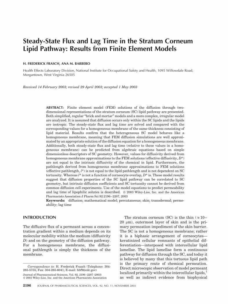
SC geometry is modeled in the simplest case as atwo-dimensional ‘‘brick and mortar’’ structure(Fig. 1), where the diffusing substance is con-strained to the ‘‘mortar.’’ The upper and lowersurfaces are not considered part of the diffusionpath. The membrane consists of N layers of cor-neocytes; the width of corneocytes is d and theirthickness is h. The lipid lamella are of thickness gin the horizontal direction and s in the vertical.
Several dimensionless parameters characterizethe geometry of this structure. Following Cussleret al.,4 the corneocyte aspect ratio is defined asa¼d/h and the slit shape is given by s¼ s/h. Fors�d, the fractional volume loading of corneo-cytes is approximated by f¼h/(hþ g). FollowingJohnson et al.,5 the corneocyte offset ratio is quan-tified by o¼ lL/lS, where lL is the length of thelongest overlapping lipid path and lS is the lengthof the shortest overlapping section (Fig. 1B). Itfollows that lLþ lS¼dþ s. o ranges from 1 forthe complete overlap ‘‘brick and mortar’’ modelof Figure 1A to 1 for the case where no overlapoccurs.
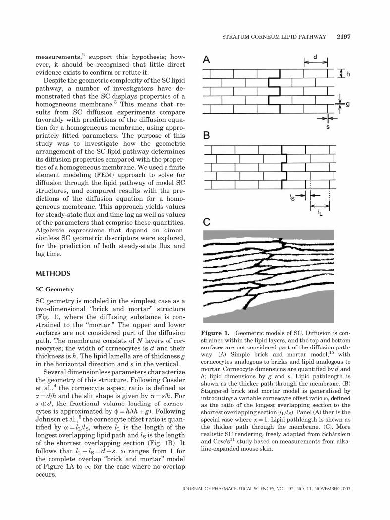
Figure 1. Geometric models of SC. Diffusion is con-strained within the lipid layers, and the top and bottomsurfaces are not considered part of the diffusion path-way. (A) Simple brick and mortar model,15 withcorneocytes analogous to bricks and lipid analogous tomortar. Corneocyte dimensions are quantified by d andh; lipid dimensions by g and s. Lipid pathlength isshown as the thicker path through the membrane. (B)Staggered brick and mortar model is generalized byintroducing a variable corneocyte offset ratio o, definedas the ratio of the longest overlapping section to theshortest overlapping section (lL/lS). Panel (A) then is thespecial case where o¼ 1. Lipid pathlength is shown asthe thicker path through the membrane. (C). Morerealistic SC rendering, freely adapted from Schatzleinand Cevc’s11 study based on measurements from alka-line-expanded mouse skin.
STRATUM CORNEUM LIPID PATHWAY 2197
JOURNAL OF PHARMACEUTICAL SCIENCES, VOL. 92, NO. 11, NOVEMBER 2003
The membrane thickness is given by l0¼Nhþ (N� 1)g; for a membrane comprised of manylayers, l0 is approximated by N(hþ g). The lipidpathlength is showngraphically as the total lengthof the thick line pathway inFigure 1AandB, and iscalculated by llip¼ l0þ (N� 1)lS. Geometric tor-tuosity is defined6 as tg¼ llip/l0. For a many-layered membrane and s�d, tg& 1þ af/(1þo).
For results presented here, we use realisticSC values of N¼ 20, h¼ 1 mm and g¼ s¼ 0.1 mm.Thus l0¼ 21.9 mm. These values were derived froma number of sources and include measurementsmade from mouse7,8 and human7,9,10 SC. o isvaried from 1 to1 and d from 20 to 60 mm to spanreported values for human SC. For typical humanSC, we take a¼ 40 (corresponding to d¼ 40 mm)and o¼ 3 (based on Talreja et al.’s6 estimate ofaverage tg for non-expanded human SC). To simu-late corneocyte swelling, we examined severalcases for h¼ 5 mm.6 To further investigate effectsof SC geometry on flux and lag time, we examinedseveral cases for g¼ s¼ 0.5 mm.
Geometric Descriptors of Irregular SC
To investigate the applicability of these geometricdescriptors to a more realistic, irregular SCstructure, we analyzed the rendering of a trans-verse section of SC shown in Figure 1C, derivedfrom hairless mouse skin after alkaline expan-sion, and freely adapted from Schatzlein andCevc.11
Statistical estimates of a, s, f, and o of themouse SC (Fig. 1C) were averaged from indepen-dent measurements made by two individuals(A.M.B. and H.F.F.) from digitized images usingsystematic sampling methods.12 The pixel wasused as the length unit. Figure 1C is 135 pixelswide and the total area of lipids þ corneocytes is10,181 square pixels. The image was divided into10 equal horizontal sections of length Dx. Arandom number r between 0 an Dxwas generated;then 10 vertical lines were placed over the image,beginning at distance r from the left edge andequally spaced Dx apart. The thickness of eachcorneocyte and each lipid layer intersecting the10 vertical lines were measured; their averagegave h and g respectively. We assumed s¼ g.Corneocyte length d was measured as the totalwidth of Figure 1C (135 pixels) minus s. Volumeratio f was measured as the ratio of the totalnumber of square pixels comprising corneocytes tototal number of square pixels comprising bothcorneocytesþ lipids. The average of the lengths of
the 10 vertical lines that intersect the SC wastaken as l0. The lipid pathlength llip wasmeasuredas the sum of the lengths of many small linesegments making a contiguous path between theupper and lower surfaces, placed by judgment ofthe analyst. Offset ratio was then calculated fromo¼ af/(tg� 1)� 1.
Finite Element Models
A commercial finite element program (ANSYS6.0, ANSYS Inc.) was used. The lipid layers oftwo-dimensional SC geometries were meshedwith quadrilateral (0.05� 0.05 mm), four-nodeelements with one degree of freedom (concentra-tion) at each node. A diffusion coefficient D0
within lipids is imposed, and it is assumed thatthe lipids are homogeneous and isotropic—thereis no difference between lateral and transversediffusivity. Whereas the complete overlap (o¼ 1)and no overlap (o¼1) models exhibit lateralsymmetry, the staggered overlap models (e.g.,Fig. 1B) have periodicity. Periodic boundaryconditions were applied using constraint equa-tions: the concentrations at corresponding loca-tions at the right edge and left edges of the lipidlayers are constrained to be equal for all times.This constraint means that the membrane istreated as being infinitely repeating.
Steady-state and transient solutions of thediffusion through these model SC lipid bilayerswere sought, with initial and boundary conditionsimposed to mimic in vitro diffusion cell experi-ments. That is, the membrane was initially at zeroconcentration throughout. At time zero, a constantconcentration difference was imposed across themembrane whereas the surface through whichdiffusing substance emerges was maintained atzero concentration. A sample ANSYS input pro-gram is available (see Supporting Information).
To demonstrate the use of this modelingapproach using a more realistic, irregular SCgeometry, we also developed an FEM based onthe rendering of a transverse section of expandedmouse SC shown in Figure 1C.Here, themesh sizewas 1 pixel� 1 pixel; lipids were again assumedhomogeneous and isotropic; and identical initialand boundary conditions were imposed, includingperiodicity.
Regression Analysis
For the accumulation of mass of permeant perunit area over time [Q(t)] under the initial and
2198 FRASCH AND BARBERO
JOURNAL OF PHARMACEUTICAL SCIENCES, VOL. 92, NO. 11, NOVEMBER 2003
boundary conditions imposed on the finite ele-ment models with a concentration difference Cmaintained between the two surfaces, the analy-tical solution for a homogeneous membrane ofthickness l is given by13:
QðtÞ ¼ ClDt
l2� 1
6� 2
p2X1n¼1
ð�1Þn
n2exp
�Dt
l2n2p2
� �" #
ð1Þ
Finite element model results are fitted to eq. (1) bynonlinear regression using SigmaPlot 2001 (SPSSInc.), which uses a Marquardt-Levenberg algo-rithm. The equation is truncated to 10 terms ofthe series. Results give estimates for the para-meters effective diffusivity (D*) and effectivepathlength (l*) and provide statistics relating togoodness of fit and variance of the parameters.Thus, D* and l* can be thought of as thediffusivity and thickness of a homogeneous mem-brane having the same permeability per unit areaand lag time as the heterogeneous SC lipidpathway.
For consistency in analysis of FEMs, the FEMsimulation time for all regressions was �10l*2/(6D*), and the maximum time step between FEMcalculated values was set at �0.5l*2/(6D*).
The FEM and regression approach was checkedby comparing results from a finite element modelof a homogeneous membrane with the analyticalsolution expressed by eq. (1). The errors inestimation of D and l were <1%. An alternateequation for flux14 [the derivative of eq. (1)], whichconvergesmore rapidly for small times thaneq. (1),was also tested. No significant differences in thevalues for D* and l* were found.
Estimates of Flux and Lag Timefrom SC Geometry
The total steady-state flux (units: mass/time)through a homogeneous membrane of area A0
and thickness l0, with concentration differenceC maintained between the two surfaces, is givenby
J0 ¼ CD0A0=l0 ð2Þ
The barrier properties of such a membrane arecharacterized by the steady-state flux and by thelag time,
tlag0 ¼ l20=ð6D0Þ ð3Þ
The analyses presented herein do not consider
any partitioning between membrane and sur-rounding vehicle.
Several estimates for flux of permeant confin-ed to the SC lipid path (J*) have been reported,based on SC geometry. The approach exemplifiedby Michaels et al.15 and Moghimi et al.16considersthe reduction in SC surface area normal to theflux and the increased tortuous lipid pathlength.For example, Moghimi et al.’s eq. (6)16 can bewritten as:
J0
J*¼ A0t
A*� a
s1þ af
1þ o
� �ð4Þ
with A0/A*&a/s. Michaels et al.’s eq. (22)15
follows a similar line of reasoning for the specialcase of complete corneocyte overlap (o¼ 1).
Cussler’s group4 derived the following ex-pression:
J0
J*¼ 1þ af
sþ a2f2
4ð1� fÞ ð5Þ
Equation (5) accounts in detail for the reduction inarea along the entire permeation pathway aswell as the increase in effective pathlength of thelipid pathway. The first term (unity) representsthe limiting case of a homogeneous membrane,where volume loading f is zero (no corneocytes).The second term represents the resistance of theshort vertical lipid slits between corneocytes inthe same horizontal plane, and the third termaccounts for resistance to diffusion of the tortuouspath around the corneocytes. See Cussler et al.4
for a complete derivation.Cussler et al.’s analysis, which was not speci-
fically related to skin, applies to the special casecorresponding to complete corneocyte overlap(Fig. 1A). Johnson et al.5 derived a factor toaccount for variable o:
J0
J*¼ 1þ af
sþ o
ð1þ oÞ2a2f2
ð1� fÞ ð6Þ
Geometry-based expressions for lag time in theSC lipid pathway have also been reported. BothFlynn et al.17 andMoghimi et al.16 have suggestedthe following:
tlag�
tlag0¼ t2 � 1þ af
1þ o
� �2
ð7Þ
whereas Cussler’s group18,19 propounds:
tlag�
tlag0¼ 1þ af
2
� �2
ð8Þ
STRATUM CORNEUM LIPID PATHWAY 2199
JOURNAL OF PHARMACEUTICAL SCIENCES, VOL. 92, NO. 11, NOVEMBER 2003

Anewexpression is proposed here. It is useful toconsider the components of the ratio J0/J* for theSC lipid pathway:
J0
J*¼ D0
l0
l*
D*
A0
A*ð9Þ
The effective pathlength l* is dominated by thelateral lipid pathways. Within each horizontallayer, transient diffusion occurs laterally to fillthe lipid lamellae. This lateral membrane capa-city significantly contributes to the observed lagtime. Hence, it seems reasonable to assert thateffective pathlength can be approximated by l*¼N(dþhþ g). This assertion will be tested later(Results). Multiplying eq. (9) by l*/l0& 1þ af,rearranging and substituting eq. (6), lag time[tlag*¼ l*2/(6D*)] can be expressed in terms of thegeometric descriptors:
tlag�
tlag0¼ sð1þ afÞ
a1þ af
aþ o
ð1þ oÞ2a2f2
ð1� fÞ
!
ð10Þ
Finite Element Results Comparedwith Geometric Estimates
Values for the parameters l* andD*, derived fromnonlinear regression of FEM model results witheq. (1), are used in eq. (9) to compare with thepredictions of eqs. (4)–(6). FEM lag times calcu-lated from l*2/(6D*) are compared with thepredictions of eqs. (7), (8), and (10).
RESULTS
Measured geometric descriptors of the SC mem-brane in Figure 1C are: a¼ 22; s¼ 0.33; f¼ 0.77;o¼ 10. We note that these values have beenderived from a rendering based on alkaline-expanded mouse SC, and do not apply to normalhuman SC.
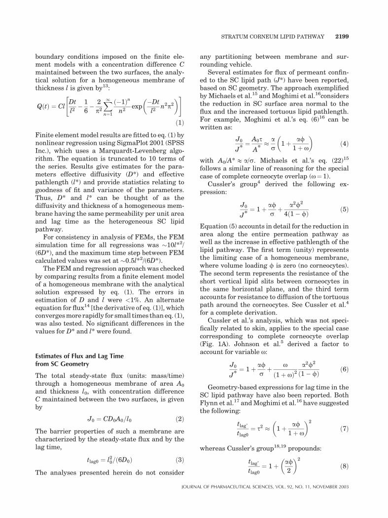
Figure 2 displays typical finite element modelresults for the diffusion simulations, along withbest-fit regression estimates using eq. (1). For allsimulations tested, the correlation coefficient (R2)exceeded 0.999. Estimates of the coefficients D*and l*were all highly significant (p<0.0001),withthe largest coefficient of variation 2.4% of theestimated value.
Figure 3 shows values for D* and l* for FEMsimulations that were performed using the brickand mortar and staggered brick and mortar
models. For clarity, values of o> 20 are notdisplayed, although simulations were performedfor o up to 1 (corresponding to no corneocyteoverlap).
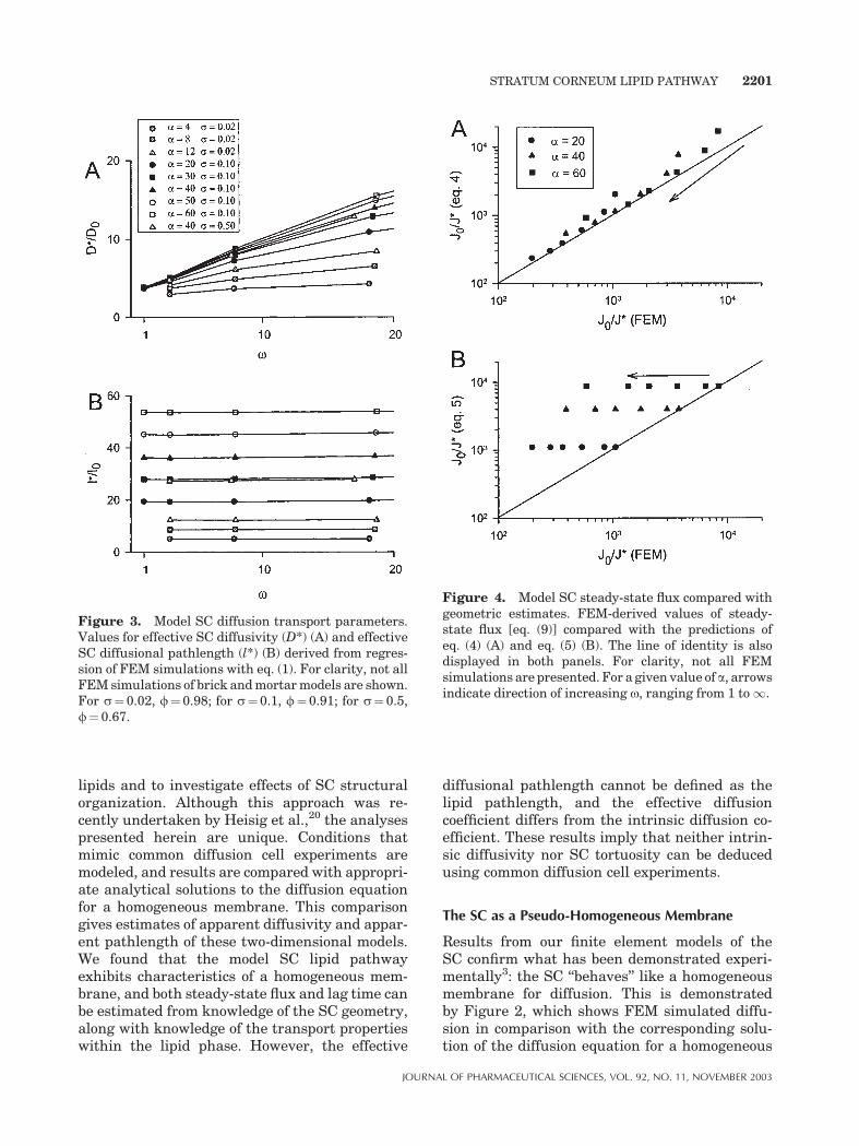
Figure 4 displays FEM-derived values ofsteady-state flux [eq. (9)] compared with thepredictions of eq. (4) (Fig. 4A) and eq. (5)(Fig. 4B). For clarity, not all FEM simulationsare presented. For a given value of a, arrowsindicate the direction of increasing o, rangingfrom 1 to 1.
Figure 5 displays FEM predictions of lag timecompared with the predictions of eq. (7) (Fig. 5A)and eq. (8) (Fig. 5B). For clarity, not all FEMsimulations are presented. For a given value of a,arrows indicate the direction of increasing o,ranging from 1 to1.
Figure 6A shows FEM-derived values for l* incomparison with the expression N(dþhþ g).Figure 6B shows steady-state flux from FEMmodels compared with the predictions of eq. (6).Figure 6C displays FEM lag time in comparisonwith the predictions of eq. (10). All FEM simula-tions are shown.
DISCUSSION
Summary
We have presented results from finite elementmodels to characterize diffusion through SC
Figure 2. Typical FEM results for diffusion simula-tions (symbols), along with best-fit regression estimatesusing eq. (1) (lines). Shown are simulations for a¼ 40,s¼ 0.1,f¼ 0.91, ando ranging from 1 to1. In all cases,the correlation coefficient (R2) betweenFEMresults andeq. (1) exceeds 0.999.
2200 FRASCH AND BARBERO
JOURNAL OF PHARMACEUTICAL SCIENCES, VOL. 92, NO. 11, NOVEMBER 2003
lipids and to investigate effects of SC structuralorganization. Although this approach was re-cently undertaken by Heisig et al.,20 the analysespresented herein are unique. Conditions thatmimic common diffusion cell experiments aremodeled, and results are compared with appropri-ate analytical solutions to the diffusion equationfor a homogeneous membrane. This comparisongives estimates of apparent diffusivity and appar-ent pathlength of these two-dimensional models.We found that the model SC lipid pathwayexhibits characteristics of a homogeneous mem-brane, and both steady-state flux and lag time canbe estimated from knowledge of the SC geometry,along with knowledge of the transport propertieswithin the lipid phase. However, the effective
diffusional pathlength cannot be defined as thelipid pathlength, and the effective diffusioncoefficient differs from the intrinsic diffusion co-efficient. These results imply that neither intrin-sic diffusivity nor SC tortuosity can be deducedusing common diffusion cell experiments.
The SC as a Pseudo-Homogeneous Membrane
Results from our finite element models of theSC confirm what has been demonstrated experi-mentally3: the SC ‘‘behaves’’ like a homogeneousmembrane for diffusion. This is demonstratedby Figure 2, which shows FEM simulated diffu-sion in comparison with the corresponding solu-tion of the diffusion equation for a homogeneous
Figure 3. Model SC diffusion transport parameters.Values for effective SC diffusivity (D*) (A) and effectiveSC diffusional pathlength (l*) (B) derived from regres-sion of FEM simulations with eq. (1). For clarity, not allFEMsimulations of brick andmortarmodels are shown.For s¼ 0.02, f¼ 0.98; for s¼ 0.1, f¼ 0.91; for s¼ 0.5,f¼ 0.67.
Figure 4. Model SC steady-state flux compared withgeometric estimates. FEM-derived values of steady-state flux [eq. (9)] compared with the predictions ofeq. (4) (A) and eq. (5) (B). The line of identity is alsodisplayed in both panels. For clarity, not all FEMsimulations are presented. For a given value of a, arrowsindicate direction of increasing o, ranging from 1 to1.
STRATUM CORNEUM LIPID PATHWAY 2201
JOURNAL OF PHARMACEUTICAL SCIENCES, VOL. 92, NO. 11, NOVEMBER 2003
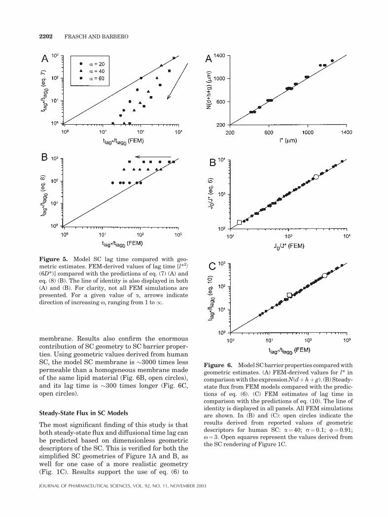
membrane. Results also confirm the enormouscontribution of SC geometry to SC barrier proper-ties. Using geometric values derived from humanSC, the model SC membrane is �3000 times lesspermeable than a homogeneous membrane madeof the same lipid material (Fig. 6B, open circles),and its lag time is �300 times longer (Fig. 6C,open circles).
Steady-State Flux in SC Models
The most significant finding of this study is thatboth steady-state flux and diffusional time lag canbe predicted based on dimensionless geometricdescriptors of the SC. This is verified for both thesimplified SC geometries of Figure 1A and B, aswell for one case of a more realistic geometry(Fig. 1C). Results support the use of eq. (6) to
Figure 5. Model SC lag time compared with geo-metric estimates. FEM-derived values of lag time [l*2/(6D*)] compared with the predictions of eq. (7) (A) andeq. (8) (B). The line of identity is also displayed in both(A) and (B). For clarity, not all FEM simulations arepresented. For a given value of a, arrows indicatedirection of increasing o, ranging from 1 to1.
Figure 6. ModelSCbarrier properties comparedwithgeometric estimates. (A) FEM-derived values for l* incomparisonwith theexpressionN(dþhþ g). (B)Steady-state flux from FEM models compared with the predic-tions of eq. (6). (C) FEM estimates of lag time incomparison with the predictions of eq. (10). The line ofidentity is displayed in all panels. All FEM simulationsare shown. In (B) and (C): open circles indicate theresults derived from reported values of geometricdescriptors for human SC: a¼ 40; s¼ 0.1; f¼ 0.91;o¼ 3. Open squares represent the values derived fromthe SC rendering of Figure 1C.
2202 FRASCH AND BARBERO
JOURNAL OF PHARMACEUTICAL SCIENCES, VOL. 92, NO. 11, NOVEMBER 2003
predict steady-state flux and eq. (10) to predicttime lag. Although the full range of values for allvariables (a, s; f, and o) was not tested, a widerange that fully spans reported values for normaland swollen SC was examined.
Figure 6B displays excellent correlation be-tween the FEM-derived values of steady-state fluxand the predictions of eq. (6). Neither eq. (4) noreq. (5) display the same level of correlation (Fig. 4).Equation (4) fails because, although it accounts forthe reduction in cross-sectional area available fordiffusion at the surfaces of the membrane, it doesnot take into consideration the diffusion areasalong the entire permeation pathway. (The equa-tion derived byMichaels et al.15 is similar to eq. (4),but slightly more complex in that Michaels et al.appear to consider the SC surface of a three-dimensional structure with a two-dimensionallipid pathway.) Equation (5) correctly accountsfor different areas for diffusion along the entirepathway but fails because it does not considercorneocyte overlap. This equation, derived byCussler et al.,4 was applied to artificial barriermembranes containing selective impermeableflakes and has undergone experimental verifica-tion.19 In applying this analysis to the SC, Johnsonet al.5 derived the dependence of flux on o andpresented a form of eq. (6), which reduces to eq. (5)for the special case of o¼ 1. The results presentedhere verify the use of eq. (6) for the prediction ofsteady-state flux and permeability.
Lag Time in SC Models
No previous expression known to the authors wasable to adequately predict lag time on the basis ofgeometric descriptors. Previous expressions16–19
fail because of incorrect assumptions regardingwhat constitutes effective diffusivity and effectivepathlength (see Appendix). The FEM resultspresented here suggest an alternate approach.An appropriate expression for l* has been devel-oped and confirmed by the FEM model results(Fig. 6A). Using this expression in eq. (9), alongwith the appropriate expression for J0/J* [eq. (6)],creates a new formula for time lag in the SC lipidpathway. Equation (10) exhibits excellent correla-tion with the lag time derived from the FEMmodels (Fig. 6C).
For o¼ 1, both eqs. (7) and (8) adequatelypredict the FEM lag times (Fig. 5). This can beunderstood by simplifying eq. (10) for conditionsthat generally hold for the SC. In making theapproximation (1þ af)/a&f (valid for af>> 1),
and noting that sf /(1�f)¼ 1 (for s¼ g), eq. (10)reduces to
tlag�
tlag0¼ sfþ af2 þ o
ð1þ oÞ2a2f2 ð11Þ
With o¼ 1, this is very close to both eqs. (7) and(8), because the second order terms dominate.
Effective Pathlength in SC Models
It is noteworthy that the effective pathlength l*derived from the FEM models is independent ofcorneocyte overlap (Fig. 3B). In other words,effective pathlength does not depend on lipidtortuosity but rather is well predicted by theexpression N(dþhþ g) (Fig. 6A), so that theratio l*/l0& 1þ af. This can be understood byconsidering transient diffusion within the lipidlamellae. Until the steady state is reached, aconcentration gradient develops along the laterallipid layers that produces a net flux of permeant:there is initially zero concentration within thelateral layers, and as permeant diffuses down-ward from the surface, a non-zero concentrationexists at the intersection of the vertical slits andthe lateral paths. A concentration gradient devel-ops along both sides of a vertical slit until netpermeant flux from the two vertical slits oneither side of it eliminates the gradient—result-ing in no further net flux. Thus, this lateralmembrane capacity contributes to the overalleffective diffusional pathlength. The total lat-eral component of l* is then given by (N� 1)dwhereas the vertical component is identical to l0.Therefore l*¼ (N� 1)(dþ g)þNh&N(dþhþ g).
This is not to say that time lag does not de-pend on corneocyte overlap. Both steady-stateflux [eq. (6)] and time lag [eq. (10)] are func-tions of o. This dependence is captured in theFEM-derived values for D*. (An expression forD* can be obtained from eqs. (9) and (6), substitut-ing the expressions for l* and for A0/A* givenpreviously.)
Significance of Results
The significance of these results is underscored byrecent theoretical21–23 and experimental2,24–27
investigations of the kinetics of percutaneousabsorption. Appropriate values for both effectivediffusivity and effective pathlength are requiredto calculate time-dependent skin diffusion phe-nomena such as time lag, membrane desorption,
STRATUM CORNEUM LIPID PATHWAY 2203
JOURNAL OF PHARMACEUTICAL SCIENCES, VOL. 92, NO. 11, NOVEMBER 2003
finite dosing, short-term exposures, and repeat-ed drug dosing. Experimental estimates of dif-fusion coefficients and pathlengths have beenmade from combined steady-state and non-steady-state diffusion measurements in isolatedSC and epidermal membranes.2,25,26 The analysisof Bunge et al.28 demonstrates some of the pitfallsencountered if, for example, no consideration isgiven to the reduction in cross-sectional areaavailable for diffusion. The results presented heresuggest further pitfalls, even if cross-sectionalarea is considered: the diffusion coefficients deriv-ed from experimental results cannot be directlyrelated to intrinsic diffusion coefficients, andthe pathlength derived from experimental resultsis not a function of SC tortuosity. Potts andFrancoeur2 estimated a diffusional pathlength of�50 times greater than SC thickness for watervapor diffusion. (Whether this value holds formore lipophilic substances is not known.) Al-though it is difficult to postulate an SC tortuosityof 50 (reasonable values6 are close to 10), 50 isclose to the value of 36 for l*/l0 predicted by theFEM model for human SC values (Fig. 3B; a¼ 40,s¼ 0.1).
Application of Model Equations to PredictPermeability and Lag Time of a Permeant
The equations presented here can be used topredict permeability and lag time of a given solutein the SC. For solutes that diffuse solely withinthe lipid phase of the SC, the steady-statepermeability coefficient is given by:
kp ¼ KL VD*
l*
A*
A0¼ KL V
J*
J0
D0
l0ð12Þ
where KL_V is the equilibrium partition coefficientbetween the lipid fraction of the SC and thesurrounding vehicle. Note that this differs fromthe more commonly reported SC-vehicle partitioncoefficient, which represents the bulk distributionof solute between the total volume of SC andvehicle:
KL V ¼ KSC V
1� fð13Þ
Therefore, permeability can be predicted based onknowledge of SC geometry, with the ratio J*/J0
given by eq. (6), and on the diffusion coefficient ofthe permeant in lipids. Reasonable values for SCgeometry are given in the text. Johnson et al.29
measured lateral diffusion coefficients in lipidsextracted from human SC. Using the fluorescence
recovery after photobleaching method, theyreport molecular weight (MW)-dependent diffu-sivities of several lipophilic fluorescent probesranging from 3.06� 10�9 to 2.34� 10�8 cm2/s, forMW of 629 and 223 Da, respectively. Additionally,the authors report a two-parameter empiricalequation that nicely correlates diffusivity as afunction of MW.
Lag time for diffusion within the SC lipids canalso bepredicted fromSCgeometry andknowledgeof D0. For a given solute, tlag* can be obtainedby multiplying the right-hand side of eq. (10) byl02/(6D0), with D0 again estimated from the resultsof Johnson et al.29
Model Limitations and Conclusions
In the models presented here, we impose theconditions that diffusion occurs only within lipidbilayers and that diffusivity within the lipids isisotropic. These simplifications are model limita-tions but may be acceptable if, as postulated byJohnson et al.,5 lateral lipid bilayer diffusionis the primary permeation pathway in the SC.Mitragotri30 recently described four possible per-meation pathways with lateral lipid transporttaking a less prominent role; however, little directexperimental evidence exists in support of a parti-cular transport mechanism in skin. Model resultspresented here are valid only for the lateral lipidbilayer diffusion pathway.
In conclusion, we have presented results fromtwo-dimensional FEMmodels for diffusion withinlipid bilayers of SC membranes. The resultsdemonstrate important implications about theeffective diffusional pathlength and effective dif-fusivity within the SC. Although the exact rela-tionship between parameters of the SC geometryand estimates of steady-state flux and lag timemay differ for a more realistic SC that has to bedescribed as a three-dimensional structure, thiswork makes it reasonable to assume that thoserelationships can be determined with additionalanalysis.
SUPPORTING INFORMATION
Supporting material is available for this articleat the following Web address: http://dx.doi.org/10.1002/jps.10466, or from the authors. It is a com-mented ANSYS input file that is used to generateand solve a finite element model of diffusionthrough SC lipids conforming to a staggered brickand mortar geometry (Fig. 1B). Note that we use a
2204 FRASCH AND BARBERO
JOURNAL OF PHARMACEUTICAL SCIENCES, VOL. 92, NO. 11, NOVEMBER 2003
heat conduction-diffusion analogy (see Crank J.1975. The mathematics of diffusion, 2nd ed.Oxford: Clarendon Press, pp 8–10).
APPENDIX
Further Discussion on Effective Diffusivityand Pathlength
Our use of the terms D* and l*—effectivediffusivity and effective pathlength—differssomewhat from commonly used terms apparentdiffusivity and apparent pathlength. Here, weexplain the differences and why the ‘‘new’’ termsare appropriate. The problem with the terms ascommonly defined is that they cannot be used tocorrectly predict both steady-state and transientdiffusion phenomena.
In a tradition that extends over �125 yearssince Maxwell, for diffusion within heterogeneousor composite media, an apparent diffusion coeffi-cient (D0) is used to incorporate the effects ofgeometry on the permeation of the diffusingcompound.31 As applied to the SC, D0 is thediffusion coefficient that is postulated when theSC is treated as a pseudo-homogeneousmembranewhose thickness is the same as the SC.28
Such an approachwould be perfectly acceptablehere if one were interested only in steady-stateflux. IfD0 as defined here were used, then the ratioof steady-state flux in thehomogeneousmembraneto that in the SC is given by
J0
J*¼ D0
D0A0
A*ðA:1Þ
Accordingly, an expression for D0 can be develop-ed in terms of our D* and l* [from eq. (9), maintext]:
D0 ¼ D*l0l*
ðA:2Þ
Use of this expression in eq. (A.1) would thensatisfy the results of the finite element models, forthe steady state.
However, if these concepts are applied to acalculation of lag time in the SC, we obtain:
tlag ¼ l206D0 ¼
l0l*
6D*6¼ tlag� ðA:3Þ
The lag time calculated from eq. (A.3) does notsatisfy the results of the FEM. Therefore, theuse of apparent diffusivity as it has been tradi-tionally defined cannot simultaneously account
for both steady-state and non-steady-state diffu-sion phenomena. If steady-state permeability isthe sole interest, which has generally been thecase in the traditional use of the concept, then theapproach is perfectly acceptable.
Another common approach is to consider anapparent pathlength in the SC as equal to the lipidpathlength. The steady-state flux ratio is thengiven by:
J0
J*¼ D0
D00llipl0
A0
A*ðA:4Þ
D00 is still an apparent diffusion coefficient, but itsvalue differs from D0. In order for eq. (A.4) tosatisfy the FEM results for steady-state flux,
D00 ¼ D*llipl*
ðA:5Þ
Applying these definitions to the calculation of lagtime in the SC:
tlag ¼l2lip6D00 ¼
llipl*
6D*6¼ tlag� ðA:6Þ
We run into the same problem as before: wecannot use the lipid pathlength to simultaneouslyaccount for both steady-state and non-steady-state phenomena.
A special case of the above approach is toconsider both that apparent pathlength is equalto the lipid pathlength, and that apparent diffu-sivity is equal to the diffusivity within the lipidphase. For this case, eq. (4) (main text) results asthe ratio of fluxes, and eq. (7) (main text) results todescribe the ratio of lag times. The analysesdescribed in the main text of this article demon-strate that these expressions fail.
Yet another approach has been taken, and thatis to lump all effects of geometry into an ‘‘effectivetortuosity’’5 as follows:
J0
J*¼ teff ðA:7Þ
Note that these authors use the term t*; however,we do not wish to confuse their terminology withours. To satisfy the FEM results for steady-stateflux, then,
teff ¼D0
D*
l*
l0
A0
A*ðA:8Þ
This concept has been applied to the calculation ofa diffusional transport time for desorption fromthe SC.26,32 This transport time equals 6� tlag.
STRATUM CORNEUM LIPID PATHWAY 2205
JOURNAL OF PHARMACEUTICAL SCIENCES, VOL. 92, NO. 11, NOVEMBER 2003
Accordingly,
tlag ¼ l0teffð Þ2
6D0¼ l*2D0
6D*2A0
A*
� �2
6¼ tlag� ðA:9Þ
Thus, the concepts of apparent diffusivity andpathlength, as traditionally used, fail to accountfor both steady-state permeability and non-steadydiffusionphenomena suchas lag time, as predictedby the finite element models of the SC lipidpathway. In the present study, the concepts ofeffective diffusivity and effective pathlength aredefined as the diffusivity and thickness of ahomogeneousmembrane, having the same perme-ability (per unit area) and lag time, as the SC lipidpathway. Used together, they are able to de-scribe both steady-state and transient diffusionphenomena.
NOMENCLATURE
List of Abbreviations Used in MathematicalDevelopment
A0 area of homogeneous membrane surfacenormal to direction of flux
A* area of lipid lamella at surface of membranenormal to direction of flux
C concentrationd corneocyte lengthD diffusivityD0 intrinsic diffusivity; diffusivity within SC
lipidsD* effective diffusivity in a membrane where
permeation occurs exclusively within lipidlamellar pathway
g lipid lamellar thickness in horizontal direc-tion
h corneocyte thicknessJ0 total steady-state flux through a specified
area of a homogeneous membraneJ* total steady-state flux through a specified
area of the exclusively lipid lamellar path-way
l diffusional pathlengthl0 homogeneous membrane thickness; SC
thicknessl* effective SC pathlength, where permeation
occurs exclusively within lipid lamellarpathway
llip length of SC lipid pathwaylL length of long sections of lipid pathway
associated with overlapping sections ofcorneocytes
lS length of short sections of lipid pathwayassociated with overlapping sections ofcorneocytes
N number of corneocyte layersQ mass of permeant per unit areas lipid lamellar thickness in vertical directiont timetlag0 lag time for diffusion within homogeneous
membranetlag* lag time for diffusion within the exclusively
lipid pathwaya corneocyte aspect ratio¼d/hs slit shape¼ s/htg geometric tortuosity of lipid pathway¼ llip/l0f volume fraction of corneocyteso corneocyte offset ratio¼ lL/lS
REFERENCES
1. Bodde HE, van den Brink I, Koerten HK, de HaanFHN. 1991. Visualization of in vitro percutaneouspenetration of mercuric chloride: Transportthrough intercellular space versus cellular uptakethrough desmosomes. J Control Release 15:227–236.
2. Potts RO, Francoeur ML. 1991. The influence ofstratum corneum morphology on water permeabil-ity. J Invest Dermatol 96:495–499.
3. Kalia YN, Pirot F, Guy RH. 1996. Homogeneoustransport in a heterogeneous membrane: Waterdiffusion across human stratum corneum in vivo.Biophys J 71:2692–2700.
4. Cussler EL, Hughes SE, Ward WJ III, Aris R. 1988.Barrier membranes. J Membr Sci 38:161–174.
5. Johnson ME, Blankschtein D, Langer R. 1997.Evaluation of solute permeation through thestratum corneum: Lateral bilayer diffusion as theprimary transport mechanism. J Pharm Sci86:1162–1172.
6. Talreja PS, Kasting GB, Kleene NK, Pickens WL,Wang T-F. 2001. Visualization of the lipid barrierand measurement of lipid pathlength in humanstratum corneum. AAPS Pharm Sci 3(2): Article 13(http://www.pharmsci.org).
7. Wildnauer RH, Miller DL, Humphries WT. 1975. Aphysicochemical approach to the characterizationof stratum corneum. In: Baier RE, editor. Appliedchemistry at protein interfaces. Washington DC:American Chemical Society, pp 74–124.
8. Menton DN. 1976. A minimum-surface mechanismto account for the organization of cells into columnsin the mamallian epidermis. Am J Anat 145:1–21.
9. Mershon MM. 1975. Barrier surfaces of skin. In:Baier RE, editor. Applied chemistry at proteininterfaces. Washington DC: American ChemicalSociety, pp 41–73.
2206 FRASCH AND BARBERO
JOURNAL OF PHARMACEUTICAL SCIENCES, VOL. 92, NO. 11, NOVEMBER 2003
10. Elias PM, Cooper ER, Korc A, Brown BE. 1981.Percutaneous transport in relation to stratumcorneum structure and lipid composition. J InvestDermatol 76:297–301.
11. Schatzlein A, Cevc G. 1998. Non-uniform cellularpacking of the stratum corneum and permeabilitybarrier function of intact skin: A high-resolutionconfocal laser scanning microscopy study usinghighly deformable vesicles (Transfersomes). Br JDermatol 138:583–592.
12. Bolender RP, Pentcheff ND. 1992. Currentmethodsin quantitative morphology. QM2000 Version 2.0.Seattle: University of Washington.
13. Crank J. 1975. The mathematics of diffusion, 2nded. Oxford: Clarendon Press, p 51.
14. Rogers WA, Buritz RS, Albert D. 1954. Diffusioncoefficient, solubility, and permeability for heliumin glass. J Appl Phys 25:868–875.
15. Michaels AS, Chandrasekaran SK, Shaw JE. 1975.Drug permeation through human skin: Theory andin vitro experimental measurement. AIChE J 21:985–996.
16. Moghimi HR, Williams AC, Barry BW. 1996. Alamellar matrix model for stratum corneum in-tercellular lipids. II. Effect of geometry of thestratum corneum on permeation of model drugs 5-fluorouracil and oestradiol. Int J Pharm 131:117–129.
17. Flynn GL, Yalkowsky SH, Roseman TJ. 1974. Masstransport phenomena and models: Theoreticalconcepts. J Pharm Sci 63:479–510.
18. Perry D, Ward WJ, Cussler EL. 1989. Unsteadydiffusion in barrier membranes. J Membr Sci44:305–311.
19. Lape NK, Yang C, Cussler EL. 2002. Flake-filledreactive membranes. J Membr Sci 209:271–282.
20. Heisig M, Lieckfeldt R, Wittum G, Mazurkevich G,Lee G. 1996. Non steady-state descriptions of drugpermeation through stratum corneum. I. Thebiphasic brick-and-mortar model. Pharm Res 13:421–426.
21. Anissimov YG, Roberts MS. 1999. Diffusion model-ing of percutaneous absorption kinetics I. Effects of
flow rate, receptor sampling rate and viable epider-mal resistance for a constant donor concentration.J Pharm Sci 88:1201–1209.
22. Anissimov YG, Roberts MS. 2001. Diffusion model-ing of percutaneous absorption kinetics. II. Finitevehicle volume and solvent deposited solids. JPharm Sci 90:504–520.
23. Kalia YN, Guy RH. 2002. Modeling transdermaldrug release. Adv Drug Deliv Rev 48:159–172.
24. Roberts MS, Triggs EJ, Anderson RA. 1975. Per-meability of solutes through biological membranesmeasured by a desorption technique. Nature 257:225–227.
25. Pellett MA, Watkinson AC, Hadgraft J, Brain KR.1997. Comparison of permeability data from tradi-tional diffusion cells and ATR-FTIR spectroscopy.Part II. Determination of diffusional pathlengths insynthetic membranes and human stratum cor-neum. Int J Pharm 154:217–227.
26. Mitragotri S. 2000. In situ determination of parti-tion and diffusion coefficients in the lipid bilayers ofstratum corneum. Pharm Res 17:1026–1029.
27. Kasting GB. 2001. Kinetics of finite dose absorptionthrough skin. I. Vanillylonanamide. J Pharm Sci90:202–212.
28. Bunge AL, Guy RH, Hadgraft J. 1999. Thedetermination of a diffusional pathlength throughthe stratum corneum. Int J Pharm 188:121–124.
29. Johnson ME, Berk DA, Blankschtein D, Golan DE,Jain RK, Langer RS. 1996. Lateral diffusion ofsmall compounds in human stratum corneum andmodel lipid bilayer systems. Biophys J 71:2656–2668.
30. Mitragotri S. 2003. Modeling skin permeabilityto hydrophilic and hydrophobic solutes based onfour permeation pathways. J Control Release 86:69–92.
31. Crank J. 1975. The mathematics of diffusion, 2nded. Oxford: Clarendon Press, pp 266–285.
32. Mitragotri S. 2001. Effect of therapeutic ultrasoundon partition and diffusion coefficients in humanstratum corneum. J Control Release 71:23–29.
STRATUM CORNEUM LIPID PATHWAY 2207
JOURNAL OF PHARMACEUTICAL SCIENCES, VOL. 92, NO. 11, NOVEMBER 2003
Erratum: Steady-State Flux and Lag Time in the StratumCorneum Lipid Pathway: Results from Finite Element Models
H. FREDERICK FRASCH, ANA M. BARBERO
Health Effects Laboratory Division, National Institute for Occupational Safety and Health, 1095 Willowdale Road,Morgantown, West Virginia 26505
Received 14 February 2003; revised 29 April 2003; accepted 1 May 2003
Published online in Wiley InterScience (www.interscience.wiley.com). DOI 10.1002/jps.20133
The original article to which this Erratum refers was published in Journal of Pharmaceutical Sciences92(11) 2003, 2196–2207.
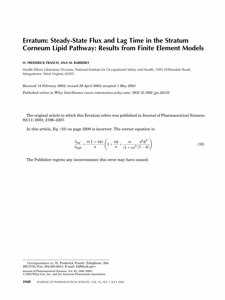
In this article, Eq. (10) on page 2200 is incorrect. The correct equation is:
tlag�
tlag0¼ sð1þ afÞ
a1þ af
sþ o
ð1þ oÞ2a2f2
ð1� fÞ
!ð10Þ
The Publisher regrets any inconvenience this error may have caused.
1940 JOURNAL OF PHARMACEUTICAL SCIENCES, VOL. 93, NO. 7, JULY 2004
Correspondence to: H. Frederick Frasch (Telephone: 304-285-5755; Fax: 304-285-6041; E-mail: [email protected])
Journal of Pharmaceutical Sciences, Vol. 93, 1940 (2004)� 2004 Wiley-Liss, Inc. and the American Pharmacists Association


![Arrangement of ceramide [EOS] in a stratum corneum lipid model matrix: new aspects revealed by neutron diffraction studies](https://static.fdokumen.com/doc/165x107/631f0e12198185cde200ea75/arrangement-of-ceramide-eos-in-a-stratum-corneum-lipid-model-matrix-new-aspects.jpg)