Crossmodal divided attention in rats: effects of chlordiazepoxide and scopolamine
Upload
uni-hamburgCategory
view
2download
0
Original Paper
Skin Pharmacol Physiol 2004;17:246–257DOI: 10.1159/000080218
SSkikinPharmacolology y
Physioliologyand
Dead but Highly Dynamic –The Stratum corneum Is Dividedinto Three Hydration Zones
T. Richterb C. Peuckertc M. Sattlera K. Koenigc I. Riemannc U. Hintzea
K.-P. Witterna R. Wiesendangerb R. Wepfa
aAnalytical Microscopy Department, R&D, Beiersdorf AG, and bDepartment of Applied Physics,University of Hamburg, Hamburg, and cCenter for Laser Microscopy, Faculty of Medicine, University of Jena,Jena, Germany
Received: January 27, 2004Accepted after revision: May 11, 2004
Roger WepfAnalytical Microscopy, PB 518, R&D, Beiersdorf AGUnnastrasse 48DE–20245 Hamburg (Germany)Tel. +49 40 4909 2588, Fax +49 40 4909 6822, E-Mail [email protected]
ABCFax + 41 61 306 12 34E-Mail [email protected]
© 2004 S. Karger AG, Basel1660–5527/04/0175–0246$21.00/0
Accessible online at:www.karger.com/spp
Key WordsFrozen hydrated W Stratum corneum W Hydration W
Osmolarity W Skin barrier
AbstractTopically applied water exerts mechanical stress on indi-vidual corneocytes as well as on the whole stratum cor-neum (SC), resulting in an alteration of barrier function.In this study we used complete skin biopsies and showedthat the SC reacts to water stress as a highly optimizedand well-regulated structure against osmotic changes.Following a relatively new cryo-processing protocol forcryo-SEM, it is possible to reliably maintain and investi-gate the hydrated state of the SC and individual corneo-cytes after treatment with solutions of different ionicstrength. Treatment with distilled water results in swell-ing of SC cells together with formation of massive waterinclusions between adjacent cell layers. Treatment with5–20% NaCl reveals three different hydration zones with-in the SC: Corneocytes near the live-dead transition zonecan swell to nearly double their thickness. The secondzone is the most compact, as the corneocytes here showthe smallest thickness variation with all treatments.Within the outermost zone, again a massive swelling andloosening of intracellular filament packing can be ob-
served. We therefore conclude that the SC itself is subdi-vided into three functional zones with individual waterpenetration and binding potentials. Since the secondzone remains nearly unaffected by water stress, we pro-pose that this zone hosts the functional SC barrier.
Copyright © 2004 S. Karger AG, Basel
Introduction
Human skin has evolved into a highly specialized andperfectly optimized organ. Its outermost layers serve as apenetration, dehydration and protection barrier againstvarious environmental hazards [1].
Hydration of the nonviable outermost layer of skin, thestratum corneum (SC), plays an important role in main-taining the metabolism, enzyme activity, mechanicalproperties, appearance and finally barrier function of theskin. Water is continuously exchanged at a very low ratebetween the viable epidermis, the SC and the atmosphere.The SC hydration state depends on the relative externalhumidity, the capacity of the epidermis to replace evapo-rative water losses, and the intrinsic water binding capaci-ty of the individual cells in the SC, the corneocytes. Theyare supposed to be responsible for the water holdingcapacity of the SC, partly due to their high content of

SC Consisting of Three Hydration Zones Skin Pharmacol Physiol 2004;17:246–257 247
hydrophilic proteins, osmolytes and natural moisturizingfactors (NMFs) such as free amino acids [2–4]. Togetherwith the cornified envelope these structures do not onlydetermine the water content but also determine the typi-cal flattened shape and stability of corneocytes. They alsohave to maintain the skin barrier function, independentof their hydration state.
Very little is known about the functional and structuralchanges in the SC due to water uptake. Data published onthe natural water content of normal SC range from 10 to20% in ex vivo investigations [5, 6] to about 35% in invivo Raman measurements [4]. Changes in the whole SCupon water uptake have been investigated by Norlén et al.[7] using confocal laser scanning microscopy: after treat-ment with distilled water SC swelling was 8% in area and26% in thickness. However, due to the typical thickness ofcorneocytes (around 200–500 nm), investigation at thelevel of single corneocytes by optical microscopy is ratherdifficult, especially if changes in thickness need to be mea-sured. Atomic force microscopy, on the other hand, hasthe resolution power but enables only investigation of sin-gle cell, which swell about 50% in thickness [8, 9], butnever the complete SC. Since both methods have shownthat corneocytes and the entire SC preferentially swellonly vertically, the question arose whether the skin barrierremains stable with such massive volume changes, espe-cially as Warner et al. [10] had shown that extended waterexposure disrupts the SC (fig. 6a).
Decreased SC water content is found in a number ofcommon skin diseases such as atopic dermatitis, eczemaor psoriasis and has an impact on the ion and pH gradientthroughout the SC [2]. Such changes influence enzymeactivity [11] and structural integrity and hence the barrierproperties. Therefore we have further investigated thestructural response of the SC to water treatments of differ-ent ionic strength, from distilled water to 20% NaCl.These investigations provide new insights into the struc-tural organization and dynamic potential of the SC uponwater uptake or loss and may also help to understand thepositive effects of salt bath for psoriasis treatment.
Materials and Methods
Pretreatment and Conditioning of Skin BiopsiesThirty-six biopsies were taken from the inside (flexor side) of the
upper arm of a healthy 73-year-old female. To minimize the biologi-cal variability within this study, all 3-mm punch biopsies were takenfrom the same area. Afterwards, they were completely immersed indifferent salt solutions (0, 5, 10, 15, or 20% NaCl in double-distilledwater). Therefore, water penetration through the sidewall of the biop-sies was also possible. Control biopsies were placed in a humidity
chamber with a saturated water atmosphere without immersing themin any liquid but with the dermis resting on filter paper (submergedin minimal essential medium). After 12 h of storage at 5 °C, all biop-sies were withdrawn from their chambers, remaining fluid was care-fully removed with a wipe. Subsequently, the samples were imme-diately frozen by plunging them at high speed into liquid ethane(temperature around –160°C) and stored in liquid nitrogen until fur-ther processing.
Control ExperimentControl experiments were done with 30 biopsies from the face,
next to the ears, of a healthy, 60-year-old female. They were treatedtopically with distilled water or 20% NaCl. The liquid exchangethrough the sidewall was prevented by lateral application of Vaseline.Comparison of these biopsies with those of the main experimentsprovides insights into water penetration pathways parallel and per-pendicular to the SC. To study the water uptake from a saturated gasatmosphere, some biopsies were placed in a humidity chamber andcompared with control samples directly frozen without any treat-ment (ambient conditions). All of these control samples were storedfor 12 h at 5°C and further processed as described above.
Unless otherwise specified, the quoted data refers to the results ofthe main experiment for the upper arm skin.
Cross Sections through the SCFrom a total of 66 biopsies, 22 were further prepared and statisti-
cally evaluated. To gain access to the inner structures of the SC, aLeica Ultramicrotome (Leica Ultracut UCT, Vienna) equipped witha diamond knife (Diatome, Switzerland) was used.
Approximately 1 mm of the sample was sectioned at a knifeadvance of 1.1 Ìm and another 30 Ìm was removed at a reducedadvance of 50 nm to prepare very homogeneous plain block faces.Knife and sample temperatures were –130°C. Until further process-ing, the cut samples were again stored in liquid nitrogen.
Preparation for Cryo-SEMFor the transfer to high vacuum, the samples were mounted on a
cryo stage and covered with a cold copper shield to trap environmen-tal water during the transfer from liquid nitrogen to the high vacuumspecimen chamber of a freeze etching device (BAF 060, Bal-Tec,Balzers, Liechtenstein). Additionally, water contaminating the sur-face was removed later by freeze etching (approximately 20 min at–105°C). The clean samples were then unidirectionally shadowedwith 10 nm of platinum/carbon at an elevation angle of 30 ° and sta-bilized by a carbon backing of about 40 nm at an elevation angle of80–90° under rotation according to the double layer techniquedescribed by Walther et al. [12]. The transfer to the scanning electronmicroscope was done under high vacuum conditions at –140°C(VCT010, Bal-Tec) [13] to prevent contamination and recrystalliza-tion of the inherent water.
Cryo-SEMImaging was performed in a field emission SEM (Leo Gemini
1530, LEO, Oberkochen, Germany) equipped with a cold stage tokeep the sample temperature at –120°C (VCT Cryostage, Bal-Tec).The combination of metal shadowing and detection of the backscat-tered electrons (acceleration voltage 7 kV, beam current F15 pA)with an annular YAG detector (AutraDet, Mecirova 17, 61200 Brno,Czech Republic) enables easy contrast enhancement of the cellboundaries and hence measurement of corneocyte thickness.

248 Skin Pharmacol Physiol 2004;17:246–257 Richter/Peuckert/Sattler/Koenig/Riemann/Hintze/Wittern/Wiesendanger/Wepf
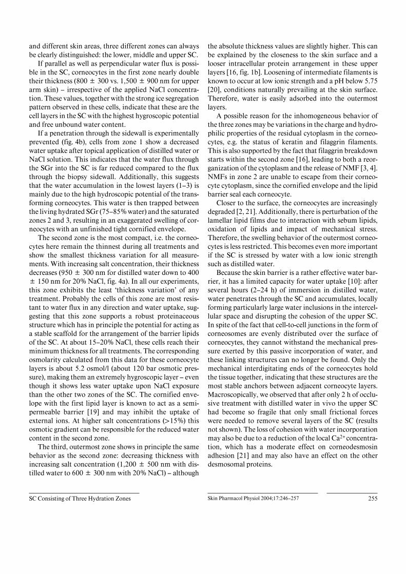
Fig. 1. Comparison between plunge-freezingand high-pressure freezing. Cryo-SEM pic-ture of the frozen hydrated skin block facesof the upper arm of a 73-year-old female; theskin sample was plunge-frozen after storingfor 12 h in a humidity chamber (a). It wascompared to abdominal skin from a 34-year-old female, high-pressure frozen, after 2 h inthe humidity chamber (b). The thickness ofeach cell layer was measured (as described in‘Materials and Methods’) at several differentlocations throughout the samples and themean values are compared (c). The asteriskdenotes the first SC layer adjacent to theSGr.
In vivo MeasurementsFor in vivo SC thickness measurements we used a multiphoton
microscope according to Koenig [14] and Peuckert [15]. Briefly, skinwas topically treated with various salt solutions for 2 and 4 h prior toimaging in vivo.
Data Selection and ProcessingTypically, 50–80 pictures with different magnifications from the
central region (width approximately 1,000 Ìm) of each preparedbiopsy (3 mm in diameter) were taken for further evaluation. Sincethe complete variation in thickness of all layers of the whole SC can-not be analyzed, data were collected from representative areasthroughout each layer: no locations were chosen in wrinkles (fig. 2d)and near hair shafts, where the SC shows strong curvature, inhomo-geneous stacking and strong interdigitation between individual cor-
neocytes (fig. 2a). Cells with large nucleus remnants – which can sig-nificantly expand the corneocytes locally – were omitted for the samereason. Furthermore, only locations where the SC was intact and hada homogeneous and parallel stacking were analyzed. Here, the indi-vidual corneocytes have a smooth and regular cell border (fig. 2b, c)and show minimal variation in thickness.
Typically, the investigated skin showed 15 or 9 cell layers in theSC for the upper arm or face, respectively. Therefore only these first15 or 9 layers of the given SC were analyzed, although up to 22 layers(upper arm) were frequently found.
To obtain enough data, the thickness (for each treatment) of eachlayer was measured at 10–30 representative locations throughout theSC using the software package KS400 (Zeiss, Göttingen, Germany)for semiautomation.

SC Consisting of Three Hydration Zones Skin Pharmacol Physiol 2004;17:246–257 249
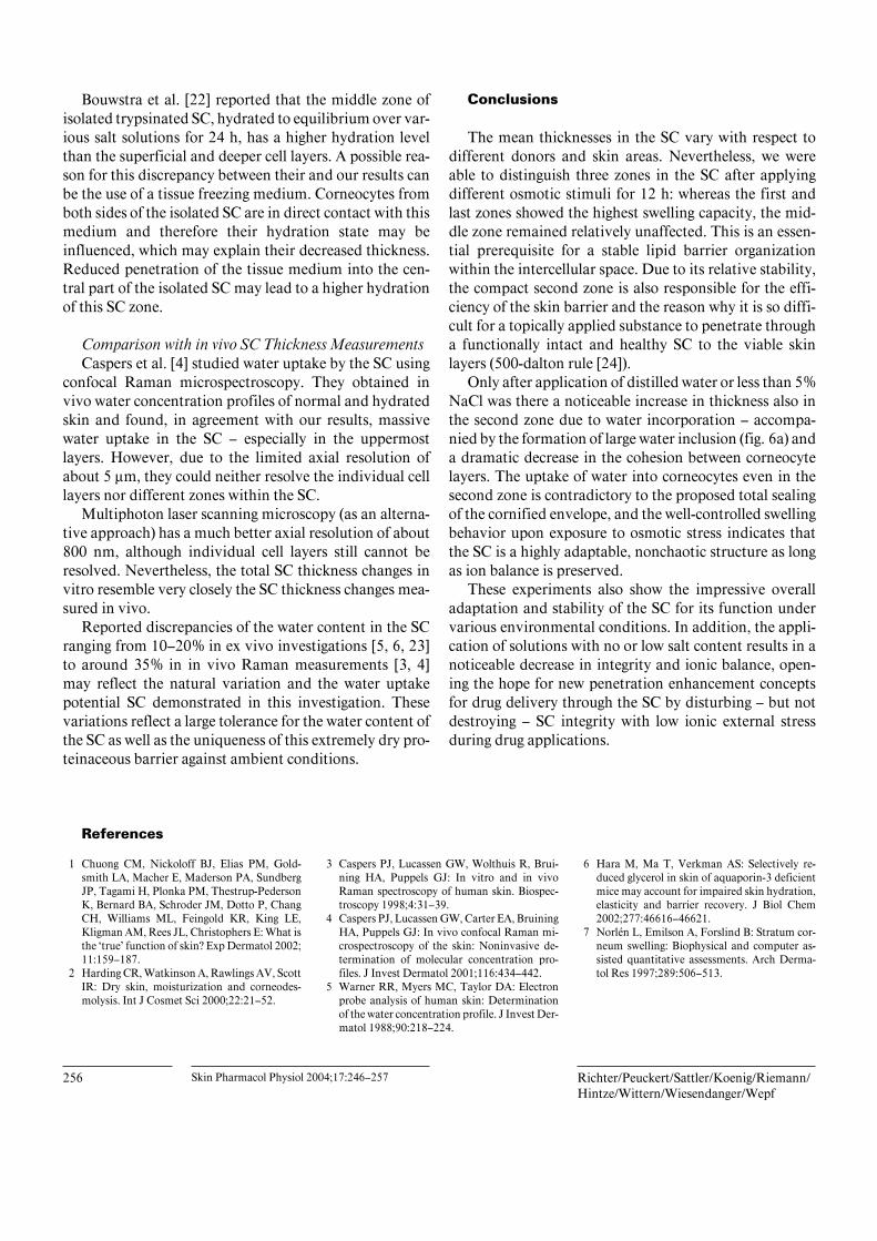
Fig. 2. Comparison of the morphology after treatment with different salt concentrations. The individual cell layers inthe SC respond directly to a changing environment: Whereas in samples (upper arm skin) from the humidity chamber(a) all corneocytes have approximately the same thickness, application of double-distilled water results in a swellingof all cell layers (b). Open box: typical segregation pattern in the first two layers of the SC just above the SGr afterplunge-freezing. Lower magnification (d). The asterisk marks a wrinkle containing sebum at the skin surface. Appli-cation of 15% NaCl results in a massive swelling of the two lowest layers but shrinkage in the middle zones (c).Cryo-SEM pictures of the block faces. Scale bars: 4 Ìm (a–c); 10 Ìm (d).
Results
SC Morphology and Free Water ContentThe sample preparation retained the overall corneo-
cyte structure and the cell-to-cell contacts throughout theSC. Usually no undulation of the corneocyte surfaces ordisruption of the SC were found after cryoprocessing. The
overall compactness of the tissue was preserved as well asthe dense organization of the SC (fig. 1, 2), as revealedafter optimal preservation by high-pressure freezing [16].
To study possible ice crystal effects on the thickness ofcorneocytes, a control sample was stored in a humiditychamber and frozen by high-pressure freezing for beststructure preservation. The individual thickness of all

250 Skin Pharmacol Physiol 2004;17:246–257 Richter/Peuckert/Sattler/Koenig/Riemann/Hintze/Wittern/Wiesendanger/Wepf
layers in the SC was measured and compared with theresults of plunge-frozen skin samples. Although differentskin areas were used, no significant difference in eitherthe morphology (fig. 1a, b) or the thickness of the individ-ual SC layers (fig. 1c) could be found [for more detailsabout the preparation see ref. 17]. Large freezing artifactsin plunge-frozen samples were only observed in the livingcell layers beneath the SC. Plunge-freezing revealed icecrystals in the SC small enough not to affect the cell thick-ness measurements by more than 10 nm, which is lessthan the 4% volume increase expected by ice crystal for-mation of pure water. Plunge-freezing allows to immobi-lize skin biopsies of 3 mm or larger and hence cell thick-ness measurements become more statistically reliable.
Structural variations were found only within the restcytoplasm of corneocytes: these structural variations weresharply edged holes of varying size within the corneocytebiomatrix. These structures were derived from ice crystalsformed during the freezing process [18]: water preferen-tially crystallizes monocrystalline, while organic as well asinorganic molecules are forced to the boundary of thegrowing ice crystal. Hence, the biomatrix will be com-pressed into so-called segregation boundaries. After re-moving part of the water by freeze-drying, the remainingstructure was a stable segregation pattern as can be seen infigure 2b (box). The size of the holes correlates with freez-ing speed and the free water available for ice crystal for-mation and hence gives insights into the content of freeunbound water within the biological structure. With in-creasing distance from the cryogen contact area (SC sur-face), the freezing rate decreases, normally resulting inlarger ice crystals. Since the ice crystals become smaller oreven undetectable in the central part of the SC, theselayers seem to contain nearly no free water compared tothe outermost and innermost layers of the SC. In addi-tion, the largest holes (in the range of 400 B 300 nm) weretypically found in the first two layers above the viable celllayers of the stratum granulosum (SGr). The size of theseice crystals was larger or similar to that of ice crystalsfound in viable cell layers, which are known to contain75–85% of free water.
These findings indicate that the content of free un-bound water was neither constant nor continuously de-creasing throughout the SC. Instead we conclude that thefree water content of corneocytes reached a minimum inthe central part of the SC.
Behavior of the SC during HydrationIn all samples the morphology and segregation patterns
of the corneocytes significantly differed from those of the
SGr. In addition, the cell boundaries appeared as denseand slightly elevated structures, thus enabling a clear dis-tinction and correct counting of all corneocyte layers.With all skin treatments the tissue remained compact andthe regularly stacked corneocytes had close contact witheach other. While the cell thickness in different layerschanged with different treatment, the cell shape itselfstayed relatively unchanged.
For the upper arm region, all layers of the samplesstored in the humidity chamber showed the same segrega-tion pattern with an individual corneocyte thickness be-tween 530 and 730 nm throughout the whole SC. In con-trast, figure 2a shows an area with a huge thickness varia-tion in individual corneocytes due to interdigitation oftheir ends. Although such areas were frequently found inthe SC, they were omitted for data acquisition (see ‘Mate-rials and Methods’).
When upper arm skin samples were immersed in dis-tilled water or NaCl solutions (5, 10, 15, 20%) and a waterflux was allowed through the SC and the biopsy sidewall,the first two layers of the SC – just above the SGr – usuallyexhibited a significant segregation pattern (fig. 2b, c). Atlower magnification this structure generated a dark signalabove the SGr layers (fig. 2d), which still contained vesic-ular structures (fig. 2b, arrow). The thickness of the cellsforming these layers was drastically wider and showed amuch larger variation than anywhere else in the SC.
At high salt concentrations (15% and above), the cor-neocytes in the central part of the SC were very compactand homogeneous in form and segregation pattern(fig. 2c). Their thickness was the lowest, typically between300 and 600 nm, and on average even lower than in thecontrol samples exposed in the humidity chamber. Cellsin the outermost part of the SC again became thicker andexhibited noticeable segregation patterns (fig. 2b, c). Incontrast, in ambient dry facial skin the thickness of theoutermost corneocytes was rather constant with a tenden-cy to decrease below 400 nm towards the skin surface(fig. 4b).
The SC Is Organized into Three Hydration ZonesTo analyze the thickness changes of the whole SC with
different treatments, we plotted the measured thickness ofall individual corneocytes as a function of the SC layer(starting with 1 at the border to the SGr and ending at theskin surface). These measurements were taken at 10–30different locations so as to give a representative thicknessdistribution over the entire SC (see fig. 3a for an exampleof upper arm skin after treatment with 15% NaCl).

SC Consisting of Three Hydration Zones Skin Pharmacol Physiol 2004;17:246–257 251
Fig. 3. Response of individual SC layers to hydration. Individual thickness values after treatment with 15% NaCl areplotted versus the respective SC layer (layer 1 denotes the lowest layer just above the viable SGr) (a). Although theseplots look rather confusing, the corresponding plot of the mean thickness values (b) allows identification of threedifferent zones within the SC. The smallest mean corneocyte thickness is found in the middle zone, which alsoexhibits the smallest variation in standard deviation. Plots of mean thickness for each zone versus salt concentration(c). No significant differences between the zones for the humidity chamber samples were found. However, afterapplication of solutions with rising salt concentrations, cells from zone 1 approximately double their thickness incontrast to those from zones 2 and 3, which are not significantly different and shrink with rising salt concentration. Alldata are for the upper arm skin.
zone 1zone 2zone 3
thic
knes
s[n
m]
500
1000
1500
2000
1 5 10 15(n=28 1928 28 28 28 28 28 28 25 24 23 21 2028 11 5 3 2) zone 2zone 1 zone 3
thic
knes
s[n
m]
500
1000
1500
2000
10%05%00%hum.chamber 20%15%
salt concentration
SC layer
a b
c

252 Skin Pharmacol Physiol 2004;17:246–257 Richter/Peuckert/Sattler/Koenig/Riemann/Hintze/Wittern/Wiesendanger/Wepf
Fig. 4. Hydration effects in different skin areas. a Upper arm skinafter topical application of different NaCl solutions. Water influxthrough the SC as well as through the sidewall of the biopsy was pos-sible in this experimental setup. Control samples were incubated in ahumidity chamber. b Facial skin after topical application of distilledwater and 20% NaCl. In this experimental setup, only topical waterinflux through the SC was possible. Controls were immediately fro-zen (ambient extracted fresh skin) or exposed to a saturated wateratmosphere in a humidity chamber prior to freezing. (SC layer 1denotes the lowest layer just above the viable SGr.)
a
b
Comparison of individual thickness curves (fig. 3a)revealed intrinsic biological variation, but plotting themean corneocyte thickness for each layer (fig. 3b) nev-ertheless allowed to distinguish three different morpho-logical zones within the SC: (a) Zone 1 (approximately thefirst three cell layers) showed a dramatic decrease in themean thickness from a few micrometers down to approxi-mately 900 nm. Overall, this was also the thickest corneo-cyte fraction within the SC. (b) Zone 2 (from layer 5 to
layer 10): Here the thickness of the individual corneocyteswas more or less the same (around 490 nm) and had thesmallest variation in thickness found in the entire SC(standard deviation 130 nm). Presumably this zone wasthe most compact zone of the SC. (c) In zone 3, the outer-most zone, the mean thickness increased again towardsthe skin surface.
After identifying the three zones within the SC, themean thickness and standard deviation were calculatedfor each zone from all cells found in a corresponding zone.The transition layers (layers 4 and 11 for upper arm skin)of two adjacent zones were not considered for these calcu-lations. The thicknesses of zones 2 and 3 were not signifi-cantly different with respect to mean thickness and itsstandard deviation. Treatment of upper arm skin with15% NaCl (fig. 3a) resulted in a mean thickness of 1,800B 900 nm in zone 1, 490 B 150 nm in zone 2, and 610 B300 nm in zone 3 (fig. 3b).
In control samples (humidity chamber), the averagedvalues of all three zones were not significantly different,either in terms of mean cell thickness or standard devia-tion. However, after treatment by submersion in salt solu-tions, the individual zones showed a significantly differ-ent behavior. In zone 1 (adjacent to the SGr), the cellsapproximately doubled their thickness and showed noclear dependence on the salt concentration, whereas cellsin zones 2 and 3 were thickest after immersion in distilledwater and their thickness decreased steadily with risingNaCl concentration. At high salt concentrations (15 and20%), the cells of zone 2 reached about the same thicknessas in the controls. The standard deviation of the thicknessmeasurements for the control sample was approximatelythe same for all three zones. Not only the thicknesschanged upon treatment with salt solution, but also a cleardifference was found in the standard deviation for eachzone: in zone 1, the deviation was nearly the same for allconcentrations. Similar behavior of the cells was found inzone 3, but the standard deviation showed a much smallervariation than in zone 1. In contrast, the mean thicknessof the cells in zone 2, as well as their standard deviation,steadily decreased until they finally reached the values ofthe control cells (humidity chamber).
The SC Regulates Its Structure upon HydrationFigure 4a presents a summary of the swelling behavior
of skin exposed to various treatments compared with thatof control skin samples, which had not been submersed inany liquid. The three zones can be clearly recognized in alltreatments. Moreover, below a salt concentration of 15%NaCl each zone independently became thicker, with the

SC Consisting of Three Hydration Zones Skin Pharmacol Physiol 2004;17:246–257 253
Fig. 5. Comparison of total SC thickness using in vivo data. The totalthickness of the SC was determined either by cryo-SEM or in vivo bymultiphoton laser scanning microscopy (mpLSM) and expressed rel-ative to the appropriate control, which was set to 100%. Both meth-ods revealed a maximum thickness increase with distilled water anddecreasing thickness with higher salt concentrations. mpLSM imag-ing revealed a greater thickness increase with distilled water and anearly linear decrease with rising salt concentration until for 20% theSC starts to shrink below the control thickness. The averaged cryo-SEM data indicate less prominent swelling with distilled water and aweaker dependence on osmotic stress. In particular, overall thicknessnever dropped below that of the control.
maximum change found in zone 1 followed by zone 3.Whereas cells from zone 2 were the thinnest after all treat-ments with NaCl solutions, they showed maximum swell-ing after direct incorporation of water and even from asaturated humidity chamber (gas phase, fig. 4a). Overall,the SC showed maximum swelling after application ofdistilled water relative to the ambient state, which showeda steady decrease of corneocyte thickness from the firstlayers to the outermost layers. The SC from the face loca-tion with 9–12 layers (fig. 4b) had significantly fewer cor-neocyte layers than the SC from the upper arm (15–22layers), but it still showed the typical three zone morphol-ogy upon NaCl treatment (here only data from 20% NaClare shown).
Comparison with in vivo SC Thickness MeasurementsIn figure 5, the total SC thickness data measured on the
upper arm skin by cryo-SEM is compared with that mea-sured with in vivo multiphoton laser scanning microscopyfor forearm skin subjected to occlusive treatment. After2 h of occlusion with the corresponding salt concentra-tion, the thickness of the SC was measured in vivo andcompared to the thickness of the SC measured before
treatment. The relative changes over the total SC are plot-ted against the relative changes of the SC thickness foundwith cryo-SEM. The total thickness of the SC reached itsmaximum with distilled water and decreased with risingsalt content also in vivo. Surprisingly, the total swelling invivo with distilled water and 5% NaCl was greater thanthat found after submersion of a biopsy in the correspond-ing solution for 12 h. Above 10% the SC thickness in vivoeven began to decrease compared to the untreated area,whereas the cryo-SEM data showed a continuous decreasein total SC thickness, but never below the SC thicknessvalue of controls.
Water Also Penetrates between Adjacent Cell LayersIn addition to the reported swelling behavior of the SC,
two completely different swelling effects could be ob-served after exposure to distilled water for 12 h: in deeperregions of the SC, approximately the first 5 layers forupper arm skin, the corneocytes showed a very homoge-neous thickness. There was a dense organization of theintracellular corneocyte matrix as well as intact cell-to-cellcontacts between neighboring corneocytes. In contrast,the upper parts of the SC showed distinctive water inclu-sions after swelling under distilled water stress (fig. 6a).Typically, they had a lens-like shape, were between 3 and25 Ìm in length and between 2 and 20 Ìm in width. Theinterior of these cavities was partially occupied by eutec-tic, the remains of the water and biomatrix segregationprocess, and appeared hollow after sublimation of waterduring the preparation process.
Such water inclusions in the intercellular space oc-curred in all investigated skin regions and could occur iso-lated or in groups, severely disrupting the dense organiza-tion of the SC. They were often found after topical appli-cation of distilled water, only very rarely after applicationof 5% NaCl and never when using higher NaCl concentra-tions.
On the other hand, the SC of untreated skin, extractedat ambient conditions and immediately frozen with high-pressure freezing for best structural preservation of the invivo state, reproducibly showed a dense, regular stackingof individual compact corneocytes with rather constantthicknesses throughout the SC (fig. 6b). Nevertheless,very small lens-like inclusions (typical sizes below 1 Ìm)could be found in such control skin, indicating naturalreservoirs for water and lipids.

254 Skin Pharmacol Physiol 2004;17:246–257 Richter/Peuckert/Sattler/Koenig/Riemann/Hintze/Wittern/Wiesendanger/Wepf
Fig. 6. Water inclusions between adjacent cell layers and loss ofcohesion. In addition to water uptake by individual cells (swelling),treatment with distilled water resulted in water cavities after partialfreeze-drying (e.g. arrow) between adjacent corneocyte layers (a).Water inclusion occurred isolated or in groups and destroyed the
local cohesion of the SC. Additionally, the nuclei (n) in the livingstratum spinosum are visible. In contrast, the ambient in vivo stateshows a regular, dense, parallel stacking of the individual corneocytelayers with occasional lens-like inclusions of typical sizes around1 Ìm (b, arrow). Magnification: scale bar 30 Ìm (a); 5 Ìm (b).
Discussion
To prevent any interference from chemical or physicalseparation of the SC, only complete skin biopsies wereused in our experiments. Furthermore to statistically eval-uate corneocyte thickness at different hydration states, 22biopsies were investigated and prepared as frozen hy-drated plane cross sections to improve the accuracy andreproducibility of the thickness measurements. Althoughin figure 4a water may have penetrated through the side-wall, no difference between the SC swelling behavior ofzones 2 and 3 was found when this water penetrationpathway was prevented (fig. 4a vs. b) The thickness of thecells in zone 1 was distinctly higher and showed a muchlarger variation than anywhere else in the SC when watercould penetrate through the sidewall of the skin biopsies.Preventing this penetration pathway (fig. 4b) drasticallyreduced water uptake in these lowest SC layers – exceptafter 20% NaCl or after gaseous water uptake in thehumidity chamber (fig. 4b).
Independent of the water uptake pathway, the in vitroswelling potential of individual corneocytes in different
layers of the human SC after exposure to solutions of dif-ferent NaCl concentration could be studied.
Swelling Behavior of the SC in Response to OsmoticStressUsually water dissipates from the viable cell layers,
driven by the water gradient, into the SC to finally evapo-rate at the skin surface. In the untreated ambient controlsamples, corneocyte thickness decreased from 900 B250 nm in the deepest layer to a thickness of about 400 B100 nm at the skin surface.
If evaporation of water is prevented by a saturatedhumidity atmosphere or occlusion by e.g. Vaseline (datanot shown), water traveling along this gradient can accu-mulate in the SC until each layer is fully saturated withwater. This results in massive water uptake and a two- orthreefold swelling mainly in zones 2 and 3 (fig. 4b), inde-pendent of whether the water comes from the atmosphereor from within the tissue. Interestingly, after applicationof distilled water or salt solutions, the situation changessignificantly: although the absolute thickness of each SClayer shows strong variations among the different donors
SC Consisting of Three Hydration Zones Skin Pharmacol Physiol 2004;17:246–257 255
and different skin areas, three different zones can alwaysbe clearly distinguished: the lower, middle and upper SC.
If parallel as well as perpendicular water flux is possi-ble in the SC, corneocytes in the first zone nearly doubletheir thickness (800 B 300 vs. 1,500 B 900 nm for upperarm skin) – irrespective of the applied NaCl concentra-tion. These values, together with the strong ice segregationpattern observed in these cells, indicate that these are thecell layers in the SC with the highest hygroscopic potentialand free unbound water content.
If a penetration through the sidewall is experimentallyprevented (fig. 4b), cells from zone 1 show a decreasedwater uptake after topical application of distilled water orNaCl solution. This indicates that the water flux throughthe SGr into the SC is far reduced compared to the fluxthrough the biopsy sidewall. Additionally, this suggeststhat the water accumulation in the lowest layers (1–3) ismainly due to the high hydroscopic potential of the trans-forming corneocytes. This water is then trapped betweenthe living hydrated SGr (75–85% water) and the saturatedzones 2 and 3, resulting in an exaggerated swelling of cor-neocytes with an unfinished tight cornified envelope.
The second zone is the most compact, i.e. the corneo-cytes here remain the thinnest during all treatments andshow the smallest thickness variation for all measure-ments. With increasing salt concentration, their thicknessdecreases (950 B 300 nm for distilled water down to 400B 150 nm for 20% NaCl, fig. 4a). In all our experiments,this zone exhibits the least ‘thickness variation’ of anytreatment. Probably the cells of this zone are most resis-tant to water flux in any direction and water uptake, sug-gesting that this zone supports a robust proteinaceousstructure which has in principle the potential for acting asa stable scaffold for the arrangement of the barrier lipidsof the SC. At about 15–20% NaCl, these cells reach theirminimum thickness for all treatments. The correspondingosmolarity calculated from this data for these corneocytelayers is about 5.2 osmol/l (about 120 bar osmotic pres-sure), making them an extremely hygroscopic layer – eventhough it shows less water uptake upon NaCl exposurethan the other two zones of the SC. The cornified enve-lope with the first lipid layer is known to act as a semi-permeable barrier [19] and may inhibit the uptake ofexternal ions. At higher salt concentrations (115%) thisosmotic gradient can be responsible for the reduced watercontent in the second zone.
The third, outermost zone shows in principle the samebehavior as the second zone: decreasing thickness withincreasing salt concentration (1,200 B 500 nm with dis-tilled water to 600 B 300 nm with 20% NaCl) – although
the absolute thickness values are slightly higher. This canbe explained by the closeness to the skin surface and alooser intracellular protein arrangement in these upperlayers [16, fig. 1b]. Loosening of intermediate filaments isknown to occur at low ionic strength and a pH below 5.75[20], conditions naturally prevailing at the skin surface.Therefore, water is easily adsorbed into the outermostlayers.
A possible reason for the inhomogeneous behavior ofthe three zones may be variations in the charge and hydro-philic properties of the residual cytoplasm in the corneo-cytes, e.g. the status of keratin and filaggrin filaments.This is also supported by the fact that filaggrin breakdownstarts within the second zone [16], leading to both a reor-ganization of the cytoplasm and the release of NMF [3, 4].NMFs in zone 2 are unable to escape from their corneo-cyte cytoplasm, since the cornified envelope and the lipidbarrier seal each corneocyte.
Closer to the surface, the corneocytes are increasinglydegraded [2, 21]. Additionally, there is perturbation of thelamellar lipid films due to interaction with sebum lipids,oxidation of lipids and impact of mechanical stress.Therefore, the swelling behavior of the outermost corneo-cytes is less restricted. This becomes even more importantif the SC is stressed by water with a low ionic strengthsuch as distilled water.
Because the skin barrier is a rather effective water bar-rier, it has a limited capacity for water uptake [10]: afterseveral hours (2–24 h) of immersion in distilled water,water penetrates through the SC and accumulates, locallyforming particularly large water inclusions in the intercel-lular space and disrupting the cohesion of the upper SC.In spite of the fact that cell-to-cell junctions in the form ofcorneosomes are evenly distributed over the surface ofcorneocytes, they cannot withstand the mechanical pres-sure exerted by this passive incorporation of water, andthese linking structures can no longer be found. Only themechanical interdigitating ends of the corneocytes holdthe tissue together, indicating that these structures are themost stable anchors between adjacent corneocyte layers.Macroscopically, we observed that after only 2 h of occlu-sive treatment with distilled water in vivo the upper SChad become so fragile that only small frictional forceswere needed to remove several layers of the SC (resultsnot shown). The loss of cohesion with water incorporationmay also be due to a reduction of the local Ca2+ concentra-tion, which has a moderate effect on corneodesmosinadhesion [21] and may also have an effect on the otherdesmosomal proteins.
256 Skin Pharmacol Physiol 2004;17:246–257 Richter/Peuckert/Sattler/Koenig/Riemann/Hintze/Wittern/Wiesendanger/Wepf
Bouwstra et al. [22] reported that the middle zone ofisolated trypsinated SC, hydrated to equilibrium over var-ious salt solutions for 24 h, has a higher hydration levelthan the superficial and deeper cell layers. A possible rea-son for this discrepancy between their and our results canbe the use of a tissue freezing medium. Corneocytes fromboth sides of the isolated SC are in direct contact with thismedium and therefore their hydration state may beinfluenced, which may explain their decreased thickness.Reduced penetration of the tissue medium into the cen-tral part of the isolated SC may lead to a higher hydrationof this SC zone.
Comparison with in vivo SC Thickness MeasurementsCaspers et al. [4] studied water uptake by the SC using
confocal Raman microspectroscopy. They obtained invivo water concentration profiles of normal and hydratedskin and found, in agreement with our results, massivewater uptake in the SC – especially in the uppermostlayers. However, due to the limited axial resolution ofabout 5 Ìm, they could neither resolve the individual celllayers nor different zones within the SC.
Multiphoton laser scanning microscopy (as an alterna-tive approach) has a much better axial resolution of about800 nm, although individual cell layers still cannot beresolved. Nevertheless, the total SC thickness changes invitro resemble very closely the SC thickness changes mea-sured in vivo.
Reported discrepancies of the water content in the SCranging from 10–20% in ex vivo investigations [5, 6, 23]to around 35% in in vivo Raman measurements [3, 4]may reflect the natural variation and the water uptakepotential SC demonstrated in this investigation. Thesevariations reflect a large tolerance for the water content ofthe SC as well as the uniqueness of this extremely dry pro-teinaceous barrier against ambient conditions.
Conclusions
The mean thicknesses in the SC vary with respect todifferent donors and skin areas. Nevertheless, we wereable to distinguish three zones in the SC after applyingdifferent osmotic stimuli for 12 h: whereas the first andlast zones showed the highest swelling capacity, the mid-dle zone remained relatively unaffected. This is an essen-tial prerequisite for a stable lipid barrier organizationwithin the intercellular space. Due to its relative stability,the compact second zone is also responsible for the effi-ciency of the skin barrier and the reason why it is so diffi-cult for a topically applied substance to penetrate througha functionally intact and healthy SC to the viable skinlayers (500-dalton rule [24]).
Only after application of distilled water or less than 5%NaCl was there a noticeable increase in thickness also inthe second zone due to water incorporation – accompa-nied by the formation of large water inclusion (fig. 6a) anda dramatic decrease in the cohesion between corneocytelayers. The uptake of water into corneocytes even in thesecond zone is contradictory to the proposed total sealingof the cornified envelope, and the well-controlled swellingbehavior upon exposure to osmotic stress indicates thatthe SC is a highly adaptable, nonchaotic structure as longas ion balance is preserved.
These experiments also show the impressive overalladaptation and stability of the SC for its function undervarious environmental conditions. In addition, the appli-cation of solutions with no or low salt content results in anoticeable decrease in integrity and ionic balance, open-ing the hope for new penetration enhancement conceptsfor drug delivery through the SC by disturbing – but notdestroying – SC integrity with low ionic external stressduring drug applications.
References
1 Chuong CM, Nickoloff BJ, Elias PM, Gold-smith LA, Macher E, Maderson PA, SundbergJP, Tagami H, Plonka PM, Thestrup-PedersonK, Bernard BA, Schroder JM, Dotto P, ChangCH, Williams ML, Feingold KR, King LE,Kligman AM, Rees JL, Christophers E: What isthe ‘true’ function of skin? Exp Dermatol 2002;11:159–187.
2 Harding CR, Watkinson A, Rawlings AV, ScottIR: Dry skin, moisturization and corneodes-molysis. Int J Cosmet Sci 2000;22:21–52.
3 Caspers PJ, Lucassen GW, Wolthuis R, Brui-ning HA, Puppels GJ: In vitro and in vivoRaman spectroscopy of human skin. Biospec-troscopy 1998;4:31–39.
4 Caspers PJ, Lucassen GW, Carter EA, BruiningHA, Puppels GJ: In vivo confocal Raman mi-crospectroscopy of the skin: Noninvasive de-termination of molecular concentration pro-files. J Invest Dermatol 2001;116:434–442.
5 Warner RR, Myers MC, Taylor DA: Electronprobe analysis of human skin: Determinationof the water concentration profile. J Invest Der-matol 1988;90:218–224.
6 Hara M, Ma T, Verkman AS: Selectively re-duced glycerol in skin of aquaporin-3 deficientmice may account for impaired skin hydration,elasticity and barrier recovery. J Biol Chem2002;277:46616–46621.
7 Norlén L, Emilson A, Forslind B: Stratum cor-neum swelling: Biophysical and computer as-sisted quantitative assessments. Arch Derma-tol Res 1997;289:506–513.

SC Consisting of Three Hydration Zones Skin Pharmacol Physiol 2004;17:246–257 257
8 Richter T, Mueller JH, Schwarz UD, Wepf R,Wiesendanger R: Investigation of the swellingof human skin cells in liquid media by tappingmode scanning force microscopy. Appl Phys2001;A72:125–128.
9 Kashibuchi N, Hirai Y, O'’Goshi K, Tagami H:Three-dimensional analyses of individual cor-neocytes with atomic force microscopy: Mor-phological changes related to age, location andto the pathologic skin conditions. Skin ResTechnol 2002;8:203–211.
10 Warner RR, Stone KJ, Boissy YL: Hydrationdisrupts human stratum corneum ultrastruc-ture. J Invest Dermatol 2003;120:275–284.
11 Ohman H, Vahlquist A: In vivo studies con-cerning a pH gradient in human stratum cor-neum and upper epidermis. Acta Derm Vene-reol 1994;74:375–379.
12 Walther P, Wehrli E, Hermann R, Müller M:Double-layer coating for high-resolution low-temperature scanning electron microscopy. JMicrosc 1995;179:229–237.
13 Ritter M, et al: A versatile high-vacuum cryo-transfer for cryo-FESEM, cryo-SPM and otherimaging techniques. Microsc Microanal 1999;5(suppl 2):424–425.
14 Koenig K: Multiphoton microscopy in lifesciences. J Microsc 2000;200:83–104.
15 Peuckert C: Anwendung von Femtosekunden-Laserpulsen im NIR-Spektralbereich für Un-tersuchungen an humaner Haut; DiplomarbeitFriedrich-Schiller Universität Jena, 2001.
16 Pfeiffer S, Vielhaber G, Vietzke JP, WitternKP, Hintze U, Wepf R: High-pressure freezingprovides new information on human epider-mis: Simultaneous protein antigen and lamel-lar lipid structure preservation. Study on hu-man epidermis by cryoimmobilization. J In-vest Dermatol 2000;114:1030–1038.
17 Richter T, et al: From single image interpreta-tion to statistic morphometric tissue analysisby cryo-SEM. J Microsc, submitted.
18 Echlin P: Low-Temperature Microscopy andAnalysis. New York, Plenum, 1992.
19 Jarnik M, Simon MN, Steven AC: Cornifiedcell envelope assembly: A model based on elec-tron microscopic determinations of thicknessand projected density. J Cell Sci 1998;111:1051–1060.
20 Aebi U, Haener M, Troncoso J, Eichner R,Engel A: Unifying principles in intermediatefilament (IF) structure and assembly. Proto-plasma 1998;145:73–81.
21 Jonca N, Guerrin M, Hadjiolova K, Caubet C,Gallinaro H, Simon M, Serre G: Corneodesmo-sin, a component of epidermal corneocyte des-mosomes, displays homophilic adhesive prop-erties. J Biol Chem 2002;277:5024–5029.
22 Bouwstra JA, de Graaff A, Gooris GS, Nijsse J,Wiechers JW, van Aelst AC: Water distribu-tion and related morphology in human stratumcorneum at different hydration levels. J InvestDermatol 2003;120:750–758.
23 Gerhardi S: Die Charakterisierung von Koh-lenhydraten in der menschlichen Hornschichtmittels Lectinen; Diplomarbeit FH Hamburg-Bergedorf, 1983, pp 23–39.
24 Bos JD, Meinardi MM: The 500 Dalton rulefor the skin penetration of chemical com-pounds and drugs. Exp Dermatol 2000;9:165–169.
Copyright © 2022 FDOKUMEN