Blue: correcting sequencing errors using consensus and context
New Developments
Splicing-Correcting Therapeutic Approaches for RetinalDystrophies: Where Endogenous Gene Regulation andSpecificity Matter
Niccolo Bacchi,1 Simona Casarosa,1,2 and Michela A. Denti1,3
1Centre for Integrative Biology (CIBIO) - University of Trento, Trento, Italy2Neuroscience Institute - National Research Council (CNR), Pisa, Italy3Neuroscience Institute - National Research Council (CNR), Padova, Italy
Correspondence: Simona Casarosa,Centre for Integrative Biology(CIBIO) - University of Trento, ViaSommarive 9, 38123 Trento, Italy;[email protected] A. Denti, Centre for Inte-grative Biology (CIBIO) - Universityof Trento, Via Sommarive 9, 38123Trento, Italy;[email protected].
SC and MAD contributed equally tothe work presented here and shouldtherefore be regarded as equivalentauthors.
Submitted: April 8, 2014Accepted: April 11, 2014
Citation: Bacchi N, Casarosa S, DentiMA. Splicing-correcting therapeuticapproaches for retinal dystrophies:where endogenous gene regulationand specificity matter. Invest Oph-thalmol Vis Sci. 2014;55:3285–3294.DOI:10.1167/iovs.14-14544
Splicing is an important and highly regulated step in gene expression. The ability to modulateit can offer a therapeutic option for many genetic disorders. Antisense-mediated splicing-correction approaches have recently been successfully exploited for some genetic diseases,and are currently demonstrating safety and efficacy in different clinical trials. Theirapplication for the treatment of retinal dystrophies could potentially solve a vast panel ofcases, as illustrated by the abundance of mutations that could be targeted and the versatility ofthe technique. In this review, we will give an insight of the different therapeutic strategies,focusing on the current status of their application for retinal dystrophies.
Keywords: splicing correction, antisense oligonucleotides, retinal dystrophy, gene therapy
Retinal dystrophies are an extremely diversified group ofgenetic diseases all characterized by visual dysfunctions
that can lead to blindness in the worst cases. Today there aremore than 200 genes responsible for syndromic and non-syndromic retinal dystrophies,1 each of them carrying severaltypes of mutations leading to very different clinical phenotypes.The development of different gene therapy approaches hasgiven a hope for the implementation of therapies for theseotherwise incurable conditions. Messenger RNA splicing is anextremely complex and fundamental cellular process that hasbeen so fare barely considered as a therapeutic target,2,3 even ifit can be seen as a highly appealing one for its importance inthe cell context. The ability to modulate splicing can in factoffer several advantages over other conventional gene replace-ment approaches, especially in the context of retinal dystro-phies. By definition, antisense-based therapeutic approachesact following base-pairing with their mRNA target, thus givingthe possibility of obtaining a great specificity of action. Sincethey act at the mRNA level, the endogenous transcriptionalregulation of the target gene is always maintained. This meansthat the therapeutic effect is obtained only where and when thetarget pre-mRNA is present. In a highly specialized andorganized tissue like the retina, it is particularly important tomaintain endogenous gene regulation. Therapeutic interven-
tions for delicate processes like the phototransduction cascadewould require the preservation of this control for desirableoutcomes.4 Splicing-correction approaches also allow a fine-tuning over the relative abundance of splicing isoformsbecause, by acting at a pre-mRNA level, it is relatively easy tomodulate their ratio. The availability of several differentmolecular tools that can be used to manipulate splicing rendersthese approaches a versatile and promising strategy for themultitude of retinal dystrophies known today. Moreover, theretina has some characteristics that make it a perfect targettissue for those therapies. First of all, it is an easy tissue toaccess, and different drug delivery routes are in use today.5,6
Being that the eye is relatively small, enclosed, and separatedfrom the rest of the body by the blood-brain barrier, itminimizes the required dose and systemic dissemination ofthe therapeutic agent, thus avoiding possible complications dueto systemic side effects of the therapy.7 The eye is also animmune-privileged organ, limiting potential immune responseto the delivered agent.8 The retina is composed by nondividingcells, thus it is easier to induce prolonged effects or transgeneexpression, without the need of using integrating vectors.Regarding gene delivery, the presence of different adeno-associated viral (AAV) vector serotypes able to efficiently and
Copyright 2014 The Association for Research in Vision and Ophthalmology, Inc.
www.iovs.org j ISSN: 1552-5783 3285

stably transduce all retina layers9 is a great advantage forsplicing-modulating genetic tools.
THE MRNA SPLICING PROCESS
Once transcription of a gene begins in the nucleus, thetranscript undergoes a complex series of cotranscriptionalprocesses all devoted to the production of a mature mRNA,collectively dubbed ‘‘mRNA processing.’’ One of these events,called mRNA splicing, consists in the removal of interveningsequences (‘‘introns’’) and the joining of the coding portions ofthe transcript (‘‘exons’’). Messenger RNA splicing is a majorway by which the cell can induce transcriptional diversity,mainly through alternative splicing, and apply a fine control onthis diversity. The proper recognition of introns and exons ismediated by cis-acting sequences and trans-acting factors. Theprincipal cis-acting elements that spatially organize the splicingreaction, consist in the splice donor (DS) site, the polypy-rimidine tract (Py), the branch-site (BS), and the spliceacceptor (AS) site. There are also other cis-acting sequences
that are fundamental for mRNA splicing10: exonic splicingenhancers (ESE) or silencers (ESS) that enhance or inhibitrecognition of the exon in which they lay; intronic splicingenhancers (ISE) or silencers (ISS), intronic sequences thatpromote or suppress recognition of the nearby exons. Trans-acting factors are instead several proteins and ribonucleopro-teins able to recognize the different cis-elements. Small nuclearRNA (snRNA) are constitutive components of the small nuclearribonucleoproteins (snRNP) U1, U2, U4, U5, U6, and allowthem to base-pair with different cis-acting sequences mediatingthe cascade of events leading to the splicing reaction. Forexample, the U1 snRNP recognizes the DS site, whereas U2binds to the branch site. The other two groups of trans-actingsplicing factors are represented by heterogeneous nuclearribonucleoproteins (hnRNPs), that have mainly a repressivefunction, and by serine- and arginine-rich (SR) proteins, thatplay an important role in splicing regulations by mainly bindingto ESE and ISE, thus promoting splicing.11–15 All these factorsassemble together in a precise temporal sequence in a complexcalled spliceosome, the cellular machinery devoted to the
TABLE. List of Genes Causing Retinal Diseases
Disease Category Involved Genes
Cone or cone-rod dystrophy/dysfunctions ABCA4 ADAM9 AIPL1 BBS12 C2orf71 C8orf37 CA4 CABP4 CACNA1F CACNA2D4
CDHR1 CERKL CNGA3 CNGB3 CNNM4 CRB1 CRX GNAT2 GUCA1A GUCY2D
KCNV2 MERTK MKS1 NR2E3 NRL OPN1LW OPN1MW PDE6C PDE6H PITPNM3
PROM1 PRPH2 RAB28 RAX2 RDH12 RIMS1 RLBP1 RPE65 RPGRIP1 TULP1
UNC119
Retinitis pigmentosa ABCA4 ARL2BP ARL6 BBS1 BEST1 C2orf71 C8orf37 CA4 CEP290 CERKL CLRN1
CNGA1 CNGB1 CRB1 CRX CYP4V2 DHDDS EMC1 EYS FAM161A FSCN2 GPR125
GRK1 GUCA1A GUCA1B GUCY2D IDH3B IMPDH1 IMPG2 KIAA1549 KLHL7
LCA5 LRAT MAK MERTK MFRP MYO7A NR2E3 NRL OFD1 PDE6A PDE6B PDE6G
PRCD PROM1 PRPF3 PRPF31 PRPF6 PRPF8 PRPH2 RBP3 RDH12 RGR RHO
RLBP1 RP1 RP1L1 RP2 RP9 RPE65 RPGR RPGRIP1 SAG SEMA4A SNRNP200
SPATA7 TOPORS TULP1 USH2A VCAN ZNF513
Leber congenital amaurosis AIPL1 BBS4 BEST1 CABP4 CEP290 CNGA3 CRB1 CRX DTHD1 GUCY2D IMPDH1
IQCB1 KCNJ13 LCA5 LRAT MERTK MYO7A NMNAT1 NRL RD3 RDH12 RPE65
RPGRIP1 RPGRIP1L SPATA7 TULP1
Macular dystrophy/degeneration ABCA4 ABCC6 BEST1 CNGB3 CRX EFEMP1 ELOVL4 GUCY2D PAX2 PROM1 PRPH2
RP1L1 TIMP3
Stargardt disease ABCA4 ELOVL4 PRPH2 CFH HMCN1
Age-related macular degeneration ABCA4 ARMS2 BEST1 C3 CFH ELOVL4 ERCC6 FBLN5 HMCN1 HTRA1 RAX2
SLC24A1
Stationary night blindness CACNA1F CABP4 GNAT1 GPR179 GRK1 GRM6 LRIT3 NYX PDE6B RHO SAG
SLC24A1 TRPM1
Color blindness CNGA3 CNGB3 GNAT2 OPN1LW OPN1MW OPN1SW PDE6C PDE6H
Usher syndrome ABHD12 CACNA1F CDH23 CIB2 CLRN1 DFNB31 GPR98 GUCY2D HARS LRAT
MYO7A PCDH15 PDZD7 TRIM32 USH1C USH1G USH2A
Chorioretinal atrophy/degeneration ABCA4 CRB1 TEAD1
Retinal dystrophies/dysfunctions/degeneration ABCC6 ABCA4 ADAMTS18 AIPL1 BEST1 C1QTNF5 CAPN5 CDHR1 CERKL CHM
CRB1 CYP4V2 FZD4 GUCA1B KCNV2 LRAT LRP5 MERTK NDP NR2E3 NRL OTX2
PANK2 PLA2G5 PROM1 PRPH2 RD3 RDH12 RDH5 RGS9 RGS9BP RLBP1 RPE65
SLC24A1 TSPAN12
Retinopathy of prematurity LRP5 NDP FZD4
Optic atrophy/aplasia MFN2 OPA1 OPA3 OTX2 SLC24A1 TMEM126A WFS1
Wagner syndrome VCAN COL2A1
Bardet-Biedl syndrome ARL6 BBS1 BBS10 BBS12 BBS2 BBS4 BBS5 BBS7BBS9 CEP290 LZTFL1 MKKS MKS1
RPGRIP1L SDCCAG8 TRIM32 TTC8 WDPCP
Other systemic/syndromic diseases involving the retina ABCC6 ABHD12 ADAMTS18 AHI1 ALMS1 ATXN7 CC2D2A CDH3 CEP290 CISD2
CLN3 COL11A1 COL2A1 COL9A1 ERCC6 FLVCR1 GNPTG IFT140 INPP5E IQCB1
ITM2B JAG1 KIF11 LRP5 NPHP1 NPHP4 OFD1 OPA3 OTX2 PANK2 PAX2 PEX1
PEX2 PEX7 PHYH RBP4 RPGRIP1L SDCCAG8 TIMM8A TMEM237 TREX1 TTPA
TTPA USH1C WFS1
All RetNet identified genes have been searched and allocated to disease categories according to HGMD Professional entries from December2013.
Splicing Correction of Retinal Dystrophies IOVS j May 2014 j Vol. 55 j No. 5 j 3286
splicing process. For a more exhaustive description of thesplicing process, we refer the reader to more detailedreviews.16–18
MUTATIONS LEADING TO RETINAL DISEASES
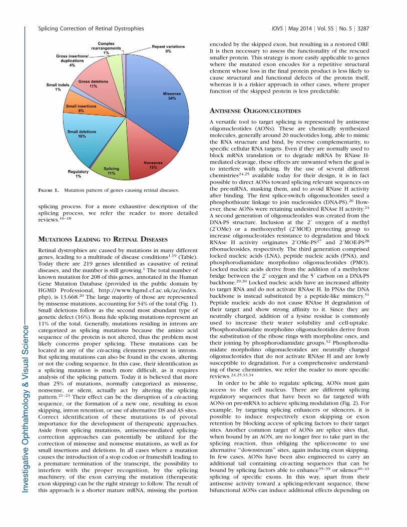
Retinal dystrophies are caused by mutations in many differentgenes, leading to a multitude of disease conditions1,19 (Table).Today there are 219 genes identified as causative of retinaldiseases, and the number is still growing.1 The total number ofknown mutation for 208 of this genes, annotated in the HumanGene Mutation Database (provided in the public domain byHGMD Professional, http://www.hgmd.cf.ac.uk/ac/index.php), is 13.668.20 The large majority of those are representedby missense mutations, accounting for 34% of the total (Fig. 1).Small deletions follow as the second most abundant type ofgenetic defect (16%). Bona fide splicing mutations represent an11% of the total. Generally, mutations residing in introns arecategorized as splicing mutations because the amino acidsequence of the protein is not altered, thus the problem mostlikely concerns proper splicing. These mutations can belocated in any of the cis-acting elements present in introns.But splicing mutations can also be found in the exons, alteringor not the coding sequence. In this case, their identification asa splicing mutation is much more difficult, as it requiresanalysis of the splicing pattern. Today it is believed that morethan 25% of mutations, normally categorized as missense,nonsense, or silent, actually act by altering the splicingpattern.21–23 Their effect can be the disruption of a cis-actingsequence, or the formation of a new one, resulting in exonskipping, intron retention, or use of alternative DS and AS sites.Correct identification of these mutations is of pivotalimportance for the development of therapeutic approaches.Aside from splicing mutations, antisense-mediated splicing-correction approaches can potentially be utilized for thecorrection of missense and nonsense mutations, as well as forsmall insertions and deletions. In all cases where a mutationcauses the introduction of a stop codon or frameshift leading toa premature termination of the transcript, the possibility tointerfere with the proper recognition, by the splicingmachinery, of the exon carrying the mutation (therapeuticexon skipping) can be the right strategy to follow. The result ofthis approach is a shorter mature mRNA, missing the portion
encoded by the skipped exon, but resulting in a restored ORF.It is then necessary to assess the functionality of the rescuedsmaller protein. This strategy is more easily applicable to geneswhere the mutated exon encodes for a repetitive structuralelement whose loss in the final protein product is less likely tocause structural and functional defects of the protein itself,whereas it is a riskier approach in other cases, where properfunction of the skipped protein is less predictable.
ANTISENSE OLIGONUCLEOTIDES
A versatile tool to target splicing is represented by antisenseoligonucleotides (AONs). These are chemically synthesizedmolecules, generally around 20 nucleotides long, able to mimicthe RNA structure and bind, by reverse complementarity, tospecific cellular RNA targets. Even if they are normally used toblock mRNA translation or to degrade mRNA by RNase H-mediated cleavage, these effects are unwanted when the goal isto interfere with splicing. By the use of several differentchemistries24,25 available today for their design, it is in factpossible to direct AONs toward splicing relevant sequences onthe pre-mRNA, masking them, and to avoid RNase H activityafter binding. The first splice-switch oligonucleotides used aphosphothioate linkage to join nucleosides (DNA-PS).26 How-ever, these AONs were retaining undesired RNAse H activity.24
A second generation of oligonucleotides was created from theDNA-PS structure. Inclusion at the 20 oxygen of a methyl(20OMe) or a methoxyethyl (20MOE) protecting group toincrease oligonucleotides resistance to degradation and blockRNAse H activity originates 2 0OMe-PS27 and 2 0MOE-PS28
ribonucleosides, respectively. The third generation comprisedlocked nucleic acids (LNA), peptide nucleic acids (PNA), andphosphorodiamidate morpholino oligonucleotides (PMO).Locked nucleic acids derive from the addition of a methylenebridge between the 20 oxygen and the 50 carbon on a DNA-PSbackbone.29,30 Locked nucleic acids have an increased affinityto target RNA and do not activate RNAse H. In PNAs the DNAbackbone is instead substituted by a peptide-like mimicry.31
Peptide nucleic acids do not cause RNAse H degradation oftheir target and show strong affinity to it. Since they areneutrally charged, addition of a lysine residue is commonlyused to increase their water solubility and cell-uptake.Phosphorodiamidate morpholino oligonucleotides derive fromthe substitution of the ribose rings with morpholine ones, andtheir joining by phosphorodiamidate groups.32 Phosphorodia-midate morpholino oligonucleotides are neutrally chargedoligonucleotides that do not activate RNAse H and are lowlysusceptible to degradation. For a comprehensive understand-ing of these chemistries, we refer the reader to more specificreviews.24,25,33,34
In order to be able to regulate splicing, AONs must gainaccess to the cell nucleus. There are different splicingregulatory sequences that have been so far targeted withAONs on pre-mRNA to achieve splicing modulation (Fig. 2). Forexample, by targeting splicing enhancers or silencers, it ispossible to induce respectively exon skipping or exonretention by blocking access of splicing factors to their targetsites. Another common target of AONs are splice sites that,when bound by an AON, are no longer free to take part in thesplicing reaction, thus obliging the spliceosome to usealternative ‘‘downstream’’ sites, again inducing exon skipping.In few cases, AONs have been also engineered to carry anadditional tail containing cis-acting sequences that can bebound by splicing factors able to enhance35–39 or silence40–43
splicing of specific exons. In this way, apart from theirantisense activity toward a splicing-relevant sequence, thesebifunctional AONs can induce additional effects depending on
FIGURE 1. Mutation pattern of genes causing retinal diseases.
Splicing Correction of Retinal Dystrophies IOVS j May 2014 j Vol. 55 j No. 5 j 3287

the sequence they carry on the tail. Since AONs act by base-pairing, they are generally believed to allow a high specificityof action for their desired target. No undesired missplicedproducts of the target gene or of chosen unrelated genes werein fact detected when investigated after therapeutic applica-tion of AONs.44,45 Even if these findings are not generalizableand proper design of the antisense molecule should always beconsidered, they underline the potentiality of AONs in thecontext of target selectivity. Splicing modulation finds its moreadvanced application in the cure of Duchenne musculardystrophy (DMD). Duchenne muscular dystrophy is an X-linked recessive disease caused by mutations in the dystrophingene. Dystrophin is an important cellular protein whose mainrole in muscle fibers is the connection of the cellularcytoskeleton with the extracellular matrix. Different mutationsin the 79 exons of the gene cause protein truncation due to theloss of the open reading frame. The majority of these mutationscan be addressed by exon skipping.46 The commonly mutatedexon 51 has been the first target for exon skipping. In theclinical trials completed so far for exon 51 skipping, thedifferent chemistries applied (2 0OMe-PS, PMOs) showedoverall efficacy and absence of serious adverse effects.44,47–49
A phase I clinical trial using a 20MOE-PS oligonucleotide forsplicing modulation has also been recently completed forspinal muscular atrophy (SMA), and a phase II trial has recentlystarted.50 Mutations in the survival motor neuron 1 (SMN1)gene are causative of the disease. In humans, SMN1 has aparalogous, named SMN2. The two genes are identical, apartfrom a silent mutation in exon 7 of SMN2. This mutation,however, causes exon 7 of SMN2 to be less recognized by thesplicing machinery. If exon 7 is not included, the protein istruncated. So a therapeutic strategy is to mask exon 7 ESS usingAONs, thus promoting exon 7 inclusion, which results in the
production of a functional full length SMN protein from SMN2that can compensate for mutations on SMN1.
Antisense oligonucleotides are known to be able to targetall retinal layers following intravitreal, subretinal, or topicaladministration.51–56 They have long been used to elicit RNAseH degradation or to block transcription in the eye for severaldifferent diseases, having been applied, for example, againstcytomegalovirus (CMV), herpes simplex virus (HSV), vascularendothelial growth factor (VEGF), transforming growth factor-b (TGF-G), fibroblast growth factor (FGF).5,57
An example of the use of splice-switch oligonucleotides inthe eye is that of vascular endothelial growth factor receptor 2(KDR). The KDR gene has two distinct products: membrane-bound KDR (mbKDR), that is prohemangiogenic, and solubleKDR (sKDR), antilymphangiogenic. Soluble KDR needs therecognition of an alternative polyadenilation site on KDRintron 13 to be translated. By intravitreal administration ofPMO directed against murine Kdr exon 13 DS site, it waspossible to increase the sKDR/mbKDR ratio at mRNA andprotein level in the retina and vitreous, interfering with thespliceosome ability of mediating intron 13 splicing58 (Fig. 3).This resulted in a block of hemangiogenesis and lymphangio-genesis in a model of choroidal neovascularization and cornealinjury.58 Another recent example of the use of AONs as splicingregulators to treat a retinal dystrophy is the one of the genecentrosomal protein 290kDa, (CEP290). Centrosomal protein
FIGURE 2. Possible AONs targets to induce splicing modulation. Schematic representation of cis-acting sequences that are possible target of AONs.Cis-acting sequences that promote exon recognition are reported in green, whereas sequences that suppress it are highlighted in red. Antisenseoligonucleotides designed to induce exon skipping are shown in the bottom part. Antisense oligonucleotides designed to promote proper exoninclusion are shown in the upper part.
FIGURE 3. Antisense oligonucleotides approach for KDR. Scheme ofaction of AON against KDR exon 13 DS site: normally mbKDR,originating from intron 23 splicing, is more abundant than sKDR—thatis, instead generated by the use of an alternative polyadenylation site inthe retained intron 13. By interfering with the E13 DS site it waspossible to increase the sKDR form, and decrease the mbKDR one.
FIGURE 4. Antisense oligonucleotides approach for CEP290. (A)Proper joining of CEP290 exon 26 and 27 (green lines and arrow) isimpaired by a mutation in intron 26 (red star). The mutation causes theaberrant inclusion of a cryptic exon in a portion of the mature mRNA(red arrows). (B) Using different AONs (black lines) it was possible toincrease the fraction of correctly spliced mRNA (black arrows).
Splicing Correction of Retinal Dystrophies IOVS j May 2014 j Vol. 55 j No. 5 j 3288

290kDa mutations are responsible for ~15%59,60 of Lebercongenital amaurosis cases, as well as for other genetic diseasessuch as Joubert syndrome, Senior-Løken syndrome, Meckel-Gruber syndrome, and Bardet-Biedl syndrome. A transition onintron 26 (c.2991!1655A>G) is among the most commonmutations of CEP290.61 The mutation introduces a new DS siteon intron 26, causing an aberrant exon to be included in themature messenger RNA between exon 26 and 27. This aberrantexon carries a stop codon, resulting in a premature truncationof the protein. By the design of 20OMe-PS directed towardpredicted ESE sequences at the 30 of the aberrant exon it waspossible to demonstrate, on patient fibroblast, its skippingfrom the mature mRNA, so to efficiently restore propersplicing between exon 26 and 2762 (Fig. 4). Another studyby Gerard X and colleagues,63 by using 20OMe-PS targeting adifferent predicted ESE sequence, came to a similar result.Moreover, they were able to show an increase of full lengthprotein levels in patient fibroblast following AON administra-tion, as well as a faster ciliation.
CHIMERIC AND ADAPTED SNRNAS
The use of AONs as a therapeutic approach for genetic diseasesposes one major problem. Their effect is time-limited, so tohave a durable effect, repeated administration is required. Inthis view, the use of engineered snRNAs offer a majoradvantage, as they can be delivered in expression cassettes inthe same way as it is done in conventional gene replacementtherapies. By using viral or nonviral delivery systems, it is infact possible to transduce or transfect target cells, and thenproduce the snRNA exploiting endogenous transcription. Likefor AONs, one of the advantages of this class of RNA moleculesis their specificity of action, as undesired activity of snRNAs hasnot been reported so far.64,65 Today there are two classes ofsnRNAs that have been successfully modified to be able tomodulate splicing: U1 and U7. The first step of spliceosomeassembly is mediated by U1 recognition of the DS site.66 U7snRNA is instead not involved in splicing, but in the processingof the 30 end of histone mRNA.67 They both can be used tomanipulate splicing exactly as AONs. Antisense U1 and U7snRNA have been applied for masking cis-acting sequences,thus inducing therapeutic exon skipping, for Duchennemuscular dystrophy.68–70 Bifunctional U7 snRNA, acting in asimilar way as bifunctional AONs, have also been designed forDMD71 and SMA.72,73
Small nuclear RNAs can also be applied to a specific set ofmutations not targetable by AONs. When a mutation disrupts aDS site, it leads to complete or partial loss of the splicingmachinery’s ability to recognize it. The design of mutation-adapted U1 snRNA able to interact by base-pairing with themutated splice site can reestablish spliceosome recogni-tion.64,74 This is possible by exploiting U1 natural function insplicing. Unfortunately, the same strategy is so far not applicablein a similar way to mutations of the AS site, It is also possible toengineer any viral vector with even a combination of differentsnRNAs, by taking advantage of their limited size.70 Tanner andcolleagues75 applied mutation-adapted U1 snRNAs to rhodopsin(RHO), one of the genes responsible for autosomal dominantretinitis pigmentosa. An exonic point mutation interfering withthe DS site was found responsible for exon 4 missplicing. Byusing minigenes as reporter systems in COS 7 cells, they wereable to show rescue of exon 4 proper recognition with anefficiency of around 90% after treatment with mutation-adaptedU1 snRNAs75 (Fig. 5). The same strategy was used for a splicedonor mutation of RPGR inducing exon 10 skipping. Properinclusion of exon 10 was achieved in patient fibroblast usingmutation-adapted U1snRNAs76 (Fig. 5). Mutations in Bardet-Biedl syndrome (BBS) 1 result in more than 20% of cases of BBS,a ciliopathy characterized by retinal dystrophy, cognitiveimpairment, obesity, polydactyly, hypogonadism, and renaldisease.77 Bardet-Biedl syndrome 1 is a member of a proteincomplex called BBSome, involved in trafficking of vesicles tothe cilia.78 Schmidt and collaborators79 identified in a familyaffected by BBS a splice donor mutation on exon 5 of BBS1causing missplicing. They were able to show, after administra-tion of mutation-adapted U1 snRNAs, restoration of propersplicing in COS-7 cells, using minigenes as splicing reporter.Similar results were obtained in patient-derived fibroblasttransduced with lentiviral vectors encoding for the modifiedU1-snRNAs (Fig. 6). A recent innovative approach has beendeveloped for the treatment of mutations occurring at position!5 of DS sites.65 The synergic use of both mutation-adapted U1and U6 snRNAs was sufficient to achieve efficient correction ofaberrant splicing caused by BBS1 mutations, whereas the onlyuse of mutation-adapted U1 snRNAs resulted in low levels ofsplicing correction.
TRANS-SPLICING
Another correction approach that acts at the splicing level isspliceosome-mediated RNA trans-splicing (SMaRT). This tech-nology is based on a cellular process, called trans-splicing. Firstdiscovered in trypanosome,80,81 trans-splicing has been alsodescribed in mammals.82,83 It consists in the ability of twodifferent pre-mRNAs to originate a chimeric mature mRNAfollowing a recombination event during splicing. Trans-splicing can be exploited to correct aberrant mRNA by usingan artificial RNA sequence, called pre–trans-splicing molecule(PTM). The PTM consists of a correct portion of the cDNA ofthe gene of interest, flanked by a region containing allimportant element for splicing and the binding domain (BD),important for specific binding of the PTM to the targetendogenous pre mRNA, mainly on an intronic sequence. Thereare three types of PTMs that can be exploited to achieve 50
trans-splicing, 30 trans-splicing or internal exon replacement,correcting respectively the 50, the 30, or a central region of atranscript (Fig. 7). Pre–trans-splicing molecules are deliveredin expressing vectors as in a normal gene transfer approach.They have been administered in vivo using different viralvectors,84–86 or nonviral delivery sistems.87–89 The peculiarityof this technique, compared with other splicing-correctionapproaches, is the fact that it is mutation-independent, thus the
FIGURE 5. Mutation-adapted U1 snRNA for RHO and RPGR DS sitemutations. The altered splicing pattern caused by the differentmutations is reported in red. The correct splicing pattern is reportedin green. The antisense sequence of the best U1 snRNA used to correctthe effect of each mutation is reported. (A) Mutation at the last base ofexon 4 of RHO causes skipping of the exon or missplicing due to theuse of an alternative DS site. (B) An intronic mutation affecting the DSsite of exon 10 of RPGR leads to exon 10 skipping.
Splicing Correction of Retinal Dystrophies IOVS j May 2014 j Vol. 55 j No. 5 j 3289

same PTM can be used to treat different mutations located inthe same region of the transcript.
Even if SMaRT has never been applied so far for thecorrection of a genetic disease of the retina, it has beensuccessfully tested in several in vivo models for spinal muscularatrophy,88 hemophilia A,90 hyper-IgM X-linked immunodefi-ciency,86 and tauopathy.84 As there are already exhaustivereviews91,92 about the first three in vivo approaches, to give anexample of possible application of SMaRT, we spend a fewwords on the last and more novel one regarding tauopathycaused by mutations in MAPT, the gene encoding tau protein.Tau, a protein important for microtubule stabilization in theCNS, is subject to active alternative splicing as it is present in
humans with six different isoforms.93 Tau exon 10 encodes fora tandem repeat and by alternative splicing originates two setsof different tau isoforms: with 4 (4R; !E10) or 3 (3R; "E10)tandem repeats. Splicing mutations that lead to a change in thelevels of E10 containing transcripts cause an imbalance in the4R/3R isoform ratio, and lead to frontotemporal dementia withparkinsonism linked to chromosome 17 (FTDP-17). Avale andcolleagues84 designed a PTM for 30 trans-splicing with the BDbinding to intron 9, and carrying the cDNA of the last threeexons of tau (E10-E11-E12). They applied the PTMs to Htaumice that express only human MAPT, resulting in equalamounts of 4R and 3R.94 Since normal adult mice expressonly the 4R isoform, the model recapitulates the effect of a
FIGURE 6. Mutation-adapted U1 snRNA for BBS1 DS site mutations. (A) Normal splicing of BBS1 (green) is altered (red) by a mutation at the end ofexon 5. The mutation causes exon 6 skipping (bottom left) or intron 5 retention (bottom right). (B) Black arrows represent the correcting effect ofthe best mutation-adapted U1 snRNA, able to partially restore proper splicing. Arrows dimension represent the amount of the different splicingproducts.
FIGURE 7. Schematic representation of the three possible trans-splicing approaches. The different PTMs are constituted by a region harboring:splicing cis-acting sequences, shown in green; the coding sequence in blue; and the BD. The mature mRNA resulting from the three trans-splicingapproaches is shown with the endogenous sequence in orange, and in blue the sequence introduced by the PTMs.
Splicing Correction of Retinal Dystrophies IOVS j May 2014 j Vol. 55 j No. 5 j 3290
splicing mutation abolishing E10 inclusion. Following deliveryof the PTM in the prefrontal cortex of Htau mice by stereotaxicinjection using lentiviral vectors, they were able to showeffective trans-splicing at RNA and protein level.
RNA INTERFERENCE
RNA interference (RNAi) is a regulatory mechanism used bythe cell to silence specific transcripts at the posttranscriptionallevel. The endogenous effectors of this mechanism are microRNAs (miRNAs). They are transcribed into primary precursors(pri-miRNA) that are then cleaved first by Drosha into a ~70-bpprecursor hairpin (pre-miRNA), and then by Dicer into a ~22-bp RNA/RNA duplex.95 One of the two strands (the guidestrand) is then loaded into the RNA-induced silencing complex(RISC) as a mature miRNA, where it can recognize comple-mentary mRNAs. Silencing by RISC is caused by translationalrepression if the complementarity between the miRNA and itstarget mRNA is not perfect. When the miRNA perfectlymatches its target, silencing is instead mediated by cleavageand subsequent degradation of the mRNA. Small interferingRNAs (siRNAs) are double-stranded RNA molecules that resultfrom processing by Dicer of exogenous double-stranded RNAs.Alternatively, they can be chemically synthetized and deliveredas such for therapeutic purposes. Small interfering RNAs aredirectly loaded into RISC. Another class of interfering RNA iscomposed by short hairpin RNA (shRNAs), stem-loop struc-tures that enter the miRNA processing pathway as substratesfor Dicer. Short hairpin RNAs are often obtained from thetranscription of a delivered DNA transgene hereby guarantee-ing stable expression. On the contrary, the effect of siRNA istransient, even if different chemistries are today available toimprove their stability. Small interfering RNAs have alreadybeen used to treat retinal diseases for a few years. The firstRNAi therapeutic applications to enter the clinical phase havein fact been two naked siRNAs developed for the treatment ofAMD: Bevasiranib, directed against VEGF; and AGN211745,targeting VEGF receptor (VEGFR1). Clinical development ofthese two drugs has been discontinued during the last years,mainly because of their failure in meeting efficacy endpoints.96
Even if other clinical trials are in progress for other nakedsiRNA,97 the lessons that we can learn from the two describedtrials is that it is a challenge to deliver naked siRNA into cells,even in an easy system such as the eye, and that their efficacy isnot mediated by RNAi, but via an off-target sequenceindependent effect on cell surface Toll-like receptor-3(TLR3).98,99 New chemistries seem to overcome these limita-tions,100 and the availability of different types of formulations(polymer- or lipid-coated nanoparticles, oligonucleotide nano-
particles, and conjugates delivery systems97) can potentiallyhelp in improving cellular uptake in the retina. However, thecurrent trend for in vivo applications is to utilize viral vectorsto deliver shRNAs to different retinal layers.101–106
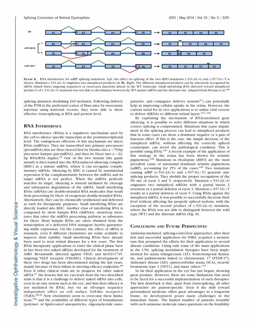
By exploiting the mechanism of RNAi-mediated genesilencing, it is possible to solve different situations in whichcorrect splicing is compromised. Mutations that cause impair-ment in the splicing process can lead to misspliced productsthat in some cases can show a dominant negative or a gain offunction effect. If this is the case, the simple decrease of themisspliced mRNA, without affecting the correctly splicedcounterpart, can revert the pathological condition. This ispossible using RNAi.107 A recent example of the application ofthis strategy in the retina has been shown for retinitispigmentosa.108 Mutations in rhodopsin (RHO) are the mostprevalent cause of autosomal dominant retinitis pigmentosa(adRP), accounting for 25% of the cases.109 Two mutationscausing adRP (c.531-2A>G and c.937-1G>T) generate mis-splicing products. They abolish the proper recognition of theDS site of exon 3 and 5, respectively. Mutation c.531-2A>Goriginates two misspliced mRNAs with a partial intron 2retention or a partial deletion of exon 3. Mutation c.937-1G>Tresults in a partial deletion of exon 5. Using RNAi against themisspliced mRNA, it was possible to successfully decrease theirlevel without affecting the properly spliced isoform, with theexception of the second product of c.531-2A>G mutation,where the RNAi was not able to distinguish between the wildtype (WT) and the aberrant mRNA (Fig. 8).
CONCLUSIONS AND FUTURE PERSPECTIVES
Antisense-mediated, splicing-correction approaches, after theirfirst and successful application for DMD, acquired a momen-tum that prompted the efforts for their application to severaldisease conditions. Citing only some of the main applicationsin the CNS, splicing modulation therapies have been imple-mented for ataxia telangiectasia (AT), frontotemporal demen-tia, and parkinsonism linked to chromosome 17 (FTDP-17),Alzheimer disease (AD), spinocerebellar ataxia (SCA), neurofi-bromatosis type 1 (NFT1), and many others.110
So far their application in the eye has just begun, showinggreat promise. However, there are some limitations that needto be faced for a successful implementation of such therapies.The first drawback is that, apart from trans-splicing, all otherapproaches are patient-specific. Even if the shift towardpersonalized medicine offers great advantages in a long-termframe, its development poses many challenges in theimmediate future. The limited number of patients treatablewith each antisense molecule raises questions on the feasibility
FIGURE 8. RNA interference for adRP splicing mutations. Left: the effect on splicing of the two RHO mutations c.531-2A>G and c.937-1G>T isshown. Mutation c.531-2A>G originates two misspliced products (A, B). Right: The different misspliced products can be selectively recognized bysiRNA (black lines) targeting sequences or exon-exon junctions absent in the WT transcript. Small interfering RNA directed toward missplicedproduct b of c.531-2A>G mutation was not able to discriminate between the WT mature mRNA and the aberrant one. Adapted from Hernan et al.108
Splicing Correction of Retinal Dystrophies IOVS j May 2014 j Vol. 55 j No. 5 j 3291

of normal clinical trials. Moreover, the current need to considerantisense therapies, even of the same class and for the samedisease, as different drugs if they are targeting differentmutations, creates a great economic obstacle to theirdevelopment. Luckily a debate between researchers andregulatory agencies has already started on these issues.111
Another problem regarding the retina is the difficulties ofhaving relevant animal models that would allow splicingmanipulation of the gene of interest. The implementation ofsuch models will definitely foster clinical application ofsplicing-correction approaches for retinal dystrophies.
Acknowledgments
The authors thank Erik Dassi for his helpful assistance in retrievingand analyzing data from HGMD.
Supported by the Italian Ministry of Health, Young ItalianResearchers Grant 2008, Project GR-2008-1136933.
Disclosure: N. Bacchi, None; S. Casarosa, None; M.A. Denti,None
References
1. Daiger SP, Rossiter BJF, Greenberg J, Christoffels AHW. Dataservices and software for identifying genes and mutationscausing retinal degeneration. Invest Ophthalmol Vis Sci.1998;39:S295.
2. Havens MA, Duelli DM, Hastings ML. Targeting RNA splicingfor disease therapy. Wiley Interdiscip Rev RNA. 2013;4:247–266.
3. Bonnal S, Vigevani L, Valcarcel J. The spliceosome as a targetof novel antitumour drugs. Nat Rev Drug Discov. 2012;11:847–859.
4. Smith AJ, Bainbridge JWB, Ali RR. Gene supplementationtherapy for recessive forms of inherited retinal dystrophies.Gene Ther. 2012;19:154–161.
5. Gomes Dos Santos AL, Bochot A, Fattal E. Intraocular deliveryof oligonucleotides. Curr Pharm Biotechnol. 2005;6:7–15.
6. Martin KR, Klein RL, Quigley HA. Gene delivery to the eyeusing adeno-associated viral vectors. Methods. 2002;28:267–275.
7. Surace EM, Auricchio A. Versatility of AAV vectors for retinalgene transfer. Vision Res. 2008;48:353–359.
8. Stein-Streilein J. Immune regulation and the eye. TrendsImmunol. 2008;29:548–554.
9. Vandenberghe LH, Auricchio A. Novel adeno-associated viralvectors for retinal gene therapy. Gene Ther. 2012;19:162–168.
10. Lam BJ, Hertel KJ. A general role for splicing enhancers inexon definition. RNA. 2002;8:1233–1241.
11. Zhou Z, Fu X-D. Regulation of splicing by SR proteins and SRprotein-specific kinases. Chromosoma. 2013;122:191–207.
12. Pozzoli U, Sironi M. Silencers regulate both constitutive andalternative splicing events in mammals. Cell Mol Life Sci.2005;62:1579–1604.
13. Kafasla P, Mickleburgh I, Llorian M, et al. Defining the rolesand interactions of PTB. Biochem Soc Trans. 2012;40:815–820.
14. Jean-Philippe J, Paz S, Caputi M. hnRNP A1: the Swiss armyknife of gene expression. Int J Mol Sci. 2013;14:18999–19024.
15. Busch A, Hertel KJ. Evolution of SR protein and hnRNPsplicing regulatory factors. Wiley Interdiscip Rev RNA. 2012;3:1–12.
16. De Conti L, Baralle M, Buratti E. Exon and intron definition inpre-mRNA splicing. Wiley Interdiscip Rev RNA. 2013;4:49–60.
17. Kornblihtt AR, Schor IE, Allo M, Dujardin G, Petrillo E, MunozMJ. Alternative splicing: a pivotal step between eukaryotic
transcription and translation. Nat Rev Mol Cell Biol. 2013;14:153–165.
18. Hoskins AA, Moore MJ. The spliceosome: a flexible,reversible macromolecular machine. Trends Biochem Sci.2012;37:179–188.
19. Berger W, Kloeckener-Gruissem B, Neidhardt J. The molec-ular basis of human retinal and vitreoretinal diseases. ProgRetin Eye Res. 2010;29:335–375.
20. Stenson PD, Ball EV, Mort M, et al. Human Gene MutationDatabase (HGMD): 2003 update. Hum Mutat. 2003;21:577–581.
21. Lopez-Bigas N, Audit B, Ouzounis C, Parra G, Guigo R. Aresplicing mutations the most frequent cause of hereditarydisease? FEBS Lett. 2005;579:1900–1903.
22. Sterne-Weiler T, Howard J, Mort M, Cooper DN, Sanford JR.Loss of exon identity is a common mechanism of humaninherited disease. Genome Res. 2011;21:1563–1571.
23. Lim KH, Ferraris L, Filloux ME, Raphael BJ, Fairbrother WG.Using positional distribution to identify splicing elements andpredict pre-mRNA processing defects in human genes. ProcNatl Acad Sci U S A. 2011;108:11093–11108.
24. Saleh AF, Arzumanov AA, Gait MJ. Overview of alternativeoligonucleotide chemistries for exon skipping. Methods MolBiol. 2012;867:365–378.
25. Jarver P, O’Donovan L, Gait MJ. A chemical view ofoligonucleotides for exon skipping and related drug applica-tions. Nucleic Acid Ther. 2013;24:37–47.
26. De Clercq E, Eckstein E, Merigan TC. Interferon inductionincreased through chemical modification of a syntheticpolyribonucleotide. Science. 1969;165:1137–1139.
27. Sproat BS, Lamond AI, Beijer B, Neuner P, Ryder U. Highlyefficient chemical synthesis of 20-O-methyloligoribonucleo-tides and tetrabiotinylated derivatives; novel probes that areresistant to degradation by RNA or DNA specific nucleases.Nucleic Acids Res. 1989;17:3373–3386.
28. Martin P. New access to 2-O-alkylated ribonucleosides andproperties of 2-O-alkylated oligoribonucleotides. Helv ChimActa. 1995;78:486–504.
29. Koshkin AA, Singh SK, Nielsen P, et al. LNA (locked nucleicacids): synthesis of the adenine, cytosine, guanine, 5-methylcytosine, thymine and uracil bicyclonucleoside mono-mers, oligomerisation, and unprecedented nucleic acidrecognition. Tetrahedron. 1998;54:3607–3630.
30. Obika S, Nanbu D, Hari Y, et al. Stability and structuralfeatures of the duplexes containing nucleoside analogueswith a fixed N-type conformation, 20 -O,40-C-methyleneribo-nucleosides. Tetrahedron Lett. 1998;39:5401–5404.
31. Nielsen PE, Egholm M, Berg RH, Buchardt O. Sequence-selective recognition of DNA by strand displacement with athymine-substituted polyamide. Science. 1991;254:1497–1500.
32. Summerton J, Weller D. Morpholino antisense oligomers:design, preparation, and properties. Antisense Nucleic AcidDrug Dev. 1997;7:187–195.
33. Kole R, Krainer AR, Altman S. RNA therapeutics: beyond RNAinterference and antisense oligonucleotides. Nat Rev DrugDiscov. 2012;11:125–140.
34. Kurreck J. Antisense technologies. Improvement throughnovel chemical modifications. Eur J Biochem. 2003;270:1628–1644.
35. Baughan T, Shababi M, Coady TH, Dickson AM, Tullis GE,Lorson CL. Stimulating full-length SMN2 expression bydelivering bifunctional RNAs via a viral vector. Mol Ther.2006;14:54–62.
36. Baughan TD, Dickson A, Osman EY, Lorson CL. Delivery ofbifunctional RNAs that target an intronic repressor andincrease SMN levels in an animal model of spinal muscularatrophy. Hum Mol Genet. 2009;18:1600–1611.
37. Owen N, Zhou H, Malygin AA, et al. Design principles forbifunctional targeted oligonucleotide enhancers of splicing.Nucleic Acids Res. 2011;39:7194–7208.
Splicing Correction of Retinal Dystrophies IOVS j May 2014 j Vol. 55 j No. 5 j 3292

38. Skordis LA, Dunckley MG, Yue B, Eperon IC, Muntoni F.Bifunctional antisense oligonucleotides provide a trans-actingsplicing enhancer that stimulates SMN2 gene expression inpatient fibroblasts. Proc Natl Acad Sci U S A. 2003;100:4114–4119.
39. Osman EY, Yen PF, Lorson CL. Bifunctional RNAs targetingthe intronic splicing silencer N1 increase SMN levels andreduce disease severity in an animal model of spinal muscularatrophy. Mol Ther. 2012;20:119–126.
40. Dickson A, Osman E, Lorson CL. A negatively actingbifunctional RNA increases survival motor neuron both invitro and in vivo. Hum Gene Ther. 2008;19:1307–1315.
41. Villemaire J, Dion I, Elela SA, Chabot B. Reprogrammingalternative pre-messenger RNA splicing through the use ofprotein-binding antisense oligonucleotides. J Biol Chem.2003;278:50031–50039.
42. Gendron D, Carriero S, Garneau D, et al. Modulation of 50
splice site selection using tailed oligonucleotides carryingsplicing signals. BMC Biotechnol. 2006;6:5.
43. Brosseau JP, Lucier JF, Lamarche AA, et al. Redirecting splicingwith bifunctional oligonucleotides. Nucleic Acids Res. 2013;42:e40.
44. Van Deutekom JC, Janson AA, Ginjaar IB, et al. Localdystrophin restoration with antisense oligonucleotidePRO051. N Engl J Med. 2007;357:2677–2686.
45. Kalbfuss B, Mabon SA, Misteli T. Correction of alternativesplicing of tau in frontotemporal dementia and parkinsonismlinked to chromosome 17. J Biol Chem. 2001;276:42986–42993.
46. Aartsma-Rus A, Fokkema I, Verschuuren J, et al. Theoreticapplicability of antisense-mediated exon skipping for Du-chenne muscular dystrophy mutations. Hum Mutat. 2009;30:293–299.
47. Kinali M, Arechavala-Gomeza V, Feng L, et al. Localrestoration of dystrophin expression with the morpholinooligomer AVI-4658 in Duchenne muscular dystrophy: asingle-blind, placebo-controlled, dose-escalation, proof-of-concept study. Lancet Neurol. 2009;8:918–928.
48. Cirak S, Arechavala-Gomeza V, Guglieri M, et al. Exonskipping and dystrophin restoration in patients with Du-chenne muscular dystrophy after systemic phosphorodiami-date morpholino oligomer treatment: an open-label, phase 2,dose-escalation study. Lancet. 2011;378:595–605.
49. Goemans NM, Tulinius M, van den Akker JT, et al. Systemicadministration of PRO051 in Duchenne’s muscular dystro-phy. N Engl J Med. 2011;364:1513–1522.
50. Zanetta C, Nizzardo M, Simone C, et al. Molecular therapeuticstrategies for spinal muscular atrophies: current and futureclinical trials. Clin Ther. 2014;36:128–140.
51. Bhisitkul RB, Robinson GS, Moulton RS, Claffey KP, Gragou-das ES, Miller JW. An antisense oligodeoxynucleotide againstvascular endothelial growth factor in a nonhuman primatemodel of iris neovascularization. Arch Ophthalmol. 2005;12:214–219.
52. Shen WY, Garrett KL, da Cruz L, Constable IJ, Rakoczy PE.Dynamics of phosphorothioate oligonucleotides in normaland laser photocoagulated retina. Br J Ophthalmol. 1999;83:852–861.
53. Dvorchik BH, Marquis JK. Disposition and toxicity of a mixedbackbone antisense oligonucleotide, targeted against humancytomegalovirus, after intravitreal injection of escalatingsingle doses in the rabbit. Drug Metab Dispos. 2000;28:1255–1261.
54. Shen WY, Rakoczy PE. Uptake dynamics and retinal toleranceof phosphorothioate oligonucleotide and its direct deliveryinto the site of choroidal neovascularization throughsubretinal administration in the rat. Antisense Nucleic AcidDrug Dev. 2001;11:257–264.
55. Cloutier F, Lawrence M, Goody R, et al. Antiangiogenicactivity of aganirsen in nonhuman primate and rodent
models of retinal neovascular disease after topical adminis-tration. Invest Ophthalmol Vis Sci. 2012;53:1195–1203.
56. Thaler S, Rejdak R, Dietrich K, et al. A selective method fortransfection of retinal ganglion cells by retrograde transfer ofantisense oligonucleotides against kynurenine aminotransfer-ase II. Mol Vis. 2006;12:100–107.
57. Fattal E, Bochot A. Ocular delivery of nucleic acids: antisenseoligonucleotides, aptamers and siRNA. Adv Drug Deliv Rev.2006;58:1203–1223.
58. Uehara H, Cho Y, Simonis J, et al. Dual suppression ofhemangiogenesis and lymphangiogenesis by splice-shiftingmorpholinos targeting vascular endothelial growth factorreceptor 2 (KDR). FASEB J. 2013;27:76–85.
59. Den Hollander AI, Roepman R, Koenekoop RK, Cremers FP.Leber congenital amaurosis: genes, proteins and diseasemechanisms. Prog Retin Eye Res. 2008;27:391–419.
60. Perrault I, Delphin N, Hanein S, et al. Spectrum of NPHP6/CEP290 mutations in Leber congenital amaurosis anddelineation of the associated phenotype. Hum Mutat.2007;28:416.
61. Den Hollander AI, Koenekoop RK, Yzer S, et al. Mutations inthe CEP290 (NPHP6) gene are a frequent cause of Lebercongenital amaurosis. Am J Hum Genet. 2006;79:556–561.
62. Collin RW, den Hollander AI, van der Velde-Visser SD,Bennicelli J, Bennett J, Cremers FP. Antisense oligonucleotide(AON)-based therapy for Leber congenital amaurosis causedby a frequent mutation in CEP290. Mol Ther Nucleic Acids.2012; 27:1:e14.
63. Gerard X, Perrault I, Hanein S, et al. AON-mediated exonskipping restores ciliation in fibroblasts harboring thecommon Leber congenital amaurosis CEP290 mutation. MolTher Nucleic Acids. 2012; 26:e29.
64. Pinotti M, Rizzotto L, Balestra D, et al. U1-snRNA-mediatedrescue of mRNA processing in severe factor VII deficiency.Blood. 2008;111:2681–2684.
65. Schmid F, Hiller T, Korner G, Glaus E, Berger W, Neidhardt J. Agene therapeutic approach to correct splice defects withmodified U1 and U6 snRNPs. Hum Gene Ther. 2013;24:97–104.
66. Patel SB, Bellini M. The assembly of a spliceosomal smallnuclear ribonucleoprotein particle. Nucleic Acids Res. 2008;36:6482–6493.
67. Dominski Z, Marzluff WF. Formation of the 30 end of histonemRNA: getting closer to the end. Gene. 2007;396:373–390.
68. Denti MA, Rosa A, D’Antona G, et al. Body-wide gene therapyof Duchenne muscular dystrophy in the mdx mouse model.Proc Natl Acad Sci U S A. 2006;103:3758–3763.
69. Denti MA, Incitti T, Sthandier O, et al. Long-term benefit ofadeno-associated virus/antisense-mediated exon skipping indystrophic mice. Hum Gene Ther. 2008;19:601–608.
70. Goyenvalle A, Wright J, Babbs A, Wilkins V, Garcia L, DaviesKE. Engineering multiple U7snRNA constructs to inducesingle and multiexon-skipping for Duchenne musculardystrophy. Mol Ther. 2012;20:1212–1221.
71. Goyenvalle A, Babbs A, van Ommen G-JB, Garcia L, DaviesKE. Enhanced exon-skipping induced by U7 snRNA carryinga splicing silencer sequence: promising tool for DMDtherapy. Mol Ther. 2009;17:1234–1240.
72. Marquis J, Meyer K, Angehrn L, Kampfer SS, Rothen-Rutishauser B, Schumperli D. Spinal muscular atrophy:SMN2 pre-mRNA splicing corrected by a U7 snRNA derivativecarrying a splicing enhancer sequence. Mol Ther. 2007;15:1479–1486.
73. Voigt T, Meyer K, Baum O, Schumperli D. Ultrastructuralchanges in diaphragm neuromuscular junctions in a severemouse model for Spinal Muscular Atrophy and theirprevention by bifunctional U7 snRNA correcting SMN2splicing. Neuromuscul Disord. 2010;20:744–752.
74. Susani L, Pangrazio A, Sobacchi C, et al. TCIRG1-dependentrecessive osteopetrosis: mutation analysis, functional identi-
Splicing Correction of Retinal Dystrophies IOVS j May 2014 j Vol. 55 j No. 5 j 3293

fication of the splicing defects, and in vitro rescue by U1snRNA. Hum Mutat. 2004;24:225–235.
75. Tanner G, Glaus E, Barthelmes D, et al. Therapeutic strategyto rescue mutation-induced exon skipping in rhodopsin byadaptation of U1 snRNA. Hum Mutat. 2009;30:255–263.
76. Glaus E, Schmid F, Da Costa R, Berger W, Neidhardt J. Genetherapeutic approach using mutation-adapted U1 snRNA tocorrect a RPGR splice defect in patient-derived cells. MolTher. 2011;19:936–941.
77. Mockel A, Perdomo Y, Stutzmann F, Letsch J, Marion V,Dollfus H. Retinal dystrophy in Bardet-Biedl syndrome andrelated syndromic ciliopathies. Prog Retin Eye Res. 2011;30:258–274.
78. Nachury MV, Seeley ES, Jin H. Trafficking to the ciliarymembrane: how to get across the periciliary diffusionbarrier? Annu Rev Cell Dev Biol. 2010;26:59–87.
79. Schmid F, Glaus E, Barthelmes D, et al. U1 snRNA-mediatedgene therapeutic correction of splice defects caused by anexceptionally mild BBS mutation. Hum Mutat. 2011;32:815–824.
80. Sutton RE, Boothroyd JC. Evidence for trans splicing intrypanosomes. Cell. 1986;47:527–535.
81. Murphy WJ, Watkins KP, Agabian N. Identification of a novel Ybranch structure as an intermediate in trypanosome mRNAprocessing: evidence for trans splicing. Cell. 1986;47:517–525.
82. Flouriot G, Brand H, Seraphin B, Gannon F. Natural trans-spliced mRNAs are generated from the human estrogenreceptor-alpha (hER alpha) gene. J Biol Chem. 2002;277:26244–26251.
83. Caudevilla C, Serra D, Miliar A, et al. Natural trans-splicing incarnitine octanoyltransferase pre-mRNAs in rat liver. ProcNatl Acad Sci U S A. 1998;95:12185–12190.
84. Avale ME, Rodrıguez-Martın T, Gallo JM. Trans-splicingcorrection of tau isoform imbalance in a mouse model oftau mis-splicing. Hum Mol Genet. 2013;22:2603–2611.
85. Chao H, Mansfield SG, Bartel RC, et al. Phenotype correctionof hemophilia A mice by spliceosome-mediated RNA trans-splicing. Nat Med. 2003;9:1015–1019.
86. Tahara M, Pergolizzi RG, Kobayashi H, et al. Trans-splicingrepair of CD40 ligand deficiency results in naturally regulatedcorrection of a mouse model of hyper-IgM X-linkedimmunodeficiency. Nat Med. 2004;10:835–841.
87. Shababi M, Glascock J, Lorson CL. Combination of SMN trans-splicing and a neurotrophic factor increases the life span andbody mass in a severe model of spinal muscular atrophy.Hum Gene Ther. 2011;22:135–144.
88. Coady TH, Lorson CL. Trans-splicing-mediated improvementin a severe mouse model of spinal muscular atrophy. JNeurosci. 2010;30:126–130.
89. Coady TH, Baughan TD, Shababi M, Passini MA, Lorson CL.Development of a single vector system that enhances trans-splicing of SMN2 transcripts. PLoS One. 2008;3:e3468.
90. Chao H, Mansfield SG, Bartel RC, et al. Phenotype correctionof hemophilia A mice by spliceosome-mediated RNA trans-splicing. Nat Med. 2003;9:1015–1019.
91. Wally V, Murauer EM, Bauer JW. Spliceosome-mediated trans-splicing: the therapeutic cut and paste. J Invest Dermatol.2012;132:1959–1966.
92. Mansfield SG, Chao H, Walsh CE. RNA repair usingspliceosome-mediated RNA trans-splicing. Trends Mol Med.2004;10:263–268.
93. Niblock M, Gallo JM. Tau alternative splicing in familial andsporadic tauopathies. Biochem Soc Trans. 2012;40:677–680.
94. Andorfer C, Kress Y, Espinoza M, et al. Hyperphosphorylationand aggregation of tau in mice expressing normal human tauisoforms. J Neurochem. 2003;86:582–590.
95. Krol J, Loedige I, Filipowicz W. The widespread regulation ofmicroRNA biogenesis, function and decay. Nat Rev Genet.2010;11;597–610.
96. Vaishnaw AK, Gollob J, Gamba-Vitalo C, et al. A status reporton RNAi therapeutics. Silence. 2010:14.
97. Kanasty R, Dorkin JR, Vegas A, Anderson D. Deliverymaterials for siRNA therapeutics. Nat Mater. 2013;12:967–977.
98. Kleinman ME, Yamada K, Takeda A, et al. Sequence- andtarget-independent angiogenesis suppression by siRNA viaTLR3. Nature. 2008;452:591–597.
99. Cho WG, Albuquerque RJ, Kleinman ME, et al. Smallinterfering RNA-induced TLR3 activation inhibits blood andlymphatic vessel growth. Proc Natl Acad Sci U S A. 2009;106:7137–7142.
100. Byrne M, Tzekov R, Wang Y, et al. Novel hydrophobicallymodified asymmetric RNAi compounds (sd-rxRNA) demon-strate robust efficacy in the eye. J Ocul Pharmacol Ther.2013;29:855–864.
101. Askou AL, Pournaras JC, Pihlmann M, et al. Reduction ofchoroidal neovascularization in mice endothelial growthfactor short hairpin RNA. J Gene Med. 2012;14:632–641.
102. Hu J, Wu Q, Li T, Chen Y, Wang S. Inhibition of high glucose-induced VEGF release in retinal ganglion cells by RNAinterference targeting G protein-coupled receptor 91. ExpEye Res. 2013;109:31–39.
103. Zhu H, Qian J, Wang W, et al. RNA interference of GADD153protects photoreceptors from endoplasmic reticulum stress-mediated apoptosis after retinal detachment. PLoS One.2013;8:e59339.
104. Chadderton N, Millington-Ward S, Palfi A, et al. Improvedretinal function in a mouse model of dominant retinitispigmentosa following AAV-delivered gene therapy. Mol Ther.2009;17:593–599
105. Yuan MK, Tao Y, Yu W-Z, Kai W, Jiang Y-R. Lentivirus-mediated RNA interference of vascular endothelial growthfactor in monkey eyes with iris neovascularization. Mol Vis.2010;16:1743–1753.
106. Jiang L, Li TZ, Boye SE, Hauswirth WW, Frederick JM, BaehrW. RNAi-mediated gene suppression in a GCAP1(L151F)cone-rod dystrophy mouse model. PLoS One. 2013;8:e57676.
107. Seyhan AA. RNAi: a potential new class of therapeutic forhuman genetic disease. Hum Genet. 2011;130:583–605.
108. Hernan I, Gamundi MJ, Planas E, Borras E, Maseras M,Carballo M. Cellular expression and siRNA-mediated interfer-ence of rhodopsin cis-acting splicing mutants associated withautosomal dominant retinitis pigmentosa. Invest OphthalmolVis Sci. 2011;52:3723–3729.
109. Fingert JH, Oh K, Chung M, et al. Association of a novelmutation in the retinol dehydrogenase 12 (RDH12) gene withautosomal dominant retinitis pigmentosa. Arch Ophthalmol.2008;126:1301–1307.
110. Siva K, Covello G. Denti MA. Exon-skipping antisenseoligonucleotides to correct missplicing in neurogeneticdiseases. Nucleic Acid Ther. 2014;24:69–86.
111. Muntoni F. The development of antisense oligonucleotidetherapies for Duchenne muscular dystrophy: report on aTREAT-NMD workshop hosted by the European MedicinesAgency (EMA), on September 25th 2009. NeuromusculDisord. 2010;20:355–362.
Splicing Correction of Retinal Dystrophies IOVS j May 2014 j Vol. 55 j No. 5 j 3294
Copyright © 2022 FDOKUMEN