Sphinxolides EG and reidispongiolide C: four new cytotoxic macrolides from the new caledonian...
10
Tetrahedron 55 (1999) 14665-14674 TETRAHEDRON Sphinxolides E-G and Reidispongiolide C: Four New Cytotoxic Macrolides from the New Caledonian Litbistida Sponges N. superstes and R coeruieu’ Sabina Carboaelli’, Angela Zampella’, Antonio Randazzo’, Cecile Debitus’, and Luigi Gomez-Paloma” ‘* ‘Dipartimento di Chimica delle Sostanze Naturali, Universits di Napoli ccFedtico II)), via D. Montesano 49,80131 Napoli, Italy bDIFARMA, Dipartimento di Scienze Farmaceutiche, Universiti di Salerno, via Ponte don Melillo, Edificio 1 l/C, 84084, Fisciano@A), Italy ‘IRD (ex ORSTOM), Centre de Noun& B.P. A5 Noun&a Cedex, New Cakdonia Dedicated to the memory of Professor Giacomino Randazzo Received 23 June 1999; revised 21 September 1999; accepted 7 October 1999 Abstracr. Four new macrolides (7-lo), structurally related to known sphinxolides (l-4) and reidispongiolides (S-6), have bean isolated following a reinvestigation of Neosiphoniu supers&s and Reidispongia coeru!ea, two Lithistida sponges collected in the deep waters off New Cakdonia. In this paper we report their structure elucidation based on spectral data (mainly ZD-NMR and MS) as well as their cytotoxicity determined on a panel of 60 human cancer cell lines (NC1 in vitro primary screen). 0 1999 Elsevier Science Ltd. All rights reserved. Key words. Marine sponge; Neosiphonia supers&x; Reidispongia coerulea; Macrolides; Cytotoxic; ZJJ-NMR. INTRODUCTION In the context of our search for new anti-tumour compounds from New Caledonian marine invertebrates, we had the opportunity to investigate two lithistida sponges, Neosiphoniu superstes and Reidispongia coerulea, whose extracts showed marked activity in cytotoxicity tests against P388, P388dox and KB tumor cell lines. Along with sphinxolide (l), previously reported from an unidentified pacific nudibranch,’ sphinxolides B-D (2-4) were first isolated from Neosiphoniu superstes.* Investigation of Reidispongia coerulea resulted in the isolation of two macrolides, named reidispongiolides A (5) and B (6), that are related to sphinxolides and co-occur with sphinxolide B and D.3 Sphinxolides and reidispongiolides, closely related from a structural point of view, are characterized by the same side chain and a very similar 26-membered macrolactone ring. They differ in that sphinxolides are hydroxylated at Cl0 position, whereas all derivatives belonging to the reidispongiolide family are not. Sphinxolides and reidispongiolides were found to be potent cytotoxins and recent pharmacological studies demonstrated that the cytotoxicity of sphinxolides is associated with cell cycle arrest in G2-M phase and induction of apoptosis.4 Like scytophycins and cytochalasins, sphinxolides have been judged potent antimitotic compounds, causing rapid loss of microfilaments in cultured cells. Moreover, they circumvent multidrug resistance, mediated by overexpression of P-glycoprotein. Therefore, these componds may be usefkl in the development of new agents for drug-resistant tumors. 0040-4020/99/$ - see front matter 0 1999 Elsevier Science Ltd. All rights reserved PII: SOO40-4020(99)009 12-6
-
Upload
independent -
Category
Documents
-
view
2 -
download
0
Transcript of Sphinxolides EG and reidispongiolide C: four new cytotoxic macrolides from the new caledonian...

Tetrahedron 55 (1999) 14665-14674
TETRAHEDRON
Sphinxolides E-G and Reidispongiolide C: Four New Cytotoxic Macrolides from the New Caledonian Litbistida Sponges N. superstes and R coeruieu’
Sabina Carboaelli’, Angela Zampella’, Antonio Randazzo’, Cecile Debitus’, and
Luigi Gomez-Paloma” ‘*
‘Dipartimento di Chimica delle Sostanze Naturali, Universits di Napoli ccFedtico II)),
via D. Montesano 49,80131 Napoli, Italy
bDIFARMA, Dipartimento di Scienze Farmaceutiche, Universiti di Salerno,
via Ponte don Melillo, Edificio 1 l/C, 84084, Fisciano@A), Italy
‘IRD (ex ORSTOM), Centre de Noun& B.P. A5 Noun&a Cedex, New Cakdonia
Dedicated to the memory of Professor Giacomino Randazzo
Received 23 June 1999; revised 21 September 1999; accepted 7 October 1999
Abstracr. Four new macrolides (7-lo), structurally related to known sphinxolides (l-4) and reidispongiolides (S-6), have bean
isolated following a reinvestigation of Neosiphoniu supers&s and Reidispongia coeru!ea, two Lithistida sponges collected in
the deep waters off New Cakdonia. In this paper we report their structure elucidation based on spectral data (mainly ZD-NMR
and MS) as well as their cytotoxicity determined on a panel of 60 human cancer cell lines (NC1 in vitro primary screen).
0 1999 Elsevier Science Ltd. All rights reserved.
Key words. Marine sponge; Neosiphonia supers&x; Reidispongia coerulea; Macrolides; Cytotoxic; ZJJ-NMR.
INTRODUCTION
In the context of our search for new anti-tumour compounds from New Caledonian marine
invertebrates, we had the opportunity to investigate two lithistida sponges, Neosiphoniu superstes and
Reidispongia coerulea, whose extracts showed marked activity in cytotoxicity tests against P388, P388dox and
KB tumor cell lines. Along with sphinxolide (l), previously reported from an unidentified pacific nudibranch,’
sphinxolides B-D (2-4) were first isolated from Neosiphoniu superstes.* Investigation of Reidispongia coerulea
resulted in the isolation of two macrolides, named reidispongiolides A (5) and B (6), that are related to
sphinxolides and co-occur with sphinxolide B and D.3 Sphinxolides and reidispongiolides, closely related from
a structural point of view, are characterized by the same side chain and a very similar 26-membered
macrolactone ring. They differ in that sphinxolides are hydroxylated at Cl0 position, whereas all derivatives
belonging to the reidispongiolide family are not. Sphinxolides and reidispongiolides were found to be potent
cytotoxins and recent pharmacological studies demonstrated that the cytotoxicity of sphinxolides is associated
with cell cycle arrest in G2-M phase and induction of apoptosis.4 Like scytophycins and cytochalasins,
sphinxolides have been judged potent antimitotic compounds, causing rapid loss of microfilaments in cultured
cells. Moreover, they circumvent multidrug resistance, mediated by overexpression of P-glycoprotein.
Therefore, these componds may be usefkl in the development of new agents for drug-resistant tumors.
0040-4020/99/$ - see front matter 0 1999 Elsevier Science Ltd. All rights reserved
PII: SOO40-4020(99)009 12-6
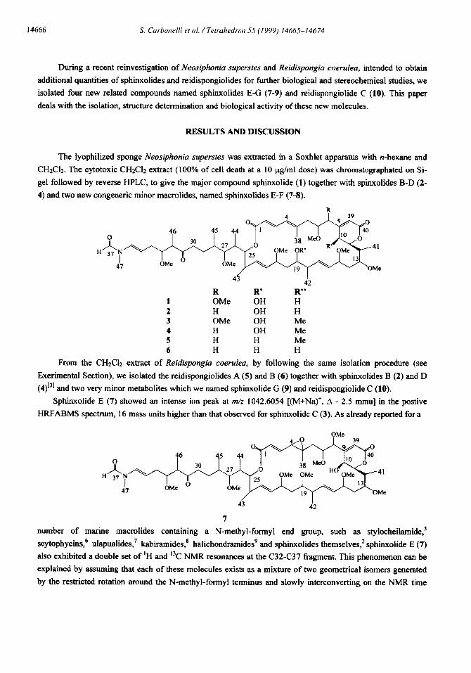
14666 S. Carhonelli et al. /Tetrahedron 55 (1999) 14665-14674
During a recent reinvestigation of Neosiphonia superstes and Reidispongia coerulea, intended to obtain
additional quantities of sphinxolides and reidispongiolides for further biological and stereochemical studies, we
isolated four new related compounds named sphinxolides E-G (7-9) and reidispongiolide C (10). This paper
deals with the isolation, structure determination and biological activity of these new molecules.
RESULTS AND DISCUSSION
The lyophilized sponge Neosiphonia superstes was extracted in a Soxhlet apparatus with n-hexane and
CH2Cl2. The cytotoxic CH2C12 extract (100% of cell death at a 10 ug/ml dose) was chromatographated on Si-
gel followed by reverse HPLC, to give the major compound sphinxolide (1) together with spinxolides B-D (2-
4) and two new congeneric minor macrolides, named sphinxolides E-F (7-8).
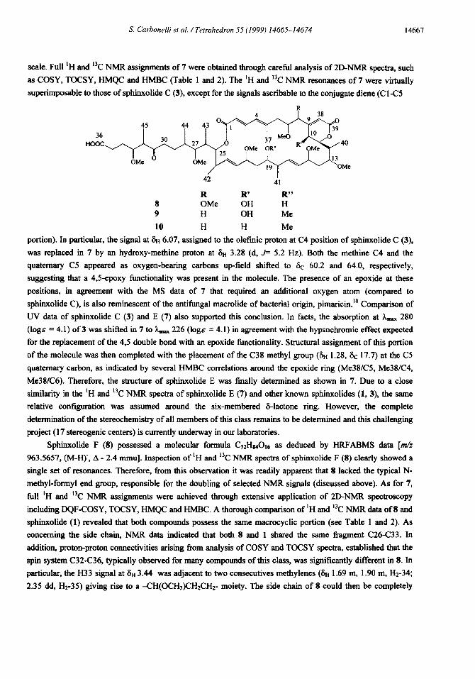
R R’ R” OMe OH H H OH H OMe OH Me H OH Me H H Me H H H
From the CHzCl2 extract of Reidispongia coerufea, by following the same isolation procedure (see
Exerimental Section), we isolated the reidispongiolides A (5) and B (6) together with sphinxolides B (2) and D
(4)13] and two very minor metabolites which we named sphinxolide G (9) and reidispongiolide C (10).
Sphinxolide E (7) showed an intense ion peak at m/z 1042.6054 [(M+Na)+, A - 2.5 mmu] in the postive
HRFABMS spectrum, 16 mass units higher than that observed for sphinxolide C (3). As already reported for a
43 42 7
number of marine macrolides containing a N-methyl-formyl end group, such as stylocheilamide.’
scytophycins,6 ulapualides? kabiramides,’ halichondramides9 and sphinxolides themselves? sphinxolide E (7)
also exhibited a double set of ‘H and 13C NMR resonances at the C32-C37 fragment. This phenomenon can be
explained by assuming that each of these molecules exists as a mixture of two geometrical isomers generated
by the restricted rotation around the N-methyl-formyl terminus and slowly interconverting on the NMR time
S. Carhonelli et al. /Tetrahedron 55 (1999) 14665-14674 14661
scale. Full ‘H and 13C NMR assignments of 7 were obtained through careful analysis of ZD-NMR spectra, such
as COSY, TOCSY, HMQC and HMBC (Table 1 and 2). The ‘H and 13C NMR resonances of 7 were virtually
superimposable to those of sphinxolide C (3), except for the signals ascribable to the conjugate diene (C 1 -C5
36 Hoot
R R’ R” 8 OMe OH H 9 H OH Me
10 H H Me
portion). In particular, the signal at 8u 6.07, assigned to the oletinic proton at C4 position of sphinxolide C (3),
was replaced in 7 by an hydroxy-methine proton at Su 3.28 (d, J= 5.2 Hz). Both the methine C4 and the
quatemary CS appeared as oxygen-bearing carbons up-field shifted to SC 60.2 and 64.0, respectively,
suggesting that a 4,5-epoxy functionality was present in the molecule. The presence of an epoxide at these
positions, in agreement with the MS data of 7 that required an additional oxygen atom (compared to
sphinxolide C), is also reminescent of the antifungal macrolide of bacterial origin, pimaricin.‘” Comparison of
W data of sphinxolide C (3) and E (7) also supported this conclusion. In facts, the absorption at L,,,,x 280
(logs = 4.1) of 3 was shifted in 7 to L.,- 226 (1og.c = 4.1) in agreement with the hypsochromic effect expected
for the replacement of the 45 double bond with an epoxide Functionality. Structural assignment of this portion
of the molecule was then completed with the placement of the C38 methyl group (Su 1.28, 6c 17.7) at the C5
quaternary carbon, as indicated by several HMBC correlations around the epoxide ring (Me38K5, Me38lC4,
Me38K6). Therefore, the structure of sphinxolide E was finally determined as shown in 7. Due to a close
similarity in the ‘H and “C NMR spectra of sphinxolide E (7) and other known sphinxolides (1, 3), the same
relative configuration was assumed around the six-membered 6-lactone ring. However, the complete
determination of the stereochemistry of all members of this class remains to be determined and this challenging
project (17 stereogenic centers) is currently underway in our laboratories.
Sphinxolide F (8) possessed a molecular formula CsrHuO’a as deduced by HRFABMS data [m/z
963.5657, (M-H), A - 2.4 mmu]. Inspection of ‘H and “C NMR spectra of sphinxolide F (8) clearly showed a
single set of resonances. Therefore, from this observation it was readily apparent that 8 lacked the typical N-
methyl-formyl end group, responsible for the doubling of selected NMR signals (discussed above). As for 7,
full ‘H and “C NMR assignments were achieved through extensive application of ZD-NMR spectroscopy
including DQF-COSY, TOCSY, HMQC and HMBC. A thorough comparison of ‘H and 13C NMR data of 8 and
sphinxolide (1) revealed that both compounds possess the same macrocyclic portion (see Table 1 and 2). As
concerning the side chain, NMR data indicated that both 8 and 1 shared the same fragment C26C33. In
addition, proton-proton connectivities arising from analysis of COSY and TOCSY spectra, established that the
spin system C32-C36, typically observed for many compounds of this class, was significantly different in 8. In
particular, the H33 signal at 8~ 3.44 was adjacent to two consecutives methylenes (8~ 1.69 m, 1.90 m, Hz-34;
2.35 dd, Hz-35) giving rise to a CH(OCHs)CH2CH2- moiety. The side chain of 8 could then be completely
I4668 S. Crwhonelli et cd. /Tetrahedron 55 (1999) 14665-14674
determined by placing a carboxyl group at C36 in agreement with the quatemary carbon signal at 6c 181.3
(C36) and the chemical shift value of H35 at on 2.35. In this view, sphinxolide F (S), can be regarded as a
truncated derivative of sphinxolide (l), arising from oxidation of the C36 aldehyde that is produced upon
hydrolysis of the N-methyl-formyl enamine. In order to give additional support to such a structural assignment,
we prepared a semisynthetic sample of 8, by exposing sphinxolide (1) to the Jones’ reagent (see Experimental
Section).
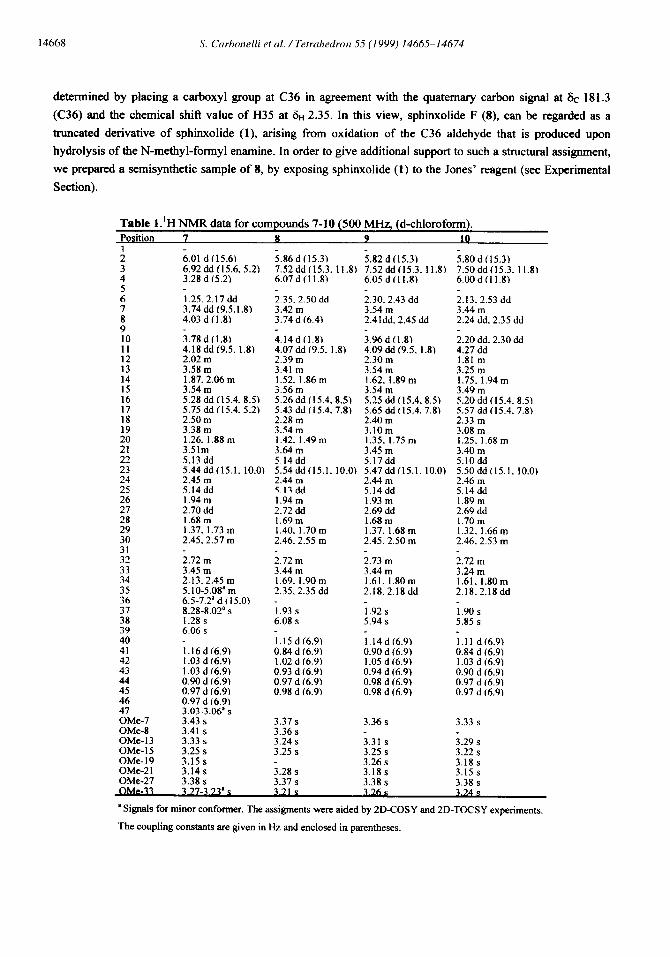
Table l.‘H NMR data for compounds 7-10 (500 MHz, (d-chloroform). Position 7 8 9 10 1
f 4 5
!: 8 9
t:
::
t: 16
t: 19
2’: 22
;:
;: 27
;; 30
::
:: 35 36
z:
:; 41
1:
z 46
ZMe-7 OMe-8 OMe- 13 OMe- 15 OMe-19 OMe-2 1 OMe-27
6.01 d (15.61 6.92 dd (15.6. 5.21 3.28 d (5.21
i.80 d f15.3) 7.50 dd (15.3. 11.81 6.00 d f 11.8)
1.25.2.17dd 3.74 dd (9.5.1.8‘1 4.03 d (I.81
2.13. 2.53 dd 3.44 m 2.24 dd. 2.35 dd
i.86 d (15.3) i.82 d (15.3) 7.52 dd (15.3. 11.81 7.52 dd (15.3. 11.8) 6.07 d (11.81 6.05 d (11.8)
2.35.2.50 dd i.30.2.43 dd 3.42 m 3.54 m 3.74 d (6.41 2.41dd. 2.45 dd
4.14 d (1.8) i.96 d (1.8) 4.07 dd (9.5. 1.8) 4.09 dd (9.5. 1.8) 2.39 m 2.30 m 3.41 m 3.54 m 1.52. 1.866 1.62. 1.89m 3.56 m 3.54 m 5.26ddc15.4. 8.5) 5.25 dd(15.4. 8.5) 5.43 dd (15.4.7.8) 5.65 dd(15.4. 7.81 2.28 m 2.40 m 3.54 m 3.10 m 1.42. 1.49m 1.35. 1.75 m 3.64 m 3.45 m
3.78 d (1.8) 4.18 dd (9.5. 1.8) 2.02 m 3.58 m t .87.2.06 m 3.54 m 5.28 dd (15.4. 8.5) 5.75 dd (15.4.5.2) 2.50 m 3.38 m 1.26. 1.88 m 3.51m 5.13 dd 5.44 dd (15.1. 10.01 2.45 m 5.14 dd 1.94m 2.70 dd 1.68 m 1.37. 1.73 m 2.45. 2.57 m
i.72 m 3.45 m 2.13. 2.45 m 5.10-5.08’m 6%7.2=d (15.0) g.28-8.02as 1.28 s 6.06 s
i.16dc6.9) 1.03 d (6.9) 1.03 d (6.9) 0.90 d (6.9) 0.97 d (6.9) 0.97 d (6.9) 3.03-3.06a s 3.43 s 3.41 s 3.33 s 3.25 s _ ._
2.20 dd. 2.30 dd 4.27 dd 1.81 m 3.25 m 1.75. 1.94 m 3.49 m 5.20 dd (15.4. 8.5) 5.57 dd (15.4. 7.8) 2.33 m 3.08 m 1.25. 1.68 m 3.40 m 5.10 dd 5.50ddf15.1. 10.0) 2.46 m 5.14 dd 1.89 m 2.69 dd 1.70m 1.32. 1.66 m 2.46.2.53 m
3.15 s 3.14 s 3.38 s
5.14 dd 5.17 dd 5.54ddt15.1. 10.0) 5.47dd(15.1.10.0) 2.44 m 5.13 dd 1.94m 2.72 dd 1.69m 1.40. 1.70 m 2.46. 2.55 m
i.72 m 3.44 m 1.69. 1.90m 2.35.2.35 dd
i.93 s 6.08 s
i.15 d (6.91 0.84 d (6.9) 1.02 d (6.9) 0.93 d (6.9) 0.97 d (6.91 0.98 d (6.9)
2.44 m 5.14dd 1.93 m 2.69 dd 1.68m 1.37. 1.68 m 2.45.2.50 m
2.73 m 3.44 m 1.61. 1.80m 2.18. 2.18 dd
2.72 m 3.24 m 1.61. 1.80m 2.18.2.18 dd
1.92 s 5.94 s
1.90s 5.85 s
1.14 d (6.9) 0.90 d (6.9) 1.05 d (6.9) 0.94 d (6.9) 0.98 d (6.91 0.98 d (6.9)
1.11 d(6.91 0.84 d (6.9) 1.03 d (6.91 0.90 d (6.91 0.97 d (6.9) 0.97 d (6.9)
3.37 s 3.36 s 3.24 s 3.25 s
i.28 s 3.37 s
3.36 s 3.33 s
3.31 s 3.25 s 3.26 s 3.18 s 3.38 s
3.29 s 3.22 s 3.18 s 3.15 s 3.38 s
Awe-33 _ ’
’ Signals for minor conformer. The assigments were aided by ZD-COSY and ZD-TOCSY experiments.
Tbe coupling constants are given in Hz and enclosed in parentheses.

S. Curhonelli et al. /Tetrahedron 55 (I 999) 141X5-14674 14669
Comparison of spectral data OIJMR and MS) of the natural product and the thus prepared truncated
derivative of 1 showed that the two compounds were indeed identical. It should be noted that a similar end
group with a carboxyl terminus has already been observed for a derivative of halicondramide.~
Sphinxolide G (9) is a minor component of the CHrClr extract of the sponge Reidispongia coeruleu and
Table 2. 13C NMR data for compounds 7-10 (125 MHz, (d-chloroform). PosltlOll . . 7 8 9 10 1
3' 4
:
8’ 9
t;
:3
t:
:76
t;
f 7 f:
;: 26
;:
32:
::
z: 35
:!:
398 40
1:
: 45 46 47 OMe-7 OMe-8 OMe- 13 OMe- 15 OMe-19 OMe-21 OMe-27
166.1 lfl7.1 124.2 120.0 142.8 140.8 60.2 125.3 64.0 146.9 37.4 40.6 81.0 78.6 82.8 85.1 155.8 155.4 62.9 60.2 84.1 83.3 35.3 35.1 75.8 78.8 33.8 34.7 79.6 78.8 127.2 131.0 141.8 137.4 39.8 41.2 79.8 72.7 35.9 38.9 78.9 82.4 130.7 129.3 138.0 138.6 39.5 40.4 75.8 75.3 34.5 36.0 87.0 87.1 34.5 34.0 23.2 23.2 41.1 40.5 213.5 212 49.0 (49.11’ 49.1 82.2 82.1 30.7 (30.5)’ 26.7 105.4 ~107.1V 32.3 130.3 (126.3)’ 181.3 162.2 060.8)’ 18.6 17.7 121.4 118.6 164.0 163.8 11.8 10.3 15.8 12.9 17.8 17.5 9.9 9.9 17.8 17.5 15.6 12.6 112.7)’ 27.6 (33.21’ 56.9 57.1 59.2 57.8 56.1 56.7 55.6 54.9 57.2 55.7 i6.7 61.8 60.8
16fi.7 166.X 120.3 120.4 140.7 140.4 125.8 126.0 145.4 145.5 43.9 44.4 77.8 77.8 39.1 40.9 156.4 157.5 62.8 31.2 82.4 79.4 35.5 39.2 78.9 77.8 34.6 33.4 79.0 78.9 130.2 130.0 138.0 139.1 38.3 37.5 81.1 80.6 36.9 36.6 79.2 79.4 130.7 130.6 138.4 138.5 40.4 40.6 75.5 75.3 36.4 36.5 87.2 87.2 34.2 34.4 23.1 23.1 40.5 40.7 213.5 214.4 49.1 49.3 82.3 82.4 26.7 26.5 32.3 32.3 181.3 181.3 18.0 17.5 119.7 117.5 164.3 165.2 10.6 9.9 14.4 14.0 17.9 17.7 9.9 10.0 17.3 17.7 12.5 12.4
57.3
i7.1 55.7 57.2 55.6 61.4
56.7
i6.7 55.4 56.7 55.3 61.4
* Signals for minor conformer. The assigments were aided by HMQC and HMBC experiments.
co-occurs with reidispongiolide A (5). The HRFABMS spectrum showed a pseudomolecular ion peak at m/z
947.5712 (M-H), (A +2.0 mmu), 16 mass units less than sphinxolide F (8). ‘H and “C NMR data of
sphinxolide G (9) were reminiscent of those of other sphinxolides. Inspection of the ‘H NMR spectrum of 9
14670 S. Curhonelli et al. /Tetrahedron 55 (1999) 14665-14674
indicated that this compound shared the same oxygenated side chain of sphinxolide F (S), whereas
modifications were present in both C&C8 and C18-C21 segments of the macrocyclic portion. Complete
assignments of the latter portion of this molecule was performed by careful analysis of TOCSY and HMBC
data (see Table 1 and 2), defining the presence of a methylene at C8 position (8” 2.41 dd, 2.45 dd), as well as a
methoxy functionality at C19. The replacement of the C19-OH in sphinxolide F (8) by a O-methyl group in
sphinxolide G (9) caused small but significative shifts in the ‘H and 13C NMR resonances of the centers around
the C 19 position, giving definitive support to the proposed structure as shown in 9.
Reidispongiolide C (10) gave rise to an intense ion peak at m/z 931 S755 [(M-H), A -2.8 mmu] in the
positive HRFABMS spectrum. MS and 13C NMR data indicated that reidispongiolide C possessed one oxygen
atom less than sphinxolide G (9). Interpretation of COSY and HMQC data revealed that C2-C8, C12-C30 and
C32-C36(COOH) portions were indentical with those of acid 9, while a pertubation was present for the
resonances relative to the six membered &lactone ring. In particular, proton-proton coupling network C 1 O-C 12
revealed that Cl0 was a methylene (8~ 2.29dd and 2.19dd). A comparison of NMR data of 10 with that of
reidispongiolide A (5), the major component of the reidispongiolide family, characterized by the presence of a
IO-deoxy-8-lactone functionality, definitively established the structure of reidispongiolide C (10) as IO-deoxy-
sphinxolide G.
Biological activity of sphinxolides E-G (7-9) and reidiapongiolide C (10)
As stated above, metabolites of both sphinxolide and reidispongiolide families, proved to be very potent
and selective cytotoxins. Preliminary cytotoxicity data were obtained for the first compounds of this series (1-6)
on several tumor cell lines.233 These encouraging results prompted reseachers from the Fox Chase Cancer
Center of Philadelphia to undertake an in-depth investigation on their mode of action4 (see introduction of the
present paper).
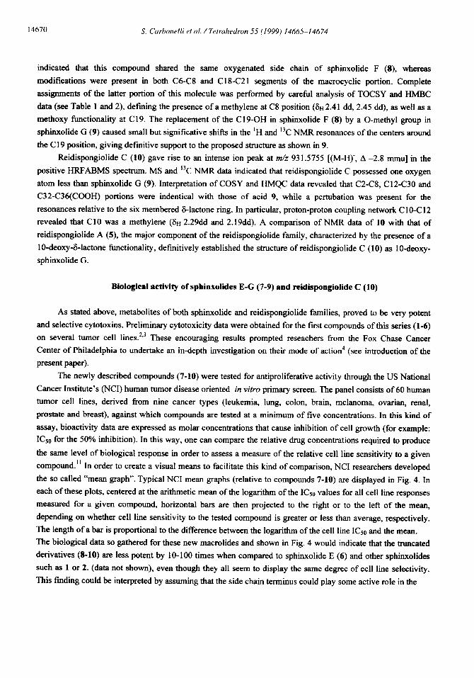
The newly described compounds (7-10) were tested for antiproliferative activity through the US National
Cancer Institute’s (NCI) human tumor disease oriented in vitro primary screen. The panel consists of 60 human
tumor cell lines, derived from nine cancer types (leukemia, lung, colon, brain, melanoma, ovarian, renal,
prostate and breast), against which compounds are tested at a minimum of five concentrations. In this kind of
assay, bioactivity data are expressed as molar concentrations that cause inhibition of cell growth (for example:
KS0 for the 50% inhibition). In this way, one can compare the relative drug concentrations required to produce
the same level of biological response in order to assess a measure of the relative cell line sensitivity to a given
compound.” In order to create a visual means to facilitate this kind of comparison, NC1 researchers developed
the so called “mean graph”. Typical NC1 mean graphs (relative to compounds 7-10) are displayed in Fig. 4. In
each of these plots, centered at the arithmetic mean of the logarithm of the I& values for all cell line responses
measured for a given compound, horizontal bars are then projected to the right or to the left of the mean,
depending on whether cell line sensitivity to the tested compound is greater or less than average, respectively.
The length of a bar is proportional to the difference between the logarithm of the cell line I&, and the mean.
The biological data so gathered for these new macrolides and shown in Fig. 4 would indicate that the truncated
derivatives (8-10) are less potent by lo-100 times when compared to sphinxolide E (6) and other sphinxolides
such as 1 or 2. (data not shown), even though they all seem to display the same degree of cell line selectivity.
This finding could be interpreted by assuming that the side chain terminus could play some active role in the

S. Carbonelli et al. /Tetrahedron 55 (1999) 14665-14674 14671
Fig. 4. I& Mean Graphs from NC1 screeni~ -6.2 (9) and -6.5 (10).
18 of compounds 7-10. Mean 1 Log ICw, (movl) for these cxxnpounds are: -8.1 (7), -7.2 (8),
(9
K-562
MOLT-4 I ii I RPM&8226
SR I hscLC
A549lATCC EKVX
HOP-62
HOP-92 NCI-H23 NCI-322M
NCI-H460
HCT-15 I 1 7 1
HT29
KM12 ii SW-620
CNS Gmcer
SF-268 SF-292 SF-539
SNR-19 SNB-75
U251 Melmloma LOX IMVI
MALME-3M Ml4 SK-MEL-2 SK-MEL-28
SK-MEL5
UACC-257 UACC-62
ovarian Cancer IGROVl OVCAR-3
ovcAR-4
OVCAR-5 OVCAR-8
SK-OV-3
Renal Cancer
786-o
A498 ACHN CAKI-1
RFX 393
SNl2C TIC-10
uO-31
PrasMe cmtcer
PC-3 DU-145 Bream Gnmr
MCF7 NCIIADR-RES
MDA-MB-231 HS 578T

14672 S. Curbmelli et al. /Tetrahedron 55 (1999) 14665-14674
mode of action of these molecules. However, this difference in bioactivity is not very high, suggesting that the
macrocyclic portion of these molecules should still have a major part in the drug-receptor recognition process.
Analogous data produced for the family of halicondrins,” that share with sphinxolides and reidispongiolides
similar (intact and truncated) side chains but different macrocyclic moieties, would lead to similar conclusions.
Further considerations of the structure-activity relationship (SAR) for these metabolites are complicated by the
limitated stereochemical and conformational information currently available.
In conclusion, four new potent antiproliferative macrolides (7-10) have been isolated and characterized.
The present compounds, along with the related and known sphinxohdes and reidispongiolides previously
described (l-6), may represent useful models for the development of new anticancer agents, also in
consideration of their interesting mode of action on the cellular cytoskeleton and their ability to circumvent
multidrug tumor resistance (MDR) mediated by overexpression of P-glycoprotein.
EXPERIMENTAL SECTION
General Experimental Procedures. All NMR measurements were performed on a Bruker AMX-500
spectrometer equipped with a Bruker X-32 computer, using the UXNMR software package.
Optical rotation were measured on a Perkin-Elmer 141 polarimeter using a sodium lamp operating at 589
run. Fast atom bombardment mass spectra (FAB MS) were recorded on a VG ZAB instrument (Argon atoms of
energy of 2-6 kV) using a glycerol-tioglycerol matrix (with lOpI of a solution of NaCl 0.1 N) in the positive
ion mode and in a glycerol-tioglycerol matrix (with triethylamine) in negative ion mode. UV spectra were
recorded on a Beckman DU70 spectrophotometer. IR spectroscopy was performed on a IFS 48 Bruker
instrument. Reversed-phase HPLC was performed on a Cl8 p-Bondapalc column (7.8 mm id. x 30 cm; flow
rate 4.5 ml/m;,) using a Waters model 6000 A pump equipped with a U6K injector and a differential
refractometer, model 40 1.
Isolation. Neosiphoniu superstes (class Demospongiae, order Lithistida, family Phymatellidae) was
collected during a dredging campaign (1994) of the ORSTOM-CNRS, Programme “Substances Marines
d’lnterest Biologique (SMIB)” on the sea mounts of the Norfolk Rise in the South of New Caledonia at a depth
of 500-515 m. Taxonomic identification was performed by L&i and Lkvi at the Museum National d’Histoire
Naturelle in Paris and reference specimens are on file in the IRD centre in Noumea (reference R1408).
Preliminary assays for cytotoxic (KB cells and P388 leukemia cells) and antifungal activities (Fusurium
oxysporum, Phythophthora hevea, Penicillium digitatum) showed a marked activity of chloroformic extract.
The organisms were freeze dried and the lyophilized material (0.5 kg) was extracted with n-hexane (3 x 1 I),
then with CH$&:MeOH 8:2 (3 x 1 1) and with MeOH (3 I) at room temperature. The dichloromethane-
methanol extract was filtered and concentrated under reduced pressure to give 2 g of a yellow cytotoxic oil
(Artemiu salina, I& 10 pg/ml).
The crude dichloromethane-methanol extract was chromatographed by MPLC on a SiO2 column (50 g)
using a solvent system from CHCl3 to CHCl3: MeOH 8:2. Fractions eluted with CHC13:MeOH 995:5 and
CHCb:MeOH 99:l (448.9 mg) were further purified by HPLC on a Waters C-18 p-Bondapak column (7.8 mm
i.d. x 30 cm) with MeOH:HzO (73:27) as eiuent (flow rate 5 ml/min) to give 24.4 mg of superstolide A &= 9.8
min.),“21 7.2 mg of superstolide B (tr = 23.1 min), [13] 81 1 mg of sphinxolide (1) (tr = 12.0 min), 57.8 mg of .
sphinxolide B (2) (h = 15.6 min), 83.3 mg of sphinxolide C (3) (tr = 19.8 min), 69.5 mg sphinxolide D (4) (tr =
21.6 min), 4.7 mg of sphinxolide E (7) (tr= 18.6 mm), 5.4 mg of sphinxolide F (8) (tr= 10.0 min.).
S. Carbonelii et al. /Tetrahedron 55 (1999) 14665-14674 14613
Isolation. Reidispongia coerulea (Levi and Levi, class Demospongiae, order Lithistida, family
Phymatellidae) was collected during the same dredging campaign, together with the Neosiphoniu superstes
sample Taxonomic identification was performed by L&i and Lkvi at the Museum National d’Histoire Naturelle
in Paris and reference specimens are on file in the IRD centre in Noumea (reference 1407). Preliminary assays
for cytotoxic (KB cells and P388 leukemia cells) and antifungal activities (Fusarium oxysporum,
Phythophthora hevea, Penicillium digitatum, Botrytis cinerea, Pyricularia oryzae and Helminthosporium
sativu) showed a marked activity of chloroformic extract. The organisms were freeze dried and the lyophilized
material (4 kg) was Soxhlet extracted with n-hexane (3 x 1 I), then with CH2C12:MeOH 82 (3 x 11) and finally
with MeOH (3 1). The dichloromethane-methanol extract was filtered and concentrated under reduced pressure
to give 2.5 g of a yellow cytotoxic oil (100% mortality at a 10 @ml dose). The crude dichloromethane-
methanol extract was chromatographed by MPLC on a SiOz column (50 g) using a solvent system from CH&
to CH2C12: MeOH 9:l. Fractions eluted with CH2Clr:MeOH 99:l and CH&:MeOH 982 (391.1 mg) were
f&her purified by HPLC on a Waters C-18 u-Bondapak column (7.8 mm i.d. x 30 cm) with MeOH:HrO (77:23)
as eluent (flow rate 4.5 ml/min) to give 165.3 mg of reidispongiolide A (5) (t, = 12.8 min) and 57.3 mg of
sphinxolide D (4) (t,= 18.0 mm), with MeOH:HzO (70:30) to give 1.5 mg of sphinxolide G (9) (G = 4 mm),
with MeOH:HrO (75:25) to give 18.8 mg of sphinxohde B (2) (tr = 13.2 min), 0.5 mg of reidispongilide B (6)
(h= 19.5 min), 5 mg of reidispongiolide C (10) (tr= 5.6 min).
Compound (7): C~~HrsNoi6 amorphous powder. [a]02’ + 2.9 (c 0.004, CHCI,); IR (KBr): vmax 1716,
1701, 1655,1099 (C-O-C); 8u (500 MHz, CDCl3) in Table 1; ); 6~ (125 MHz, CDCl3) in Table 2; UVNis
(Methanol): k, (log E): 226 nm (4.1); HRMS (FAB positive): MNa’, found 1042.6054. C&HssNOi&a
requires 1042.6079.
Compound (8): &Hs~O~~ amorphous powder. [a] nzo+ 24.3 (c 0.001, CHCl,); IR (KBr): v, 1720, 1700,
1680; 8u (500 MHz, CDCl3) in Table 1; 6c (125 MHz, CDCl3) in Table 2; UVNis (Methanol): h,, (log E):
280 run (4.0), 208 nm (3.9); HRMS (FAB negative): M-H-, found 963.5657. C~Hr5016 requires 963.5681.
Compound (9): C~Hs40is amorphous powder. [a]02’ t 8.0 (c 0.004, CHCI,); IR (KBr): vmax 1710, 1700,
1690; 8u (500 MHz, CDC&) in Table 1; 8c (125 MHz, CDCl3) in Table 2; UVNis (Methanol) : h,, (log E):
280 mn (4.0), 212 nm (3.9); HRMS (FAB negative): M-K, found 947.5712. C52Hs5015 requires 947.5732.
Compound (10): C&&$014 amorphous powder. [a]02’+ 9” (c 0.006, CHCl3); IR (KBr): vmax 1725, 1700,
1690; 8~ (500 MHz, CDCl3) in Table 1; 6~ (125 MHz, CDClx) in Table 2; UVNis (Methanol): h (log E):
276 mn (4.1), 212 nm (3.9); HRMS (FAB negative): M-H-, found 931.5755. C52H&4requires 931.5783.
Conversion of sphiaxolide A to sphiaxolide F. Sphinxolide A (1) (20 mg) was dissolved in dry acetone
(300 ~1) and cooled to 0 “C, and Jones’reagent (7.7 pl) was added. After being stirred for 2 h under argon, the
reaction was quenched with 2-propanol (2 ml). Chloroform (5 ml) and water (5 ml) were added. The organic
soluble portion was removed, and the aqueous layer was washed with additional chloroform (2 x 5 ml). The
combined chloroform extracts were dried over anhydrous sodium sulfate, and the solvent was removed in vucuo
to give a green oil that was purified by HPLC chromatography on a Waters C-18 u-Bondapak column (7.8 mm
i.d. x 30 cm) with MeOH:HrO (70:30) to obtain the sphinxolide F (8) (3.5 mg, 18% of yield) that was identical
by ‘H and 13C NMR spectroscopy with the natural product.
ACKNOWLEDGEMENTS
We thank the crew of the R/V ALIS (IRD Centre de Noumea) for their help during the dredging, and Pr. C.
Levi of the Museum Nationale d’Histoire Naturelle, Paris, France for the identification of the sponge. The

14614 S. Curbonelli et al. /Tetrahedron 55 (1999) 14665-14674
chemical work was supported by the Italian National Research Council (CNR, Rome, Italy) and the Ministry of
University and Scientific Research and Technology (MURST, Rome, Italy). Mass and NMR spectra were
recorded at CRIAS, Centro Interdipartimentale di Analisi Strumentale, Faculty of Pharmacy, University of
Naples Federico II.
1.
2.
3.
4.
5.
6.
7.
8.
9.
10.
11.
12.
13.
REFERENCES
Guella, G.; Man&i, I.; Chiasera, G.; Pietra, F.; Helv. Chim. Acta 1989, 72,237-246.
D’Auria, M. V.; Gomez Paloma, L.; Minale, L.; Zampella, A. Verbist, J. F.; Roussakis, C.; Debitus, C.;
Tetrahedron 1993,49, 8657-8664.
D’Auria, M. V.; Gomez Paloma, L.; Minale, L.; Zampella, A. Verbist, J. F.; Roussakis, C.; Debitus, C.;
Patissou, J.; Tetrahedron 1994,50,4829-4834.
Zhang, X.; Minale, L.; Zampella, A.; Smith, C. D. Cancer Research, 1997, 57, 3751-3758.
Rose, A. F.; Scheuer, P. J.; Springer, J. P.; Clardy, J. J. Am. Chem. Sot. 1978,100,7665-7670.
a) Moore, R. E.; Patterson, G. M. L.; Mynderse, J. S.; Barchi, J., Jr; Norton, T. R.; Furosawa, E.;
Furosawa, S. Pure Appl. Chem. 1986, 58, 263-271. b) Ishibashi, M.; Moore, R. E.; Patterson, G. M. L. J.
Org. Chem. 1986,51,5300-5306.
Roesener, J. A.; Scheuer, P. J. J. Am. Chem. Sot. 1986,108,846-847.
a) Matsunaga, S.; Fusetani, N.; Hashimoto, K.; Koseki, K.; Noma, M. J. Am. Chem. Sot. 1986, 108, 847-
849. b) Matsunaga, S.; Fusetani, N.; Hashimoto, K.; Koseki, K.; Noma, M.; Noguchi, H.; Sankawa, U. J.
Org. Chem. 1989,54,1360-1363.
a) Keman, M. R.; Faulkner, D. J.; Tetrahedron Lett. 1987,52,2809-2812. b) Keman, M. R.; Molinski,T.
F.; Faulkner, D. J. J. Org. Chem. 1988,53, 5014-5020.
Lancelin, J. M.; Beau, J. M. J. Am. Chem. Sot. 1990, 112.4060-4061 and references cited therein.
Boyd, M. R. Principles and Practices of Oncology 1989, 3, 1 - 12.
D’Auria, M. V.; Debitus, C.; Gomez Paloma, L.; Minale, L.; Zampella, A. J. Am. Chem. Sot. 1994, I I6,
6658-6663.
D’Auria, M. V.; Gomez Paloma, L.; Minale, L.; Zampella, A.; Debitus, C. J. Nat Prod. 1994, 57, 1595-
1597.