Speech from Another: The Mechanized Pursuit of the Human Voice
Upload
khangminh22Category
view
1download
0
Institutionen för klinisk vetenskap, intervention och teknik (CLINTEC)
Speech and voice characteristics in multiple sclerosis and cervical spinal cord injury: descriptive studies and effects of respiratory training
AKADEMISK AVHANDLING som för avläggande av medicine doktorsexamen vid Karolinska Institutet offentligen försvaras i föreläsningssal R 64, Rehabgatan 64, plan 6, Karolinska universitetssjukhuset, Huddinge
Fredagen den 29 november 2013, kl 09.00
av Kerstin Johansson Leg logoped
Huvudhandledare:
Medicine doktor Ellika Schalling
Karolinska Institutet
Institutionen för klinisk vetenskap, intervention
och teknik (CLINTEC)
Enheten för logopedi
Bihandledare:
Professor Lena Hartelius
Göteborgs universitet
Sahlgrenska akademin
Institutionen för neurovetenskap och fysiologi
Sektionen för klinisk neurovetenskap och
rehabilitering
Enheten för logopedi
Professor Sten Fredrikson
Karolinska Institutet
Institutionen för klinisk neurovetenskap
Sektionen för neurologi
Fakultetsopponent:
Medicine doktor Jenny Iwarsson
Köpenhamns universitet
Institutionen för nordiska studier och
språkvetenskap
Betygsnämnd:
Professor Jörgen Borg
Karolinska Institutet
Institutionen för kliniska vetenskaper, DS
Docent Stellan Hertegård
Karolinska Institutet
Institutionen för klinisk vetenskap, intervention
och teknik (CLINTEC)
Enheten för öron-, näs- och halssjukdomar
Docent Kristina Hansson
Lunds universitet
Institutionen för kliniska vetenskaper
Logopedi, foniatri och audiologi
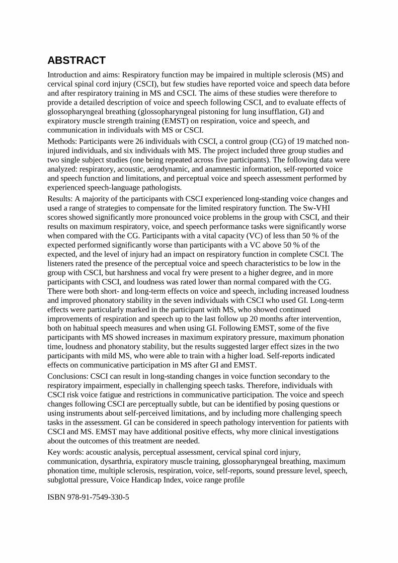
ABSTRACT
Introduction and aims: Respiratory function may be impaired in multiple sclerosis (MS) and
cervical spinal cord injury (CSCI), but few studies have reported voice and speech data before
and after respiratory training in MS and CSCI. The aims of these studies were therefore to
provide a detailed description of voice and speech following CSCI, and to evaluate effects of
glossopharyngeal breathing (glossopharyngeal pistoning for lung insufflation, GI) and
expiratory muscle strength training (EMST) on respiration, voice and speech, and
communication in individuals with MS or CSCI.
Methods: Participants were 26 individuals with CSCI, a control group (CG) of 19 matched non-
injured individuals, and six individuals with MS. The project included three group studies and
two single subject studies (one being repeated across five participants). The following data were
analyzed: respiratory, acoustic, aerodynamic, and anamnestic information, self-reported voice
and speech function and limitations, and perceptual voice and speech assessment performed by
experienced speech-language pathologists.
Results: A majority of the participants with CSCI experienced long-standing voice changes and
used a range of strategies to compensate for the limited respiratory function. The Sw-VHI
scores showed significantly more pronounced voice problems in the group with CSCI, and their
results on maximum respiratory, voice, and speech performance tasks were significantly worse
when compared with the CG. Participants with a vital capacity (VC) of less than 50 % of the
expected performed significantly worse than participants with a VC above 50 % of the
expected, and the level of injury had an impact on respiratory function in complete CSCI. The
listeners rated the presence of the perceptual voice and speech characteristics to be low in the
group with CSCI, but harshness and vocal fry were present to a higher degree, and in more
participants with CSCI, and loudness was rated lower than normal compared with the CG.
There were both short- and long-term effects on voice and speech, including increased loudness
and improved phonatory stability in the seven individuals with CSCI who used GI. Long-term
effects were particularly marked in the participant with MS, who showed continued
improvements of respiration and speech up to the last follow up 20 months after intervention,
both on habitual speech measures and when using GI. Following EMST, some of the five
participants with MS showed increases in maximum expiratory pressure, maximum phonation
time, loudness and phonatory stability, but the results suggested larger effect sizes in the two
participants with mild MS, who were able to train with a higher load. Self-reports indicated
effects on communicative participation in MS after GI and EMST.
Conclusions: CSCI can result in long-standing changes in voice function secondary to the
respiratory impairment, especially in challenging speech tasks. Therefore, individuals with
CSCI risk voice fatigue and restrictions in communicative participation. The voice and speech
changes following CSCI are perceptually subtle, but can be identified by posing questions or
using instruments about self-perceived limitations, and by including more challenging speech
tasks in the assessment. GI can be considered in speech pathology intervention for patients with
CSCI and MS. EMST may have additional positive effects, why more clinical investigations
about the outcomes of this treatment are needed.
Key words: acoustic analysis, perceptual assessment, cervical spinal cord injury,
communication, dysarthria, expiratory muscle training, glossopharyngeal breathing, maximum
phonation time, multiple sclerosis, respiration, voice, self-reports, sound pressure level, speech,
subglottal pressure, Voice Handicap Index, voice range profile
ISBN 978-91-7549-330-5
From DEPARTMENT OF CLINICAL SCIENCE, INTERVENTION AND TECHNOLOGY
Division of Speech and Language Pathology Karolinska Institutet, Stockholm, Sweden
SPEECH AND VOICE CHARACTERISTICS IN
MULTIPLE SCLEROSIS AND CERVICAL SPINAL CORD
INJURY: DESCRIPTIVE STUDIES AND EFFECTS OF RESPIRATORY TRAINING
Kerstin Johansson
Stockholm 2013


All previously published papers were reproduced with permission from the publisher.
Published by Karolinska Institutet. Printed by Universitetsservice US-AB
© Kerstin Johansson, 2013
ISBN 978-91-7549-330-5
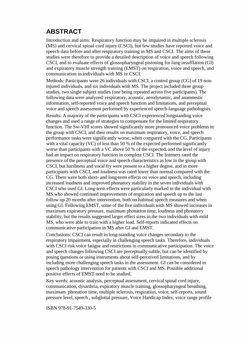
ABSTRACT
Introduction and aims: Respiratory function may be impaired in multiple sclerosis
(MS) and cervical spinal cord injury (CSCI), but few studies have reported voice and
speech data before and after respiratory training in MS and CSCI. The aims of these
studies were therefore to provide a detailed description of voice and speech following
CSCI, and to evaluate effects of glossopharyngeal pistoning for lung insufflation (GI)
and expiratory muscle strength training (EMST) on respiration, voice and speech, and
communication in individuals with MS or CSCI.
Methods: Participants were 26 individuals with CSCI, a control group (CG) of 19 non-
injured individuals, and six individuals with MS. The project included three group
studies, two single subject studies (one being repeated across five participants). The
following data were analyzed: respiratory, acoustic, aerodynamic, and anamnestic
information, self-reported voice and speech function and limitations, and perceptual
voice and speech assessment performed by experienced speech-language pathologists.
Results: A majority of the participants with CSCI experienced longstanding voice
changes and used a range of strategies to compensate for the limited respiratory
function. The Sw-VHI scores showed significantly more pronounced voice problems in
the group with CSCI, and their results on maximum respiratory, voice, and speech
performance tasks were significantly worse, when compared with the CG. Participants
with a vital capacity (VC) of less than 50 % of the expected performed significantly
worse than participants with a VC above 50 % of the expected, and the level of injury
had an impact on respiratory function in complete CSCI. The listeners rated the
presence of the perceptual voice and speech characteristics as low in the group with
CSCI, but harshness and vocal fry were present to a higher degree, and in more
participants with CSCI, and loudness was rated lower than normal compared with the
CG. There were both short- and long-term effects on voice and speech, including
increased loudness and improved phonatory stability in the seven individuals with
CSCI who used GI. Long-term effects were particularly marked in the individual with
MS who showed continued improvements of respiration and speech up to the last
follow up 20 months after intervention, both on habitual speech measures and when
using GI. Following EMST, some of the five individuals with MS showed increases in
maximum expiratory pressure, maximum phonation time, loudness and phonatory
stability, but the results suggested larger effect sizes in the two individuals with mild
MS, who were able to train with a higher load. Self-reports indicated effects on
communicative participation in MS after GI and EMST.
Conclusions: CSCI can result in long-standing voice changes secondary to the
respiratory impairment, especially in challenging speech tasks. Therefore, individuals
with CSCI risk voice fatigue and restrictions in communicative participation. The voice
and speech changes following CSCI are perceptually subtle, but can be identified by
posing questions or using instruments about self-perceived limitations, and by
including more challenging speech tasks in the assessment. GI can be considered in
speech pathology intervention for patients with CSCI and MS. Possible additional
positive effects of EMST need to be studied.
Key words: acoustic analysis, perceptual assessment, cervical spinal cord injury,
communication, dysarthria, expiratory muscle training, glossopharyngeal breathing,
maximum phonation time, multiple sclerosis, respiration, voice, self-reports, sound
pressure level, speech , subglottal pressure, Voice Handicap Index, voice range profile
ISBN 978-91-7549-330-5
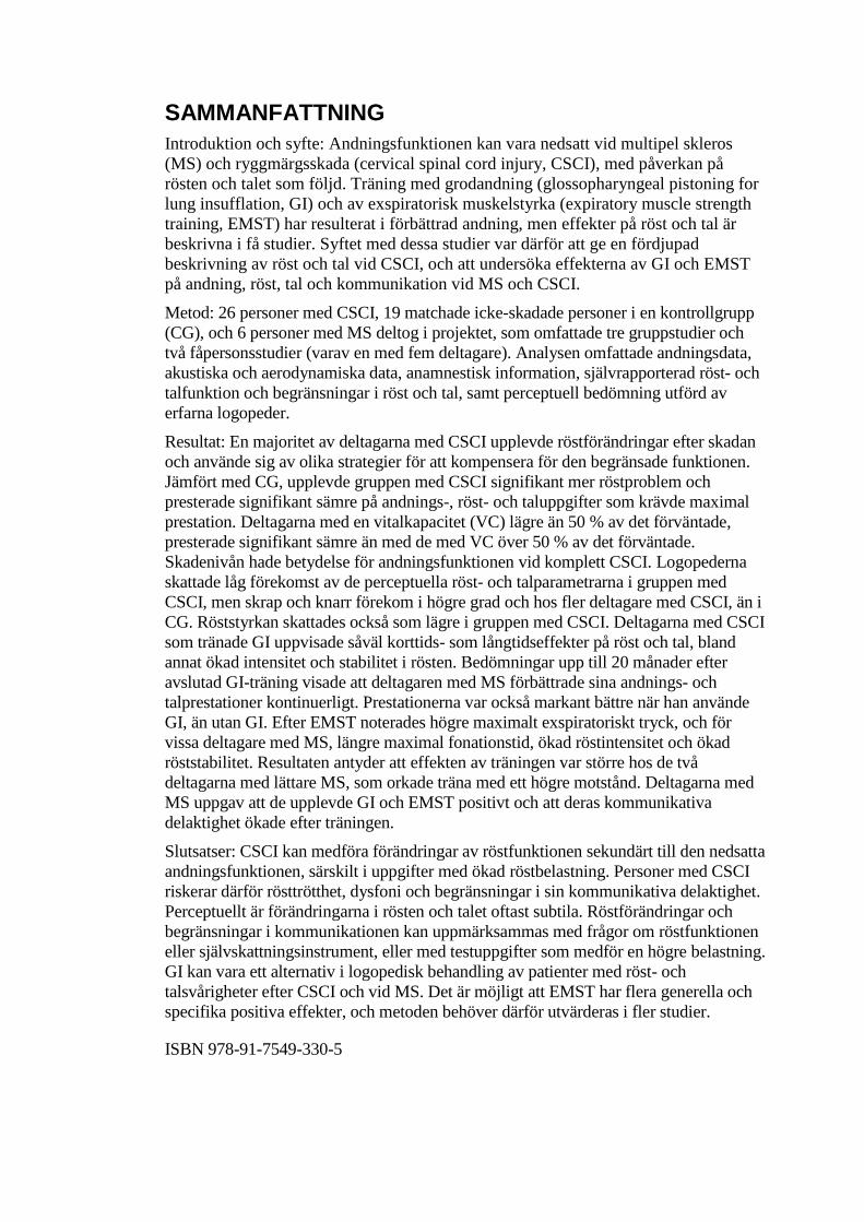
SAMMANFATTNING
Introduktion och syfte: Andningsfunktionen kan vara nedsatt vid multipel skleros
(MS) och ryggmärgsskada (cervical spinal cord injury, CSCI), med påverkan på
rösten och talet som följd. Träning med grodandning (glossopharyngeal pistoning for
lung insufflation, GI) och av exspiratorisk muskelstyrka (expiratory muscle strength
training, EMST) har resulterat i förbättrad andning, men effekter på röst och tal är
beskrivna i få studier. Syftet med dessa studier var därför att ge en fördjupad
beskrivning av röst och tal vid CSCI, och att undersöka effekterna av GI och EMST
på andning, röst, tal och kommunikation vid MS och CSCI.
Metod: 26 personer med CSCI, 19 matchade icke-skadade personer i en kontrollgrupp
(CG), och 6 personer med MS deltog i projektet, som omfattade tre gruppstudier och
två fåpersonsstudier (varav en med fem deltagare). Analysen omfattade andningsdata,
akustiska och aerodynamiska data, anamnestisk information, självrapporterad röst- och
talfunktion och begränsningar i röst och tal, samt perceptuell bedömning utförd av
erfarna logopeder.
Resultat: En majoritet av deltagarna med CSCI upplevde röstförändringar efter skadan
och använde sig av olika strategier för att kompensera för den begränsade funktionen.
Jämfört med CG, upplevde gruppen med CSCI signifikant mer röstproblem och
presterade signifikant sämre på andnings-, röst- och taluppgifter som krävde maximal
prestation. Deltagarna med en vitalkapacitet (VC) lägre än 50 % av det förväntade,
presterade signifikant sämre än med de med VC över 50 % av det förväntade.
Skadenivån hade betydelse för andningsfunktionen vid komplett CSCI. Logopederna
skattade låg förekomst av de perceptuella röst- och talparametrarna i gruppen med
CSCI, men skrap och knarr förekom i högre grad och hos fler deltagare med CSCI, än i
CG. Röststyrkan skattades också som lägre i gruppen med CSCI. Deltagarna med CSCI
som tränade GI uppvisade såväl korttids- som långtidseffekter på röst och tal, bland
annat ökad intensitet och stabilitet i rösten. Bedömningar upp till 20 månader efter
avslutad GI-träning visade att deltagaren med MS förbättrade sina andnings- och
talprestationer kontinuerligt. Prestationerna var också markant bättre när han använde
GI, än utan GI. Efter EMST noterades högre maximalt exspiratoriskt tryck, och för
vissa deltagare med MS, längre maximal fonationstid, ökad röstintensitet och ökad
röststabilitet. Resultaten antyder att effekten av träningen var större hos de två
deltagarna med lättare MS, som orkade träna med ett högre motstånd. Deltagarna med
MS uppgav att de upplevde GI och EMST positivt och att deras kommunikativa
delaktighet ökade efter träningen.
Slutsatser: CSCI kan medföra förändringar av röstfunktionen sekundärt till den nedsatta
andningsfunktionen, särskilt i uppgifter med ökad röstbelastning. Personer med CSCI
riskerar därför rösttrötthet, dysfoni och begränsningar i sin kommunikativa delaktighet.
Perceptuellt är förändringarna i rösten och talet oftast subtila. Röstförändringar och
begränsningar i kommunikationen kan uppmärksammas med frågor om röstfunktionen
eller självskattningsinstrument, eller med testuppgifter som medför en högre belastning.
GI kan vara ett alternativ i logopedisk behandling av patienter med röst- och
talsvårigheter efter CSCI och vid MS. Det är möjligt att EMST har flera generella och
specifika positiva effekter, och metoden behöver därför utvärderas i fler studier.
ISBN 978-91-7549-330-5
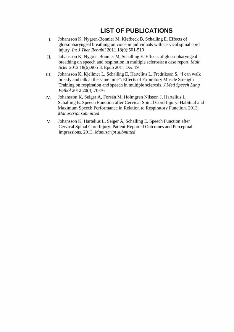
LIST OF PUBLICATIONS
I. Johansson K, Nygren-Bonnier M, Klefbeck B, Schalling E. Effects of
glossopharyngeal breathing on voice in individuals with cervical spinal cord
injury. Int J Ther Rehabil 2011 18(9):501-510
II. Johansson K, Nygren-Bonnier M, Schalling E. Effects of glossopharyngeal
breathing on speech and respiration in multiple sclerosis: a case report. Mult
Scler 2012 18(6):905-8. Epub 2011 Dec 19
III. Johansson K, Kjellmer L, Schalling E, Hartelius L, Fredrikson S. “I can walk
briskly and talk at the same time”: Effects of Expiratory Muscle Strength
Training on respiration and speech in multiple sclerosis. J Med Speech Lang
Pathol 2012 20(4):70-76
IV. Johansson K, Seiger Å, Forsén M, Holmgren Nilsson J, Hartelius L,
Schalling E. Speech Function after Cervical Spinal Cord Injury: Habitual and
Maximum Speech Performance in Relation to Respiratory Function. 2013.
Manuscript submitted
V. Johansson K, Hartelius L, Seiger Å, Schalling E. Speech Function after
Cervical Spinal Cord Injury: Patient-Reported Outcomes and Perceptual
Impressions. 2013. Manuscript submitted
CONTENTS
1 Introduction ................................................................................................... 1
1.1 Respiration and speech in MS and CSCI ........................................... 1
1.1.1 Respiratory function in MS .................................................... 3
1.1.2 Speech symptoms related to respiration in MS ..................... 4
1.1.3 Compensatory strategies ........................................................ 6
1.1.4 Respiratory function in CSCI ................................................. 6
1.1.5 Speech symptoms related to respiration in CSCI .................. 7
1.1.6 Self-reports regarding voice and speech in MS and CSCI .... 9
1.2 Intervention targeting speech dysfunction related to respiration .... 10
1.3 Intervention targeting respiration ..................................................... 11
1.3.1 Glossopharyngeal breathing ................................................. 11
1.3.2 Expiratory muscle strength training, EMST ........................ 12
1.4 Rationale for the included studies .................................................... 13
2 Aims ............................................................................................................ 14
3 Material and methods ................................................................................. 15
3.1 Participants ........................................................................................ 15
3.1.1 Participants with MS ............................................................ 15
3.1.2 Participants with CSCI ......................................................... 15
3.1.3 Control group ........................................................................ 15
3.2 Data collection .................................................................................. 16
3.2.1 Procedure .............................................................................. 16
3.2.2 Collection of probe data and data at follow-ups .................. 18
3.3 Audio recordings and analyses of speech samples .......................... 20
3.3.1 Instrumentation ..................................................................... 20
3.3.2 Acoustic analyses ................................................................. 20
3.3.3 Intelligibility ......................................................................... 21
3.3.4 Perceptual assessment .......................................................... 21
3.4 Estimation of subglottal pressure, PS ............................................... 23
3.5 Self-reported information and questionnaires .................................. 24
3.6 Respiratory intervention ................................................................... 25
3.6.1 Glossopharyngeal breathing ................................................. 25
3.6.2 Expiratory Muscle Strength Training, EMST ..................... 25
3.7 Statistical analyses ............................................................................ 26
3.8 Ethical approvals .............................................................................. 27
4 Results ......................................................................................................... 28
4.1 Study I ............................................................................................... 28
4.2 Study II .............................................................................................. 28
4.3 Study III ............................................................................................ 30
4.4 Study IV ............................................................................................ 31
4.5 Study V ............................................................................................. 31
5 Discussion ................................................................................................... 33
5.1 Selection of participants ................................................................... 33
5.2 Subglottal pressure, PS ...................................................................... 34
5.3 Habitual vs maximum performance ................................................. 36
5.4 Compensatory strategies ................................................................... 36

5.5 Perceptual assessments ..................................................................... 37
5.5.1 Reliability ............................................................................. 37
5.5.2 Perceptual voice and speech characteristics ........................ 39
5.6 Effects of glossopharyngeal breathing on speech ............................ 40
5.7 Effects of EMST on speech .............................................................. 42
6 Conclusions ................................................................................................. 44
7 Further studies ............................................................................................ 45
8 Acknowledgements .................................................................................... 46
9 References ................................................................................................... 49
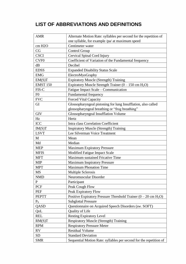
LIST OF ABBREVIATIONS AND DEFINITIONS
AMR Alternate Motion Rate: syllables per second for the repetition of
one syllable, for example /pa/ at maximum speed
cm H2O Centimeter water
CG Control Group
CSCI Cervical Spinal Cord Injury
CVF0 Coefficient of Variation of the Fundamental frequency
dB Decibel
EDSS Expanded Disability Status Scale
EMG ElectroMyoGraphy
EM(S)T Expiratory Muscle (Strength) Training
EMST 150 Expiratory Muscle Strength Trainer (0 – 150 cm H2O)
FIS-C Fatigue Impact Scale – Communication
F0 Fundamental frequency
FVC Forced Vital Capacity
GI Glossopharyngeal pistoning for lung Insufflation, also called
glossopharyngeal breathing or “frog breathing”
GIV Glossopharyngeal Insufflation Volume
Hz Hertz
ICC Intra class Correlation Coefficient
IM(S)T Inspiratory Muscle (Strength) Training
LSVT Lee Silverman Voice Treatment
M Mean
Md Median
MEP Maximum Expiratory Pressure
MFIS Modified Fatigue Impact Scale
MFT Maximum sustained Fricative Time
MIP Maximum Inspiratory Pressure
MPT Maximum Phonation Time
MS Multiple Sclerosis
NMD Neuromuscular Disorder
P Participant
PCF Peak Cough Flow
PEF Peak Expiratory Flow
PEPTT Positive Expiratory Pressure Threshold Trainer (0 – 20 cm H2O)
PS Subglottal Pressure
QASD Questionnaire on Acquired Speech Disorders (sw. SOFT)
QoL Quality of Life
REL Resting Expiratory Level
RM(S)T Respiratory Muscle (Strength) Training
RPM Respiratory Pressure Meter
RV Residual Volume
SD Standard Deviation
SMR Sequential Motion Rate: syllables per second for the repetition of
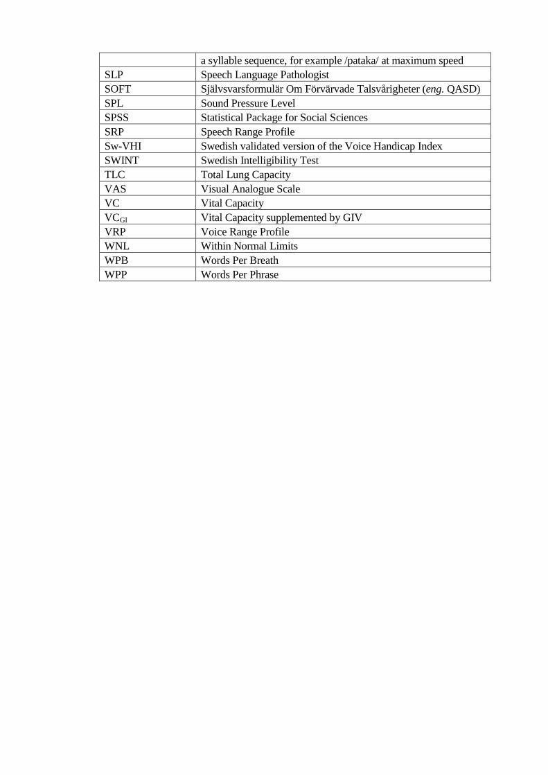
a syllable sequence, for example /pataka/ at maximum speed
SLP Speech Language Pathologist
SOFT Självsvarsformulär Om Förvärvade Talsvårigheter (eng. QASD)
SPL Sound Pressure Level
SPSS Statistical Package for Social Sciences
SRP Speech Range Profile
Sw-VHI Swedish validated version of the Voice Handicap Index
SWINT Swedish Intelligibility Test
TLC Total Lung Capacity
VAS Visual Analogue Scale
VC Vital Capacity
VCGI Vital Capacity supplemented by GIV
VRP Voice Range Profile
WNL Within Normal Limits
WPB Words Per Breath
WPP Words Per Phrase
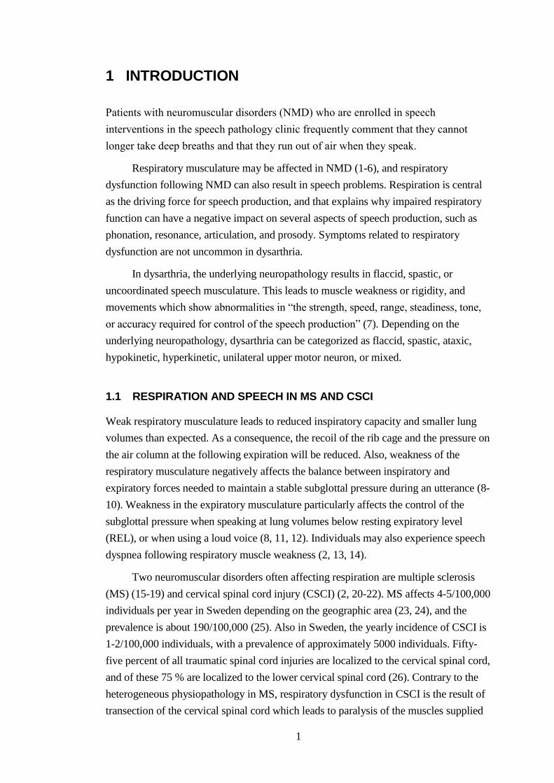
1
1 INTRODUCTION
Patients with neuromuscular disorders (NMD) who are enrolled in speech
interventions in the speech pathology clinic frequently comment that they cannot
longer take deep breaths and that they run out of air when they speak.
Respiratory musculature may be affected in NMD (1-6), and respiratory
dysfunction following NMD can also result in speech problems. Respiration is central
as the driving force for speech production, and that explains why impaired respiratory
function can have a negative impact on several aspects of speech production, such as
phonation, resonance, articulation, and prosody. Symptoms related to respiratory
dysfunction are not uncommon in dysarthria.
In dysarthria, the underlying neuropathology results in flaccid, spastic, or
uncoordinated speech musculature. This leads to muscle weakness or rigidity, and
movements which show abnormalities in “the strength, speed, range, steadiness, tone,
or accuracy required for control of the speech production” (7). Depending on the
underlying neuropathology, dysarthria can be categorized as flaccid, spastic, ataxic,
hypokinetic, hyperkinetic, unilateral upper motor neuron, or mixed.
1.1 RESPIRATION AND SPEECH IN MS AND CSCI
Weak respiratory musculature leads to reduced inspiratory capacity and smaller lung
volumes than expected. As a consequence, the recoil of the rib cage and the pressure on
the air column at the following expiration will be reduced. Also, weakness of the
respiratory musculature negatively affects the balance between inspiratory and
expiratory forces needed to maintain a stable subglottal pressure during an utterance (8-
10). Weakness in the expiratory musculature particularly affects the control of the
subglottal pressure when speaking at lung volumes below resting expiratory level
(REL), or when using a loud voice (8, 11, 12). Individuals may also experience speech
dyspnea following respiratory muscle weakness (2, 13, 14).
Two neuromuscular disorders often affecting respiration are multiple sclerosis
(MS) (15-19) and cervical spinal cord injury (CSCI) (2, 20-22). MS affects 4-5/100,000
individuals per year in Sweden depending on the geographic area (23, 24), and the
prevalence is about 190/100,000 (25). Also in Sweden, the yearly incidence of CSCI is
1-2/100,000 individuals, with a prevalence of approximately 5000 individuals. Fifty-
five percent of all traumatic spinal cord injuries are localized to the cervical spinal cord,
and of these 75 % are localized to the lower cervical spinal cord (26). Contrary to the
heterogeneous physiopathology in MS, respiratory dysfunction in CSCI is the result of
transection of the cervical spinal cord which leads to paralysis of the muscles supplied
2
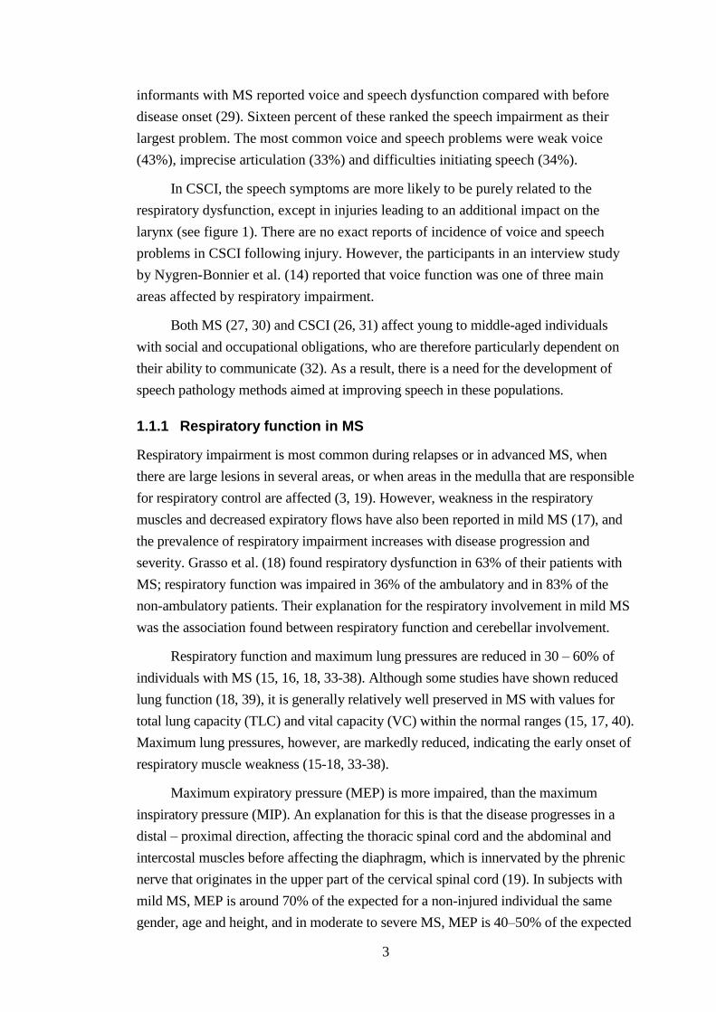
by the spinal nerves under the level of injury (2). At the level of injury of C4 and
below, the innervation of the diaphragm is left intact and the patients can breathe
without a ventilator. However, the muscles in the chest and abdomen are paralyzed,
which explains why both inspiration and expiration are limited (2, 22). Respiratory
dysfunction is therefore a frequent problem for patients with CSCI (2, 22), possibly
resulting in a negative impact on speech function (1).
MS is an inflammatory disease resulting in multiple areas of demyelination in the
nervous system. Because of this, nerve impulse conduction is slowed, causing muscle
atrophy and weakness. The clinical picture is often complex and symptoms vary
depending on the localization of the lesions (27, 28). Respiratory impairment is often
described as a symptom in the late stages of MS, but studies have also shown that the
respiratory musculature is already affected in mild MS resulting in lower inspiratory
and expiratory pressures than expected (17). Respiratory involvement in MS seems to
be more common in individuals where the lesions involve the cerebellum (18).
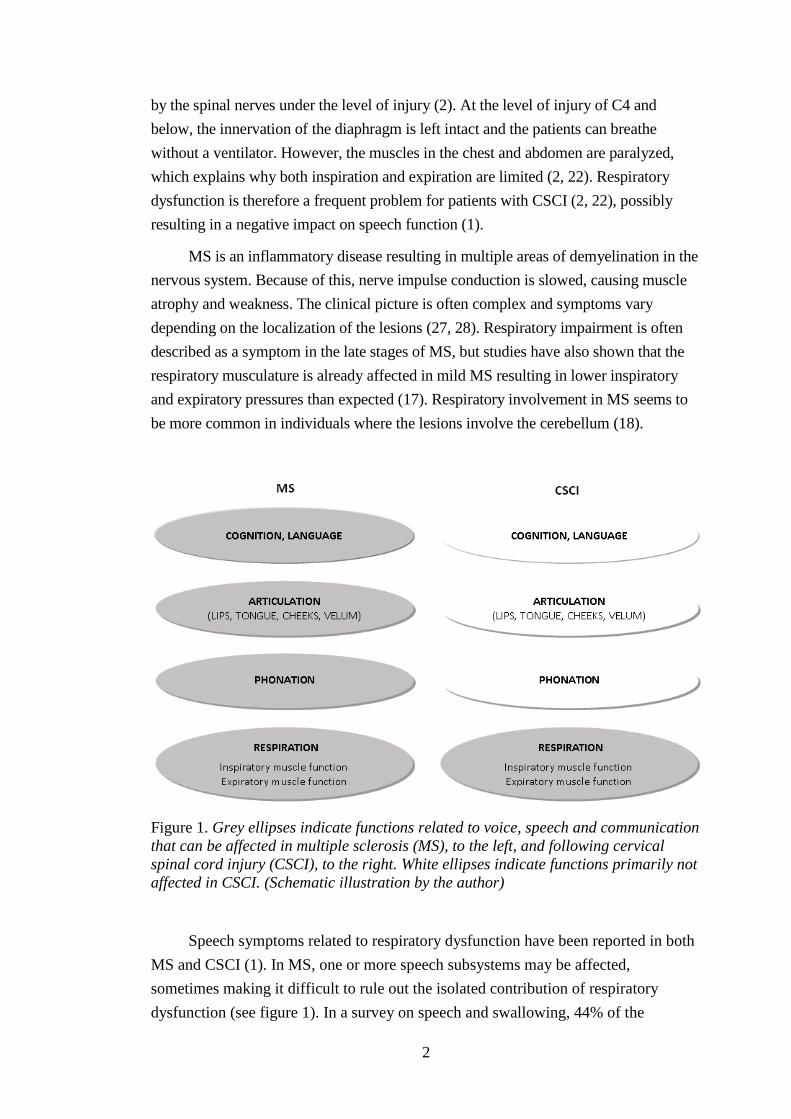
Figure 1. Grey ellipses indicate functions related to voice, speech and communication
that can be affected in multiple sclerosis (MS), to the left, and following cervical
spinal cord injury (CSCI), to the right. White ellipses indicate functions primarily not
affected in CSCI. (Schematic illustration by the author)
Speech symptoms related to respiratory dysfunction have been reported in both
MS and CSCI (1). In MS, one or more speech subsystems may be affected,
sometimes making it difficult to rule out the isolated contribution of respiratory
dysfunction (see figure 1). In a survey on speech and swallowing, 44% of the
3
informants with MS reported voice and speech dysfunction compared with before
disease onset (29). Sixteen percent of these ranked the speech impairment as their
largest problem. The most common voice and speech problems were weak voice
(43%), imprecise articulation (33%) and difficulties initiating speech (34%).
In CSCI, the speech symptoms are more likely to be purely related to the
respiratory dysfunction, except in injuries leading to an additional impact on the
larynx (see figure 1). There are no exact reports of incidence of voice and speech
problems in CSCI following injury. However, the participants in an interview study
by Nygren-Bonnier et al. (14) reported that voice function was one of three main
areas affected by respiratory impairment.
Both MS (27, 30) and CSCI (26, 31) affect young to middle-aged individuals
with social and occupational obligations, who are therefore particularly dependent on
their ability to communicate (32). As a result, there is a need for the development of
speech pathology methods aimed at improving speech in these populations.
1.1.1 Respiratory function in MS
Respiratory impairment is most common during relapses or in advanced MS, when
there are large lesions in several areas, or when areas in the medulla that are responsible
for respiratory control are affected (3, 19). However, weakness in the respiratory
muscles and decreased expiratory flows have also been reported in mild MS (17), and
the prevalence of respiratory impairment increases with disease progression and
severity. Grasso et al. (18) found respiratory dysfunction in 63% of their patients with
MS; respiratory function was impaired in 36% of the ambulatory and in 83% of the
non-ambulatory patients. Their explanation for the respiratory involvement in mild MS
was the association found between respiratory function and cerebellar involvement.
Respiratory function and maximum lung pressures are reduced in 30 – 60% of
individuals with MS (15, 16, 18, 33-38). Although some studies have shown reduced
lung function (18, 39), it is generally relatively well preserved in MS with values for
total lung capacity (TLC) and vital capacity (VC) within the normal ranges (15, 17, 40).
Maximum lung pressures, however, are markedly reduced, indicating the early onset of
respiratory muscle weakness (15-18, 33-38).
Maximum expiratory pressure (MEP) is more impaired, than the maximum
inspiratory pressure (MIP). An explanation for this is that the disease progresses in a
distal – proximal direction, affecting the thoracic spinal cord and the abdominal and
intercostal muscles before affecting the diaphragm, which is innervated by the phrenic
nerve that originates in the upper part of the cervical spinal cord (19). In subjects with
mild MS, MEP is around 70% of the expected for a non-injured individual the same
gender, age and height, and in moderate to severe MS, MEP is 40–50% of the expected
4
(15, 16). The corresponding percentages for MIP are 84% and 74% of the expected in
subjects with mild MS and moderate to severe MS, respectively (15, 17).
1.1.2 Speech symptoms related to respiration in MS
Dysarthria is present in about 40 to 60% of the population of MS (29, 41-43) and there
is a large variation in the symptomatology because of the variety of localization of
lesions. Several types of dysarthria are possible, but a mixed ataxic – spastic dysarthria
is believed to be the most common (44).
Descriptions of speech in MS include several respiratory-related aspects (see
table 1). One of the earliest analyses of speech in MS found “irregular” breathing
affecting voice intensity (45). Reduced respiratory support for speech and loudness
control have been described by Farmakides and Boone (46), Darley et al. (38),
Murdoch et al. (40), and Hartelius et al. (47). The prevalence of deviating perceptual
voice and speech characteristics in two large studies is shown in table 1. Perceptually,
impaired loudness control has been found to appear together with deviating pitch
control in individuals with low VC (38).
Impaired respiratory support is expected to reduce the duration of maximum
phonation and of breath phrases (1). Individuals with MS sustain a vowel with a
significantly shorter duration, than healthy controls (38, 48, 49), and the participants
with low VC in particular had shorter maximum phonation times (MPT) (38). Hartelius
et al. (47) described both increased durations and decreased intra-utterance variability
(more isochronous syllables) in combination with increased inter-utterance variability
in participants with MS. For inter-stress-intervals there were increased durations and
increased variability. These findings reflect inflexibility, as well as instability of
temporal control due to cerebellar involvement.
The majority of studies of speech in MS have focused on different aspects of
phonatory function. Various aspects of phonatory dysfunction, such as pitch breaks and
harsh voice quality, as well as vocal fatigue, have been reported in 18–70% of
individuals with mild – moderate MS (48-50), and in up to 90% of individuals with
severe MS (51). Phonatory instability is also common in MS, with studies reporting a
higher presence of jitter and shimmer in individuals with MS than in healthy control
individuals (38, 46, 48-50, 52-54). Perceptually, a predominant voice characteristic in
MS is harshness (38, 51).
5
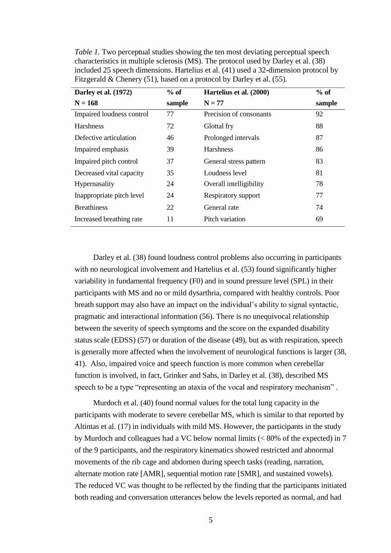
Table 1. Two perceptual studies showing the ten most deviating perceptual speech
characteristics in multiple sclerosis (MS). The protocol used by Darley et al. (38)
included 25 speech dimensions. Hartelius et al. (41) used a 32-dimension protocol by
Fitzgerald & Chenery (51), based on a protocol by Darley et al. (55).
Darley et al. (1972)
N = 168
% of
sample
Hartelius et al. (2000)
N = 77
% of
sample
Impaired loudness control 77 Precision of consonants 92
Harshness 72 Glottal fry 88
Defective articulation 46 Prolonged intervals 87
Impaired emphasis 39 Harshness 86
Impaired pitch control 37 General stress pattern 83
Decreased vital capacity 35 Loudness level 81
Hypernasality 24 Overall intelligibility 78
Inappropriate pitch level 24 Respiratory support 77
Breathiness 22 General rate 74
Increased breathing rate 11 Pitch variation 69
Darley et al. (38) found loudness control problems also occurring in participants
with no neurological involvement and Hartelius et al. (53) found significantly higher
variability in fundamental frequency (F0) and in sound pressure level (SPL) in their
participants with MS and no or mild dysarthria, compared with healthy controls. Poor
breath support may also have an impact on the individual’s ability to signal syntactic,
pragmatic and interactional information (56). There is no unequivocal relationship
between the severity of speech symptoms and the score on the expanded disability
status scale (EDSS) (57) or duration of the disease (49), but as with respiration, speech
is generally more affected when the involvement of neurological functions is larger (38,
41). Also, impaired voice and speech function is more common when cerebellar
function is involved, in fact, Grinker and Sahs, in Darley et al. (38), described MS
speech to be a type “representing an ataxia of the vocal and respiratory mechanism” .
Murdoch et al. (40) found normal values for the total lung capacity in the
participants with moderate to severe cerebellar MS, which is similar to that reported by
Altintas et al. (17) in individuals with mild MS. However, the participants in the study
by Murdoch and colleagues had a VC below normal limits (< 80% of the expected) in 7
of the 9 participants, and the respiratory kinematics showed restricted and abnormal
movements of the rib cage and abdomen during speech tasks (reading, narration,
alternate motion rate [AMR], sequential motion rate [SMR], and sustained vowels).
The reduced VC was thought to be reflected by the finding that the participants initiated
both reading and conversation utterances below the levels reported as normal, and had
6
slightly lower lung volume excursions compared with a group of healthy controls. The
participants also showed irregular (bizarre) movements during inspiration and
expiration, which were thought to be related to the breakdown in coordination between
the rib cage compartment and the abdomen.
Apart from the neurophysiological impact on speech production, factors such as
cognitive load and fatigue may affect an individual’s ability to speak. In healthy
individuals, speech tasks with a cognitive-linguistic loading affect speech breathing
patterns, resulting in slower speaking rate, reduced number of syllables produced per
breath group, greater lung volume expended per syllable than under a lower demanding
condition, and a smaller volume of the abdomen at breath group termination (58).
Demanding speech tasks could therefore pose a communication problem for individuals
with MS.
1.1.3 Compensatory strategies
In NMD, there are also speech symptoms which are the result of the individual’s
conscious or unconscious way of coping with impaired speech function. For example,
Scripture (45) found prolonged pauses secondary to irregular breathing in participants
with MS, and named these efforts of coping with ataxia “anataxia”. Maladaptive
breathing strategies are, for example, producing few syllables or words when there is
enough respiratory support for longer breath groups (7). An individual with limited
respiratory function may compensate for insufficient respiratory support by
“downstream” adaptations (1). Such coping strategies are, for example, on a laryngeal
level, increasing vocal fold adduction, thereby increasing laryngeal resistance in order
to extend the expiratory phase. This will however affect voice quality and intensity.
Another strategy for economizing expiration may be increasing resistance in the oral
and pharyngeal cavities by changing articulation. This will affect the distinctness of
speech articulation and voice intensity, and result in decreased intelligibility (1). From
the above, it can be hypothesized, that the maladaptive compensatory strategies
themselves can cause exertion, which can lead to increased fatigue, and may further
impair voice function and speech.
1.1.4 Respiratory function in CSCI
Vital capacity is decreased by 30-50% of the predicted based on gender and height
following CSCI (2). Respiratory function is related to the level of injury and the
completeness of injury (21), with higher levels of injury showing more restricted VC.
However, as the proportion used for speech is estimated to be about 20% of the VC in
healthy individuals (10), respiratory function will probably be sufficient for
conversational speech (59). Poor respiratory support could however affect speech
production in more demanding speech tasks. One way in which people with CSCI
7
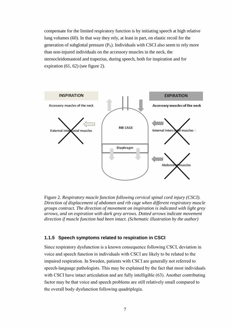
compensate for the limited respiratory function is by initiating speech at high relative
lung volumes (60). In that way they rely, at least in part, on elastic recoil for the
generation of subglottal pressure (PS). Individuals with CSCI also seem to rely more
than non-injured individuals on the accessory muscles in the neck, the
sternocleidomastoid and trapezius, during speech, both for inspiration and for
expiration (61, 62) (see figure 2).
Figure 2. Respiratory muscle function following cervical spinal cord injury (CSCI).
Direction of displacement of abdomen and rib cage when different respiratory muscle
groups contract. The direction of movement on inspiration is indicated with light grey
arrows, and on expiration with dark grey arrows. Dotted arrows indicate movement
direction if muscle function had been intact. (Schematic illustration by the author)
1.1.5 Speech symptoms related to respiration in CSCI
Since respiratory dysfunction is a known consequence following CSCI, deviation in
voice and speech function in individuals with CSCI are likely to be related to the
impaired respiration. In Sweden, patients with CSCI are generally not referred to
speech-language pathologists. This may be explained by the fact that most individuals
with CSCI have intact articulation and are fully intelligible (63). Another contributing
factor may be that voice and speech problems are still relatively small compared to
the overall body dysfunction following quadriplegia.
8
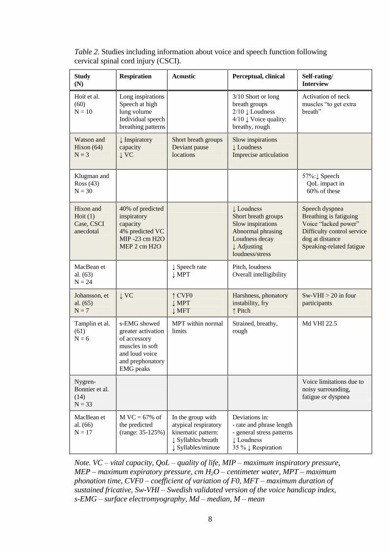
Table 2. Studies including information about voice and speech function following
cervical spinal cord injury (CSCI).
Study
(N)
Respiration Acoustic Perceptual, clinical Self-rating/
Interview
Hoit et al.
(60)
N = 10
Long inspirations
Speech at high
lung volume
Individual speech
breathing patterns
3/10 Short or long
breath groups
2/10 ↓ Loudness
4/10 ↓ Voice quality:
breathy, rough
Activation of neck
muscles “to get extra
breath”
Watson and
Hixon (64)
N = 3
↓ Inspiratory
capacity
↓ VC
Short breath groups
Deviant pause
locations
Slow inspirations
↓ Loudness
Imprecise articulation
Klugman and
Ross (43)
N = 30
57%:↓ Speech
QoL impact in
60% of these
Hixon and
Hoit (1)
Case, CSCI
anecdotal
40% of predicted
inspiratory
capacity
4% predicted VC
MIP -23 cm H2O
MEP 2 cm H2O
↓ Loudness
Short breath groups
Slow inspirations
Abnormal phrasing
Loudness decay
↓ Adjusting
loudness/stress
Speech dyspnea
Breathing is fatiguing
Voice “lacked power”
Difficulty control service
dog at distance
Speaking-related fatigue
MacBean et
al. (63)
N = 24
↓ Speech rate
↓ MPT
Pitch, loudness
Overall intelligibility
Johansson, et
al. (65)
N = 7
↓ VC ↑ CVF0
↓ MPT
↓ MFT
Harshness, phonatory
instability, fry
↑ Pitch
Sw-VHI > 20 in four
participants
Tamplin et al.
(61)
N = 6
s-EMG showed
greater activation
of accessory
muscles in soft
and loud voice
and prephonatory
EMG peaks
MPT within normal
limits
Strained, breathy,
rough
Md VHI 22.5
Nygren-
Bonnier et al.
(14)
N = 33
Voice limitations due to
noisy surrounding,
fatigue or dyspnea
MacBean et
al. (66)
N = 17
M VC = 67% of
the predicted
(range: 35-125%)
In the group with
atypical respiratory
kinematic pattern:
↓ Syllables/breath
↓ Syllables/minute
Deviations in:
- rate and phrase length
- general stress patterns
↓ Loudness
35 % ↓ Respiration
Note. VC – vital capacity, QoL – quality of life, MIP – maximum inspiratory pressure,
MEP – maximum expiratory pressure, cm H2O – centimeter water, MPT – maximum
phonation time, CVF0 – coefficient of variation of F0, MFT – maximum duration of
sustained fricative, Sw-VHI – Swedish validated version of the voice handicap index,
s-EMG – surface electromyography, Md – median, M – mean
9
Although voice and speech issues in CSCI risk being neglected because they are
relatively minor in comparison with the other sequelae of CSCI, speech function in
individuals with CSCI has been investigated in studies using respiratory kinematics,
electromyography (EMG), acoustic and perceptual analysis, and self-reports in order to
to describe different aspects (see table 2). Speech following CSCI has been described as
being characterized by mild dysprosody, reduced speech rate, and phonatory deviations
(63). Individuals with CSCI have been found to speak at high relative lung volumes,
thus relying on elastic recoil for the generation of subglottal pressure (60).
Clinical observations of speech following CSCI include reduced loudness (60,
64), imprecise consonants and slow inspirations (64), and also changes in voice quality
parameters such as increased strain (61), breathiness, and roughness (60, 61).
Acoustic analysis of speech in individuals with CSCI has shown shorter duration
of a maximally sustained vowel, lower sound pressure levels (66), shorter breath groups
(64, 66), and lower speech rate (63, 66) than control subjects or compared to reference
data. MacBean et al. (63) found less intelligible words per minute and thus lower
communication efficiency ratio (CER) values for a group with CSCI, in comparison to
a healthy control group.
1.1.6 Self-reports regarding voice and speech in MS and CSCI
Poor respiratory drive for speech can have a negative impact on an individual’s ability
to communicate, making it difficult to talk in a noisy environment, or fatiguing when
talking for a long time. Also, in MS, other symptoms such as fatigue, depression,
cognitive problems, and a lack of social support have been found to have an impact on
the ability to participate in communicative situations (67, 68).
Participants with CSCI in a study by Nygren-Bonnier et al. (14) reported the
consequences of impaired respiratory function in daily life. Although voice and speech
symptoms following CSCI are often relatively mild, the study revealed that voice
function was affected by the respiratory impairment, resulting in limitations such as
difficulties being heard in a noisy surrounding, or when calling for help or calling
children. When communicating in social situations, such as during phone calls and
dinner parties, speech was affected by reduced endurance. They also perceived speech-
related fatigue and dyspnea.
Small-sample studies have shown that individuals with CSCI present with
relatively high ratings on the Voice Handicap Index (VHI) (61, 65), indicating that the
respiratory dysfunction affects voice and speech to such a degree that it results in
limitations in communicative participation.

10
1.2 INTERVENTION TARGETING SPEECH DYSFUNCTION RELATED TO
RESPIRATION
Speech pathology interventions for respiratory – phonatory dysfunction in dysarthria
include methods aimed at improving respiration, voice and speech function and
reducing limitations in activity and in participation (1, 32, 69). A rule of thumb is that if
the patient manages to produce an adequate loudness level and to adjust his or her
speech breathing according to the speech demands, then respiratory function is
probably sufficient for speech purposes, and intervention can focus on voice,
articulation, speech naturalness, and compensatory strategies (1, 7, 32).
Farmakides and Boone (46) prescribed the use of expiratory exercises for patients
with MS and sufficient respiratory drive for speech, so that “the patient can learn to
utilize exhaled air effectively to produce louder phonation”. They also reported that the
different speech interventions were planned from the individual needs of each of their
68 patients with MS in the study. The authors recommended using “the most effective
technique for improving speech: to speak louder.” They also recommended working
with specific respiratory exercises to develop an understanding of the normal process of
respiration, and respiratory drills were used to extend the expiratory phase of
respiration.
The Lee Silverman Voice Treatment (LSVT) is a method that also focuses on
increased phonatory effort in to achieve increased sound pressure levels. Secondary
positive effects, for example on articulation and speech rate, have also been shown as a
result of LSVT (70). LSVT was originally developed for patients with hypokinetic
dysarthria secondary to Parkinson’s disease, but positive outcomes have also been
shown in patients with dysarthria of other etiologies, for example in ataxic dysarthria in
MS (71).
If the initial assessment indicates weak respiration, therapy should focus on
improving respiratory function and respiratory support for speech, before any treatment
for improved voice, speech and communicative participation is introduced. The
intervention methods may focus on increasing the strength, timing, and endurance of
the respiratory musculature, in order to increase and regulate the lung volume, and the
subglottal pressure.
For example, training can focus on increased strength of respiratory muscles, so
called Respiratory Muscle (Strength) Training (RM(S)T) (72), targeting the expiratory
musculature (EM(S)T), or the inspiratory (IM(S)T). EMST is described in more detail
later.
In a recent attempt to improve respiratory function, speech function, and mood in
individuals with CSCI, a 12-week singing training program (3 times a week) was used
11
(73). The participants in the group that received singing therapy sustained a vowel with
significantly longer duration and with a significantly higher sound pressure level when
singing than participants in a control group, but no significant improvement was found
in respiratory function. Both groups demonstrated improvements in mood after the 12-
week intervention period. Thus, music and singing have positive effects on mood, but,
although larger portions of the VC are used for singing, than in speech, singing
probably does not challenge the respiratory mechanism enough to increase muscle
strength.
When improved muscle function is not a possible goal, the treatment will instead
often focus on optimizing respiratory function, and may include postural adjustments,
respiratory prostheses, and respiratory techniques (1, 32). Examples of behavioral
compensation are to initiate speech at relatively larger lung volumes, and terminate
speech earlier in the expiratory phase (32), or train “inspiratory checking”, which is a
technique that was described by Netsell (74) for balancing the expiratory forces and
thereby being able to produce longer breath phrases. An important part of therapy is to
identify and train patients to substitute maladaptive compensatory breathing strategies
with more well-functioning strategies (1, 44), such as glossopharyngeal breathing
(described below) for individuals with paralyzed respiratory musculature.
1.3 INTERVENTION TARGETING RESPIRATION
1.3.1 Glossopharyngeal breathing
Glossopharyngeal breathing, also called glossopharyngeal pistoning for lung
insufflation (GI), or “frog breathing” because of the way in which a person performing
the breathing looks, is a method used to increase VC when inspiratory capacity is
limited due to paralysis of the inspiratory muscles (75). The technique has been used
for many years by breath-hold divers, to increase their lung volume above their total
lung capacity, thus making it possible for them to stay under water for a longer time
(76). In NMD, glossopharyngeal breathing has been used since the 1950s by patients
with polio and respiratory dysfunction due to paresis of the inspiratory musculature (77,
78). By using glossopharyngeal breathing, these patients managed to increase alveolar
ventilation, and to back-up artificial ventilation in case of failure, or simply make it
possible to remain off a ventilator for short periods of time. After learning the
technique, individuals have demonstrated a capacity to add liters of air to their VC,
even beyond their inspiratory capacity (79). The presumable risks, such as fainting,
following glossopharyngeal air stacking, are reported to be minimal (79).
Glossopharyngeal breathing is performed by first inhaling to total lung capacity.
Thereafter, small volumes of air are gulped into the lungs with the movements of the
12
lips, tongue and cheeks to increase VC (77, 80, 81). One gulp of air takes
approximately 0.6 sec and has to be repeated about 10 times to get a good ventilation
(77). On average, individuals perform 14-20 gulps per breath (78, 79). The technique is
relatively easy to learn, and there are instructional videos available on the internet (79,
82).
Glossopharyngeal breathing has resulted in larger lung volumes and capacities in
healthy individuals (83, 84), and in patients, for example those with post-polio (77),
CSCI (79, 85), Duchenne’s muscular dystrophy (86) and stroke (87).
Patients with poliomyelitic paralysis who used glossopharyngeal breathing
increased their ability to perform a functional cough and clear secretions, as well as
increasing the volume of their voice and the length of their breath phrases (77).
McKeever and Miller (87) found improved respiration and speech at assessment after a
5-month intervention when a patient with flaccid dysarthria and impaired respiratory
support after a stroke was trained in glossopharyngeal breathing. The patient was able
to sustain a vowel for significantly longer after the intervention, and could produce
significantly longer utterances; it was suggested that increased lung volumes accounted
for these improvements. VC was further increased when the participant maximized his
VC capacity with glossopharyngeal breathing, but there were no speech measurements
when using glossopharyngeal breathing.
The only report on implementing glossopharyngeal breathing into conversational
speech was provided by Hixon and Hoit (1). The patient, who was highly skilled in
using glossopharyngeal breathing, was able to extend utterances by using
glossopharyngeal breathing intermittently in running speech.
1.3.2 Expiratory muscle strength training, EMST
The goal in EMST is increased strength in the expiratory muscles in order to increase
the ability to generate intra-thoracic pressure, which can be used for coughing, the
clearance of secretions, and for singing and speaking (10, 72). EMST is performed by
exhaling forcefully from TLC against a resistance, using threshold resistance trainers.
For the treatment to have effect, training is performed intensively at resistance levels of
70-80% of the individual’s maximum muscle strength (72).
Since conversational speech is normally produced on lung volumes above REL in
healthy individuals (8), increased expiratory muscle strength could have an impact on
the production of loud voice and long utterances (1). Individuals could also potentially
benefit from increased muscle strength for the production of prosodic aspects that
require fast changes of the subglottal pressure, such as stress and inflections,
particularly at phrase endings. As contraction of the diaphragm is dependent on
abdominal muscle contraction, increased expiratory muscle strength could improve

13
inspiratory capacity, particularly in advanced stages of NMD, when pronounced
weakness in the respiratory muscles is common and reduced lung function a frequent
symptom (3, 5, 6, 10, 88). Also, increased expiratory muscle strength could have
effects on the checking action of conversational speech (74), which is presumably more
important in individuals with limited VC.
Significant improvement in respiratory muscle strength and lung function after
EMST has been found in sedentary elders (89), and for patients with diagnoses, such as
respiratory disorders (90). Improvements have also been seen in patients with weakness
or loss of function in the respiratory musculature secondary to, for example,
Parkinson’s disease (91), Lance-Adams syndrome (92), MS (34-36, 93, 94), and in
patients with spinal cord injury (95). The effects of EMST on speech and voice were
studied in 17 individuals with MS and EDSS 1.5–6.5 (35). After the training period
expiratory muscle strength improved significantly, and there were numerical
differences in the voice parameters; however, no significant changes were shown.
Also, strength training of the inspiratory musculature has resulted in positive
effects on muscle strength and respiration in patients with mild to moderate MS (33)
and with severe MS (96), as well as in CSCI (97, 98).
To date, the proportion of individuals with MS or CSCI who are offered and
receive speech pathology services is not known. Hartelius and Svensson (29) found that
44% of the participants with MS experienced voice and speech problems, but only 2%
had received speech therapy. In the study by Johansson et al. (99) the participants with
CSCI reported that voice and speech issues had not been identified by health-care
professionals. Possibly because individuals with MS and CSCI have many physical
symptoms that are more prominent and have greater impacts on daily life, voice and
speech functions risk being neglected, even though impairment in this area may limit
the ability to communicate.
1.4 RATIONALE FOR THE INCLUDED STUDIES
This doctoral project was motivated by previously established knowledge about
respiratory impairment in MS and CSCI and the relationship between respiratory
function and speech and voice production. The limited number of studies of effects of
respiratory training on speech and voice function in MS and CSCI further motivated
the following studies.
14
2 AIMS
The overall aim of this project was to investigate voice, speech and communication in
individuals with MS and CSCI before and after respiratory training using objective
respiratory, acoustic, aerodynamic and perceptual methods of analysis. Also, self-
reported data including interviews and self-rating questionnaires were included. The
study was motivated by the fact that only a minority of individuals with voice and
speech problems following MS and CSCI are identified, and there have been few
descriptions of voice, speech and communication in MS and CSCI before and after
respiratory training.
More specifically the aims were:
1) To provide an in-depth description of voice and speech function in individuals
with CSCI, as measured with respiratory, acoustic, and aerodynamic methods
(Study I and IV)
2) To evaluate effects on respiration, voice, speech and communication
following GI in individuals with CSCI or MS (Study I and II)
3) To evaluate effects on respiration, voice, speech and communication of EMST
in individuals with MS (Study III)
4) To describe how individuals with MS or CSCI perceive their voice, speech
and communicative function (Study I, II, III, and V)
5) To investigate how experienced listeners rate voice and speech perceptual
parameters in speech in individuals with CSCI (Study I and V).
15
3 MATERIAL AND METHODS
3.1 PARTICIPANTS
3.1.1 Participants with MS
In studies II and III, the participants with MS were recruited via the Department of
Neurology at Karolinska University Hospital. Background data included a measure of
disease severity (EDSS, 57), self-rated fatigue (100), and self-rated impact of fatigue
on communication (101). All participants gave their written informed consent. In
study II, the participant was a 47-year old male severely restricted by his MS (EDSS
9.0), with quadriplegia and severely affected respiration and dysphonia. The
participants in study III had mild–moderate MS: EDSS was 6.5–8 for participants 1–
3, and 2–3 for participants 4 and 5 (see table 3).
3.1.2 Participants with CSCI
The participants with CSCI in study I were recruited via a study investigating effects
of glossopharyngeal breathing in CSCI (79). Inclusion criteria were CSCI >12
months post-injury, level of injury C7 and above with at least partly spared
diaphragm function and no other conditions that could affect respiration, voice and
speech, and age 18–65 years. Background data in the group with CSCI included
gender, age, height, weight, time post-injury, level and completeness of injury.
The participants with CSCI in studies IV and V were recruited via Spinalis,
Rehab Station, Stockholm, a clinic with a near complete prevalence population
available. Individuals with CSCI were first asked by their primary physician at
Spinalis if they were interested in being contacted about the study. Those who agreed
(N=25) were contacted by the project leader, informed about the study and asked to
participate. Twenty-two individuals agreed to participate, and of these one dropped
out before assessment because of work, one withdrew because of disease-related
medical problems, and one was excluded because her age was over 65 years. In all,
19 individuals with CSCI participated. The reasons for declining to participate in the
group with CSCI included lack of time, participation in other studies, no economic
compensation, or did not find the study of importance.
3.1.3 Control group
For studies IV and V, non-injured individuals were recruited to form a control group
(CG) via professional and social networks. For minimal inter-group variability in the
independent variables, the control participants were carefully matched on an individual
level with the participants with CSCI. The participants were matched for gender, age,
16
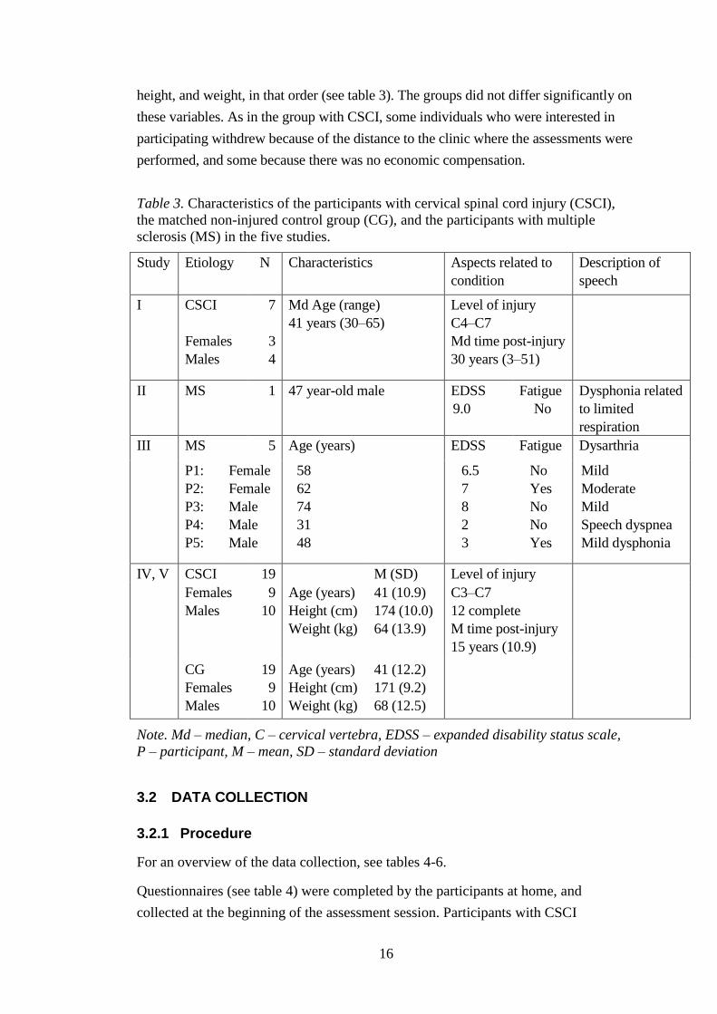
height, and weight, in that order (see table 3). The groups did not differ significantly on
these variables. As in the group with CSCI, some individuals who were interested in
participating withdrew because of the distance to the clinic where the assessments were
performed, and some because there was no economic compensation.
Table 3. Characteristics of the participants with cervical spinal cord injury (CSCI),
the matched non-injured control group (CG), and the participants with multiple
sclerosis (MS) in the five studies.
Study Etiology N Characteristics Aspects related to
condition
Description of
speech
I CSCI
Females
Males
7
3
4
Md Age (range)
41 years (30–65)
Level of injury
C4–C7
Md time post-injury
30 years (3–51)
II MS 1 47 year-old male EDSS
9.0
Fatigue
No
Dysphonia related
to limited
respiration
III MS 5 Age (years) EDSS Fatigue Dysarthria
P1:
P2:
P3:
P4:
P5:
Female
Female
Male
Male
Male
58
62
74
31
48
6.5
7
8
2
3
No
Yes
No
No
Yes
Mild
Moderate
Mild
Speech dyspnea
Mild dysphonia
IV, V CSCI 19 M (SD) Level of injury
Females
Males
9
10
Age (years)
Height (cm)
Weight (kg)
41 (10.9)
174 (10.0)
64 (13.9)
C3–C7
12 complete
M time post-injury
15 years (10.9)
CG
Females
Males
19
9
10
Age (years)
Height (cm)
Weight (kg)
41 (12.2)
171 (9.2)
68 (12.5)
Note. Md – median, C – cervical vertebra, EDSS – expanded disability status scale,
P – participant, M – mean, SD – standard deviation
3.2 DATA COLLECTION
3.2.1 Procedure
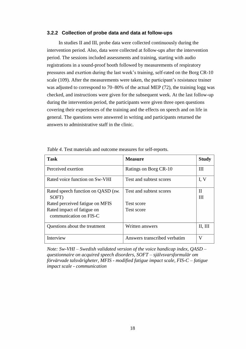
For an overview of the data collection, see tables 4-6.
Questionnaires (see table 4) were completed by the participants at home, and
collected at the beginning of the assessment session. Participants with CSCI
17
completed a Swedish validated version of the Voice Handicap Index (Sw-VHI) (102),
and the participants with MS used the Questionnaire on Acquired Speech Disorders
(QASD, sw. SOFT) for self-ratings (103). The Participants with MS also performed
self-ratings on the Modified Fatigue Impact Scale (MFIS) (100) and the Fatigue
Communication Scale (FIS-C) (101). Information on relevant background aspects
were collected via questions during an interview.
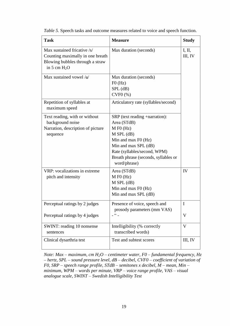
Speech samples (see table 5) were recorded in a sound-proof booth. The
recordings followed standardized routines, making it possible to perform computer-
based acoustic analyses of the speech signal. The speech and voice samples included
three trials of a maximum sustained /a/ and three of a maximum sustained /s/, two
repetitions of syllables at high rate (SMR/AMR), reading of phrases for the
assessment of articulation, and nonsense-sentences for the assessment of
intelligibility (104), two trials of counting as far as possible in one breath, text
reading with and without background noise, narrative speech, and vocalizations for
the voice range profiles (VRP). The participants performed the VRP according to
Hallin et al. (105). For the elicitation of the VRP, repeated vocalizations were
performed in a soft and loud voice across the whole pitch range to reach the extreme
F0 and SPL-levels of the participant’s voice range (106).
For the estimation of PS, the intra-oral pressure during the production of a string
of 7 syllables consisting of voiceless stops and vowels, /pae/, in three loudness
conditions – habitual, loud and soft – was registered. The audio signal was recorded
simultaneously (see table 6).
Respiratory testing included spirometry and measurement of respiratory
pressures (see table 6). Respiratory measures were collected with a MicroLoop
Spirometer (Care Fusion, San Diego, USA), and the spirometry was performed
according to the American Thoracic Society Standards (107). The participants
performed maximum exhalations, forceful maximum exhalations, and coughing, until
there were three trials resulting in values within a 10% range for each respiratory
parameter. In study I, VC after a maximal GI- cycle was measured to obtain the VCGI,
which is the VC with the glossopharyngeal insufflation volume (GIV) added (79). For
each of the respiratory measures VC, forced vital capacity (FVC), peak expiratory
flow (PEF), peak cough flow (PCF), MEP and MIP, the best performance of three
within a 10% range was used. In study III, respiratory pressures were tested using a
respiratory pressure meter Micro RPM (MicroMedical, Basingstoke, UK).
For the evaluation of speech function, a Swedish standardized dysarthria test
was used (108).
18
3.2.2 Collection of probe data and data at follow-ups
In studies II and III, probe data were collected continuously during the
intervention period. Also, data were collected at follow-ups after the intervention
period. The sessions included assessments and training, starting with audio
registrations in a sound-proof booth followed by measurements of respiratory
pressures and exertion during the last week’s training, self-rated on the Borg CR-10
scale (109). After the measurements were taken, the participant’s resistance trainer
was adjusted to correspond to 70–80% of the actual MEP (72), the training logg was
checked, and instructions were given for the subsequent week. At the last follow-up
during the intervention period, the participants were given three open questions
covering their experiences of the training and the effects on speech and on life in
general. The questions were answered in writing and participants returned the
answers to administrative staff in the clinic.
Table 4. Test materials and outcome measures for self-reports.
Task Measure Study
Perceived exertion Ratings on Borg CR-10 III
Rated voice function on Sw-VHI Test and subtest scores I, V
Rated speech function on QASD (sw.
SOFT)
Rated perceived fatigue on MFIS
Rated impact of fatigue on
communication on FIS-C
Test and subtest scores
Test score
Test score
II
III
Questions about the treatment Written answers II, III
Interview Answers transcribed verbatim V
Note: Sw-VHI – Swedish validated version of the voice handicap index, QASD –
questionnaire on acquired speech disorders, SOFT – självsvarsformulär om
förvärvade talsvårigheter, MFIS - modified fatigue impact scale, FIS-C – fatigue
impact scale - communication
19
Table 5. Speech tasks and outcome measures related to voice and speech function.
Task Measure Study
Max sustained fricative /s/
Counting maximally in one breath
Blowing bubbles through a straw
in 5 cm H2O
Max duration (seconds)
I, II,
III, IV
Max sustained vowel /a/ Max duration (seconds)
F0 (Hz)
SPL (dB)
CVF0 (%)
Repetition of syllables at
maximum speed
Articulatory rate (syllables/second)
Text reading, with or without
background noise
Narration, description of picture
sequence
SRP (text reading +narration):
Area (STdB)
M F0 (Hz)
M SPL (dB)
Min and max F0 (Hz)
Min and max SPL (dB)
Rate (syllables/second, WPM)
Breath phrase (seconds, syllables or
word/phrase)
VRP: vocalizations in extreme
pitch and intensity
Area (STdB)
M F0 (Hz)
M SPL (dB)
Min and max F0 (Hz)
Min and max SPL (dB)
IV
Perceptual ratings by 2 judges Presence of voice, speech and
prosody parameters (mm VAS)
I
Perceptual ratings by 4 judges - “ - V
SWINT: reading 10 nonsense
sentences
Intelligibility (% correctly
transcribed words)
V
Clinical dysarthria test Test and subtest scores III, IV
Note: Max – maximum, cm H2O – centimeter water, F0 – fundamental frequency, Hz
– hertz, SPL – sound pressure level, dB – decibel, CVF0 – coefficient of variation of
F0, SRP – speech range profile, STdB – semitones x decibel, M – mean, Min –
minimum, WPM – words per minute, VRP – voice range profile, VAS – visual
analogue scale, SWINT – Swedish Intelligibility Test
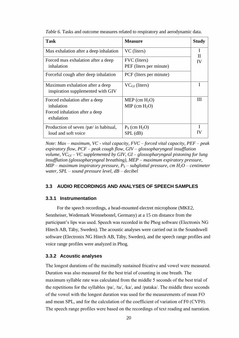
20
Table 6. Tasks and outcome measures related to respiratory and aerodynamic data.
Task Measure Study
Max exhalation after a deep inhalation VC (liters) I
II
IV Forced max exhalation after a deep
inhalation
FVC (liters)
PEF (liters per minute)
Forceful cough after deep inhalation PCF (liters per minute)
Maximum exhalation after a deep
inspiration supplemented with GIV
VCGI (liters) I
Forced exhalation after a deep
inhalation
Forced inhalation after a deep
exhalation
MEP (cm H2O)
MIP (cm H2O)
III
Production of seven /pæ/ in habitual,
loud and soft voice
PS (cm H2O)
SPL (dB)
I
IV
Note: Max – maximum, VC - vital capacity, FVC – forced vital capacity, PEF – peak
expiratory flow, PCF – peak cough flow, GIV – glossopharyngeal insufflation
volume, VCGI – VC supplemented by GIV, GI – glossopharyngeal pistoning for lung
insufflation (glossopharyngeal breathing), MEP – maximum expiratory pressure,
MIP – maximum inspiratory pressure, PS – subglottal pressure, cm H2O – centimeter
water, SPL – sound pressure level, dB – decibel
3.3 AUDIO RECORDINGS AND ANALYSES OF SPEECH SAMPLES
3.3.1 Instrumentation
For the speech recordings, a head-mounted electret microphone (MKE2,
Sennheiser, Wedemark Wennebostel, Germany) at a 15 cm distance from the
participant’s lips was used. Speech was recorded in the Phog software (Electronix NG
Hitech AB, Täby, Sweden). The acoustic analyses were carried out in the Soundswell
software (Electronix NG Hitech AB, Täby, Sweden), and the speech range profiles and
voice range profiles were analyzed in Phog.
3.3.2 Acoustic analyses
The longest durations of the maximally sustained fricative and vowel were measured.
Duration was also measured for the best trial of counting in one breath. The
maximum syllable rate was calculated from the middle 5 seconds of the best trial of
the repetitions for the syllables /pa/, /ta/, /ka/, and /pataka/. The middle three seconds
of the vowel with the longest duration was used for the measurements of mean FO
and mean SPL, and for the calculation of the coefficient of variation of F0 (CVF0).
The speech range profiles were based on the recordings of text reading and narration.
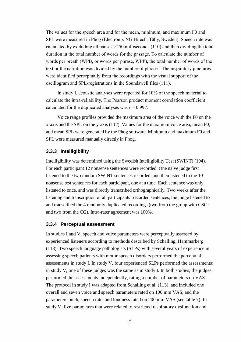
21
The values for the speech area and for the mean, minimum, and maximum F0 and
SPL were measured in Phog (Electronix NG Hitech, Täby, Sweden). Speech rate was
calculated by excluding all pauses >250 milliseconds (110) and then dividing the total
duration in the total number of words for the passage. To calculate the number of
words per breath (WPB, or words per phrase, WPP), the total number of words of the
text or the narration was divided by the number of phrases. The inspiratory junctures
were identified perceptually from the recordings with the visual support of the
oscillogram and SPL-registrations in the Soundswell files (111).
In study I, acoustic analyses were repeated for 10% of the speech material to
calculate the intra-reliability. The Pearson product moment correlation coefficient
calculated for the duplicated analyses was r = 0.997.
Voice range profiles provided the maximum area of the voice with the F0 on the
x-axis and the SPL on the y-axis (112). Values for the maximum voice area, mean F0,
and mean SPL were generated by the Phog software. Minimum and maximum F0 and
SPL were measured manually directly in Phog.
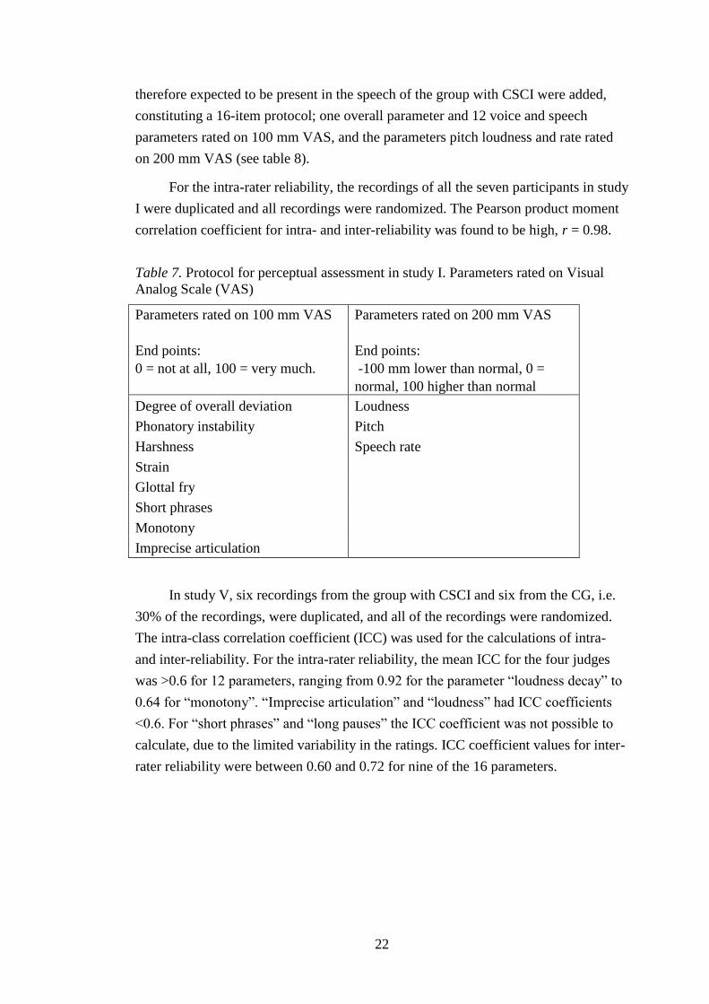
3.3.3 Intelligibility
Intelligibility was determined using the Swedish Intelligibility Test (SWINT) (104).
For each participant 12 nonsense sentences were recorded. One naïve judge first
listened to the two random SWINT sentences recorded, and then listened to the 10
nonsense test sentences for each participant, one at a time. Each sentence was only
listened to once, and was directly transcribed orthographically. Two weeks after the
listening and transcription of all participants’ recorded sentences, the judge listened to
and transcribed the 4 randomly duplicated recordings (two from the group with CSCI
and two from the CG). Intra-rater agreement was 100%.
3.3.4 Perceptual assessment
In studies I and V, speech and voice parameters were perceptually assessed by
experienced listeners according to methods described by Schalling, Hammarberg
(113). Two speech language pathologists (SLPs) with several years of experience in
assessing speech patients with motor speech disorders performed the perceptual
assessments in study I. In study V, four experienced SLPs performed the assessments;
in study V, one of these judges was the same as in study I. In both studies, the judges
performed the assessments independently, rating a number of parameters on VAS.
The protocol in study I was adapted from Schalling et al. (113), and included one
overall and seven voice and speech parameters rated on 100 mm VAS, and the
parameters pitch, speech rate, and loudness rated on 200 mm VAS (see table 7). In
study V, five parameters that were related to restricted respiratory dysfunction and
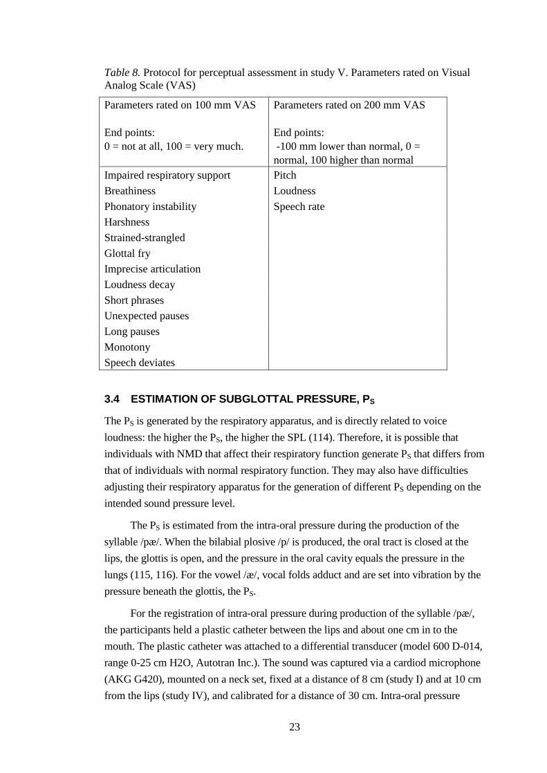
22
therefore expected to be present in the speech of the group with CSCI were added,
constituting a 16-item protocol; one overall parameter and 12 voice and speech
parameters rated on 100 mm VAS, and the parameters pitch loudness and rate rated
on 200 mm VAS (see table 8).
For the intra-rater reliability, the recordings of all the seven participants in study
I were duplicated and all recordings were randomized. The Pearson product moment
correlation coefficient for intra- and inter-reliability was found to be high, r = 0.98.
Table 7. Protocol for perceptual assessment in study I. Parameters rated on Visual
Analog Scale (VAS)
Parameters rated on 100 mm VAS
End points:
0 = not at all, 100 = very much.
Parameters rated on 200 mm VAS
End points:
-100 mm lower than normal, 0 =
normal, 100 higher than normal
Degree of overall deviation Loudness
Phonatory instability Pitch
Harshness Speech rate
Strain
Glottal fry
Short phrases
Monotony
Imprecise articulation
In study V, six recordings from the group with CSCI and six from the CG, i.e.
30% of the recordings, were duplicated, and all of the recordings were randomized.
The intra-class correlation coefficient (ICC) was used for the calculations of intra-
and inter-reliability. For the intra-rater reliability, the mean ICC for the four judges
was >0.6 for 12 parameters, ranging from 0.92 for the parameter “loudness decay” to
0.64 for “monotony”. “Imprecise articulation” and “loudness” had ICC coefficients
<0.6. For “short phrases” and “long pauses” the ICC coefficient was not possible to
calculate, due to the limited variability in the ratings. ICC coefficient values for inter-
rater reliability were between 0.60 and 0.72 for nine of the 16 parameters.
23
Table 8. Protocol for perceptual assessment in study V. Parameters rated on Visual
Analog Scale (VAS)
Parameters rated on 100 mm VAS
End points:
0 = not at all, 100 = very much.
Parameters rated on 200 mm VAS
End points:
-100 mm lower than normal, 0 =
normal, 100 higher than normal
Impaired respiratory support Pitch
Breathiness Loudness
Phonatory instability Speech rate
Harshness
Strained-strangled
Glottal fry
Imprecise articulation
Loudness decay
Short phrases
Unexpected pauses
Long pauses
Monotony
Speech deviates
3.4 ESTIMATION OF SUBGLOTTAL PRESSURE, PS
The PS is generated by the respiratory apparatus, and is directly related to voice
loudness: the higher the PS, the higher the SPL (114). Therefore, it is possible that
individuals with NMD that affect their respiratory function generate PS that differs from
that of individuals with normal respiratory function. They may also have difficulties
adjusting their respiratory apparatus for the generation of different PS depending on the
intended sound pressure level.
The PS is estimated from the intra-oral pressure during the production of the
syllable /pæ/. When the bilabial plosive /p/ is produced, the oral tract is closed at the
lips, the glottis is open, and the pressure in the oral cavity equals the pressure in the
lungs (115, 116). For the vowel /æ/, vocal folds adduct and are set into vibration by the
pressure beneath the glottis, the PS.
For the registration of intra-oral pressure during production of the syllable /pæ/,
the participants held a plastic catheter between the lips and about one cm in to the
mouth. The plastic catheter was attached to a differential transducer (model 600 D-014,
range 0-25 cm H2O, Autotran Inc.). The sound was captured via a cardiod microphone
(AKG G420), mounted on a neck set, fixed at a distance of 8 cm (study I) and at 10 cm
from the lips (study IV), and calibrated for a distance of 30 cm. Intra-oral pressure
24
during the production of /pæ/, was registered in Swell in study I, and in Pico Scope
(Pico Technology, Cambridgeshire, UK) in study IV, and the sound was recorded in
Phog (Electronix NG Hitech AB, Täby, Sweden) in both studies.
The registration and analysis of the PS and the SPL followed a protocol used
earlier (114-117). The participants produced a string of 7 /pæ/ in habitual loudness and
at a steady pace of 1.5 syllables per second. This was repeated in a loud voice and in a
soft voice, until three sets of seven adequate pressure curves, in each loudness
condition, were registered. Simultaneous feedback was provided on the monitor,
allowing the quality of the registered pressure curves to be determined.
PS was estimated from the maximum points of the middle five pressure peaks in
the syllable string, in Swell (study I) and in Pico Scope (study IV). The same settings in
Swell and in Pico Scope were used for all measurements. Maximum SPL was
measured for each of the vowels from the SPL-channel in Swell. Mean PS and mean
SPL were calculated for all three syllable strings in each loudness condition (habitual,
loud, and soft voice).
3.5 SELF-REPORTED INFORMATION AND QUESTIONNAIRES
All five studies included the collection of background data, to control for inclusion
and exclusion criteria and to obtain information related to the neurological condition,
such as duration of the condition, magnitude of symptoms etc. In study V, 19
predefined questions were formulated for background information. Questions
included personal data (age, height, and weight), data on the CSCI (time post injury,
level and completeness of injury), social factors (family situation, profession, and
employment), and history of voice and speech problems and on professional help
regarding voice and speech issues. The answers were transcribed verbatim at the time
of the interview.
Studies I and V used Sw-VHI to obtain information about the participants’ self-
perceived voice problems. Sw-VHI is a Swedish validated version of the VHI (118),
which consists of thirty statements divided into three subscales: a physical, a
functional and an emotional subscale. Each statement is rated on a five-point interval
scale; 0 represents never, 1 = almost never, 2 = sometimes, 3 = almost always, and 4
= always. The maximum total score is 120, which indicates severe problems related
to voice dysfunction. A total score >20 indicates a voice disorder (102).
The participants with MS in studies II and III rated their perceived speech
problems using QASD (sw. SOFT; Självsvarsformulär Om Förvärvade
Talsvårigheter) (103). QASD consists of thirty statements, with each statement rated
on a four-point scale with 0 = definitely false, 1 = sometimes true, 2 = mostly true,
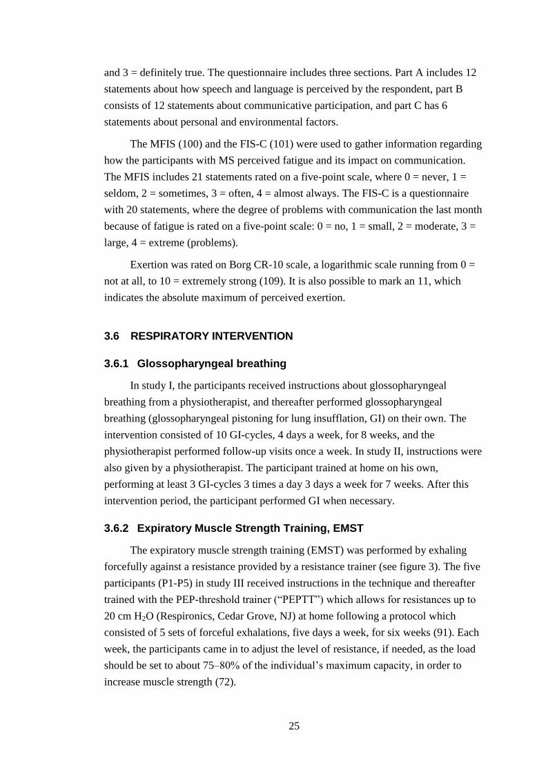
25
and 3 = definitely true. The questionnaire includes three sections. Part A includes 12
statements about how speech and language is perceived by the respondent, part B
consists of 12 statements about communicative participation, and part C has 6
statements about personal and environmental factors.
The MFIS (100) and the FIS-C (101) were used to gather information regarding
how the participants with MS perceived fatigue and its impact on communication.
The MFIS includes 21 statements rated on a five-point scale, where 0 = never, 1 =
seldom, 2 = sometimes, 3 = often, 4 = almost always. The FIS-C is a questionnaire
with 20 statements, where the degree of problems with communication the last month
because of fatigue is rated on a five-point scale: 0 = no, 1 = small, 2 = moderate, 3 =
large, 4 = extreme (problems).
Exertion was rated on Borg CR-10 scale, a logarithmic scale running from 0 =
not at all, to 10 = extremely strong (109). It is also possible to mark an 11, which
indicates the absolute maximum of perceived exertion.
3.6 RESPIRATORY INTERVENTION
3.6.1 Glossopharyngeal breathing
In study I, the participants received instructions about glossopharyngeal
breathing from a physiotherapist, and thereafter performed glossopharyngeal
breathing (glossopharyngeal pistoning for lung insufflation, GI) on their own. The
intervention consisted of 10 GI-cycles, 4 days a week, for 8 weeks, and the
physiotherapist performed follow-up visits once a week. In study II, instructions were
also given by a physiotherapist. The participant trained at home on his own,
performing at least 3 GI-cycles 3 times a day 3 days a week for 7 weeks. After this
intervention period, the participant performed GI when necessary.
3.6.2 Expiratory Muscle Strength Training, EMST
The expiratory muscle strength training (EMST) was performed by exhaling
forcefully against a resistance provided by a resistance trainer (see figure 3). The five
participants (P1-P5) in study III received instructions in the technique and thereafter
trained with the PEP-threshold trainer (“PEPTT”) which allows for resistances up to
20 cm H2O (Respironics, Cedar Grove, NJ) at home following a protocol which
consisted of 5 sets of forceful exhalations, five days a week, for six weeks (91). Each
week, the participants came in to adjust the level of resistance, if needed, as the load
should be set to about 75–80% of the individual’s maximum capacity, in order to
increase muscle strength (72).
26
Figure 3. Components of a resistance trainer. (Schematic illustration by the author)
P1–P3 completed phase B. After a period with no training, P4 and P5, who had
lower EDSS scores (2 and 3), resumed training using the “EMST 150”, allowing for
resistances up to 150 cm H2O (Aspire Products, Gainesville, FL). P4 and P5 trained
until their performance reached a plateau.
To obtain information regarding perceived training load, the participants rated
the degree of perceived exertion on the Borg CR-10 scale (79, 109). At each follow-
up, they were asked to rate the perceived exertion during the previous weeks’
training.
3.7 STATISTICAL ANALYSES
Statistical analyses included descriptive statistics and comparisons pre and post
treatment. Calculations of median and range were performed in studies I and III.
Calculations of mean and standard deviations were performed in studies II, IV and V.
In study III Cohen’s d (SD2 – SD1) /(M2 – M1) was used to indicate the effect size of
change in performance after intervention. In study I, non-parametric statistical
methods were used for the comparison between performance pre- and post-treatment.
In study IV, parametric methods were used for the comparison between groups. For
the analyses of inter- and intra-rater reliability, the Pearson product moment
correlation coefficient was used in study I, and the ICC in study V. Regression
analysis was used in study IV to investigate the relation between background factors,
respiratory variables and speech variables.
All statistical analyses were performed in Excel and in Statistical Package for
Social Sciences (SPSS) (119).

27
3.8 ETHICAL APPROVALS
The studies in the doctoral project were approved by the Regional Ethics
Committee of Stockholm: Dnr 2006/127/32 (study I), Dnr 2009/565-31 (studies II-
III) and Dnr 2010/2088-31/2 (study IV and V), and Dnr 2013/328-32 (additional
application covering inclusion of the CG).
28
4 RESULTS
4.1 STUDY I
The aim of this study was to describe voice, speech and self-rated communication in 7
individuals with CSCI before and after an 8-week GI-intervention, and when using GI.
The main findings before the 8-week GI-intervention were that phonatory
stability, as measured with CVF0 was higher than reference data (113), and the
maximum duration of sustained phonation and of sustained fricative were shorter than
reference data (120, 121). The perceptual parameters phonatory instability, harshness,
and glottal fry were present to a higher degree than other voice and speech parameters.
Also, the perceptual ratings showed vocal loudness lower than normal and pitch that
was higher than normal. After the 8-week GI-intervention the participants used a
significantly louder voice in the production of a 7-syllable string and when reading with
background noise. Also, phonation was stabilized, as shown by the significant decrease
in CVF0, to levels of vocally healthy reference individuals, and the participants’ breath
phrases increased from 9.5 to 12.8 words per breath during reading. Perceptually, pitch
was normalized. Self-ratings on the Sw-VHI indicated voice disorder in four
participants before, and in three participants after the 8-week intervention.
With maximized VC after a GI-cycle, VC increased by 0.6 liters and PS in loud
voice by 4.5 cm H2O, and SPL increased by 4.6 dB in habitual loudness and 7.2 dB in
loud voice.
4.2 STUDY II
The aim of this study was to evaluate effects of GI on respiration and speech in a 47-
year old male with quadriplegia due to advanced MS. The participant was referred for
voice therapy, but was not able to change his breathing or his voice behavior in any
way, such as taking a deeper breath to improve voice quality, increasing loudness or
prolonging phonation. Results from GI in individuals with MS and quadriplegia have
not been previously reported.
The study used a single-subject study design, with baseline measurements before
and after a 7-week GI-intervention. A physiotherapist instructed the participant in the
GI-technique. The participant quickly learned how to ”frog breathe”, and trained 3 x 3
GI-cycles 3 days per week for 7 weeks. After that, he used GI when necessary, which
he reported to be typically in the morning and before making phone calls. Respiration
and speech were always better after having maximized his VC with a GI-cycle. When
maximized with GI, VC increased by up to 60%, he could sustain a vowel 2.5 times
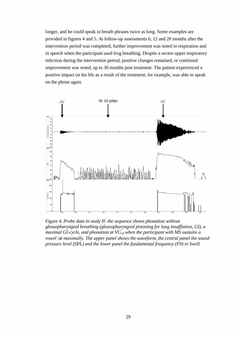
29
longer, and he could speak in breath phrases twice as long. Some examples are
provided in figures 4 and 5. At follow-up assessments 6, 12 and 20 months after the
intervention period was completed, further improvement was noted in respiration and
in speech when the participant used frog breathing. Despite a severe upper respiratory
infection during the intervention period, positive changes remained, or continued
improvement was noted, up to 30 months post treatment. The patient experienced a
positive impact on his life as a result of the treatment; for example, was able to speak
on the phone again.
Figure 4. Probe data in study II: the sequence shows phonation without
glossopharyngeal breathing (glossopharyngeal pistoning for lung insufflation, GI), a
maximal GI-cycle, and phonation at VCGI when the participant with MS sustains a
vowel /a/ maximally. The upper panel shows the waveform, the central panel the sound
pressure level (SPL) and the lower panel the fundamental frequency (F0) in Swell.
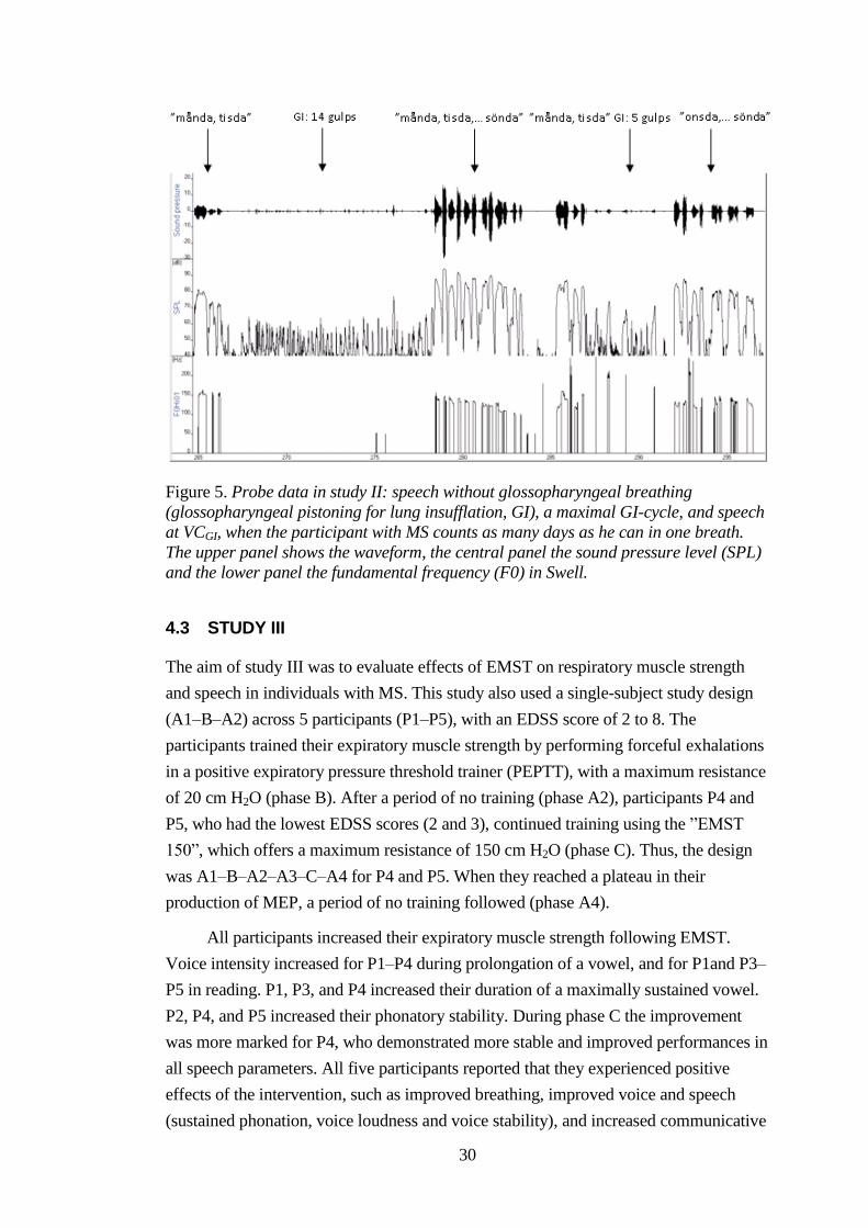
30
Figure 5. Probe data in study II: speech without glossopharyngeal breathing
(glossopharyngeal pistoning for lung insufflation, GI), a maximal GI-cycle, and speech
at VCGI, when the participant with MS counts as many days as he can in one breath.
The upper panel shows the waveform, the central panel the sound pressure level (SPL)
and the lower panel the fundamental frequency (F0) in Swell.
4.3 STUDY III
The aim of study III was to evaluate effects of EMST on respiratory muscle strength
and speech in individuals with MS. This study also used a single-subject study design
(A1–B–A2) across 5 participants (P1–P5), with an EDSS score of 2 to 8. The
participants trained their expiratory muscle strength by performing forceful exhalations
in a positive expiratory pressure threshold trainer (PEPTT), with a maximum resistance
of 20 cm H2O (phase B). After a period of no training (phase A2), participants P4 and
P5, who had the lowest EDSS scores (2 and 3), continued training using the ”EMST
150”, which offers a maximum resistance of 150 cm H2O (phase C). Thus, the design
was A1–B–A2–A3–C–A4 for P4 and P5. When they reached a plateau in their
production of MEP, a period of no training followed (phase A4).
All participants increased their expiratory muscle strength following EMST.
Voice intensity increased for P1–P4 during prolongation of a vowel, and for P1and P3–
P5 in reading. P1, P3, and P4 increased their duration of a maximally sustained vowel.
P2, P4, and P5 increased their phonatory stability. During phase C the improvement
was more marked for P4, who demonstrated more stable and improved performances in
all speech parameters. All five participants reported that they experienced positive
effects of the intervention, such as improved breathing, improved voice and speech
(sustained phonation, voice loudness and voice stability), and increased communicative
31
participation. Also, P5 spontaneously mentioned a decrease in nocturnal coughing and
in misdirected swallows of saliva.
4.4 STUDY IV
Study I raised questions about how voice and speech function was affected by CSCI.
Therefore, the aims of studies IV and V were to obtain in-depth analyses of voice and
speech function in a larger sample with CSCI, and to compare the results in this group
with those of a matched non-injured CG. In study IV, respiratory, aerodynamic,
acoustic and clinical data were analyzed using quantitative methods.
Performances varied in both groups, but the individuals with CSCI performed
significantly worse than the CG on maximum performance respiratory, voice, and
speech tasks. The group with CSCI had smaller lung volume and softer voice; they
produced a significantly smaller maximum voice area, and they reached significantly
lower maximum levels of F0 and SPL during the vocalizations. The participants with a
VC of less than 50% of the expected performed significantly worse on several speech
tasks, compared with participants who had a VC that was greater than 50% of the
expected. The level of injury had an impact on respiratory function in individuals with
a complete injury, but other physical aspects did not correlate with respiration or
speech. Data for PS showed that the individuals with CSCI initiated speech (a 7-syllable
train) with a numerically higher PS than the CG, and that PS decayed over the phrase
more than for the CG. Despite this, the group with CSCI produced a significantly softer
voice than the CG.
4.5 STUDY V
The aim of this study was to further investigate some of the findings from study I,
indicating higher self-perceived voice problems and an increased presence of some
perceptual parameters following CSCI, by investigating patient-reported and perceptual
assessments of voice and speech in a larger sample with CSCI, as compared with a
non-injured CG.
A major part of the group with CSCI reported longstanding changes in voice
function and limitations in participation related to these changes. The reports included
changes in voice and speech (89%), limitations related to restrictions in voice and
speech (39%), 74% mentioned compensatory strategies to adapt speech to the restricted
respiratory function, and 58% experienced occupational limitations related to their
voice function. One out of 19 had been referred to an SLP. As a comparison, only a few
participants in the CG reported limitations related to their voice and speech.
32
The Sw-VHI total score and the scores for the two subscales “Functional” and
“Physical” were significantly higher in the group with CSCI than in the CG, which
indicates that individuals with CSCI have more voice problems than non-injured
individuals. The perceptual mean ratings of the four judges (in mm VAS) indicated the
presence of more pronounced irregular phonation, such as harshness (6.1 mm) and
glottal fry (8.9 mm). Loudness was perceived as lower than normal (-7.9 mm) in the
group with CSCI. Some individuals with CSCI were also perceived to have impaired
respiratory support. The results of the perceptual ratings have to be interpreted
cautiously, since the ICC coefficient for inter-rater reliability was low for several of the
perceptual parameters. However, the results indicate that perceptual parameters
representing aspects of irregular voice quality are the most prominent in both the group
with CSCI and in the CG and that these parameters are also present in a larger number
of the group with CSCI. The results showed large variations in self-ratings on the Sw-
VHI and of perceptual assessments in both the group with CSCI and in the CG.
33
5 DISCUSSION
This doctoral project has investigated voice, speech and communicative participation
following MS and CSCI, both before and after respiratory training. The main findings
were that: 1) the components of speech production affected in CSCI were, above all,
related to challenging speech tasks and situations, 2) habitual and maximum
performance, as well as performance optimized with various compensatory strategies,
risk straining the voice mechanism, thereby putting the individual at risk for increased
voice fatigue and dysphonia, 3) there were both instant and long-term effects on speech
when applying GI, and 4) expiratory muscle strength training (EMST) seems to
positively affect some components of speech in individuals with MS.
The findings indicate that individuals with MS and CSCI who have speech
problems as a result of impaired respiration should be identified, since respiratory
training can improve their voice and speech function, and thus increase communicative
participation. The findings of this study add to the body of evidence for respiratory
intervention, and also raise questions for further research in the area of speech
restrictions related to respiratory dysfunction following neurological conditions.
5.1 SELECTION OF PARTICIPANTS
Not all of the individuals with CSCI who were contacted by their primary physician
about participation in the studies IV and V wanted to be contacted again to receive
more information about the study. Subsequently, about 80% of those who had agreed to
get more information about the study participated, and 20% declined. Studies in various
populations on participants’ reasons for deciding to participate or not have included the
following barriers for participating: additional demands on the patient (appointments,
travelling and additional costs), concerns about information and consent (122), current
health status, or personal factors (123). A major reason for participating in research
studies seems to be altruism, that participating is seen as a civic duty, but is also a way
of benefiting the health of family members and friends (124).
Some reported their decision to decline to participate due to a lack of time, or
because there was no economic compensation. Some indicated that they had decided
not to participate in studies, because of their health condition, while others mentioned
that they did not find the purpose of the study motivating. Regardless of the individual
reason for participating or not, there is a risk that the individuals agreeing to participate
were those who were the least affected by their injury, or were experiencing voice
problems. Vice versa, some individuals may have engaged in the study because they
did not experience voice problems, but wanted to get a “voice check”, particularly since
34
voice and speech function following CSCI, to date, is not routinely identified by the
health-care practice.
Thus, a larger sample would have been desirable to minimize this risk of biases.
However, because of the size of the population of individuals with CSCI in the
Stockholm area, the possible number of individuals meeting the inclusion criteria for
this study was limited. Similar aspects regarding the recruitment of participants were
described in Nygren-Bonnier et al. (79), a study with similar inclusion criteria and a
similar sample size.
The non-injured individuals invited to participate in the control group (CG)
reported the same reasons for not participating as the individuals with CSCI, with the
exception of health condition. As for participants with CSCI, those in the CG may also
have had different reasons for accepting to participate. For example, individuals with
some voice issues may have been more motivated to participate. This could have been
the case in study I, where all participants were asked if they wanted an SLP assessment
of their voice and speech in addition to the respiratory measurements, but only about
one third accepted the offer. It is possible that these individuals were motivated because
of limitations in their voice and speech function. The same might be true for some of
the participants in the CG, who also had ratings above the cut-off for voice disorder on
the Sw-VHI. This further emphasizes the importance of considering factors that may
influence inclination to participate, as this may have an impact on the generalizability
of the study results.
5.2 SUBGLOTTAL PRESSURE, PS
One important task of the respiratory system is to generate and maintain a stable PS
during speech production (8, 59). The threshold subglottal pressure (PS) for phonation
is 2–3 cm H2O (114) and a basic demand for speech production is the ability to sustain
a PS of 5 cm H2O for at least 5 seconds (59). The mean PS generated for the five middle
syllables in the seven-syllable train by the participants with CSCI in studies I and IV
was within normal limits (114), and the mean PS generated by the group with CSCI for
the production of the syllable-train did not differ significantly from that of the CG.
However, the resulting SPL was significantly lower for all three loudness conditions in
the group with CSCI compared with the CG.
Figure 6 shows mean PS and mean SPL for each of the analyzed five middle
syllables in the registered 7-syllable string. The group with CSCI tended to initiate the
syllable train at a higher PS than the CG, particularly in the production of a loud voice.
It can also be observed that the decay was steeper for the individuals with CSCI than
for the CG. This may reflect findings similar to those reported by Hoit et al. (60), who
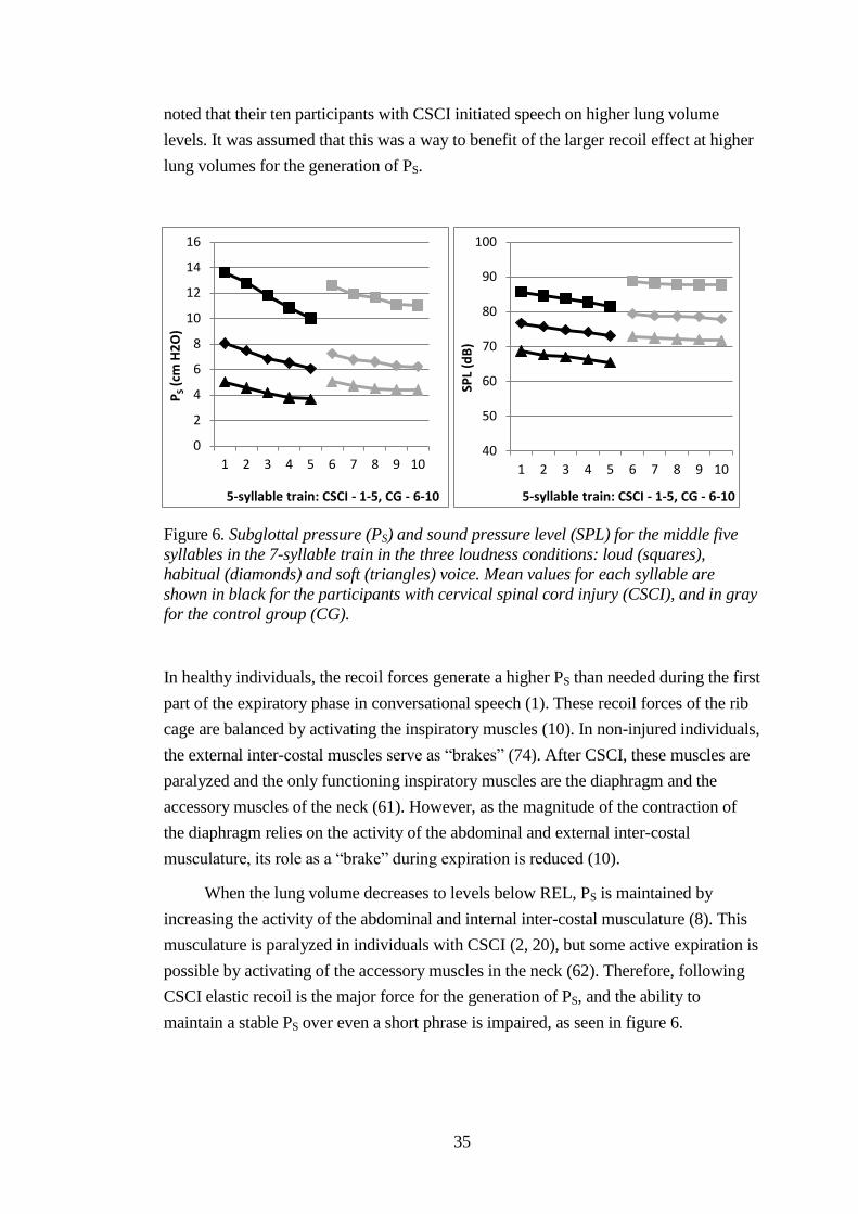
35
noted that their ten participants with CSCI initiated speech on higher lung volume
levels. It was assumed that this was a way to benefit of the larger recoil effect at higher
lung volumes for the generation of PS.
Figure 6. Subglottal pressure (PS) and sound pressure level (SPL) for the middle five
syllables in the 7-syllable train in the three loudness conditions: loud (squares),
habitual (diamonds) and soft (triangles) voice. Mean values for each syllable are
shown in black for the participants with cervical spinal cord injury (CSCI), and in gray
for the control group (CG).
In healthy individuals, the recoil forces generate a higher PS than needed during the first
part of the expiratory phase in conversational speech (1). These recoil forces of the rib
cage are balanced by activating the inspiratory muscles (10). In non-injured individuals,
the external inter-costal muscles serve as “brakes” (74). After CSCI, these muscles are
paralyzed and the only functioning inspiratory muscles are the diaphragm and the
accessory muscles of the neck (61). However, as the magnitude of the contraction of
the diaphragm relies on the activity of the abdominal and external inter-costal
musculature, its role as a “brake” during expiration is reduced (10).
When the lung volume decreases to levels below REL, PS is maintained by
increasing the activity of the abdominal and internal inter-costal musculature (8). This
musculature is paralyzed in individuals with CSCI (2, 20), but some active expiration is
possible by activating of the accessory muscles in the neck (62). Therefore, following
CSCI elastic recoil is the major force for the generation of PS, and the ability to
maintain a stable PS over even a short phrase is impaired, as seen in figure 6.
0
2
4
6
8
10
12
14
16
1 2 3 4 5 6 7 8 9 10
PS
(cm
H2
O)
5-syllable train: CSCI - 1-5, CG - 6-10
40
50
60
70
80
90
100
1 2 3 4 5 6 7 8 9 10 SP
L (d
B)
5-syllable train: CSCI - 1-5, CG - 6-10
36
5.3 HABITUAL VS MAXIMUM PERFORMANCE
The difficulties with controlling PS can explain why the individuals with CSCI in study
IV performed worse on several speech tasks than the participants in the CG. The group
with CSCI had a significantly lower speech rate in reading when compared with the
CG. Also, the speech range profiles showed that men with CSCI produced significantly
lower maximum F0-levels compared with men in the CG. The rest of the speech
measures for habitual performance did not differ significantly between the group with
CSCI and the CG, but the speech range profiles, which were based on the text reading
and the narration in habitual loudness, showed numerically smaller speech areas in men
with CSCI than in men in the CG. The larger number of complete injuries in men than
in women with CSCI could explain this numerical difference only existing for men.
Above all, the individuals with CSCI performed significantly worse than the
participants in the CG on the maximum performance tasks, such as using loud voice,
sustaining speech sounds maximally, and vocalizing at extreme F0 and SPL levels.
Larger lung volume is used for loud speaking conditions (8, 12, 120). While the lung
volume required for conversational speech corresponds to about 20% of the VC, public
speaking uses about 65% of the VC (8). As the participants with CSCI had VC ranging
from 30% to 65% of the expected volume, several of them can be expected to have
great difficulties producing a loud enough voice and maintaining a loud voice for
longer phrases. These problems were also reported by the participants with CSCI in
study V. Also, the fact that larger lung volume is needed to generate the PS up to 20 cm
H2O required for challenging speech, explains why the group with a VC below 50% of
the expected performed significantly worse on several speech measures than those
individuals with a VC >50% of the expected.
Thus, the reduced performance on both habitual and maximum performance
speech tasks in individuals with CSCI, as compared with non-injured individuals, could
be explained by the difficulty described by individuals with CSCI with regard to
generating and maintaining a sufficient PS.
5.4 COMPENSATORY STRATEGIES
The participants in study V reported a variety of ways to deal with their limited
respiration during the production of speech, and the pattern of initiating speech at a
higher PS may reflect at least two of these strategies.
Some participants with CSCI reported working harder or forcing themselves to
produce speech in demanding speech tasks. When respiratory muscles are paralyzed, it
may be difficult to adjust the respiratory apparatus for the coordination of respiration
and phonation during a specific speech task. Therefore, individuals with CSCI have to
37
rely on a limited respiratory apparatus for the production of speech. Also, impaired
sensation could result in distorted feedback from the musculature. One possible way to
compensate for these difficulties in controlling the musculature could be to increase the
physical effort. This strategy could be used for the production of a loud enough voice
over a longer phrase. As reported by the individuals with CSCI, it was necessary to try
to inhale deeper when they planned to utter a long phrase.
Even during the relatively short test phrase, a 7-syllable string for a total duration
of 3–4 seconds, participants with CSCI had difficulties maintaining a steady PS.
Considering that the mean utterance length in a reading task is about 13 syllables per
breath in healthy subjects (125), it is possible that the PS at phrase endings in running
speech declines to pressure levels that are not sufficient to generate loud enough
phonation.
In summary, the above include demands for excessive effort in individuals who
have to rely on the accessory muscles of the neck for speech production (61, 62).
Tamplin et al. (61) found pre-phonatory activity in the accessory muscles of the neck.
This indicates that people with CSCI use a lot of effort even before speech is initiated.
Strain in these muscles may lead to further fatigue, but the tension may also spread into
the muscles of the throat and of the larynx. Therefore, individuals with CSCI risk
straining their laryngeal muscles and damaging their vocal folds (126, 127).
5.5 PERCEPTUAL ASSESSMENTS
5.5.1 Reliability
The results of the perceptual assessment have to be interpreted with caution. While
inter-reliability was excellent in study I, as calculated with Pearson’s correlation
coefficient, the inter-rater reliability in study V, as calculated with the ICC, was low for
a majority of the perceptual parameters. In study I, two SLPs with several years of
experience in assessing speech in patients with dysarthria performed the perceptual
assessments, while the assessments in study V were performed by four highly
experienced SLPs. One of the two judges in study I also participated in the perceptual
assessment in study V. In both studies, the judges independently performed the
assessments, rating a number of parameters on VAS. Intra-judge reliability was good in
both studies, which may reflect the fact that the listeners were experienced in
perceptual assessment. Thus, the procedure and the listener experience were very
similar in both studies.
There are several possible explanations for the low inter-rater reliability in study
V (128). Firstly, the changes in voice and speech following CSCI were relatively small,
and therefore difficult to identify perceptually. The parameters that received high
38
ratings were essentially the same in the group with CSCI as in the CG. This indicates
that these aspects may also be present in the speech of non-injured individuals, and
therefore regarded to be within normal limits.
Also, 2 of the 19 in the CG scored >20, a cut-off point for voice disorders (102),
which may indicate that the CG also included some individuals with subtle voice
problems. There was a large variability in both the group with CSCI and the CG, but
the individuals with CSCI scored significantly higher on the Sw-VHI than the CG on a
group level. Altogether, this shows that the groups overlapped regarding voice
handicap, which might explain the unexpected high prevalence of perceptual
dysfunction in the CG and thus, possibly regarding the presence of perceptual aspects.
It is possible that this overlap in perceptual characteristics between the group with
CSCI and the CG could have been reduced if there been an inclusion criterion of a Sw-
VHI score <20 for the CG.
Secondly, the variability in the ratings could also reflect the judges’ limited
experience in assessing voice and speech in this particular population. Listener training
before the perceptual assessment could have resulted in different ratings and may have
strengthened the inter-rater reliability (129, 130). It is also possible that an initial
procedure where the listeners identify prominent parameters in the speech of
individuals with CSCI could have resulted both in a partially different set of perceptual
parameters, and higher agreement between the judges. A similar procedure was used in
Schalling et al. (113).
Yet another explanation for the difference in inter-rater reliability is that voice
and speech were possibly more affected in the participants in study I than in those in
study V. In study I, the participants were recruited from a larger study (79). All
participants were asked if they were also interested in seeing an SLP for measurements
of voice and speech, in addition to the respiratory measurements. About one third of the
participants in the study enrolled in study I, and it is possible that part of their
motivation was caused by perceived voice problems.
Therefore, these results should be taken as an indicator for further work in
perceptual assessments of speech following CSCI. Perceptual assessment of voice and
speech in CSCI has not been reported in many studies. Similar results were found in a
study of 24 participants with CSCI and 31 healthy controls (63). The authors did not
find significant differences at a group level for the majority of perceptual speech
dimensions, but the results indicated that speech was characterized by deviations in
variation and maintenance of pitch, general stress pattern, breath support for speech,
intermittent breathiness, and hyponasality. Rating speech and voice parameters
perceptually can be considered a natural, effective, and clinically useful and accessible
method (131). However, in the assessment of voice and speech following CSCI,
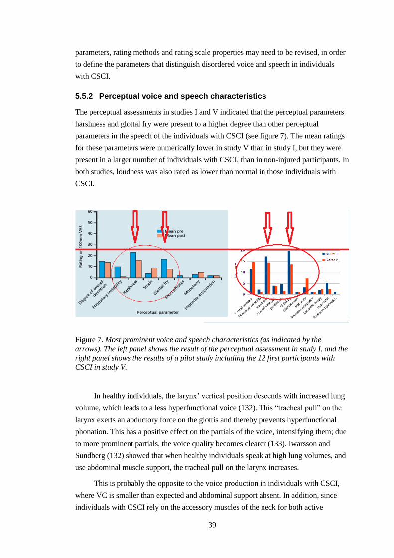
39
parameters, rating methods and rating scale properties may need to be revised, in order
to define the parameters that distinguish disordered voice and speech in individuals
with CSCI.
5.5.2 Perceptual voice and speech characteristics
The perceptual assessments in studies I and V indicated that the perceptual parameters
harshness and glottal fry were present to a higher degree than other perceptual
parameters in the speech of the individuals with CSCI (see figure 7). The mean ratings
for these parameters were numerically lower in study V than in study I, but they were
present in a larger number of individuals with CSCI, than in non-injured participants. In
both studies, loudness was also rated as lower than normal in those individuals with
CSCI.
Figure 7. Most prominent voice and speech characteristics (as indicated by the
arrows). The left panel shows the result of the perceptual assessment in study I, and the
right panel shows the results of a pilot study including the 12 first participants with
CSCI in study V.
In healthy individuals, the larynx’ vertical position descends with increased lung
volume, which leads to a less hyperfunctional voice (132). This “tracheal pull” on the
larynx exerts an abductory force on the glottis and thereby prevents hyperfunctional
phonation. This has a positive effect on the partials of the voice, intensifying them; due
to more prominent partials, the voice quality becomes clearer (133). Iwarsson and
Sundberg (132) showed that when healthy individuals speak at high lung volumes, and
use abdominal muscle support, the tracheal pull on the larynx increases.
This is probably the opposite to the voice production in individuals with CSCI,
where VC is smaller than expected and abdominal support absent. In addition, since
individuals with CSCI rely on the accessory muscles of the neck for both active
40
inspiration (61) and active expiration (62), a large amount of muscular effort is used in
the neck area for the control of the PS, thereby risking muscular tension spreading into
the muscles of the throat and larynx (134).
When air leaks out, caused by the recoil forces, one method of compensating for
an insufficient respiratory support is to save air by increasing the resistance in the
larynx or in the pharyngeal and oral cavities (1). Strategies such as this may explain the
higher presence of harshness and glottal fry in those individuals with CSCI in studies I
and V.
In contrast, an individual could use glottal abduction as a strategy, when the PS is
too high because of the recoil forces. The voice might, in that case, be characterized by
a higher degree of breathiness, as air leaks out. In study V, breathiness was perceived in
some individuals with CSCI.
5.6 EFFECTS OF GLOSSOPHARYNGEAL BREATHING ON SPEECH
Speech results after GI in individuals with CSCI, and with quadriplegia following MS,
have not been previously reported. Existing descriptions of speech following GI are
limited to two studies: Hixon and Hoit (1) reported on a participant with quadriplegia
who used GI in running speech, while the other study reported one patient with flaccid
dysarthria following bilateral medullary stroke (87). In this individual, respiratory
function was identified as the main contributing cause for the dysarthria, and oral motor
function was sufficient for GI. Speech outcomes were assessed over a 5-month GI-
intervention. The participant increased his VC, MPT and the length of his breath
phrases during reading and conversation, and VC was larger when maximized with GI.
Thus, GI may lead to an increased duration of phonation and of phrases, and louder
speech, at least following stroke.
It takes seconds to perform a maximal GI-cycle of about 10–14 gulps of air,
which means that considerable training may be needed to incorporate GI into running
speech. These two factors probably contribute to the limited use of this technique in
speech pathology. Another important factor is that the clinician needs to be familiar
with the technique and how to teach it. For the individual reported by (1), the use of GI
was automatized and the patient was not always aware of using the technique. Some
individuals may, however, need extensive speech training to implement the GI
technique fully into their speech.
The participant with MS in study II was completely quadriplegic, and had very
poor expiratory drive for speech. The results of the study showed dramatic
improvement. The participant was assessed at follow-ups up to 20 months after
completion of the 7-week intervention. His voice and speech improved over time, with
41
and without GI. As a result of the marked effects that GI had on his voice and speech,
the participant took up speaking on the telephone again. He typically used GI in the
mornings “to pump myself up”. The study design with several follow-up assessments
up to 20 months after the intervention period made it possible to show that the effect of
GI on respiration and speech increased over time.
Also, the results indicate that GI may be a means to maintain respiratory function
in individuals with quadriplegia. The participant had a bad cold during most of the
intervention period. This may be reflected in his values for FVC and PCF, which
became worse during habitual performance than before the intervention period.
However, with VC maximized with GI, his values increased. The findings suggest that
this individual was to be able to maintain his respiratory function and speech function
with the help of GI.
Not surprisingly the increase in lung volume after a GI-cycle has immediate
effects on PS and SPL. The more the rib cage is stretched by the GIV, the larger the
elastic forces of the maximally extended rib cage will be. Therefore, since the
participants with CSCI in study I already produced median PS within normal limits, the
PS generated after a maximal GI-cycle exceeded 20 cm H2O in some individuals. This
is higher than necessary for conversational speech, but may be of use for loud speech.
The participant with MS in study II also increased his SPL after a maximal GI-cycle,
but with his markedly restricted VC; the gain in SPL resulted in normal levels for
conversational speech.
Nygren-Bonnier et al. (79) reported positive effects of GI on pulmonary function
and chest expansion up to three months after intervention period in their participants
with CSCI. Although the chest expansion was not measured in the participant in study
II, it is possible that such changes accounted for the long-term effects on respiration and
speech. Given the positive effects on the stretching of the rib cage, it is possible that GI
could have effects in other populations with NMD, such as Parkinson’s disease, where
rigidity of the respiratory musculature is common (135). Since relatively well-
preserved orofacial, pharyngeal and laryngeal function is required for GI, individuals
with Parkinson’s disease would probably need to learn the technique early in the course
of their disease, when muscle function in the face, mouth, pharynx and larynx are not
yet too severely affected.
To date, there are no reports on the effects of GI in patients with different NMDs
who start training early in the disease progression. It has been shown that respiratory
muscle strength and ventilation are affected early in MS (17). As weak respiratory
muscles lead to decreased inspiratory capacity, there is a risk that this in turn leads to
decreased respiratory function. It would therefore be interesting to evaluate the effects
on ventilation, fatigue, and voice and speech in individuals with mild MS.
42
5.7 EFFECTS OF EMST ON SPEECH
Healthy adults performing EMST for 4 weeks increased their MEP by 29 to 41% (35,
136). Similar relative increases in MEP (31% to 41%), following EMST, have been
reported for individuals with mild to moderate MS (34, 94). In individuals with
moderate to severe MS somewhat smaller relative increases in MEP (18% to 36%)
have been observed (36, 93). Thus, there is evidence suggesting that EMST is a method
with the possibility to reduce the effects of muscle weakness in MS.
Few studies have evaluated the effects of EMST on voice and speech (35, 92,
134). In Roy et al. (134), there was no effect on self-rated VHI following a 6-week
EMST in 20 teachers with voice disorders. Jones (92) showed increases in MPT, WPM
and communicative efficiency score (CES) after a 6-month intervention with EMST in
their participant with Lance-Adams Syndrome and flaccid dysarthria. Chiara, Martin
(35) found a numerical, but not significant, increase in MPT and WPM in their group
with 17 individuals with mild to moderate MS after an 8-week intervention with
EMST. They hypothesized that incoordination of the laryngeal and respiratory muscle
could explain the lack of significant voice and speech changes.
In study III, P1–P3 with moderate MS trained with a lower resistance, about 40–
50% of their MEP. Despite this rather low resistance, they had to work hard to exhale
forcefully. The changes in MEP were the largest in P4 and P5 when they trained with
the “EMST 150”, which offers resistance up to 150 cm H2O. However, even these
participants with mild MS had difficulty reaching the levels of resistance recommended
for an improvement to occur (136). Despite the sub-optimal training load, all five
participants with MS improved their expiratory muscle strength, as indicated by their
increased MEP, and the training resulted in positive effects on some speech parameters
(MPT, CVF0, and SPL). With the single-subject study design it was possible to
visualize a decrease in variability in the participants’ performances. Also, the
participants’ self-reports indicated clinical changes in both improved voice and speech
function, and improved participation.
There are two possible explanations for the observed and perceived changes in
voice, speech and communication in study III: improved coordination of expiration and
an overall effect on ventilation.
Cerebellar involvement, which is frequent in MS, may lead to instability in the
voice and speech production (47, 53). During the assessments in study III, it was often
noted that the participants with MS had difficulties when performing coordinated
exhalations through the mouth-piece of the resistance trainer. The intensive training
with many repetitions of forceful expiration could therefore have contributed to a better
control of the act of exhaling, and resulted in a decreased variability in performance.
43
This was particularly noted in P4, who markedly reduced his variability of F0 when
sustaining a vowel maximally.
Ventilation is reduced in individuals with MS (17). EMST consists of multiple
repetitions of inhaling to TLC and then exhaling forcefully, performed several days a
week. It is possible that the participants’ ventilation improved as an effect of these deep
breaths, and this could have resulted in positive effects on the participants’ general
well-being. Such effects could have accounted for the changes noted in P1 and P3 with
moderate MS, and in P5 who perceived restrictions in participation from his fatigue.
An interesting finding was the spontaneous report of P5 that his nocturnal
coughing and misdirected saliva swallows had disappeared. Similar findings have been
reported in individuals with Parkinson’s disease following EMST (135). It is believed
that transference accounts for these effects on other muscle groups and functions than
those which are the primary goal for EMST (72).
In summary, the results of study III suggest the need for further evaluation of
speech effects of EMST in individuals with NMD, in isolation or in conjunction with
specific voice and speech exercises. It is possible that EMST can have several different
effects on voice, speech and communication in individuals with MS, depending on the
neurological involvement of the disease. The goals of the treatment may be different for
a severely restricted population than for individuals in early stages of MS. When
considering EMST for speech pathology intervention in MS, it is therefore important to
specify in what ways the training is expected to contribute to the improvement of voice,
speech, or communication.
In addition, EMST is an interesting training method because compliance with this
method is high. The participants in study III all expressed their satisfaction with the
intervention, which they found easy to perform. EMST is easy to learn and to perform
independently or with assistance, changes in performance are easily quantified (92),
and the cognitive load is minimal. Feedback on performance is immediate (valve opens
or not). An additional factor is intensity: with several training sessions per week, it is
possible that the training can develop into a routine.
44
6 CONCLUSIONS
The following conclusions were drawn:
Following CSCI, there are longstanding voice changes, resulting in limitations
in voice and speech function, and restrictions in communicative participation.
Above all, the restricted respiratory function after CSCI leads to limitations in
challenging speech tasks, such as loud speech or long phrases, and individuals
with a VC <50% perform significantly worse on demanding speech tasks, than
individuals with a VC >50%.
Individuals with complete injuries seem to have most voice, speech and
communication difficulties, and may be at greater risk for voice fatigue and
damage.
Individuals with CSCI adapt to their voice dysfunction by using a variety of
strategies, including speech breathing strategies, compensatory strategies, and
avoidance.
The compensatory strategies used by individuals with CSCI put them at further
risk of increased voice fatigue, and possibly of functional organic voice
damage.
GI-training may have positive short- and long-term effects on respiration, voice
and speech in individuals with CSCI and MS.
The changes in some voice and speech measures indicate the positive effects of
EMST in individuals with MS: these changes may be more marked in
individuals in the early stages of MS.
The answers to the questions on social validity raise questions about a more
general effect from EMST on ventilation, and thereby positive effects on well-
being and overall function in the individuals with MS.
Valid assessment tasks must be defined, in order to identify individuals with
CSCI who would benefit from speech therapy interventions (assessment,
counseling, voice and speech training).
45
7 FURTHER STUDIES
Further research should investigate the effects of speech pathology intervention
on voice function, voice fatigue, and communicative participation in individuals
with CSCI.
The results of studies I, IV and V indicate the importance of identifying
individuals with CSCI experiencing voice changes and voice fatigue. In
particular, studies should focus on individuals with a VC below 50% of the
expected, to assess the long-term effects on voice and speech, and possible cut-off
for a voice disorder.
Test materials to detect voice and speech changes in CSCI need to be carefully
selected and further developed.
The results of the extended follow up period in study II suggest that
glossopharyngeal breathing may be important for the maintenance of respiratory
function and voice function in severe MS, despite the progression of disease. The
study should be duplicated with additional subjects with the same condition.
It is possible that the perceived positive effects on communication were partly due
to the more general effects of EMST, for example improved ventilation, in the
individuals with MS. This motivates further investigation into the additional
effects of EMST in individuals with MS.
As there were larger effects of EMST in participants with MS and lower EDSS-
scores, it would be interesting to study effects of EMST in a group of mild MS.
Also, the effects of EMST using low resistance load should be assessed in
individuals with moderate to advanced MS.
Since stiffness in respiratory muscles is common in disorders such as Parkinson’s
disease (PD), the long-term effects of GI on respiration and voice and speech
function in patients who start to train in early stages of PD should be investigated.
Also, investigations combining respiratory training methods, such as GI and
EMST, with speech pathology intervention, such as voice training, should be
studied.
46
8 ACKNOWLEDGEMENTS
A doctoral project is a process extending over a long period of time, including years of
preparation before the formal doctoral education. I am grateful for the encounters with
patients in the speech pathology clinic over the years, and discussions with colleagues,
students, and friends, as these have led to the formulation of questions and the planning
of the studies in this project.
Particularly, I want to thank:
- The participants in the studies of this project: without you, these studies would not
have been possible!
- The Promobilia Foundation, the Norrbacka-Eugenia Foundation, the Aina
Börjeson’s Foundation, the Department of Clinical Science, Intervention and
Technology at Karolinska Institutet, Capio Travel Grant and the HÖST
scholarship for financial support making it possible for me to complete these
studies, participate at international conferences and visit leading researchers
abroad.
- My main supervisor and co-author Ellika Schalling, also inspiring colleague in the
clinic and in the teaching at the Speech Pathology Program at Karolinska Institutet,
and friend since many years. Ellika, thank you for your support and interest, for
your sharp mind, and for always being there! Thank you for all fun professional
and private discussions, all the coaching and the constructive discussions at the
Karolinska University Hospital, at our many “walkie-talkies” and in fantastic
places such as the greenhouse at Björstorp and the Canary Islands. Above all, I
will always remember our creative research plans taking form at the round table, in
the bay window in a beautiful 19th
century tea room, in Savannah, in 2010.
- My secondary supervisors and co-authors, Lena Hartelius and Sten Fredrikson, for
all your constructive feedback on study planning, data analysis, and manuscript
writing. Lena, also my supervisor at the Speech Pathology Program back in 1989,
and since then a great inspirer in many ways. Sten, for your expertise in multiple
sclerosis and for your subtle support.
- My external mentor and former teacher at Institutet för Handledning och
Konsultation, Anders Wächter, for reflecting talks early in the doctoral process,
and for contributing to unexpected shifts in the direction and content of thoughts.
- My former employer Christina Blom, and my recent employer Elisabet
Lundström, who have supported me immensely. I feel extremely lucky to work at
the Speech Language Pathology Department at the Karolinska University Hospital,
where you have contributed to the friendly and creative atmosphere!
47
- Anette Lohmander, head of the Division of Speech and Language Pathology at
Karolinska Institutet, for your support and for your critical eye, and for your
unique way of making others feel involved.
- My co-authors Malin Nygren-Bonnier and Brita Klefbeck, for your generous
sharing of your knowledge and work on respiration in neuromuscular disorders;
Lisen Kjellmer, for your solid knowledge on single-subject methodology, for your
enthusiasm in the “single-subject-project” at our department, and for many good
discussions; Åke Seiger, for your help with the recruitment of participants, for your
participation in the data analysis and manuscript writing, and for showing such
interest for my project. Thank you all, for your cooperation and inspiration, and for
your generous guiding in this interesting and creative process!
- Malin Forsén and Jeanette Holmgren Nilsson, co-authors and former students, for
your work with your thesis project on cervical spinal cord injury.
- The members of the MS-team at the Karolinska University Hospital in Huddinge
for your help in recruiting participants for the studies.
- My doctoral fellows, especially Ulrika Nygren and Päivikki Aarne, for never-
failing support and interesting talks about life in general and research more
specifically, and Renata Bogo and the rest of the doctoral research program HÖST
at Karolinska Institutet for the best research retreats ever!
- Jessica Huber, Purdue University, and Christine Sapienza, University of Florida,
for generously sharing your time and knowledge during my study visit.
- The number of people at the Karolinska University Hospital and Karolinska
Institutet who have helped me in different ways during my doctoral years: Anita
McAllister, Britta Hammarberg, Christina Hedestedt, Eva Holmberg, Hans
Larsson, Maria Södersten, Per Östberg, and many more.
- Warm-hearted colleagues at the Department of Speech Language Pathology who
have accompanied me on my journey during this project, to mention a few:
Therese Engström, Joakim Gustafson, Sanna Jansson, Victoria Kelly, Robert
Liljebäck, Matilda Lindsten, Helena Ljungman, Anna Lundblad, Ann Malmenholt,
Liisi Raud, Helena Zetterman, and Karin Wier.
- Colleagues at the ENT-department at the Karolinska University Hospital,
Huddinge, for stimulating cooperation in the FEES-business over the years: Per-
Åke Lindestad, Riitta Möller, Helene Lindqvist, and Kia Nyberg, to mention some.
I look forward to coming back to the dysphagia work!
Outside the Karolinska University Hospital and Karolinska Institutet, several persons
have shared my doctoral study experience in one way or another:
48
- My friend since the study years in Göteborg, Christina Samuelsson, for our plans
in 1989 to do research – you proceeded faster, but I finally also got there! Thank
you for all the things we have shared these years: children, vacations, your
superior cooking to mention a few nice ingredients. Also, thanks to Martin
Samuelsson, for being a most warm-hearted and generous person!
- Petra Bovide, also a friend from the study years: thank you for being such a good
listener and inspirer, and for the reflective dialogue, which has meant a lot for my
professional development and has led me onto different paths, the doctoral project
being one.
- All friends, neighbors, amateur musician friends, book circle members, and former
colleagues, for all the good company and fruitful discussions at late evening walks,
dinners, travels, and rehearsals: Cecilia Aginger, Mats Andersson, Ingebjørg Barth
Magnus, Gunilla Cröde-Widsell, Jessica Elfsberg, Ulrika Ferm, Åsa Goudeau,
Annika Malmrud, Per Malmrud, Stefan Nilsson, Philippa Nordmark, Helga Rudhe,
Eva Sonered, Maria Szönyi, and Gunilla Zaar, to mention some. You mean a lot to
me!
And last, but not least, I am immensely grateful to my family for having put up with my
fears, as well as joys, during the years of my doctoral studies:
- Ulf, for always offering the greatest support, always handling the everyday work in
the household in the quiet, for all the good discussions and the superior coaching.
- The lights of my life: Axel, I really so much appreciate all our late kitchen-talks
over a cup of tea, all the humor links you’ve fed me with over the years, and your
marvelous self-coaching tips; Vida, thank you for all your wide, deep and often
eye-opening reflections, and the discussions we’ve had over the years; Klara,
thank you for all our forest walks with or without käpphästar, our piano and
drawing sessions, and our morning breakfasts on the front porch, to mention some
of all the things that bring such joy!
- My close family: my sister Anne and niece Ella, my mother Ingert and my aunt
Ingalill, for all good laughs, and for always being more than a hundred percent
supportive and encouraging.
Some people have meant much to me and in different ways stimulated me in my
doctoral studies, but are sadly not here anymore: my dad Nisse, my old friends Anna
and Sten, and my parents-in-law, Eivor and Per. You are missed!
49
9 REFERENCES 1. Hixon T, Hoit J. Evaluation and management of speech breathing disorders:
principles and methods. Tucson, AZ: Redington Brown; 2005.
2. Brown R, DiMarco AF, Hoit JD, Garshick E. Respiratory dysfunction and management in spinal cord injury. Respir Care. 2006;51(8):853-68;discussion 69-70. Epub 2006/07/27.
3. Nogues MA, Roncoroni AJ, Benarroch E. Breathing control in neurological diseases. Clinical autonomic research : official journal of the Clinical Autonomic Research Society. 2002;12(6):440-9. Epub 2003/02/25.
4. De Troyer A, Borenstein S, Cordier R. Analysis of lung volume restriction in patients with respiratory muscle weakness. Thorax. 1980;35(8):603-10.
5. Annoni J, Chevrolet J, Kesselring J. Respiratory problems in chronic neurological disorders. Crit Rev Phys Rehab Med. 1993;5(2):155-92.
6. Aboussouan LS. Respiratory disorders in neurologic diseases. Cleveland Clinic journal of medicine. 2005;72(6):511-20.
7. Duffy J. Motor speech disorders: substrates, differential diagnosis, and management. 2 ed. St. Louis, MO, USA: Elsevier Mosby; 2005.
8. Aronson A. Clinical voice disorders. 6th edition ed1990.
9. Weismer G. Motor speech disorders. 1 edition ed: Plural Publishing Inc 2006.
10. Hixon TJ, Mead J, Goldman MD. Dynamics of the chest wall during speech production: function of the thorax, rib cage, diaphragm, and abdomen. Journal of speech and hearing research. 1976;19(2):297-356.
11. Hixon TJ, editor. Respiratory function in speech and song: College-Hill Press; 1987.
12. Proctor DF. Breathing, speech, and song. Wien New York: Springer-Verlag; 1980.
13. Hoit JD, Lansing, R. W., Dean, K., Yarkosky, M., Lederle, A. Nature and evaluation of dyspnea in speaking and swallowing. Seminars in speech and language. 2011;32(1):5-20.
14. Nygren-Bonnier M, Normi LL, Klefbeck B, Biguet G. Experiences of decreased lung function in people with cervical spinal cord injury. Disability and rehabilitation. 2011;33(6):530-6.
15. Smeltzer SC, Skurnick JH, Troiano R, Cook SD, Duran W, Lavietes MH. Respiratory function in multiple sclerosis. Utility of clinical assessment of respiratory muscle function. Chest. 1992;101(2):479-84. Epub 1992/02/01.
16. Tantucci C, Massucci M, Piperno R, Betti L, Grassi V, Sorbini CA. Control of breathing and respiratory muscle strength in patients with multiple sclerosis. Chest. 1994;105(4):1163-70. Epub 1994/04/01.
17. Altintas A, Demir T, Ikitimur HD, Yildirim N. Pulmonary function in multiple sclerosis without any respiratory complaints. Clin Neurol Neurosurg. 2007;109(3):242-6. Epub 2006/10/19.
18. Grasso MG, Lubich S, Guidi L, Rinnenburger D, Paolucci S. Cerebellar deficit and respiratory impairment: a strong association in multiple sclerosis? Acta Neurol Scand. 2000;101(2):98-103. Epub 2000/02/24.
19. Smeltzer SC, Utell MJ, Rudick RA, Herndon RM. Pulmonary function and dysfunction in multiple sclerosis. Archives of neurology. 1988;45(11):1245-9.
50
20. Baydur A, Adkins RH, Milic-Emili J. Lung mechanics in individuals with spinal cord injury: effects of injury level and posture. Journal of applied physiology. 2001;90(2):405-11. Epub 2001/02/13.
21. Winslow C, Rozovsky J. Effect of spinal cord injury on the respiratory system. American journal of physical medicine & rehabilitation / Association of Academic Physiatrists. 2003;82(10):803-14. Epub 2003/09/26.
22. Fugl-Meyer A. Effects of respiratory muscle paralysis in tetraplegic and paraplegic patients. Scandinavian journal of rehabilitation medicine. 1971;3:141-50.
23. Svenningsson A, Runmarker B, Lycke J, Andersen O. Incidence of MS during two fifteen-year periods in the Gothenburg region of Sweden. Acta Neurol Scand. 1990;82(3):161-8. Epub 1990/09/01.
24. Sundstrom P, Nystrom L, Forsgren L. Incidence (1988-97) and prevalence (1997) of multiple sclerosis in Vasterbotten County in northern Sweden. Journal of neurology, neurosurgery, and psychiatry. 2003;74(1):29-32. Epub 2002/12/18.
25. Ahlgren C, Oden A, Lycke J. High nationwide prevalence of multiple sclerosis in Sweden. Mult Scler. 2011;17(8):901-8. Epub 2011/04/05.
26. Levi R, Holtz, A., editor. Ryggmärgsskador. Lund: Studentlitteratur AB; 2006.
27. Hillert J, Lycke J. Neurologi. 5 ed. Stockholm: Liber AB; 2012.
28. Howard RS, Wiles CM, Hirsch NP, Loh L, Spencer GT, Newsom-Davis J. Respiratory involvement in multiple sclerosis. Brain : a journal of neurology. 1992;115 ( Pt 2):479-94. Epub 1992/04/01.
29. Hartelius L, Svensson P. Speech and swallowing symptoms associated with Parkinson's disease and multiple sclerosis: a survey. Folia Phoniatr Logop. 1994;46(1):9-17. Epub 1994/01/01.
30. Mackenzie IS, Morant SV, Bloomfield GA, Macdonald TM, O'Riordan J. Incidence and prevalence of multiple sclerosis in the UK 1990-2010: a descriptive study in the General Practice Research Database. Journal of neurology, neurosurgery, and psychiatry. 2013. Epub 2013/09/21.
31. Wyndaele M, Wyndaele JJ. Incidence, prevalence and epidemiology of spinal cord injury: what learns a worldwide literature survey? Spinal cord. 2006;44(9):523-9. Epub 2006/01/04.
32. Yorkston KM, Beukelman DR, Strand EA, Hakel M. Managements of motor speech disorders in chkdren and adults. 3 ed. Austin, TX: Pro-ed; 2010.
33. Fry DK, Pfalzer LA, Chokshi AR, Wagner MT, Jackson ES. Randomized control trial of effects of a 10-week inspiratory muscle training program on measures of pulmonary function in persons with multiple sclerosis. J Neurol Phys Ther. 2007;31(4):162-72. Epub 2008/01/04.
34. Chiara T, Martin AD, Davenport PW, Bolser DC. Expiratory muscle strength training in persons with multiple sclerosis having mild to moderate disability: effect on maximal expiratory pressure, pulmonary function, and maximal voluntary cough. Archives of physical medicine and rehabilitation. 2006;87(4):468-73. Epub 2006/03/31.
35. Chiara T, Martin D, Sapienza C. Expiratory muscle strength training: speech production outcomes in patients with multiple sclerosis. Neurorehabil Neural Repair. 2007;21(3):239-49. Epub 2007/03/14.
36. Gosselink R, Kovacs L, Ketelaer P, Carton H, Decramer M. Respiratory muscle weakness and respiratory muscle training in severely disabled multiple sclerosis patients. Archives of physical medicine and rehabilitation. 2000;81(6):747-51. Epub 2000/06/17.
51
37. Gosselink R, Kovacs L, Decramer M. Respiratory muscle involvement in multiple sclerosis. The European respiratory journal. 1999;13(2):449-54. Epub 1999/03/05.
38. Darley FL, Brown JR, Goldstein NP. Dysarthria in multiple sclerosis. Journal of speech and hearing research. 1972;15(2):229-45. Epub 1972/06/01.
39. Rasova K, Brandejsky P, Havrdova E, Zalisova M, Rexova P. Spiroergometric and spirometric parameters in patients with multiple sclerosis: are there any links between these parameters and fatigue, depression, neurological impairment, disability, handicap and quality of life in multiple sclerosis? Mult Scler. 2005;11(2):213-21.
40. Murdoch BE, Chenery HJ, Stokes PD, Hardcastle WJ. Respiratory kinematics in speakers with cerebellar disease. Journal of speech and hearing research. 1991;34(4):768-80. Epub 1991/08/01.
41. Hartelius L, Runmarker B, Andersen O. Prevalence and characteristics of dysarthria in a multiple-sclerosis incidence cohort: relation to neurological data. Folia Phoniatr Logop. 2000;52(4):160-77. Epub 2000/04/27.
42. Merson RM, Rolnick MI. Speech-language pathology and dysphagia in multiple sclerosis. Physical medicine and rehabilitation clinics of North America. 1998;9(3):631-41. Epub 1999/01/20.
43. Klugman TM, Ross E. Perceptions of the impact of speech, language, swallowing, and hearing difficulties on quality of life of a group of South African persons with multiple sclerosis. Folia Phoniatr Logop. 2002;54(4):201-21. Epub 2002/08/10.
44. Duffy J. Motor speech disorders: substrates, differential diagnosis, and management. 3rd ed. St.Louis, MO, USA: Elsevier Mosby; 2013.
45. Scripture E. Records of speech in disseminated sclerosis. Brain : a journal of neurology. 1916;39(3-4):456-77.
46. Farmakides M, Boone D. Speech problems of patients with Multiple Sclerosis. Journal of speech and hearing disorders. 1960;25:385-90.
47. Hartelius L, Runmarker B, Andersen O, Nord L. Temporal speech characteristics of individuals with multiple sclerosis and ataxic dysarthria: 'scanning speech' revisited. Folia Phoniatr Logop. 2000;52(5):228-38. Epub 2000/08/31.
48. Dogan M, Midi I, Yazici MA, Kocak I, Gunal D, Sehitoglu MA. Objective and subjective evaluation of voice quality in multiple sclerosis. J Voice. 2007;21(6):735-40. Epub 2006/07/04.
49. Yamout B, Fuleihan N, Hajj T, Sibai A, Sabra O, Rifai H, et al. Vocal symptoms and acoustic changes in relation to the expanded disability status scale, duration and stage of disease in patients with multiple sclerosis. European archives of oto-rhino-laryngology : official journal of the European Federation of Oto-Rhino-Laryngological Societies. 2009;266(11):1759-65.
50. Feijo AV, Parente MA, Behlau M, Haussen S, de Veccino MC, Martignago BC. Acoustic analysis of voice in multiple sclerosis patients. J Voice. 2004;18(3):341-7. Epub 2004/08/28.
51. Fitzgerald FJM, B.E. Chenery, H.J. Multiple sclerosis: Associated speech and language disorders. Australian Journal of Human Communication Disorders. 1987;9:50-66.
52. Konstantopoulos K, Vikelis M, Seikel JA, Mitsikostas DD. The existence of phonatory instability in multiple sclerosis: an acoustic and electroglottographic study. Neurological sciences : official journal of the Italian Neurological Society and of the Italian Society of Clinical Neurophysiology. 2010;31(3):259-68.
52
53. Hartelius L, Buder EH, Strand EA. Long-term phonatory instability in individuals with multiple sclerosis. Journal of speech, language, and hearing research : JSLHR. 1997;40(5):1056-72. Epub 1997/11/05.
54. Hartelius L, Buder EH, Strand EA. Long-Term Phonatory Instability in Individuals with Multiple-Sclerosis and Normal Control Subjects. Folia Phoniatrica Et Logopaedica. 1995;47(2):75-.
55. Darley FL, Aronson AE, Brown JR. Differential diagnostic patterns of dysarthria. Journal of speech and hearing research. 1969;12(2):246-69.
56. Rutter B. Reduced breath support in a speaker with dysarthria secondary to multiple sclerosis: Its impact on cenversational speech. Journal of Interactional Research in Communication Disorders. 2013;4(1):27-44.
57. Kurtzke JF. Rating neurologic impairment in multiple sclerosis: an expanded disability status scale (EDSS). Neurology. 1983;33(11):1444-52. Epub 1983/11/01.
58. Mitchell HL, Hoit JD, Watson PJ. Cognitive-linguistic demands and speech breathing. Journal of speech and hearing research. 1996;39(1):93-104. Epub 1996/02/01.
59. Netsell R, Hixon TJ. A noninvasive method for clinically estimating subglottal air pressure. The Journal of speech and hearing disorders. 1978;43(3):326-30.
60. Hoit JD, Banzett RB, Brown R, Loring SH. Speech breathing in individuals with cervical spinal cord injury. Journal of speech and hearing research. 1990;33(4):798-807. Epub 1990/12/01.
61. Tamplin J, Brazzale DJ, Pretto JJ, Ruehland WR, Buttifant M, Brown DJ, et al. Assessment of breathing patterns and respiratory muscle recruitment during singing and speech in quadriplegia. Archives of physical medicine and rehabilitation. 2011;92(2):250-6. Epub 2011/01/29.
62. De Troyer A, Estenne M, Heilporn A. Mechanism of active expiration in tetraplegic subjects. The New England journal of medicine. 1986;314(12):740-4.
63. MacBean N, Ward E, Murdoch B, Cahill L, Solley M, Geraghty T. Characteristics of speech following cervical spinal cord injury. Journal of Medical Speech-Language Pathology. 2006;14(3):167-84.
64. Watson PJ, Hixon TJ. Effects of abdominal trussing on breathing and speech in men with cervical spinal cord injury. Journal of speech, language, and hearing research : JSLHR. 2001;44(4):751-62. Epub 2001/08/28.
65. Johansson K, Nygren-Bonnier M, Klefbeck B, Schalling E. Effects of glossopharyngeal breathing on voice and speech in individuals with cervical spinal cord injury. Int J Ther Rehabil. 2011;18(9):501-11.
66. MacBean N, Ward E, Murdoch B, Cahill L, Geraughty T. Respiratory function and speech breathing patterns in non-ventilator-dependent individuals after cervical spinal cord injury. J Med Speech-Lang Pathol. 2011;19(4):73-87.
67. Baylor C, Yorkston K, Bamer A, Britton D, Amtmann D. Variables associated with communicative participation in people with multiple sclerosis: a regression analysis. American journal of speech-language pathology / American Speech-Language-Hearing Association. 2010;19(2):143-53. Epub 2009/12/02.
68. Blaney BE, Lowe-Strong A. The impact of fatigue on communication in multiple sclerosis. The insider's perspective. Disability and rehabilitation. 2009;31(3):170-80. Epub 2008/07/09.
69. Yorkston K, Spencer K, Duffy J. Behavioral management of respiratory/phonatory dysfunction from dysarthria: A systematic review of the
53
evidence. Journal of Medical Speech Language Pathology. 2003;11(2):xiii-xxxvii.
70. Ramig LO, Countryman S, Thompson LL, Horii Y. Comparison of two forms of intensive speech treatment for Parkinson disease. Journal of speech and hearing research. 1995;38(6):1232-51. Epub 1995/12/01.
71. Sapir S, Spielman J, Ramig LO, Hinds SL, Countryman S, Fox C, et al. Effects of intensive voice treatment (the Lee Silverman Voice Treatment [LSVT]) on ataxic dysarthria: a case study. American journal of speech-language pathology / American Speech-Language-Hearing Association. 2003;12(4):387-99. Epub 2003/12/09.
72. Burkhead LM, Sapienza CM, Rosenbek JC. Strength-training exercise in dysphagia rehabilitation: principles, procedures, and directions for future research. Dysphagia. 2007;22(3):251-65.
73. Tamplin J, Baker FA, Grocke D, Brazzale DJ, Pretto JJ, Ruehland WR, et al. Effect of singing on respiratory function, voice, and mood after quadriplegia: a randomized controlled trial. Archives of physical medicine and rehabilitation. 2013;94(3):426-34. Epub 2012/10/30.
74. Netsell R. Inspiratory checking in therapy for individuals wih speech breathing dysfunction. American Speech-Language-Hearing Association Annual Convention San Antonio, TX1992.
75. Dail CW. "Glossopharyngeal breathing" by paralyzed patients; a preliminary report. California medicine. 1951;75(3):217-8. Epub 1951/09/01.
76. Lindholm P, Nyren S. Studies on inspiratory and expiratory glossopharyngeal breathing in breath-hold divers employing magnetic resonance imaging and spirometry. European journal of applied physiology. 2005;94(5-6):646-51. Epub 2005/06/09.
77. Dail CW, Affeldt JE, Collier CR. Clinical aspects of glossopharyngeal breathing; report of use by one hundred postpoliomyelitic patients. J Am Med Assoc. 1955;158(6):445-9. Epub 1955/06/11.
78. Affeldt JE, Dail CW, Collier CR, Farr AF. Glossopharyngeal breathing: ventilation studies. Journal of applied physiology. 1955;8(1):111-3. Epub 1955/07/01.
79. Nygren-Bonnier M, Wahman K, Lindholm P, Markstrom A, Westgren N, Klefbeck B. Glossopharyngeal pistoning for lung insufflation in patients with cervical spinal cord injury. Spinal cord. 2009;47(5):418-22.
80. Metcalf VA. Vital capacity and glossopharyngeal breathing in traumatic quadriplegia. Physical therapy. 1966;46(8):835-8. Epub 1966/08/01.
81. Collier CR, Dail CW, Affeldt JE. Mechanics of glossopharyngeal breathing. Journal of applied physiology. 1956;8(6):580-4. Epub 1956/05/01.
82. RTP. Grodandning. 2010; Available from: http://rtp.se/index.php?id=534.
83. Nygren-Bonnier M, Gullstrand L, Klefbeck B, Lindholm P. Effects of glossopharyngeal pistoning for lung insufflation in elite swimmers. Medicine and Science in Sports and Exercise. 2007;39(5):836-41.
84. Bach JR, Alba AS, Bodofsky E, Curran FJ, Schultheiss M. Glossopharyngeal breathing and noninvasive aids in the management of post-polio respiratory insufficiency. Birth defects original article series. 1987;23(4):99-113. Epub 1987/01/01.
85. Bach JR. New approaches in the rehabilitation of the traumatic high-level quadriplegic. American Journal of Physical Medicine & Rehabilitation. 1991;70(1):13-9.
54
86. Bach JR, Bianchi C, Vidigal-Lopes M, Turi S, Felisari G. Lung inflation by glossopharyngeal breathing and "air stacking" in Duchenne muscular dystrophy. American journal of physical medicine & rehabilitation / Association of Academic Physiatrists. 2007;86(4):295-300. Epub 2007/04/07.
87. McKeever SL, Miller RM. Glossopharyngeal breathing to improve functional vital capacity and speech production in a patient with flaccid dysarthria. Journal of Medical Speech-Language Pathology. 2002;10(4):307-11.
88. Hixon TJ. Kinematics of the chest wall during speech production: volume displacements of the rib cage, abdomen, and lung. Journal of speech and hearing research. 1973;16(1):78-115. Epub 1973/03/01.
89. Kim J, Sapienza CM. Implications of expiratory muscle strength training for rehabilitation of the elderly: Tutorial. J Rehabil Res Dev. 2005;42(2):211-24. Epub 2005/06/10.
90. Weiner P, Magadle R, Beckerman M, Weiner M, Berar-Yanay N. Comparison of specific expiratory, inspiratory, and combined muscle training programs in COPD. Chest. 2003;124(4):1357-64.
91. Saleem AF, Sapienza, C.M., Okun, M.S. Respiratory muscle strength training: Treatment and response duration in a patient with early idiopathic Parkinson's disease. Neuro Rehabil. 2005;20:323-33.
92. Jones HN, Donovan, N.J., Sapienza, C.M., Shrivastav, R., Fernandez, H.H., Rosenbek, J.R. Expiratory Muscle Strength Training in the Treatment of Mixed Dysarthria in a Patient with Lance-Adams Syndrome. J Med Speech-Lang Pathol. 2006;14(3):207-17.
93. Smeltzer SC, Lavietes MH, Cook SD. Expiratory training in multiple sclerosis. Archives of physical medicine and rehabilitation. 1996;77(9):909-12. Epub 1996/09/01.
94. Oligiati R, Girr A, Hügi L, Haegi V. Respiratory muscle training in multiple sclerosis: a pilot study. MS - Forschertagung; October 29, 1988; Zürich1988.
95. Roth EJ, Stenson KW, Powley S, Oken J, Primack S, Nussbaum SB, et al. Expiratory muscle training in spinal cord injury: a randomized controlled trial. Archives of physical medicine and rehabilitation. 2010;91(6):857-61.
96. Klefbeck B, Hamrah Nedjad J. Effect of inspiratory muscle training in patients with multiple sclerosis. Archives of physical medicine and rehabilitation. 2003;84(7):994-9.
97. Rutchik A, Weissman AR, Almenoff PL, Spungen AM, Bauman WA, Grimm DR. Resistive inspiratory muscle training in subjects with chronic cervical spinal cord injury. Archives of physical medicine and rehabilitation. 1998;79(3):293-7.
98. Cruzada D. Resistive inspiratory muscle training improves inspiratory muscle strength in subjects with cervical spinal cord injury. Neurology Report [Internet]. 2002:[7 p.].
99. Johansson K, Nygren-Bonnier, M., Klefbeck, B., Schalling, E. Effects of glossopharyngeal breathing on voice in cervical spinal cord injury. Int J Ther Rehabil. 2011;18(9):501-10.
100. Fisk JD, Doble SE. Construction and validation of a fatigue impact scale for daily administration (D-FIS). Qual Life Res. 2002;11(3):263-72. Epub 2002/06/21.
101. Hartelius L, Burge Å, Johansson A, Ljungsfors A, Mattsson A, Winkworth A, et al. How does fatigue affect communication? The influence of fatigue on cognitive, physical, psychosocial and communicative ability in individuals with multiple sclerosis. Int J MS Care. 2004;6:39-51.
55
102. Ohlsson AC, Dotevall H. Voice handicap index in Swedish. Logopedics, phoniatrics, vocology. 2009;34(2):60-6. Epub 2009/03/25.
103. Hartelius L, Elmberg M, Holm R, Lovberg AS, Nikolaidis S. Living with dysarthria: evaluation of a self-report questionnaire. Folia Phoniatr Logop. 2008;60(1):11-9. Epub 2007/12/07.
104. Lillvik MA, E.; Karlström, P.; Hartelius, L. Intelligibility of dysarthric speech in words and sentences: development of a computerized assessment procedure in Swedish. Logop Phoniatr Vocol. 1999;24(3):107-19.
105. Hallin AE, Frost K, Holmberg EB, Sodersten M. Voice and speech range profiles and Voice Handicap Index for males - methodological issues and data. Logop Phoniatr Voco. 2012;37(2):47-61.
106. Pabon P. Voice Profiler Users Group. A step by step outline how to record a Voice Range Profile (VRP)/Phonetogram. 2012; Available from: http://kc.koncon.nl/staff/pabon/VRP/VPusersGroupStandardProtocolApril2012PPabon.pdf.
107. Standardization of Spirometry, 1994 Update. American Thoracic Society. American journal of respiratory and critical care medicine. 1995;152(3):1107-36. Epub 1995/09/01.
108. Hartelius L, Svensson, P. Dysartritest. Stockholm: Psykologiförlaget; 1990.
109. Borg GA. Psychophysical bases of perceived exertion. Medicine and science in sports and exercise. 1982;14(5):377-81.
110. Goldman-Eisler F. Psycholinguistics: Experiment in Spontaneous-Speech. New York, NJ: Academic Press; 1968.
111. Iwarsson J. Effects of inhalatory abdominal wall movement on vertical laryngeal position during phonation. J Voice. 2001;15(3):384-94.
112. Titze IR. Acoustic interpretation of the voice range profile (phonetogram). Journal of speech and hearing research. 1992;35(1):21-34. Epub 1992/02/01.
113. Schalling E, Hammarberg B, Hartelius L. Perceptual and acoustic analysis of speech in individuals with spinocerebellar ataxia (SCA). Logop Phoniatr Voco. 2007;32(1):31-46.
114. Holmberg EB, Hillman RE, Perkell JS. Glottal airflow and transglottal air pressure measurements for male and female speakers in soft, normal, and loud voice. The Journal of the Acoustical Society of America. 1988;84(2):511-29.
115. Lofqvist A, Carlborg B, Kitzing P. Initial validation of an indirect measure of subglottal pressure during vowels. The Journal of the Acoustical Society of America. 1982;72(2):633-5. Epub 1982/08/01.
116. Smitheran JR, Hixon TJ. A clinical method for estimating laryngeal airway resistance during vowel production. The Journal of speech and hearing disorders. 1981;46(2):138-46. Epub 1981/05/01.
117. Aronsson C, Bohman M, Ternstrom S, Sodersten M. Loud voice during environmental noise exposure in patients with vocal nodules. Logopedics, phoniatrics, vocology. 2007;32(2):60-70. Epub 2007/07/07.
118. Jacobson R, Johnson AB, Grywalski C, Silbergleit AK, Jacobson G, Benninger M, et al. The voice handicap index (VHI): development and validation. American journal of speech-language pathology / American Speech-Language-Hearing Association. 1997;6(3):66-70.
119. IBM. IBM SPSS Statistics for Windows, Version 20.0. Armonk, NY, USA: IBM Corp; 2011.
56
120. Kent RD, Kent JF, Rosenbek JC. Maximum performance tests of speech production. The Journal of speech and hearing disorders. 1987;52(4):367-87.
121. Goy H, Fernandes DN, Pichora-Fuller MK, van Lieshout P. Normative voice data for younger and older adults. J Voice. 2013;27(5):545-55.
122. Ross S, Grant A, Counsell C, Gillespie W, Russell I, Prescott R. Barriers to participation in randomised controlled trials: a systematic review. Journal of clinical epidemiology. 1999;52(12):1143-56.
123. Costenbader KH, Brome D, Blanch D, Gall V, Karlson E, Liang MH. Factors determining participation in prevention trials among systemic lupus erythematosus patients: a qualitative study. Arthritis and rheumatism. 2007;57(1):49-55.
124. Boyle T, Landrigan J, Bulsara C, Fritschi L, Heyworth J. Increasing study participation. Epidemiology. 2011;22(2):279.
125. Huber JE, Darling M, Francis EJ, Zhang D. Impact of typical aging and Parkinson's disease on the relationship among breath pausing, syntax, and punctuation. American journal of speech-language pathology / American Speech-Language-Hearing Association. 2012;21(4):368-79. Epub 2012/08/01.
126. Welham NV, Maclagan MA. Vocal fatigue: current knowledge and future directions. J Voice. 2003;17(1):21-30.
127. Mathieson L. Greene & Mathieson's The voice & its disorders. 6th ed. Philadelphia, PA, USA: Whurr publisher; 2001. 750 p.
128. Johansson K, Seiger A, Hartelius L, Schalling E. Speech function after cervical spinal cord injury: patient-reported outcomes and perceptual impressions. 2013.
129. Bunton K, Kent RD, Duffy JR, Rosenbek JC, Kent JF. Listener agreement for auditory-perceptual ratings of dysarthria. Journal of speech, language, and hearing research : JSLHR. 2007;50(6):1481-95. Epub 2007/12/07.
130. Iwarsson J, Petersen Reinholt N. Effects of consensus training on the reliability of auditory perceptual ratings of voice quality. J Voice. 2011;26(3):304-12.
131. Kreiman J, Gerratt BR, Kempster GB, Erman A, Berke GS. Perceptual evaluation of voice quality: review, tutorial, and a framework for future research. Journal of speech and hearing research. 1993;36(1):21-40. Epub 1993/02/01.
132. Iwarsson J, Sundberg J. Effects of lung volume on vertical larynx position during phonation. J Voice. 1998;12(2):159-65. Epub 1998/07/02.
133. Sundberg J. Röstlära: Studentlitteratur, Lund; 2001.
134. Roy N, Weinrich B, Gray SD, Tanner K, Stemple JC, Sapienza CM. Three treatments for teachers with voice disorders: a randomized clinical trial. Journal of speech, language, and hearing research : JSLHR. 2003;46(3):670-88.
135. Pitts T, Bolser, D., Rosenbek, J., Troche, M., Okun, M.S., Sapienza, C.M. Impact of expiratory muscle strength training on voluntary cough and swallow function in Parkinson disease. Chest. 2009;135:1301-8.
136. Baker S, Davenport P, Sapienza C. Examination of strength training and detraining effects in expiratory muscles. Journal of speech, language, and hearing research : JSLHR. 2005;48(6):1325-33. Epub 2006/02/16.
Copyright © 2022 FDOKUMEN