Spectroscopic (NMR, UV, FT-IR and FT-Raman) analysis and theoretical investigation of nicotinamide...
9
Spectrochimica Acta Part A 83 (2011) 250–258 Contents lists available at SciVerse ScienceDirect Spectrochimica Acta Part A: Molecular and Biomolecular Spectroscopy jou rn al hom epa ge: www.elsevier.com/locate/saa Spectroscopic (NMR, UV, FT-IR and FT-Raman) analysis and theoretical investigation of nicotinamide N-oxide with density functional theory Ahmet Atac a,∗ , Mehmet Karabacak b , Etem Kose a , Caglar Karaca a a Department of Physics, Celal Bayar University, Manisa, Turkey b Department of Physics, Afyon Kocatepe University, Afyonkarahisar, Turkey a r t i c l e i n f o Article history: Received 4 July 2011 Received in revised form 3 August 2011 Accepted 18 August 2011 Keywords: Nicotinamide N-oxide DFT FT-IR FT-Raman 1 H and 13 C NMR UV spectra a b s t r a c t The spectroscopic properties of the nicotinamide N-oxide (abbreviated as NANO, C 6 H 6 N 2 O 2 ) were exam- ined by FT-IR, FT-Raman, NMR and UV techniques. FT-IR and FT-Raman spectra in solid state were observed in the region 4000–400 cm −1 and 3500–50 cm −1 , respectively. The 1 H and 13 C NMR spectra were recorded in DMSO. The UV absorption spectrum of the compound that dissolved in water was recorded in the range of 200–800 nm. The structural and spectroscopic data of the molecule in the ground state were calculated by using Density Functional Theory (DFT) employing B3LYP methods with the 6-311++G(d,p) basis set. The geometry of the molecule was fully optimized, vibrational spectra were calculated and fun- damental vibrations were assigned on the basis of the total energy distribution (TED) of the vibrational modes, calculated with scaled quantum mechanics (SQM) method and PQS program. The optimized struc- ture of compound was interpreted and compared with the reported experimental values. The observed vibrational wavenumbers, absorption wavelengths and chemical shifts were compared with calculated values. As a result, the optimized geometry and calculated spectroscopic data show a good agreement with the experimental results. © 2011 Elsevier B.V. All rights reserved. 1. Introduction Nicotinamide (also known as 3-pyridinecarboxylic acid amide and vitamin B3) and its derivatives have been subjected to many types of scientific studies due to its importance of biological activ- ity [1–3]. For example, nicotinamide prevents immune suppression caused by UV-A and UV-B radiations. It is essential for many aspects of health and biological systems, including energy metabolism, growth and development, hormone synthesis and healthy skin, genetics, digestive tract, blood cells, brain and nervous system [4–10]. Extensive experimental and theoretical investigations have been focused on elucidating the structure and normal vibrations of nicotinamide and its derivatives. Takeshima et al. [11] deter- mined the gas phase molecular structure of nicotinamide by using electron diffraction combined with MP2 calculations. Wright and King [12] have determined the crystal structure of nicotinamide by X-ray method. Miwa et al. [13] determined the crystal struc- ture of nicotinamide by X-ray and neutron diffraction methods. The amide rotational barriers of nicotinamide and picolinamide have been studied using NMR and ab initio methods by Olsen et al. [2]. ∗ Corresponding author. Tel.: +90 236 241 2151. E-mail address: [email protected] (A. Atac). The molecular structure and vibrational spectra of zinc (II) halide complexes of nicotinamide [14] were investigated by computa- tional vibrational study and scaled quantum mechanical (SQM) analysis. The solid state FT-IR and micro-Raman spectra of nicoti- namide have been recorded in the range of 650–4000 cm −1 and 100–1200 cm −1 , respectively [15]. FT-IR spectrum of nicotinamide has been reported in the range 400–4000 cm −1 and vibrational assignments have been done for all observed IR bands [16]. Akalin and Akyuz [17] calculated the optimized geometry and vibrational fundamentals of nicotinamide employing DFT method and the FT- IR experimental technique. The molecular structure and force field of nicotinamide ı 0 and ı 2 have been studied using IR spectra, ab initio and DFT calculations [18]. Borba et al. [19] have investigated the aggregation of nicotinamide using matrix-isolation, supersonic jet and solid state. Nicotinamide N-oxide has been recognized as an excretory product of nicotinamide in the mouse and the hog [20]. The biologi- cal reduction of various N-oxide compounds has also been reported although very little is known about the enzyme, or enzymes, involved [21]. The enzymatic reduction of nicotinamide was inves- tigated by Murray et al. [1]. Biological effects of nicotinamide N-oxide and nicotinic acid were researched by Fukuwatari et al. [22]. Kumar et al. [23] have present FT-IR spectra of nicotinamide and its N-oxide has been recorded and analyzed in the range 400–4000 cm −1 . 1386-1425/$ – see front matter © 2011 Elsevier B.V. All rights reserved. doi:10.1016/j.saa.2011.08.027
Transcript of Spectroscopic (NMR, UV, FT-IR and FT-Raman) analysis and theoretical investigation of nicotinamide...

Si
Aa
b
a
ARRA
KNDFF1
U
1
aticogg[
bomeKbtab
1d
Spectrochimica Acta Part A 83 (2011) 250– 258
Contents lists available at SciVerse ScienceDirect
Spectrochimica Acta Part A: Molecular andBiomolecular Spectroscopy
jou rn al hom epa ge: www.elsev ier .com/ locate /saa
pectroscopic (NMR, UV, FT-IR and FT-Raman) analysis and theoreticalnvestigation of nicotinamide N-oxide with density functional theory
hmet Ataca,∗, Mehmet Karabacakb, Etem Kosea, Caglar Karacaa
Department of Physics, Celal Bayar University, Manisa, TurkeyDepartment of Physics, Afyon Kocatepe University, Afyonkarahisar, Turkey
r t i c l e i n f o
rticle history:eceived 4 July 2011eceived in revised form 3 August 2011ccepted 18 August 2011
eywords:icotinamide N-oxideFT
a b s t r a c t
The spectroscopic properties of the nicotinamide N-oxide (abbreviated as NANO, C6H6N2O2) were exam-ined by FT-IR, FT-Raman, NMR and UV techniques. FT-IR and FT-Raman spectra in solid state wereobserved in the region 4000–400 cm−1 and 3500–50 cm−1, respectively. The 1H and 13C NMR spectra wererecorded in DMSO. The UV absorption spectrum of the compound that dissolved in water was recorded inthe range of 200–800 nm. The structural and spectroscopic data of the molecule in the ground state werecalculated by using Density Functional Theory (DFT) employing B3LYP methods with the 6-311++G(d,p)basis set. The geometry of the molecule was fully optimized, vibrational spectra were calculated and fun-
T-IRT-RamanH and 13C NMRV spectra
damental vibrations were assigned on the basis of the total energy distribution (TED) of the vibrationalmodes, calculated with scaled quantum mechanics (SQM) method and PQS program. The optimized struc-ture of compound was interpreted and compared with the reported experimental values. The observedvibrational wavenumbers, absorption wavelengths and chemical shifts were compared with calculatedvalues. As a result, the optimized geometry and calculated spectroscopic data show a good agreement
sults.
with the experimental re. Introduction
Nicotinamide (also known as 3-pyridinecarboxylic acid amidend vitamin B3) and its derivatives have been subjected to manyypes of scientific studies due to its importance of biological activ-ty [1–3]. For example, nicotinamide prevents immune suppressionaused by UV-A and UV-B radiations. It is essential for many aspectsf health and biological systems, including energy metabolism,rowth and development, hormone synthesis and healthy skin,enetics, digestive tract, blood cells, brain and nervous system4–10].
Extensive experimental and theoretical investigations haveeen focused on elucidating the structure and normal vibrationsf nicotinamide and its derivatives. Takeshima et al. [11] deter-ined the gas phase molecular structure of nicotinamide by using
lectron diffraction combined with MP2 calculations. Wright anding [12] have determined the crystal structure of nicotinamide
y X-ray method. Miwa et al. [13] determined the crystal struc-ure of nicotinamide by X-ray and neutron diffraction methods. Themide rotational barriers of nicotinamide and picolinamide haveeen studied using NMR and ab initio methods by Olsen et al. [2].∗ Corresponding author. Tel.: +90 236 241 2151.E-mail address: [email protected] (A. Atac).
386-1425/$ – see front matter © 2011 Elsevier B.V. All rights reserved.oi:10.1016/j.saa.2011.08.027
© 2011 Elsevier B.V. All rights reserved.
The molecular structure and vibrational spectra of zinc (II) halidecomplexes of nicotinamide [14] were investigated by computa-tional vibrational study and scaled quantum mechanical (SQM)analysis.
The solid state FT-IR and micro-Raman spectra of nicoti-namide have been recorded in the range of 650–4000 cm−1 and100–1200 cm−1, respectively [15]. FT-IR spectrum of nicotinamidehas been reported in the range 400–4000 cm−1 and vibrationalassignments have been done for all observed IR bands [16]. Akalinand Akyuz [17] calculated the optimized geometry and vibrationalfundamentals of nicotinamide employing DFT method and the FT-IR experimental technique. The molecular structure and force fieldof nicotinamide ı0 and ı2 have been studied using IR spectra, abinitio and DFT calculations [18]. Borba et al. [19] have investigatedthe aggregation of nicotinamide using matrix-isolation, supersonicjet and solid state.
Nicotinamide N-oxide has been recognized as an excretoryproduct of nicotinamide in the mouse and the hog [20]. The biologi-cal reduction of various N-oxide compounds has also been reportedalthough very little is known about the enzyme, or enzymes,involved [21]. The enzymatic reduction of nicotinamide was inves-tigated by Murray et al. [1]. Biological effects of nicotinamide
N-oxide and nicotinic acid were researched by Fukuwatari et al.[22]. Kumar et al. [23] have present FT-IR spectra of nicotinamideand its N-oxide has been recorded and analyzed in the range400–4000 cm−1.
a Acta
efatwnpBtccsidst
2
AtPcrsw1naNt(es4aPab
3
om6piftcwstmbsbocPct
A. Atac et al. / Spectrochimic
Literature survey reveals that to the best of authors’ knowl-dge no literature has been yet available on the structural and toully determine the molecular structure with FT-IR, FT-Raman, 1Hnd 13C NMR, UV–Vis spectra. Therefore, the aim of this study iso fully determine the molecular structure, vibrational modes andavenumbers, isotropic chemical shifts and absorption bands oficotinamide N-oxide experimentally and theoretically. For com-utations, we have carried out DFT calculations with the combinedecke’s three-parameter exchange functional in combination withhe Lee, Yang and Parr correlation functional (B3LYP) [24]. All cal-ulations have been studied trans form and cis form of NANO. Thesealculations are valuable for providing insight into the vibrationalpectrum and the geometric structure, vibrational energies, chem-cal shifts, absorption wavelengths, excitation energies and electricipole moment of NANO. Detailed interpretations of the vibrationalpectra of our compound have been made based on the calculatedotal energy distribution (TED).
. Experimental
The compound NANO in solid state was purchased from Sigmaldrich Company with a stated purity of 98%. The FT-IR spec-
rum of NANO was recorded between 4000 and 400 cm−1 on aerkin-Elmer FT-IR System Spectrum BX spectrometer, which wasalibrated using KBr disc technique. The spectrum was recorded atoom temperature, with a scanning speed of 10 cm−1 min−1 and thepectral resolution of 4.0 cm−1. FT-Raman spectrum of the sampleas recorded on a Bruker RFS 100/S FT-Raman instrument using
064 nm excitation from an Nd:YAG laser. The detector is a liquiditrogen cooled Ge detector. Five hundred scans were accumulatedt 4 cm−1 resolution using a laser power of 100 mW. 1H and 13CMR spectra were performed in Varian Infinity Plus spectrome-
er at 300 K. The compound was dissolved in dimethyl sulfoxideDMSO). Chemical shifts were reported in ppm relative to tetram-thylsilane (TMS) for 1H and 13C NMR spectra. 1H and 13C NMRpectra were obtained at a base frequency of 75 MHz for 13C and00 MHz for 1H nuclei. The ultraviolet absorption spectra of NANOre examined in the range 200–800 nm using Shimadzu UV-2101C, UV–VIS recording Spectrometer. The UV pattern is taken from
10−5 molar solution of NANO, solved in water. Data are analyzedy UV PC personal spectroscopy software, version 3.91.
. Quantum chemical computations
The first task for the computational work was to determine theptimized geometry of the compound. The hybrid B3LYP [25,26]ethod based on Becke’s three parameter functional of DFT and
-311++G(d,p) basis set level were chosen. Optimized structuralarameters were used in the vibrational frequency, isotropic chem-
cal shift and calculations of electronic properties. However, therequency values computed at these levels contain known sys-ematic errors [27]. Therefore, it is customary to scale down thealculated harmonic frequencies in order to improve the agreementith the experiment. In our study, we have followed two different
caling factors, i.e. 0.983 up to 1700 cm−1 and 0.958 for greaterhan 1700 cm−1 [28]. Analytic frequency calculations at the opti-
ized geometry were done to confirm the optimized structures toe an energy minimum and to obtain the theoretical vibrationalpectra. The stability of the optimized geometries was confirmedy frequency calculations, which give positive values for all thebtained frequencies. The total energy distribution (TED) was cal-
ulated by using the scaled quantum mechanics (SQM) method andQS program [29,30] and the fundamental vibrational modes wereharacterized by their TED. The 1H and 13C NMR chemical shifts ofhe compound were calculated using the gauge-invariant atomicPart A 83 (2011) 250– 258 251
orbital (GIAO) method. The GIAO approach allows the computationof absolute chemical shielding due to the electronic environmentof the individual nuclei and this method, that one of the most com-mon approaches for calculating nuclear magnetic shielding tensors,is often more accurate than those calculated with other approachesfor the same basis set size. The time dependent DFT (TD-DFT)[24,31,32] is proved to be a powerful and effective computationaltool for the study of ground and excited state properties by compar-ison to the available experimental data. Hence, we used TD-B3LYPto obtain wavelengths �max and compare with the experimentalUV absorption spectra of NANO.
3.1. Prediction of Raman intensities
The Raman activities (SRa) calculated with Gaussian 03 program[24] converted to relative Raman intensities (IRa) using the fol-lowing relationship derived from the intensity theory of Ramanscattering [33,34]:
Ii = f (v0 − vi)4Si
vi[1 − exp(−hcvi/kT)]
where �0 is the laser exciting wavenumber in cm−1 (in this work,we have used the excitation wavenumber �0 = 9398.5 cm−1, whichcorresponds to the wavelength of 1064 nm of a Nd:YAG laser), �ithe vibrational wavenumber of the ith normal mode (cm−1), whileSi is the Raman scattering activity of the normal mode �i. f (is a con-stant equal to 10−12) is a suitably chosen common normalizationfactor for all peak intensities. h, k, c and T are Planck and Boltzmannconstants, speed of light and temperature in Kelvin, respectively.The simulation of calculated FT-Raman spectra was plotted usingpure Lorentizian band shape with a bandwidth of Full Width andHalf Maximum (FWHM) of 10 cm−1.
4. Results and discussion
The calculated geometric parameters of NANO have been seento be equal in size for trans and cis. Calculated energies and energydifference for two conformers of title molecule, determined byDFT/B3LYP/6-311++G(d,p) are presented in Table 1. The formationof hydrogen bonding between an amide group cause the structureof the conformers trans and cis. Using the energy of the lowestenergy (trans) as reference point, the relative energy of the otherconformers was as: �E = Etrans − Ecis. From DFT calculations, thetrans conformation was predicted to be 0.8723 kcal/mol smallerthan cis conformer.
4.1. Geometrical structures
The optimized structures of two conformers of NANO are shownin Fig. 1 with numbering of the atoms. Table 2 compares thecalculated bond lengths and angles for NANO with those of exper-imentally available from X-ray data for nicotinamide [12]. Fromthe theoretical values we can find that most of the optimizedbond lengths are slightly larger than the experimental values atthe calculated values using B3LYP level, due to that the theoret-ical calculations belong to nicotinamide and isolated molecule ingaseous phase and the experimental results belong to molecule insolid state. The molecule of NANO, which have two substituentssuch as the oxygen atom and amide group (CONH2), attached toa planar pyridine ring. Amide group and pyridine ring have non-planar structures, therefore C1 point group symmetry is used for
computation.Several authors [35,36] have explained the changes in frequencyor bond lengths of the C–H bond on substitution due to a changein the charge distribution on the carbon atom of the ring. Title

252 A. Atac et al. / Spectrochimica Acta Part A 83 (2011) 250– 258
Table 1Calculated energies and energies difference for two conformers (trans & cis) of NANO by DFT/B3LYP/6-311++G(d,p).
Conformers Energy Energy differencesa Dipole moment
Hartree kcal/mol Hartree kcal/mol Debye
Trans −492.29830941 −308921.8660 0.0000 0.0000 6.9054
mrdgTameowTltoe
siaat
actabtmda
4
m
Fc
and the proposed vibrational assignments are given in Table 3.Modes are numbered from biggest to smallest frequency withineach fundamental wavenumbers, �. In the last column is given a
Table 2Experimental and optimized bond lengths (Å) and bond angles (◦) by using B3LYP/6-311++G(d,p).
Parameters Cis Trans
X-raya B3LYP B3LYP
Bond lengths (Å)C1–C2 1.384 1.386 1.386C1–N6 1.341 1.374 1.371C1–H8 1.074 1.080 1.080C2–C3 1.388 1.398 1.397C2–C12 1.492 1.508 1.508C3–C4 1.385 1.394 1.391C3–H7 1.092 1.082 1.081C4–C5 1.384 1.379 1.381C4–H9 1.082 1.083 1.083C5–N6 1.338 1.368 1.371C5–H10 1.077 1.080 1.080N6–O11 – 1.274 1.277C12–O13 1.230 1.217 1.218C12–N14 1.337 1.369 1.367N14–H15 1.015 1.009 1.009N14–H16 1.000 1.007 1.007Bond Angles (◦)
Cis −492.29691934 −308920.9937
a Energies between the most stable trans conformer and cis conformer.
olecule bond lengths for amide group have shown positive cor-elation for experimental values and literature. Worth mentioningifferences are C–N bond lengths both pyridine ring and amideroup. These lengths calculated bigger than experimental values.his enhancement could be due to the presence of the oxygentom near the pyridine and amide group. In the ring part opti-ized geometry of the molecule shows very good agreement with
xperiment. For example, the CC bond lengths of the pyridine ringf nicotinamide were observed in the range of 1.384–1.388 A [12]hich calculated in the range 1.381–1.397 A for trans structure (see
able 2). The optimized NH (amino group) bond length were calcu-ated as 1.009 A and 1.007 A by DFT with 6-311++G(d,p) method forwo structures. By comparing these values with experimental valuef 1.000 and 1.015 A for nicotinamide, it is observed that B3LYPstimates good.
The structural parameters are found to be similar for twotructures (trans and cis) which are in good agreement with exper-mental values [12]. We can also enounce most of the bond anglesnd distances are almost the same for two conformers but we havessessed energy and general correlation trans structure could behe most stable structure.
The asymmetry of the pyridine ring is also evident from the neg-tive and positive deviations from the normal value of 120◦. Thislearly shows that because of electronegative oxygen atom (O11),he C1–N6–C5 angle is the smallest value (118.0 and 118.6 for exp.nd cal., respectively) in the pyridine ring. Also, for C4–C5–H10ond angle is bigger than experimental values, this can be due tohe presence of oxygen atom attached at pyridine ring in our title
olecule. Therefore some bond angles near the oxygen atom areifferent experimental values. Also the optimized values of NANOre in very good agreement with the literature [12–19].
.2. Vibrational spectra
The molecule of NANO consists of 16 atoms, so it has 42 nor-al vibrational modes. On the assumption of Cs symmetry the
ig. 1. The theoretical geometric structures of NANO molecule for (a) trans and (b)is.
0.0014 0.8723 2.4641
numbers of vibration modes of the 42 fundamental vibrations willbe distributed as 29A′ + 13A′ ′. The vibrations of the A′ species arein plane and those of the A′ ′ species are out of plane. The experi-mental wavenumbers tabulated in Table 3 are given together withthe calculated wavenumbers for trans and cis of studied molecule.The resulting vibrational frequencies for the optimized geometries
C2–C1–N6 119.5 121.6 121.7C2–C1–H8 – 123.3 125.2N6–C1–H8 – 115.1 113.1C1–C2–C3 – 119.9 119.8C1–C2–C12 117.9 116.3 121.9C3–C2–C12 124.8 123.7 118.3C2–C3–C4 123.1 118.0 118.2C2–C3–H7 118.4 121.5 119.5C4–C3–H7 122.0 120.4 122.3C3–C4–C5 118.3 120.6 120.7C3–C4–H9 121.5 121.0 121.0C5–C4–H9 120.2 118.4 118.4C4–C5–N6 123.1 121.2 121.1C4–C5–H10 120.4 125.0 125.0N6–C5–H10 – 113.8 113.9C1–N6–C5 118.0 118.6 118.6C1–N6–O11 – 120.9 120.5C5–N6–O11 – 120.5 120.9C2–C12–O13 119.7 121.7 121.1C2–C12–N14 117.8 115.7 116.3O13–C12–N14 – 122.6 122.6C12–N14–H15 118.5 116.6 116.7C12–N14–H16 121.3 121.4 121.9H15–N14–H16 – 117.6 117.7Selected dihedral angles (◦)C3–C2–C12–O13 – 24.5 −23.5C2–C12–N14–H16 – 18.7 −17.7O13–C12–N14–H15 – −6.3 5.3O13–C12–N14–H16 – −162.3 163.1
a X-ray data from Ref [12].

A. Atac et al. / Spectrochimica Acta Part A 83 (2011) 250– 258 253
Table 3Comparison of the calculated harmonic wavenumbers and experimental (FT-IR and FT-Raman) wavenumbers (cm−1) using by B3LYP method 6-311++G(d,p) basis set for twoconformers (trans & cis) of NANO.
Modes no. Experimental Trans structure Cis structure TED (≥10%)
FT-IR FT-Raman Unscaled freq. Scaled freq.a Unscaled freq. Scaled freq.a
�1 3526 3711 3555 3710 3555 �NH2 (100) asym.�2 3432 3586 3436 3585 3434 �NH2 (100) sym.�3 3182 3241 3105 3250 3114 �CHring (96)�4 3234 3098 3242 3106 �CHring (99)�5 3091 3221 3086 3205 3070 �CHring (99)�6 3065 3194 3060 3190 3056 �CHring (98)�7 1684 1719 1756 1682 1758 1684 �C O (78)�8 1642 1645 1617 1647 1619 �CC (56) + ˇCNH (10)�9 1609 1624 1596 1620 1593 �NH2 (57) + ˇC–NH2 (30)�10 1568 1594 1588 1561 1583 1556 �CC (48) + �CN (15) + ˇCCH (14)�11 1509 1512 1487 1510 1484 �CC (32) + ˇCCH (30) + ˇCNH (16)�12 1436 1442 1460 1435 1462 1437 �CC (24) + ˇCNH (21) + ˇCCH (17)�13 1369 1345 1364 1341 �CH (27) + �CC (20) + �NO (12)�14 1304 1334 1312 1331 1308 ˇCCH (48) + �NO (19) + ˇCNH (16)�15 1298 1276 1304 1282 �NO (34) + ˇCCH (26)�16 1236 1231 1253 1232 1257 1236 �CN (55) + �CC (35)�17 1141 1183 1163 1186 1166 ˇCCH (76) + �CC (14)�18 1118 1118 1135 1116 1128 1108 �CC (25) + �CN (10) + ˇCNH (23) + ˇCCH (13)�19 1107 1089 1114 1095 �CC (33) + ˇCCH (33)�20 1031 1087 1068 1088 1070 �CN (27) + ˇCNH (26) + ˇCNC (18)�21 1022 1025 1008 1024 1007 �CC (25) + �CN (17) + ˇCCC (19) + ˇCCH (18)�22 983 966 966 950 �CHCH (59) + �CCCH (19) + �CNCH (10)�23 938 936 958 941 955 939 �C–NH2 (35) + �C–CONH (14) + �NO (11)�24 907 891 890 875 �CCCH (46) + �CHCH (26) + �COCH (12)�25 870 855 885 870 �CCCH (54) + �CHNO (17)�26 810 796 800 787 �CCCH (32) + �CNCH (22) + �CNCO (15)�27 748 746 764 751 760 747 �CCCN (30) + �CNCH (16) + �CCCH (12) + �CCCO (10)�28 742 745 732 743 730 �CC (22) + �CN (16) + ˇCCC (13) + ˇCCN (10)�29 653 678 666 674 663 �CCCN (31) + �CCCH (27) + �CCCC (15) + �CNCH (15)�30 648 640 630 628 617 ˇCCO (23) + ˇCNO (21) + ˇCCC (10)�31 590 580 594 584 �CONH (29) + �CCNH (22)�32 554 557 560 551 567 558 ˇCCC (18) + ˇCNC (17) + �CNO (13) + ˇCCN (10)�33 548 539 546 537 ˇCNO (23) + ˇCCN (15) + ˇCCC (14) + �CCNH (13)�34 505 496 503 495 ˇCNO (21) + �CCNH (19) + �CCCC (13)�35 444 436 455 447 ˇCNO (19) + �CCCN (15) + �CCCC (10)�36 404 403 396 405 398 ˇCCN (22) + �CCCN (16) + ˇCCO (10)�37 377 366 360 363 356 �CC (30) + ˇCON (13) + ˇCCC (11)�38 353 347 352 346 �CONH (40) + �CCNH (37)�39 213 210 211 207 �CCNO (37) + �CCNC (20) + �CCNH (13)�40 186 183 185 182 ˇCCring of CONH (93)�41 144 147 144 150 147 �CCCN (34) + �CCCC (19) + �CCCH (16)�42 69 56 55 55 54 �CCCN (49) + �CCCO (46)
TED: total energy distribution; � stretching; bending; � scissoring; � torsion.a Wavenumbers in the ranges from 4000 to 1700 cm−1 and lower than 1700 cm−1 are scaled with 0.958 and 0.983 for B3LYP/6-311++G(d,p) basis set, respectively [28].
dd
edwt
Rcq
atsttaCa
etailed description of the normal modes based on the total energyistribution (TED).
Calculations were made for free molecule in vacuum, whilexperiments were performed for solid samples, so there areisagreements between calculated and observed vibrationalavenumbers. All frequencies are calculated, however some of
hem are not observed in the FT-IR and FT-Raman spectra.Fig. 2 presents the experimental and calculated Infrared and
aman spectra. The calculated IR and Raman spectra where thealculated intensity is plotted against the harmonic vibrational fre-uencies were drawn for comparison.
In Table 3 there is great mixing of the ring vibrational modesnd between the ring and substituent modes. The descriptions ofhe modes are very complex because of the low symmetry of thetudied molecule. Especially, bending modes and torsion modes arehe most difficult to assign due to mixing with the ring modes. But
here are some strong frequencies useful to characterize in the IRnd Raman spectra. There are stretching vibrational modes (NH,H, CO, CC and CN), bending modes (CNH, CCH, CCC, CCN, CNO)nd torsion modes for title molecule.4.2.1. NH2 vibrationsThe title molecule is a type of amino pyridine which is comprised
of carbonyl and amino groups. The molecule under investigationpossesses only one NH2 group and hence expects one symmet-ric and one asymmetric N–H stretching vibrations in NH2 group.The asymmetric stretching for the NH2, CH2 and CH3 has mag-nitude higher than the symmetric stretching. In all the primaryaromatic amines the N–H stretching frequency occurs in the region3300–3500 cm−1 [37]. In this region, the bands are not affectedappreciably by the nature of the substituent. N–H asymmetricand symmetric stretching vibrations were assigned at 3526 and3432 cm−1, respectively in NH2 group. These vibrations were cal-culated at 3555 and 3436 cm−1 for trans form of title molecule. Asexpected these two modes are pure stretching modes as it is evi-dent from TED column, they are almost contributing 100%. Theseobservations agree well with the earlier reports [38]. For NH2 scis-
soring modes are suggested in the region 1590–1650 cm−1 [37–41].The NH2 scissoring mode is observed at 1609 cm−1 in the FT-Ramanand calculated 1596 cm−1 using B3LYP method. The computed NH2scissoring vibration is in excellent agreement with the recorded
254 A. Atac et al. / Spectrochimica Acta Part A 83 (2011) 250– 258
rared
smc
4
scvbsNp(oa
abtiiaCtmra[os1
Fig. 2. Calculated and experimental inf
pectral data observed by Kumar et al. [23]. The C–NH2 stretchingode were observed at 938 cm−1 in FT-IR (936 cm−1 in FT-Raman),
alculated 941 and 939 cm−1 for trans and cis, respectively.
.2.2. C–H vibrationsThe heteroaromatic structure shows the presence of the C–H
tretching vibrations in the 3000–3100 cm−1 range which is theharacteristic region for the ready identification of C–H stretchingibrations [42]. In this region, the bands are not affected apprecia-ly by the nature of the substituents. Accordingly, in the presenttudy, the four adjacent hydrogen atoms left around the ring theANO give rise four C–H stretching modes (�3−�6), four C–H inlane bending (�14, �15, �17, �19) and four C–H out-of plane bending�22, �24−�26) vibrations which corresponds to stretching modesf C5–H, C1–H, C3–H and C4–H units. The vibrations (�3−�6) weressigned to as an aromatic C–H stretching in this region.
In aromatic compounds, the C–H in plane bending frequenciesppear in the range of 1000–1300 cm−1 and the C–H out-of-planeending vibration in the range 750–1000 cm−1 [43–48]. Accordingo TED, bending modes which were assigned �CCH were calculatedn the range 1089–1312 cm−1 with in very good agreement exper-mental values. The C−H in plane modes were assigned in FT-IRnd FT-Raman spectra at 1304 and 1141 cm−1, respectively. The−H out-of-plane bends are assigned from 751 to 907 cm−1. Bothhe in-plane and out-of-plane bending vibrations are described as
ixed modes. This band seems to be diagnostic for the pyridineing since a similar band of medium intensity has been observedt 1250 cm−1 in the FT-IR spectrum of 5-bromo-2-nitropyridine
27]. The ring stretching frequencies of the pyridine molecule werebserved in the range 990–1585 cm−1 by Wilmshurst and Bern-tein [49]. Kumar et al. [23] have predicted this band in the range055–1630 cm−1 for the nicotinamide molecule. Similarly, Gambiand Raman spectra of NANO molecule.
and Ghersetti [50] observed the six ring stretching frequencies inthe range 1015–1605 cm−1 for pyridine N-oxide and calculated inthe range of 955–1645 cm−1 for NANO.
4.2.3. C O vibrationsThe interaction of carbonyl group with amide group did not
produce such a drastic and characteristic changes in the fre-quency of C O stretch as did by interaction of N–H stretch. Thecarbon–oxygen double bond is formed by p�–p� between car-bon and oxygen. Because of the different electro-negativities ofcarbon and oxygen atoms, the bonding electrons are not equallydistributed between the two atoms. The lone pair of electrons onoxygen also determines the nature of the carbonyl group. The posi-tion of the C O stretching vibration is very sensitive to variousfactors such as the physical state, electronic effects by substituents,ring strains [51]. Normally carbonyl group vibrations occur in theregion 1850–1600 cm−1 [52]. The C O stretching vibrations of xan-thine are observed at 1693, 1672 cm−1 in FT-IR and 1689 cm−1 inthe FT-Raman spectrum [53]. The C O stretching mode was calcu-lated as a strong IR and medium Raman intensities at 1755 cm−1
for NA and its N-oxide by Kumar et al. [23]. In the present work,the � (C O) mode is observed at 1684 cm−1 with very strong IRintensity in FT-IR and 1719 cm−1 with FT-Raman. That vibrationwas calculated at 1682 cm−1 which is in very good agreement withexperimental value and above the other authors’ works [23,51–53].
4.2.4. C–C vibrationsThe ring stretching vibrations are very much important in the
spectrum of the aromatic rings. The ring carbon–carbon stretch-ing vibrations appear in the region 1400–1650 cm−1 in benzenederivates [44–48]. In general, the bands are of variable intensity andare observed at 1625–1590, 1590–1575, 1540–1470, 1465–1430

a Acta Part A 83 (2011) 250– 258 255
a[acatdbT(
wsioc
4
ss1oba1Cptl1soF7wm
4
agaimb�(naiT
fi6rTmmma
4
s
Table 4Experimental and theoretical, 1H and 13C NMR isotropic chemical shifts (withrespect to TMS) of NANO by DFT (B3LYP/6-311++G(d,p)) method.
Atoms Carbon Atoms Hydrogen
Exp. B3LYP Exp. B3LYP
C(1) 138.42 144.29 H(7) 7.733 7.761C(2) 134.17 139.05 H(8) 8.570 8.484C(3) 124.98 129.02 H(9) 7.493 7.549C(4) 127.07 131.46 H(10) 8.319 8.359
A. Atac et al. / Spectrochimic
nd 1380–1280 cm−1 from the frequency ranges given by Varsanyi48] for the five bands in this region. It follows from Table 3, the C–Cromatic stretching mode, known as semicircle stretching modesalculated at 1437–1619 cm−1 (extensive region) is in excellentgreement with experimental values. The most intense modes ofhese �8, �10–�12, showed excellent agreement with experimentalata. This is also supported by the literature data [54–60]. The CCending modes assigned ˇCCC, ˇCCN and ˇCCO using by TED inable 3 and numbered �28, �30, �32 and �33 have weak intensityca. 15–20%) and as mixed modes.
The ring-breathing mode at 848 cm−1 coincides satisfactorilyith the very strong FT-Raman band at 861 cm−1 [55]. We can also
ee in Table 3 �CC (–CONH2) stretching modes assigned accord-ng to TED calculations as modes �23 and �37. These modes werebserved at 936–377 cm−1 FT-Raman and 938 cm−1 FT-IR and cal-ulated 941 and 360 cm−1 respectively.
.2.5. C–N vibrationsThe identification of C–N vibrations is a very difficult task,
ince the mixing of several bands is possible in this region. Silver-tein et al. [44] assigned C–N stretching absorption in the region382–1266 cm−1 for aromatic amines. In benzamide the bandbserved at 1368 cm−1 is assigned due to C–N stretching [61]. Inenzotriazole, the C–N stretching bands are found to be presentt 1382 and 1307 cm−1. Sundaraganesan et al. [56] observed at485 and 1395 cm−1 in FT-IR spectrum have been assigned to C N,–N stretching vibrations, respectively. The theoretically com-uted values of C N and C–N stretching vibrations also fall inhe region 1464–1254 cm−1 [56]. Kumar et al. [23] have calcu-ated the frequency for the CN stretching mode lies in the region350–1370 cm−1 for both NA and its N-oxide with strong IR inten-ity. In our present work, C N and C–N stretching vibrations arebserved at 742–1568 cm−1 in FT-IR and at 936–1594 cm−1 inT-Raman spectra, respectively. This band has been calculated at32–1561 cm−1 by using B3LYP method in very good agreementith experimental values. It is worth mentioning that, �16 and �23odes the highest contribute for this vibration at about 1231 cm−1.
.2.6. N–O vibrationsThe most characteristic bands in the spectra of nitro compounds
re due to NO2 stretching vibrations, which are the most usefulroup wavenumbers, not only because of their spectral position butlso for their strong intensity [62]. According to TED, N–O stretch-ng mode is observed at 938 and 936 cm−1 FT-IR and FT-Raman (�23
ode), respectively. It should be emphasized that the wavenum-ers calculated by the B3LYP/6-311++G(d,p) method for the modes23 (941 cm−1) and �15 (1276 cm−1) with a major contribution34%) from the N–O stretching vibration. N–O stretching mode isot observed in our study (�15), Kumar et al. [23] have observedt 1279 cm−1 IR spectrum. �CNO modes are assigned wide rangen the fingerprint region at 350–650 cm−1 shown in last column ofable 3.
Empirical assignments of vibrational modes for peaks in thengerprint region are difficult. In the wavenumber region of00–1660 cm−1, the spectrum observed in the experiments closelyesembles the calculated spectrum, except for differences in details.hese wavenumbers in the same range are in reasonable agree-ent with experimental results for nicotinamide N-oxide. Torsionodes, other not mentioned vibrations and the detailed assign-ent of the remaining bands in the vibrational spectra of NANO
re shown in Table 3.
.3. NMR spectra and calculations
The experimental 1H and 13C NMR spectra of NANO arehown in Fig. 3a and b. The recorded and calculated 1H and 13C
C(5) 141.40 148.97 H(15) 4.102 5.139C(12) 164.77 170.27 H(16) 4.102 5.672
chemical shifts in the dimethylsulfoxide (DMSO) solvent are gath-ered in Table 4. The atom positions were numbered accordingto Fig. 1. The isotropic chemical shifts are frequently used as anaid in identification of reactive ionic species. It is recognized thataccurate predictions of molecular geometries are essential for reli-able calculations of magnetic properties. Therefore, full geometryoptimization of present compound was performed at the gradientcorrected DFT/B3LYP/6-311++G(d,p) method. The isotropic shield-ing values were used to calculate the isotropic chemical shifts ıwith respect to tetramethylsilane (TMS), ıTMS (ıX
iso = �TMSiso − �X
iso).The values of �TMS
iso are 182.46 ppm and 31.88 ppm for C and H NMRspectra, respectively.
It is clear from Table 4 that the agreement with experimentaldata is good, even the trends in relative values are well repro-duced. As in Fig. 1, the studied molecule shows six differentcarbon atoms. The range of the 13C NMR chemical shifts for atypical organic molecule is usually >100 ppm [63–65] and theaccuracy ensures reliable interpretation of spectroscopic param-eters. In the present paper, the 13C NMR chemical shifts for thetitle molecule are >100 ppm, as they would be expected. Thelargest differentiation in 13C chemical shifts is observed amount-ing to about 8 ppm. The pyridine ring include nitrogen atom whichshows electronegative property therefore the chemical shift val-ues of C1 (138.42 ppm) and C5 (141.40 ppm) are bigger than C2(134.17 ppm), C3 (124.98 ppm) and C4 (127.07 ppm). They havebeen calculated 144.29 and 148.97 ppm for C1 and C5, and 139.05,129.02 and 131.46 ppm for C2, C3 and C4 respectively (in Table 4).Similarly, other carbon peak (which bonded oxygen and nitro-gen atoms, O C–NH2) is observed at 164.77 ppm and calculatedat 170.27. That is too high, because of electronegative oxygen andnitrogen atoms.
The studied molecule has four hydrogen atoms in the ring, twohydrogen atoms attached to the nitrogen atom of amino group.Signals for protons were observed at 7.493–8.570 ppm for ringhydrogen atoms. But the chemical shifts obtained and calculatedfor the hydrogen atoms of amino groups are quite low. Two sig-nals result from NH2 group (H15 and H16) are observed combinedone peak as a coupling (4.102 ppm) which calculated 5.139 and5.672 ppm.
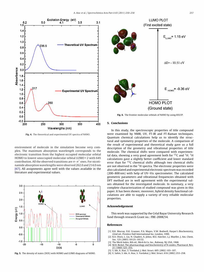
4.4. UV spectrum and electronic properties
Ultraviolet spectra analysis of NANO have been investigatedby theoretical calculation. The UV spectrum of NANO is shown inFig. 4, was measured in water solution and the density of states(DOS) with HOMO and LUMO diagrams for NANO can be seen inFig. 5. Calculations of the molecular orbital geometry show thatthe visible absorption maxima of this molecule correspond to theelectron transition between frontier orbitals such as translation
from HOMO to LUMO. The HOMO energy characterizes the abilityof electron giving, the LUMO characterizes the ability of electronaccepting, and the gap between HOMO and LUMO characterizesthe molecular chemical stability [66]. The energy gap between the
256 A. Atac et al. / Spectrochimica Acta Part A 83 (2011) 250– 258
ANO
HlcfoouAsarncmpaf
number of overlapping transitions. These absorption maxima val-ues are observed 260.42 and 217.67 nm those show agreement intheoretical values. The deviation between experiment and theorymay be resulted from solvent effects. Solvent make the chemical
Table 5Experimental and calculated (cis/6-311++(d,p)) wavelengths � (nm), excitationenergies (eV), oscillator strengths (f), in water solution.
Exp. Calculated Major contribution (≥10%)
Fig. 3. (a) 13C NMR spectrum of N
OMOs and LUMOs is a critical parameter in determining molecu-ar electrical transport properties because it is a measure of electrononductivity. These results are illustrated in Fig. 6. Surfaces for therontier orbitals were drawn to understand the bonding schemef present compound. We examine the two important molecularrbitals (MO) for NANO: the highest occupied MOs and the lowestnoccupied MOs which we denote HOMO and LUMO, respectively.ccording to Fig. 6, the HOMO of free NANO presents a charge den-ity localized mainly on the pyridine ring and N O double bondnd LUMO is characterized by a charge distribution on all pyridineing, C O double bond and NH2 group. Absorption maxima (�max;m) for lower-lying singlet states of the molecule have been cal-ulated by DFT/B3LYP method. The calculated visible absorption
axima of � which are functions of the electron availability wereresented in Table 5. As can be seen from the Table 5, the calculatedbsorption maxima values have been found to be 236.51, 223.32 nmor in water solution at DFT/B3LYP/6-311++G(d,p) method. The
. (b) 1H NMR spectrum of NANO.
spectrum shows two broad bands, a long wavelength band cover-ing the 280–240 nm region and a short wavelength one coveringthe 240–200 nm region. Each of these bands corresponds to a
� (nm) � (nm) E (eV) f
217.67 223.3 5.55361 0.0068 H → L + 2 (64%)260.42 236.5 5.24668 0.2189 H-1 → L + 2 (24%), H → L + 4 (47%)
H, HOMO; L, LUMO.
A. Atac et al. / Spectrochimica Acta Part A 83 (2011) 250– 258 257
epeHcn[l
F
Fig. 4. The theoretical and experimental UV spectra of NANO.
nvironment of molecule in the simulation become very com-lex. The maximum absorption wavelength corresponds to thelectronic transition from the highest occupied molecular orbitalOMO to lowest unoccupied molecular orbital LUMO + 2 with 64%ontribution. All the observed transitions are �–�* ones. For nicoti-amide absorption wavelengths were observed 262.0 and 214.0 nm67]. All assignments agree well with the values available in the
iterature and experimental values.ig. 5. The density of states (DOS) with HOMO and LUMO diagrams of NANO.
Fig. 6. The frontier molecular orbitals of NANO by using B3LYP.
5. Conclusions
In this study, the spectroscopic properties of title compoundwere examined by NMR, UV, FT-IR and FT-Raman techniques.Quantum chemical calculations help us to identify the struc-tural and symmetry properties of the molecule. A comparison ofthe result of experimental and theoretical study gave us a fulldescription of the geometry and vibrational properties of titlemolecule. The chemical shifts were compared with experimen-tal data, showing a very good agreement both for 13C and 1H. 1Hcalculations gave a slightly better coefficient and lower standarderror than for 13C chemical shifts although two chemical shiftsare not observed in the 1H spectra. The electronic properties werealso calculated and experimental electronic spectrum was recorded(200–800 nm) with help of UV–Vis spectrometer. The calculatedgeometric parameters and vibrational frequencies obtained withDFT method are in well agreement with the experimental val-ues obtained for the investigated molecule. In summary, a verycomplete characterization of studied compound was given in thispaper. It has been shown; moreover, hybrid density functional cal-culations are able to supply a variety of very reliable molecularproperties.
Acknowledgement
This work was supported by the Celal Bayar University Researchfund through research Grant no.: FBE-2008/54.
References
[1] R.K. Murray, D.K. Granner, P.A. Mayes, V.W. Rodwell, Harper’s Biochemistry,22nd ed., Prentice Hall International Inc, London, 1990.
[2] R.A. Olsen, L. Liu, N. Ghaderi, A. Johns, M.E. Hatcher, L.J. Mueller, J. Am. Chem.Soc. 125 (2003) 10125–10132.
[3] The Merk Index, 8th ed., Merk & Co. Inc., Rahway, NJ, USA, 1968.[4] M.H. Bickel, The pharmacology and biochemistry of N-oxides, Pharmacol. Rev.
21 (1969) 325–355.[5] S. Ide, A. Atac, S. Yurdakul, J. Mol. Struct. 605 (2002) 103–107.[6] E. Sahin, S. Ide, A. Atac, S. Yurdakul, J. Mol. Struct. 616 (2002) 253–258.

2 a Acta
[[
[[
[[[[[[
[[[
[
[[[[[
[[
[[[
[
[[[
[[[[[
[
[
[
[[[
[[[
[
[[[[
[[[[
[[
[
[
58 A. Atac et al. / Spectrochimic
[7] C.S. Mojumdar, M. Melnik, E. Jona, J. Anal. Appl. Pyrolysis 53 (2000) 149–160.[8] S. Chlopicki, J. Swies, A. Mogielnicki, W. Buczko, M. Bartus, M. Lomnicka, J.
Adamus, J. Gebicki, Br. J. Pharmacol. 152 (2007) 230–239.[9] J.P. Kamat, T.P. Devasagayam, Redox Rep. 4 (1999) 179–184.10] R.W. Pero, B. Axelasson, D. Riemann, Mol. Cell. Biochem. 193 (1999) 119–125.11] T. Takeshima, H. Takeuchi, T. Egawa, S. Konaka, J. Mol. Struct. 644 (2003)
197–205.12] W.B. Wright, G.S.D. King, Acta Crystrallogr. 7 (1954) 283–288.13] Y. Miwa, T. Mizuno, K. Tsuchiya, T. Taga, Y. Iwata, Acta Crystallogr. B 55 (1999)
78–84.14] O. Bolukbasi, S. Akyuz, J. Mol. Struct. 744–747 (2005) 961–971.15] M. Bakiler, O. Bolukbasi, A. Yilmaz, J. Mol. Struct. 826 (2007) 6–16.16] S. Bayari, A. Atac, S. Yurdakul, J. Mol. Struct. 655 (2003) 163–170.17] E. Akalin, S. Akyuz, Vib. Spectrosc. 42 (2006) 333–340.18] E.A. Velcheva, L.I. Daskalova, J. Mol. Struct. 741 (2005) 85–92.19] A. Borba, M. Albrecht, A. Gomez-Zavaglia, L. Lapinski, M.J. Nowak, M.A. Suhm,
R. Fausto, Phys. Chem. Chem. Phys. 10 (2008) 7010–7021.20] S. Chaykin, M. Dagani, L. Johnson, M. Samli, J. Biol. Chem. 240 (1965) 932.21] N.M. Keith, S. Chaykin, J. Bio. Chem. 241/9 (1966) 10–18.22] T. Fukuwatari, H. Wada, R. Sasaki, K. Shibata, Biosci. Biotechnol. Biochem. 68
(1) (2004) 44–50.23] M. Kumar, S. Jaiswal, R. Singh, G. Srivastav, P. Singh, T.N. Yadav, R.A Yadav,
Spectrochim. Acta A 75 (2010) 281–292.24] M.J. Frisch, et al., GAUSSIAN 03 (Revision A.9), Gaussian, Inc., Pittsburgh, 2003.25] A.D. Becke, J. Chem. Phys. 98 (1993) 5648–5652.26] C. Lee, W. Yang, R.G. Parr, Phys. Rev. B37 (1988) 785–789.27] B.J. Orr, J.F. Ward, Mol. Phys. 20 (1970) 513–526.28] N. Sundaraganesan, S. Ilakiamani, H. Saleem, P.M. Wojciechowski, D. Michalska,
Spectrochim. Acta A 61 (2005) 2995–3001.29] J. Baker, A.A. Jarzecki, P. Pulay, J. Phys. Chem. A102 (1998) 1412–1424.30] P. Pulay, J. Baker, K. Wolinski, 2013 Green AcresRoad, Suite A, Fayettevile, AR
72703, USA.31] R. Bauernschmitt, Ahlrichs Chem. Phys. Lett. 256 (1996) 454–464.32] C. Jamorski, M.E. Casida, D.R. Salahub, J. Chem. Phys. 104 (1996) 5134–5150.33] G. Keresztury, S. Holly, J. Varga, G. Besenyei, A.Y. Wang, J.R. Durig, Spectrochim.
Acta 49 A (1993) 2007–2017.34] G. Keresztury, Raman spectroscopy: theory, in: J.M. Chalmers, P.R. Griffith
(Eds.), Handbook of Vibrational Spectroscopy, vol. 1, John Wiley & Sons Ltd.,
New York, 2002.35] J.V. Prasad, S.B. Rai, S.N. Thakur, Chem. Phys. Lett. 164 (6) (1989) 629–645.36] M.K. Ahmed, B.R. Henry, J. Phys. Chem. 90 (1986) 1737–1739.37] L.J. Bellamy, The Infrared Spectra of Complex Molecules, vol. 2, Chapman and
Hall, London, 1980.
[[[
Part A 83 (2011) 250– 258
38] M.A. Palafox, Ind. J. Pure Appl. Phys. 31 (1993) 90–98.39] R.A. Yadav, I.S. Sing, Ind. J. Pure Appl. Phys. 23 (1985) 626–627.40] K.B. Wiberg, A. Shrake, Spectrochim. Acta A 29 (1973) 583–594.41] S. Mancy, W.L. Peticoles, R.S. Toblas, Spectrochim. Acta A 35 (1979) 315.42] T.S. Planka, B. Gyurcsik, N.V. Nagy, A. Rockenbauer, R. Sipos, J. Sima, M. Menlik,
J. Inorg. Biochem. 102 (2008) 101–109.43] K. Rastogi, M.A. Palafox, R.P. Tanwar, L. Mittal, Spectrochim. Acta A 58 (2002)
1987–2004.44] M. Silverstein, G. Clayton Basseler, C. Morill, Spectrometric Identification of
Organic Compounds, Wiley, New York, 1981.45] G. Socrates, Infrared and Raman Characteristic Group Frequencies Tables and
Charts, 3rd ed., Wiley, New York, 2001.46] M. Karabacak, M. Kurt, J. Mol. Struct. 919 (2009) 215–222.47] M. Karabacak, E. Kose, M. Kurt, J. Raman Spectr. 41 (9) (2010) 1085–1097.48] G. Varsanyi, Assignments of Vibrational Spectra of 700 Benzene Derivatives,
Wiley, New York, 1974.49] J.K. Wilmshurst, H.J. Bernstein, Can. J. Chem. 35 (1957) 1183–1194.50] A. Gambi, S. Ghersetti, Spectrosc. Lett. 8 (1977) 627–632.51] D.N. Sathyanarayana, Vibrational Spectroscopy, Theory and Applications, New
Age International Publishers, New Delhi, 2004.52] R.L. Prasad, A. Kushwaha, M. Suchita, R.A. Kumar, R. Yadav, Spectrochim. Acta
A 69 (2008) 304–311.53] M. Arivazhagan, S. Jeyavijayan, Spectrochim. Acta A 79 (1) (2011) 161–168.54] Y. Kishore, S.N. Shatrma, C.P.D. Dwivedi, Ind. J. Phys. 48 (1974) 412–417.55] N.P. Sing, R.A. Yadav, Ind. J. Phys. B 75 (4) (2001) 347–355.56] N. Sundaraganesan, S. Ilakiamani, B. Ananda, H. Saleem, B. Dominic Joshua,
Spectrochim. Acta A 64 (2006) 586–594.57] Y. Akkaya, S. Akyuz, Vib. Spectrosc. 42 (2006) 292–301.58] H.I.S. Nogueira, Spectrochim. Acta A 54 (1998) 1461–1470.59] M. Karabacak, M. Cinar, M. Kurt, J. Mol. Struct. 885 (2008) 28–35.60] N. Sundaraganesan, B.D. Joshua, K. Settu, Spectrochim. Acta A 66 (2007)
381–388.61] R. Shanmugam, D. Sathyanarayana, Spectrochim. Acta A 40 (1984) 764.62] N.P.G. Roeges, A Guide to the Complete Interpretation of Infrared Spectra of
Organic Structures, Wiley, New York, USA, 1994.63] H.O. Kalinowski, S. Berger, S. Braun, C13 NMR Spectroscopy, John Wiley & Sons,
Chichester, 1988.64] K. Pihlaja, E. Kleinpeter, Carbon-13 Chemical Shifts in Structural and Stereo
Chemical Analysis, VCH Publishers, Deerfield Beach, FL, 1994.65] Y. Erdogdu, M.T. Gulluoglu, M. Kurt, J. Raman Spectrosc. 40 (2009) 1615–1623.66] K. Fukui, Science 218 (1982) 747–754.67] R.H. Abu Eittah, M.M. Hamed, A.A. Mohamed, Int. J. Quant. Chem. 64 (1997)
689–701.


![Vibrational spectroscopic (FT-IR and FT-Raman) studies, HOMO-LUMO, NBO analysis and MEP of 6-methyl-1-({[(2E)-2-methyl-3-phenyl-prop-2-en-1-yl]oxy}methyl)-1,2,3,4-tetra-hydroquinazoline-2,4-dione,](https://static.fdokumen.com/doc/165x107/633494f441100cab3c07ce05/vibrational-spectroscopic-ft-ir-and-ft-raman-studies-homo-lumo-nbo-analysis.jpg)