Specificity and Plasticity of Thalamocortical Connections in Sema6A Mutant Mice
15
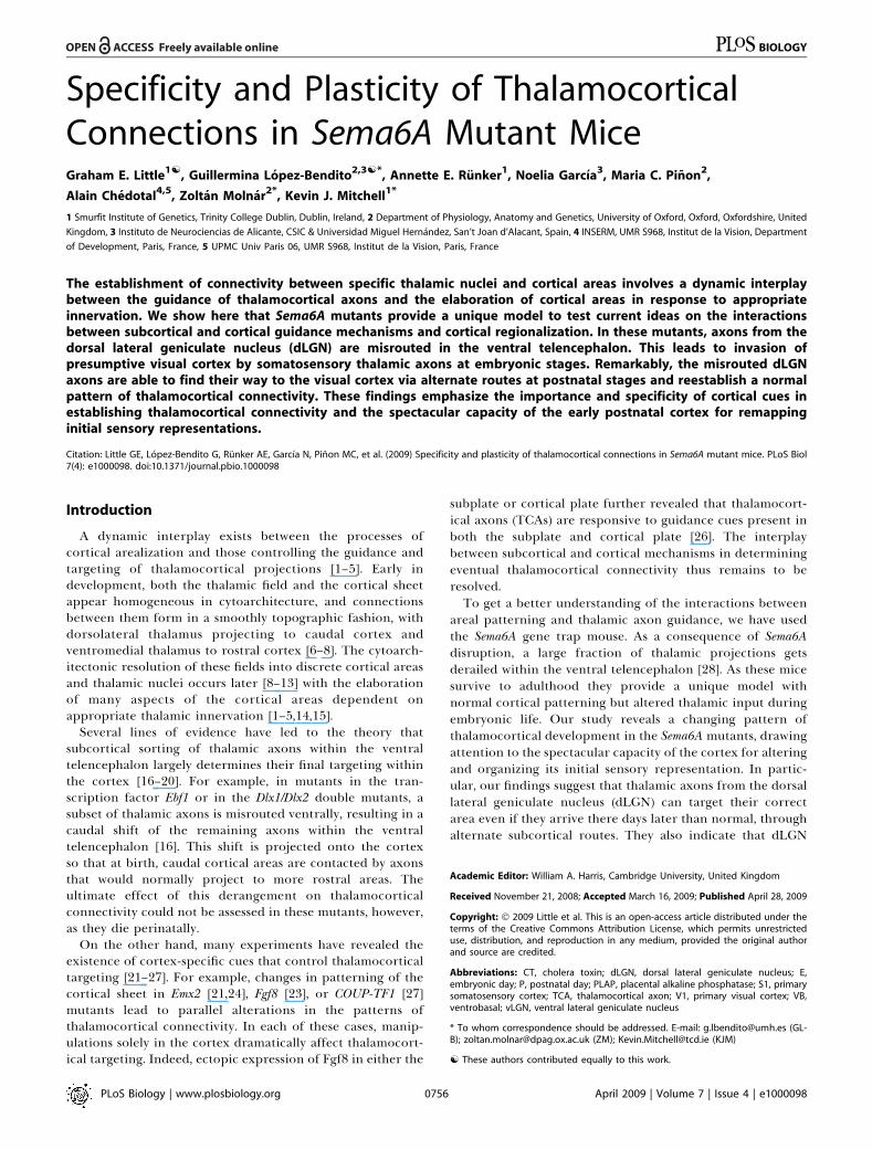
Specificity and Plasticity of Thalamocortical Connections in Sema6A Mutant Mice Graham E. Little 1[ , Guillermina Lo ´ pez-Bendito 2,3[* , Annette E. Ru ¨ nker 1 , Noelia Garcı ´a 3 , Maria C. Pin ˜ on 2 , Alain Che ´ dotal 4,5 , Zolta ´ n Molna ´r 2* , Kevin J. Mitchell 1* 1 Smurfit Institute of Genetics, Trinity College Dublin, Dublin, Ireland, 2 Department of Physiology, Anatomy and Genetics, University of Oxford, Oxford, Oxfordshire, United Kingdom, 3 Instituto de Neurociencias de Alicante, CSIC & Universidad Miguel Herna ´ndez, San’t Joan d’Alacant, Spain, 4 INSERM, UMR S968, Institut de la Vision, Department of Development, Paris, France, 5 UPMC Univ Paris 06, UMR S968, Institut de la Vision, Paris, France The establishment of connectivity between specific thalamic nuclei and cortical areas involves a dynamic interplay between the guidance of thalamocortical axons and the elaboration of cortical areas in response to appropriate innervation. We show here that Sema6A mutants provide a unique model to test current ideas on the interactions between subcortical and cortical guidance mechanisms and cortical regionalization. In these mutants, axons from the dorsal lateral geniculate nucleus (dLGN) are misrouted in the ventral telencephalon. This leads to invasion of presumptive visual cortex by somatosensory thalamic axons at embryonic stages. Remarkably, the misrouted dLGN axons are able to find their way to the visual cortex via alternate routes at postnatal stages and reestablish a normal pattern of thalamocortical connectivity. These findings emphasize the importance and specificity of cortical cues in establishing thalamocortical connectivity and the spectacular capacity of the early postnatal cortex for remapping initial sensory representations. Citation: Little GE, Lo ´ pez-Bendito G, Ru ¨ nker AE, Garcı ´a N, Pin ˜ on MC, et al. (2009) Specificity and plasticity of thalamocortical connections in Sema6A mutant mice. PLoS Biol 7(4): e1000098. doi:10.1371/journal.pbio.1000098 Introduction A dynamic interplay exists between the processes of cortical arealization and those controlling the guidance and targeting of thalamocortical projections [1–5]. Early in development, both the thalamic field and the cortical sheet appear homogeneous in cytoarchitecture, and connections between them form in a smoothly topographic fashion, with dorsolateral thalamus projecting to caudal cortex and ventromedial thalamus to rostral cortex [6–8]. The cytoarch- itectonic resolution of these fields into discrete cortical areas and thalamic nuclei occurs later [8–13] with the elaboration of many aspects of the cortical areas dependent on appropriate thalamic innervation [1–5,14,15]. Several lines of evidence have led to the theory that subcortical sorting of thalamic axons within the ventral telencephalon largely determines their final targeting within the cortex [16–20]. For example, in mutants in the tran- scription factor Ebf1 or in the Dlx1/Dlx2 double mutants, a subset of thalamic axons is misrouted ventrally, resulting in a caudal shift of the remaining axons within the ventral telencephalon [16]. This shift is projected onto the cortex so that at birth, caudal cortical areas are contacted by axons that would normally project to more rostral areas. The ultimate effect of this derangement on thalamocortical connectivity could not be assessed in these mutants, however, as they die perinatally. On the other hand, many experiments have revealed the existence of cortex-specific cues that control thalamocortical targeting [21–27]. For example, changes in patterning of the cortical sheet in Emx2 [21,24], Fgf8 [23], or COUP-TF1 [27] mutants lead to parallel alterations in the patterns of thalamocortical connectivity. In each of these cases, manip- ulations solely in the cortex dramatically affect thalamocort- ical targeting. Indeed, ectopic expression of Fgf8 in either the subplate or cortical plate further revealed that thalamocort- ical axons (TCAs) are responsive to guidance cues present in both the subplate and cortical plate [26]. The interplay between subcortical and cortical mechanisms in determining eventual thalamocortical connectivity thus remains to be resolved. To get a better understanding of the interactions between areal patterning and thalamic axon guidance, we have used the Sema6A gene trap mouse. As a consequence of Sema6A disruption, a large fraction of thalamic projections gets derailed within the ventral telencephalon [28]. As these mice survive to adulthood they provide a unique model with normal cortical patterning but altered thalamic input during embryonic life. Our study reveals a changing pattern of thalamocortical development in the Sema6A mutants, drawing attention to the spectacular capacity of the cortex for altering and organizing its initial sensory representation. In partic- ular, our findings suggest that thalamic axons from the dorsal lateral geniculate nucleus (dLGN) can target their correct area even if they arrive there days later than normal, through alternate subcortical routes. They also indicate that dLGN Academic Editor: William A. Harris, Cambridge University, United Kingdom Received November 21, 2008; Accepted March 16, 2009; Published April 28, 2009 Copyright: Ó 2009 Little et al. This is an open-access article distributed under the terms of the Creative Commons Attribution License, which permits unrestricted use, distribution, and reproduction in any medium, provided the original author and source are credited. Abbreviations: CT, cholera toxin; dLGN, dorsal lateral geniculate nucleus; E, embryonic day; P, postnatal day; PLAP, placental alkaline phosphatase; S1, primary somatosensory cortex; TCA, thalamocortical axon; V1, primary visual cortex; VB, ventrobasal; vLGN, ventral lateral geniculate nucleus * To whom correspondence should be addressed. E-mail: [email protected] (GL- B); [email protected] (ZM); [email protected] (KJM) [ These authors contributed equally to this work. PLoS Biology | www.plosbiology.org April 2009 | Volume 7 | Issue 4 | e1000098 0756 P L o S BIOLOGY
Transcript of Specificity and Plasticity of Thalamocortical Connections in Sema6A Mutant Mice
Specificity and Plasticity of ThalamocorticalConnections in Sema6A Mutant MiceGraham E. Little
1[, Guillermina Lopez-Bendito
2,3[*, Annette E. Runker
1, Noelia Garcıa
3, Maria C. Pinon
2,
Alain Chedotal4,5
, Zoltan Molnar2*
, Kevin J. Mitchell1*
1 Smurfit Institute of Genetics, Trinity College Dublin, Dublin, Ireland, 2 Department of Physiology, Anatomy and Genetics, University of Oxford, Oxford, Oxfordshire, United
Kingdom, 3 Instituto de Neurociencias de Alicante, CSIC & Universidad Miguel Hernandez, San’t Joan d’Alacant, Spain, 4 INSERM, UMR S968, Institut de la Vision, Department
of Development, Paris, France, 5 UPMC Univ Paris 06, UMR S968, Institut de la Vision, Paris, France
The establishment of connectivity between specific thalamic nuclei and cortical areas involves a dynamic interplaybetween the guidance of thalamocortical axons and the elaboration of cortical areas in response to appropriateinnervation. We show here that Sema6A mutants provide a unique model to test current ideas on the interactionsbetween subcortical and cortical guidance mechanisms and cortical regionalization. In these mutants, axons from thedorsal lateral geniculate nucleus (dLGN) are misrouted in the ventral telencephalon. This leads to invasion ofpresumptive visual cortex by somatosensory thalamic axons at embryonic stages. Remarkably, the misrouted dLGNaxons are able to find their way to the visual cortex via alternate routes at postnatal stages and reestablish a normalpattern of thalamocortical connectivity. These findings emphasize the importance and specificity of cortical cues inestablishing thalamocortical connectivity and the spectacular capacity of the early postnatal cortex for remappinginitial sensory representations.
Citation: Little GE, Lopez-Bendito G, Runker AE, Garcıa N, Pinon MC, et al. (2009) Specificity and plasticity of thalamocortical connections in Sema6A mutant mice. PLoS Biol7(4): e1000098. doi:10.1371/journal.pbio.1000098
Introduction
A dynamic interplay exists between the processes ofcortical arealization and those controlling the guidance andtargeting of thalamocortical projections [1–5]. Early indevelopment, both the thalamic field and the cortical sheetappear homogeneous in cytoarchitecture, and connectionsbetween them form in a smoothly topographic fashion, withdorsolateral thalamus projecting to caudal cortex andventromedial thalamus to rostral cortex [6–8]. The cytoarch-itectonic resolution of these fields into discrete cortical areasand thalamic nuclei occurs later [8–13] with the elaborationof many aspects of the cortical areas dependent onappropriate thalamic innervation [1–5,14,15].
Several lines of evidence have led to the theory thatsubcortical sorting of thalamic axons within the ventraltelencephalon largely determines their final targeting withinthe cortex [16–20]. For example, in mutants in the tran-scription factor Ebf1 or in the Dlx1/Dlx2 double mutants, asubset of thalamic axons is misrouted ventrally, resulting in acaudal shift of the remaining axons within the ventraltelencephalon [16]. This shift is projected onto the cortexso that at birth, caudal cortical areas are contacted by axonsthat would normally project to more rostral areas. Theultimate effect of this derangement on thalamocorticalconnectivity could not be assessed in these mutants, however,as they die perinatally.
On the other hand, many experiments have revealed theexistence of cortex-specific cues that control thalamocorticaltargeting [21–27]. For example, changes in patterning of thecortical sheet in Emx2 [21,24], Fgf8 [23], or COUP-TF1 [27]mutants lead to parallel alterations in the patterns ofthalamocortical connectivity. In each of these cases, manip-ulations solely in the cortex dramatically affect thalamocort-ical targeting. Indeed, ectopic expression of Fgf8 in either the
subplate or cortical plate further revealed that thalamocort-ical axons (TCAs) are responsive to guidance cues present inboth the subplate and cortical plate [26]. The interplaybetween subcortical and cortical mechanisms in determiningeventual thalamocortical connectivity thus remains to beresolved.To get a better understanding of the interactions between
areal patterning and thalamic axon guidance, we have usedthe Sema6A gene trap mouse. As a consequence of Sema6Adisruption, a large fraction of thalamic projections getsderailed within the ventral telencephalon [28]. As these micesurvive to adulthood they provide a unique model withnormal cortical patterning but altered thalamic input duringembryonic life. Our study reveals a changing pattern ofthalamocortical development in the Sema6A mutants, drawingattention to the spectacular capacity of the cortex for alteringand organizing its initial sensory representation. In partic-ular, our findings suggest that thalamic axons from the dorsallateral geniculate nucleus (dLGN) can target their correctarea even if they arrive there days later than normal, throughalternate subcortical routes. They also indicate that dLGN
Academic Editor: William A. Harris, Cambridge University, United Kingdom
Received November 21, 2008; Accepted March 16, 2009; Published April 28, 2009
Copyright: � 2009 Little et al. This is an open-access article distributed under theterms of the Creative Commons Attribution License, which permits unrestricteduse, distribution, and reproduction in any medium, provided the original authorand source are credited.
Abbreviations: CT, cholera toxin; dLGN, dorsal lateral geniculate nucleus; E,embryonic day; P, postnatal day; PLAP, placental alkaline phosphatase; S1, primarysomatosensory cortex; TCA, thalamocortical axon; V1, primary visual cortex; VB,ventrobasal; vLGN, ventral lateral geniculate nucleus
* To whom correspondence should be addressed. E-mail: [email protected] (GL-B); [email protected] (ZM); [email protected] (KJM)
[ These authors contributed equally to this work.
PLoS Biology | www.plosbiology.org April 2009 | Volume 7 | Issue 4 | e10000980756
PLoS BIOLOGY

axons can out-compete invading axons from inappropriatethalamic nuclei, establishing a surprisingly normal adultcortical representation.
Results
Sema6A Is Strongly Expressed in the Developing Thalamusand Ventral Telencephalon
Sema6A is broadly expressed in the thalamus at embryonicday (E)14.5, a time when thalamic neurons are extendingaxons towards their cortical targets [28], with expressionhighest in the dorsolateral aspect (n ¼ 5; Figure 1A and 1B).Sema6A is also strongly expressed in the amygdala and theventral telencephalon, and weakly expressed in the neocortexat this age, localized to the most superficial compartments(Figure 1A and 1B). At late embryonic stages, Sema6A is alsoexpressed by deep cortical plate neurons, eventually layer 5(unpublished data). Staining with the axonal marker placentalalkaline phosphatase (PLAP), encoded on the gene trapcassette in this Sema6A allele, revealed thalamic axonsextending from the thalamus through the internal capsuleand towards the neocortex (Figure 1C and 1D).
Lack of Sema6A Leads to Abnormalities in ThalamocorticalPathfinding
A previous study using PLAP staining showed that manythalamic axons were misrouted in the Sema6A�/� brains atembryonic stages [28]. To further examine the guidance ofTCAs in the absence of functional Sema6A protein, weperformed carbocyanine dye tracing studies. Broad injectionsof DiI in the thalamus (including the dorsolateral aspect) atE15.5 revealed a prominent derailment of thalamic axons atthe surface of the ventral telencephalon and amygdala inSema6A�/� embryos (n¼ 4/4; Figure 2D and 2G–2I), comparedto the normal route of navigation through the internalcapsule towards the neocortex observed in wild-type embryos
(n ¼ 4/4; Figure 2A and 2C). The derailment of a largeproportion of TCA fibers at the ventral telencephalon inSema6A�/� brains at E16.5 was confirmed by neurofilament(NF) immunohistochemistry (n¼ 3; Figure 2E and 2F) and byPLAP staining (Figure S1A and S1C). Overlaid consecutiveserial sections of Sema6A�/� brains at E17.5 stained for NFreveal more clearly the extent of the TCA derailment (n¼ 3;Figure 2H).
Only dLGN Axons Are Misrouted within the VentralTelencephalonTo identify the precise origin of the misrouted thalamic
axons within the thalamus, we placed small DiI crystals at thesite of the derailed fibers near the ventral surface of thetelencephalon (Figure 3A) in wild-type (n ¼ 4) and Sema6A�/�
(n ¼ 4) postnatal day (P)0 brains. Whereas no back-labeledcells were observed in any thalamic nuclei in wild-type brains(Figure 3B and 3C), several axon bundles were retrogradely
Figure 1. Sema6A Is Highly Expressed in the Developing Embryonic
Thalamus
(A and B) Coronal section of an E14.5 mouse brain showing in situhybridization for Sema6A in the forebrain (A). Sema6A is expressed inneurons and axons (arrowheads) of the dorsal geniculate nucleus (dLGN)and ventrobasal complex (VB) of the developing dorsal thalamus (dTh;[B]).(C and D) PLAP staining on comparable coronal sections of Sema6Aþ/�
mouse brains. PLAP-positive axons, presumably TCAs, can be seenprojecting from the dTh through the internal capsule towards theneocortex (NCx; arrowheads).Hyp, hypothalamus; vLGN, ventrolateral geniculate nucleus. Scale bars in(A and C) indicate 500 lm; in (B and D) 200 lm.doi:10.1371/journal.pbio.1000098.g001
PLoS Biology | www.plosbiology.org April 2009 | Volume 7 | Issue 4 | e10000980757
Sema6A and Thalamocortical Development
Author Summary
During brain development, the emergence of distinct areas in thecerebral cortex involves an interplay between patterning of thecortical sheet in the early embryo and later influences of incomingconnections made from other brain areas, namely the thalamus.Connectivity between the thalamus and the cortex is initiallysmooth and graded, and a prominent model for how thalamocort-ical connectivity is established proposes thalamic axons are topo-graphically sorted as they course through subcortical regions andthen passively delivered to appropriate areas of the cortical sheet.We have used mutant mice lacking the guidance moleculeSemaphorin-6A to test this model. In these mutants, Semaphorin-6A axons from the visual part of the thalamus are subcorticallymisrouted and fail to innervate the presumptive visual cortex, whichis instead invaded by somatosensory thalamic axons. Despite thismajor disruption in initial connectivity, many visual thalamic axonsfind their way specifically to visual cortex, arriving several days laterthan usual. These late-arriving axons often follow alternate routes,and upon arrival are able to out-compete earlier-arriving somato-sensory axons to reestablish grossly normal thalamocorticalconnectivity. These results argue strongly against an essential rolefor early subcortical targeting in the establishment of thalamocort-ical connectivity patterns and suggest instead the existence ofhighly specific target-selection mechanisms that match thalamicaxons with appropriate cortical areas.

traced to the dorsolateral aspect of the thalamus in Sema6A�/�
brains (Figure 3D and 3E). Retrogradely labeled cells werespecifically found in the presumptive dLGN (Figure 3G and3H). Some labeled bundles were also observed ascendinglaterally towards the cortex (Figure 3E). Moreover, inSema6A�/� brains, at more-caudal telencephalic levels, DiI-labeled axons were observed running through the intermedi-
ate zone of the primary visual cortical area (Figure 3F and 3I;and unpublished data), suggesting that some dLGN axons thatfollow this abnormal route might still reach the visual cortex.Indeed, whereas a DiA crystal placed in the internal capsulezone of wild-type brains at E17.5 back-labeled cells through-out the dorsal thalamus (n¼ 2/2; Figure S2A–S2C), a similarlyplaced DiA crystal in Sema6A�/� brains at the same age
Figure 2. Early Thalamocortical Guidance Defects in Sema6A�/� Mouse
Coronal sections showing half the brain or close-ups of ventral telencephalon.(A and B) TCAs follow a normal route along the ventral telencephalon and striatum (Str) in wild-type embryos as revealed by DiI tracing from the dorsalthalamus (dTh, [A]) as well as by neurofilament staining (B).(D–H) In Sema6A�/� embryos, a large proportion of TCAs are derailed at the ventral telencephalon (filled arrowheads in [D and H]), as shown by DiItracing from the internal capsule (ic) at E15.5 (D) and neurofilament staining at E16.5 (E and F), and an overlaid series of images at E17.5 (G and H). Notethat some TCAs follow their normal route through the striatum (Str) in the Sema6A�/� brains (open arrowhead in [D]). Also, note that misrouted TCAs inthe Sema6A�/� brains appear to bifurcate in the ventral telencephalon (open and filled arrowheads in [H]).(C and I) Schematic diagrams of the trajectory of TCAs in wild-type (C) and Sema6A�/� brains (I).Scale bars in (A and D) indicate 1 mm; in (B and E) 500 lm, in (F) 300 lm; and in (G and H) 250 lm.Hyp, hypothalamus; NCx, neocortex; Str, striatum.doi:10.1371/journal.pbio.1000098.g002
PLoS Biology | www.plosbiology.org April 2009 | Volume 7 | Issue 4 | e10000980758
Sema6A and Thalamocortical Development
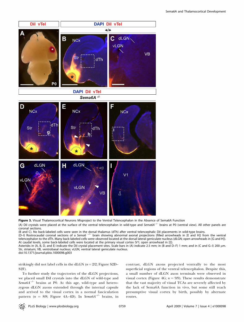
strikingly did not label cells in the dLGN (n¼2/2; Figure S2D–S2F).
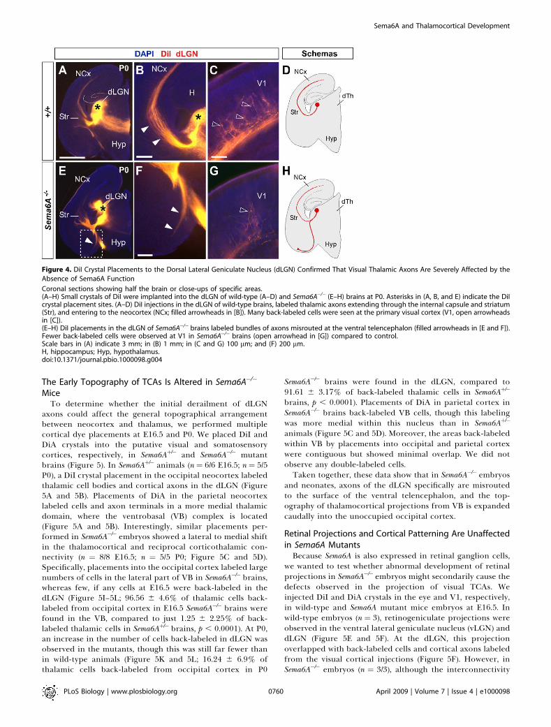
To further study the trajectories of the dLGN projections,we placed small DiI crystals into the dLGN of wild-type andSema6A�/� brains at P0. At this age, wild-type and hetero-zygous dLGN axons extended through the internal capsuleand arrived to the visual cortex in a normal fasciculationpattern (n ¼ 8/8; Figure 4A–4D). In Sema6A�/� brains, in
contrast, dLGN axons projected ventrally to the mostsuperficial regions of the ventral telencephalon. Despite this,a small number of dLGN axon terminals were observed invisual cortex (Figure 4G; n ¼ 9/9). These results demonstratethat the vast majority of visual TCAs are severely affected bythe lack of Sema6A function in vivo, but some still reachpresumptive visual cortex by birth, possibly by alternateroutes.
Figure 3. Visual Thalamocortical Neurons Misproject to the Ventral Telencephalon in the Absence of Sema6A Function
(A) DiI crystals were placed at the surface of the ventral telencephalon in wild-type and Sema6A�/� brains at P0 (ventral view). All other panels arecoronal sections.(B and C). No back-labeled cells were seen in the dorsal thalamus (dTh) after ventral telencephalic DiI placements in wild-type brains.(D–I) Rostrocaudal coronal sections of a Sema6 �/� brain showing abnormal axonal projections (filled arrowheads in [E and H]) from the ventraltelencephalon to the dTh. Many back-labeled cells were observed located at the dorsal lateral geniculate nucleus (dLGN; open arrowheads in [G and H]).At caudal levels, some back-labeled cells were located at the primary visual cortex (V1; open arrowhead in [I]).Asterisks in (A, B, D, and E) indicate the DiI crystal placement sites. Scale bars in (A) indicate 2.5 mm; in (B and D–F) 1 mm; and in (C and G–I) 200 lm.Str, striatum; VB, ventrobasal nucleus; vLGN, ventral lateral geniculate nucleus.doi:10.1371/journal.pbio.1000098.g003
PLoS Biology | www.plosbiology.org April 2009 | Volume 7 | Issue 4 | e10000980759
Sema6A and Thalamocortical Development
The Early Topography of TCAs Is Altered in Sema6A�/�
MiceTo determine whether the initial derailment of dLGN
axons could affect the general topographical arrangementbetween neocortex and thalamus, we performed multiplecortical dye placements at E16.5 and P0. We placed DiI andDiA crystals into the putative visual and somatosensorycortices, respectively, in Sema6Aþ/� and Sema6A�/� mutantbrains (Figure 5). In Sema6Aþ/� animals (n ¼ 6/6 E16.5; n¼ 5/5P0), a DiI crystal placement in the occipital neocortex labeledthalamic cell bodies and cortical axons in the dLGN (Figure5A and 5B). Placements of DiA in the parietal neocortexlabeled cells and axon terminals in a more medial thalamicdomain, where the ventrobasal (VB) complex is located(Figure 5A and 5B). Interestingly, similar placements per-formed in Sema6A�/� embryos showed a lateral to medial shiftin the thalamocortical and reciprocal corticothalamic con-nectivity (n ¼ 8/8 E16.5; n ¼ 5/5 P0; Figure 5C and 5D).Specifically, placements into the occipital cortex labeled largenumbers of cells in the lateral part of VB in Sema6A�/� brains,whereas few, if any cells at E16.5 were back-labeled in thedLGN (Figure 5I–5L; 96.56 6 4.6% of thalamic cells back-labeled from occipital cortex in E16.5 Sema6A�/� brains werefound in the VB, compared to just 1.25 6 2.25% of back-labeled thalamic cells in Sema6Aþ/� brains, p , 0.0001). At P0,an increase in the number of cells back-labeled in dLGN wasobserved in the mutants, though this was still far fewer thanin wild-type animals (Figure 5K and 5L; 16.24 6 6.9% ofthalamic cells back-labeled from occipital cortex in P0
Sema6A�/� brains were found in the dLGN, compared to91.61 6 3.17% of back-labeled thalamic cells in Sema6Aþ/�
brains, p , 0.0001). Placements of DiA in parietal cortex inSema6A�/� brains back-labeled VB cells, though this labelingwas more medial within this nucleus than in Sema6Aþ/�
animals (Figure 5C and 5D). Moreover, the areas back-labeledwithin VB by placements into occipital and parietal cortexwere contiguous but showed minimal overlap. We did notobserve any double-labeled cells.Taken together, these data show that in Sema6A�/� embryos
and neonates, axons of the dLGN specifically are misroutedto the surface of the ventral telencephalon, and the top-ography of thalamocortical projections from VB is expandedcaudally into the unoccupied occipital cortex.
Retinal Projections and Cortical Patterning Are Unaffectedin Sema6A MutantsBecause Sema6A is also expressed in retinal ganglion cells,
we wanted to test whether abnormal development of retinalprojections in Sema6A�/� embryos might secondarily cause thedefects observed in the projection of visual TCAs. Weinjected DiI and DiA crystals in the eye and V1, respectively,in wild-type and Sema6A mutant mice embryos at E16.5. Inwild-type embryos (n ¼ 3), retinogeniculate projections wereobserved in the ventral lateral geniculate nucleus (vLGN) anddLGN (Figure 5E and 5F). At the dLGN, this projectionoverlapped with back-labeled cells and cortical axons labeledfrom the visual cortical injections (Figure 5F). However, inSema6A�/� embryos (n ¼ 3/3), although the interconnectivity
Figure 4. DiI Crystal Placements to the Dorsal Lateral Geniculate Nucleus (dLGN) Confirmed That Visual Thalamic Axons Are Severely Affected by the
Absence of Sema6A Function
Coronal sections showing half the brain or close-ups of specific areas.(A–H) Small crystals of DiI were implanted into the dLGN of wild-type (A–D) and Sema6A�/� (E–H) brains at P0. Asterisks in (A, B, and E) indicate the DiIcrystal placement sites. (A–D) DiI injections in the dLGN of wild-type brains, labeled thalamic axons extending through the internal capsule and striatum(Str), and entering to the neocortex (NCx; filled arrowheads in [B]). Many back-labeled cells were seen at the primary visual cortex (V1, open arrowheadsin [C]).(E–H) DiI placements in the dLGN of Sema6A�/� brains labeled bundles of axons misrouted at the ventral telencephalon (filled arrowheads in [E and F]).Fewer back-labeled cells were observed at V1 in Sema6A�/� brains (open arrowhead in [G]) compared to control.Scale bars in (A) indicate 3 mm; in (B) 1 mm; in (C and G) 100 lm; and (F) 200 lm.H, hippocampus; Hyp, hypothalamus.doi:10.1371/journal.pbio.1000098.g004
PLoS Biology | www.plosbiology.org April 2009 | Volume 7 | Issue 4 | e10000980760
Sema6A and Thalamocortical Development

Figure 5. Early Topographical Defects in Thalamocortical Connectivity in Sema6A�/� Mice
(A–J) Coronal sections showing back-labeling in dorsal thalamus.(A and B) In Sema6Aþ/� brains, DiI crystal placement in the occipital cortex back-labels cells in the dorsal lateral geniculate nucleus (dLGN; filledarrowheads), whereas a DiA crystal placement in parietal cortex back-labels cells in the ventrobasal complex (VB; open arrowheads).(C–D) In Sema6A�/� brains, identical dye crystal placements resulted in back-labeled red (DiI) and green (DiA) cells in the VB of the dorsal thalamus,indicating a medial shift of visual thalamocortical connectivity in these mice. Note that at E16.5 (C), the visual shift was more pronounced than at P0stages (D).(E–H) In Sema6Aþ/� brains at E16.5, retinothalamic projections, labeled by a DiI crystal placed in the retina, entered the ventral and dorsal lateralgeniculate nucleus (vLGN and dLGN, respectively; open arrowheads in [E and F]). A DiA crystal placement in the occipital cortex back-labeled dLGN cells(filled arrowheads in [E and F]). In contrast, in Sema6A�/� brains, whereas the retinal projection enters the vLGN and dLGN (open arrowheads [G and H])normally, thalamic cells in the VB are back-labeled from a DiA crystal in the occipital cortex (filled arrowheads, [G and H]).(I–L) At early stages (E16.5), the vast majority of cells back-labeled from the occipital cortex are found in the dLGN in Sema6Aþ/�mice, with few if any,being found in the VB. In Sema6A�/�mice at the same age, the majority of cells back-labeled from the occipital cortex are found in the VB, with very fewbeing found in the dLGN.These differences are highly statistically significant (triple asterisks [***] indicates p ,0.0001).At P0, the proportion of cells back-labeled from the occipital cortex and found in the dLGN is significantly greater than that at E16.5 but still significantlyless than in Sema6Aþ/�mice (p , 0.0001). By P4, there was no significant difference between wild-type and Sema6A�/�mice in the proportion of cellsback-labeled from the occipital cortex and found in the dLGN. Error bars indicate standard error mean (s.e.m.).Scale bars in (A–D) indicate 200 lm; in (E and G) 200 lm; in (F and H) 200 lm; and in (I and J) 100 lm.MeA, medial amygdala.doi:10.1371/journal.pbio.1000098.g005
PLoS Biology | www.plosbiology.org April 2009 | Volume 7 | Issue 4 | e10000980761
Sema6A and Thalamocortical Development

between visual cortex and thalamus was shifted medially,retinogeniculate projections still arrived to the vLGN anddLGN in a normal fashion (Figure 5G and 5H). Thus, thedefects observed in the development of visual TCAs are notdue to abnormal development of the projection from theretina or to abnormal differentiation of the LGN, which canstill act as a specific target for retinal axons.
Similarly, to test whether changes in cortical patterningcould account for the topographical defect observed in TCAsconnectivity in Sema6A�/� mutants, we performed in situhybridization of specific cortical area markers on both wild-type (n ¼ 9) and Sema6A�/� (n ¼ 8) brains at P0. In wild-typebrains EphA7 is expressed both rostrally and caudally in thecortex, but is largely absent from parietal cortex, whereasEphrinA5 is expressed in a complementary pattern (FigureS3A and S3C). We did not find any changes in the expressionof EphA7 and EphrinA5 in Sema6A�/� brains (Figure S3B andS3D). The expression of other cortical area markers (Cad6,Cad8, and RZRb) was also unaltered in Sema6A�/� brains at P0(unpublished data). At earlier (E14.5 and E16.5) and later (P7)developmental stages, there were also no differences observedin the expression of cortical area markers between wild-typeand Sema6A�/� brains (unpublished data). These data indicatethat intrinsic cortical patterning is unaltered in Sema6Amutants.
Postnatal Recovery of Grossly Normal ThalamocorticalConnectivity
We investigated thalamocortical connectivity at P4 usingmultiple cortical dye placements. Small crystals of DiI andDiA were placed in the primary visual (V1) and primarysomatosensory (S1) cortices, respectively, in wild-type andSema6A�/� brains (Figure 6). In wild-type brains (n¼ 2/2), a DiIplacement in V1 labeled thalamic cell bodies and corticalaxons in the dLGN, whereas a DiA placement in S1 labeledcell bodies in the VB (Figure 6A and 6B). Interestingly, inSema6A�/� brains (n ¼ 2/2), a DiI placement in V1 mainlylabeled thalamic cell bodies in the dLGN (Figures 6C, 6D, and5J; 79.49 6 25.3% of thalamic cells back-labeled fromoccipital cortex in P4 Sema6A�/� brains were found in thedLGN, compared to 99.49 6 1.51% of back-labeled thalamiccells in wild-type brains, p ¼ 0.0428), whereas far fewer cellswere labeled in VB, in sharp contrast to results from similartracing experiments at E16.5 and P0 (Figure 5K. In Sema6A�/�
brains at P4, just 20.5 6 25.3% of cells back-labeled from theoccipital cortex were found in the VB, compared to 96.56 6
4.68% and 83.65 6 6.9% in Sema6A�/� brains at E16.5 and P0,respectively, p , 0.0001). A DiA placement in S1 labeled cellbodies in the VB as expected (Figure 6C and 6D). Thisapparent rapid recovery of the normal thalamocorticalconnectivity was observed despite a persistent misprojectionof TCAs from the dLGN to the ventral telencephalon inSema6A�/� brains at P4 (unpublished data). Although thetopographical sorting of TCAs was apparently restored at thisstage, fewer back-labeled cells were detected in the dLGNafter visual cortical dye placements in Sema6A�/� compared towild-type brains (Figure 6B and 6D).
To characterize the extent to which any shift in thalamo-cortical connectivity persists in the adult Sema6A�/�mouse, weperformed in vivo stereotaxic tracing studies. We injected redand green cholera toxin (CT) dyes in V1 and S1, respectively,in wild-type (n¼ 6) and Sema6A�/� (n¼ 8) mice at P60 (Figure
6E, 6F, 6H, and 6I). At this adult stage, injections of red andgreen CT showed a normal topographical arrangement of thethalamocortical connectivity in Sema6A�/� brains (Figure 6I)when compared with wild-type brains (Figure 6F).We also used two different colors of retrograde tracing
microspheres, injected in V1 (red) and S1 (green) in wild-typeor heterozygous (n¼ 5) and Sema6A�/� (n¼ 6) mice at P60. Theback-labeled thalamic cells of each color were plotted in thecorresponding thalamic nuclei, dLGN or VB, in wild-type(Figure 6G) and Sema6A�/� brains (Figure 6J). These datademonstrate that the early embryonic shift in TCAs con-nectivity, observed at E16.5 and P0, is partially recovered atP4 and totally compensated in the adult Sema6A�/� mouse.
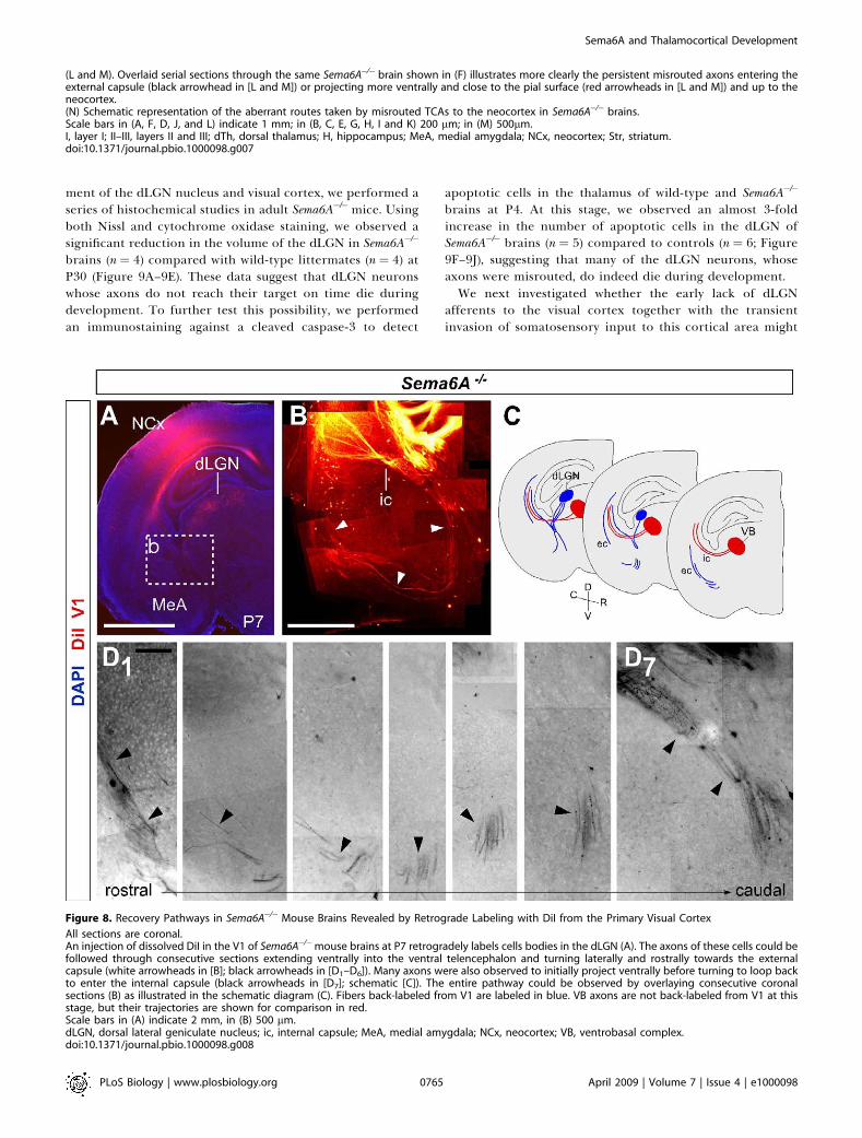
Recovery Pathways for Misrouted TCAsTo investigate whether the misrouted TCAs from the dLGN
persist in the adult mouse, we performed PLAP stainingstudies to reveal the pathway of Sema6A-positive axons inboth Sema6Aþ/� (n¼ 4) and Sema6A�/� (n¼ 4) brains. Sema6A isexpressed in oligodendrocytes in adults, and this staining thuslabels all myelinated fibers. Surprisingly, in Sema6A�/� brainsat P60, a misrouted bundle of axons was still observed at theventral-most region of the telencephalon (Figure 7F, 7L, and7M) in a similar location to the misrouted TCAs shown atearlier developmental stages. Similar ectopic bundles werenever observed in any heterozygous adult brains (Figure 7A).Interestingly, we could follow some PLAP-labeled axons up
to the level of the visual cortex in Sema6A�/� brains (Figure7H–7M). Misrouted labeled axons appear to follow one of twoalternate routes: (1) they either turn laterally through theamygdala and join the external capsule, or (2) they continueto project ventrally and extend along the superficial marginof the telencephalon (Figure 7L–7N). In some cases, abnormalbundles could be followed either through the marginal zoneor the external capsule up to the cortex (Figure 7L). At morecaudal regions, misrouted axons can be seen close to the pialsurface of the cortex, including the visual cortex, of Sema6A�/�
brains (Figure 7K).To confirm the recovery pathways observed in PLAP-
stained adult Sema6A�/� brains, a solution of DiI was injectedinto the primary visual cortex of wild-type or Sema6A�/�
brains at P7. These injections back-labeled cell bodies in thedLGN specifically of both wild-type (n¼ 4; unpublished data)and Sema6A�/� brains (n¼ 3; Figure 8A). In wild-type animals,the axons of these back-labeled cells could be followed as theyprojected through the internal capsule to the cortex(unpublished data). In contrast, in Sema6A�/� brains, manylabeled axons were observed projecting ventrally androstrally in the ventral telencephalon, turning laterally toreach the external capsule at more rostral levels (Figure 8B–8D). Additionally, many axons were also seen to project in amore canonical fashion through the internal capsule, withsome axons initially projecting ventrally before turningdorsally again to loop back and enter the internal capsule(Figure 8D7), suggesting that many initially misrouted dLGNaxons may extend a branch at later stages and extend throughthe internal capsule to reach the cortex.
Reduction in the Size of the dLGN and Visual Cortex inAdult Sema6A�/� MiceTo investigate whether the massive misrouting of TCAs
during early embryonic stages has an impact on the develop-
PLoS Biology | www.plosbiology.org April 2009 | Volume 7 | Issue 4 | e10000980762
Sema6A and Thalamocortical Development

Figure 6. Postnatal Recovery of Visual Thalamocortical Connectivity in Sema6A�/� Mice
All sections are coronal.(A and B) In wild-type brains, DiI and DiA crystal placement in occipital and parietal cortex, respectively, back-labeled cells in the dorsal lateral geniculatenucleus (dLGN; filled arrowhead) and the ventrobasal complex (VB) (open arrowhead) at P4. Dashed box indicates the area in (A) that is magnified in (B).(C and D) In contrast to what happened at earlier stages, in Sema6A�/� brains at P4, DiI crystal placement in the visual cortex back-labeled cellspredominantly in the dLGN (filled arrowhead). DiA crystal placement in the somatosensory cortex back-labeled cells in the VB (open arrowhead).Dashed box indicates the area in (C) that is magnified in (D).(E, F, H, and I) Red and green cholera toxin (CT) was injected into the visual (V1, open arrowhead in [H]) and somatosensory (S1, open arrowhead in [E])cortex, respectively, of wild-type and Sema6A�/� adult mice. In wild-type and Sema6A�/�mice, red CT back-labeled cells and axon terminals in the dLGN(filled arrowheads in [F and I]), and green CT back-labeled cells and axon terminal in the VB (open arrowheads in [F and I]).(G and J) Schematic diagrams showing the location of cell somata containing either red or green retrobeads, from injections in the visual (red) andsomatosensory (green) cortices of adult wild-type and Sema6A�/�brains. In wild-type and Sema6A�/�brains, red retrobeads were found in the dLGN andnever the VB in the dorsal thalamus. Green retrobeads were only ever observed in the VB.Scale bars in (A and C) indicate 1 mm; in (B and D) 500 lm; in (E and H) 1 mm; and in (F and I) 300 lm.MeA, medial amygdala; NCx, neocortex.doi:10.1371/journal.pbio.1000098.g006
PLoS Biology | www.plosbiology.org April 2009 | Volume 7 | Issue 4 | e10000980763
Sema6A and Thalamocortical Development

Figure 7. PLAP Staining Reveals Potential Recovery Pathways of Visual TCAs in Sema6A�/� Adult Brains
All sections are coronal.(A and F) Low-power images of coronal sections of adult Sema6Aþ/� (A) and Sema6A�/� (F) brains, stained for PLAP. Misrouted TCAs are clearly visible inthe ventral telencephalon of Sema6A�/� brains (arrowheads in [F]).(B, C, G–I) High-power view of these aberrant axon tracts (G), some of which project towards and into the external capsule, whereas others take a moreventral position and can be seen extending dorsally, close to the pial surface, up to the neocortex ([F, H, and I] red arrowheads). These axon tracts arenever seen in wild-type or Sema6Aþ/� brains (B and C).(D, E, J, and K) Caudal coronal sections, showing the primary visual cortex (V1) of adult Sema6Aþ/� (D) and Sema6A�/� (J) brains. Note the presence ofmisrouted axons close to the pial surface of V1 in Sema6A�/� brains (red arrowheads in [K]). These axon tracts are never seen in wild-type or Sema6Aþ/�
brains (E).
PLoS Biology | www.plosbiology.org April 2009 | Volume 7 | Issue 4 | e10000980764
Sema6A and Thalamocortical Development
ment of the dLGN nucleus and visual cortex, we performed aseries of histochemical studies in adult Sema6A�/�mice. Usingboth Nissl and cytochrome oxidase staining, we observed asignificant reduction in the volume of the dLGN in Sema6A�/�
brains (n ¼ 4) compared with wild-type littermates (n ¼ 4) atP30 (Figure 9A–9E). These data suggest that dLGN neuronswhose axons do not reach their target on time die duringdevelopment. To further test this possibility, we performedan immunostaining against a cleaved caspase-3 to detect
apoptotic cells in the thalamus of wild-type and Sema6A�/�
brains at P4. At this stage, we observed an almost 3-foldincrease in the number of apoptotic cells in the dLGN ofSema6A�/� brains (n ¼ 5) compared to controls (n ¼ 6; Figure9F–9J), suggesting that many of the dLGN neurons, whoseaxons were misrouted, do indeed die during development.We next investigated whether the early lack of dLGN
afferents to the visual cortex together with the transientinvasion of somatosensory input to this cortical area might
(L and M). Overlaid serial sections through the same Sema6A�/� brain shown in (F) illustrates more clearly the persistent misrouted axons entering theexternal capsule (black arrowhead in [L and M]) or projecting more ventrally and close to the pial surface (red arrowheads in [L and M]) and up to theneocortex.(N) Schematic representation of the aberrant routes taken by misrouted TCAs to the neocortex in Sema6A�/� brains.Scale bars in (A, F, D, J, and L) indicate 1 mm; in (B, C, E, G, H, I and K) 200 lm; in (M) 500lm.I, layer I; II–III, layers II and III; dTh, dorsal thalamus; H, hippocampus; MeA, medial amygdala; NCx, neocortex; Str, striatum.doi:10.1371/journal.pbio.1000098.g007
Figure 8. Recovery Pathways in Sema6A�/� Mouse Brains Revealed by Retrograde Labeling with DiI from the Primary Visual Cortex
All sections are coronal.An injection of dissolved DiI in the V1 of Sema6A�/�mouse brains at P7 retrogradely labels cells bodies in the dLGN (A). The axons of these cells could befollowed through consecutive sections extending ventrally into the ventral telencephalon and turning laterally and rostrally towards the externalcapsule (white arrowheads in [B]; black arrowheads in [D1–D6]). Many axons were also observed to initially project ventrally before turning to loop backto enter the internal capsule (black arrowheads in [D7]; schematic [C]). The entire pathway could be observed by overlaying consecutive coronalsections (B) as illustrated in the schematic diagram (C). Fibers back-labeled from V1 are labeled in blue. VB axons are not back-labeled from V1 at thisstage, but their trajectories are shown for comparison in red.Scale bars in (A) indicate 2 mm, in (B) 500 lm.dLGN, dorsal lateral geniculate nucleus; ic, internal capsule; MeA, medial amygdala; NCx, neocortex; VB, ventrobasal complex.doi:10.1371/journal.pbio.1000098.g008
PLoS Biology | www.plosbiology.org April 2009 | Volume 7 | Issue 4 | e10000980765
Sema6A and Thalamocortical Development

Figure 9. Effects of Loss of Sema6A on the Structure and Extension of Both dLGN and the Visual Cortex
(A–E) Coronal sections of wild-type (A and B) and Sema6A�/� (D and E) adult mouse brains stained with Nissl (A and D) and cytochrome oxidase (B andE). Note the large reduction in size and volume of the dorsal lateral geniculate nucleus (dLGN) in Sema6A�/� brains (C).(F–J) Increased apoptosis in the dLGN in Sema6A�/� brains at P4 detected by caspase 3 antibody (filled arrowheads in [I and J]).(H) Graphical representation of the increased levels of apoptosis in the dLGN of Sema6A�/� compared to wild-type mice.(K and L) Tangential sections of wild-type (K) and Sema6A�/� (L) mouse brains stained for serotonin immunohistochemistry to reveal the cortical sensorydomains at P7. Rostrocaudal and mediolateral directions are indicated by the cross-bars. Note that the size of the primary visual cortex (V1) inSema6A�/� brains is reduced compared to that of wild-type brains.
PLoS Biology | www.plosbiology.org April 2009 | Volume 7 | Issue 4 | e10000980766
Sema6A and Thalamocortical Development
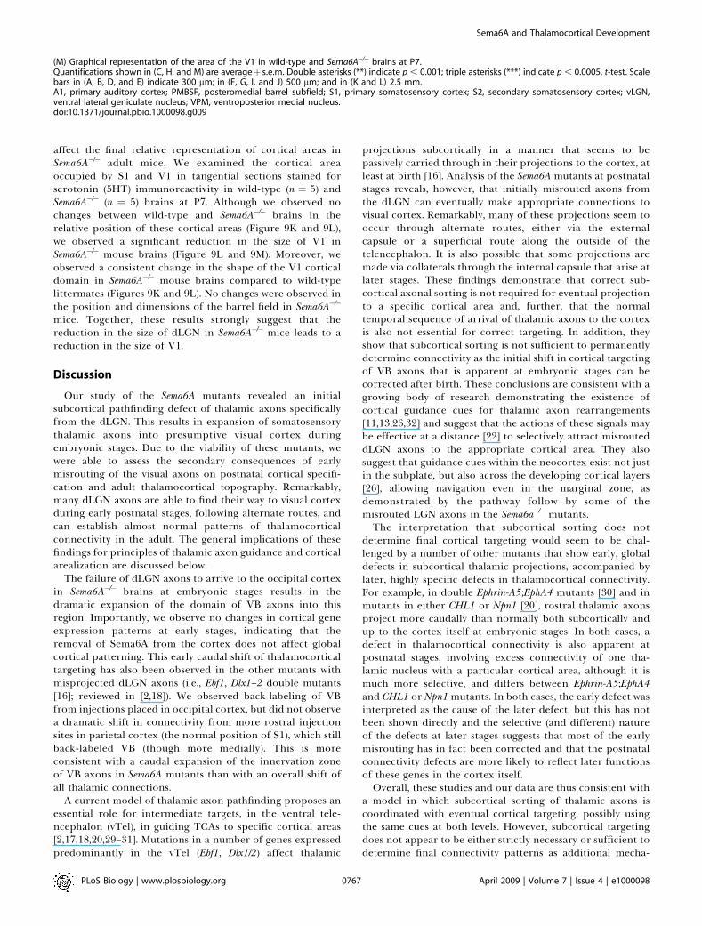
affect the final relative representation of cortical areas inSema6A�/� adult mice. We examined the cortical areaoccupied by S1 and V1 in tangential sections stained forserotonin (5HT) immunoreactivity in wild-type (n ¼ 5) andSema6A�/� (n ¼ 5) brains at P7. Although we observed nochanges between wild-type and Sema6A�/� brains in therelative position of these cortical areas (Figure 9K and 9L),we observed a significant reduction in the size of V1 inSema6A�/� mouse brains (Figure 9L and 9M). Moreover, weobserved a consistent change in the shape of the V1 corticaldomain in Sema6A�/� mouse brains compared to wild-typelittermates (Figures 9K and 9L). No changes were observed inthe position and dimensions of the barrel field in Sema6A�/�
mice. Together, these results strongly suggest that thereduction in the size of dLGN in Sema6A�/� mice leads to areduction in the size of V1.
Discussion
Our study of the Sema6A mutants revealed an initialsubcortical pathfinding defect of thalamic axons specificallyfrom the dLGN. This results in expansion of somatosensorythalamic axons into presumptive visual cortex duringembryonic stages. Due to the viability of these mutants, wewere able to assess the secondary consequences of earlymisrouting of the visual axons on postnatal cortical specifi-cation and adult thalamocortical topography. Remarkably,many dLGN axons are able to find their way to visual cortexduring early postnatal stages, following alternate routes, andcan establish almost normal patterns of thalamocorticalconnectivity in the adult. The general implications of thesefindings for principles of thalamic axon guidance and corticalarealization are discussed below.
The failure of dLGN axons to arrive to the occipital cortexin Sema6A�/� brains at embryonic stages results in thedramatic expansion of the domain of VB axons into thisregion. Importantly, we observe no changes in cortical geneexpression patterns at early stages, indicating that theremoval of Sema6A from the cortex does not affect globalcortical patterning. This early caudal shift of thalamocorticaltargeting has also been observed in the other mutants withmisprojected dLGN axons (i.e., Ebf1, Dlx1–2 double mutants[16]; reviewed in [2,18]). We observed back-labeling of VBfrom injections placed in occipital cortex, but did not observea dramatic shift in connectivity from more rostral injectionsites in parietal cortex (the normal position of S1), which stillback-labeled VB (though more medially). This is moreconsistent with a caudal expansion of the innervation zoneof VB axons in Sema6A mutants than with an overall shift ofall thalamic connections.
A current model of thalamic axon pathfinding proposes anessential role for intermediate targets, in the ventral tele-ncephalon (vTel), in guiding TCAs to specific cortical areas[2,17,18,20,29–31]. Mutations in a number of genes expressedpredominantly in the vTel (Ebf1, Dlx1/2) affect thalamic
projections subcortically in a manner that seems to bepassively carried through in their projections to the cortex, atleast at birth [16]. Analysis of the Sema6A mutants at postnatalstages reveals, however, that initially misrouted axons fromthe dLGN can eventually make appropriate connections tovisual cortex. Remarkably, many of these projections seem tooccur through alternate routes, either via the externalcapsule or a superficial route along the outside of thetelencephalon. It is also possible that some projections aremade via collaterals through the internal capsule that arise atlater stages. These findings demonstrate that correct sub-cortical axonal sorting is not required for eventual projectionto a specific cortical area and, further, that the normaltemporal sequence of arrival of thalamic axons to the cortexis also not essential for correct targeting. In addition, theyshow that subcortical sorting is not sufficient to permanentlydetermine connectivity as the initial shift in cortical targetingof VB axons that is apparent at embryonic stages can becorrected after birth. These conclusions are consistent with agrowing body of research demonstrating the existence ofcortical guidance cues for thalamic axon rearrangements[11,13,26,32] and suggest that the actions of these signals maybe effective at a distance [22] to selectively attract misrouteddLGN axons to the appropriate cortical area. They alsosuggest that guidance cues within the neocortex exist not justin the subplate, but also across the developing cortical layers[26], allowing navigation even in the marginal zone, asdemonstrated by the pathway follow by some of themisrouted LGN axons in the Sema6a�/� mutants.The interpretation that subcortical sorting does not
determine final cortical targeting would seem to be chal-lenged by a number of other mutants that show early, globaldefects in subcortical thalamic projections, accompanied bylater, highly specific defects in thalamocortical connectivity.For example, in double Ephrin-A5;EphA4 mutants [30] and inmutants in either CHL1 or Npn1 [20], rostral thalamic axonsproject more caudally than normally both subcortically andup to the cortex itself at embryonic stages. In both cases, adefect in thalamocortical connectivity is also apparent atpostnatal stages, involving excess connectivity of one tha-lamic nucleus with a particular cortical area, although it ismuch more selective, and differs between Ephrin-A5;EphA4and CHL1 or Npn1mutants. In both cases, the early defect wasinterpreted as the cause of the later defect, but this has notbeen shown directly and the selective (and different) natureof the defects at later stages suggests that most of the earlymisrouting has in fact been corrected and that the postnatalconnectivity defects are more likely to reflect later functionsof these genes in the cortex itself.Overall, these studies and our data are thus consistent with
a model in which subcortical sorting of thalamic axons iscoordinated with eventual cortical targeting, possibly usingthe same cues at both levels. However, subcortical targetingdoes not appear to be either strictly necessary or sufficient todetermine final connectivity patterns as additional mecha-
(M) Graphical representation of the area of the V1 in wild-type and Sema6A�/� brains at P7.Quantifications shown in (C, H, and M) are averageþ s.e.m. Double asterisks (**) indicate p , 0.001; triple asterisks (***) indicate p , 0.0005, t-test. Scalebars in (A, B, D, and E) indicate 300 lm; in (F, G, I, and J) 500 lm; and in (K and L) 2.5 mm.A1, primary auditory cortex; PMBSF, posteromedial barrel subfield; S1, primary somatosensory cortex; S2, secondary somatosensory cortex; vLGN,ventral lateral geniculate nucleus; VPM, ventroposterior medial nucleus.doi:10.1371/journal.pbio.1000098.g009
PLoS Biology | www.plosbiology.org April 2009 | Volume 7 | Issue 4 | e10000980767
Sema6A and Thalamocortical Development
nisms exist to restore thalamocortical connectivity to aspecific cortical area when alterations during embryonicdevelopment occur.
The recovery of the dLGN projection to visual cortex, inspite of previous occupation of this territory by VB axonssuggests that dLGN axons have an advantage in theinnervation of that particular cortical area. This must be inaddition to selective axon guidance to this region as arrival ofVB axons to this area is clearly not sufficient to enable themto make permanent connections, at least when faced withcompetition from later-arriving dLGN axons. A model toexplain this would be that dLGN axons and presumptivevisual cortex express some matching label(s) that confer thisadvantage. One candidate for such a cue is the neurotrophinNT-3, which is specifically required for dLGN axons to invadethe cortical plate in V1 [33]. NT-3 has been shown to be moststrongly expressed in presumptive visual cortex (V1) fromaround P0 [34], while its receptor TrkC, is selectivelyexpressed by neurons in the dLGN. If such a matched cue isessential then VB axons that at early stages project into thesubplate of occipital cortex may not be able to invade thecortical plate, allowing later-arriving dLGN axons to do so.Indeed, if the function of NT-3 in this context sharessimilarities with trophic signaling [35] then dLGN axonsmight actively secrete factors that promote withdrawal of VBaxons. Axon–axon interactions mediated by surface receptorsand cell adhesion molecules [36] might also actively mediatesegregation of visual and somatosensory axons [37].
Activity-dependent mechanisms mediating the competitiveadvantage of dLGN axons for presumptive visual cortex mustalso be considered, especially as the process takes placeduring the first few postnatal days, by which time thalamicaxons have normally entered into the cortex and formed fullyfunctional synapses [32,38,39]. A number of studies haveexamined the potential role of electrical activity in arealtargeting of thalamic axons. Intracranial infusion of thesodium channel blocker tetrodotoxin (TTX) caused dLGNaxons to inappropriately innervate the subplate of corticalareas that they would normally bypass [40]. This could betaken as an instructive role for patterned activity inestablishing areal connectivity but could alternatively beexplained by an earlier effect of TTX on biochemicalsignaling pathways downstream of guidance receptors [41],or by feedback onto the expression levels of guidancemolecules [42]. This interpretation is more consistent withthe known specificity of thalamic axon targeting from theearliest stages [10,11,43] and the lack of effects in arealtargeting observed in embryonic SNAP-25 mutants [39,44].
Finally, although our study demonstrates spectacularplasticity of thalamocortical connectivity during early post-natal life, there are some changes in the cortical architecturethat persist into adulthood. The reduction in size and changein shape of V1 in Sema6A mutants, which are far more subtlethan those observed in enucleation experiments [1,5,45,46],suggest that they may be an interesting model to study someless well-characterized processes, including the separation ofthe termination zones of primary thalamic axons intodiscrete areas, the innervation of intervening areas by axonsfrom secondary nuclei, the formation of distinct borders andthe hierarchical dependence of secondary and higher-orderareas on correct specification of primary areas (reviewed in:[47,48]).
Materials and Methods
Animals. All animal procedures were performed to relevantnational and international licensing agreements and in accordancewith institutional guidelines. Sema6A mutants were identified in agene trap screen, as described previously [49]. Insertion of the genetrap vector pGT1PFS into intron 17 results in a fusion of upstreamexons of Sema6A with TM-b-galactosidase-neomycin phosphotrans-ferase. This fusion protein is sequestered intracellularly [28]. PLAP iscotranscribed but translated independently from an internal ribo-some entry site. No wild-type transcripts are produced from thisallele [28].
Dye tracing studies. Brains from E16.5 (n¼ 18), P0 (n¼ 45), P4 (n¼8), P7 (n ¼ 6), and P30 (n ¼ 26) were used in the study. To labelthalamic and corticofugal fibers, single crystals of 1,19-dioctadecyl-3,3,39,39-tetramethylindocarbocyanine perchlorate (DiI) and 4-(4-(dihexadecylamino) styryl)-N-methylpyridinium iodide (DiA) (Molec-ular Probes) were placed with a stainless steel electrode into the visualand somatosensory dorsal thalamic nuclei or the visual andsomatosensory cerebral cortex of both hemispheres of each brain.After injections, brains were kept in 2% paraformaldehyde forbetween 3 wk and 2 mo at room temperature in the dark. Back-labeling of dLGN neurons and their axons in P7 animals wasperformed under hypothermia-induced anesthesia. A small incisionwas made in the scalp to reveal the skull, and a fine needle was used topierce the skull above the primary visual cortex. A Hamilton syringewas used to inject 0.5 ll of a 10% solution of DiI in absolute ethanol,into the primary visual cortex. The scalp was bonded with tissueadhesive (Dermabond) and animals were allowed to survive for 24–48h to allow for adequate retrograde labeling before being sacrificed.Dissected brains were postfixed for 24 h in 4% PFA at 4 8C. Brainswere washed in PBS (0.1 M, pH 7.4), embedded into 4% agarose(Sigma), and cut at 100 lm with a Vibroslicer (Leica, VT1000S).Sections were counterstained with 2.5 lg/ml of bis-benzimide (Sigma)or with 0.5 lg/ml DAPI (49-6-diamidino-2-phenylindole), mounted inPBS/glycerol or AquaPolymount (Polysciences) onto slides, andanalyzed using an epifluorescence microscope (Leica, DMR, or Zeiss)and a laser scanning confocal microscope (Leica, DMRE).
Histochemistry and immunohistochemistry. Mice were perfusedwith 4% paraformaldehyde or a mixture of 1% paraformaldehyde/1.5% glutaraldehyde (for the cytochrome oxidase staining) in PBS.Brains were removed, postfixed in the same fixative overnight at 4 8Cand embedded in 4% agarose. Serial 50- or 100-lm sections were cuton a vibratome (Leica; VT1000S) and processed for PLAP staining aspreviously described [28]. Alternatively, following perfusion, brainswere postfixed in the same fixative for 3 h and cryoprotected with30% sucrose in PBS. Serial 40-lm sections were cut on a freezingmicrotome and processed for Nissl staining (0.5% cresyl violetsolution; Sema6Aþ/�: P0, n¼ 2; P30, n¼ 6, and Sema6A�/�: P0, n¼ 2; P30,n ¼ 6). For cytochrome oxidase staining, cortical hemispheres weredissected from adult mice (Sema6Aþ/�, n ¼ 9; Sema6A�/�, n ¼ 9),postfixed between glass slides and cryoprotected before sectioningand processing. For immunohistochemistry, dissected brains werepostfixed in 4% paraformaldehyde for 24 h, washed in PBS,embedded in 4% agarose, and sectioned (40–60 lm) on a vibratome.P 4 Sema6Aþ/þ (n¼ 7) and Sema6A�/� (n¼ 5) mouse brains were treatedfor immunofluorescence with rabbit antibody to cleaved caspase-3(1:200; Cell Signaling Technologies). Similarly, immunofluorescencewith mouse antibody to neurofilament (1:100; DHSB) was detected onsections from E16.5 Sema6Aþ/þ (n ¼ 2) and Sema6A�/� (n ¼ 2) mousebrains. Tangential sections were cut from flattened cortical hemi-spheres of P7 Sema6Aþ/þ (n ¼ 5) and Sema6A knockout (KO; n ¼ 10)mouse brains and incubated with antibody to serotonin (1:50,000;Immunostar), which was then detected with biotinylated secondaryantibodies using the Elite ABC kit (Vector). Results were documentedusing a digital camera (Leica DC500; Canon Powershot S40) or anepifluorescence microscope (Zeiss) and digital camera (Olympus), andthe images compiled with Adobe Photoshop 8.0 or Adobe PhotoshopCS software.
Retrograde tracing with cholera toxin and fluorescent latexmicrospheres. Green and red fluorescent latex microspheres (Luma-flor) were used to labeled axonal projection from somatosensory andvisual cortical areas, respectively, to the corresponding thalamicnuclei in Sema6Aþ/þ (n ¼ 4) and Sema6A�/� (n ¼ 4) mice. Animals wereanesthetized with 2.7 mg/kg Hypnovel (Roche), Hypnorm (Janssen),and distilled H2O mixture (1:1:2 volume ratio), which was deliveredintraperitoneally, and placed in a stereotaxis frame. After the skinwas disinfected and incised, a microdrill was used to perform acraniotomy. Glass micropipettes (Clark Electromedical Instruments)and a binocular stereo-microscope (Zeiss) were used to inject a single
PLoS Biology | www.plosbiology.org April 2009 | Volume 7 | Issue 4 | e10000980768
Sema6A and Thalamocortical Development

injection of 0.3–1.0 ll of CT or microspheres into S1 or V1. Animalswere allowed to survive for 24 to 48 h to permit adequate retrogradetransport of the CT or microspheres to thalamic cell somata.
In situ hybridization. In situ hybridization was performed on 50-lm vibratome sections of E14.5 Sema6A (wild-type [WT]: n ¼ 2,Sema6Aþ/� [HT]: n¼4 and KO: n¼4), P0 Sema6A (HT: n¼9, KO: n¼8),and P7 Sema6A (HT: n ¼ 4, KO: n ¼ 4) mouse brains, as previouslydescribed [50]. The following digoxigenin-labeled RNA probes wereused: Sema6a (a gift from W. Snider); EphA7, EphrinA5, Cadherin6, andRZRb (kindly provided by J. Rubenstein, with permission from theoriginal researchers); Cadherin8 (241–1,481 of mouse Cad8; GenBankaccession number X95600; obtained by reverse transcription [RT]-PCR).
Quantification. The number of cells in the dLGN and the VB back-labeled from the occipital cortex were manually counted inconsecutive 100-lm sections of E16.5 Sema6Aþ/� (n ¼ 30 sections, 4animals) and Sema6A�/� (n¼ 41 sections, 6 animals), P0 Sema6Aþ/� (n¼11 sections, 2 animals) and Sema6A�/� (n¼ 10 sections, 2 animals), andP4 wild-type (n¼ 9 sections, 2 animals) and Sema6A�/� (n¼12 sections,2 animals) brains. The numbers of cells back-labeled to either thedLGN or VB for animals of a given age were compared using theWilcoxon two-sample test and found to be significant at 99.9%confidence limits at E16.5 and P0. As the absolute number of cellsback-labeled is dependent on the size of dye crystal used, we alsoanalyzed the proportion of labeled cells in either the dLGN or VB ofa given section. The proportional values were Arcsine transformedfor statistical analysis by Wilcoxon two-sample tests. The area of thedLGN and vLGN thalamic nuclei was measured in 40-lm cytochromeoxidase serial sections from Sema6Aþ/� (n ¼ 5) and Sema6A�/� (n ¼ 4)brains using SigmaScan Pro software (SigmaScan). The volume of thedLGN and vLGN was calculated using the Cavalieri method. Therelative area of V1 was measured in tangential sections of P7 ofSema6Aþ/� (n¼ 5) and Sema6A�/� (n¼ 10) brains, stained for serotoninimmunohistochemistry, using Cell A software (Soft Image System).
Supporting Information
Figure S1. PLAP Staining Reveals a Large Bundle of Misrouted TCAsat Caudal Levels in Sema6A�/� Brains at E16.5
(A–D) Rostral–caudal consecutive sections of PLAP-stained Sema6A�/�
mouse brain at E16.5 reveals a large bundle of misrouted thalamo-cortical axons at more caudal levels. Whereas many labeled TCAsproject normally through the internal capsule towards the cortex(black arrowheads in [A and B]), misrouted axons can be seen toproject deep into the ventral telencephalon and project along itsmost superficial aspect (red arrowheads). Scale bars indicate 500 lm.
Found at doi:10.1371/journal.pbio.1000098.sg001 (1.44 MB TIF).
Figure S2. DiI and DiA Crystal Placements in the Ventral Tele-ncephalon and Internal Capsule Confirm That dLGN Axons Do NotEnter the Internal Capsule in Sema6A�/� Brains at E17.5
Sections are at 458 to the coronal plane to encompass the internalcapsule and dorsal thalamus.
(A and B) A DiA crystal placed in the internal capsule of wild-typebrains at E17.5 broadly labels cells throughout the dorsal thalamus.(C) In these same animals, a DiI crystal in the ventral telencephalonfails to label any cells in the dorsal thalamus, indicating that all TCAsproject through the internal capsule in these animals.(D–F) In Sema6A�/� brains at the same age (D and E), a DiA crystal inthe internal capsule labels cell bodies in the VB but specifically not inthe dLGN, whereas a DiI crystal in the ventral telencephalon doeslabel dLGN neurons specifically, indicating that dLGN axons in theseanimals do not project at all to the internal capsule at this age (F).Scale bars in (A–D) indicate 500 lm.
Found at doi:10.1371/journal.pbio.1000098.sg002 (1.90 MB TIF).
Figure S3. Cortical Area Markers Show No Defect in the RelativePosition of Principal Cortical Areas in Sema6A�/� Brains at P0
(A and D) In situ hybridization with DIG-labeled probes for EphA7 (Aand C) and EphrinA5 (B and D) on sagittal sections of wild-type (A andB) and Sema6A�/� (C and D) brains. EphA7 is expressed in the rostraland occipital neocortex (NCx), but is absent from the putativesomatosensory cortex. In contrast, EphrinA5 is expressed is highlyexpressed in the somatosensory cortex. dTh, dorsal thalamus. Scalebars in (A–D) indicate 500 lm.
Found at doi:10.1371/journal.pbio.1000098.sg003 (1.17 MB TIF).
Acknowledgments
We thank Jackie Dolan and Mary Phillips for excellent technicalassistance. The monoclonal anti-NF165kD antibody (2H3) wasdeveloped by T. M. Jessell and J. Dodd and obtained through theDevelopmental Studies Hybridoma Bank (University Iowa). We aregrateful to members of the Lopez-Bendito, Molnar, Chedotal, andMitchell labs for stimulating discussions.
Author contributions. GEL, GL-B, AER, ZM, and KJM conceivedand designed the experiments. GEL, GL-B, AER, NG, MCP, AC, andZM performed the experiments. GEL, GL-B, AER, NG, MCP, ZM, andKJM analyzed the data. AC contributed reagents/materials/analysistools. GEL, GL-B, ZM, and KJM wrote the paper.
Funding. This work was supported by grants from ScienceFoundation Ireland (01/F1/B006) to KJM, Spanish Ministry of Scienceand Innovation (grants BFU2006–00408 and CONSOLIDER-INGEN-IO 2010 CSD2007–00023) to GLB, and a Medical Research Council(MRC) Career Establishment Award (G0300200) and Project Grant(G0700377) to ZM. AC is supported by the Fondation pour laRecherche Medicale (programme equipe FRM). GL has beensupported by a Government of Ireland Scholarship, awarded by theIrish Research Council of Science, Engineering and Technology. ARwas supported by a Health Research Board Postdoctoral Fellowship.The funders had no role in study design, data collection and analysis,decision to publish, or preparation of the manuscript.
Competing interests. The authors have declared that no competinginterests exist.
References1. Rakic P (1998) Images in neuroscience. Brain development, VI: radial
migration and cortical evolution. Am J Psychiatry 155: 1150–1151.2. Lopez-Bendito G, Molnar Z (2003) Thalamocortical development: how are
we going to get there? Nat Rev Neurosci 4: 276–289.3. O’Leary DD, Chou SJ, Sahara S (2007) Area patterning of the mammalian
cortex. Neuron 56: 252–269.4. Sur M, Rubenstein JL (2005) Patterning and plasticity of the cerebral
cortex. Science 310: 805–810.5. Krubitzer L, Kahn DM (2003) Nature versus nurture revisited: an old idea
with a new twist. Prog Neurobiol 70: 33–52.6. Molnar Z, Adams R, Goffinet AM, Blakemore C (1998) The role of the first
postmitotic cortical cells in the development of thalamocortical innerva-tion in the reeler mouse. J Neurosci 18: 5746–5765.
7. Caviness VS Jr, Frost DO (1980) Tangential organization of thalamicprojections to the neocortex in the mouse. J Comp Neurol 194: 335–367.
8. Catalano SM, Robertson RT, Killackey HP (1996) Individual axonmorphology and thalamocortical topography in developing rat somato-sensory cortex. J Comp Neurol 367: 36–53.
9. Molnar Z, Blakemore C (1995) Guidance of thalamocortical innervation.Ciba Found Symp 193: 127–149.
10. Molnar Z, Adams R, Blakemore C (1998) Mechanisms underlying the earlyestablishment of thalamocortical connections in the rat. J Neurosci 18:5723–5745.
11. Krug K, Smith AL, Thompson ID (1998) The development of topography inthe hamster geniculo-cortical projection. J Neurosci 18: 5766–5776.
12. Naegele JR, Jhaveri S, Schneider GE (1988) Sharpening of topographicalprojections and maturation of geniculocortical axon arbors in the hamster.J Comp Neurol 277: 593–607.
13. Rebsam A, Seif I, Gaspar P (2002) Refinement of thalamocortical arborsand emergence of barrel domains in the primary somatosensory cortex: astudy of normal and monoamine oxidase a knock-out mice. J Neurosci 22:8541–8552.
14. Paysan J, Kossel A, Bolz J, Fritschy JM (1997) Area-specific regulation ofgamma-aminobutyric acid type A receptor subtypes by thalamic afferentsin developing rat neocortex. Proc Natl Acad Sci U S A 94: 6995–7000.
15. Lukaszewicz A, Cortay V, Giroud P, Berland M, Smart I, et al. (2006) Theconcerted modulation of proliferation and migration contributes to thespecification of the cytoarchitecture and dimensions of cortical areas.Cereb Cortex 16 Suppl 1: i26–34.
16. Garel S, Yun K, Grosschedl R, Rubenstein JL (2002) The early topography ofthalamocortical projections is shifted in Ebf1 and Dlx1/2 mutant mice.Development 129: 5621–5634.
17. Seibt J, Schuurmans C, Gradwhol G, Dehay C, Vanderhaeghen P, et al.(2003) Neurogenin2 specifies the connectivity of thalamic neurons bycontrolling axon responsiveness to intermediate target cues. Neuron 39:439–452.
18. Vanderhaeghen P, Polleux F (2004) Developmental mechanisms patterning
PLoS Biology | www.plosbiology.org April 2009 | Volume 7 | Issue 4 | e10000980769
Sema6A and Thalamocortical Development

thalamocortical projections: intrinsic, extrinsic and in between. TrendsNeurosci 27: 384–391.
19. Dufour A, Egea J, Kullander K, Klein R, Vanderhaeghen P (2006) Geneticanalysis of EphA-dependent signaling mechanisms controlling topographicmapping in vivo. Development 133: 4415–4420.
20. Wright AG, Demyanenko GP, Powell A, Schachner M, Enriquez-Barreto L,et al. (2007) Close homolog of L1 and neuropilin 1 mediate guidance ofthalamocortical axons at the ventral telencephalon. J Neurosci 27: 13667–13679.
21. Mallamaci A, Muzio L, Chan CH, Parnavelas J, Boncinelli E (2000) Areaidentity shifts in the early cerebral cortex of Emx2-/- mutant mice. NatNeurosci 3: 679–686.
22. Frappe I, Gaillard A, Roger M (2001) Attraction exerted in vivo by grafts ofembryonic neocortex on developing thalamic axons. Exp Neurol 169: 264–275.
23. Fukuchi-Shimogori T, Grove EA (2001) Neocortex patterning by thesecreted signaling molecule FGF8. Science 294: 1071–1074.
24. Bishop KM, Rubenstein JL, O’Leary DD (2002) Distinct actions of Emx1,Emx2, and Pax6 in regulating the specification of areas in the developingneocortex. J Neurosci 22: 7627–7638.
25. Fukuchi-Shimogori T, Grove EA (2003) Emx2 patterns the neocortex byregulating FGF positional signaling. Nat Neurosci 6: 825–831.
26. Shimogori T, Grove EA (2005) Fibroblast growth factor 8 regulatesneocortical guidance of area-specific thalamic innervation. J Neurosci 25:6550–6560.
27. Armentano M, Chou SJ, Tomassy GS, Leingartner A, O’Leary DD, et al.(2007) COUP-TFI regulates the balance of cortical patterning betweenfrontal/motor and sensory areas. Nat Neurosci 10: 1277–1286.
28. Leighton PA, Mitchell KJ, Goodrich LV, Lu X, Pinson K, et al. (2001)Defining brain wiring patterns and mechanisms through gene trapping inmice. Nature 410: 174–179.
29. Metin C, Godement P (1996) The ganglionic eminence may be anintermediate target for corticofugal and thalamocortical axons. J Neurosci16: 3219–3235.
30. Dufour A, Seibt J, Passante L, Depaepe V, Ciossek T, et al. (2003) Areaspecificity and topography of thalamocortical projections are controlled byephrin/Eph genes. Neuron 39: 453–465.
31. Lopez-Bendito G, Cautinat A, Sanchez JA, Bielle F, Flames N, et al. (2006)Tangential neuronal migration controls axon guidance: a role forneuregulin-1 in thalamocortical axon navigation. Cell 125: 127–142.
32. Higashi S, Hioki K, Kurotani T, Kasim N, Molnar Z (2005) Functionalthalamocortical synapse reorganization from subplate to layer IV duringpostnatal development in the reeler-like mutant rat (shaking rat Kawasaki).J Neurosci 25: 1395–1406.
33. Ma L, Harada T, Harada C, Romero M, Hebert JM, et al. (2002)Neurotrophin-3 is required for appropriate establishment of thalamocort-ical connections. Neuron 36: 623–634.
34. Vigers AJ, Baquet ZC, Jones KR (2000) Expression of neurotrophin-3 in the
mouse forebrain: insights from a targeted LacZ reporter. J Comp Neurol416: 398–415.
35. Deppmann CD, Mihalas S, Sharma N, Lonze BE, Niebur E, et al. (2008) Amodel for neuronal competition during development. Science 320: 369–373.
36. Luo L, Flanagan JG (2007) Development of continuous and discrete neuralmaps. Neuron 56: 284–300.
37. Bolz J, Kossel A, Bagnard D (1995) The specificity of interactions betweenthe cortex and the thalamus. Ciba Found Symp 193: 173–191.
38. Higashi S, Molnar Z, Kurotani T, Toyama K (2002) Prenatal development ofneural excitation in rat thalamocortical projections studied by opticalrecording. Neuroscience 115: 1231–1246.
39. Molnar Z, Higashi S, Lopez-Bendito G (2003) Choreography of earlythalamocortical development. Cereb Cortex 13: 661–669.
40. Catalano SM, Shatz CJ (1998) Activity-dependent cortical target selectionby thalamic axons. Science 281: 559–562.
41. Nicol X, Voyatzis S, Muzerelle A, Narboux-Neme N, Sudhof TC, et al. (2007)cAMP oscillations and retinal activity are permissive for ephrin signalingduring the establishment of the retinotopic map. Nat Neurosci 10: 340–347.
42. Serizawa S, Miyamichi K, Takeuchi H, Yamagishi Y, Suzuki M, et al. (2006) Aneuronal identity code for the odorant receptor-specific and activity-dependent axon sorting. Cell 127: 1057–1069.
43. Agmon A, Yang LT, Jones EG, O’Dowd DK (1995) Topological precision inthe thalamic projection to neonatal mouse barrel cortex. J Neurosci 15:549–561.
44. Molnar Z, Lopez-Bendito G, Small J, Partridge LD, Blakemore C, et al.(2002) Normal development of embryonic thalamocortical connectivity inthe absence of evoked synaptic activity. J Neurosci 22: 10313–10323.
45. Dehay C, Giroud P, Berland M, Killackey H, Kennedy H (1996)Contribution of thalamic input to the specification of cytoarchitectoniccortical fields in the primate: effects of bilateral enucleation in the fetalmonkey on the boundaries, dimensions, and gyrification of striate andextrastriate cortex. J Comp Neurol 367: 70–89.
46. Rakic P, Suner I, Williams RW (1991) A novel cytoarchitectonic areainduced experimentally within the primate visual cortex. Proc Natl AcadSci U S A 88: 2083–2087.
47. Bargary G, Mitchell KJ (2008) Synaesthesia and cortical connectivity.Trends Neurosci 31: 335–342.
48. Guillery RW (2005) Is postnatal neocortical maturation hierarchical?Trends Neurosci 28: 512–517.
49. Mitchell KJ, Pinson KI, Kelly OG, Brennan J, Zupicich J, et al. (2001)Functional analysis of secreted and transmembrane proteins critical tomouse development. Nat Genet 28: 241–249.
50. Dolan J, Walshe K, Alsbury S, Hokamp K, O’Keeffe S, et al. (2007) Theextracellular leucine-rich repeat superfamily; a comparative survey andanalysis of evolutionary relationships and expression patterns. BMCGenomics 8: 320.
PLoS Biology | www.plosbiology.org April 2009 | Volume 7 | Issue 4 | e10000980770
Sema6A and Thalamocortical Development