Broad Surveys of DNA Viral Diversity Obtained through Viral Metagenomics of Mosquitoes

Research Paper
Specific Delivery of Corroles to Cells via Noncovalent Conjugateswith Viral Proteins
Hasmik Agadjanian,1 Jeremy J. Weaver,2 Atif Mahammed,3 Altan Rentsendorj,1 Sam Bass,1 Jihee Kim,4
Ivan J. Dmochowski,5 Ruth Margalit,5 Harry B. Gray,2 Zeev Gross,3 and Lali K. Medina-Kauwe1,6,11
Received July 14, 2005; accepted October 21, 2005
Purpose. Corroles are amphiphilic macrocycles that can bind and transport metal ions, and thus may be
toxic to cells. We predicted that anionic corroles would poorly enter cells due to the negatively charged
cell membrane, but could be ideal tumor-targeted drugs if appropriate carriers enabled delivery into
tumor cells. In this work, we test the hypothesis that recombinant cell penetrating proteins of the
adenovirus (Ad) capsid form noncovalent conjugates with corroles to facilitate target-specific delivery
and cell death.
Methods. Corroles mixed with recombinant proteins were tested for conjugate assembly, cell penetration,
stability, targeted binding, and cell killing in vitro.
Results. Sulfonated corroles entered cells only with carrier proteins, and formed stable complexes with
recombinant Ad capsid proteins. ErbB receptor-targeted conjugates were cytotoxic to ErbB2-positive
but not ErbB2-negative breast cancer cells, whereas molar equivalents of free corrole had no effect on
these cells.
Conclusions. Sulfonated corroles are cytotoxic to ErbB2-positive breast cancer cells when delivered by a
targeted cell penetrating protein. The relatively low dose required to accomplish this compared to
untargeted compounds suggests that corroles may lend themselves to targeted therapy. Importantly, the
amphiphilicity of corroles enables a unique approach to bioconjugate formation whereby the carrier and
drug form a stable complex by noncovalent assembly.
KEY WORDS: capsid; corrole; metal; penton base; targeting.
INTRODUCTION
We report the use of corroles as novel drugs that canreadily bind shuttle proteins noncovalently and induce tar-geted toxicity to certain cells in culture. Corroles are macro-cyclic compounds with structural similarity to porphyrins,and, likewise, can chelate a large variety of metal ions.Whereas porphyrins have been used extensively in cancertherapy (1Y3), corroles have not been explored until recently,when the once-complicated methodology for their synthesisbecame simplified (4,5). The production of amphiphilic,
water-soluble corroles now enables investigations of thesecompounds for application in physiological environments.The unique features of corroles that should lend themselvesto bioconjugate therapy are: tight noncovalent binding toproteins in solution, highly intense fluorescence, and poorcell uptake without a carrier molecule.
A hallmark feature shared by several recently intro-duced corroles is amphiphilicity, enabled by negativelycharged moieties positioned on one side of the macrocycle.These negative charges are likely to prevent nonspecific up-take of corroles into cells without the aid of a carrier. Manyserum molecules could be potential carriers of corrolessystemically, as porphyrins and related compounds areknown to transport into cells by serum proteins (6,7). It hasbeen shown that the sulfonated corroles explored in thisstudy bind rapidly and noncovalently to human serumalbumin (HSA), forming extremely stable complexes that re-main associated during high-performance liquid chromatog-raphy (HPLC) separation (8).
To take advantage of the capacity for such stable inter-actions with proteins, we asked in this study whether corrolescould form noncovalent conjugates with specific receptor-targeted proteins known to penetrate into cells. We alsoasked whether ligand-directed delivery of corroles could in-duce targeted cell death. Here we show that corroles readilyinteract with the recombinant proteins tested in this study,
367 0724-8741/06/0200-0367/0 # 2006 Springer Science + Business Media, Inc.
Pharmaceutical Research, Volume 23, No. 2, February 2006 (# 2006)DOI: 10.1007/s11095-005-9225-1
1 The Gene Therapeutics Research Institute, Cedars-Sinai Medical
Center, 8700 Beverly Blvd, Davis 5092, Los Angeles, California
90048, USA.2 Department of Chemistry, California Institute of Technology,
Pasadena, California 91125, USA.3 Department of Chemistry, Technion Y Israel Institute of Technol-
ogy, Haifa 32000, Israel.4 IntraGene Sciences, Institute for Genetic Medicine, Los Angeles,
California 90033, USA.5 Department of Chemistry, University of Pennsylvania, Philadel-
phia, Pennsylvania 19104, USA.6 To whom correspondence should be addressed. (e-mail: MedinaL@
cshs.org)

but do not transfer to serum albumin after conjugate assem-bly. Importantly, we show that these recombinant proteinsdirect the receptor-targeted cell binding and cell entry ofcorroles, eliciting specific cell death after corrole delivery.
Among the recombinant carrier proteins tested here arederivatives of the Adenovirus serotype 5 (Ad5) capsidpenton base protein: PB, PBK10, and HerPBK10. PB is arecombinant protein encompassing the entire amino acidsequence of the wild-type Ad5 penton base (9,10), and isknown to mediate cell entry of the virus during infection.PBK10 carries the wild-type penton base sequence followedby a carboxy (C)-terminal decalysine (9). Both PB andPBK10 interact with cellular integrins and undergo integrinreceptor-mediated cell entry (9). A heregulin-targeted ver-sion, HerPBK10, specifically binds heregulin, or ErbB,receptors that are amplified on the cell surface of ErbB2+human breast cancer cells, including MDA-MB-435 andMDA-MB-453 cell lines, and undergoes receptor-mediatedcell entry (11Y13). Importantly, these proteins were derivedfrom the penton base because of its capacity for endosomo-lysis and translocation into the cytosol, which shouldfacilitate targeted entry of the corrole into the cell interior.Also relevant to this study is the protein, Her, which en-compasses the receptor binding domain of heregulin-a andcompetitively inhibits HerPBK10 binding to heregulin recep-tors (11).
We have previously demonstrated that HerPBK10delivers plasmid DNA molecules to ErbB2+ cells through amechanism involving ErbB receptor-specific binding andendocytosis, and endosomal escape, likely via the PB domainof HerPBK10 (11). Delivery of plasmid DNA, however, pre-sents certain barriers to potential therapy. For example, thelarge size of the DNA molecule requires that DNA con-densing agents be added to assembled complexes to collapsethe plasmid into a particle (14,15). Such agents are likely tobind DNA very tightly and may, ironically, hamper geneexpression as a result of this interaction (16). Moreover, evenif such agents are able to release DNA inside the cell cyto-plasm, the uncondensed molecule is unable to migratethrough the crowded cytosol toward the nucleus (17). Theattachment of a cytotoxic molecule to HerPBK10, on theother hand, may streamline conjugate design and assembly.Attachment via noncovalent interactions, as tested here,would greatly simplify delivery of therapeutic molecules.
This study presents three areas of novelty: (1) the testingof corroles on a biological system; (2) the noncovalent as-sembly of corroles with recombinant cell targeting proteins;(3) the demonstration that corroles can be targeted tospecific cell surface receptors, eliciting cell death. Here wepresent our in vitro findings.
MATERIALS AND METHODS
Materials
Phosphate buffered saline (PBS), PBS: 137 mM NaCl,2.7 mM KCl, 4.3 mM Na2PO4, 1.4 mM KH2PO4. PBS, Mg2+/Ca2+: PBS supplemented with 0.01% final CaCl2 and MgCl2.The corrole compounds S2FB, S2Ga, and S2Mn weresynthesized as described elsewhere (4,5). Corroles werereconstituted in PBS and concentrations quantified byobtaining the lmax absorption using UVYVis spectroscopyand applying the equation: (absorbance at lmax / corroleextinction coefficient) � dilution factor = concentration (M).The extinction coefficients at lmax of each corrole are listedin Table I. HeLa, 293, MDA-MB-453 (ErbB2+), MDA-MB-435 (ErbB2+), and MDA-MB-231 (ErbB2j) cells weremaintained in complete media, consisting of DMEMsupplemented with 10% fetal bovine serum and penicillin/streptomycin at 37-C, 5% CO2. PB, PBK10, HerPBK10, andHer recombinant proteins were produced as histidine-taggedfusion proteins, as described previously (9,11,13). After metalchelate affinity chromatography of HerPBK10, the proteineluate was further purified from smaller molecular weightproducts by HPLC using a size exclusion column (TSKgel G3000 SWXL, 7.8 mm � 30 cm; Tosoh Bioscience,Montgomeryville, PA, USA). Elution of proteins wasmonitored at 280 nm at a flow rate of 1 mL minj1, and thefraction eluting at 6 min (delineating full-length HerPBK10)was used for subsequent conjugate assembly. Proteinconcentrations quantified using a Micro-BCA protein assaykit (Pierce Biotechnology, Inc., Rockford, IL, USA) andmeasuring absorbance at 560 nm.
Corrole Uptake Assay
HeLa, or 293 (1 � 104/well), or MDA-MB-453 (5 � 104/well) were plated in 96-well dishes and grown overnight. Thenext morning, the media was replaced with 0.1 mL finalvolume of 27 HM S2FB in PBS, Mg2+/Ca2+ with or without5 HM PB, or Lipofectin (2 Hg) per well. The cells were in-cubated for 30 min to 1 h at 37-C, then observed immediatelyby fluorescence microscopy without prior washing. In previ-ous experiments, we tried washing the cells to remove thebackground fluorescence, but most of the cells were looselyattached to the substratum because of the PB binding tointegrins in protein-free buffer. It is well established that thepenton base can bind integrins and thus cause cell lifting,whereas the cells otherwise remain viable (18,19). Ourexperience with this protein suggests that this effect is less
Table I. Summary of Corroles
Name of corrole Fluorescent Metallated Type of metal lmax Absorbance
Extinction coefficient
(cmj1 Mj1)
S2FB Yes No Y 430 60,000
S2Ga Yes Yes Gallium 424 74,700
S2Mn No Yes Manganese 420 21,000
368 Agadjanian et al.

pronounced in complete (i.e., serum-containing) cell mediathan in protein-free buffer.
For uptake in complete (serum-containing) media, S2Gawas added at 3 HM final concentration directly to the cellmedium, then cells were incubated at 37-C in the dark at 5%CO2 for at least 30 min, after which the cells were directlyvisualized under the microscope. To measure cytotoxicityafter treatment in complete media, each cell line was platedin seven 96-well plates (to measure one plate/day) at 1 � 104
cells wellj1 in 0.2 mL wellj1, and allowed to grow for 24 hbefore treatment. On day 0, the media was aspirated andfresh media added containing the indicated concentration ofcorrole in 0.2 mL wellj1, in triplicate wells. Importantly, allcells were incubated in the dark and did not receiveprolonged exposure to light. On each subsequent day (i.e.,day 1, day 2, etc.), an ELISA-based BrdU incorporationassay was performed on sequential plates following themanufacturer’s instructions (Exalpha Biological, Inc.,Watertown, MA, USA). The reactions were measured at460 nm using a SpectraMax M2 plate reader (MolecularDevices Corp., Sunnyvale, CA, USA).
Conjugate Assembly
Proteins were conjugated with sulfonated Ga, Mn, andfreebase corroles by separately incubating indicated proteinswith 10 times molar excess of corroles at room temperaturefor 1 h on a slow shaker, protected from light. The unboundcorroles were removed by ultrafiltration through 10 K MWcutoff spin column filters (Millipore Corporation, Billerica,MA, USA) according to the manufacturer’s procedures fordiafiltration, and the conjugates washed with PBS until thefiltrate clarified. The retentates retained their bright greencolor (indicative of corrole pigment) throughout the filtrationprocess. The retentates were resuspended in PBS andmeasured for absorbance at the lmax of the corrole to obtainthe corrole concentration as described earlier. Whereas thelmax of S2Ga, for example, shifts from 424 to 429 nm whenbound to proteins, this does not dramatically change theestimation of corrole concentration in complexes.
Testing for Equilibration with Bovine Serum Albumin
The wells of a 96-well dish were prepared using estab-lished methodology for standard immunoassays. Briefly,wells were coated with bovine serum albumin (BSA) at2 Hg wellj1 and allowed to adsorb overnight. The nextmorning, the wells were emptied and rinsed with PBS.PBK10-S2Ga conjugates were prepared by incubating 20 HgPBK10 with 60 nmoles S2Ga and ultrafiltering as describedearlier. PBK10-S2Ga was added to triplicate wells of theBSA-coated dish at an equivalent final concentration of 2 Hgof PBK10 per well. A separate triplicate set of wells receivedS2Ga alone at 15 nmoles of S2Ga per well. The dishes wereincubated at room temperature for 30 min with agitation.The samples were then removed from the wells and theirabsorbances at 424 nm were measured to determine theremaining corrole concentration in each sample afterincubation with BSA-coated wells. The absorbance at 424nm of the wells was also measured after sample removal to
determine if there was any residual corrole left in the wellsafter sample removal.
To test for transfer of corroles to BSA from immobilizedconjugates, PBK10-S2Ga conjugates were prepared as de-scribed earlier, then bound to PBS-equilibrated Ni-NTAagarose beads (Qiagen, Inc., Valencia, CA, USA) in 0.2 mLvolumes (in triplicate) by incubation in tubes on ice withagitation for 1 h. The tubes were centrifuged at 1,000 � g for2 min, washed with PBS to removed unbound conjugate, thenincubated with either 0.5 mg mLj1 BSA in PBS or PBS alonefor 30 min at room temperature with gentle agitation. Thetubes were centrifuged and supernatants (Bfiltrates^) col-lected. The beads were then incubated with elution buffer(9,11,13) to elute the conjugates from the beads, and theeluates were collected. The absorbance of filtrates andeluates were measured at 424 nm on a plate reader (Spec-traMax M2; Molecular Devices).
Cell Targeting Assays
To assay the cellular uptake of complexes, we plated 5 �104 MDA-MB-453 per well (in 100 HL wellj1) in 96-welldishes and grew the cells overnight. We added 20 HL (36pmol HerPBK10-S2Ga complex) to wells on ice (to promotereceptor binding but prevent receptor internalization). Thecells were then warmed to 37-C up to 45 min (to promoteendocytosis of receptors and their bound ligands). The cellswere given a mild acid wash at the following time points ofwarming: 0, 5, 15, 30, and 45 min at 37-C to remove anynonspecifically bound protein. Specifically, wells received 100HL of 0.25 M acetic acid, 0.5 M NaCl, then after 10 s,neutralized with 50 HL of 1 M Na acetate. After washing withPBS, Mg2+/Ca2+, the cells were observed under fluorescencemicroscopy. To obtain cells with complexes that are boundon the cell surface only, and not internalized, a set of cellswere incubated on ice with the complexes, then washed withPBS, Mg2+/Ca2+ without receiving a mild acid wash.
To test the competitive inhibition of receptor bindingand uptake, we plated 5 � 104 MDA-MB-453 in 96-welldishes and incubated the cells overnight. The dishes werethen placed on ice and we coincubated 36, 360, or 1,800 pmolHer on cells with 36 pmol of either HerPBK10-S2Ga orBSA-S2Ga. After 30 min on ice, the cells were washed withPBS, Mg2+/Ca2+ to remove nonspecifically bound proteins,then warmed to 37-C for 5 min to promote internalization.Afterwards, complexes remaining on the cell surface wereremoved by a mild acid wash, as described earlier. A solutionof 5� diluted trypan blue was added to the cells in PBS atroom temperature for 5 min, then the cells were washed inPBS, Mg2+, visualized live under brightfield and fluorescencemicroscopy, and then quantified by scoring. We scored Q100cells per field in three independent fields per treatment, anddetermined the percentage of red fluorescent cells out of thetotal cells scored from each field.
To test targeted toxicity, MDA-MB-435 and MDA-MB-231 cells were first plated in 96-well plates at 1 � 104 cellswellj1. At 48 h after plating, sets of wells were aspirated andreceived 0.1 mL of the following: media only (untreatedcontrol) or 0.5 HM (with respect to corrole concentration) ofHerPBK10-S2Ga or HerPBK10-S2Mn. For the wells receiv-ing the Her competitive inhibitor, the cells were preincu-
369Corrole Bioconjugates

bated with Her at a 10-fold molar excess (in comparison toHerPBK10 in the conjugates) for 1 h on ice to promote re-ceptor binding, after which the conjugates were added andincubated on the cells for 4 h at 37-C. Afterwards, free con-jugates were aspirated and 0.2 mL of fresh media added. Forcomparison against nontargeted corroles, cells received equi-valent concentrations (0.5 HM) of S2Ga alone or as a BSA-S2Ga conjugate. All wells received this treatment daily,whereas each day a separate set of wells were measured forcell survival using a kit for measuring metabolic activity(BioAssay Systems, Hayward, CA, USA).
Microscopy
Fluorescent cells were visualized using an OlympusIMT-2 inverted microscope fitted with a Texas Red filter.Confocal imaging of cells containing corroles were obtainedusing an inverted laser scanning confocal microscope (LeicaMicrosystems Heidelberg, Mannheim, Germany). Samples
were excited by UV and captured in a 600Y656 emissionwindow.
To visualize intracellular corrole by confocal microsco-py, HeLa cells were plated on coverslips at 1 � 105 cells perwell in 6-well dishes. At 48 h after plating, S2Ga was addeddirectly to the cell media at 15 HM final concentration, andincubated for 45 min at 37-C, 5% CO2, in the dark. The cellswere then aspirated of media, washed with PBS, Mg2+, thencoverslips were directly placed on slides and viewed by con-focal microscopy. HeLa cells were chosen for this microscopybecause of their flat nature and large cytoplasm. We haveattempted to visualize MDA-MB-453 cells by confocal mi-croscopy; however, these cells are not morphologicallyconducive to this methodology because they are too round,making the cytoplasm difficult to visually isolate.
RESULTS
Carrier Molecules Appear to Facilitate Corrole Uptake
The corroles under investigation carry two sulfonates,and have been synthesized as a nonmetallated freebasecompound, designated S2FB (Fig. 1A), and as metallatedcompounds, designated S2Ga and S2Mn (Fig. 1B). S2FB is abrightly fluorescent freebase molecule (20). S2Ga is bound toa gallium metal group and is even more fluorescent (20).S2Mn carries manganese and is nonfluorescent (Table I).
To test whether corroles readily penetrate the cellmembrane, we took advantage of the fluorescence of theS2FB molecule to track uptake. Several cell lines (HeLa, 293,and MDA-MB-453 cells) were incubated in protein-freebuffer containing S2FB (27 HM) alone (Fig. 2AYC), or withthe cell entry protein, PB (at 5 HM) (Fig. 2DYF). In theabsence of PB, the concentration of corrole in the media
Fig. 1. Chemical structure of the (A) nonmetallated free base and
(B) metallated corroles tested in this study.
Fig. 2. Corrole uptake requires a carrier. (AYI) Cells were treated by corroles in
buffer only (Neg) or with recombinant penton base (PB) or Lipofectin (Lipo).
Images were obtained of live cells at 10� magnification using a fluorescence
inverted microscope. Fluorescence settings were kept constant between imaging,
and images were captured at constant exposure and gain settings. C and F are
slightly enlarged views. Bar, õ20 Hm.
370 Agadjanian et al.

seemed much greater than in the cells. In contrast, coincu-bation with PB yielded an enhanced cellular corrole fluores-cence. Although S2FB seemed to adhere to the surface ofMDA-MB-453 cells in the absence of PB, coincubation withPB, once again, seemed to enhance cellular corrole fluores-cence. The transfection agent, Lipofectin (at 2 Hg), which isnormally used to facilitate cell entry of exogenous nucleicacids, produced similar results (Fig. 2GYI).
Complete (i.e., serum-containing) cell medium alonealso seemed to facilitate the uptake of S2FB (not shown), andS2Ga into MDA-MB-453, 293, and HeLa cells (Fig. 3AYC).To confirm whether we were observing intracellular, as op-posed to cell-bound, S2Ga fluorescence, we examined HeLacells live using laser scanning confocal fluorescence micros-copy after uptake in complete medium. The flat morphologyand large cytoplasm of HeLa cells, and the brighter fluo-rescence intensity of S2Ga together facilitated confocal im-aging. Imaging a section of the cell through both thecytoplasm and nucleus, we observed that the corrole accu-mulated in the cytoplasm, but remained excluded from thenucleus (Fig. 3C).
After several days in complete medium, high (30 HM)concentrations of S2Ga reduced 293, HeLa, and MDA-MB-
435 cell numbers by 95Y100%, whereas low concentrations(3Y0.3 HM) had modest to no effect (Fig. 3DYF). Identicalconcentrations of S2FB had no effect on MDA-MB-435 cells,whereas S2Mn had no effect on 293 and HeLa cells (notshown).
These initial findings suggested that corroles couldinduce cell death after cell entry, and could enter the celleither by fluid co-uptake with a carrier, or as a bound con-jugate to a carrier. As corroles are capable of noncovalentinteractions with proteins, we investigated whether corrolescould form conjugates with targeted cell entry proteins(Table II).
Corroles Form Stable Conjugates With RecombinantCell Entry Proteins
To determine whether noncovalent conjugates form be-tween S2Ga and recombinant proteins, we incubated thefollowing proteins with S2Ga: (1) PB, (2) PBK10, (3)HerPBK10, (4) Her, and (5) BSA. We mixed 20 Hg of eachprotein with the indicated concentrations of S2Ga and inc-ubated the mixes for 30 min at room temperature to allowthe corrole to bind with each protein. Free, unbound corrole
Fig. 3. S2Ga uptake and cell death. (A, B) Fluorescence microscopy of live cells 30 min after addition of
3 HM final S2Ga directly to cell media, as described in Materials and Methods. (C) Live cell confocal
fluorescence microscopy of HeLa cell after S2Ga (15 HM final) uptake in complete media, as described in
Materials and Methods. (DYF) Analysis of cell survival by cell proliferation assay on (D) MDA-MB-435,
(E) 293, and (F) HeLa cells. Cells were treated daily with 0.3 HM (triangles), 3 HM (circles), and 30 HM
(diamonds) S2Ga added directly to cell media, as described in Materials and Methods.
Table II. Summary of Carrier Molecules
Name of reagent Description Purpose
BSA Bovine serum albumin Nonspecific carrier
Lipofectin Transfection reagent Nonspecific permeabilization of cells
PB Ad5 capsid penton Integrin binding, cell entry, and endosomolysis
PBK10a Polylysine-tagged penton Integrin binding, cell entry, and endosomolysis
Herb Heregulin receptor ligand Heregulin receptor binding and cell entry
HerPBK10 c Heregulin-targeted penton Heregulin receptor binding, cell entry, and endosomolysis
a Medina-Kauwe et al. (9).b Medina-Kauwe et al. (13).c Medina-Kauwe and Chen (12).
371Corrole Bioconjugates

was removed by ultrafiltration through a size exclusion mem-brane. As determined by measuring the absorbances of re-tentates at specified wavelengths, it was evident that eachprotein molecule could bind and retain multiple corrole mol-ecules throughout the filtration process (Table III). Impor-tantly, these findings suggest that the binding of S2Ga toproteins is strong enough to withstand ultrafiltration andbuffer exchange.
As a further verification of conjugate formation, wemixed HerPBK10 and S2FB, filtered the mixture as de-scribed above, then ran the retentate through a size exclusioncolumn by HPLC, monitoring elution at 280 and 422 nm toidentify protein and corrole maximum absorbances, respec-tively. HerPBK10 and S2FB alone were also each appliedseparately to the column. S2FB alone elutes at õ12 min, butwhen combined with HerPBK10, the elution time of S2FB isshifted to õ6 min (Fig. 4B), which is the same elution time asthat of HerPBK10 alone (Fig. 4A). Importantly, although thefull-length HerPBK10 protein elutes at 6 min whereassmaller-sized products elute at later time points, the fractioncollected only at 6 min was used for subsequent conjugateassembly.
The retention of corrole binding to protein duringultrafiltration and HPLC purification suggests that theinteraction is highly stable. As an additional examination ofstability, we determined whether corroles transfer from theconjugate to serum proteins. For this assay, we first formedcomplexes between PBK10 and S2Ga as just describedearlier. The complexes were then applied to BSA-coatedwells at 2 Hg of PBK10 per well, with each well containing2 Hg of immobilized BSA. PBK10 and BSA have similarmolecular weights, and were thus used in equivalent molarconcentrations in this assay. Noncoated wells were includedas controls for nonspecific binding of corroles or conjugatesto the well surface. After incubation in the wells for 30 min,we removed the complexes and measured their absorbance at424 nm. We also measured the absorbance of the wellscontaining the immobilized BSA after complex removal. We
reasoned that if the corrole was released from PBK10 andbound to the BSA, we would lose significant corrole absor-bance to the immobilized BSA and see a marked reduction incomplex absorbance in comparison to complexes not exposedto the BSA. Our findings show a 9.4% gain of absorbance bythe BSA and nearly 90% retention of absorbance by PBK10,indicating little exchange of corrole between the proteins(Fig. 5A). This is in agreement with previous studies showingnanomolar dissociation constants for corroleYprotein com-plexes (8). If the conjugate nonspecifically bound to the wellsurface, and the BSA blocked these adsorption sites, therewould be significant increase in corrole absorbance by thenoncoated (jBSA) well compared to the coated (+BSA)well, and likewise significant loss of absorbance from theconjugate sample after removal from the jBSA compared tothe +BSA well. However, this did not seem to be the case.
As a confirmation of stability, we immobilized PBK10-S2Ga complexes on nickel beads via the polyhistidine affinitytag engineered into the PBK10 protein. The beads werewashed with either PBS alone or PBS containing 0.5 mgmLj1 BSA, and the filtrate absorbances were measured at424 nm. The complexes were then eluted from the nickelbeads and eluate absorbances also measured. Consistent withthe previous findings, there was little to no gain of ab-sorbance by the BSA wash compared to PBS alone. Like-wise, the BSA solution produced little to no loss ofabsorbance from the conjugates compared to PBS alone(Fig. 5B).
Table III. Assembly of ProteinYCorrole Complexes
Protein
(name)
Protein
(Hg)
S2Ga
input
(nmol)
S2Ga
retentate
(nmol)
S2Ga/Protein
(mol:mol)
BSA 20 7 5.6 18.7
Her 20 7 5, 3.2 5.6, 3.6
HerPBK10 20 7 4.9, 1.9 24.5, 9.5
PB 20 7 8, 3 26.7, 10
PBK10 20 7 4.1, 9.4 13.7, 31.3
The indicated concentrations of reagents were mixed together in a
0.5-mL final volume of PBS and processed according to the protocol
described in Materials and Methods for conjugate assembly. Except
for the BSA conjugate, each experiment was performed twice,
independently. Thus, the results for the S2Ga retentates and
corrole/protein ratios are given as two numbers, one from each
independent experiment. Except for BSA, each complex was
assembled twice and measured by UVYVis absorbance. Although in
two cases, it appears that the retentates contain more corrole than
the original input amount, these findings suggest that the actual input
concentration of corrole may be slightly higher than the theoretical
input amount.
Fig. 4. Assembly of corrole conjugates. Conjugate assembly was
assessed by HPLC of conjugates (HerPBK10-S2Ga) or separate
conjugate components (HerPBK10, S2Ga) through a size exclusion
column. Elution of compounds was monitored at (A) 280 nm and (B)
422 nm to measure protein and corrole, respectively. In panel (B), the
absorbance of the conjugate or corrole alone is measured against the
left or right y-axis, respectively.
372 Agadjanian et al.

In the remaining studies, the name of each conjugate isdesignated by the name of the protein hyphenated with thename of the corrole.
Corrole Conjugates Undergo Receptor-TargetedCell Binding and Cell Entry
To test the binding and uptake of HerPBK10-S2Ga com-plexes on MDA-MB-453 cells, the complexes were incubatedwith the cells at 4-C to promote receptor binding but notinternalization. The cells were then incubated up to 45 min at37-C to promote endocytosis of receptors and their boundligands. The cells were given a mild acid wash at 0 and 45 minof warming at 37-C to remove any protein remaining on thecell surface, then observed by microscopy. Cells that werekept at 4-C and not given a mild acid wash displayed fluo-rescence around the cell peripheries but not internally,suggesting that HerPBK10-S2Ga was bound on the cellsurfaces only and not internalized (Fig. 6A and B). Endocyticinhibition at 4-C is characteristic of receptor-mediated endo-cytosis into clathrin-coated pits and vesicles (21). At 37-C,fluorescence can be observed within cells, indicating that thecomplexes have internalized (Fig. 6C and D). Importantly,this fluorescence is observed after mild acid washing, indi-cating that complexes have entered the cells and are pro-tected from the acid treatment.
To test whether cell entry of HerPBK10-S2Ga isreceptor-specific, we used the recombinant protein, Her, asa competitive inhibitor for heregulin receptor binding (11).HerPBK10-S2Ga complexes were coincubated with a 50�molar excess of Her (compared to HerPBK10) on MDA-MB-453 cells on ice for 30 min, then washed with buffer toremove proteins not bound to receptors. After a 5-minincubation at 37-C to induce receptor-mediated endocytosis,cells were treated with a mild acid wash to remove remaining
cell surface complexes, and observed by microscopy. Cellswere scored for percentage of fluorescent cells out of thetotal number of cells. To determine the percentage of cellstaking up conjugates due to membrane damage or necrosis,cells were treated with trypan blue and scored accordingly.Whereas 10Y20% of all cells took up trypan blue, nearly 80%of BSA-S2Ga-positive cells were damaged or necrotic,suggesting that cell damage accounts for a large proportionof BSA-S2Ga uptake. In contrast, nearly 20% of HerPBK10-S2Ga-positive cells were damaged, suggesting that celldamage accounts for a minority of HerPBK10-S2Ga uptake.Uptake of S2Ga in the remaining intact cells was enhancedby HerPBK10 nearly 10 times over BSA. Our findings showthat a 50� molar excess of Her to HerPBK10 results in a72% decrease in fluorescence uptake of HerPBK10-S2Ga(Fig. 6E). The same concentration of Her has no effect onBSA-S2Ga cell entry (Fig. 6E).
Altogether, these findings suggest that HerPBK10-S2Gauptake is inhibited by low temperature, supporting a temper-ature-dependent receptor-mediated endocytic mechanism.Furthermore, cell binding is specific through heregulin recep-tors, as an excess of free Her specifically inhibits HerPBK10-mediated, but not BSA-mediated, uptake.
Fig. 5. Corrole binds tightly to carrier, with negligible transfer to
BSA. Corrole loss from PBK10-S2Ga conjugate is reflected by the
difference between the filled and open bar of the BRetained^category. Likewise, corrole gain by BSA is reflected by the difference
between the filled and open bar of the BResidual^ category. The
presence of S2Ga was detected by measuring absorbance at 424 nm.
Fig. 6. HerPBK10 targets S2Ga (red fluorescence) to breast cancer
cells. MDA-MB-453 cells were either: (A, B) kept at 4-C after cell
binding of HerPBK10-S2Ga (BBound^), or (C, D) warmed to 37-C
after cell binding of HerPBK10-S2Ga (BInternalized^). Arrow points
to a representative cell bearing internal red fluorescence after
removal of cell surface molecules by acid wash. Micrographs were
obtained from live cells at 10� magnification using an inverted
fluorescence microscope. Bar, õ8 Hm. (E) Receptor specificity of
HerPBK10-S2Ga. Conjugate uptake was determined by scoring cells
for red fluorescence, and damaged cells were assessed by uptake of
trypan blue, as described in Materials and Methods.
373Corrole Bioconjugates
Corrole Conjugates Induce Targeted Toxicity
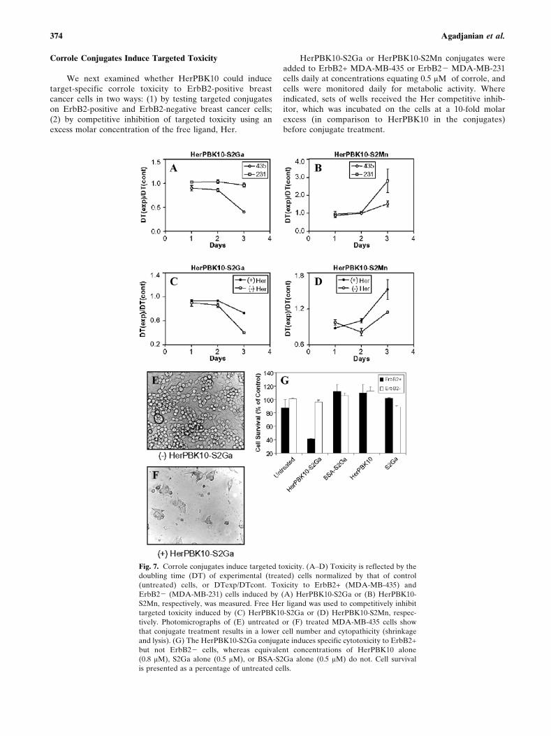
We next examined whether HerPBK10 could inducetarget-specific corrole toxicity to ErbB2-positive breastcancer cells in two ways: (1) by testing targeted conjugateson ErbB2-positive and ErbB2-negative breast cancer cells;(2) by competitive inhibition of targeted toxicity using anexcess molar concentration of the free ligand, Her.
HerPBK10-S2Ga or HerPBK10-S2Mn conjugates wereadded to ErbB2+ MDA-MB-435 or ErbB2j MDA-MB-231cells daily at concentrations equating 0.5 HM of corrole, andcells were monitored daily for metabolic activity. Whereindicated, sets of wells received the Her competitive inhib-itor, which was incubated on the cells at a 10-fold molarexcess (in comparison to HerPBK10 in the conjugates)before conjugate treatment.
Fig. 7. Corrole conjugates induce targeted toxicity. (AYD) Toxicity is reflected by the
doubling time (DT) of experimental (treated) cells normalized by that of control
(untreated) cells, or DTexp/DTcont. Toxicity to ErbB2+ (MDA-MB-435) and
ErbB2j (MDA-MB-231) cells induced by (A) HerPBK10-S2Ga or (B) HerPBK10-
S2Mn, respectively, was measured. Free Her ligand was used to competitively inhibit
targeted toxicity induced by (C) HerPBK10-S2Ga or (D) HerPBK10-S2Mn, respec-
tively. Photomicrographs of (E) untreated or (F) treated MDA-MB-435 cells show
that conjugate treatment results in a lower cell number and cytopathicity (shrinkage
and lysis). (G) The HerPBK10-S2Ga conjugate induces specific cytotoxicity to ErbB2+
but not ErbB2j cells, whereas equivalent concentrations of HerPBK10 alone
(0.8 HM), S2Ga alone (0.5 HM), or BSA-S2Ga alone (0.5 HM) do not. Cell survival
is presented as a percentage of untreated cells.
374 Agadjanian et al.

By day 3 of the experiment, HerPBK10-S2Ga reducedthe cell survival of MDA-MB-435 cells by 60% whereassurvival of MDA-MB-231 cells remained unaffected (Fig. 7Aand G). Likewise, HerPBK10-S2Mn slowed the growth ofMDA-MB-435 cells by õ70% compared to MDA-MB-231(Fig. 7B). As a confirmation of the targeted toxicity of theconjugates, we demonstrate here that the free ligand, Her,inhibited MDA-MB-435 cell death from HerPBK10-S2Ga byõ60Y70% (Fig. 7C), and from HerPBK10-S2Mn byõ80Y85% (Fig. 7D). Importantly, the conjugates seemed toinduce death and lysis of the cells (Fig. 7F) as opposed toinducing cytostasis. Also importantly, neither HerPBK10alone nor S2Ga alone produced cell death when each wasapplied at the equivalent molar concentrations as its cognatein the conjugate (Fig. 7G).
The toxicity induced when 0.5 HM of corrole is deliveredby a targeted vehicle starkly contrasts with the toxicityinduced by corroles when they are not targeted, such aswhen delivered alone in complete cell media, or as a BSA-S2Ga conjugate (Fig. 7G). Moreover, our previous assaysindicate that cell death from nontargeted corroles did notensue until day 6 of treatment. Taken altogether, our findingsshow that S2Ga can induce nearly 60% MDA-MB-435 celldeath by 3 days at 0.5 HM when it is targeted, whereas theuntargeted compound must be exposed to the cells for nearlya week at nearly an order of magnitude higher concentrationto accomplish the same effect.
DISCUSSION
Existing approaches to targeting therapeutic compoundsnormally require covalent attachment of the drug to thecarrier. Such an approach of chemical modification may notonly complicate the preparation of the conjugate, but alsocompromise the potency of the drug and abrogate the activityor specificity of the carrier molecule. Instead, a system inwhich the therapeutic can be incorporated with a carrier andthen released into the target cell would be ideal. In this study,we have explored the use of a new type of bioconjugatewhereby corroles are noncovalently assembled with recom-binant viral proteins having unique cell binding and entryfeatures.
Until now, there has been little research on the effect ofcorroles in biological systems. Here we present the firststudies on targeting noncovalent corrole assemblies to spe-cific cell-surface receptors. In particular, the carrier mole-cules tested here include recombinant derivatives of the Ad5penton base, and exhibit the functions of receptor-specificbinding, cell entry, and translocation to the cytosol. Using aheregulin-directed penton base protein, we demonstrate thatreceptor-specific targeting of corroles to ErbB2-positivebreast cancer cells, and target-specific cell death is possible.
The most significant finding in these studies is that tar-geted corroles induce cell death at submicromolar concen-trations, whereas equivalent concentrations of the untargetedcompound do not. Importantly, the protein targeting vehiclealone did not induce cell death. These findings support thepremise that sulfonated corroles induce cytotoxicity onlywhen they are able to translocate into the cell via a cell-penetrating carrier, and underscore the importance of using a
targeted cell entry vehicle for effective delivery. Takentogether, these findings suggest that targeting corroles usingspecialized carrier proteins such as HerPBK10 should enableeffective dosing at reasonable concentrations while avoidingnonspecific toxicity, should the corrole release prematurelyfrom the carrier. Given the high degree of binding stability toendure ultrafiltration, HPLC, and nonexchange with serumprotein, however, this is unlikely to happen.
Among the proteins we have tested here as carriers arerecombinant derivatives of the Ad5 penton base protein: PB,PBK10, and HerPBK10. The proteins PB and PBK10 entercells by integrin receptor binding and endocytosis, andtranslocate into the cytoplasm by lysis of cellular endosomes(9,10). The protein, HerPBK10, utilizes the endosomolyticactivity of the penton base but specifically binds and entersErbB2-positive cells (11), which display amplified levels ofthe heregulin receptor on the cell surface (22,23). Taking ad-vantage of the intense fluorescence of the S2Ga compound,we could detect both receptor-specific binding and cell entryof HerPBK10-S2Ga conjugates on ErbB2-positive humanbreast cancer cells.
Our findings indicate that multiple corroles noncova-lently bind the recombinant proteins studied here, likely atmultiple sites on the same protein molecule. The corrolebinding sites on HerPBK10 or any of the other carrier mol-ecules tested here remain to be determined. It is possible thatthe polyhistidine tag appended to the recombinant proteinsPB, PBK10, HerPBK10, and Her, which facilitates proteinpurification by metal chelate chromatography, may contrib-ute to binding. It is also possible that the positively chargedpolylysine on PBK10 and HerPBK10 may enable chargeinteractions with the sulfonates on the corroles, much thesame way as these polylysines interact with the phosphatebackbone of DNA (9,11). Determining which residues con-tribute to binding, however, is beyond the scope of this pre-sent study.
To demonstrate that HerPBK10-corrole conjugates werespecifically targeted to ErbB receptors, we competitively in-hibited receptor binding using excessive concentrations offree Her ligand. These concentrations equated 8Y18 HM offree Her, which would be in far excess of the presumed cir-culating levels of heregulin (24Y26). Moreover, the molarityof HerPBK10 itself when delivered as a conjugate still ex-ceeds these levels, and thus does not present a scenario thatwould suggest that circulating heregulin could competitivelyinhibit conjugate delivery. Although Her reduced the cyto-toxocity of targeted conjugates, our data suggest that S2Mnmay stimulate cell doubling, which would not be a desirableeffect. This apparent stimulation warrants further investiga-tion outside the scope of this study. Considering that it maytake several days to begin observing substantial reductions incell number (Figs. 3 and 7), one possible explanation is thatthe cells undergo a delay in cytotoxicity, and thus may havean opportunity to divide before undergoing cell death. Thisfinding brings up another important consideration, which isunderstanding the mechanism of cell death. Although cor-roles are capable of binding metal ions, it is unknown wheth-er metal chelation is the actual mechanism of cytotoxicity.The actual intracellular targets of toxicity also remain to beidentified. It is possible that corroles may interact with keyenzymes, or may simply tightly bind intracellular protein and
375Corrole Bioconjugates

thus impede function. Our future studies aim at elucidatingthe cell death mechanism. Meanwhile, the present studies inpart explore the potential of corroles as cytotoxic drugs, andthus these findings can help us select which corroles are bet-ter candidates as anticancer compounds.
Currently used chemotherapy drugs such as doxorubicinor cisplatin exert their effect by entering the cell nucleus andbinding to DNA, stabilizing certain proteinYDNA complexesand resulting in inhibition of replication (27). As such, thesedrugs are nondiscriminatory: they can penetrate cell mem-branes, but induce the most potent damage on dividing cells.One of the potential advantages of amphiphilic, negativelycharged corroles is their cell impermeability, as seen here.The negative charges on sulfonated corroles likely explainswhy these compounds are unable to enter cells without acarrier in protein-free conditions, as the net-negative charge onthe cell surface would repel such corroles. Likewise, a pos-itively charged corrole enters cells presumably because thepositive charges facilitate binding to the cell surface (28). Thenegative charge of sulfonated corroles thus plays up an addi-tional benefit for targeted therapy by reducing nonspecificbinding to cell surfaces. This should allow for control of cellentry by conjugating corroles to certain carrier molecules.
Previous studies have tested the strategy of covalentlycoupling shuttle molecules, such as antibodies, to drugs butthe chemical modifications introduced by this approach canreduce the activity of either component (29Y31). Recombi-nant fusion of proteins, such as toxins, to shuttle peptidesrepresents another strategy, but often the toxin requiresspecial processing to become active (30,32,33). Our approachof assembling complexes as noncovalent conjugates may besuperior to targeting strategies utilizing toxin fusions orcovalently linked drugs, as assembly did not seem to abrogatetargeting activity and, in fact, enhanced the toxicity of thecorrole payload. These studies represent the first example, toour knowledge, of assembling and testing targeted corroleconjugates. These encouraging in vitro findings shouldprovide a foundation for further development of targetedcorrole conjugates for application in cancer therapy.
ACKNOWLEDGMENTS
We thank the following people for valuable discussionson this work: Nori Kasahara, Larry Kedes, and RenataStripecke. This work was supported, in part, through acollaborative research agreement with IntraGene SciencesInc., and by grants to L.K.M.-K. from the NIH (CA102126),the Susan G. Komen Breast Cancer Foundation (BCTR02-01194), and the Donna and Jesse Garber Award; to Z.G.from the STAR (Chicago) foundation; to H.B.G. from theNational Science Foundation; and to I.J.D. from the HelenHay Whitney Foundation.
REFERENCES
1. T. J. Dougherty, C. J. Gomer, B. W. Henderson, G. Jori,D. Kessel, M. Korbelik, J. Moan, and Q. Peng. Photodynamictherapy. J. Natl. Cancer Inst. 90:889Y905 (1998).
2. L. D. Via and S. M. Magno. Photochemotherapy in thetreatment of cancer. Curr. Med. Chem. 8:1405Y1418 (2001).
3. T. J. Dougherty. A brief history of clinical photodynamic ther-apy development at Roswell Park Cancer Institute. J. Clin.Laser Med. 14:219Y221 (1996).
4. A. Mahammed, I. Goldberg, and Z. Gross. Highly selectivechlorosulfonation of tris(pentafluorophenyl)corrole as a synthet-ic tool for the preparation of amphiphilic corroles and metalcomplexes of chiral planarity. Org. Lett. 3:3443Y3446 (2001).
5. I. Saltsman, A. Mahammed, I. Goldberg, E. Tkachenko, M.Botoshansky, and Z. Gross. Selective substitution of corroles:nitration, hydroformylation, and chlorosulfonation. J. Am.Chem. Soc. 124:7411Y7420 (2002).
6. T. G. Gantchev, R. Ouellet, and J. E. Liervan. Binding inter-actions and conformational changes induced by sulfonated alu-minum phthalocyanines in human serum albumin. Arch.Biochem. Biophys. 366:21Y30 (1999).
7. R. K. Pandey, S. Constantine, T. Tsuchida, G. Zheng, C. J.Medforth, M. Aoudia, A. N. Kozyrev, M. A. Rodgers, H. Kato,K. M. Smith, and T. J. Dougherty. Synthesis, photophysical pro-perties, in vivo photosensitizing efficacy, and human serumalbumin binding properties of some novel bacteriochlorins.J. Med. Chem. 40:2770Y2779 (1997).
8. A. Mahammed, H. B. Gray, J. J. Weaver, K. Sorasaenee, andZ. Gross. Amphiphilic corroles bind tightly to human serumalbumin. Bioconjug. Chem. 15:738Y746 (2004).
9. L. K. Medina-Kauwe, N. Kasahara, and L. Kedes. 3PO, a novelnon-viral gene delivery system using engineered Ad5 pentonproteins. Gene Ther. 8:795Y803 (2001).
10. L. K. Medina-Kauwe. Endocytosis of adenovirus and adenoviruscapsid proteins. Adv. Drug Deliv. Rev. 55:1485Y1496 (2003).
11. L. K. Medina-Kauwe, M. Maguire, N. Kasahara, and L. Kedes.Non-viral gene delivery to human breast cancer cells by targetedAd5 penton proteins. Gene Ther. 8:1753Y1761 (2001).
12. L. K. Medina-Kauwe and X. Chen. Using GFPYligand fusionsto measure receptor-mediated endocytosis in living cells. In G.Litwack (ed.), Vitamins and Hormones, Vol. 65, Elsevier, SanDiego, 2002, pp. 81Y95.
13. L. K. Medina-Kauwe, V. Leung, L. Wu, and L. Kedes. Assessingthe binding and endocytosis activity of cellular receptors usingGFPYligand fusions. BioTechniques 29:602Y609 (2000).
14. G. S. Manning. The molecular theory of polyelectrolyte solu-tions with applications to the electrostatic properties of polynu-cleotides. Q. Rev. Biophys. 11:179Y246 (1978).
15. M. X. Tang and F. C. Szoka Jr. Characterization of polycationcomplexes with DNA. In A. V. Kabanov, P. L. Felgner, L. W.Seymour (eds.), Self-Assembling Complexes for Gene Delivery,Wiley, Chichester, 1998.
16. J. Zabner, A. J. Fasbender, T. Moninger, K. A. Poellinger, andM. J. Welsh. Cellular and molecular barriers to gene transfer bya cationic lipid. J. Biol. Chem. 270:18997Y19007 (1995).
17. G. L. Lukacs, P. Haggie, O. Seksek, D. Lechardeur, N.Freedman, and A. S. Verkman. Size-dependent DNA mobil-ity in cytoplasm and nucleus. J. Biol. Chem. 275:1625Y1629 (2000).
18. L. Karayan, B. Gay, J. Gerfaux, and P. A. Boulanger. Oligo-merization of recombinant penton base of adenovirus type 2 andits assembly with fiber in baculovirus-infected cells. Virology202:782Y795 (1994).
19. T. J. Wickham, P. Mathias, D. A. Cheresh, and G. R. Nemerow.Integrins avb3 and avb5 promote adenovirus internalization butnot virus attachment. Cell 73:309Y319 (1993).
20. A. Mahammed and Z. Gross. Aluminum corrolin, a novelchlorophyll analogue. J. Inorg. Biochem. 88:305Y309 (2002).
21. S. Mukherjee, R. N. Ghosh, and F. R. Maxfield. Endocytosis.Physiol. Rev. 77:759Y803 (1997).
22. D. J. Slamon, G. M. Clark, S. G. Wong, W. J. Levin, A. Ullrich,and W. L. McGuire. Human breast cancer: correlation of relapseand survival with amplification of the HER-2/neu oncogene.Science 235:177Y182 (1987).
23. D. J. Slamon and G. M. Clark. Amplification of c-erbB-2 andaggressive human breast tumors? Science 240:1795Y1798 (1988).
24. W. E. Holmes, M. X. Sliwkowski, R. W. Akita, W. J. Henzel,J. Lee, J. W. Park, D. Yansura, N. Abadi, H. Raab, and G. D.Lewis et al. Identification of heregulin, a specific activator ofp185erbB2. Science 256:1205Y1210 (1992).
25. S. D. Chan, D. M. Antoniucci, K. S. Fok, M. L. Alajoki, R. N.Harkins, S. A. Thompson, and H. G. Wada. Heregulin activation
376 Agadjanian et al.

of extracellular acidification in mammary carcinoma cells isassociated with expression of HER2 and HER3. J. Biol. Chem.270:22608Y22613 (1995).
26. V. R. Schelfhout, E. D. Coene, B. Delaey, A. A. Waeytens,L. De Rycke, M. Deleu, and C. R. De Potter. The role ofheregulin-alpha as a motility factor and amphiregulin as agrowth factor in wound healing. J. Pathol. 198:523Y533 (2002).
27. L. H. Hurley. DNA and its associated processes as targets forcancer therapy. Nat. Rev. 2:188Y200 (2002).
28. D. Aviezer, S. Cotton, M. David, A. Segev, N. Khaselev,N. Galili, Z. Gross, and A. Yayon. Porphyrin analogues asnovel antagonists of fibroblast growth factor and vascularendothelial growth factor receptor binding that inhibit endothe-lial cell proliferation, tumor progression, and metastasis. CancerRes. 60:2973Y2980 (2000).
29. K. A. Chester, J. Bhatia, G. Boxer, S. P. Cooke, A. A. Flynn,A. Huhalov, A. Mayer, R. B. Pedley, L. Robson, S. K. Sharma,
D. I. Spencer, and R. H. Begent. Clinical applications of phage-derived sFvs and sFv fusion proteins. Dis. Markers 16:53Y62(2000).
30. A. E. Frankel, R. J. Kreitman, and E. A. Sausville. Targetedtoxins. Clin. Cancer Res. 6:326Y334 (2000).
31. M. E. Reff and C. Heard. A review of modifications to recom-binant antibodies: attempt to increase efficacy in oncologyapplications. Crit. Rev. Oncol. Hematol. 40:25Y35 (2001).
32. M. Schmidt, M. Maurer-Gebhard, B. Groner, G. Kohler,G. Brochmann-Santos, and W. Wels. Suppression of metastasisformation by a recombinant single chain antibody-toxin targetedto full-length and oncogenic variant EGF receptors. Oncogene18:1711Y1721 (1999).
33. M. Jeschke, W. Wels, W. Dengler, R. Imber, E. Stocklin, andB. Groner. Targeted inhibition of tumor-cell growth by recom-binant heregulinYtoxin fusion proteins. Int. J. Cancer 60:730Y739(1995).
377Corrole Bioconjugates
Copyright © 2022 FDOKUMEN