Specific Binding Sites for Triiodothyronine in the Plasma ...
8
Specific Binding Sites for Triiodothyronine in the Plasma Membrane of Rat Thymocytes CORRELATION WITH BIOCHEMICAL RESPONSES JOSEPH SEGAL and SIDNEY H. INGBAR, Charles A. Dana Research Institute and the Harvard-Thorndike Laboratory of Beth Israel Hospital, Department of Medicine, Beth Israel Hospital and Harvard Medical School, Boston, Massachusetts 02215 A B S T R A C T As a prerequisite to studies of whether the plasma membrane of the rat thymocyte contains specific, saturable binding sites for the thyroid hor- mone 3,5,3'-triiodothyronine (T3), a method was de- veloped for the isolation of a plasma membrane frac- tion from these cells. As judged from both electron microscopic and marker enzyme studies, the fraction was composed principally of plasma membrane vesi- cles, was free of nuclear contaminants, and was only slightly contaminated with other subcellular compo- nents. At 37°C and pH 7.4, binding of ['25I]T3 by the fresh membrane preparation was rapid, reaching a maximum at 5 min and then declining with time, so that by 60 min binding was virtually nil. Decreased binding with time was due to a loss of functional bind- ing sites, but did not reflect desensitization, since the decrease in binding activity with time was indepen- dent of the presence or absence of T3. Scatchard anal- ysis of saturation studies revealed the presence of two binding sites, one with an apparent dissociation con- stant (Kd) of 0.95 nM and a maximum capacity of 5.3 X 10¹⁰ sites/100 ,ug protein, and the other with an ap- parent Kd of 25 nM and a binding capacity of 1.4 X 10¹² sites/100 ,g protein. Measurement of the ability of several thyronine analogues to inhibit the binding of [1251I]T3 revealed the following rank order of potency: L-T3 > L-T4 > D-T3 = D-T4 > L-3,5-T2 > rT3 > D,L- th*ronine. Binding of T3 was inhibited by the omission of calcium from the medium or by the addition of the This work was supported in part by grant AM-18416 from the National Institute of Arthritis, Metabolism, and Digestive Diseases, and grant AGO-1346 from the National Institute of Aging, National Institutes of Health, Bethesda, MD. Received for publication 13 April 1982 and in revised form 2 August 1982. beta adrenergic antagonist alprenolol. As judged from studies of the lower affinity binding site, these manip- ulations decreased the affinity, but not the number, of binding sites for T3. The relative potencies of thyro- nine analogues to inhibit the binding of ['25I]T3 were generally parallel to their previously reported poten- cies in stimulating the uptake of the sugar analogue 2-deoxy-glucose (2-DG) in intact rat thymocytes in vitro. Further, the inhibition of T3-binding produced by L-alprenolol or by excluding calcium from the me- dium resembled the previously reported inhibition that these manipulations produce with respect to T3- induced enhancement of 2-DG uptake. These findings suggest that the binding sites for T3 present in the plasma membrane of rat thymocytes act as functional receptors linked to the stimulation of 2-DG uptake that T3 induces in these cells. INTRODUCTION We have previously demonstrated that 3,5,3'-triio- dothyronine (T3)' enhances the uptake of the glucose analogue 2-deoxy-D-glucose (2-DG) in rat thymocytes in vitro (1). Several lines of evidence indicate that T3 induces its effect by acting at the level of the plasma membrane. Thus, the effect is rapid, is independent of new protein synthesis, and is mediated via an in- crease in cellular cyclic (c)AMP concentration, as a consequence of an increase in the activity of adenylate cyclase (2-4), an enzyme that resides within the plasma membrane. Furthermore, epinephrine, which is known to initiate its action upon binding to its I Abbreviations used in this paper: T3, 3,5,3'-triiodothy- ronine; T4, 3,5,3',5'-tetraiodothyronine (thyroxine); rT3, 3,3',5'- triiodothyronine; 3,5-T2, 3,5-diiodothyronine, 2-DG, 2-deoxy- D-glucose. J. Clin. Invest. C) The American Society for Clinical Investigation, Inc. * 0021-9738/82/11/0919/08 $1.00 Volume 70 November 1982 919-926 919
-
Upload
khangminh22 -
Category
Documents
-
view
2 -
download
0
Transcript of Specific Binding Sites for Triiodothyronine in the Plasma ...
Specific Binding Sites for Triiodothyronine in the PlasmaMembrane of Rat Thymocytes
CORRELATION WITH BIOCHEMICAL RESPONSES
JOSEPH SEGAL and SIDNEY H. INGBAR, Charles A. Dana Research Institute and theHarvard-Thorndike Laboratory of Beth Israel Hospital, Department ofMedicine, Beth Israel Hospital and Harvard Medical School, Boston,Massachusetts 02215
A B S T R A C T As a prerequisite to studies of whetherthe plasma membrane of the rat thymocyte containsspecific, saturable binding sites for the thyroid hor-mone 3,5,3'-triiodothyronine (T3), a method was de-veloped for the isolation of a plasma membrane frac-tion from these cells. As judged from both electronmicroscopic and marker enzyme studies, the fractionwas composed principally of plasma membrane vesi-cles, was free of nuclear contaminants, and was onlyslightly contaminated with other subcellular compo-nents. At 37°C and pH 7.4, binding of ['25I]T3 by thefresh membrane preparation was rapid, reaching amaximum at 5 min and then declining with time, sothat by 60 min binding was virtually nil. Decreasedbinding with time was due to a loss of functional bind-ing sites, but did not reflect desensitization, since thedecrease in binding activity with time was indepen-dent of the presence or absence of T3. Scatchard anal-ysis of saturation studies revealed the presence of twobinding sites, one with an apparent dissociation con-stant (Kd) of 0.95 nM and a maximum capacity of 5.3X 10¹⁰ sites/100 ,ug protein, and the other with an ap-parent Kd of 25 nM and a binding capacity of 1.4X 10¹² sites/100 ,g protein. Measurement of the abilityof several thyronine analogues to inhibit the bindingof [1251I]T3 revealed the following rank order of potency:L-T3 > L-T4 > D-T3 = D-T4 > L-3,5-T2 > rT3 > D,L-th*ronine. Binding of T3 was inhibited by the omissionof calcium from the medium or by the addition of the
This work was supported in part by grant AM-18416 fromthe National Institute of Arthritis, Metabolism, and DigestiveDiseases, and grant AGO-1346 from the National Instituteof Aging, National Institutes of Health, Bethesda, MD.
Received for publication 13 April 1982 and in revisedform 2 August 1982.
beta adrenergic antagonist alprenolol. As judged fromstudies of the lower affinity binding site, these manip-ulations decreased the affinity, but not the number, ofbinding sites for T3. The relative potencies of thyro-nine analogues to inhibit the binding of ['25I]T3 weregenerally parallel to their previously reported poten-cies in stimulating the uptake of the sugar analogue2-deoxy-glucose (2-DG) in intact rat thymocytes invitro. Further, the inhibition of T3-binding producedby L-alprenolol or by excluding calcium from the me-dium resembled the previously reported inhibitionthat these manipulations produce with respect to T3-induced enhancement of 2-DG uptake. These findingssuggest that the binding sites for T3 present in theplasma membrane of rat thymocytes act as functionalreceptors linked to the stimulation of 2-DG uptake thatT3 induces in these cells.
INTRODUCTIONWe have previously demonstrated that 3,5,3'-triio-dothyronine (T3)' enhances the uptake of the glucoseanalogue 2-deoxy-D-glucose (2-DG) in rat thymocytesin vitro (1). Several lines of evidence indicate that T3induces its effect by acting at the level of the plasmamembrane. Thus, the effect is rapid, is independentof new protein synthesis, and is mediated via an in-crease in cellular cyclic (c)AMP concentration, as aconsequence of an increase in the activity of adenylatecyclase (2-4), an enzyme that resides within theplasma membrane. Furthermore, epinephrine, whichis known to initiate its action upon binding to its
I Abbreviations used in this paper: T3, 3,5,3'-triiodothy-ronine; T4, 3,5,3',5'-tetraiodothyronine (thyroxine); rT3, 3,3',5'-triiodothyronine; 3,5-T2, 3,5-diiodothyronine, 2-DG, 2-deoxy-D-glucose.
J. Clin. Invest. C) The American Society for Clinical Investigation, Inc. * 0021-9738/82/11/0919/08 $1.00Volume 70 November 1982 919-926
919
plasma membrane receptors and thence activating ad-enylate cyclase, produces with T3 a synergistic increasein the 2-DG uptake (5). If T3 does indeed act at thelevel of the thymocyte plasma membrane, there shouldbe specific saturable binding sites for the hormonepresent in the plasma membrane of these cells. Wehave now shown that the plasma membrane of ratthymocytes does contain specific binding sites for T3,and have found several properties of the binding ofT3 to these sites that strengthen the likelihood that theyare linked to the actions of T3 described above.
METHODSMaterials used were purchased from the following sources:L-alprenolol bitartrate, epinephrine bitartrate, sodium tar-trate, Tris-ATP, ascorbic acid, disodium 5'-AMP, DNA, 2,6-dichlorophenol indophenol, diphenylamine, disodium D-glu-cose-6-phosphate, B-glycerophosphate, Hepes, phenazinemethosulfate, and DL-thyronine from Sigma Chemical Co.,St. Louis, MO; ammonium molybdate, paraformaldehyde,lead nitrate, sodium succinate, and uranyl acetate fromFisher Scientific Co., Pittsburgh, PA; L-T4, D-T4, L-T3, D-T3,and L-3,5-T2 from Henning Berlin GMBH, West Germany;rT3 from Calbiochem-Behring Co. La Jolla, CA; sodium cac-odylate (sodium dimethyl arsenate) from ICN Pharmaceu-tical Inc., Cleveland, OH; 50% glutaraldehyde solution fromElectron Microscopy Sciences, Fort Washington, PA; Epon812 from Ted Pella, Inc.; propylene oxide from the EastmanKodak Co., Rochester, NY; and [1251IT3 (sp act ranging from1,106 to 1,250 MCi/Mg) from New England Nuclear, Bos-ton, MA.Female Sprague-Dawley rats of the CD strain, 26-28 d
old and purchased from Charles River Breeding Laboratories(Wilmington, MA), served as the source of thymocytes. Thy-mocytes were isolated according to a method previouslydescribed (1).
Preparation of plasma membranes. Plasma membraneswere prepared at 0-4°C by the following technique. Isolatedthymocytes were resuspended in hypotonic homogenizationbuffer (10 mM Tris-HCl; 2.5 mM MgCl2; 1 mM EGTA), pH7.4, 1 m1/5 thymus glands, and were allowed to stand for15 min to permit cells to become swollen, thereby facilitatingtheir disruption. Cells were then transferred into a glass ho-mogenizer and were homogenized until >95% were dis-rupted. This usually required 40-50 strokes. The homoge-nate was centrifuged at 300 g for 10 min, the supernate wascollected, and the pellet was resuspended in homogenizationbuffer and recentrifuged at 300 g for 10 min. The two su-pernatants were combined and centrifuged at 4,000 g for 15min. The supernate was collected and centrifuged at 20,000g for 30 min. The yellow pellet obtained was suspended in40% sucrose-10 mM Tris-HCl, pH 7.4, and was transferredinto a cellulose-nitrate tube. An equivalent volume of 30%sucrose-10 mM Tris-HCI, pH 7.4, was carefully overlayeredon the 40% sucrose-membrane fraction, and the sucrose-gra-dient was centrifuged at 80,000 g for 4 h, during which timeplasma membranes were concentrated in a narrow milkyband at the interphase of the 30-40% sucrose solutions. Theplasma membranes were harvested and transferred into acentrifugation tube into which 10-20 vol of Tris-HCl, pH7.4, were added. The suspension was then centrifuged at20,000 g for 60 min. The pellet containing the plasma mem-branes was collected and kept on ice until samples weretaken for electron microscopic studies, assays for cellularmarkers, and measurements of T3-binding.
Measurement of [125I]T3binding. Freshly prepared mem-branes were always used in the T3-binding assay. The plasmamembranes were suspended in Krebs-Ringer-Tris buffer, (20mM Tris-HCl, 5 mM Tris-base, 120 mM NaCl, 5 mM KCI,1 mM CaCl2, 1.5 mM NaH2PO4, 2.5 mM MgC92), pH 7.4.This is the same buffer as that used in studies on the effectsof T3 in the intact cell, except that Hepes is omitted (Hepeswas omitted from the buffer because it interferes with themeasurement of protein concentration.). In some cases,where so indicated, membranes were suspended in Ca2+-freemedium, i.e., the standard medium from which calcium saltswere omitted. Ca2+-free medium contained 5 sM Ca2' be-cause of contamination by calcium in the other buffer salts.The suspended membranes were transferred into a glass ho-mogenizer and 10 strokes were then applied to obtain a ho-mogenous membrane suspension. Membrane suspensionswere then transferred into plastic tubes (12-ml capacity)containing buffer, placed in a shaking water bath, and equil-ibrated for 10 min at 37°C in air. Thereafter, 50 Ml of buffercontaining 75 nCi of ['25I]T3 was added. As appropriate tothe experiment, varying concentrations of unlabeled T3 orother thyronine analogues were added at the same time,keeping the final volume of the reaction mixture constant.Nonspecific binding of [251I]T3 was assessed in specimens con-taining unlabeled T3 in a concentration of 10 MM, and values'for [1251]T3 found therein were subtracted from total bindingto determine the saturable binding of ['251IT3.
At the end of the incubation period, triplicate 250-Ml al-iquots of reaction mixture were quickly removed and ap-plied onto 0.3-um Millipore filters (Millipore Corp., Bedford,MA) under vacuum. The membranes were then quicklywashed three times with 2.5 ml of ice-cold buffer. The filterswere then dried, transferred into tubes, and were countedin a gamma counter.
Kinetic parameters of T3 binding were obtained in satu-ration studies using Scatchard plots and the iterative tech-nique described by Minneman (6).
Electron microscopy. Plasma membrane fractions wereprepared for examination under the electron microscopeaccording to the technique described by Stemerman (7).
Cellular markers. DNA content in homogenates andmembrane preparations was measured according to a methoddescribed by Burton (8); 5'-nucleotidase activity accordingto the method of Bosmann (9); succinate dehydrogenase ac-tivity by the method of Hatefi (10); Na+,K+-ATPase, activityby the method described by Wallach and Kamat (11); acidphosphatase activity by the method of Baudhuin (12); andglucose-6-phosphatase activity, as well as inorganic phos-phate content, by methods described by Ames (13).
Purity of [1"5I]T3. Purity of preparations of [1251]T3 wasassessed by descending filter paper chromatography in ahexane-tertiary amyl alcohol-N-ammonia (1:10:11) solventsystem according to a technique described by Borgeset al. (14).
Statistical analysis. The following statistical methodswere used: Dunnet's test for comparisons between multiplegroups and a single control; analysis of variance, followedby the Newman-Keuls multiple range test, for comparisonsof multiple groups with each other; and analysis of covari-ance, followed by Dunnet's test, for comparisons of multipleregression lines with a single control line (15).
RESULTS
Purity of plasma membrane preparations. Bothultramicroscopic appearance and biochemical markersindicated that plasma membrane preparations were
920 J. Segal and S. H. Ingbar
highly purified. Electron microscopic studies revealeda homogenous plasma membrane preparation, com-prising closed vesicles of varying sizes, ranging from0.25 to 1.2 Mm Diam. Small quantities of ribosomes
were present, but nuclear, mitochondrial, and lyso-somal contaminants were not evident.A comparison of the relative concentrations of spe-
cific cellular markers in the initial homogenates andthe membrane preparations also indicated that thepreparations were highly enriched in plasma mem-branes and were essentially devoid of other cellularcomponents (Table I). DNA content was virtually nil,indicating the absence of nuclei. The plasma mem-brane markers, 5'-nucleotidase and Na',K'-ATPase,were enriched by 15- and 12.5-fold, respectively. Thepreparation contained a small amount microsomes, asindicated by glucose-6-phosphatase; smaller amountsof mitochondria, as indicated by succinate dehydro-genase; and negligible amounts of lysosomes, as indi-cated by acid phosphatase. In terms of both the yieldof protein (-0.43% of initial total protein) and of therelative enrichment of specific marker enzymes, thepresent preparations of rat thymocyte plasma mem-branes were quite comparable to those derived fromthymocytes of calf and rabbit, as described by others(16-19).Binding of ['25I]T3: effect of membrane concentra-
tion and time. Binding of ['25I]T3 to various concen-trations of plasma membranes was linear between 20and 240 Mg protein/ml in the reaction medium (datanot shown). In the ensuing experiments, plasma mem-branes were used in a concentration of 100-200 MAgprotein/ml.
TABLE IBiochemical Characterization of Thymocyte
Plasma Membrane Preparations'
Plasma membrane§Markert Homogenate fraction
DNA 348.8±28.5 0.0005'-nucleotidase 0.34±0.01 5.13±0.05Na+,K+-ATPase 0.90±0.02 11.26±0.74Glucose-6-phosphatase 1.33±0.17 0.51±0.03Acid phosphatase 2.11±0.20 0.48±0.04Succinate dehydrogenase 1.06±0.01 0.03±0.01
* Homogenates and plasma membrane fractions derived therefromwere prepared as described in Methods. Values for the varioussubcellular markers are the mean±SD of those obtained in threeseparate preparations.t DNA micrograms per milligram protein; enzymes, microgramsproduct per milligram protein/60 min.§ Plasma membranes fractions contained 83.7±11.7 ng protein/109original thymocytes (mean±SD, n = 19), representing 0.43% of theoriginal protein content.
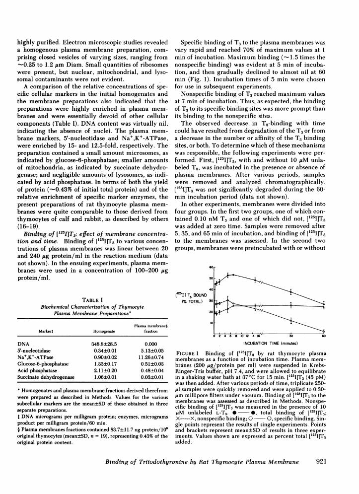
Specific binding of T3 to the plasma membranes wasvary rapid and reached 70% of maximum values at 1min of incubation. Maximum binding (- 1.5 times thenonspecific binding) was evident at 5 min of incuba-tion, and then gradually declined to almost nil at 60min (Fig. 1). Incubation times of 5 min were chosenfor use in subsequent experiments.
Nonspecific binding of T3 reached maximum valuesat 7 min of incubation. Thus, as expected, the bindingof T3 to its specific binding sites was more prompt thanits binding to the nonspecific sites.The observed decrease in T3-binding with time
could have resulted from degradation of the T3 or froma decrease in the number or affinity of the T3 bindingsites, or both. To determine which of these mechanismswas responsible, the following experiments were per-formed. First, ['25I]T3, with and without 10 MM unla-beled T3, was incubated in the presence or absence ofplasma membranes. After various periods, sampleswere removed and analyzed chromatographically.[1251I]T3 was not significantly degraded during the 60-min incubation period (data not shown).
In other experiments, membranes were divided intofour groups. In the first two groups, one of which con-tained 0.10 nM T3 and one of which did not, [t25I]T3was added at zero time. Samples were removed after5, 35, and 65 min of incubation, and binding of ['25I]T3to the membranes was assessed. In the second twogroups, membranes were preincubated with or without
60r
50
40
('25I) TX BOUND(% TOTAL) 30
20
0 2 4 6 8 10 12 14 i6 30
INCUBATION TIME (minutes)
60
FIGURE 1 Binding of [1251]T3 by rat thymocyte plasmamembranes as a function of incubation time. Plasma mem-branes (200 ug/protein per ml) were suspended in Krebs-Ringer-Tris buffer, pH 7.4, and were allowed to equilibratein a shaking water bath at 37°C for 15 min. [125I]T3 (45 pM)was then added. After various periods of time, triplicate 250-,ul samples were quickly removed and were applied to 0.30-Am millipore filters under vacuum. Binding of [1251]T3 to themembranes was assessed as described in Methods. Nonspe-cific binding of [t251]T3 was measured in the presence of 10IAM unlabeled L-T3. 0 0, total binding of [125I]T3;X-----X, nonspecific binding; 0 0, specific binding. Sin-gle points represent the results of single experiments. Pointsand brackets represent mean±SD of results in three exper-iments. Values shown are expressed as percent total [125I]T3added.
Binding of Triiodothyronine by Rat Thymocyte Plasma Membrane
.1
921
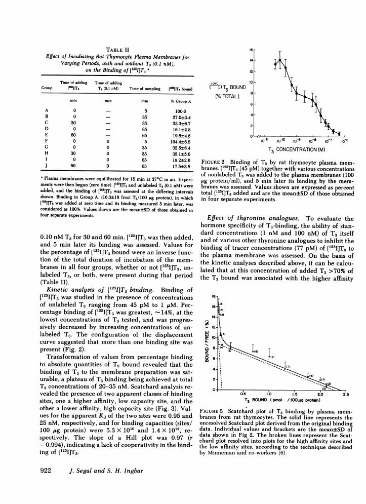
TABLE IIEffect of Incubating Rat Thymocyte Plasma Membranes for
Varying Periods, with and without T3 (0.1 nM),on the Binding of ['2I]T3.
Time of adding Time of addingGroup "I 1IrrS T3 (0.1 nM) Time of sampling ['"1IT3 bound
min min min % Group A
A 0 - 5 100.0B 0 - 35 37.0±3.4C 30 - 35 33.3±6.7D 0 - 65 16.1±2.8E 60 - 65 19.8±4.6P 0 0 5 104.4±6.5G 0 0 35 32.5±6.4H 30 0 35 35.1±5.0I 0 0 65 18.2±2.6J 60 0 65 17.5±3.8
Plasma membranes were equilibrated for 15 min at 37°C in air. Experi-ments were then begun (zero time). [l25sITs and unlabeled T3 (0.1 nM) wereadded, and the binding of [lssI]Ts was assessed at the differing intervalsshown. Binding in Group A. (16.3±18 fmol T3/100 sg protein), in which['25IJT was added at zero time and its binding measured 5 min later, wasconsidered as 100%. Values shown are the mean±SD of those obtained infour separate experiments.
0.10 nM T3 for 30 and 60 min. ['25I]T3 was then added,and 5 min later its binding was assessed. Values forthe percentage of [125I]T3 bound were an inverse func-tion of the total duration of incubation of the mem-branes in all four groups, whether or not [251I]T5, un-labeled T3, or both, were present during that period(Table II).
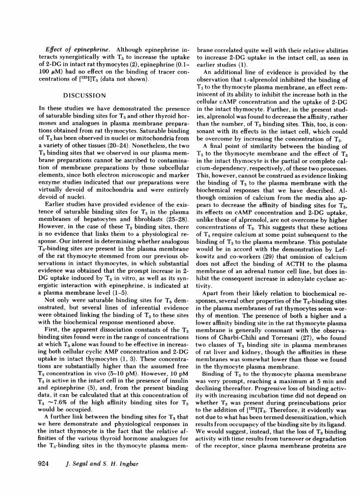
Kinetic analysis of [1251]T3 binding. Binding of[I251]T3 was studied in the presence of concentrationsof unlabeled T3 ranging from 45 pM to 1 ,uM. Per-centage binding of [1251]T3 was greatest, -14%, at thelowest concentrations of T3 tested, and was progres-sively decreased by increasing concentrations of un-labeled T3. The configuration of the displacementcurve suggested that more than one binding site waspresent (Fig. 2).
Transformation of values from percentage bindingto absolute quantities of T3 bound revealed that thebinding of T3 to the membrane preparation was sat-urable, a plateau of T3 binding being achieved at totalT3 concentrations of 20-35 nM. Scatchard analysis re-vealed the presence of two apparent classes of bindingsites, one a higher affinity, low capacity site, and theother a lower affinity, high capacity site (Fig. 3). Val-ues for the apparent Kd of the two sites were 0.95 and25 nM, respectively, and for binding capacities (sites/100 ,ug protein) were 5.3 X 10¹⁰ and 1.4 X 10¹², re-spectively. The slope of a Hill plot was 0.97 (r= 0.994), indicating a lack of cooperativity in the bind-ing of ['25I]T3.
( I)T3 BOUNDu
(% TOTAL) 86
4
2
lo b0100o 9 &0 8 1o 7 16-6
T3 CONCENTRATION (M)
FIGURE 2 Binding of T3 by rat thymocyte plasma mem-branes. [1251]T3 (45 pM) together with various concentrationsof nonlabeled T3 was_added to the plasma membranes (100,ug protein/ml), and 5 min later its binding by the mem-branes was assessed. Values shown are expressed as percenttotal [1251]T3 added and are the mean±SD of those obtainedin four separate experiments.
Effect of thyronine analogues. To evaluate thehormone specificity of T3-binding, the ability of stan-dard concentrations (1 nM and 100 nM) of T3 itselfand of various other thyronine analogues to inhibit thebinding of tracer concentrations (77 pM) of [I251]T3 tothe plasma membrane was assessed. On the basis ofthe kinetic analyses described above, it can be calcu-lated that at this concentration of added T3 >70% ofthe T3 bound was associated with the higher affinity
0
w
wIL
z
m
18
(8)(3)
14- (2)
12- ;7
I0-i~~~~
"')I
O- 1,()-8l
1--(2(7GII~~~~~~~~~~~~ 55
I (7
0.5 1.0 1.5 2.0T3 BOUND (pmol /lOO,ug protein)
2.5
FIGURE 3 Scatchard plot of T3 binding by plasma mem-branes from rat thymocytes. The solid line represents theunresolved Scatchard plot derived from the original bindingdata. Individual values and brackets are the mean±SD ofdata shown in Fig 2. The broken lines represent the Scat-chard plot resolved into plots for the high affinity sites andthe low affinity sites, according to the technique describedby Minneman and co-workers (6).
922 J. Segal and S. H. Ingbar

binding sites. In rank order of decreasing inhibitorypotency, the following sequence was obtained: L-T3> L-T4 > D-T3 = D-T4 > L-3,5-T2> rT3 > DL-thyronine(Table III). This rank-order agreed quite closely withthat of the potency of these agents to increase the up-take of 2-DG in the intact cell (1).
Effect of calcium. To ascertain the role, if any, ofCa21 in the binding of T3, experiments were performedin which membranes were suspended in standard orcalcium-free media, in some cases with or without 1mM EGTA. Some experiments were performed attracer concentrations of [I251]T3, as above, while otherexperiments were performed at T3 concentrations of10 nM-1 1zM, over which range >90% of T3-bindingoccurs at the low affinity site.
In the presence of tracer concentrations of T3, bind-ing of T3 in calcium-free medium was significantlydecreased to about two-thirds of control values(8.67±0.53 fmol/100 gg protein). Addition of 1 mMEGTA to the calcium-free medium produced no fur-ther effect (Fig. 4). In studies that examined bindingof T3 to the low affinity site, omission of calcium wasalso found to decrease T3Lbinding. Scatchard analysisrevealed this to be due to a reduction in the bindingaffinity, rather than the number of binding sites (Fig.5). Thus, as would be expected, the inhibitory effectof excluding calcium became proportionately less asthe concentration of T3 was progressively increased.
Effect of L-alprenolol. Experiments to determinethe effects of L-alprenolol on T3-binding were also con-ducted at tracer and at relatively high concentrationsof T3, as in experiments with calcium-free medium.
TABLE IIIInhibition of the Binding of [lssI]T3 to Thymocyte Plasma
Membranes by Various Thyronine Analogues'
Inhibition of ["'IJT, binding (%)
Thyronine analogue Analogue concentration
1lo M 1o-' M
L-T3 44.2±4.4 69.6±2.6L-T4 28.4±2.4 43.1±5.5D-T3 12.8±3.0 34.1±3.1D-T4 15.3±3.0 34.5±2.6L-3,5-T2 ----- 16.7±2.8rT3 ----- 11.8±1.9DL-Thyronine ----- 10.9±1.8
* Plasma membranes were incubated for 5 min with ['"IT3(77 pM), with or without two concentrations (1 nM and 0.1 AM)of various thyronine analogues, including T3. Values represent thepercent inhibition of [l'I]T.3-binding, relative to the binding ob-tained in control specimens incubated with [lssIJT3 alone. Valuesare mean±SD of those obtained in four separate experiments. L-T4contained 0.5% or less of L-T3 as a contaminant.
100
801-
(125)T3 BOUND 60
(% CONTROL) 40
20
++ V _Ca (M)
ALPRENOLOL
± 1414-
1o3 5x 10 5x10⁶ o-3(+ EGTA)
0, O 0 id-"
r±1
FIGURE 4 The effect of calcium depletion and of L-alpren-olol on the binding of tracer concentrations of ['25I]T3 by ratthymocyte plasma membranes. Plasma membranes were sus-pended in either standard medium (containing 1 mM Ca2")or Ca2"-free medium, in the presence or absence of 1 mMEGTA. ['251]T3 (77 pM) was added, with or without variousconcentrations of L-alprenolol, and 5 min later binding of[125I]T3 by the plasma membranes were assessed. Results areshown as percent ['251]T3 binding in the corresponding con-trol group. Values shown are mean±SD of those obtainedin from three to six separate experiments.
In the former experiments, L-alprenolol tartrate pro-duced a dose-related inhibition of T3 binding (Fig. 4).Sodium tartrate (0.1 mM) had no effect (data notshown). At the higher concentrations of T3, 10 ,M L-alprenolol was also inhibitory to T3 binding, and thiseffect, like that of excluding calcium, was to decreasethe binding affinity, rather than the number of bindingsites (Fig. 5).
0.086
0.07
0.06 \ALPRENOLOL CONTROL
BF 0.05
/F 004 C--FREE CONTROL- a0.03\-
0.0a-002 0
0001.0.00 0 2 3 4 0 2 3 4
T3 BOUND (pmol /lOOpg protein)
FIGURE5 Scatchard plot of T3-binding to rat thymocyteplasma membranes in the presence and absence of calciumand L-alprenolol. Plasma membranes were suspended instandard medium (1 mM Ca2+), or in Ca2+-free medium (5MM Ca2+). [125I]T3, together with various concentrations ofnonlabeled T3, with or without 10 AM alprenolol, was thenadded and 5 min later binding of [1251I]T3 by the plasmamembranes was measured. Values shown are mean±SD ofthose obtained in four separate experiments. Analysis of co-variance followed by the Dunnet's test was used for com-parisons between the alprenolol or Ca2 -free groups and thecontrol group.
Binding of Triiodothyronine by Rat Thymocyte Plasma Membrane
IO-3IO-5
923
Effect of epinephrine. Although epinephrine in-teracts synergistically with T3 to increase the uptakeof 2-DG in intact rat thymocytes (2), epinephrine (0.1-100 ,uM) had no effect on the binding of tracer con-centrations of [1251]T3 (data not shown).
DISCUSSION
In these studies we have demonstrated the presenceof saturable binding sites for T3 and other thyroid hor-mones and analogues in plasma membrane prepara-tions obtained from rat thymocytes. Saturable bindingof T3 has been observed in nuclei or mitochondria froma variety of other tissues (20-24). Nonetheless, the twoT3 binding sites that we observed in our plasma mem-brane preparations cannot be ascribed to contamina-tion of membrane preparations by those subcellularelements, since both electron microscopic and markerenzyme studies indicated that our preparations. werevirtually devoid of mitochondria and were entirelydevoid of nuclei.
Earlier studies have provided evidence of the exis-tence of saturable binding sites for T3 in the plasmamembranes of hepatocytes and fibroblasts (25-28).However, in the case of these T3 binding sites, thereis no evidence that links them to a physiological re-sponse. Our interest in determining whether analogousT3-binding sites are present in the plasma membraneof the rat thymocyte stemmed from our previous ob-servations in intact thymocytes, in which substantialevidence was obtained that the prompt increase in 2-DG uptake induced by T3 in vitro, as well as its syn-ergistic interaction with epinephrine, is indicated ata plasma membrane level (1-5).Not only were saturable binding sites for T3 dem-
onstrated, but several lines of inferential evidencewere obtained linking the binding of T3 to these siteswith the biochemical response mentioned above.
First, the apparent dissociation constants of the T3binding sites found were in the range of concentrationsat which T3 alone was found to be effective in increas-ing both cellular cyclic AMP concentration and 2-DGuptake in intact thymocytes (1, 3). These concentra-tions are substantially higher than the assumed freeT3 concentration in vivo (5-10 pM). However, 10 pMT3 is active in the intact cell in the presence of insulinand epinephrine (5), and, from the present bindingdata, it can be calculated that at this concentration ofT3 -7.6% of the high affinity binding sites for T3would be occupied.A further link between the binding sites for T3 that
we here demonstrate and physiological responses inthe intact thymocyte is the fact that the relative af-finities of the various thyroid hormone analogues forthe T3-binding sites in the thymocyte plasma mem-
brane correlated quite well with their relative abilitiesto increase 2-DG uptake in the intact cell, as seen inearlier studies (1).An additional line of evidence is provided by the
observation that L-alprenolol inhibited the binding ofT3 to the thymocyte plasma membrane, an effect rem-iniscent of its ability to inhibit the increase both in thecellular cAMP concentration and the uptake of 2-DGin the intact thymocyte. Further, in the present stud-ies, alprenolol was found to decrease the affinity, ratherthan the number, of T3 binding sites. This, too, is con-sonant with its effects in the intact cell, which couldbe overcome by increasing the concentration of T3.A final point of similarity between the binding of
T3 to the thymocyte membrane and the effect of T3in the initact thymocyte is the partial or complete cal-cium-dependency, respectively, of these two processes.This, however, cannot be construed as evidence linkingthe binding of T3 to the plasma membrane with thebiochemical responses that we have described. Al-though omission of calcium from the media also ap-pears to decrease the affinity of binding sites for T3,its effects on cAMP concentration and 2-DG uptake,unlike those of alprenolol, are not overcome by higherconcentrations of T3. This suggests that these actionsof T3 require calcium at some point subsequent to thebinding of T3 to the plasma membrane. This postulatewould be in accord with the demonstration by Lef-kowitz and co-workers (29) that omission of calciumdoes not affect the binding of ACTH to the plasmamembrane of an adrenal tumor cell line, but does in-hibit the consequent increase in adenylate cyclase ac-tivity.
Apart from their likely relation to biochemical re-sponses, several other properties of the T3-binding sitesin the plasma membranes of rat thymocytes seem wor-thy of mention. The presence of both a higher and alower affinity binding site in the rat thymocyte plasmamembrane is generally consonant with the observa-tions of Gharbi-Chihi and Torresani (27), who foundtwo classes of T3 binding site in plasma membranesof rat liver and kidney, though the affinities in thesemembranes was somewhat lower than those we foundin the thymocyte plasma membrane.
Binding of T3 to the thymocyte plasma membranewas very prompt, reaching a maximum at 5 min anddeclining thereafter. Progressive loss of binding activ-ity with increasing incubation time did not depend onwhether T3 was present during preincubations priorto the addition of ['25I]T3. Therefore, it evidently wasnot due to what has been termed desensitization, whichresults from occupancy of the binding site by its ligand.We would suggest, instead, that the loss of T3 bindingactivity with time results from turnover or degradationof the receptor, since plasma membrane proteins are
924 J. Segal and S. H. Ingbar
known to undergo rapid turnover (30); alternatively,it may reflect a change in the conformation of themembrane that leads to a loss of available binding sites.
Finally, one may consider the mechanisms by whichalprenolol and calcium inhibit the binding of T3. Theinhibitory effects of alprenolol could result from eithera direct competition with T3 for a common bindingsite or an effect on the T3-binding site secondary tobinding of alprenolol to its own binding site elsewhereon the plasma membrane. It is unlikely that the effectof L-alprenolol can be ascribed to a nonspecific deter-gent-like effect on the membrane, since, in the intactthymocyte, comparable concentrations of D-alprenololdo not affect cell viability or the accumulation of 2-DG, which functions are highly sensitive to pertur-bation of the plasma membrane (31). With respect tothe inhibitory effect of calcium, it is well known thatplasma membranes have specific binding sites for cal-cium (32-34) and that calcium increases the bindingof particular ligands to the plasma membranes of sev-eral tissues (35, 36), but it is not known whether thebinding of calcium initiates the latter responses.
ACKNOWLEDGMENTSWe are grateful to Dr. M. Stemerman and Dr. S. Rosen fortheir helpful advice and to Mrs. K. Rave for her excellenttechnical assistance in the preparation and evaluation of theelectron micrographs.
REFERENCES
1. Segal, J., and S. H. Ingbar. 1979. Stimulation by triio-dothyronine of the in vitro uptake of sugars by rat thy-mocytes. J. Clin. Invest. 63: 507-515.
2. Segal, J., and S. H. Ingbar. 1980. Stimulation of 2-deoxy-D-glucose uptake in rat thymocytes in vitro by physio-logical concentrations of triiodothyronine, insulin, orepinephrine. Endocrinology. 107: 1354-1358.
3. Segal, J., and S. H. Ingbar. 1981. Studies on the mech-anism by which 3,5,3'-triiodothyronine stimulates 2-de-oxyglucose uptake in rat thymocytes in vitro. Role ofcalcium and adenosine 3':5'-monophosphate. J. Clin. In-vest. 68: 103-110.
4. Segal, J., and, S. H. Ingbar. 1982. Enhancement of ad-enylate cyclase activity by 3,5,3'-triiodothyronine (T3)in thymocyte plasma membranes. Clin. Res. 30: 276A(Abstr.).
5. Segal, J., and S. H. Ingbar. 1980. Direct and synergisticinteractions of 3,5,3'-triiodothyronine and the adrener-gic system in stimulating sugar transport by rat thy-mocytes. J. Clin. Invest. 65: 958-966.
6. Minneman, K. P., L. R. Hegstrand, and P. Molinoff.1979. Simultaneous determination of beta-1 and beta-2adrenergic receptors in tissues containing both receptorsubtypes. Mol. Pharmacol. 16: 34-46.
7. Stemerman, M B., and R. Ross. 1972. Experimental ar-teriosclerosis. I. Fibrous plaque formation in primates,an electron microscope study. J. Exp. Med. 136: 769-789.
8. Burton, K. 1966. Determination of DNA concentrationwith diphenylamine. Methods Enzymol. 12: 163-166.
9. Bosmann, H. B., and.C. Z. Pike. 1971. Membrane marker
enzymes: isolation, purification and properties of 5'-nu-cleotidase from rat cerebellum. Biochim. Biophys. Acta.227: 402-412.
10. Hatefi, Y. 1978. Regulation of complex II and isolationof succinate dehydrogenase. Methods Enzymol. 53: 27-35.
11. Wallach, D. F. H., and V. B. Kamat. 1966. Preparationof plasma-membrane fragments from mouse ascites tu-mor cells. Methods Enzymol. 8: 164-169.
12. Baudhuin, P. 1974. Isolation of rat liver peroxisomes.Methods Enzymol. 81: 356-368.
13. Ames, B. N. 1966. Assay of inorganic phosphate, totalphosphate and phosphatases. Methods Enzymol. 8: 115-118.
14. Borges, M., Z. Eisenstein, A. G. Burger, and S. H. Ingbar.1981. Immunosequestration: A new technique for study-ing peripheral iodothyronine metabolism in vitro. En-docrinology. 108: 1665-1671.
15. Zar, J. H. 1974. Multiple comparison. In BiostatisticalAnalysis. Prentice-Hall, Inc, Englewood Cliffs, NJ. 151-162.
16. Van Blitterswijk, W. J., P. Emmelot, and C. A. Feltkamp.1973. Studies on plasma membranes. xix. Isolation of andcharacterization of a plasma membrane fraction fromcalf thymocytes. Biochim. Biophys. Acta. 298: 577-592.
17. Kornfeld, R., and C. Siemers. 1974. Large scale isolationand characterization of calf thymocyte plasma mem-branes. J. Biol. Chem. 249: 1295-1301.
18. Kabir, S. 1978. The isolation and characterization ofplasma membranes from calf thymocytes. Prep. Biochem.8: 155-169.
19. Schmidt-Ullrich, R., E. Ferber, H. Kniifermann, H.Fischer, and D. F. H. Wallach. 1974. Analysis of theproteins in thymocyte plasma membrane and smoothendoplasmic reticulum by sodium deodecylsulfate-gelelectrophoresis. Biochim. Biophys. Acta. 332: 175-191.
20. Oppenheimer, J. H. 1979. Thyroid hormone action atthe cellular level. Science (Wash., DC). 203: 971-979.
21. Samuels, H. H., and J. S. Tsai. 1973. Thyroid hormoneaction in cell culture: demonstration of nuclear receptorsin intact cells and isolated nuclei. Proc. Natl. Acad. Sci.USA 70: 3488-3492.
22. Spindler, B. J., K. M. McLeod, J. Ring, and J. D. Baxter.1975. Thyroid hormone receptors. Binding characteris-tics and lack of hormonal dependency for nuclear lo-calization. J. Biol. Chem. 250: 4113-4119.
23. Sterling, K. 1979. Thyroid hormone action at the celllevel. N. Engl. J. Med. 300: 173-177.
24. Sterling, K., P. 0. Milch, M. A. Brenner, and J. H. La-zarus. 1980. Thyroid hormone action: the mitochondrialpathway. Science (Wash., DC). 197: 996-999.
25. Pliam, N. B., and I. D. Goldfine. 1977. High affinitythyroid hormone binding sites on purified rat liverplasma membranes. Biochem. Biophys. Res. Commun.79: 166-172.
26. Gharbi, J., and J. Torresani. 1979. High affinity thyrox-ine binding to purified rat liver plasma membranes.Biochem. Biophys. Res. Commun. 88: 170-177.
27. Gharbi-Chihi, J., and Y. Torresani. 1981. Thyroid hor-mone binding to plasma membrane preparations: studiesin different thyroid states and tissues. J. Endocrinol. In-vest. 4: 177-183.
28. Maxfield, F. R., M. C. Willingham, I. Pastan, P. Drag-sten, and S. Y. Cheng. 1981. Binding and mobility of thecell surface receptors for 3,3',5-triiodo-L-thyronine. Sci-ence (Wash., DC). 211: 63-65.
29. Lefkowitz, R. J., Y. Ross, and I. Pastan. 1970. Effects of
Binding of Triiodothyronine by Rat Thymocyte Plasma Membrane 925

calcium on ACTH stimulation of the adrenal: separationof hormone binding from adenyl cyclase activation.Nature (Lond.). 228: 864-866.
30. Doljanski, F., and M. Kapeller. 1976. Cell surface shed-ding-the phenomenon and its possible significance. J.Theor. Biol. 62: 253-270.
31. Segal, J., and S. H. Ingbar. 1981. A postulated mecha-nism for the coordinate effects of A23187 on calciumuptake and cell viability. Biochim. Biophys. Acta. 684:7-11.
32. McDonald, J. M., D. E. Bruns, and L. Jarett. 1976. Cal-cium binding to adipocyte plasma membrane. J. Biol.Chem. 251: 5345-5351.
33. Tolone, G., L. Bonasera, F. Vitale, and G. M. Pontieri.
1980. Calcium binding to mastocyte plasma membrane.Int. Arch. Allergy Appl. Immunol. 63: 236-239.
34. Mikkelsen, R. B., and D. L. Freerksen. 1980. Calciumbinding to plasma membranes from normal and SV40transformed hamster lymphocytes. J. Receptor Res. 1:239-260.
35. Desai, K. S., B. Zinman, G. Steiner, and C. H. Hollen-berg. 1978. Effect of calcium on ["WI] insulin binding torat adipocytes. Can. J. Biochem. 56: 843-848.
36. Williams, L. T., D. Mullikin, and R. J. Lefkowitz. 1978.Magnesium dependence of agonist binding to adenylatecyclase-coupled hormone receptors. J. Biol. Chem. 253:2984-2989.
926 J. Segal and S. H. Ingbar

![Separate [3H]-nitrendipine binding sites in mitochondria and plasma membranes of bovine adrenal medulla](https://static.fdokumen.com/doc/165x107/63443fe2df19c083b10781df/separate-3h-nitrendipine-binding-sites-in-mitochondria-and-plasma-membranes-of.jpg)