Sizing up Septoria - CORE
84
Copyright CBS-KNAW Fungal Biodiversity Centre. You are free to share - to copy, distribute and transmit the work, under the following conditions: Attribution: You must attribute the work in the manner specified by the author or licensor (but not in any way that suggests that they endorse you or your use of the work). Non-commercial: You may not use this work for commercial purposes. No derivative works: You may not alter, transform, or build upon this work. For any reuse or distribution, you must make clear to others the license terms of this work, which can be found at http://creativecommons.org/licenses/by-nc-nd/3.0/legalcode. Any of the above conditions can be waived if you get permission from the copyright holder. Nothing in this license impairs or restricts the author’s moral rights. Studies in Mycology 307 STUDIES IN MYCOLOGY 75: 307–390. Sizing up Septoria W. Quaedvlieg 1,2 , G.J.M. Verkley 1 , H.-D. Shin 3 , R.W. Barreto 4 , A.C. Alfenas 4 , W.J. Swart 5 , J.Z. Groenewald 1 , and P.W. Crous 1,2,6* 1 CBS-KNAW Fungal Biodiversity Centre, Uppsalalaan 8, 3584 CT Utrecht, The Netherlands; 2 Wageningen University and Research Centre (WUR), Laboratory of Phytopathology, Droevendaalsesteeg 1, 6708 PB Wageningen, The Netherlands; 3 Utrecht University, Department of Biology, Microbiology, Padualaan 8, 3584 CH Utrecht, The Netherlands; 2 Microbiology, Department of Biology, Utrecht University, Padualaan 8, 3584 CH Utrecht, the Netherlands; 3 Division of Environmental Science and Ecological Engineering, Korea University, Seoul 136-701, Korea; 4 Departamento de Fitopatologia, Universidade Federal de Viçosa, 36750 Viçosa, Minas Gerais, Brazil; 5 Department of Plant Sciences, University of the Free State, P.O. Box 339, Bloemfontein 9300, South Africa; 6 Wageningen University and Research Centre (WUR), Laboratory of Phytopathology, Droevendaalsesteeg 1, 6708 PB Wageningen, The Netherlands *Correspondence: Pedro W. Crous, [email protected] Abstract: Septoria represents a genus of plant pathogenic fungi with a wide geographic distribution, commonly associated with leaf spots and stem cankers of a broad range of plant hosts. A major aim of this study was to resolve the phylogenetic generic limits of Septoria, Stagonospora, and other related genera such as Sphaerulina, Phaeosphaeria and Phaeoseptoria using sequences of the the partial 28S nuclear ribosomal RNA and RPB2 genes of a large set of isolates. Based on these results Septoria is shown to be a distinct genus in the Mycosphaerellaceae, which has mycosphaerella-like sexual morphs. Several septoria-like species are now accommodated in Sphaerulina, a genus previously linked to this complex. Phaeosphaeria (based on P. oryzae) is shown to be congeneric with Phaeoseptoria (based on P. papayae), which is reduced to synonymy under the former. Depazea nodorum (causal agent of nodorum blotch of cereals) and Septoria avenae (causal agent of avenae blotch of barley and rye) are placed in a new genus, Parastagonospora, which is shown to be distinct from Stagonospora (based on S. paludosa) and Phaeosphaeria. Partial nucleotide sequence data for five gene loci, ITS, LSU, EF-1α, RPB2 and Btub were generated for all of these isolates. A total of 47 clades or genera were resolved, leading to the introduction of 14 new genera, 36 new species, and 19 new combinations. Key words: Capnodiales, Multi-Locus Sequence Typing (MLST), Mycosphaerella, Mycosphaerellaceae, Phaeoseptoria, Phaeosphaeria, Phaeosphaeriaceae, Pleosporales, Septoria, Sphaerulina, Stagonospora, systematics. Taxonomic novelties: New genera – Acicuseptoria Quaedvlieg, Verkley & Crous, Cylindroseptoria Quaedvlieg, Verkley & Crous, Kirstenboschia Quaedvlieg, Verkley & Crous, Neoseptoria Quaedvlieg, Verkley & Crous, Neostagonospora Quaedvlieg, Verkley & Crous, Parastagonospora Quaedvlieg, Verkley & Crous, Polyphialoseptoria Quaedvlieg, R.W. Barreto, Verkley & Crous, Ruptoseptoria Quaedvlieg, Verkley & Crous, Septorioides Quaedvlieg, Verkley & Crous, Setoseptoria Quaedvlieg, Verkley & Crous, Stromatoseptoria Quaedvlieg, Verkley & Crous, Vrystaatia Quaedvlieg, W.J. Swart, Verkley & Crous, Xenobotryosphaeria Quaedvlieg, Verkley & Crous, Xenoseptoria Quaedvlieg, H.D. Shin, Verkley & Crous. New species – Acicuseptoria rumicis Quaedvlieg, Verkley & Crous, Caryophylloseptoria pseudolychnidis Quaedvlieg, H.D. Shin, Verkley & Crous, Coniothyrium sidae Quaedvlieg, Verkley, R.W. Barreto & Crous, Corynespora leucadendri Quaedvlieg, Verkley & Crous, Cylindroseptoria ceratoniae Quaedvlieg, Verkley & Crous, Cylindroseptoria pistaciae Quaedvlieg, Verkley & Crous, Kirstenboschia diospyri Quaedvlieg, Verkley & Crous, Neoseptoria caricis Quaedvlieg, Verkley & Crous, Neostagonospora caricis Quaedvlieg, Verkley & Crous, Neostagonospora elegiae Quaedvlieg, Verkley & Crous, Paraphoma dioscoreae Quaedvlieg, H.D. Shin, Verkley & Crous, Parastagonospora caricis Quaedvlieg, Verkley & Crous, Parastagonospora poae Quaedvlieg, Verkley & Crous, Phlyctema vincetoxici Quaedvlieg, Verkley & Crous, Polyphialoseptoria tabebuiae- serratifoliae Quaedvlieg, Alfenas & Crous, Polyphialoseptoria terminaliae Quaedvlieg, R.W. Barreto, Verkley & Crous, Pseudoseptoria collariana Quaedvlieg, Verkley & Crous, Pseudoseptoria obscura Quaedvlieg, Verkley & Crous, Sclerostagonospora phragmiticola Quaedvlieg, Verkley & Crous, Septoria cretae Quaedvlieg, Verkley & Crous, Septoria glycinicola Quaedvlieg, H.D. Shin, Verkley & Crous, Septoria oenanthicola Quaedvlieg, H.D. Shin, Verkley & Crous, Septoria pseudonapelli Quaedvlieg, H.D. Shin, Verkley & Crous, Setophoma chromolaenae Quaedvlieg, Verkley, R.W. Barreto & Crous, Setoseptoria phragmitis Quaedvlieg, Verkley & Crous, Sphaerulina amelanchier Quaedvlieg, Verkley & Crous, Sphaerulina pseudovirgaureae Quaedvlieg, Verkley & Crous, Sphaerulina viciae Quaedvlieg, H.D. Shin, Verkley & Crous, Stagonospora duoseptata Quaedvlieg, Verkley & Crous, Stagonospora perfecta Quaedvlieg, Verkley & Crous, Stagonospora pseudocaricis Quaedvlieg, Verkley, Gardiennet & Crous, Stagonospora pseudovitensis Quaedvlieg, Verkley & Crous, Stagonospora uniseptata Quaedvlieg, Verkley & Crous, Vrystaatia aloeicola Quaedvlieg, Verkley, W.J. Swart & Crous, Xenobotryosphaeria calamagrostidis Quaedvlieg, Verkley & Crous, Xenoseptoria neosaccardoi Quaedvlieg, H.D. Shin, Verkley & Crous. New combinations – Parastagonospora avenae (A.B. Frank) Quaedvlieg, Verkley & Crous, Parastagonospora nodorum (Berk.) Quaedvlieg, Verkley & Crous, Phaeosphaeria papayae (Speg.) Quaedvlieg, Verkley & Crous, Pseudocercospora domingensis (Petr. & Cif.) Quaedvlieg, Verkley & Crous, Ruptoseptoria unedonis (Roberge ex Desm.) Quaedvlieg, Verkley & Crous, Septorioides pini-thunbergii (S. Kaneko) Quaedvlieg, Verkley & Crous, Sphaerulina abeliceae (Hiray.) Quaedvlieg, Verkley & Crous, Sphaerulina azaleae (Voglino) Quaedvlieg, Verkley & Crous, Sphaerulina berberidis (Niessl) Quaedvlieg, Verkley & Crous, Sphaerulina betulae (Pass.) Quaedvlieg, Verkley & Crous, Sphaerulina cercidis (Fr.) Quaedvlieg, Verkley & Crous, Sphaerulina menispermi (Thüm.) Quaedvlieg, Verkley & Crous, Sphaerulina musiva (Peck) Quaedvlieg, Verkley & Crous, Sphaerulina oxyacanthae (Kunze & J.C. Schmidt) Quaedvlieg, Verkley & Crous, Sphaerulina patriniae (Miura) Quaedvlieg, Verkley & Crous, Sphaerulina populicola (Peck) Quaedvlieg, Verkley & Crous, Sphaerulina quercicola (Desm.) Quaedvlieg, Verkley & Crous, Sphaerulina rhabdoclinis (Butin) Quaedvlieg, Verkley & Crous, Stromatoseptoria castaneicola (Desm.) Quaedvlieg, Verkley & Crous. Typifications: Epitypifications – Phaeosphaeria oryzae I. Miyake, Phaeoseptoria papayae Speg.; Neotypification – Hendersonia paludosa Sacc. & Speg. doi:10.3114/sim0017. Hard copy: June 2013. INTRODUCTION Fungal species belonging to Septoria are among the most common and widespread leaf-spotting fungi worldwide. Septoria Sacc. (Mycosphaerella, Capnodiales, Dothideomycetes) is based on Septoria cytisi, which was first described by Desmazières (1847) as a pathogen of Cytisus laburnum (= Laburnum anagyroides). The genus Septoria is extremely large, and during the past 150 years more than 2000 taxa have been ascribed to this asexual genus (Verkley & Priest 2000, Verkley et al. 2004). Presently, Septoria s.lat. represents a polyphyletic assembly of genera that cluster mostly in the Mycosphaerellaceae (a family incorporating many plant pathogenic coelomycetes), although fungi with septoria-like morphology have also evolved outside this family Open access under CC BY-NC-ND license.
-
Upload
khangminh22 -
Category
Documents
-
view
2 -
download
0
Transcript of Sizing up Septoria - CORE

Copyright CBS-KNAW Fungal Biodiversity Centre.
You are free to share - to copy, distribute and transmit the work, under the following conditions:Attribution: You must attribute the work in the manner specified by the author or licensor (but not in any way that suggests that they endorse you or your use of the work). Non-commercial: You may not use this work for commercial purposes. No derivative works: You may not alter, transform, or build upon this work. For any reuse or distribution, you must make clear to others the license terms of this work, which can be found at http://creativecommons.org/licenses/by-nc-nd/3.0/legalcode. Any of the above conditions can be waived if you get permission from the copyright holder. Nothing in this license impairs or restricts the author’s moral rights.
Stud
ies
in M
ycol
ogy
307
StudieS in Mycology 75: 307–390.
Sizing up Septoria
W. Quaedvlieg1,2, G.J.M. Verkley1, H.-D. Shin3, R.W. Barreto4, A.C. Alfenas4, W.J. Swart5, J.Z. Groenewald1, and P.W. Crous1,2,6*
1CBS-KNAW Fungal Biodiversity Centre, Uppsalalaan 8, 3584 CT Utrecht, The Netherlands; 2Wageningen University and Research Centre (WUR), Laboratory of Phytopathology, Droevendaalsesteeg 1, 6708 PB Wageningen, The Netherlands; 3Utrecht University, Department of Biology, Microbiology, Padualaan 8, 3584 CH Utrecht, The Netherlands; 2Microbiology, Department of Biology, Utrecht University, Padualaan 8, 3584 CH Utrecht, the Netherlands; 3Division of Environmental Science and Ecological Engineering, Korea University, Seoul 136-701, Korea; 4Departamento de Fitopatologia, Universidade Federal de Viçosa, 36750 Viçosa, Minas Gerais, Brazil; 5Department of Plant Sciences, University of the Free State, P.O. Box 339, Bloemfontein 9300, South Africa; 6Wageningen University and Research Centre (WUR), Laboratory of Phytopathology, Droevendaalsesteeg 1, 6708 PB Wageningen, The Netherlands
*Correspondence: Pedro W. Crous, [email protected]
Abstract: Septoria represents a genus of plant pathogenic fungi with a wide geographic distribution, commonly associated with leaf spots and stem cankers of a broad range of plant hosts. A major aim of this study was to resolve the phylogenetic generic limits of Septoria, Stagonospora, and other related genera such as Sphaerulina, Phaeosphaeria and Phaeoseptoria using sequences of the the partial 28S nuclear ribosomal RNA and RPB2 genes of a large set of isolates. Based on these results Septoria is shown to be a distinct genus in the Mycosphaerellaceae, which has mycosphaerella-like sexual morphs. Several septoria-like species are now accommodated in Sphaerulina, a genus previously linked to this complex. Phaeosphaeria (based on P. oryzae) is shown to be congeneric with Phaeoseptoria (based on P. papayae), which is reduced to synonymy under the former. Depazea nodorum (causal agent of nodorum blotch of cereals) and Septoria avenae (causal agent of avenae blotch of barley and rye) are placed in a new genus, Parastagonospora, which is shown to be distinct from Stagonospora (based on S. paludosa) and Phaeosphaeria. Partial nucleotide sequence data for five gene loci, ITS, LSU, EF-1α, RPB2 and Btub were generated for all of these isolates. A total of 47 clades or genera were resolved, leading to the introduction of 14 new genera, 36 new species, and 19 new combinations.
Key words: Capnodiales, Multi-Locus Sequence Typing (MLST), Mycosphaerella, Mycosphaerellaceae, Phaeoseptoria, Phaeosphaeria, Phaeosphaeriaceae, Pleosporales, Septoria, Sphaerulina, Stagonospora, systematics.Taxonomic novelties: New genera – Acicuseptoria Quaedvlieg, Verkley & Crous, Cylindroseptoria Quaedvlieg, Verkley & Crous, Kirstenboschia Quaedvlieg, Verkley & Crous, Neoseptoria Quaedvlieg, Verkley & Crous, Neostagonospora Quaedvlieg, Verkley & Crous, Parastagonospora Quaedvlieg, Verkley & Crous, Polyphialoseptoria Quaedvlieg, R.W. Barreto, Verkley & Crous, Ruptoseptoria Quaedvlieg, Verkley & Crous, Septorioides Quaedvlieg, Verkley & Crous, Setoseptoria Quaedvlieg, Verkley & Crous, Stromatoseptoria Quaedvlieg, Verkley & Crous, Vrystaatia Quaedvlieg, W.J. Swart, Verkley & Crous, Xenobotryosphaeria Quaedvlieg, Verkley & Crous, Xenoseptoria Quaedvlieg, H.D. Shin, Verkley & Crous. New species – Acicuseptoria rumicis Quaedvlieg, Verkley & Crous, Caryophylloseptoria pseudolychnidis Quaedvlieg, H.D. Shin, Verkley & Crous, Coniothyrium sidae Quaedvlieg, Verkley, R.W. Barreto & Crous, Corynespora leucadendri Quaedvlieg, Verkley & Crous, Cylindroseptoria ceratoniae Quaedvlieg, Verkley & Crous, Cylindroseptoria pistaciae Quaedvlieg, Verkley & Crous, Kirstenboschia diospyri Quaedvlieg, Verkley & Crous, Neoseptoria caricis Quaedvlieg, Verkley & Crous, Neostagonospora caricis Quaedvlieg, Verkley & Crous, Neostagonospora elegiae Quaedvlieg, Verkley & Crous, Paraphoma dioscoreae Quaedvlieg, H.D. Shin, Verkley & Crous, Parastagonospora caricis Quaedvlieg, Verkley & Crous, Parastagonospora poae Quaedvlieg, Verkley & Crous, Phlyctema vincetoxici Quaedvlieg, Verkley & Crous, Polyphialoseptoria tabebuiae-serratifoliae Quaedvlieg, Alfenas & Crous, Polyphialoseptoria terminaliae Quaedvlieg, R.W. Barreto, Verkley & Crous, Pseudoseptoria collariana Quaedvlieg, Verkley & Crous, Pseudoseptoria obscura Quaedvlieg, Verkley & Crous, Sclerostagonospora phragmiticola Quaedvlieg, Verkley & Crous, Septoria cretae Quaedvlieg, Verkley & Crous, Septoria glycinicola Quaedvlieg, H.D. Shin, Verkley & Crous, Septoria oenanthicola Quaedvlieg, H.D. Shin, Verkley & Crous, Septoria pseudonapelli Quaedvlieg, H.D. Shin, Verkley & Crous, Setophoma chromolaenae Quaedvlieg, Verkley, R.W. Barreto & Crous, Setoseptoria phragmitis Quaedvlieg, Verkley & Crous, Sphaerulina amelanchier Quaedvlieg, Verkley & Crous, Sphaerulina pseudovirgaureae Quaedvlieg, Verkley & Crous, Sphaerulina viciae Quaedvlieg, H.D. Shin, Verkley & Crous, Stagonospora duoseptata Quaedvlieg, Verkley & Crous, Stagonospora perfecta Quaedvlieg, Verkley & Crous, Stagonospora pseudocaricis Quaedvlieg, Verkley, Gardiennet & Crous, Stagonospora pseudovitensis Quaedvlieg, Verkley & Crous, Stagonospora uniseptata Quaedvlieg, Verkley & Crous, Vrystaatia aloeicola Quaedvlieg, Verkley, W.J. Swart & Crous, Xenobotryosphaeria calamagrostidis Quaedvlieg, Verkley & Crous, Xenoseptoria neosaccardoi Quaedvlieg, H.D. Shin, Verkley & Crous. New combinations – Parastagonospora avenae (A.B. Frank) Quaedvlieg, Verkley & Crous, Parastagonospora nodorum (Berk.) Quaedvlieg, Verkley & Crous, Phaeosphaeria papayae (Speg.) Quaedvlieg, Verkley & Crous, Pseudocercospora domingensis (Petr. & Cif.) Quaedvlieg, Verkley & Crous, Ruptoseptoria unedonis (Roberge ex Desm.) Quaedvlieg, Verkley & Crous, Septorioides pini-thunbergii (S. Kaneko) Quaedvlieg, Verkley & Crous, Sphaerulina abeliceae (Hiray.) Quaedvlieg, Verkley & Crous, Sphaerulina azaleae (Voglino) Quaedvlieg, Verkley & Crous, Sphaerulina berberidis (Niessl) Quaedvlieg, Verkley & Crous, Sphaerulina betulae (Pass.) Quaedvlieg, Verkley & Crous, Sphaerulina cercidis (Fr.) Quaedvlieg, Verkley & Crous, Sphaerulina menispermi (Thüm.) Quaedvlieg, Verkley & Crous, Sphaerulina musiva (Peck) Quaedvlieg, Verkley & Crous, Sphaerulina oxyacanthae (Kunze & J.C. Schmidt) Quaedvlieg, Verkley & Crous, Sphaerulina patriniae (Miura) Quaedvlieg, Verkley & Crous, Sphaerulina populicola (Peck) Quaedvlieg, Verkley & Crous, Sphaerulina quercicola (Desm.) Quaedvlieg, Verkley & Crous, Sphaerulina rhabdoclinis (Butin) Quaedvlieg, Verkley & Crous, Stromatoseptoria castaneicola (Desm.) Quaedvlieg, Verkley & Crous. Typifications: Epitypifications – Phaeosphaeria oryzae I. Miyake, Phaeoseptoria papayae Speg.; Neotypification – Hendersonia paludosa Sacc. & Speg.
doi:10.3114/sim0017. Hard copy: June 2013.
INTroducTIoN
Fungal species belonging to Septoria are among the most common and widespread leaf-spotting fungi worldwide. Septoria Sacc. (Mycosphaerella, Capnodiales, Dothideomycetes) is based on Septoria cytisi, which was first described by Desmazières (1847) as a pathogen of Cytisus laburnum (= Laburnum anagyroides).
The genus Septoria is extremely large, and during the past 150 years more than 2000 taxa have been ascribed to this asexual genus (Verkley & Priest 2000, Verkley et al. 2004). Presently, Septoria s.lat. represents a polyphyletic assembly of genera that cluster mostly in the Mycosphaerellaceae (a family incorporating many plant pathogenic coelomycetes), although fungi with septoria-like morphology have also evolved outside this family
Open access under CC BY-NC-ND license.

Quaedvlieg et al.
308
(Crous et al. 2009a, c). Although many species of Septoria have mycosphaerella-like sexual states, the name Mycosphaerella does not apply to them, and should not be used in this context.
Following a proposal accepted by the International Code of Nomenclature for algae, fungi and plants (ICN), the generic name Septoria Sacc. was conserved over the older synonym Septaria Fr. (original spelling). The arguments preceding the typification of Septoria and subsequent proposals for name conservation by Wakefield (1940), Rogers (1949) and Donk (1964) between Septoria sensu Saccardo or Septaria Fries were various. In the end the committee for fungi appointed by the ICN followed the recommendation of Donk (1964), and decided on Septoria Sacc. over Septaria Fr., arguing that Septoria Sacc. had already been in prevalent use for many years, and should therefore be accepted as the correct name.
After examining several herbarium specimens of S. cytisi, Sutton (1980) circumscribed Septoria as follows: Mycelium immersed, branched, septate, pale brown. Conidiomata pycnidial, immersed, separate or aggregated (but not confluent), globose, papillate (or not), brown, thin-walled of pale brown textura angularis, often with a smaller-celled inner layer, somewhat darker and more thick-walled around the ostiole. Conidiophores reduced to conidiogenous cells. Conidiogenous cells holoblastic, either determinate or indeterminate, with a limited number of sympodial proliferations. Each locus has a broad, flat, unthickened scar, discrete, hyaline, smooth, ampulliform, doliiform or lageniform to short cylindrical. Conidia hyaline, multiseptate, filiform, smooth and either continuous or constricted at septa. Later work by Constantinescu (1984), Sutton & Pascoe (1987, 1989) and Farr (1991, 1992) augmented Sutton’s previous generic circumscription by also including species with sympodial, enteroblastic and percurrent conidial proliferation. Furthermore, based on similarities in conidiomatal development, von Arx (1983) and Braun (1995) adopted an even wider concept of Septoria that included the acervular forms normally accommodated in Phloeospora.
Morphological traits in Septoria are generally conserved, and specific morphological characters by which to describe and identify Septoria and septoria-like species are limited. This lack of specific morphological characters caused Septoria taxonomy to be largely dependent on associated host data, leading to many of the described species only being identifiable by host plant, and by variation in informative supplementary characters like conidial length, width and septation (Jørstad 1965, 1967, Sutton 1980). Of these supplementary characters, conidial width appears to be the most stable (i.e. it shows the least amount of intraspecific variation) and in most Septoria species, intraspecific conidial width rarely varies more than 1 μm (Priest 2006).
This reliance on host data in Septoria taxonomy is far from perfect, and should be avoided for identification purposes (see Verkley et al. 2013, this volume). Extensive host inoculation experiments by Beach (1919) and Teterevnikova-Babayan (1987) have shown that identification of Septoria spp. by host specificity alone is error prone because many Septoria species are not restricted to a single specific host (i.e. several taxa have broader host ranges). Septoria species like S. lactucicola and S. lycopersici can not only infect multiple plant species within the same genus, but can also infect plants belonging to closely allied families and genera. In contrast to this, morphologically well distinguishable Septoria species can also parasitise the same hosts (e.g. multiple distinct Septoria species can be found on both Chrysanthemum and Rubus hosts) (Demaree & Wilcox 1943, Punithalingam 1976, Shin & Sameva 2004). Because host specificity has been
one of the main criteria used for describing new, morphologically indistinguishable Septoria species over the past 150 years, one can expect that a certain number of described taxa are in fact synonyms of species from related hosts.
Septoria and septoria-like genera in the molecular era
Although it had previously been speculated by Sutton (1980) that Septoria was in fact polyphyletic, definitive proof of this hypothesis awaited the introduction of molecular techniques. Cunfer & Ueng (1999) were the first to use rDNA sequence data of the internal transcribed spacer region (ITS) to postulate that Zymoseptoria tritici (then known as Septoria tritici) and several Stagonospora spp. (a morphologically similar genus, previously linked to Septoria) actually belonged to two distinct genera. Verkley et al. (2004) extended this study by employing a combination of 28S nrDNA (LSU) and ITS data to prove that Septoria was in fact both poly- and paraphyletic. Their work showed that septoria-like species such as Z. tritici and Z. passerinii were more closely related to Ramularia than to the majority of the other Septoria species used in their datasets.
Feau et al. (2006) were the first to use a multi-locus polyphasic sequencing approach to reliably identify Septoria spp. Besides ITS and LSU sequence data, they also used β-tubulin (Btub) sequence data to separate closely related species into distinct monophyletic groups that frequently correlated with their respective host families. These results supported the approach of using multi-gene sequence data for studying a large collection of Septoria strains at species level.
Septoria s. str. was finally demarcated when Quaedvlieg et al. (2011) managed to obtain both ITS and LSU sequence data from S. cytisi herbarium specimens. Phylogenetic analysis of the obtained S. cytisi LSU sequence data clearly proved that Z. tritici and Z. passerinii [as previously indicated by Cunfer & Ueng (1999) and Verkley et al. (2004)] did not belong to Septoria s. str., but in fact belonged to a separate genus, closely related to Ramularia. These two species were subsequently split off from Septoria and placed in a new genus, Zymoseptoria (named for the yeast-like state produced in culture). Since the initial Zymoseptoria paper, five additional species from members of Poaceae have been described in this genus (Crous et al. 2012a, Stukenbrock et al. 2012).
Septoria-like asexual genera
Since the description of Septoria by Desmazières (1847), several additional septoria-like genera (pycnidial/acervular/stromatic conidioma with filiform conidia) have been described which could be mistaken for Septoria s. str.
The two economically most important septoria-like genera are probably Zymoseptoria (sexual morph mycosphaerella-like) and Parastagonospora (sexual morph phaeosphaeria-like; see below). Both of these genera are pathogenic on Poaceae (grasses) and are directly or indirectly responsible for significant annual crop losses worldwide on cereals such as barley and wheat (Eyal et al. 1987). Quaedvlieg et al. (2011) determined that Zymoseptoria formed a distinct clade in the Mycosphaerellaceae, while Stagonospora was found to cluster in the Phaeosphaeriaceae within the Pleosporales, near other genera like Phoma and Phaeosphaeria (Cunfer & Ueng 1999, Solomon et al. 2006) which contain important plant pathogens. However, besides Zymoseptoria and Parastagonospora there are many other, lesser-known septoria-like genera awaiting

www.studiesinmycology.org
Sizing up Septoria
309
elucidation. The goal of the present study is therefore to conduct an in-depth morphological and molecular analysis of these septoria-like genera, and resolve the affinities of Stagonospora and its purported sexual morph, Phaeosphaeria. To this end a collection of 370 Septoria and septoria-like isolates (Table 1) were subjected to morphological examination and multi-gene DNA analyses.
MaTErials aNd METhods
Isolates
Symptomatic leaves were incubated in moist chambers for up to 1 wk to enhance sporulation before single conidial colonies were established on 2 % malt extract agar (MEA) (Crous et al. 2009d). Leaf spots bearing ascomata were soaked in water for approximately 2 h, after which they were attached to the inner surface of Petri dish lids over plates containing MEA. Ascospore germination patterns were examined after 24 h, and single ascospore cultures established as described previously (Crous et al. 1991, Crous 1998). Colonies were sub-cultured onto synthetic nutrient-poor agar (SNA) containing sterile Hordeum vulgare (barley) and Urtica dioica (stinging nettle) stems, potato-dextrose agar (PDA), oatmeal agar (OA), and MEA (Crous et al. 2009d), and incubated at 25 °C under continuous near-ultraviolet light to promote sporulation. Isolates were also obtained from the culture collections of the CBS-KNAW Fungal Biodiversity Centre (CBS) in Utrecht, and the working collection of Pedro Crous (CPC). Reference strains were deposited CBS (Table 1).
dNa extraction, amplification and sequencing
Genomic DNA was extracted from fungal mycelium growing on MEA, using the UltraClean® Microbial DNA Isolation Kit (Mo Bio Laboratories, Inc., Solana Beach, CA, USA). Strains (Table 1) were screened for five loci (β-tubulin (Btub), internal transcribed spacer (ITS), Translation elongation factor 1-alpha (EF-1α) 28S nrDNA (LSU) and RNA polymerase II second largest subunit (RPB2) using the primer sets listed in Table 2. The PCR amplifications were performed in a total volume of 12.5 µL solution containing 10–20 ng of template DNA, 1 × PCR buffer, 0.7 µL DMSO (99.9 %), 2 mM MgCl2, 0.4 µM of each primer, 25 µM of each dNTP and 1.0 U Taq DNA polymerase (GoTaq, Promega). PCR amplification conditions were set as follows: an initial denaturation temperature of 96 °C for 2 min, followed by 40 cycles of denaturation temperature of 96 °C for 45 s, primer annealing at the temperature stipulated in Table 2, primer extension at 72 °C for 90 s and a final extension step at 72 °C for 2 min. The resulting fragments were sequenced using the PCR primers together with a BigDye Terminator Cycle Sequencing Kit v. 3.1 (Applied Biosystems, Foster City, CA). Sequencing reactions were performed as described by Cheewangkoon et al. (2008). All novel sequences were deposited in NCBI’s GenBank database and alignments and phylogenetic trees in TreeBASE.
Phylogenetic analyses
A basic alignment of the obtained sequence data was first done using MAFFT v. 7 [(http://mafft.cbrc.jp/alignment /server/index. html) (Katoh et al. 2002)] and if necessary, manually improved in BioEdit v. 7.0.5.2 (Hall 1999). To check the congruency of the RPB2 and LSU dataset, a 70 % neighbour-joining (NJ) reciprocal bootstrap
method with maximum likelihood distance was performed (Mason-Gamer & Kellogg 1996, Lombard et al. 2010). Bayesian analyses (critical value for the topological convergence diagnostic set to 0.01) were performed on the concatenated loci using MrBayes v. 3.2.1 (Huelsenbeck & Ronquist 2001) as described by Crous et al. (2006) using nucleotide substitution models that were selected using MrModeltest v.2.3 (Table 3) (Nylander 2004). In order to keep the trees manageable for publication, two separate Bayesian trees were run. The first tree was run with all the Septoria and septoria-like isolates that either belonged to, or where more closely related to the Mycosphaerellaceae (Fig. 1) while the second tree contained all the septoria-like isolates either belonging to, or being more closely related to the Phaeosphaeriaceae (Fig. 2). Parastagonospora nodorum (CBS 259.49) was used as outgroup for the Mycosphaerellaceae dataset, while Dothistroma pini (CBS 121005) was used as outgroup for the Phaeosphaeriaceae dataset. As the novel genera and species described in this study were already clearly distinquishable in the LSU/RPB2 trees, the ITS, EF-1α and Btub sequence data of these isolates were deposited in GenBank without their subsequent trees being published in this paper.
Taxonomy
Taxonomic descriptions were based on isolates sporulating in culture. Diseased leaf tissue was viewed under a Zeiss V20 Discovery stereo-microscope, while a Zeiss Axio Imager 2 light microscope with differential interference contrast (DIC) illumination and an AxioCam MRc5 camera with Zen software was used to capture morphological structures. Adobe Photoshop CS3 was used for the final editing of acquired images and photographic preparations. For measurements, 30–50 replicates of all relevant morphological features were made at ×1000 magnification. Colony characters and pigment production were noted after 2–4 wk of growth on MEA, PDA and OA (Crous et al. 2009d) incubated at 25 ºC in the dark. Colony colours (surface and reverse) were rated according to the colour charts of Rayner (1970).
rEsulTs
dNa sequencing and phylogenetic analysis
The RPB2 and LSU sequence datasets did not show any conficts in both the Mycosphaerellaceae and Phaeosphaeriaceae tree topologies for the 70 % reciprocal bootstrap trees, allowing us to combine them in the multigene analyses. For the Mycosphaerellaceae tree, the gene boundaries were: 1–327 bp for RPB2 and 332–1120 bp for LSU. For the Phaeosphaeriaceae tree (Fig. 2), the gene boundaries were 1–777 bp for LSU and 782–1108 bp for RPB2. During the generation of the Mycosphaerellaceae tree (Fig. 1), a total of 57 048 trees were sampled out of the generated 76 062 trees (75 %). During the generation of the Phaeosphaeriaceae tree (Fig. 2), a total of 2844 trees were sampled out of the generated 3792 trees (75 %).
Taxonomy
A total of 347 isolates representing 170 species were subjected to DNA analysis and morphological comparison. Phylogenetic analyses based on the LSU and RPB2 genes resolved a total of 47 clades of

Quaedvlieg et al.
310
Tabl
e 1. C
ollec
tion d
etails
and G
enBa
nk ac
cess
ion nu
mber
s of is
olates
inclu
ded i
n this
stud
y.Sp
ecies
Isolat
e no.
1ho
st
loca
tion
colle
ctor
GenB
ank a
cces
sion
no.2
EF-1
αBt
ubrP
B2ls
uIT
SAc
icuse
ptor
ia ru
mici
sCB
S 52
2.78
Rum
ex a
lpinu
sFr
ance
H.A.
van d
er A
aKF
2531
05KF
2526
43KF
2521
53KF
2516
48KF
2511
44Bo
erem
ia te
lephii
CBS
1354
15; S
670
Lava
tera
thur
ingiac
aGe
rman
yU.
Dam
m–
KF25
2644
KF25
2154
KF25
1649
KF25
1145
Cary
ophy
llose
ptor
ia lyc
hnidi
sCB
S 10
9098
Silen
e pr
aten
sisAu
stria
G.J.M
. Ver
kley
KF25
3234
KF25
2768
KF25
2292
KF25
1790
KF25
1286
CBS
1090
99Si
lene
prat
ensis
Austr
iaG.
J.M. V
erkle
yKF
2532
35KF
2527
69KF
2522
93KF
2517
91KF
2512
87
CBS
1091
01Si
lene
prat
ensis
Austr
iaG.
J.M. V
erkle
yKF
2532
36KF
2527
70KF
2522
94KF
2517
92KF
2512
88
CBS
1091
02Si
lene
prat
ensis
Austr
iaG.
J.M. V
erkle
yKF
2532
37KF
2527
71KF
2522
95KF
2517
93KF
2512
89
Car. p
seud
olych
nidis
CBS
1286
14Ly
chnis
cogn
ata
South
Kor
eaH.
D. S
hinKF
2532
38KF
2527
72KF
2522
96KF
2517
94KF
2512
90
CBS
1286
30Ly
chnis
cogn
ata
South
Kor
eaH.
D. S
hinKF
2532
39KF
2527
73KF
2522
97KF
2517
95KF
2512
91
Car. s
ilene
sCB
S 10
9100
Silen
e nu
tans
Austr
iaG.
J.M. V
erkle
yKF
2532
40KF
2527
74KF
2522
98KF
2517
96KF
2512
92
CBS
1091
03Si
lene
prat
ensis
Austr
iaG.
J.M. V
erkle
yKF
2532
41KF
2527
75KF
2522
99KF
2517
97KF
2512
93
Car. s
perg
ulae
CBS
397.5
2Di
anth
us ca
ryop
hyllu
sNe
therla
nds
Scho
uten
KF25
3243
KF25
2777
KF25
2301
KF25
1799
KF25
1295
CBS
1090
10Sp
ergu
la m
oriso
niiNe
therla
nds
A. A
ptroo
tKF
2532
42KF
2527
76KF
2523
00KF
2517
98KF
2512
94
Cerc
ospo
ra b
etico
laCB
S 12
4.31;
CPC
5070
Beta
vulga
ris
Roma
nia–
KF25
3106
KF25
2645
KF25
2155
KF25
1650
KF25
1146
Cer. c
apsic
iCB
S 11
8712
– Fij
iP.
Tyler
KF25
3244
KF25
2778
KF25
2302
KF25
1800
KF25
1296
Cer. z
ebrin
aCB
S 13
7.56
Hedy
saru
m co
rona
rium
Italy
M. R
ibaldi
KF25
3245
KF25
2779
KF25
2303
KF25
1801
KF25
1297
CBS
1187
90; IM
I 262
766
Trifo
lium
subt
erra
neum
Austr
alia
M.J.
Barb
etti
KF25
3107
KF25
2646
KF25
2156
KF25
1651
KF25
1147
Chae
tosp
haer
onem
a his
pidulu
mCB
S 21
6.75
Anth
yllis
vulne
raria
Germ
any
R. S
chne
ider
KF25
3108
KF25
2647
KF25
2157
KF25
1652
KF25
1148
Conio
thyr
ium ca
rteri
CBS
105.9
1Qu
ercu
s rob
urGe
rman
yH.
Sch
illKF
2531
65KF
2527
00KF
2522
14KF
2517
12KF
2512
09
CBS
1016
33Qu
ercu
s sp.
Nethe
rland
s–
KF25
3166
KF25
2701
KF25
2215
KF25
1713
KF25
1210
Con.
glyc
inico
la CB
S 12
4141
Glyc
ine m
axZim
babw
eC.
Lavy
KF25
3167
KF25
2702
KF25
2216
KF25
1714
KF25
1211
Con.
sida
eCB
S 13
5108
; CPC
1960
2Si
da sp
.Br
azil
R.W
. Bar
reto
KF25
3109
KF25
2648
KF25
2158
KF25
1653
KF25
1149
Cory
nesp
ora
leuca
dend
riCB
S 13
5133
; CPC
1934
5Le
ucad
endr
on sp
.So
uth A
frica
S. Le
eKF
2531
10KF
2526
39KF
2521
59KF
2516
54KF
2511
50Cy
lindr
osep
toria
cera
tonia
eCB
S 47
7.69
Cera
tonia
siliq
uaSp
ainH.
A. va
n der
Aa
KF25
3111
KF25
2649
KF25
2160
KF25
1655
KF25
1151
Cyl. p
istac
iaeCB
S 47
1.69
Pista
cia le
ntisc
usSp
ainH.
A. va
n der
Aa
KF25
3112
KF25
2650
KF25
2161
KF25
1656
KF25
1152
Cyto
stago
nosp
ora
mar
tinian
aCB
S 13
5102
; CPC
1772
7Ac
acia
pycn
anth
aAu
strali
aP.W
. Cro
usKF
2531
13KF
2526
51KF
2521
62KF
2516
57KF
2511
53Di
ssoc
onium
com
mun
eCP
C 12
397
Euca
lyptu
s glob
ulus
Austr
alia
I. Smi
thKF
2531
90KF
2527
24KF
2522
42KF
2517
40KF
2512
37Do
thist
rom
a pin
iCB
S 11
6484
Pinu
s nigr
aUS
AG.
Ada
msJX
9016
22JX
9021
93JX
9019
48JX
9018
24JX
9017
36
CBS
1164
85Pi
nus n
igra
USA
G. A
dams
JX90
1625
JX90
2196
JX90
1951
JX90
1827
JX90
1739
CBS
1164
87Pi
nus n
igra
USA
G. A
dams
JX90
1620
JX90
2191
JX90
1946
JX90
1822
GU21
4532
CBS
1210
05Pi
nus p
allas
iana
Russ
iaT.
S. B
ulgak
ovKF
2531
15KF
2526
53–
KF25
1659
KF25
1155
CBS
1210
11Pi
nus p
allas
iana
Russ
iaA.
C. U
siche
nko
KF25
3250
–KF
2523
07KF
2518
06KF
2513
02
Dot.
sept
ospo
rum
CBS
383.7
4Pi
nus c
oulte
riFr
ance
M. M
orele
tKF
2532
51–
KF25
2308
KF25
1807
KF25
1303

www.studiesinmycology.org
Sizing up Septoria
311
Tabl
e 1. (
Conti
nued
).Sp
ecies
Isolat
e no.
1ho
st
loca
tion
colle
ctor
GenB
ank a
cces
sion
no.2
EF-1
αBt
ubrP
B2ls
uIT
SCP
C 16
798
Pinu
s mug
o ‘R
ostra
ta’
Nethe
rland
sW
. Qua
edvli
egJX
9016
27JX
9021
98JX
9019
53JX
9018
29JX
9017
41
CPC
1679
9Pi
nus m
ugo
Nethe
rland
sW
. Qua
edvli
egJX
9016
28JX
9021
99JX
9019
54JX
9018
30JX
9017
42Ki
rste
nbos
chia
diosp
yri
CBS
1349
11; C
PC 19
869
Dios
pyro
s why
tean
aSo
uth A
frica
P.W. C
rous
KF25
3116
KF25
2640
KF25
2164
KF25
1660
KF25
1156
CPC
1987
0Di
ospy
ros w
hyte
ana
South
Afric
aP.W
. Cro
usKF
2531
17KF
2526
41KF
2521
65KF
2516
61KF
2511
57
Leca
nosti
cta a
cicola
CBS
322.3
3–
– P.V
. Sigg
ers
JX90
1639
JX90
2213
JX90
1968
JX90
1844
JX90
1755
CBS
1337
91Pi
nus s
trobu
sUS
AB.
Ostr
ofsky
KC01
3002
KC01
3008
KC01
3014
KC01
3017
KC01
2999
Lec.
brev
ispor
aCB
S 13
3601
Pinu
s sp.
Mexic
oJ.Y
. Mor
ales
JX90
1649
JX90
2224
JX90
1979
JX90
1855
JX90
1763
Lec.
guat
amale
nsis
IMI 2
8159
8Pi
nus o
ocar
paGu
atema
la H.
C. E
vans
JX90
1650
JX90
2225
JX90
1980
JX90
1856
JX90
1764
Lec.
longis
pora
CBS
1336
02Pi
nus s
p.Me
xico
J.Y. M
orale
sJX
9016
51JX
9022
27JX
9019
82JX
9018
58JX
9017
66Le
ptos
phae
ria a
lbopu
ncta
taCB
S 25
4.64
Spar
tina
alter
niflor
aUS
AJ.
Kohlm
eyer
KF25
3118
KF25
2654
KF25
2166
KF25
1662
KF25
1158
Myc
osph
aere
lla b
rass
icico
laCB
S 22
8.32
Bras
sica
olera
cea
Denm
ark
C.A.
Jörg
ense
nKF
2532
52KF
2527
83KF
2523
09KF
2518
08KF
2513
04
CBS
267.5
3Br
assic
a ole
race
aNe
therla
nds
F. Qu
akKF
2532
53KF
2527
84KF
2523
10KF
2518
09KF
2513
05
Myc
osph
aere
lla sp
.CB
S 13
5464
; CPC
1167
7Dr
aba
nem
oros
a var.
he
beca
rpa
South
Kor
eaH.
D. S
hin–
KF25
2786
KF25
2312
KF25
1811
KF25
1307
Neos
epto
ria ca
ricis
CBS
1350
97; S
653
Care
x acu
tiform
isNe
therla
nds
W. Q
uaed
vlieg
––
KF25
2167
KF25
1663
KF25
1159
Neos
etop
hom
a sa
mar
orum
CBS
138.9
6Ph
lox p
anicu
lata
Nethe
rland
s–
KF25
3119
KF25
2655
KF25
2168
KF25
1664
KF25
1160
CBS
139.9
6Po
a sp
.Ne
therla
nds
–KF
2531
20KF
2526
56KF
2521
69KF
2516
65KF
2511
61
CBS
568.9
4Ur
tica
dioica
Nethe
rland
sG.
J.M. V
erkle
yKF
2531
21KF
2526
57KF
2521
70KF
2516
66KF
2511
62
Neos
tago
nosp
ora
caric
isCB
S 13
5092
; S61
6Ca
rex a
cutifo
rmis
Nethe
rland
sW
. Qua
edvli
eg–
KF25
2658
KF25
2171
KF25
1667
KF25
1163
Neos
t. ele
giae
CBS
1351
01; C
PC 16
977
Eleg
ia cu
spida
taSo
uth A
frica
S. Le
eKF
2531
22KF
2526
59KF
2521
72KF
2516
68KF
2511
64Pa
raph
oma
chry
sant
hem
icola
CBS
172.7
0Ch
rysa
nthe
mum
mor
ifoliu
mNe
therla
nds
R. S
chne
ider
KF25
3123
KF25
2660
KF25
2173
KF25
1669
KF25
1165
CBS
522.6
6Ch
rysa
nthe
mum
mor
ifoliu
mUK
–KF
2531
24KF
2526
61KF
2521
74KF
2516
70KF
2511
66
Para
p. d
iosco
reae
CBS
1351
00; C
PC 11
357
Dios
core
a to
koro
South
Kor
eaH.
D. S
hinKF
2531
25KF
2526
62KF
2521
75KF
2516
71KF
2511
67
CPC
1135
5Di
osco
rea
toko
roSo
uth K
orea
H.D.
Shin
KF25
3126
KF25
2663
KF25
2176
KF25
1672
KF25
1168
CPC
1136
1Di
osco
rea
toko
roSo
uth K
orea
H.D.
Shin
KF25
3127
KF25
2664
KF25
2177
KF25
1673
KF25
1169
Para
p. fim
eti
CBS
170.7
0Ap
ium g
rave
olens
Nethe
rland
sM.
A. de
Waa
rdKF
2531
28KF
2526
65KF
2521
78KF
2516
74KF
2511
70
CBS
368.9
1Ju
niper
us co
mm
unis
Switz
erlan
d–
KF25
3129
KF25
2666
KF25
2179
KF25
1675
KF25
1171
Para
p. ra
dicina
CBS
111.7
9M
alus s
ylves
tris
Nethe
rland
sG.
H. B
oere
maKF
2531
30KF
2526
67KF
2521
80KF
2516
76KF
2511
72
CBS
1028
75Ly
cope
rsico
n es
culen
tum
Germ
any
–KF
2531
31KF
2526
68KF
2521
81KF
2516
77KF
2511
73
Para
stago
nosp
ora
aven
aeCB
S 28
9.69
Loliu
m p
eren
neGe
rman
yU.
G. S
chlös
ser
KF25
3132
KF25
2669
KF25
2182
KF25
1678
KF25
1174
CBS
290.6
9Lo
lium
per
enne
Germ
any
U.G.
Sch
lösse
rKF
2531
33KF
2526
70KF
2521
83KF
2516
79KF
2511
75
Para
s. ca
ricis
CBS
1356
71; S
615
Care
x acu
tiform
isNe
therla
nds
W. Q
uaed
vlieg
KF25
3134
KF25
2671
KF25
2184
KF25
1680
KF25
1176

Quaedvlieg et al.
312
Tabl
e 1. (
Conti
nued
).Sp
ecies
Isolat
e no.
1ho
st
loca
tion
colle
ctor
GenB
ank a
cces
sion
no.2
EF-1
αBt
ubrP
B2ls
uIT
SPa
ras.
nodo
rum
CBS
1101
09Lo
lium
per
enne
Denm
ark
M.P.S
. Câm
ara
KF25
3135
KF25
2672
KF25
2185
KF25
1681
KF25
1177
Para
s. “n
odor
um”
CBS
259.4
9Tr
iticum
sp.
Cana
da–
KF25
3143
KF25
2679
KF25
2192
KF25
1688
KF25
1185
Para
s. po
aeCB
S 13
5089
; S60
6Po
a sp
.Ne
therla
nds
S.I.R
. Vide
iraKF
2531
36KF
2526
73KF
2521
86KF
2516
82KF
2511
78
CBS
1350
91; S
613
Poa
sp.
Nethe
rland
sS.
I.R. V
ideira
KF25
3137
KF25
2674
KF25
2187
KF25
1683
KF25
1179
Pass
alora
dep
ress
aCP
C 14
915
Ange
lica
gigas
South
Kor
eaH.
D. S
hinKF
2532
56KF
2527
88KF
2523
14KF
2518
13KF
2513
09Pa
s. dio
scor
eae
CBS
1354
60; C
PC 10
855
Dios
core
a to
koro
South
Kor
eaH.
D. S
hinKF
2532
57KF
2527
89KF
2523
15KF
2518
14KF
2513
10
CBS
1354
63; C
PC 11
513
Dios
core
a te
nuipe
sSo
uth K
orea
H.D.
Shin
KF25
3258
KF25
2790
KF25
2316
KF25
1815
KF25
1311
Phae
ophle
ospo
ra e
ugen
iaeCP
C 15
143
Euge
nia u
niflor
aBr
azil
A.C.
Alfe
nas
KF25
3138
KF25
2642
–JX
9018
75KF
2511
80
CPC
1515
9Eu
genia
unifl
ora
Braz
ilA.
C. A
lfena
sJX
9016
67JX
9022
45JX
9019
99JX
9018
76FJ
4931
89“P
haeo
spha
eria”
alpi
naCB
S 45
6.84
Phleu
m a
lpinu
mSw
itzer
land
A. Le
uchtm
ann
KF25
3139
KF25
2675
KF25
2188
KF25
1684
KF25
1181
Phae
os. c
arici
cola
CBS
603.8
6Ca
rex p
endu
laSw
itzer
land
A. Le
uchtm
ann
KF25
3140
KF25
2676
KF25
2189
KF25
1685
KF25
1182
Phae
os. ju
ncico
la CB
S 11
0108
Phlox
sp.
Nethe
rland
sM.
P.S. C
âmar
aKF
2531
41KF
2526
77KF
2521
90KF
2516
86KF
2511
83Ph
aeos
. nigr
ans
CBS
307.7
9Ze
a m
ays
Switz
erlan
d–
KF25
3142
KF25
2678
KF25
2191
KF25
1687
KF25
1184
Phae
os. o
ryza
eCB
S 11
0110
Oryz
a sa
tiva
South
Kor
eaL.
Haus
ch–
KF25
2680
KF25
2193
KF25
1689
KF25
1186
Phae
os. p
apay
aeCB
S 13
5416
Caric
a pa
paya
Braz
ilA.
C. A
lfena
s–
KF25
2681
KF25
2194
KF25
1690
KF25
1187
“Pha
eos.”
phr
agm
iticola
CBS
459.8
4Ph
ragm
ites a
ustra
lisSw
itzer
land
A. Le
uchtm
ann
KF25
3144
KF25
2682
KF25
2195
KF25
1691
KF25
1188
“Pha
eos.”
pon
tiform
is CB
S 11
7487
–Ne
therla
nds
J. Ha
rrak
KF25
3145
KF25
2683
KF25
2196
KF25
1692
KF25
1189
Phae
osph
aeria
sp.
CBS
206.8
7Ze
a m
ays
Gabo
nJ.L
. Nott
eghe
mKF
2531
46KF
2526
84KF
2521
97KF
2516
93KF
2511
90
CBS
1354
65; C
PC 11
894
Zea
may
sSo
uth A
frica
P.W. C
rous
KF25
3147
KF25
2685
KF25
2198
KF25
1694
KF25
1191
“Pha
eos.”
typh
arum
CBS
296.5
4Na
rdus
stric
taSw
itzer
land
L.E. W
ehme
yer
KF25
3148
KF25
2686
KF25
2199
KF25
1695
KF25
1192
“Pha
eos.”
vaga
nsCB
S 60
4.86
Calam
agro
stis a
rund
inace
aSw
eden
A. Le
uchtm
ann
KF25
3149
KF25
2687
KF25
2200
KF25
1696
KF25
1193
phae
osph
aeria
-like s
p.CB
S 12
3.76
Prun
us d
omes
tica
Serb
iaM.
Arse
ijevic
KF25
3150
KF25
2688
KF25
2201
KF25
1697
KF25
1194
CBS
1354
61; C
PC 11
231
Mus
a sp
.Ma
uritiu
sY.
Jaufe
erall
y-Fak
imKF
2531
51KF
2526
89KF
2522
02KF
2516
98KF
2511
95
CBS
1354
66; C
PC 12
131
Acac
ia cr
assic
arpa
Thail
and
W. H
imam
anKF
2531
53KF
2526
91KF
2522
04KF
2517
00KF
2511
97
CBS
1354
69; C
PC 12
881
Pinu
s mon
ticola
USA
G. N
ewco
mbe &
R.G
. Ga
nley
KF25
3154
KF25
2692
KF25
2205
KF25
1701
KF25
1198
CPC
1213
0Ac
acia
cras
sicar
paTh
ailan
dW
. Him
aman
KF25
3152
KF25
2690
KF25
2203
KF25
1699
KF25
1196
Phae
osph
aerio
psis
glauc
opun
ctata
CBS
653.8
6Ru
scus
acu
leatu
sSw
itzer
land
A. Le
uchtm
ann
KF25
3155
KF25
2693
KF25
2206
KF25
1702
KF25
1199
Phloe
ospo
ra u
lmi
CBS
344.9
7Ul
mus
glab
raAu
stria
W. G
ams
KF25
3158
KF25
2696
–KF
2517
05KF
2512
02
CBS
613.8
1Ul
mus
sp.
Austr
iaH.
A. va
n der
Aa
KF25
3159
KF25
2697
KF25
2208
KF25
1706
KF25
1203
CBS
1015
64Ul
mus
sp.
Nethe
rland
sH.
A. va
n der
Aa
KF25
3156
KF25
2694
KF25
2207
KF25
1703
KF25
1200
CBS
1098
35Ul
mus
sp.
Nethe
rland
sG.
J.M. V
erkle
yKF
2531
57KF
2526
95–
KF25
1704
KF25
1201
Phlog
icylin
drium
euc
alypt
orum
CBS
1116
80Eu
calyp
tus n
itens
Austr
alia
P.W. C
rous
KF25
3160
KF25
2698
KF25
2209
KF25
1707
KF25
1204

www.studiesinmycology.org
Sizing up Septoria
313
Tabl
e 1. (
Conti
nued
).Sp
ecies
Isolat
e no.
1ho
st
loca
tion
colle
ctor
GenB
ank a
cces
sion
no.2
EF-1
αBt
ubrP
B2ls
uIT
SCB
S 11
1689
Euca
lyptu
s nite
nsAu
strali
a P.W
. Cro
usKF
2531
61–
KF25
2210
KF25
1708
KF25
1205
Phlyc
tem
a vin
ceto
xici
CBS
1237
26Vi
ncet
oxicu
m o
fficina
leCz
ech R
epub
licG.
J.M. V
erkle
yKF
2531
62KF
2526
99KF
2522
11KF
2517
09KF
2512
06
CBS
1237
27Vi
ncet
oxicu
m o
fficina
leCz
ech R
epub
licG.
J.M. V
erkle
yKF
2531
63–
KF25
2212
KF25
1710
KF25
1207
CBS
1237
43Vi
ncet
oxicu
m o
fficina
leCz
ech R
epub
licG.
J.M. V
erkle
yKF
2531
64–
KF25
2213
KF25
1711
KF25
1208
Phom
a he
rbar
umCB
S 61
5.75
Rosa
mult
iflora
Nethe
rland
sG.
H. B
oere
maKF
2531
68KF
2527
03KF
2522
17KF
2517
15KF
2512
12Po
lyphia
losep
toria
tabe
buiae
-se
rratifo
liae
CBS
1126
50Ta
bebu
ia se
rratifo
liaBr
azil
A.C.
Alfe
nas
KF25
3169
KF25
2704
KF25
2218
KF25
1716
KF25
1213
Pol. t
erm
inalia
eCB
S 13
5106
; CPC
1961
1Te
rmina
lia ca
tapp
aBr
azil
R.W
. Bar
reto
KF25
3170
KF25
2705
KF25
2219
KF25
1717
KF25
1214
CBS
1354
75; C
PC 19
487
Term
inalia
cata
ppa
Braz
ilR.
W. B
arre
toKF
2531
71–
KF25
2220
KF25
1718
KF25
1215
Pseu
doce
rcos
pora
chian
gmaie
nsis
CBS
1232
44Eu
calyp
tus c
amald
uren
sisTh
ailan
dR.
Che
ewan
gkoo
nJX
9016
76JX
9022
54JX
9020
08JX
9018
85JX
9017
81Ps
e. e
ucaly
ptor
umCB
S 11
6303
Euca
lyptu
s nite
nsSo
uth A
frica
P.W. C
rous
KF25
3172
KF25
2706
KF25
2221
KF25
1719
KF25
1216
CPC
1381
6Eu
calyp
tus g
lauce
scen
sUK
S. D
enma
nKF
2532
30KF
2527
64KF
2522
88KF
2517
86KF
2512
82
Pse.
mad
agas
carie
nsis
CBS
1241
55Eu
calyp
tus c
amald
ulens
isMa
daga
scar
M.J.
Wing
field
KF25
3265
–KF
2523
22KF
2518
22KF
2513
18Ps
e. n
atale
nsis
CBS
1110
69Eu
calyp
tus n
itens
South
Afric
aT.
Couti
nho
KF30
2389
KF30
2384
KF30
2393
KF30
2405
KF30
2399
Pse.
nor
chien
sisCB
S 12
0738
Euca
lyptu
s sp.
Italy
W. G
ams
JX90
1684
JX90
2263
JX90
2017
JX90
1894
JX90
1785
Pse.
robu
staCB
S 11
1175
Euca
lyptu
s rob
urMa
laysia
M.J.
Wing
field
JX90
1694
JX90
2273
JX90
2027
JX90
1904
DQ30
3081
Pse.
schiz
olobii
CBS
1200
29Sc
hizolo
bium
par
ahyb
umEc
uado
rM.
J. W
ingfie
ldKF
2532
69KF
2527
98KF
2523
26KF
2518
26KF
2513
22Ps
e. te
retic
ornis
CPC
1329
9Eu
calyp
tus t
eret
icorn
isAu
strali
aP.W
. Cro
usJX
9017
01JX
9022
80JX
9020
34JX
9019
11GQ
8527
70Ps
eudo
cerc
ospo
rella
caps
ellae
CBS
127.2
9–
– K.
Toga
shi
KF25
3273
KF25
2801
KF25
2330
KF25
1830
KF25
1326
CBS
1120
32Br
assic
a sp.
UKR.
Eva
nsKF
2532
67KF
2527
97KF
2523
24KF
2518
24KF
2513
20
CBS
1120
33Br
assic
a sp.
UKR.
Eva
nsKF
2532
54KF
2527
85KF
2523
11KF
2518
10KF
2513
06
CBS
1184
12Br
assic
a sp
.Ne
w Ze
aland
C.F.
Hill
KF25
3272
KF25
2800
KF25
2329
KF25
1829
KF25
1325
“Pell
a.” m
agnu
siana
CBS
1147
35Ge
raniu
m si
lvatic
umSw
eden
E. G
unne
rbec
kKF
2532
74KF
2528
02–
KF25
1831
KF25
1327
Pella
. pas
tinac
aeCB
S 11
4116
Lase
rpitiu
m la
tifoliu
mSw
eden
L. Ho
lmKF
2532
75KF
2528
03KF
2523
31KF
2518
32KF
2513
28Ps
eudo
sept
oria
colla
riana
CBS
1351
04; C
PC 18
119
Bam
buso
ideae
sp.
Iran
A. M
irzad
i Goh
ari
KF25
3174
KF25
2707
KF25
2223
KF25
1721
KF25
1218
Pseu
dos.
obsc
ura
CBS
1351
03; C
PC 18
118
Bam
buso
ideae
sp.
Iran
A. M
irzad
i Goh
ari
KF25
3175
KF25
2708
KF25
2224
KF25
1722
KF25
1219
Ram
ularia
end
ophy
llaCB
S 11
3265
Quer
cus r
obur
Nethe
rland
sG.
J.M. V
erkle
yKF
2531
76KF
2527
09KF
2522
25KF
2517
23KF
2512
20
Ram
. euc
alypt
iCB
S 12
0726
Euca
lyptu
s gra
ndis
var.
gran
diflor
a M
aiden
Italy
W. G
ams
KF25
3177
KF25
2710
KF25
2226
KF25
1724
KF25
1221
Ram
. lam
iiCP
C 11
312
Leon
urus
sibir
icus
South
Kor
eaH.
D. S
hinKF
2531
78KF
2527
11KF
2522
27KF
2517
25KF
2512
22Ra
m. p
rate
nsis
CPC
1129
4Ru
mex
crisp
usSo
uth K
orea
– KF
2531
79KF
2527
12KF
2522
28KF
2517
26KF
2512
23Ra
mula
ria sp
.CB
S 11
5913
Cera
stium
sem
ideca
ndru
mNe
therla
nds
A. A
ptroo
tKF
2531
80–
KF25
2229
KF25
1727
KF25
1224
Read
eriel
la an
gusti
aCB
S 12
4998
Euca
lyptu
s dele
gate
nsis
Austr
alia
B.A.
Sum
mere
lKF
2531
81KF
2527
13KF
2522
30KF
2517
28KF
2512
25Re
a. e
ucaly
pti
CPC
1340
1Eu
calyp
tus s
p.Po
rtuga
lP.W
. Cro
usKF
2531
73–
KF25
2222
KF25
1720
KF25
1217

Quaedvlieg et al.
314
Tabl
e 1. (
Conti
nued
).Sp
ecies
Isolat
e no.
1ho
st
loca
tion
colle
ctor
GenB
ank a
cces
sion
no.2
EF-1
αBt
ubrP
B2ls
uIT
SRe
a. re
ader
iellop
hora
CPC
1292
0Eu
calyp
tus s
p.Au
strali
aA.
Car
negie
KF25
3114
KF25
2652
KF25
2163
KF25
1658
KF25
1154
Rupt
osep
toria
une
donis
CBS
355.8
6Ar
butu
s une
doFr
ance
H.A.
van d
er A
a–
KF25
2715
KF25
2233
KF25
1731
KF25
1228
CBS
755.7
0Ar
butu
s une
doCr
oatia
J.A. v
on A
rx–
KF25
2716
KF25
2234
KF25
1732
KF25
1229
Scler
osta
gono
spor
a ph
ragm
iticola
CBS
338.8
6Ph
ragm
ites a
ustra
lisFr
ance
H.A.
van d
er A
aKF
2531
84KF
2527
17KF
2522
35KF
2517
33KF
2512
30Se
ptor
ia ab
eiCB
S 12
8598
Hibis
cus s
yriac
usSo
uth K
orea
H.D.
Shin
KF25
3280
KF25
2805
KF25
2336
KF25
1837
KF25
1333
Sep.
“agr
opyr
ina”
CBS
387.6
4–
Japa
n–
KF30
2392
KF30
2387
KF30
2398
KF30
2410
KF30
2404
Sep.
ant
hrisc
iCB
S 10
9020
Anth
riscu
s sp.
Austr
iaG.
J.M. V
erkle
yKF
2532
86KF
2528
11KF
2523
40KF
2518
43KF
2513
39Se
p. a
nthu
riiCB
S 34
6.58
Anth
urium
sche
rzer
ianum
Germ
any
R. S
chne
ider
KF25
3288
KF25
2813
KF25
2342
KF25
1845
KF25
1341
Sep.
apii
cola
CBS
400.5
4Ap
ium g
rave
olens
Nethe
rland
s J.A
. von
Arx
KF25
3292
KF25
2817
KF25
2346
KF25
1849
KF25
1345
“Sep
.” ar
undin
acea
CBS
133.6
8Ph
ragm
ites a
ustra
lisNe
therla
nds
H.A.
van d
er A
aKF
2531
85KF
2527
18KF
2522
36KF
2517
34KF
2512
31
CBS
281.7
2Ph
ragm
ites a
ustra
lisNe
therla
nds
J.W. V
eenb
aas-R
ijks
KF25
3186
KF25
2719
KF25
2237
KF25
1735
KF25
1232
Sep.
aste
ricola
CBS
1285
93As
ter y
omen
aSo
uth K
orea
H.D.
Shin
KF25
3294
KF25
2819
KF25
2348
KF25
1851
KF25
1347
Sep.
astr
agali
CBS
1091
16As
traga
lus sp
.Au
stria
G.J.M
. Ver
kley
KF25
3298
KF25
2823
KF25
2352
KF25
1855
KF25
1351
CBS
1238
78As
traga
lus g
lycyp
hyllo
sCz
ech R
epub
licG.
J.M. V
erkle
yKF
2532
97KF
2528
22KF
2523
51KF
2518
54KF
2513
50
Sep.
atro
purp
urea
CBS
348.5
8As
ter c
anus
Germ
any
R. S
chne
ider
KF25
3299
KF25
2824
KF25
2353
KF25
1856
KF25
1352
Sep.
bot
hrios
perm
iCB
S 12
8599
Both
riosp
erm
um te
nellu
mSo
uth K
orea
H.D.
Shin
KF25
3301
KF25
2826
KF25
2355
KF25
1858
KF25
1354
Sep.
bup
leuric
olaCB
S 12
8603
Buple
urum
falca
tum
South
Kor
eaH.
D. S
hinKF
2533
03KF
2528
28KF
2523
57KF
2518
60KF
2513
56Se
p. ca
lendu
laeCB
S 34
9.58
Calen
dula
arve
nsis
Italy
R. S
chne
ider
KF25
3304
KF25
2829
KF25
2358
KF25
1861
KF25
1357
Sep.
callis
teph
iCB
S 12
8590
Callis
teph
us ch
inens
isSo
uth K
orea
H.D.
Shin
KF25
3305
KF25
2830
KF25
2359
KF25
1862
KF25
1358
Sep.
cam
panu
laeCB
S 12
8604
Cam
panu
la ta
kesim
ana
South
Kor
eaH.
D. S
hinKF
2533
08KF
2528
33KF
2523
62KF
2518
65KF
2513
61Se
p. ce
rasti
iCB
S 12
8612
Cera
stium
holo
steoid
esSo
uth K
orea
H.D.
Shin
KF25
3311
KF25
2836
KF25
2365
KF25
1868
KF25
1364
Sep.
cf. a
grim
oniic
olaCB
S 12
8585
Agrim
onia
pilos
aSo
uth K
orea
H.D.
Shin
KF25
3283
KF25
2808
KF25
2337
KF25
1840
KF25
1336
CBS
1286
02Ag
rimon
ia pil
osa
South
Kor
eaH.
D. S
hinKF
2532
84KF
2528
09KF
2523
38KF
2518
41KF
2513
37
Sep.
cf. r
ubi
CBS
1286
46Ru
bus c
rata
egifo
lius
South
Kor
eaH.
D. S
hinKF
2533
14KF
2528
39KF
2523
68KF
2518
71KF
2513
67Se
p. cf
. sta
chyd
icola
CBS
1286
68St
achy
s ried
eri v
ar. ja
ponic
aSo
uth K
orea
H.D.
Shin
KF25
3512
KF25
3033
KF25
2558
KF25
2070
KF25
1565
Sep.
cheli
donii
CBS
1286
07Ch
elido
nium
maju
sSo
uth K
orea
H.D.
Shin
KF25
3319
KF25
2844
KF25
2373
KF25
1876
KF25
1372
Sep.
chro
mola
enae
CBS
1133
73Ch
rom
olaen
a od
orat
aCu
baS.
Nes
erKF
2533
21KF
2528
46KF
2523
75KF
2518
78KF
2513
74Se
p. ch
rysa
nthe
mell
aCB
S 12
8622
Chry
sant
hem
um b
orea
leSo
uth K
orea
H.D.
Shin
KF25
3323
KF25
2848
KF25
2377
KF25
1880
KF25
1376
CBS
1287
16–
South
Afric
aE.
Oh
KF25
3325
KF25
2850
KF25
2379
KF25
1882
KF25
1378
Sep.
cirs
iiCB
S 12
8621
Cirs
ium se
tiden
sSo
uth K
orea
H.D.
Shin
KF25
3328
KF25
2853
KF25
2382
KF25
1885
KF25
1381
Sep.
citri
cola
CBS
356.3
6Ci
trus s
inens
isIta
lyG.
Rug
gieri
KF25
3329
KF25
2854
KF25
2383
KF25
1886
KF25
1382
Sep.
clem
atidi
sCB
S 10
8983
Clem
atis
vitalb
aGe
rman
yG.
J.M. V
erkle
yKF
2533
30KF
2528
55KF
2523
84KF
2518
87KF
2513
83Se
p. co
dono
psidi
sCB
S 12
8620
Codo
nops
is lan
ceola
taSo
uth K
orea
H.D.
Shin
KF25
3333
KF25
2858
KF25
2387
KF25
1890
KF25
1386

www.studiesinmycology.org
Sizing up Septoria
315
Tabl
e 1. (
Conti
nued
).Sp
ecies
Isolat
e no.
1ho
st
loca
tion
colle
ctor
GenB
ank a
cces
sion
no.2
EF-1
αBt
ubrP
B2ls
uIT
SSe
p. co
nvolv
uliCB
S 12
8627
Calys
tegia
solda
nella
South
Kor
eaH.
D. S
hinKF
2533
36KF
2528
61KF
2523
90KF
2518
93KF
2513
89Se
p. co
pros
ma
CBS
1133
91Co
pros
ma
robu
staNe
w Ze
aland
G.J.M
. Ver
kley
KF25
3255
KF25
2787
KF25
2313
KF25
1812
KF25
1308
Sep.
crep
idis
CBS
1286
08Yo
ungia
japo
nica
South
Kor
eaH.
D. S
hinKF
2533
37KF
2528
62KF
2523
91KF
2518
94KF
2513
90
CBS
1286
19Yo
ungia
japo
nica
South
Kor
eaH.
D. S
hinKF
2533
38KF
2528
63KF
2523
92KF
2518
95KF
2513
91
Sep.
cret
aeCB
S 13
5095
; CPC
651
Neriu
m o
leand
erGr
eece
U. D
amm
–KF
2527
20KF
2522
38KF
2517
36KF
2512
33Se
p. cr
uciat
aeCB
S 12
3747
Galiu
m o
dora
tum
Czec
h Rep
ublic
G.J.M
. Ver
kley
KF25
3340
KF25
2865
KF25
2394
KF25
1897
KF25
1393
Sep.
cucu
bali
CBS
1023
86Sa
pona
ria o
fficina
lisNe
therla
nds
G.J.M
. Ver
kley
KF25
3344
KF25
2869
KF25
2398
KF25
1901
KF25
1397
Sep.
cucu
rbita
cear
umCB
S 17
8.77
Cucu
rbita
max
ima
New
Zeala
ndH.
J. Bo
esew
inkel
KF25
3346
–KF
2524
00KF
2519
03KF
2513
99Se
p. d
earn
essii
CBS
1286
24An
gelic
a da
huric
aSo
uth K
orea
H.D.
Shin
KF25
3347
KF25
2871
KF25
2401
KF25
1904
KF25
1400
Sep.
digi
talis
CBS
391.6
3Di
gitali
s lan
ata
Czec
h Rep
ublic
V. H
olubo
váKF
2533
49KF
2528
73KF
2524
03KF
2519
06KF
2514
02Se
p. d
ysen
teric
aeCB
S 13
1892
; CPC
1232
8In
ula b
ritan
nica
South
Kor
eaH.
D. S
hinKF
2533
53KF
2528
77KF
2524
06KF
2519
10KF
2514
06Se
p. e
pam
bros
iaeCB
S 12
8629
Ambr
osia
trifid
aSo
uth K
orea
H.D.
Shin
KF25
3356
KF25
2880
KF25
2407
KF25
1913
KF25
1409
Sep.
epil
obii
CBS
1090
84Ep
ilobiu
m fle
ische
riAu
stria
G.J.M
. Ver
kley
KF25
3358
KF25
2882
KF25
2409
KF25
1915
KF25
1411
CBS
1090
85Ep
ilobiu
m fle
ische
riAu
stria
G.J.M
. Ver
kley
KF25
3359
KF25
2883
KF25
2410
KF25
1916
KF25
1412
Sep.
erig
eron
tisCB
S 18
6.93
Erige
ron
annu
usIta
lyM.
Vur
roKF
2533
64KF
2528
87KF
2525
37KF
2520
48KF
2515
43
CBS
1090
94Er
igero
n an
nuus
Austr
iaG.
J.M. V
erkle
yKF
2533
60KF
2528
84KF
2524
11KF
2519
17KF
2514
13
CBS
1318
93; C
PC 12
340
Erige
ron
annu
usSo
uth K
orea
H.D.
Shin
KF25
3363
KF25
2888
KF25
2414
KF25
1920
KF25
1416
Sep.
euc
alypt
orum
CBS
1185
05Eu
calyp
tus s
p.Ind
iaW
. Gam
sKF
2533
65KF
2528
89KF
2524
15KF
2519
21KF
2514
17Se
p. e
xotic
aCB
S 16
3.78
Hebe
spec
iosa
New
Zeala
ndH.
J. Bo
esew
inkel
KF25
3366
KF25
2890
KF25
2416
KF25
1922
KF25
1418
Sep.
gale
opsid
isCB
S 19
1.26
Galeo
psis
sp.
– C.
Killi
anKF
2533
70KF
2528
94KF
2524
20KF
2519
26KF
2514
22
CBS
1023
14Ga
leops
is te
trahit
Nethe
rland
sG.
J.M. V
erkle
yKF
2533
71KF
2528
95KF
2524
21KF
2519
27KF
2514
23
CBS
1024
11Ga
leops
is te
trahit
Nethe
rland
sG.
J.M. V
erkle
yKF
2533
72KF
2528
96KF
2524
22KF
2519
28KF
2514
24
Sep.
gen
tiana
eCB
S 12
8633
Gent
iana
scab
raSo
uth K
orea
H.D.
Shin
KF25
3374
KF25
2898
KF25
2424
KF25
1930
KF25
1426
“Sep
.” gla
dioli
CBS
121.2
0–
– W
.J. K
aiser
KF
2533
75KF
2528
99KF
2524
25KF
2519
31KF
2514
27
CBS
353.2
9–
Nethe
rland
s J.C
. Wen
tKF
2533
76KF
2529
00KF
2524
26KF
2519
32KF
2514
28
Sep.
glyc
inico
la CB
S 12
8618
Glyc
ine m
axSo
uth K
orea
H.D.
Shin
KF25
3378
KF25
2902
KF25
2427
KF25
1934
KF25
1430
Sep.
heli
anth
iCB
S 12
3.81
Helia
nthu
s ann
uus
– M.
Mun
tañola
KF25
3379
KF25
2903
KF25
2428
KF25
1935
KF25
1431
Sep.
hibi
scico
laCB
S 12
8615
Hibis
cus s
yriac
usSo
uth K
orea
H.D.
Shin
KF25
3382
KF25
2906
KF25
2431
KF25
1938
KF25
1434
Sep.
hipp
ocas
tani
CBS
411.6
1Ae
sculu
s hipp
ocas
tanu
mGe
rman
yW
. Ger
lach
KF25
3383
KF25
2907
KF25
2432
KF25
1939
KF25
1435
CPC
2310
3; MP
11Ae
sculu
s sp.
Nethe
rland
sS.
I.R. V
ideira
KF25
3510
KF25
3031
KF25
2556
KF25
2068
KF25
1563
Sep.
justi
ciae
CBS
1286
25Ju
sticia
pro
cum
bens
South
Kor
eaH.
D. S
hinKF
2533
85KF
2529
09KF
2524
34KF
2519
41KF
2514
37Se
p. la
ctuca
eCB
S 35
2.58
Lactu
ca sa
tiva
Germ
any
G. S
örge
lKF
2533
88KF
2529
12KF
2524
37KF
2519
44KF
2514
40

Quaedvlieg et al.
316
Tabl
e 1. (
Conti
nued
).Sp
ecies
Isolat
e no.
1ho
st
loca
tion
colle
ctor
GenB
ank a
cces
sion
no.2
EF-1
αBt
ubrP
B2ls
uIT
SCB
S 10
8943
Lactu
ca sa
tiva
Nethe
rland
sP.
Groo
teman
KF25
3387
KF25
2911
KF25
2436
KF25
1943
KF25
1439
Sep.
lam
iicola
CBS
1238
84La
mium
sp.
Czec
h Rep
ublic
G.J.M
. Ver
kley
KF25
3397
KF25
2921
KF25
2446
KF25
1953
KF25
1449
Sep.
lepid
iicola
CBS
1286
35Le
pidium
virg
inicu
mSo
uth K
orea
H.D.
Shin
KF25
3398
KF25
2922
KF25
2447
KF25
1954
KF25
1450
Sep.
lept
osta
chya
eCB
S 12
8613
Phry
ma
lepto
stach
yaSo
uth K
orea
H.D.
Shin
KF25
3399
KF25
2923
KF25
2448
KF25
1955
KF25
1451
CBS
1286
28Ph
rym
a lep
tosta
chya
South
Kor
eaH.
D. S
hinKF
2534
00KF
2529
24KF
2524
49KF
2519
56KF
2514
52
Sep.
leuc
anth
emi
CBS
1090
90Ch
rysa
nthe
mum
leu
cant
hem
umAu
stria
G.J.M
. Ver
kley
KF25
3403
KF25
2927
KF25
2452
KF25
1959
KF25
1455
Sep.
limon
umCB
S 41
9.51
Citru
s lim
onum
Italy
G. G
oidán
ichKF
2534
07KF
2529
31KF
2524
56KF
2519
63KF
2514
59Se
p. lin
icola
CBS
316.3
7Lin
um u
sitat
issim
um–
H.W
. Holl
enwe
ber
KF25
3408
KF25
2932
KF25
2457
KF25
1964
KF25
1460
Sep.
lyco
ctoni
CBS
1090
89Ac
onitu
m vu
lparia
Austr
iaG.
J.M. V
erkle
yKF
2534
09KF
2529
33KF
2524
58KF
2519
65KF
2514
61Se
p. ly
cope
rsici
CBS
1286
54Ly
cope
rsico
n es
culen
tum
South
Kor
eaH.
D. S
hinKF
2534
10KF
2529
34KF
2524
59KF
2519
66KF
2514
62Se
p. ly
copic
olaCB
S 12
8651
Lyco
pus r
amos
issim
usSo
uth K
orea
H.D.
Shin
KF25
3412
KF25
2936
KF25
2461
KF25
1968
KF25
1464
Sep.
lysim
achia
eCB
S 10
2315
Lysim
achia
vulga
risNe
therla
nds
G.J.M
. Ver
kley
KF25
3413
KF25
2937
KF25
2462
KF25
1969
KF25
1465
CBS
1237
95Ly
simac
hia sp
.Cz
ech R
epub
licG.
J.M. V
erkle
yKF
2534
17KF
2529
41KF
2524
66KF
2519
73KF
2514
69
Sep.
mala
gutii
CBS
106.8
0So
lanum
sp.
Peru
G.H.
Boe
rema
KF25
3418
–KF
2524
67KF
2519
74KF
2514
70Se
p. m
atric
ariae
CBS
1090
01M
atric
aria
disco
idea
Nethe
rland
sG.
J.M. V
erkle
yKF
2534
20KF
2529
43KF
2524
69KF
2519
76KF
2514
72Se
p. m
azi
CBS
1287
55M
azus
japo
nicus
South
Kor
eaH.
D. S
hinKF
2534
22KF
2529
45KF
2524
71KF
2519
78KF
2514
74Se
p. m
eliss
aeCB
S 10
9097
Meli
ssa
offic
inalis
Nethe
rland
sH.
A. va
n der
Aa
KF25
3423
KF25
2946
KF25
2472
KF25
1979
KF25
1475
Sep.
nap
elli
CBS
1091
05Ac
onitu
m n
apell
usAu
stria
G.J.M
. Ver
kley
KF25
3426
KF25
2949
KF25
2474
KF25
1982
KF25
1478
Sep.
obe
saCB
S 35
4.58
Chry
sant
hem
um in
dicum
Germ
any
R. S
chne
ider
KF25
3431
–KF
2524
79KF
2519
87KF
2514
83
CBS
1285
88Ar
tem
isia
lavan
dulae
folia
South
Kor
eaH.
D. S
hinKF
2534
28KF
2529
51KF
2524
76KF
2519
84KF
2514
80
CBS
1286
23Ch
rysa
nthe
mum
indic
umSo
uth K
orea
H.D.
Shin
KF25
3429
KF25
2952
KF25
2477
KF25
1985
KF25
1481
Sep.
oen
anth
icola
CBS
1286
49Oe
nant
he ja
vanic
aSo
uth K
orea
H.D.
Shin
KF25
3187
KF25
2721
KF25
2239
KF25
1737
KF25
1234
Sep.
oen
anth
isCB
S 12
8667
Cicu
ta vi
rosa
South
Kor
eaH.
D. S
hinKF
2534
32KF
2529
53KF
2524
81KF
2519
89KF
2514
85Se
p. o
rchid
earu
mCB
S 45
7.78
Liste
ra o
vata
Fran
ceH.
A. va
n der
Aa
KF25
3435
KF25
2956
KF25
2483
KF25
1991
KF25
1487
CBS
1286
31Cy
clam
en fa
trens
eSo
uth K
orea
H.D.
Shin
KF25
3434
KF25
2955
KF25
2482
KF25
1990
KF25
1486
Sep.
pac
hysp
ora
CBS
1286
52Zy
atho
xylum
schin
ifoliu
mSo
uth K
orea
H.D.
Shin
KF25
3437
KF25
2958
KF25
2485
KF25
1993
KF25
1488
Sep.
par
idis
CBS
1091
08Vi
ola sp
.Au
stria
G.J.M
. Ver
kley
KF25
3440
KF25
2961
KF25
2488
KF25
1996
KF25
1491
CBS
1091
11Pa
ris q
uadr
ifolia
Austr
iaG.
J.M. V
erkle
yKF
2534
38KF
2529
59KF
2524
86KF
2519
94KF
2514
89
Sep.
pas
siflor
aeCB
S 10
2701
Pass
iflora
edu
lisNe
w Ze
aland
C.F.
Hill
KF25
3442
KF25
2963
KF25
2490
KF25
1998
KF25
1493
Sep.
per
illae
CBS
1286
55Pe
rilla
frute
scen
sSo
uth K
orea
H.D.
Shin
KF25
3444
KF25
2965
KF25
2491
KF25
2000
KF25
1495
Sep.
pet
rose
lini
CBS
182.4
4Pe
trose
linum
sativ
umNe
therla
nds
S.D.
de W
itKF
2534
46KF
2529
67KF
2524
93KF
2520
02KF
2514
97Se
p. p
hlogis
CBS
1286
63Ph
lox p
anicu
lata
South
Kor
eaH.
D. S
hinKF
2534
48KF
2529
69KF
2524
95KF
2520
04KF
2514
99

www.studiesinmycology.org
Sizing up Septoria
317
Tabl
e 1. (
Conti
nued
).Sp
ecies
Isolat
e no.
1ho
st
loca
tion
colle
ctor
GenB
ank a
cces
sion
no.2
EF-1
αBt
ubrP
B2ls
uIT
SSe
p. p
olygo
noru
mCB
S 34
7.67
Polyg
onum
per
sicar
iaNe
therla
nds
H.A.
van d
er A
aKF
2534
55KF
2529
76KF
2525
02KF
2520
11KF
2515
06
CBS
1098
34Po
lygon
um p
ersic
aria
Nethe
rland
sG.
J.M. V
erkle
yKF
2534
53KF
2529
74KF
2525
00KF
2520
09KF
2515
04
Sep.
pos
onien
sisCB
S 12
8645
Chry
sosp
lenium
japo
nicum
South
Kor
eaH.
D. S
hinKF
2534
56KF
2529
77KF
2525
03KF
2520
12KF
2515
07Se
p. p
rote
arum
CBS
177.7
7Fr
agar
ia sp
.Ne
w Ze
aland
H.J.
Boes
ewink
elKF
2534
63KF
2529
84KF
2525
09KF
2520
19KF
2515
14
CBS
390.5
9Lig
ustru
m vu
lgare
Italy
M. R
ibaldi
KF25
3467
KF25
2987
KF25
2513
KF25
2023
KF25
1518
CBS
566.8
8He
dera
heli
xFr
ance
H.A.
van d
er A
aKF
2534
70KF
2529
90KF
2525
15KF
2520
26KF
2515
21
CBS
778.9
7Pr
otea
cyna
roide
sSo
uth A
frica
L. Vi
ljoen
KF25
3472
KF25
2992
KF25
2517
KF25
2028
KF25
1523
CBS
1354
77; C
PC 19
675
Zant
edes
chia
aeth
iopica
South
Afric
aP.W
. Cro
usKF
2534
73KF
2529
93KF
2525
18KF
2520
29KF
2515
24
Sep.
pse
udon
apell
iCB
S 12
8664
Acon
itum
pse
udola
eve
var.
erec
tum
South
Kor
eaH.
D. S
hinKF
2534
75KF
2529
95KF
2525
20KF
2520
31KF
2515
26
Sep.
put
rida
CBS
1090
88Se
necio
nem
oren
sisAu
stria
G.J.M
. Ver
kley
KF25
3477
KF25
2997
KF25
2522
KF25
2033
KF25
1528
Sep.
rum
icum
CBS
503.7
6Ru
mex
ace
tosa
Fran
ceH.
A. va
n der
Aa
KF25
3478
KF25
2998
KF25
2523
KF25
2034
KF25
1529
Sep.
sacc
ardo
iCB
S 12
8756
Lysim
achia
vulga
risSo
uth K
orea
H.D.
Shin
KF25
3479
KF25
2999
KF25
2524
KF25
2035
KF25
1530
Sep.
scab
iosico
laCB
S 10
2334
Knau
tia a
rven
sisNe
therla
nds
G.J.M
. Ver
kley
KF25
3481
KF25
3001
KF25
2526
KF25
2037
KF25
1532
CBS
1023
36Kn
autia
arv
ensis
Nethe
rland
sG.
J.M. V
erkle
yKF
2534
83KF
2530
03KF
2525
28KF
2520
39KF
2515
34
CBS
1089
81Kn
autia
arv
ensis
Germ
any
G.J.M
. Ver
kley
KF25
3484
KF25
3004
KF25
2529
KF25
2040
KF25
1535
CBS
1090
93Kn
autia
dips
acifo
liaAu
stria
G.J.M
. Ver
kley
KF25
3487
KF25
3007
KF25
2532
KF25
2043
KF25
1538
Sep.
sene
cionis
CBS
1023
66Se
necio
fluvia
tilis
Nethe
rland
sG.
J.M. V
erkle
yKF
2534
92KF
2530
12KF
2525
38KF
2520
49KF
2515
44
CBS
1023
81Se
necio
fluvia
tilis
Nethe
rland
sG.
J.M. V
erkle
yKF
2534
93KF
2530
13KF
2525
39KF
2520
50KF
2515
45
Sep.
sieg
esbe
ckiae
CBS
1286
59Si
eges
beck
ia gla
bres
cens
South
Kor
eaH.
D. S
hinKF
2534
94KF
2530
14KF
2525
40KF
2520
51KF
2515
46
CBS
1286
61Si
eges
beck
ia pu
besc
ens
South
Kor
eaH.
D. S
hinKF
2534
95KF
2530
15KF
2525
41KF
2520
52KF
2515
47
Sep.
sii
CBS
1023
70Be
rula
erec
taNe
therla
nds
G.J.M
. Ver
kley
KF25
3497
KF25
3017
KF25
2543
KF25
2054
KF25
1549
Sep.
sisy
rinch
iiCB
S 11
2096
Sysir
inchiu
m sp
.Ne
w Ze
aland
C.F.
Hill
KF25
3499
KF25
3019
KF25
2545
KF25
2056
KF25
1551
Sept
oria
sp.
CBS
1286
50Ta
raxa
cum
offic
inale
South
Kor
eaH.
D. S
hinKF
2535
04KF
2530
24KF
2525
50KF
2520
61KF
2515
56
CBS
1286
58Ch
ryso
splen
ium ja
ponic
umSo
uth K
orea
H.D.
Shin
KF25
3505
KF25
3025
KF25
2551
KF25
2062
KF25
1557
CBS
1287
57So
nchu
s asp
erSo
uth K
orea
H.D.
Shin
KF25
3500
KF25
3020
KF25
2546
KF25
2057
KF25
1552
CBS
1354
72; C
PC 19
304
Vign
a un
guicu
lata s
sp.
sesq
uiped
alis
Austr
iaP.W
. Cro
usKF
2535
06KF
2530
26KF
2525
52KF
2520
63KF
2515
58
CBS
1354
74; C
PC 19
485
Cony
za ca
nade
nsis
Braz
ilR.
W. B
arre
toKF
2535
07KF
2530
27KF
2525
53KF
2520
64KF
2515
59
CBS
1354
78; C
PC 19
716
Euca
lyptu
s sp.
India
W. G
ams
KF25
3188
KF25
2722
KF25
2240
KF25
1738
KF25
1235
CBS
1354
79; C
PC 19
793
Syzy
gium
cord
atum
South
Afric
aP.W
. Cro
us–
KF25
3029
KF25
2555
KF25
2066
KF25
1561
CPC
1997
6Fe
ijoa
sello
wian
aIta
lyG.
Poli
zzi
KF25
3509
KF25
3030
–KF
2520
67KF
2515
62

Quaedvlieg et al.
318
Tabl
e 1. (
Conti
nued
).Sp
ecies
Isolat
e no.
1ho
st
loca
tion
colle
ctor
GenB
ank a
cces
sion
no.2
EF-1
αBt
ubrP
B2ls
uIT
SCP
C 21
105
Cluv
ia sp
.So
uth A
frica
P.W. C
rous
––
KF30
2396
KF30
2408
KF30
2402
CPC
2310
4–
Italy
E. va
n Agtm
aal
KF25
3511
KF25
3032
KF25
2557
KF25
2069
KF25
1564
Sep.
stac
hydis
CBS
347.5
8As
ter c
anus
Germ
any
R. S
chne
ider
KF25
3295
KF25
2820
KF25
2349
KF25
1852
KF25
1348
CBS
1023
26St
achy
s sylv
atica
Nethe
rland
sG.
J.M. V
erkle
yKF
2535
14KF
2530
35KF
2525
60KF
2520
72KF
2515
67
CBS
1091
15Ca
mpa
nula
glom
erat
aAu
stria
G.J.M
. Ver
kley
KF25
3502
KF25
3022
KF25
2548
KF25
2059
KF25
1554
CBS
1091
27St
achy
s sylv
atica
Austr
iaG.
J.M. V
erkle
yKF
2535
17KF
2530
38KF
2525
63KF
2520
75KF
2515
70
Sep.
stell
ariae
CBS
1023
76St
ellar
ia m
edia
Nethe
rland
sG.
J.M. V
erkle
yKF
2535
21KF
2530
42KF
2525
67KF
2520
79KF
2515
74“S
ep.”
stevia
e CB
S 12
0132
Stev
ia re
baud
iana
Japa
nJ.
Ishiba
KF25
3191
–KF
2522
43KF
2517
41KF
2512
38“S
ep.”
tana
ceti
CBS
358.5
8Ta
nace
tum
vulga
reGe
rman
yR.
Sch
neide
rKF
2531
92–
KF25
2244
KF25
1742
KF25
1239
Sep.
tara
xaci
CBS
567.7
5Ta
raxa
cum
sp.
Arme
niaH.
A. va
n der
Aa
KF25
3524
KF25
3045
KF25
2570
KF25
2082
KF25
1577
Sep.
tincto
riae
CBS
1291
54Se
rratu
la co
rona
taSo
uth K
orea
H.D.
Shin
KF25
3525
KF25
3046
KF25
2571
KF25
2083
KF25
1578
Sep.
torm
entill
aeCB
S 12
8643
Pote
ntilla
frag
arioi
des
South
Kor
eaH.
D. S
hinKF
2535
26KF
2530
47KF
2525
72KF
2520
84KF
2515
79
CBS
1286
47Po
tent
illa fr
agar
ioide
sSo
uth K
orea
H.D.
Shin
KF25
3527
KF25
3048
KF25
2573
KF25
2085
KF25
1580
Sep.
urti
cae
CBS
1023
16Gl
echo
ma
hede
race
aNe
therla
nds
G.J.M
. Ver
kley
KF25
3528
KF25
3049
KF25
2574
KF25
2086
KF25
1581
CBS
1023
75Ur
tica
dioica
Nethe
rland
sG.
J.M. V
erkle
yKF
2535
30KF
2530
51KF
2525
76KF
2520
88KF
2515
83
Sep.
verb
ascic
olaCB
S 10
2401
Verb
ascu
m n
igrum
Nethe
rland
sG.
J.M. V
erkle
yKF
2535
31KF
2530
52KF
2525
77KF
2520
89KF
2515
84Se
p. ve
rben
aeCB
S 11
3438
Verb
ena
offic
inalis
New
Zeala
ndG.
J.M. V
erkle
yKF
2535
32KF
2530
53KF
2525
78KF
2520
90KF
2515
85Se
p. vi
llars
iaeCB
S 51
4.78
Nym
phoid
es p
eltat
aNe
therla
nds
H.A.
van d
er A
aKF
2535
34KF
2530
55KF
2525
80KF
2520
92KF
2515
87Se
p. vi
olae-
palus
tris
CBS
1286
44Vi
ola se
lkirk
iiSo
uth K
orea
H.D.
Shin
KF25
3537
KF25
3058
KF25
2583
KF25
2095
KF25
1590
CBS
1286
60Vi
ola ye
doen
sisSo
uth K
orea
H.D.
Shin
KF25
3538
KF25
3059
KF25
2584
KF25
2096
KF25
1591
septo
ria-lik
e sp.
CBS
1349
10; C
PC 19
500
Tibou
china
her
bace
aBr
azil
D.F.
Parre
iraKF
3023
91KF
3023
86KF
3023
97KF
3024
09KF
3024
03
CBS
1354
71; C
PC 19
294
Cory
mbia
gum
mife
raAu
strali
a P.W
. Cro
usKF
2531
93KF
2527
25KF
2522
45KF
2517
43KF
2512
40
CBS
1354
73; C
PC 19
311
Phra
gmite
s sp.
USA
–KF
2531
94KF
2527
26KF
2522
46KF
2517
44KF
2512
41
CBS
1354
81; C
PC 22
154;
S672
Polyg
onat
um sp
. Ne
therla
nds
U. D
amm
––
KF25
2247
KF25
1745
KF25
1242
Sept
orioi
des p
ini-th
unbe
rgii
CBS
473.9
1Pi
nus t
hunb
ergii
Japa
nS.
Kan
eko &
Y. Z
inno
–KF
2527
27KF
2522
48KF
2517
46KF
2512
43Se
toph
oma
chro
mola
enae
CBS
1351
05; C
PC 18
553
Chro
mola
ena
odor
ata
Braz
ilR.
W. B
arre
toKF
2531
95KF
2527
28KF
2522
49KF
2517
47KF
2512
44Se
top.
sacc
hari
CBS
333.3
9Sa
ccha
rum
offic
inaru
mBr
azil
A.A.
Bita
ncou
rt–
–KF
2522
50KF
2517
48KF
2512
45Se
top.
terre
stris
CBS
335.2
9Al
lium
sativ
umUS
AH.
N. H
anse
nKF
2531
96KF
2527
29KF
2522
51KF
2517
49KF
2512
46
CBS
335.8
7Al
lium
cepa
Sene
gal
–KF
2531
97KF
2527
30KF
2522
52KF
2517
50KF
2512
47
CBS
377.5
2Al
lium
cepa
–R.
H. La
rson
KF25
3198
KF25
2731
KF25
2253
KF25
1751
KF25
1248
CBS
1354
70; C
PC 18
417
Zea
may
sSo
uth A
frica
S. La
mpre
cht
KF25
3189
KF25
2723
KF25
2241
KF25
1739
KF25
1236

www.studiesinmycology.org
Sizing up Septoria
319
Tabl
e 1. (
Conti
nued
).Sp
ecies
Isolat
e no.
1ho
st
loca
tion
colle
ctor
GenB
ank a
cces
sion
no.2
EF-1
αBt
ubrP
B2ls
uIT
SSe
tose
ptor
ia ph
ragm
itisCB
S 11
4802
Phra
gmite
s aus
tralis
Hong
Kon
gK.
D. H
yde
KF25
3199
KF25
2732
KF25
2254
KF25
1752
KF25
1249
CBS
1149
66Ph
ragm
ites a
ustra
lisHo
ng K
ong
K.D.
Hyd
eKF
2532
00KF
2527
33KF
2522
55KF
2517
53KF
2512
50
Spha
eruli
na a
belic
eae
CBS
1285
91Ze
lkova
serra
taSo
uth K
orea
H.D.
Shin
KF25
3539
–KF
2525
85KF
2520
97KF
2515
92Sp
h. a
ceris
CBS
687.9
4Ac
er p
seud
oplat
anus
Nethe
rland
s G.
J.M. V
erkle
yKF
2535
42KF
2530
61KF
2525
88KF
2521
00KF
2515
95Sp
h. a
mela
nchie
rCB
S 10
2063
Actin
idia
delic
iosa
New
Zeala
ndC.
F. Hi
llKF
2535
81KF
2530
96KF
2526
27KF
2521
40KF
2516
35
CBS
1351
10; M
P8Am
elanc
hier s
p.Ne
therla
nds
S.I.R
. Vide
iraKF
2535
43KF
2530
62KF
2525
89KF
2521
01KF
2515
96
CPC
2310
5; MP
22Qu
ercu
s sp.
Nethe
rland
sS.
I.R. V
ideira
KF25
3544
KF25
3063
KF25
2590
KF25
2102
KF25
1597
CPC
2310
6; MP
7Ca
stane
a sp
.Ne
therla
nds
S.I.R
. Vide
iraKF
2535
45KF
2530
64KF
2525
91KF
2521
03KF
2515
98
CPC
2310
7; MP
9Be
tula
sp.
Nethe
rland
sS.
I.R. V
ideira
KF25
3583
KF25
3098
KF25
2626
KF25
2139
KF25
1634
Sph.
aza
leae
CBS
352.4
9Rh
odod
endr
on sp
.Be
lgium
J. va
n Hold
erKF
2535
47KF
2530
66KF
2525
93KF
2521
05KF
2516
00
CBS
1286
05Rh
odod
endr
on sp
.So
uth K
orea
H.D.
Shin
KF25
3546
KF25
3065
KF25
2592
KF25
2104
KF25
1599
Sph.
ber
berid
isCB
S 32
4.52
Berb
eris
vulga
risSw
itzer
land
E. M
üller
KF25
3548
KF25
3067
KF25
2594
KF25
2106
KF25
1601
Sph.
bet
ulae
CBS
1167
24Be
tula
pube
scen
sNe
therla
nds
S. G
reen
KF25
3549
KF25
3068
KF25
2595
KF25
2107
KF25
1602
CBS
1286
00Be
tula
platyp
hylla
var. j
apon
icaSo
uth K
orea
H.D.
Shin
KF25
3552
KF25
3071
KF25
2598
KF25
2110
KF25
1605
Sph.
cerc
idis
CBS
501.5
0Ce
rcis
siliqu
astru
mNe
therla
nds
G. va
n den
End
eKF
2535
56KF
2530
75KF
2526
01KF
2521
13KF
2516
08
CBS
1189
10Eu
calyp
tus s
p.Fr
ance
P.W. C
rous
KF25
3553
KF25
3072
KF25
2602
KF25
2114
KF25
1609
CBS
1286
34Ce
rcis
siliqu
astru
mAr
genti
naH.
D. S
hinKF
2535
54KF
2530
73KF
2525
99KF
2521
11KF
2516
06
CBS
1291
51Ce
rcis
siliqu
astru
mAr
genti
naH.
D. S
hinKF
2535
55KF
2530
74KF
2526
00KF
2521
12KF
2516
07
Sph.
corn
icola
CBS
1023
24Co
rnus
sp.
Nethe
rland
sA.
van I
pere
nKF
2535
57KF
2530
76KF
2526
03KF
2521
15KF
2516
10
CBS
1023
32Co
rnus
sp.
Nethe
rland
sA.
van I
pere
nKF
2535
58KF
2530
77KF
2526
04KF
2521
16KF
2516
11
Sph.
fron
dicola
CBS
391.5
9Po
pulus
pyr
amida
lisGe
rman
yR.
Sch
neide
rKF
2535
72–
KF25
2617
KF25
2130
KF25
1625
Sph.
gei
CBS
1023
18Ge
um u
rban
umNe
therla
nds
G.J.M
. Ver
kley
KF25
3560
KF25
3079
KF25
2605
KF25
2118
KF25
1613
CBS
1286
32Ge
um ja
ponic
umSo
uth K
orea
H.D.
Shin
KF25
3562
KF25
3081
KF25
2607
KF25
2120
KF25
1615
Sph.
hyp
erici
CBS
1023
13Hy
peric
um sp
.Ne
therla
nds
G.J.M
. Ver
kley
KF25
3563
KF25
3082
KF25
2608
KF25
2121
KF25
1616
Sph.
men
isper
mi
CBS
1286
66M
enisp
erm
um d
auric
umSo
uth K
orea
H.D.
Shin
KF25
3564
KF25
3083
KF25
2609
KF25
2122
KF25
1617
CBS
1287
61M
enisp
erm
um d
auric
umSo
uth K
orea
H.D.
Shin
KF25
3565
KF25
3084
KF25
2610
KF25
2123
KF25
1618
Sph.
mus
ivaCB
S 13
0570
Popu
lus d
eltoid
esCa
nada
J. Le
Boldu
sJX
9017
25JX
9023
04JX
9020
58JX
9019
35JX
9018
12Sp
h. m
yriad
eaCB
S 12
4646
Quer
cus d
enta
taJa
pan
K. Ta
naka
KF
2532
01KF
2527
34KF
2522
56KF
2517
54KF
2512
51Sp
h. o
xyac
anth
aeCB
S 13
5098
; S65
4Cr
atae
gus s
p.Ne
therla
nds
W. Q
uaed
vlieg
KF25
3202
KF25
2735
KF25
2257
KF25
1755
KF25
1252
Sph.
pat
riniae
CBS
1286
53Pa
trinia
scab
iosae
folia
South
Kor
eaH.
D. S
hinKF
2535
70KF
2530
87KF
2526
15KF
2521
28KF
2516
23Sp
h. p
opuli
cola
CBS
1000
42Po
pulus
trich
ocar
paUS
AG.
New
comb
eKF
2535
73–
KF25
2618
KF25
2131
KF25
1626
Sph.
pse
udov
irgau
reae
CBS
1351
09; S
669
Solid
ago
gigan
tea
Nethe
rland
sS.
I.R. V
ideira
KF25
3203
KF25
2736
KF25
2258
KF25
1756
KF25
1253

Quaedvlieg et al.
320
Tabl
e 1. (
Conti
nued
).Sp
ecies
Isolat
e no.
1ho
st
loca
tion
colle
ctor
GenB
ank a
cces
sion
no.2
EF-1
αBt
ubrP
B2ls
uIT
SSp
h. q
uerc
icola
CBS
663.9
4Qu
ercu
s rob
urNe
therla
nds
H.A.
van d
er A
aKF
2535
77KF
2530
92KF
2526
22KF
2521
35KF
2516
30
CBS
1090
09Qu
ercu
s rub
raNe
therla
nds
G.J.M
. Ver
kley
KF25
3574
KF25
3089
KF25
2619
KF25
2132
KF25
1627
CBS
1150
16Qu
ercu
s rob
urNe
therla
nds
G.J.M
. Ver
kley
KF25
3575
KF25
3090
KF25
2620
KF25
2133
KF25
1628
CBS
1151
36Qu
ercu
s rob
urNe
therla
nds
G.J.M
. Ver
kley
KF25
3576
KF25
3091
KF25
2621
KF25
2134
KF25
1629
CBS
1151
37Qu
ercu
s rob
urNe
therla
nds
G.J.M
. Ver
kley
KF30
2390
KF30
2385
KF30
2394
KF30
2406
KF30
2400
Sph.
socia
CBS
355.5
8Ro
sa sp
.–
– KF
2535
79KF
2530
94KF
2526
24KF
2521
37KF
2516
32
CBS
357.5
8Ch
rysa
nthe
mum
leuc
anth
emum
Germ
any
R. S
chne
ider
KF25
3580
KF25
3095
KF25
2625
KF25
2138
KF25
1633
Sph.
tirole
nsis
CBS
1090
17Ru
bus i
daeu
sAu
stria
G.J.M
. Ver
kley
KF25
3584
KF25
3099
KF25
2629
KF25
2142
KF25
1637
CBS
1090
18Ru
bus i
daeu
sAu
stria
G.J.M
. Ver
kley
KF25
3585
KF25
3100
KF25
2630
KF25
2143
KF25
1638
Sph.
vicia
eCB
S 13
1898
Vicia
am
uren
seSo
uth K
orea
H.D.
Shin
KF25
3586
KF25
3101
KF25
2631
KF25
2144
KF25
1639
Sph.
wes
tend
orpii
CBS
1174
78Ru
bus f
rutic
osus
Nethe
rland
sG.
J.M. V
erkle
yKF
2535
89KF
2531
04KF
2526
34KF
2521
47KF
2516
42St
agon
ospo
ra cf
. palu
dosa
CBS
1300
05Ca
rex s
p.Ru
ssia
–KF
2532
04KF
2527
37KF
2522
59KF
2517
57KF
2512
54St
a. d
uose
ptat
aCB
S 13
5093
; S61
8Ca
rex a
cutifo
rmis
Nethe
rland
sW
. Qua
edvli
egKF
2532
05KF
2527
38KF
2522
60KF
2517
58KF
2512
55“S
ta.”
foliic
ola
CBS
1101
11Ph
alaris
aru
ndina
cea
USA
N. O
’Neil
KF25
3206
KF25
2739
KF25
2261
KF25
1759
KF25
1256
Sta.
palu
dosa
CBS
1350
88; S
601
Care
x acu
tiform
isNe
therla
nds
W. Q
uaed
vlieg
KF25
3207
KF25
2740
KF25
2262
KF25
1760
KF25
1257
Sta.
per
fecta
CBS
1350
99; S
656
Care
x acu
tiform
isNe
therla
nds
W. Q
uaed
vlieg
KF25
3208
–KF
2522
63KF
2517
61KF
2512
58St
a. p
seud
ocar
icis
CBS
1351
32; S
610
Care
x acu
tiform
isFr
ance
A. G
ardie
nnet
KF25
3210
KF25
2742
KF25
2265
KF25
1763
KF25
1260
CBS
1354
14; S
609
Care
x acu
tiform
isFr
ance
A. G
ardie
nnet
–KF
3023
83KF
3023
95KF
3024
07KF
3024
01St
a. p
seud
ovite
nsis
CBS
1350
94; S
620
Care
x acu
tiform
isNe
therla
nds
W. Q
uaed
vlieg
KF25
3211
KF25
2743
KF25
2266
KF25
1764
KF25
1261
S602
Care
x acu
tiform
isNe
therla
nds
W. Q
uaed
vlieg
KF25
3212
KF25
2744
KF25
2267
KF25
1765
KF25
1262
Stag
onos
pora
sp.
CBS
1350
96; 6
52Ca
rex a
cutifo
rmis
Fran
ceA.
Gar
dienn
et–
–KF
2522
68KF
2517
66KF
2512
63St
a. u
nisep
tata
CBS
1350
90; S
611
Care
x acu
tiform
isNe
therla
nds
W. Q
uaed
vlieg
–KF
2527
45KF
2522
69KF
2517
67KF
2512
64
CPC
2215
0; S6
08Ca
rex a
cutifo
rmis
Nethe
rland
sW
. Qua
edvli
egKF
2532
14KF
2527
47KF
2522
71KF
2517
69KF
2512
66
CPC
2215
1; S6
07Ca
rex a
cutifo
rmis
Nethe
rland
sW
. Qua
edvli
egKF
2532
13KF
2527
46KF
2522
70KF
2517
68KF
2512
65
stago
nosp
ora-
like s
p.CB
S 51
6.74
Tritic
um a
estiv
umBr
azil
Y.R. M
ehta
KF25
3215
KF25
2748
KF25
2272
KF25
1770
KF25
1267
CBS
1354
82; C
PC 22
155;
S526
Poa
sp.
Nethe
rland
sW
. Qua
edvli
egKF
2532
16KF
2527
49KF
2522
73KF
2517
71KF
2512
68
CBS
1354
83; C
PC 22
157;
S617
Ca
rex a
cutifo
rmis
Nethe
rland
sW
. Qua
edvli
egKF
2532
17KF
2527
50KF
2522
74KF
2517
72KF
2512
69
S619
Care
x acu
tiform
isNe
therla
nds
W. Q
uaed
vlieg
KF25
3218
KF25
2751
KF25
2275
KF25
1773
KF25
1270
Stro
mat
osep
toria
casta
neico
laCB
S 10
2322
Casta
nea
sativ
aNe
therla
nds
G.J.M
. Ver
kley
KF25
3219
KF25
2752
KF25
2276
KF25
1774
KF25
1271
CBS
1023
77Ca
stane
a sa
tiva
Nethe
rland
sG.
J.M. V
erkle
yKF
2532
20KF
2527
53KF
2522
77KF
2517
75KF
2512
72
Tera
tosp
haer
ia juv
enali
sCB
S 11
1149
Euca
lyptu
s clad
ocaly
xSo
uth A
frica
P.W. C
rous
KF25
3221
KF25
2754
KF25
2278
KF25
1776
KF25
1273

www.studiesinmycology.org
Sizing up Septoria
321
Tabl
e 1. (
Conti
nued
).Sp
ecies
Isolat
e no.
1ho
st
loca
tion
colle
ctor
GenB
ank a
cces
sion
no.2
EF-1
αBt
ubrP
B2ls
uIT
STe
r. moll
erian
aCB
S 11
1164
Euca
lyptu
s glob
ulus
Portu
gal
M.J.
Wing
field
KF25
3222
KF25
2755
KF25
2279
KF25
1777
KF25
1274
Ter. p
arva
CBS
1199
01Eu
calyp
tus g
lobulu
sEt
hiopia
A. G
ezah
gne
KF25
3223
KF25
2756
KF25
2280
KF25
1778
KF25
1275
Ter. p
seud
oeuc
alypt
iCB
S 12
4577
Euca
lyptu
s gra
ndis
×
E. ca
mald
ulens
isAu
strali
aV.
And
jicKF
2532
24KF
2527
57KF
2522
81KF
2517
79KF
2512
76
Ter. s
uber
osa
CPC
1310
6Eu
calyp
tus d
unnii
Austr
alia
A.J.
Carn
egie
KF25
3183
–KF
2522
32KF
2517
30KF
2512
27Te
r. tole
dana
CBS
1133
13Eu
calyp
tus s
p.Sp
ainP.W
. Cro
us &
G. B
illsKF
2532
25KF
2527
58KF
2522
82KF
2517
80KF
2512
77Vr
ysta
atia
aloeic
olaCB
S 13
5107
; CPC
2061
7Al
oe m
acula
taSo
uth A
frica
P.W. C
rous
& W
.J. S
wart
–KF
2527
59KF
2522
83KF
2517
81KF
2512
78Xe
nobo
tryos
phae
ria ca
lamag
rosti
disCB
S 30
3.71
Calam
agro
stis s
p.Ita
lyG.
A. H
edjar
oude
KF25
3226
KF25
2760
KF25
2284
KF25
1782
KF25
1279
Xeno
sept
oria
neos
acca
rdoi
CBS
120.4
3Cy
clam
en p
ersic
umNe
therla
nds
Rood
enbu
rgKF
2532
27KF
2527
61KF
2522
85KF
2517
83KF
2512
80CB
S 12
8665
Lysim
achia
vulga
ris va
r. da
vuric
aSo
uth K
orea
H.D.
Shin
KF25
3228
KF25
2762
KF25
2286
KF25
1784
KF25
1281
Zasm
idium
ant
huriic
olaCB
S 11
8742
Anth
urium
sp.
Thail
and
C.F.
Hill
KF25
3229
KF25
2763
KF25
2287
KF25
1785
FJ83
9626
Zas.
citri
CPC
1346
7Eu
calyp
tus s
p.Th
ailan
dW
. Him
aman
KF25
3182
KF25
2714
KF25
2231
KF25
1729
KF25
1226
Zas.
lonice
ricola
CBS
1250
08Lo
nicer
a jap
onica
South
Kor
eaH.
D. S
hinKF
2532
31KF
2527
65KF
2522
89KF
2517
87KF
2512
83Za
s. no
coxi
CBS
1250
09Tw
ig de
bris
USA
P.W. C
rous
KF25
3232
KF25
2766
KF25
2290
KF25
1788
KF25
1284
Zas.
scae
volic
olaCB
S 12
7009
Scae
vola
tacc
ada
Austr
alia
R.G.
Shiv
as &
P.W
Cro
usKF
2532
33KF
2527
67KF
2522
91KF
2517
89KF
2512
85Zy
mos
epto
ria b
revis
CBS
1288
53Ph
alaris
mino
r Ira
n–
JQ73
9777
JF70
0968
JF70
0799
JQ73
9833
JF70
0867
CPC
1810
9Ph
alaris
par
adox
a Ira
n–
JQ73
9779
JF70
0970
JF70
0801
JQ73
9835
JF70
0869
CPC
1811
2Ph
alaris
par
adox
a Ira
n–
JQ73
9782
JF70
0973
JF70
0804
JQ73
9838
JF70
0872
Zym
. halo
phila
CBS
1288
54; C
PC 18
105
Hord
eum
glau
cum
Iran
M. R
azav
iKF
2535
92–
JF70
0808
KF25
2150
KF25
1645
Zym
. pas
serin
iiCB
S 12
0384
Hord
eum
vulga
reUS
AS.
War
eJQ
7397
88JF
7008
78JF
7009
79JQ
7398
44JF
7008
10
CBS
1203
85Ho
rdeu
m vu
lgare
USA
S. W
are
JQ73
9789
JF70
0980
JF70
0811
JQ73
9845
JF70
0879
Zym
. pse
udot
ritici
CBS
1309
76Da
ctylis
glom
erat
aIra
nM.
Java
n-Ni
kkha
hJQ
7397
72JN
9824
84JN
9824
82JQ
7398
28JN
9824
80Zy
m. t
ritici
CPC
1811
7Av
ena
sp.
Iran
– JQ
7398
01JF
7009
86JF
7008
17JQ
7398
57JF
7008
851 CB
S: C
BS-K
NAW
Fun
gal B
iodive
rsity
Centr
e, Ut
rech
t, The
Neth
erlan
ds; C
PC: C
ultur
e coll
ectio
n of P
edro
Cro
us, h
ouse
d at C
BS; IM
I: Inte
rnati
onal
Myco
logica
l Insti
tute,
CABI
-Bios
cienc
e, Eg
ham,
Bak
eham
Lane
, U.K
.; MP:
Wor
king c
ollec
tion o
f San
dra
Vide
ira; S
: Wor
king c
ollec
tion o
f Willi
am Q
uaed
vlieg
.2 Bt
ub: β
-tubu
lin; E
F-1α
: Tra
nslat
ion el
onga
tion f
actor
1-alp
ha; IT
S: in
terna
l tran
scrib
ed sp
acer
s and
inter
venin
g 5.8S
nrDN
A; LS
U: 28
S lar
ge su
bunit
of th
e nrR
NA ge
ne; R
PB2:
RNA
polym
eras
e II s
econ
d lar
gest
subu
nit.

Quaedvlieg et al.
322
Fig. 1. A Bayesian 50 % majority rule RPB2/LSU consensus tree containing all Septoria and septoria-like taxa available at the CBS, which cluster in or near the Mycosphaerellaceae. Bayesian posterior probabilities support values for the respective nodes are displayed in the tree. A stop rule (set to 0.01) for the critical value for the topological convergence diagnostic was used for the Bayesian analysis. The tree was rooted to Phaeosphaeria nodorum (CBS 259.49). The scalebar indicates 0.1 expected changes per site.
0.58
0.8
0.56
0.96
0.95
1
0.98
1
1
Septoria saccardoi CBS 128756Septoria cf. stachydicola CBS 128668
Septoria leucanthemi CBS 109090
Septoria cirsii CBS 128621
Septoria lamiicola CBS 123884
Septoria epambrosiae CBS 128629
Septoria senecionis CBS 102366 Septoria senecionis CBS 102381
Septoria chrysanthemella CBS 128622
Septoria lycoctoni CBS 109089 Septoria napelli CBS 109105Septoria violae-palustris CBS 128660
Septoria galeopsidis CBS 102314 Septoria calendulae CBS 349.58 Septoria epilobii CBS 109084 Septoria epilobii CBS 109085 Septoria galeopsidis CBS 102411 Septoria galeopsidis CBS 191.26 Septoria melissae CBS 109097 Septoria orchidearum CBS 457.78 Septoria verbascicola CBS 102401 Septoria taraxaci CBS 567.75
Septoria chelidonii CBS 128607 Septoria digitalis CBS 391.63 Septoria sp. CPC 23104
Septoria exotica CBS 163.78 Septoria posoniensis CBS 128645
Septoria stachydis CBS 347.58Septoria orchidearum CBS 128631 Septoria siegesbeckiae CBS 128659 Septoria siegesbeckiae CBS 128661 Septoria stachydis CBS 102326 Septoria cerastii CBS 128612 Septoria violae-palustris CBS 128644
Septoria phlogis CBS 128663 Septoria polygonorum CBS 109834 Septoria polygonorum CBS 347.67
Septoria stachydis CBS 109115 Septoria stachydis CBS 109127
Septoria callistephi CBS 128590 Septoria convolvuli CBS 128627 Septoria perillae CBS 128655 Septoria dysentericae CPC 12328 Septoria pachyspora CBS 128652 Septoria sp. CBS 128650Septoria glycinicola CBS 128618 Septoria villarsiae CBS 514.78
Septoria urticae CBS 102316 Septoria urticae CBS 102375 Septoria crepidis CBS 128619 Septoria crepidis CBS 128608
Septoria codonopsidis CBS 128620
Septoria tormentillae CBS 128643 Septoria tormentillae CBS 128647
Septoria leptostachyae CBS 128613 Septoria leptostachyae CBS 128628
0.1
1
Mycosphaerellaceae
1 Septoria

www.studiesinmycology.org
Sizing up Septoria
323
0.56
0.67
0.71
0.74
1
0.83
0.59
0.76 0.98
Septoria helianthi CBS 123.81 Septoria matricariae CBS 109001
Septoria atropurpurea CBS 348.58
Septoria eucalyptorum CBS 118505
Septoria petroselini CBS 182.44 Septoria anthrisci CBS 109020
Septoria astericola CBS 128593
Septoria bothriospermi CBS 128599
Septoria lysimachiae CBS 102315 Septoria lysimachiae CBS 123795
Septoria erigerontis CBS 109094 Septoria erigerontis CBS 186.93 Septoria erigerontis CPC 12340
Septoria abei CBS 128598 Septoria hibiscicola CBS 128615
Septoria malagutii CBS 106.80 Septoria lycopersici CBS 128654
Septoria putrida CBS 109088 Septoria lycopicola CBS 128651
Septoria cucurbitacearum CBS 178.77 Septoria apiicola CBS 400.54
Septoria sp. CBS 128658
Septoria tinctoriae CBS 129154
Septoria protearum CBS 566.88 Septoria lepidiicola CBS 128635 Septoria cruciatae CBS 123747 Septoria verbenae CBS 113438
Septoria passiflorae CBS 102701
Septoria chrysanthemella CBS 128716Septoria pseudonapelli CBS 128664
Septoria obesa CBS 354.58
Septoria paridis CBS 109111 Septoria paridis CBS 109108
Septoria cf. agrimoniicola CBS 128585
Septoria justiciae CBS 128625
Septoria cucubali CBS 102386 Septoria sp. CPC 19793
Septoria sp. CPC 19304
Septoria anthurii CBS 346.58
Septoria obesa CBS 128588 Septoria obesa CBS 128623
Septoria cf. rubi CBS 128646 Septoria cf. agrimoniicola CBS 128602
Septoria sp. CPC 19976 Septoria protearum CBS 177.77 Septoria protearum CBS 390.59 Septoria limonum CBS 419.51 Septoria protearum CBS 778.97 Septoria protearum CBS 135477 Septoria sp. CPC 21105 Septoria citricola CBS 356.36
Septoria linicola CBS 316.37 Septoria cretae CBS 135095
Septoria sisyrinchii CBS 112096 Septoria chromolaenae CBS 113373
1
1
1
1
1
1
Mycosphaerellaceae
1 Septoria (continued)
0.1
Fig. 1. (Continued).

Quaedvlieg et al.
324
Fig. 1. (Continued).
0.99
1
0.64
0.53
0.55
0.85
0.86
1
0.55
1
0.91
0.94
0.97
0.73
0.98
0.84
1
0.55
0.98
0.97
1
Septoria sp. CPC 19716
“Pseudocercosporella” magnusiana CBS 114735 Septoria bupleuricola CBS 128603 Septoria clematidis CBS 108983
Sphaerulina betulae CBS 116724Sphaerulina betulae CBS 128600
Septoria stellariae CBS 102376 Septoria scabiosicola CBS 102336 Septoria scabiosicola CBS 108981 Septoria scabiosicola CBS 109093 Septoria scabiosicola CBS 102334
Septoria coprosma CBS 113391 Septoria campanulae CBS 128604
Septoria sp. CPC 19485 Septoria dearnessii CBS 128624
Sphaerulina socia CBS 355.58Sphaerulina socia CBS 357.58
Sphaerulina aceris CBS 687.94
Sphaerulina quercicola CBS 115136 Sphaerulina quercicola CBS 115016
Septoria rumicum CBS 503.76
Septoria gentianae CBS 128633Septoria oenanthicola CBS 128649
Septoria mazi CBS 128755
Septoria sp. CBS 128757
Sphaerulina berberidis CBS 324.52Sphaerulina tirolensis CBS 109018Sphaerulina tirolensis CBS 109017
Sphaerulina frondicola CBS 391.59
Sphaerulina quercicola CBS 109009 Sphaerulina quercicola CBS 663.94 Sphaerulina quercicola CBS 115137
Septoria oenanthis CBS 128667 Septoria sii CBS 102370
Septoria lactucae CBS 108943 Septoria lactucae CBS 352.58
Sphaerulina azaleae CBS 128605Sphaerulina azaleae CBS 352.49
Sphaerulina hyperici CBS 102313
Sphaerulina populicola CBS 100042 Sphaerulina musiva CBS 130570
Septoria hippocastani CPC 23103 Septoria hippocastani CBS 411.61
Septoria astragali CBS 109116 Septoria astragali CBS 123878
Sphaerulina patriniae CBS 128653Sphaerulina cercidis CBS 129151Sphaerulina cercidis CBS 501.50Sphaerulina cercidis CBS 128634Sphaerulina cercidis CBS 118910
Sphaerulina menispermi CBS 128761Sphaerulina menispermi CBS 128666
Sphaerulina viciae CBS 131898
Sphaerulina pseudovirgaureae CBS 135109 Sphaerulina gei CBS 102318 Sphaerulina gei CBS 128632
Sphaerulina amelanchier CPC 23106 Sphaerulina amelanchier CBS 135110 Sphaerulina amelanchier CPC 23107Sphaerulina amelanchier CPC 23105Sphaerulina amelanchier CBS 102063
1
1
1
1
1
1
1
1
1
1
1
Mycosphaerellaceae
1 Septoria (continued)
2 Sphaerulina
0.1

www.studiesinmycology.org
Sizing up Septoria
325
Fig. 1. (Continued).
1
1
1
1
1
1
0.98
1
1
1
0.55
0.99
1
1
0.94
0.59
0.82
0.57
0.99
1
0.96
1
0.69
0.75
1
0.58
0.98
1
Neoseptoria caricis CBS 135097
Ramularia endophylla CBS 113265
Phloeospora ulmi CBS 344.97 Phloeospora ulmi CBS 613.81
Pseudocercosporella pastinacae CBS 114116
Sphaerulina westendorpii CBS 117478 Sphaerulina myriadea CBS 124646
‘Septoria’ gladioli CBS 353.29 ‘Septoria’ gladioli CBS 121.20
Passalora dioscoreae CPC 10855 Passalora dioscoreae CPC 11513
Pseudocercospora tereticornis CPC 13299
Pseudocercospora norchiensis CBS 120738
Zymoseptoria halophila CPC 18105
Phloeospora ulmi CBS 101564 Phloeospora ulmi CBS 109835
Passalora depressa CPC 14915
Sphaerulina oxyacanthae CBS 135098 Sphaerulina abeliceae CBS 128591
Sphaerulina cornicola CBS 102332 Sphaerulina cornicola CBS 102324
Cercospora beticola CBS 124.31 Cercospora beticola CPC 5070
Caryophylloseptoria pseudolychnidis CBS 128614 Caryophylloseptoria pseudolychnidis CBS 128630Caryophylloseptoria pseudolychnidis CBS 128630
Pseudocercospora madagascariensis CBS 124155
Pseudocercospora natalensis CBS 111069
Ramularia eucalypti CBS 120726 Ramularia pratensis CPC 11294
Ramularia lamii CPC 11312 Ramularia sp. CBS 115913
Zymoseptoria brevis CPC 18109 Zymoseptoria pseudotritici CBS 130976 Zymoseptoria tritici CPC 18117
Zymoseptoria passerinii CBS 120385 Zymoseptoria passerinii CBS 120384
Mycosphaerella sp. CPC 11677
Cercospora capsici CBS 118712 Cercospora capsici CBS 118712
Cercospora zebrina CBS 118790 Cercospora zebrina CBS 137.56 Cercospora zebrina IMI 262766
Caryophylloseptoria spergulae CBS 397.52 Caryophylloseptoria spergulae CBS 109010
Pseudocercospora chiangmaiensis CBS 123244 Pseudocercospora schizolobii CBS 120029
Pseudocercospora robusta CBS 111175
Zymoseptoria brevis CBS 128853 Zymoseptoria brevis CPC 18112
Mycosphaerella brassicicola CBS 228.32 Mycosphaerella brassicicola CBS 267.53
Pseudocercosporella capsellae CBS 112032 Pseudocercosporella capsellae CBS 112033 Pseudocercosporella capsellae CBS 118412 Pseudocercosporella capsellae CBS 127.29
Caryophylloseptoria silenes CBS 109100 Caryophylloseptoria silenes CBS 109103
Caryophylloseptoria lychnidis CBS 109099 Caryophylloseptoria lychnidis CBS 109102 Caryophylloseptoria lychnidis CBS 109098 Caryophylloseptoria lychnidis CBS 109101
Pseudocercospora eucalyptorum CBS 116303 Pseudocercospora eucalyptorum CPC 13816
1
1
1
1
1
1
1
1
1
1
7 septoria-like
8 passalora-like9 Neoseptoria
Mycosphaerellaceae
2 Sphaerulina (continued)
3 Caryophylloseptoria
4 pseudocercosporella-like
5 Cercospora
6 Phloeospora
10 Pseudocercospora
11 Zymoseptoria
12 Ramularia
0.1

Quaedvlieg et al.
326
Fig. 1. (Continued).
1
1
0.73
1
1
1
1
0.98
1
1
1
1
1
0.91
0.66
1
1
1
1
1
0.92
1
0.95
1
0.51
Phaeosphaeria “nodorum” CBS 259.49
septoria-like sp. CBS 134910 Cylindroseptoria ceratoniae CBS 477.69
Cylindroseptoria pistaciae CBS 471.69 Pseudoseptoria obscura CBS 135103
Pseudoseptoria collariana CBS 135104
D. commune CPC 12397
Teratosphaeria parva CBS 119901
Teratosphaeria juvenalis CBS 111149 Teratosphaeria pseudoeucalypti CBS 124577
Cy. martiniana CBS 135102
Ruptoseptoria unedonis CBS 355.86 Ruptoseptoria unedonis CBS 755.70
T. suberosa CPC 13106
Teratosphaeria molleriana CBS 111164 Teratosphaeria toledana CBS 113313
Stromatoseptoria castaneicola CBS 102322 Stromatoseptoria castaneicola CBS 102377
Po. tabebuiae-serratifoliae CBS 112650
Readeriella angustia CBS 124998
L. brevispora CBS 133601 Ph. eugeniae CPC 15143 Ph. eugeniae CPC 15159
Dothistroma pini CBS 116484 Dothistroma pini CBS 116485 Dothistroma pini CBS 116487 Dothistroma pini CBS 121005 Dothistroma pini CBS 121011
Dothistroma septosporum CPC 16799 Dothistroma septosporum CBS 383.74 Dothistroma septosporum CPC 16798
Po. terminaliae CBS 135475Po. terminaliae CBS 135106
Z. anthuriicola CBS 118742 Z. lonicericola CBS 125008
Z. nocoxi CBS 125009
Readeriella eucalypti CPC 13401 Readeriella readeriellophora CPC 12920
L. acicola CBS 322.33L. acicola CBS 133791
L. guatamalensis IMI 281598 L. longispora CBS 133602
Z. citri CPC 13467 Z. scaevolicola CBS 127009
1
1
1
1
1
4x
8x
14 Stromatoseptoria
17 Cytostagonospora
19 Polyphialoseptoria
20 Ruptoseptoria21 Dissoconium
24 septoria-like25 Cylindroseptoria
26 Pseudoseptoria
16 Phaeophleospora
Mycosphaerellaceae
Terato-sphaeriaceae
Dissoconiaceae
Incertae sedis
Dothideaceae
Dothioraceae
13 Dothistroma
15 Lecanosticta
18 Zasmidium
22 Readeriella
23 Teratosphaeria
0.1
Table 2. Primer combinations used during this study for generic amplification and sequencing.
locus Primer Primer sequence 5’ to 3’ Annealing temperature (°c)
orientation reference
Translation elongation factor-1α EF1-728F CATCGAGAAGTTCGAGAAGG 52 Forward Carbone & Kohn (1999)EF-2 GGARGTACCAGTSATCATGTT 52 Reverse O’Donnell et al. (1998)
β-tubulin T1 AACATGCGTGAGATTGTAAGT 52 Forward O’Donnell & Cigelnik (1997)β-Sandy-R GCRCGNGGVACRTACTTGTT 52 Reverse Stukenbrock et al. (2012)
RNA polymerase II second largest subunit
fRPB2-5F GAYGAYMGWGATCAYTTYGG 49 Forward Liu et al. (1999)
fRPB2-414R ACMANNCCCCARTGNGWRTTRTG 49 Reverse Quaedvlieg et al. (2011)LSU LSU1Fd GRATCAGGTAGGRATACCCG 52 Forward Crous et al. (2009a)
LR5 TCCTGAGGGAAACTTCG 52 Reverse Vilgalys & Hester (1990)ITS ITS5 GGAAGTAAAAGTCGTAACAAGG 52 Forward White et al. (1990)
ITS4 TCCTCCGCTTATTGATATGC 52 Reverse White et al. (1990)

www.studiesinmycology.org
Sizing up Septoria
327
which 26 contained species belonging to the Septoria (-like) complex. These 47 resolved clades belong to a multitude of different families within the Dothidiomycetes ranging from the Mycosphaerellaceae in the Capnodiales to the Lentitheciaceae in the Pleosporales. It is still unclear within the Dothidiomycetes where the phylogenetic family borders are located, or even how many phylogenetically substainable families there actually are. The family annotation in the phylogenetic trees (Figs 1, 2) is therefore based on the closest LSU neighbour that was available in GenBank, with clades treated as incertae sedis if no closer relationship than 97 % could be found.
Septoria and septoria-like genera
In addition to Septoria s. str., numerous septoria-like genera (pycnidial/acervular/stromatic conidioma with filiform conidia) have since been described. Although the majority of these have no ex-type culture available for DNA analysis, many have type material deposited in herbaria, which were available for morphological examination. A summary of these genera is provided below.
Pycnidial forms
Cytostagonospora Bubák, Ann. Mycol. 14: 150. 1916. Fig. 3.
Mycelium immersed, dark brown, branched, septate. Conidiomata pycnidial, amphigenous, separate, globose, dark brown to black, immersed, unilocular, thick-walled, clypeate; walls of dark brown, thick-walled textura angularis to textura globulosa, becoming hyaline towards the conidiogenous region, extending in the upper part to become a circular clypeus of similar thickness to the wall. Ostiole central, circular, papillate to shortly rostrate, depressed, situated immersed within the clypeus. Conidiophores reduced to conidiogenous cells. Conidiogenous cells holoblastic, determinate, discrete, lageniform, hyaline, smooth, formed from the inner cells of the pycnidial wall. Conidia hyaline, 0–2-euseptate, not constricted at septa, base truncate, apex obtuse, thin-walled, eguttulate, smooth, filiform, often curved (Sutton 1980).
Type species: C. photiniicola Bubák, Ann. Mycol. 14(3–4): 150. 1916.
Notes: Von Arx (1983) and Sutton (1980) disagreed about the link of Cytostagonospora to Septoria. Von Arx treated it as a synonym of Septoria, while Sutton retained it as a separate genus.
Dearnessia Bubák, Hedwigia 58: 25. 1916.
Mycelium hyaline to brown, branched, septate. Conidiomata pycnidial, amphigenous, separate, globose, immersed, brown; wall of thin-walled textura angularis. Ostiole central, circular, papillate. Setae ostiolar, approximately straight, unbranched, tapered towards apex, dark brown, smooth, thin-walled, septate. Conidiogenous cells holoblastic, determinate, discrete, doliiform to ampulliform, hyaline, smooth and formed from the inner layer of the pycnidial wall. Conidia cylindrical to irregular, hyaline, 1–multi-transversely euseptate, rarely with 1–2 longitudinal eusepta, continuous or constricted, often tapered at the apex, base truncate, thin-walled, smooth, guttulate or not (Sutton 1980).
Type species: D. apocyni Bubák, Hedwigia 58: 25. 1916.
Dearnessia apocyni Bubák, Hedwigia 58: 25. 1916. Figs 4, 5.
Leaf spots amphigenous, irregular, feathery to angular, dark brown, 3–6 mm diam, surrounded by a wide chlorotic zone up to 3 mm diam. Conidiomata epiphyllous, pycnidial, erumpent, up to 150 µm diam, with central ostiole; wall of 3–6 layers of brown textura angularis. Conidiogenous cells doliiform, globose to subcylindrical, hyaline, smooth, thin-walled, mode of proliferation obscure, 5–10 × 4–6 µm. Conidia hyaline, smooth, subcylindrical to obclavate, apex obtuse, base truncate to subobtuse, straight to irregular (lateral swellings?), 1–4-septate, 16–33 × 5–8 µm.
Specimen examined: canada, Ontario, London, on leaves of Apocynum androsaemifolium (Apocynaceae), 11 Aug. 1910, J. Dearness, holotype F43227.
Notes: Because the specimen is in poor condition, no definite conclusion could be reached about its potential relationships. However, D. apocyni does appear septoria-like in general morphology.
Jahniella Petr., Ann. Mycol. 18: 123. 1921. [1920]. Figs 6, 7.
Mycelium branched, immersed, septate, brown. Conidiomata pycnidial, superficial on epidermis, immersed, separate, globose, papillate, dark brown, thick-walled, sclerenchymatic; wall consisting of an outer layer of dark brown, thick-walled textura angularis, a middle layer of 8 cells thick, of hyaline to pale brown, thick-walled cells, and an inner layer of thin-walled, hyaline, irregular cells. Ostiole single, circular, with a distinct channel and hyaline
Table 3. Amplification success, phylogenetic data and the substitution models used in the phylogenetic analysis, per locus.
locus rPB2 lsuAmplification succes (%) 99.20 % 100 %Number of characters 327 792Unique site patterns 197 216Substitution model used GTR-I-gamma GTR-I-gammaNumber of generations (1000×) 2575Total number of trees (n) 5152Sampled trees (n) 3864

Quaedvlieg et al.
328
Fig. 2. A Bayesian 50 % majority rule RPB2/LSU consensus tree containing all Septoria and septoria-like taxa available at the CBS, which cluster in or near the Phaeosphaeriaceae. Bayesian posterior probabilities support values for the respective nodes are displayed in the tree. A stop rule (set to 0.01) for the critical value for the topological convergence diagnostic was used for the Bayesian analysis. The tree was rooted to Dothistroma pini (CBS 121005). The scalebar indicates 0.01 expected changes per site.
0.98
0.86
1
0.93
1
1
0.78
1
0.91
0.55
0.71
1
1
1
0.94
0.77
1
1
1
0.59
1
0.83
1
0.79
0.94
0.97
1
1
0.96
1
0.98
1
0.96
1
1
0.99
0.70
1
1
1
0.84
1
1
1
0.99
0.98
0.66
1
1
1
0.83
1
Vrystaatia aloeicola CBS 135107
Setophoma sacchari CBS 333.39 Setophoma chromolaenae CBS 135105
Setophoma terrestris CBS 335.29 Setophoma terrestris CBS 377.52
Xenoseptoria neosaccardoi CBS 120.43 Xenoseptoria neosaccardoi CBS 128665
Setophoma terrestris CBS 135470 Setophoma terrestris CBS 335.87
stagonospora-like sp. CBS 516.74
“Phaeosphaeria” typharum CBS 296.54 “Phaeosphaeria” alpina CBS 456.84
Paraphoma fimeti CBS 170.70 Paraphoma fimeti CBS 368.91
Neostagonospora caricis CBS 135092
“Stagonospora” foliicola CBS 110111
Phaeosphaeria juncicola CBS 110108 Phaeosphaeriopsis glaucopunctata CBS 653.86
Phaeosphaeria caricicola CBS 603.86
Phaeosphaeria sp. CBS 135465
“Phaeosphaeria” phragmiticola CBS 459.84 phaeosphaeria-like sp. CBS 135466phaeosphaeria-like sp. CPC 12130
Neosetophoma samarorum CBS 139.96 Neosetophoma samarorum CBS 568.94 Neosetophoma samarorum CBS 138.96
Paraphoma dioscoreae CPC 11361 Paraphoma dioscoreae CBS 135100 Paraphoma dioscoreae CPC 11355
“Septoria” arundinacea CBS 133.68 Neostagonospora elegiae CBS 135101
“Septoria” agropyrina CBS 387.64
phaeosphaeria-like sp. CBS 135469 Chaetosphaeronema hispidulum CBS 216.75
stagonospora-like sp. CBS 135482
Sclerostagonospora phragmiticola CBS 338.86
phaeosphaeria-like sp. CBS 123.76 phaeosphaeria-like sp. CBS 135461
Paraphoma chrysanthemicola CBS 172.70 Paraphoma chrysanthemicola CBS 522.66 Paraphoma radicina CBS 111.79 Paraphoma radicina CBS 102875
Parastagonospora “nodorum” CBS 259.49
Phaeosphaeria oryzae CBS 110110
Leptosphaeria albopunctata CBS 254.64
Parastagonospora poae CBS 135089 Parastagonospora poae CBS 135091
Phaeosphaeria sp. CBS 206.87 Phaeosphaeria papayae CBS 135416
Parastagonospora caricis CBS 135671 Parastagonospora nodorum CBS 110109 Phaeosphaeria nigrans CBS 307.79
“Phaeosphaeria” vagans CBS 604.86 “Phaeosphaeria” pontiformis CBS 117487 “Septoria” arundinacea CBS 281.72
Parastagonospora avenae CBS 289.69 Parastagonospora avenae CBS 290.69
stagonospora-like sp. CBS 135483 stagonospora-like sp. S619
0.01
27 Parastagonospora
29 Phaeosphaeriopsis
31 Phaeosphaeria
32 Neosetophoma
33 Paraphoma
36 Setophoma
28 Neostagonospora
30 Incertae sedis
34 Xenoseptoria
35 Vrystaatia
Phaeosphaeriaceae

www.studiesinmycology.org
Sizing up Septoria
329
periphysoid cells. Conidiophores reduced to conidiogenous cells. Conidiogenous cells holoblastic, determinate, discrete, hyaline, ampulliform, lining the wall of the pycnidium. Conidia straight or slightly curved, hyaline, thin-walled, smooth, 3–4-euseptate, eguttulate, truncate at the base, slightly tapered to the apex (Sutton 1980).
Type species: J. bohemica Petr., Ann. Mycol. 18(4–6): 123. 1921. [1920]
Specimen examined: czech republic, Bohemia, on stems of Scrophularia nodosa (Scrophulariaceae), 18 Mar. 1916, J. Jahn, holotype K(M) 180917, slides ex BPI.
Note: The specimen correlates closely with the description provided by Sutton (1980), except that the conidiomata are superficial, not immersed in the epidermis.
Megaloseptoria Naumov, Morbi Plantarum 14: 144. 1925. Figs 8, 9.
Mycelium immersed, branched, septate, brown. Conidiomata pycnidial, separate, globose, slightly papillate, dark brown to
black, superficial, sessile, often aggregated in groups, unilocular, thick-walled; wall of several cell layers of brown textura angularis, more darkly pigmented on the outside. Ostiole single, circular. Conidiophores hyaline, branched, septate (mainly at the base), smooth, straight or irregular, formed from the inner cells of the pycnidial wall. Conidiogenous cells enteroblastic, determinate, discrete or integrated, doliiform, ampulliform or irregularly cylindrical, hyaline, smooth, collarette evident, channel wide, periclinal thickening present. Conidia hyaline to pale brown with several transverse eusepta, continuous, tapered near the obtuse apex and truncate base, thin-walled, smooth, cylindrical, straight or slightly curved, often with 2 guttules in each cell (Sutton 1980).
Type species: M. mirabilis Naumov, Morbi Plant. Script. Sect. Phytopath. Hort. Bot. Prince. USSR 14: 144. 1925.
Megaloseptoria mirabilis Naumov, Morbi Plantarum 14: 144. 1925.
Conidiomata aggregated in a black stroma at the ends of branchlets, globose, black, smooth, with central ostiole, up to 600 µm diam, papillate; wall of 3–8 layers of dark brown textura
Fig. 2. (Continued).
1
0.93
1
1
0.99
1
1
0.93
0.95
1
0.54
0.59
1
1
1
0.82
1
1
0.97
1
0.97
0.94
1
0.62
1
0.89
1
1
1
Dothistroma pini CBS 121005
Septorioides pini-thunbergii CBS 473.91
“Septoria” tanaceti CBS 358.58 septoria-like sp. CBS 135471
Kirstenboschia diospyri CBS 134911 Kirstenboschia diospyri CPC 19870
Phlogicylindrium eucalyptorum CBS 111680 Phlogicylindrium eucalyptorum CBS 111689
septoria-like sp. CBS 135481
Xenobotryo. calamagrostidis CBS 303.71
Acicuseptoria rumicis CBS 522.78
septoria-like sp. CBS 135473
Corynespora leucadendri CBS 135133
Phlyctema vincetoxici CBS 123743
Coniothyrium glycinicola CBS 124141 Coniothyrium sidae CBS 135108
Phoma herbarum CBS 615.75
Setoseptoria phragmitis CBS 114966 Setoseptoria phragmitis CBS 114802
Phlyctema vincetoxici CBS 123727 Phlyctema vincetoxici CBS 123726
Coniothyrium carteri CBS 101633 Coniothyrium carteri CBS 105.91
“Septoria” steviae CBS 120132 Boeremia telephii CBS 135415
Stagonospora duoseptata CBS 135093 Stagonospora uniseptata CPC 22151Stagonospora uniseptata CPC 22150 Stagonospora uniseptata CBS 135090
Stagonospora cf. paludosa CBS 130005
Stagonospora sp. CBS 135096
Stagonospora paludosa CBS 135088
Stagonospora perfecta CBS 135099
Stagonospora pseudocaricis CBS 135132 Stagonospora pseudocaricis CBS 135414
Stagonospora pseudovitensis S602 Stagonospora pseudovitensis CBS 135094
0.01
39 Phoma
37 Coniothyrium
41 Stagonospora
38 Xenobotryosphaeria
40 Acicuseptoria
42 Corynespora 43 Setoseptoria
44 Septorioides
45 Phlyctema
46 Kirstenboschia
45 Phlogicylindrium 3x
3.8x
1.4x
1.6x
1.2x
Coniothyriaceae
Didymellaceae
Lentitheciaceae
Botryo- sphaeriaceae
Dermateaceae
Incertae sedis
Incertae sedis
Sordariomycetes incertae sedis
Massarinaceae
Lepto-sphaeriaceae
Pleosporales
Coryne-sporascaceae

Quaedvlieg et al.
330
Fig. 3. Conidia and conidiogenous cells of Cytostagonospora photiniicola (redrawn from Sutton 1980). Scale bar = 10 µm.
Fig. 4. Conidia and conidiogenous cells of Dearnessia apocyni (F43227). Scale bars = 10 µm.
Fig. 5. Dearnessia apocyni (F43227). A. Leaf spot. B, C. Conidiogenous cells. D. Conidia. Scale bars = 10 µm.

www.studiesinmycology.org
Sizing up Septoria
331
Fig. 6. Conidia and conidiogenous cells of Jahniella bohemica (redrawn from Sutton 1980). Scale bar = 10 µm.
Fig. 8. Conidia and conidiogenous cells of Megaloseptoria mirabilis (MA-Fungi 6978-1). Scale bars = 10 µm.
Fig. 7. Jahniella bohemica [K(M) 180917]. A. Vertical section through conidioma. B. Ostiolar region with loose cells. C. Conidiogenous cells. D. Conidia. Scale bars = 10 µm.
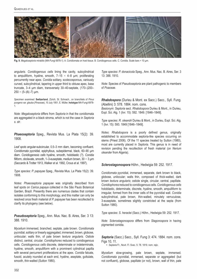
Quaedvlieg et al.
332
angularis. Conidiogenous cells lining the cavity, subcylindrical to ampulliform, hyaline, smooth, 7–15 × 4–8 µm; proliferating percurrently near apex. Conidia solitary, scolecosporous, variously curved, subcylindrical, tapering in upper third to obtuse apex, base truncate, 3–4 µm diam, transversely 30–40-septate, (170–)200–250 × (5–)6(–7) µm.
Specimen examined: Switzerland, Zürich, St. Schnach., on branchlets of Pinus pungens var. glauba (Pinaceae), 10 July 1951, E. Müller, holotype MA-Fungi 6978-1.
Note: Megaloseptoria differs from Septoria in that the conidiomata are aggregated in a black stroma, which is not the case in Septoria s. str.
Phaeoseptoria Speg., Revista Mus. La Plata 15(2): 39. 1908.
Leaf spots angular-subcircular, 0.5–3 mm diam, becoming confluent. Conidiomata pycnidial, epiphyllous, subepidermal, black, 60–90 µm diam. Conidiogenesis cells hyaline, smooth, holoblastic (?). Conidia filiform, obclavate, smooth, 1–3-euseptate, medium brown, 30 × 3 µm (Saccardo & Trotter 1913, Walker et al. 1992, Crous et al. 1997).
Type species: P. papayae Speg., Revista Mus. La Plata 15(2): 39. 1908.
Notes: Phaeoseptoria papayae was originally described from leaf spots on Carica papaya collected in the São Paulo Botanical Garden, Brazil. Presently there are numerous clades that contain isolates conforming to this morphology, and this matter can only be resolved once fresh material of P. papayae has been recollected to clarify its phylogeny (see below).
Pseudoseptoria Speg., Ann. Mus. Nac. B. Aires, Ser. 3 13: 388. 1910.
Mycelium immersed, branched, septate, pale brown. Conidiomata pycnidial, solitary or linearly aggregated, immersed, brown, globose, unilocular; walls thin, of pale brown textura angularis. Ostiole distinct, central, circular. Conidiophores reduced to conidiogenous cells. Conidiogenous cells discrete, determinate or indeterminate, hyaline, smooth, ampulliform with a prominent cylindrical papilla with several percurrent proliferations at the apex. Conidia falcate, fusoid, acutely rounded at each end, hyaline, aseptate, guttulate, smooth, thin-walled (Sutton 1980).
Type species: P. donacicola Speg., Ann. Mus. Nac. B. Aires, Ser. 3 13: 388. 1910.
Note: Species of Pseudoseptoria are plant pathogenic to members of Poaceae.
Rhabdospora (Durieu & Mont. ex Sacc.) Sacc., Syll. Fung. (Abellini) 3: 578. 1884. nom. cons.Basionym: Septoria sect. Rhabdospora Durieu & Mont., in Durieu, Expl. Sci. Alg. 1 (livr. 15): 592. 1849. [1846–1849].
Type species: R. oleandri Durieu & Mont., in Durieu, Expl. Sci. Alg. 1 (livr. 15): 593. 1849 [1846–1849].
Notes: Rhabdospora is a poorly defined genus, originally established to accommodate septoria-like species occurring on stems (Priest 2006). Of the 11 species treated by Sutton (1980), most are currently placed in Septoria. This genus is in need of revision pending the recollection of fresh material (on Nerium oleander from Algeria).
Sclerostagonospora Höhn., Hedwigia 59: 252. 1917.
Conidiomata pycnidial, immersed, separate, dark brown to black, globose, unilocular; walls thin, composed of thick-walled, dark brown textura angularis; ostiole single, circular, central, papillate. Conidiophores reduced to conidiogenous cells. Conidiogenous cells holoblastic, determinate, discrete, hyaline, smooth, ampulliform to irregular, formed from the inner cells of the pycnidial wall. Conidia subcylindrical, pale brown, thin-walled, minutely verruculose, 3-euseptate, sometimes slightly constricted at the septa (from Sutton 1980).
Type species: S. heraclei (Sacc.) Höhn., Hedwigia 59: 252. 1917.
Note: Sclerostagonospora differs from Stagonospora in having pigmented conidia.
Septoria (Sacc.) Sacc., Syll. Fung 3: 474. 1884. nom. cons. Figs 10, 11.
= Septaria Fr., Novit. Fl. Svec. 5: 78. 1819. nom. rejic.
Mycelium slow-growing, pale brown, septate, immersed. Conidiomata pycnidial, immersed, separate or aggregated (but not confluent), globose, papillate (or not), brown, wall of thin, pale
Fig. 9. Megaloseptoria mirabilis (MA-Fungi 6978-1). A. Conidiomata on host tissue. B. Conidiogenous cells. C. Conidia. Scale bars = 10 µm.

www.studiesinmycology.org
Sizing up Septoria
333
brown textura angularis, inner layer of flattened, hyaline textura angularis, frequently somewhat darker and more thick-walled around the ostiole. Ostiole single, circular, central. Conidiophores reduced to conidiogenous cells. Conidiogenous cells holoblastic, either determinate or indeterminate, proliferating sympodially and/or percurrently, hyaline, smooth, ampulliform, dolliform or lageniform to short cylindrical; scars unthickened. Conidia hyaline, multiseptate, filiform, smooth, continuous or constricted at septa. Sexual states are mycosphaerella-like.
Type species: S. cytisi Desm., Ann. Sci. Nat. Bot. 8: 24. 1847.
Specimen examined: Slovakia, Muehlthal near Bratislava (Pressburg), on leaves of Laburnum anagyroides (Leguminosae), 1884, J.A. Baeumler, BPI USO 378994.
Note: The ITS and LSU sequences of this specimen were published respectively under GenBank accession numbers JF700932 and JF700954.
Stagonospora (Sacc.) Sacc., Syll. Fung. (Abellini) 3: 445. 1884. nom. cons.Basionym: Hendersonia subgen. Stagonospora Sacc., Michelia 2 (no. 6): 8. 1880.
Conidiomata pycnidial, immersed, unilocular, globose, separate, ostiolate; walls of dark brown, thick-walled textura angularis, and on the inside of hyaline, thin-walled, flattened cells. Conidiophores reduced to conidiogenous cells. Conidiogenous cells doliiform, hyaline, with several percurrent proliferations at the apex, formed from the inner cells of the pycnidial wall. Conidia hyaline, smooth to finely verruculose, 1–multiseptate, cylindrical or fusoid-ellipsoidal, straight or slightly curved, often guttulate, constricted or not at septa.
Type species: S. paludosa (Sacc. & Speg.) Sacc., Syll. Fung. (Abellini) 3: 453. 1884.
Stenocarpella Syd. & P. Syd., Ann. Mycol. 15(3–4): 258. 1917. Fig. 12.
Mycelium immersed, brown, branched, septate. Conidiomata pycnidial, separate or sometimes confluent, globose or elongated, dark brown, subepidermal, unilocular, thick-walled; walls composed of dark brown, thick-walled textura angularis. Ostiole single, circular, papillate, protruding. Conidiophores usually absent. Conidiogenous cells cylindrical, hyaline, determinate, discrete, phialidic, with collarette and minute periclinal thickening, lining the inner layer of the pycnidial wall. Conidia subcylindrical, straight or curved, fusiform, apex obtuse, base tapered, truncate, thick-walled, smooth-walled, granular, pale to medium brown, 0–3-euseptate. Beta conidia hyaline, scolecosporous, curved (Crous et al. 2006, Lamprecht et al. 2011).
Type species: S. zeae Syd. & P. Syd., Ann. Mycol. 15(3–4): 258. 1917. [= S. macrospora (Earle) B. Sutton]
Specimens examined: South Africa, KwaZulu-Natal, Hlabisa, rain-damaged Bt Zea mays hybrid (Poaceae), 2003-04 season, J. Rheeder (ex-epitype, CBS 117560 = MRC 8615, designated in Crous et al. 2006); KwaZulu-Natal, Zea mays kernels, 2005, P. Caldwell, CPC 11863 = CBS 128560.
Notes: Stenocarpella presently contains two species, S. macrospora and S. maydis, both causing “Diplodia ear rot of maize”. These two taxa were previously assigned to Diplodia and Macrodiplodia, respectively (Petrak & Sydow 1927, Sutton 1964). Several years later, Sutton re-examining these taxa and placed them in their own genus, Stenocarpella (Sutton 1977, 1980). Recent phylogenetic studies confirmed that these taxa indeed cluster by themselves
Fig. 10. Conidia and conidiogenous cells of Septoria cytisi (BPI USO 378994). Scale bars = 10 µm.
Fig. 11. Septoria cytisi (BPI USO 378994). A. Leaf with symptoms. B. Close-up of leaf spot with conidiomata. C, D. Conidiogenous cells giving rise to conidia. E. Conidia. Scale bars = 10 µm.

Quaedvlieg et al.
334
within the Diaporthales (Crous et al. 2006, Lamprecht et al. 2011), supporting the decision of Sutton (1980).
Trichoseptoria Cavara, Atti Ist. Bot. Univ. Lab. Crittog. Pavia 2: 40. 1892.
Type species: T. alpei Cavara, Atti Ist. Bot. Univ. Lab. Crittog. Pavia 2: 40. 1892.
Notes: Not much is known about this septoria-like genus, except that it is distinguished from Septoria by having setae on its pycnidia with 1–2-septate, hyaline conidia. This genus is in further need of revision once fresh material has been recollected (Citrus vulgaris, Belgiojoso, Alps).
Zymoseptoria Quaedvlieg & Crous, Persoonia 26: 64. 2011.
Conidiomata pycnidial, semi-immersed to erumpent, dark brown to black, subglobose, with central ostiole; wall of 3–4 layers of brown textura angularis. Conidiophores hyaline and smooth, 1–2-septate, or reduced to conidiogenous cells, lining the inner cavity. Conidiogenous cells are tightly aggregated and ampulliform to doliiform or subcylindrical, phialidic with periclinal thickening, or with 2–3 inconspicuous, percurrent proliferations at the apex. Conidia (Type I) solitary, hyaline, smooth, guttulate, narrowly cylindrical to subulate, tapering towards acutely rounded apex, with bluntly rounded to truncate base, transversely euseptate; hila not thickened nor darkened. On OA and PDA media plates the aerial hyphae disarticulate into phragmospores (Type II conidia) that again give rise to Type I conidia via microcyclic conidiation; yeast-like growth and microcyclic conidiation (Type III conidia) common on agar media (Quaedvlieg et al. 2011).
Type species: Z. tritici (Desm.) Quaedvlieg & Crous, Persoonia 26: 67. 2011.
Notes: Zymoseptoria was split off from Septoria s. str. and redescribed by Quaedvlieg et al. (2011) based on LSU sequence data when said authors delimitated Septoria s. str. by sequencing the ITS and LSU sequences out of S. cytisi herbarium material. Phylogenetic analysis showed that Zymoseptoria species cluster within a distinct clade inside the Mycosphaerellaceae that is closely related to Ramularia, but located distant from Septoria s. str.
Acervular forms
Asteromidium Speg., Ann. Soc. cient. argent. 26(1): 66. 1888. Figs 13, 14.
Mycelium immersed, branched, septate, hyaline. Conidiomata acervular, subcuticular, separate or confluent, pulvinate to doliiform, at the base, composed of hyaline to pale brown, thin-walled textura angularis which extends laterally, finally with separate cells dispersed in a mucilaginous matrix to form the overlying wall; cuticle discoloured and occasionally pseudoparenchymatous, walls adjacent to the upper epidermal wall also discoloured; dehiscence irregular. Conidiogenous cells holoblastic, discrete, indeterminate, ± cylindrical, hyaline, smooth, with 1–2 sympodial proliferations, scars unthickened, flat, formed from the basal and lateral walls. Conidia cylindrical to fusoid, gently tapered at each end, apex obtuse, base truncate, thin-walled, guttulate to granular, hyaline, 3-septate (Sutton 1980).
Type species: A. imperspicuum Speg., Ann. Soc. cient. argent. 26(1): 66. 1888.
Specimen examined: Paraguay, on leaves of Sapindaceae, 1883, isotype K(M) 180228, ex B. Balansa Pl. du Paraguay No. 4085.
Notes: This genus has to be recollected (Sapindaceae, Paraguay) to allow for a molecular comparison to other existing genera in this
Fig. 12. Stenocarpella maydis (top) and S. macrospora (bottom) (redrawn from Sutton 1980). Scale bars = 10 µm.
Fig. 13. Conidia and conidiogenous cells of Asteromidium imperspicuum (redrawn from Sutton 1980). Scale bar = 10 µm.

www.studiesinmycology.org
Sizing up Septoria
335
complex. The morphology of the specimen examined correlates well with the description provided by Sutton (1980).
Ciferriella Petr., Ann. Mycol. 28(5/6): 409. 1930.
Type species: C. domingensis Petr. & Cif., Ann. Mycol. 28(5/6): 409. 1930.
= Pseudocercospora Speg., Anales Mus. Nac. Hist. Nat. B. Aires, Ser. 3, 20: 437. 1910.
Pseudocercospora domingensis (Petr. & Cif.) Quaedvlieg, Verkley & Crous, comb. nov. MycoBank MB804401. Figs 15, 16.Basionym: Ciferriella domingensis Petr. & Cif., Ann. Mycol. 28(5/6): 409. 1930.
Leaf spots amphigenous, subcircular, medium brown with dark purple margin, 1.5–6 mm diam. Sporulation hypophyllous, fasciculate to sporodochial, brown, arising from a brown stroma, up to 50 µm diam. Conidiophores medium brown, smooth, subcylindrical, 0–2-septate, straight to once geniculate, 15–20 × 3–5 µm. Conidiogenous cells terminal, brown, smooth to finely verruculose, ampulliform to subcylindrical, proliferating sympodially or percurrently, tapering to a truncate apex, 2 µm diam, 10–15 × 3–4 µm. Conidia brown, smooth, straight to slightly curved, obclavate, apex subobtuse, base obconically truncate, 0–3-septate, 35–60 × 3–4 µm.
Specimen examined: dominican republic, on Vitex umbrosa (Lamiaceae), 26 May 1929, coll. R. Ciferri, det. F. Petrak, holotype ex N.Y. Bot. Gard. No 01048475.
Notes: The dimensions of the conidia and conidiophores correlate with the observations of Sutton (1980). However, the conidiomata are sporodochial to fasciculate, and not acervular. Ciferriella domingensis is a typical Pseudocercospora sensu Crous et al. (2013). Based on the species presently known from Vitex (Crous & Braun 2003), it appears to represent a distinct taxon, for which a new combination in Pseudocercospora is proposed.
Colletogloeum Petr., Sydowia 7: 368. 1953.
Mycelium immersed, branched, septate, hyaline to pale brown. Conidiomata acervular, epidermal to subepidermal, separate, occasionally confluent, composed of hyaline to pale brown, thin-walled textura angularis. Conidiophores hyaline to pale brown, sparsely branched, septate, smooth, cylindrical or slightly irregular, formed from
the upper cells of the acervulus. Conidiogenous cells integrated or discrete, indeterminate, cylindrical or doliiform, with several percurrent proliferations at apex. Conidia hyaline to pale brown, 0-multiseptate, straight, curved or irregular, truncate at the base, obtuse at the apex, usually thin-walled, smooth, guttulate or not.
Type species: C. dalbergiae (S. Ahmad) Petr., Sydowia 7: 369. 1953. [= C. sissoo (Syd.) B. Sutton, Mycol. Pap. 97: 14. 1964.]
Notes: The exact taxonomic position of Colletogloeum was unclear for a long time as it included many species that appear to represent asexual morphs of Teratosphaeria. Crous et al. (2009a–c) used ITS sequence data from a specimen representative of C. sissoo (IMI 119162) to demonstate that the type of Colletogloeum clustered near the Pseudocercospora complex within the Mycosphaerellaceae.
Cylindrosporium Grev., Scott. crypt. fl. (Edinburgh) 1: pl. 27. 1822.
= Cylindrodochium Bonord., Handb. Allgem. mykol. (Stuttgart): 132. 1851.
Mycelium immersed, branched, septate, hyaline. Conidiomata acervular, white, slimy, subcuticular, separate or confluent, formed of pale brown to hyaline, thin-walled textura angularis; dehiscence irregular. Conidiophores hyaline, parallel, branched
Fig. 14. Asteromidium imperspicuum [K(M) 180228]. A. Conidiomata on host surface. B. Section through conidioma. C, D. Conidiogenous cells and conidia. Scale bars: B = 75 µm, all others = 10 µm.
Fig. 15. Conidia and conidiogenous cells of Pseudocercospora domingensis (NY No 01048475). Scale bars = 10 µm.

Quaedvlieg et al.
336
only at the base, 1–2-septate, smooth, formed from the upper pseudoparenchyma. Conidiogenous cells enteroblastic, phialidic, integrated, cylindrical, hyaline, smooth. Conidia straight or slightly curved, aseptate, cylindrical, thin-walled, smooth, hyaline, eguttulate (Sutton 1980).
Type species: C. concentricum Unger, Exanth. Pflanzen (Wien) 2: 9. 1833.
Notes: Sutton (1980), Von Arx (1983), Deighton (1987) and Braun (1990) could not agree on the taxonomic status of this genus, which is associated with light leaf spot of oil seed rape (sexual morph Pyrenopeziza brassicae). This genus is in need of revision, awaiting the recollection of fresh material of C. concentricum (on Pulmonaria officinalis, Germany).
Phloeospora Wallr., Fl. Crypt. Germ. (Norimbergae) 2: 176. 1833.
Mycelium immersed, septate, hyaline. Conidiomata acervular, subepidermal, circular, discrete or confluent, composed of hyaline to pale brown, thin-walled textura angularis; dehiscence irregular. Conidiophores reduced to conidiogenous cells or with one or two supporting cells, branched at base or not. Conidiogenous cells holoblastic, annellidic, occasionally also sympodial, discrete, indeterminate hyaline, smooth, cylindrical, with several apical inconspicuous annellations, formed from the upper cells of the acervuli. Conidia hyaline, septate, smooth, guttulate or not, cylindrical, curved, attenuated towards the apices, apex obtuse to subobtuse, base truncate, with minute marginal frill.
Type species: P. ulmi (Fr.) Wallr., Fl. Crypt. Germ. (Norimbergae) 2: 177. 1833.
Notes: Sexual morphs of Phloeospora have been linked to genera that resemble the concepts of Mycosphaerella, Didymella and Sphaerulina. Verkley & Priest (2000) already noted that this genus is heterogeneous and in need of revision. The phylogenetic analysis performed in this study confirmed that Phloeospora (based on P. ulmi) clusters close to, but separate from Septoria s. str. (Fig. 1).
Phloeosporella Höhn., Ann. Mycol. 22: 201. 1924. Fig. 17.
Mycelium immersed, branched, septate, hyaline. Conidiomata acervular, subepidermal, ± circular, discrete, composed of hyaline
to pale brown, thin-walled textura angularis. Conidiogenous cells holoblastic, sympodial, discrete, indeterminate, hyaline, smooth, lageniform to cylindrical, with 1–2 broad, flat unthickened apical scars, formed from the upper pseudoparenchyma. Conidia hyaline, 2-euseptate, thin-walled, smooth, guttulate, straight, curved or irregular, tapered gradually to an obtuse apex and abruptly to a truncate base (Sutton 1980).
Type species: P. ceanothi (Ellis & Everh.) Höhn., Ann. Mycol. 22(1–2): 201. 1924.
Notes: Not much is known of the sexual state of this genus, but P. padi has been linked to Blumeriella jaapii (Sutton 1980). A phylogenetic analysis performed on available isolates (unpubl. data) indicated that Phloeosporella is polyphyletic. However, as the type is not known from culture (on Ceanothus, California, USA), this matter could not be resolved.
Septogloeum Sacc., Michelia 2(6): 11. 1880.
Mycelium immersed, branched, septate, hyaline. Conidiomata acervular, epidermal to subepidermal, separate or confluent, formed
Fig. 17. Conidia and conidiogenous cells of Phloeosporella ceanothi (redrawn from Sutton 1980). Scale bar = 10 µm.
Fig. 16. Pseudocercospora domingensis (NY No 01048475). A. Leaf spot. B, C. Conidiogenous cells. D. Conidia. Scale bars = 10 µm.

www.studiesinmycology.org
Sizing up Septoria
337
of pale brown thin-walled pseudoparenchyma. Conidiophores short, stout, 1–2-septate, hyaline, smooth, branched at the base, formed from the upper pseudoparenchyma. Conidiogenous cells phialidic, discrete or integrated, determinate, cylindrical, doliiform to obpyriform, hyaline, smooth, with minute collarette and prominent periclinal thickening. Conidia hyaline, 1–3-euseptate, thin-walled, smooth, eguttulate, base truncate, apex obtuse, straight or curved, constricted, obovoid (Sutton 1980).
Type species: S. carthusianum (Sacc.) Sacc., Michelia 2(6): 11. 1880.
Notes: Although more than 120 species of Septogloeum have been described, the genus was reduced to just two species by Sutton & Pollack (1974). Sexual morphs have been linked to Pleuroceras in the Diaporthales (Monod 1983). The genus is in need of revision pending fresh collections.
Xenocylindrosporium Crous & Verkley, Fungal Planet 44. 2009.
Conidiomata immersed, black, opening by irregular rupture, acervuloid, up to 300 μm diam; wall consisting of 3–4 layers of pale brown textura angularis. Conidiophores hyaline, smooth, subcylindrical, branched, septate, or reduced to ampulliform conidiogenous cells. Conidiogenous cells hyaline, smooth, ampulliform to subcylindrical, terminal or lateral on septate conidiophores, monophialidic with minute periclinal thickening. Conidia solitary, hyaline, smooth, curved, widest in middle, tapering to acutely rounded apex and truncate base, 0 –1-septate.
Type species: X. kirstenboschense Crous & Verkley, Fungal Planet 44. 2009.
Stromatic forms
Dothistroma Hulbary, Bull. Ill. Nat. Hist. Surv. 21: 235. 1941.
Mycelium immersed, branched, septate, pale brown to hyaline. Conidiomata sometimes acervular, initially subepidermal later erumpent, composed of pale brown, thin-walled textura angularis, sometimes eustromatic, multilocular and of darker brown, thick-walled tissue. Stromata are strongly erumpent and finally pulvinate. Conidiogenous cells holoblastic, discrete, determinate, ampulliform, hyaline, smooth, non-proliferating, formed from the upper cells of stroma or from inner cells of the locular walls. Conidia hyaline, straight or curved, filiform, 1–5-euseptate, continuous, thin-walled and smooth (Barnes et al. 2004).
Type species: D. pini Hulbary, Bull. Ill. Nat. Hist. Surv. 21: 235. 1941.
Notes: Dothistroma sexual morphs are mycosphaerella-like (Evans 1984), and the two species of Dothistroma that have been subjected to DNA sequencing (D. septosporum and D. pini) cluster together in the “Dothistroma clade” as described by Crous et al. (2009a, c). Because of a lack of recognisable morphological characteristics, it is virtually impossible to discriminate between D. septosporum and D. pini without molecular tools (Barnes et al. 2004). Multiple morphological varieties of both D. septosporum and D. pini have
been described based on differences in conidia length alone (e.g. D. septosporum var. keniense). However, controversy exists as to whether spore size represents an adequate characteristic to distinguish among these Dothistroma varieties, as since the introduction of molecular tools only D. septosporum and D. pini have been confirmed as distinct species.
Phlyctaeniella Petr., Ann. Mycol. 20: 323. 1922. Fig. 18.
Mycelium immersed, branched, septate, hyaline. Conidiomata eustromatic, separate, immersed, pale brown, globose, unilocular, scarcely erumpent; side wall and base of several cell layers of hyaline, thin-walled textura angularis, above of larger pale brown tissue. Ostiole indistinct, and dehiscence by rupture of the upper wall. Conidiophores hyaline, smooth, septate, irregularly branched, especially at the base, formed from the inner cells of the stroma wall. Conidiogenous cells phialidic, integrated or discrete, determinate, hyaline, markedly tapered at the apices, smooth, with apical or lateral apertures, collarette minute, with periclinal thickening; only rarely becoming percurrent. Conidia hyaline, smooth, thin-walled, irregularly guttulate, filiform, straight, curved or irregular, multiseptate (Sutton 1980).
Type species: P. polonica Petr., Ann. Mycol. 20: 323. 1922.
Note: Fresh material needs to be collected of this taxon (on Aruncus silvestris, Austria), before its taxonomy can be resolved.
Septocyta Petr., Ann. Mycol. 25: 330. 1927. Figs 19, 20.
Mycelium immersed, branched, septate, hyaline to pale brown. Conidiomata eustromatic, immersed, separate, erumpent, dark brown to black, finally opening widely, unilocular, multilocular or convoluted, thick-walled; wall of pale brown, thin-walled textura angularis except in the dehiscent region which is darker brown and more thick-walled. Ostiole absent, dehiscence by breakdown of the upper wall. Conidiogenous cells are holoblastic, sympodial with 1–3
Fig. 18. Conidia and conidiogenous cells of Phlyctaeniella humuli (IMI 202260) (redrawn from Sutton 1980). Scale bar = 10 µm.

Quaedvlieg et al.
338
apical, scarcely protruding, unthickened denticles, indeterminate, discrete, ampulliform to lageniform, hyaline, smooth, formed from the inner cells of the locular walls. Conidia hyaline, 1–3 euseptate, smooth, straight or slightly curved, acicular, apex obtuse, base truncate, often with minute guttules associated with septa (Sutton 1980).
Type species: S. ramealis (Roberge ex Desm.) Petr., Ann. Mycol. 25: 330. 1927.
Septocyta ramealis (Roberge ex Desm.) Petr., Ann. Mycol. 25: 330. 1927.
Conidiomata eustromatic to pycnidial, black, up to 160 µm diam, aggregated in clusters, erumpent through ruptures in epidermis, convulated; wall of 3–8 layers of brown textura angularis. Conidiophores lining the inner cavity, reduced to conidiogenous cells, or one or two supporting cells. Conidiogenous cells hyaline, smooth, ampulliform, proliferating sympodially and percurrently near apex, also with lateral denticle-like protrusions, 6–12 × 2.5–4 µm. Conidia hyaline, smooth, guttulate, (9–)20–30(–35) × 1.5(–2) µm, 1(–3)-septate, irregularly curved, subcylindrical, apex obtuse, base tapering slightly to truncate hilum, 0.5 µm diam.
Specimen examined: Germany, Brandenberg, on Rubus fructicosus (Rosaceae), 7 June 1923, coll. P. Sydow, det. H. Sydow, Sydow Mycoth. Germ. PDD 51271.
Notes: Septocyta ramealis, the type of Septocyta, has a long list of synonyms. The specimen examined here (PDD 51271), differs somewhat from the description provided by Sutton (1980), and appears to represent a species of Septoria s. str., as the mode of conidiogenesis is not that different. Presently there is a single ITS sequence labelled as S. ruborum available on GenBank (JN133277.1), placing it in the middle of Septoria s. str. As no type material of S. ramealis could be located, this matter remains unresolved.
Septopatella Petr., Ann. Mycol. 23: 128. 1925.
Mycelium immersed, branched, septate, hyaline to subhyaline. Conidiomata superficial, often subtended by a superficial, pale brown, septate, branched mycelium, pulvinate, separate to occasionally aggregated, dark brown to black, finally opening widely, cupulate; basal wall of small-celled, brown, thin-walled textura angularis, becoming textura porrecta as it merges into the periclinal walls; a hypostroma attaches the conidioma to the substrate; Ostiole absent. Conidiophores hyaline, septate, branched at the base, thin-walled, cylindrical, formed from the gelatinized basal wall of the conidioma. Conidiogenous cells holoblastic, sympodial, integrated, indeterminate, cylindrical, hyaline, smooth, produced as 2–3 branches from the apex of the conidiophores. Conidia hyaline, 3–4-euseptate, thin-walled, smooth, minutely guttulate, straight or curved, occasionally irregularly filiform (Dyko & Sutton 1979, Sutton 1980).
Type species: S. septata (Jaap) Petr., Ann. Mycol. 23: 129. 1925.
Note: Not much is known about this genus, and as no cultures of S. septata are presently available (on Pinus montana, Czech Republic) this matter cannot be resolved.
Stictosepta Petr., Sydowia 17: 230. 1964. [1963]. Fig. 21.
Mycelium immersed, branched, septate, hyaline. Conidiomata eustromatic, immersed, globose to collabent, papillate, unilocular, often convoluted, hyaline; walls thick, of hyaline, thin-walled
Fig. 19. Conidia and conidiogenous cells of Septocyta ramealis (PDD 51271). Scale bars = 10 µm.
Fig. 20. Septocyta ramealis (PDD 51271). A. Conidiomata on host tissue. B, C. Conidiogenous cells. D. Conidia. Scale bar = 10 µm.

www.studiesinmycology.org
Sizing up Septoria
339
textura intricata. Ostiole central and circular, single, furfuraceous. Conidiophores hyaline, septate, branched, anastomosing, formed from the inner cells of the locular wall. Conidiogenous cells sympodial or synchronous, integrated, indeterminate, hyaline, thin-walled, with usually two small, unthickened, apical, slightly protuberant conidiogenous loci. Conidia hyaline, thin-walled, smooth, multiseptate, slightly constricted at the septa, each cell medianly guttulate, straight or curved, base truncate, apex obtuse (Sutton 1980).
Type species: S. cupularis Petr., Sydowia 17: 230. 1964. [1963].
Note: Not much is known about this genus, but as no isolate of S. cupularis is presently available (on stems of Fraxinus, Czech Republic), it will not be treated here.
Sexual morphs linked to Septoria
Several sexual genera have been linked to Septoria and allied genera in literature, but very few have been confirmed in culture. Most sexual states cluster in the Mycosphaerella complex.
Mycosphaerella Johanson, Öfvers. K. Svensk. Vetensk.-Akad. Förhandl. 41(no. 9): 163. 1884.
= Ramularia Unger, Exanth. Pflanzen (Wien): 119. 1833.
Mycelium immersed to superficial, septate, hyaline, branched. Caespituli usually whitish to greyish on host tissue. Conidiophores fasciculate to synnematal, rarely solitary, or forming small sporodochia, emerging through stomata, from inner hyphae or stromata; conidiophores straight, subcylindric to geniculate-sinuous, aseptate or septate, hyaline, occasionally branched, smooth, rarely rough. Conidiogenous cells integrated, terminal, polyblastic, elongating sympodially, apex more or less straight to geniculate-sinuous or strongly curved, cicatrized, conidial scars hardly to conspicuously thickened, but always darkened, refractive. Conidia solitary to catenate, sometimes in branched chains, 0–4(–multi)-septate, hyaline, ellipsoid-ovoid to cylindrical-fusoid, rarely filiform, occasionally constricted at the septa, smooth
to verruculose-echinulate; hila distinct, slightly to conspicuously thickened, darkened, refractive; conidial secession schizolytic. Ascomata immersed to superficial, uniloculate, globose to subglobose with papillate, central, periphysate ostiole, dark brown to black, scattered or gregarious. Peridium of 3–6 layers of thin- to thick-walled textura angularis, dark brown to black. Hamathecium dissolves at maturity, and no stromatic tissue remains between the asci. Asci bitunicate, fissitunicate, 8-spored, cylindrical to cylindric-clavate, ovoid to ampulliform or saccate, sessile to subsessile, apex rounded with distinct or indistinct ocular chamber. Ascospores bi- to tri- or multiseriate, ellipsoid-fusoid to obclavate or subcylindrical, hyaline, medianly 1-septate, often constricted at the septum, smooth-walled, granular to guttulate, mostly lacking any sheath.
Type species: Ramularia pusilla Unger, Exanth. Pflanzen (Wien): 169. 1833.
Notes: Species of Ramularia (including the Mycosphaerella sexual morph) have evolved over a broad developmental and physiological adaptation range that includes endophytes, saprophytes and symbionts. However, for a major part Ramularia consists of a wide range of narrow host range, foliicolous plant pathogens which are the cause of significant economical losses in both temperate and tropical crops worldwide (Crous et al. 2001). Verkley et al. (2004) showed that Mycosphaerella s. str. (linked to M. punctiformis) was in fact restricted to species with Ramularia anamorphs, leaving many “Mycosphaerella” species to be disposed to other genera. In employing the one fungus = one name concept (Hawksworth et al. 2011, Wingfield et al. 2012), the choice is to use Ramularia over Mycosphaerella, as the former is monophyletic and recently monographed (Braun 1995, 1998), while Mycosphaerella is poly- and paraphyletic, and consists of more than 40 genera, many as yet untreated (Crous et al. 2009c)
Sphaerulina Sacc., Michelia 1(no. 4): 399. 1878.
Ascomata pseudothecial, immersed, subepidermal, erumpent at the top, single to clustered, globose, papillate. Ostiole central, with hyaline periphyses; wall of textura angularis, composed of 2–4 layers of brown cells. Hamathecium dissolving at maturity. Asci bitunicate, fissitunicate, clustered, cylindrical to obclavate, rounded at apex, with or without a shallow apical chamber, short-stipitate or sessile, with 8 biseriate to triseriate ascospores. Ascospores subcylindrical to fusiform, rounded at ends, slightly tapered, straight or slightly curved, 1–3-septate, with a primary septum nearly median, hyaline, smooth, without sheath or appendages.
Type species: Sphaerulina myriadea (DC.) Sacc., Michelia 1(no. 4): 399. 1878.
Notes: The genus Sphaerulina was chiefly separated from Mycosphaerella on the basis of ascospore septation (Crous et al. 2011). Sphaerulina myriadea, which occurs on hosts in the Fagaceae, appears to be a species complex. Results in this paper show that Sphaerulina myriadea clusters together with many septoria-like species in a clade that is distinct, but very closely related to Septoria s. str. The septoria-like species in this Sphaerulina clade were subsequently rediscribed in Sphaerulina. Species including ones with 1-septate ascospores and septoria-like asexual morphs are treated below and by Verkley et al. (2013).
Fig. 21. Conidia and conidiogenous cells of Stictosepta cupularis (redrawn from Sutton 1980). Scale bar = 10 µm.

Quaedvlieg et al.
340
Treatment of phylogenetic clades
Based on the phylogenetic data generated in this study, we were able to delineate several clades in the Septoria complex. Recognised clades, as well as novel species and genera, are described and discussed below. Taxa with descriptions that are freely available online in MycoBank or open access journals, are not repeated here.
clade 1: Septoria
Description: See above.
Type species: S. cytisi Desm., Ann. Sci. Nat. Bot. 8: 24. 1847.
Septoria cf. agrimoniicola Bondartsev, Mater. mikol. obslêed. Ross. 2: 6. 1921.
Leaf spots on the upper leaf surface, distinct, scattered, brown with purplish margin, circular to angular, sometimes vein-limited, discrete lesions 2–4 mm diam, reaching 10 mm wide when confluent, finally the center becoming pale colored to nearly whitish; on the lower leaf surface similar but discoloured (Shin & Sameva 2004). On sterile Carex leaves on WA. Conidiomata pycnidial, separate but frequently aggregated and linked by brown stromatic tissue in a stroma; globose, black, exuding a creamy conidial mass via a central ostiole; conidiomata up to 350 µm diam; wall of 6–12 layers of dark brown, thick-walled textura angularis. Conidiophores reduced to conidiogenous cells or 1–2 supporting cells, hyaline, subcylindrical, lining the inner layer of conidioma. Conidiogenous cells hyaline, smooth, subcylindrical to ampulliform, 10–17 × 3–4 µm; proliferating sympodially but also percurrently near apex. Conidia hyaline, smooth, guttulate, filiform, apex subobtuse, base long obconically truncate, 1–4-septate, (20–)25–35(–40) × 1.5–2(–2.5) µm; microcyclic conidiation observed.
Culture characteristics: Colonies on PDA flat, undulate with sparse, white aerial mycelium, surface olivaceous-black, reverse olivaceous-black, after 14 d, 3.5 cm diam; on MEA with sparse white aerial mycelium, surface olivaceous-black, reverse olivaceous-black, after 14 d, 5 cm diam; on OA with sparse white aerial mycelium, surface olivaceous, reverse olivaceous, after 14 d, 3 cm diam.
Specimen examined: South Korea, Guri, on leaves of Agrimonia pilosa (Rosaceae), 11 Jul. 2009, H.D. Shin (CBS H-21279, culture CBS 128602 = KACC 44644 = SMKC 24292).
Notes: This fungus was first reported from Korea by Shin & Sameva (2002) as S. agrimoniicola, and fits well with the original description of this European taxon. However, fresh European collections and cultures are required for comparison, as S. agrimoniicola may well be restricted to Europe.
Septoria cf. stachydicola Hollós, Mathem. Természettud. Közlem. Magg. Tudom. Akad. 35(1): 60. 1926.
Leaf spots on the upper leaf surface distinct, scattered, brown with purplish margin, circular to angular, sometimes vein-limited, discrete lesions 2–4 mm diam, reaching 10 mm wide when
confluent, finally the center becoming paler or nearly whitish; on the lower leaf surface similar but discoloured (Shin & Sameva 2004). On OA. Conidiomata solitary to aggregated, black, globose, becoming somewhat papillate, up to 250 µm diam, opening by means of central ostiole, up to 40 µm diam; wall of 6–8 layers of thick-walled, brown textura angularis; exuding a creamy conidial mass. Conidiophores reduced to conidiogenous cells. Conidiogenous cells lining the inner wall layer, hyaline, discrete, ampulliform to lageniform, 4–10 × 3–5 µm, proliferating sympodially or percurrently with inconspicuous proliferations. Conidia filiform, curved or flexuous, rarely straight, (60–)65–75(–90) × 1.5–2(–3) µm, hyaline, guttulate, 4–7(–11)–septate, apex subobtuse, slightly tapering from basal septum to truncate base, 1.5–2 µm.
Culture characteristics: Colonies on PDA erumpent, with feathery margin, with sparse white aerial mycelium, surface greenish-black, reverse olivaceous-black, after 14 d, 2.5 cm diam; on MEA with sparse white aerial mycelium, surface cinnamon to olivaceous-black in the younger patches, reverse cinnamon to olivaceous-black in patches, after 14 d, 4 cm diam; on OA with sparse white aerial mycelium, surface greenish-black, reverse fuscous-black, after 14 d, 3 cm diam.
Specimen examined: South Korea, Incheon, leaf of Stachys riederi var. japonica (Lamiaceae), 14 Aug. 2008, H.D. Shin (CBS H-21278, culture CBS 128668 = KACC 44796 = SMKC 24663).
Note: The Korean collection was originally identified as Septoria stachydicola, which fits the original description provided for this taxon (Shin & Sameva 2004). However, authentic European material is required for a comparison to confirm this identification, as we suspect S. stachydicola may be restricted to Europe.
Septoria cretae Quaedvlieg, Verkley & Crous, sp. nov. MycoBank MB804402. Figs 22, 23.
Etymology: Named after Crete, the island from where it was collected.
On sterile Carex leaves on WA. Conidiomata up to 250 µm diam, brown, immersed, subepidermal, pycnidial, subglobose with central
Fig. 22. Conidia and conidiogenous cells of Septoria cretae (CBS 135095). Scale bar = 10 µm.

www.studiesinmycology.org
Sizing up Septoria
341
ostiole, exuding creamy conidial mass; wall of 2–3 layers of brown textura angularis. Conidiophores reduced to conidiogenous cells, or with a supporting cell that gives rise to several conidiogenous cells. Conidiogenous cells phialidic, hyaline, smooth, aggregated, lining the inner cavity, ampulliform to subcylindrical, straight to curved, proliferating sympodially near apex, 10–20 × 2–3.5 µm. Conidia hyaline, smooth, thin-walled, subcylindrical to narrowly obclavate, granular, with subobtuse apex and obconically truncate to truncate base, 1–3-septate, (8–)15–22(–27) × 2(–3) µm.
Culture characteristics: Colonies on PDA erumpent, with feathery margin, lacking aerial mycelium, surface fuscous-black, reverse olivaceous-black, after 14 d, 3.5 cm diam; on MEA surface fuscous-black, reverse olivaceous-black, after 14 d, 4 cm diam; on OA surface fuscous-black, reverse fuscous-black, after 14 d, 3.5 cm diam.
Specimen examined: Greece, Crete, on leaves of Nerium oleander (Apocynaceae), 7 Jul. 2012, U. Damm, (holotype CBS H-21277, culture ex-type CBS 135095).
Notes: Several species of Septoria are known on Nerium oleander, namely S. juliae [conidia 1–6(–7)-septate, 26–54 × 2.5–5.5 µm], S.
neriicola (conidia 1-septate, 30–40 × 0.7–1 µm), S. oleandriicola [conidia 1–3-septate, 12.5–22.5–37.5(–40) × 2.5–3(–4.5) µm], S. oleandrina (conidia 0–1-septate, 9–19 × 1–1.5 µm), and S. roll-hansenii (conidia 0–4-septate, 25–39 × 3–4 µm) (Bedlan 2011), which differ from S. cretae based on conidial dimensions and septation.
Septoria glycinicola Quaedvlieg, H.D. Shin, Verkley & Crous, sp. nov. MycoBank MB804403. Fig. 24.
Etymology: Named after the host genus on which it was collected, Glycine.
On OA. Conidiomata forming in concentric circles, pycnidial, separate, black, globose, up to 150 µm diam, opening by a central ostiole, up to 30 µm diam, exuding a creamy conidial mass; wall consisting of 3–6 layers of brown textura angularis. Conidiophores reduced to conidiogenous cells. Conidiogenous cells lining the inner cavity, hyaline, smooth, ampulliform, 10–16 × 2.5–3.5 µm, proliferating sympodially near apex, holoblastic.
Fig. 23. Septoria cretae (CBS 135095). A. Colony sporulating in culture. B–F. Conidiophores and conidiogenous cells giving rise to conidia. G. Conidia. Scale bars = 10 µm.
Fig. 24. Septoria glycinicola (CBS 128618). A, B. Colonies sporulating on PDA. C. Conidiogenous cells. D. Conidia. Scale bars = 10 µm.

Quaedvlieg et al.
342
Conidia hyaline, smooth, guttulate to granular, subcylindrical to narrowly obclavate, irregularly to gently curved, apex subobtuse, base long obconically truncate, 3–6-septate, (33–)45–55(–65) × (1.5–)2 µm.
Culture characteristics: Colonies on PDA flat, circular, with sparse black aerial mycelium with black tufts, surface patches of olivaceous-black to fawn in the younger parts, reverse with patches of olivaceous-black in the older parts to mouse-grey and pale purplish grey in the younger mycelium, after 14 d, 6.5 cm diam, pinkish exudate; on OA lobate, with sparse white aerial mycelium, surface patches of vinaceous to olivaceous-black, reverse fuscous-black to vinaceous-buff; after 14 d, 8.5 cm diam, pinkish exudate; on MEA with radial lobes, very short white aerial mycelium, surface fuscous-black, reverse olivaceous-black; after 14 d, 4.5 cm diam.
Specimen examined: South Korea, Namyangju, on leaves of Glycine max (Fabaceae), 22 Sep. 2008, H.D. Shin (holotype CBS H-21270, culture ex-type CBS 128618 = KACC 43091 = SMKC 22879).
Notes: Septoria glycines is the common Septoria species associated with brown spot of soybeans. Septoria glycinicola is distinct from S. glycines (conidia 1–4 septate, 21–45 × 1.5–2 µm) in that it has larger conidia.
Septoria oenanthicola Quaedvlieg, H.D. Shin, Verkley & Crous, sp. nov. MycoBank MB804405. Fig. 25.
Etymology: Named after the host genus from which it was collected, Oenanthe.
On sterile Carex leaves on WA. Conidiomata pycnidial, separate but aggregated, black, globose, up to 200 µm diam, opening by central ostiole, up to 20 µm diam, exuding a creamy conidial mass; wall consisting of dark brown, thickened, 6–10 layers of textura angularis. Conidiophores reduced to conidiogenous cells or to one
supporting cell. Conidiogenous cells hyaline, smooth, 3–5 × 3–7 µm, ampulliform, proliferating sympodially near apex, holoblastic. Conidia hyaline, smooth, guttulate, subcylindrical to narrowly obclavate, apex subobtuse, base long obconically truncate, 1–6-septate, (17–)25–45(–55) × (2–)2.5(–3) µm.
Culture characteristics: Colonies on PDA flat, undulate with sparse, white aerial mycelium, surface olivaceous-grey, reverse olivaceous, after 14 d, 2.5 cm diam; on MEA with sparse, white aerial mycelium, surface olivaceous-grey, reverse olivaceous-black, after 14 d, 5 cm diam; on OA with sparse white aerial mycelium, surface olivaceous-grey, reverse olivaceous, after 14 d, 3 cm diam.
Specimen examined: South Korea, Yangpyeong, on leaves of Oenanthe javanica (Apiaceae), 25 May 2006, H.D. Shin (holotype CBS H-21281, culture ex-type CBS 128649 = KACC 42394 = SMKC 21807).
Notes: This fungus was originally recorded from Korea by Shin (1998) as Septoria oenanthis. However, conidia of Korean specimens (30–60 ×1.5–2.5 µm; Shin & Sameva 2004) are much larger than that of the American type collection (20–35 × 1.5–2 µm; Saccardo 1895), and therefore better treated as a separate taxon.
Septoria pseudonapelli Quaedvlieg, H.D. Shin, Verkley & Crous, sp. nov. MycoBank MB804404. Fig. 26.
Etymology: Named after its morphological similarity to Septoria napelli.
Leaf spots on the upper leaf surface, scattered to confluent, distinct, angular to irregular, usually vein-limited, small to large, up to 30 mm when confluent, at first appearing small angular brown discoloration, later turning blackish brown with or without distinct border line, finally central area becoming blackish and surrounded by pale greenish margin; on the lower leaf surface similar but discoloured (Shin
Fig. 25. Septoria oenanthicola (CBS 128649). A. Colony sporulating on MEA. B. Section through conidiomata. C–G. Conidiogenous cells. H. Conidia. Scale bars: B = 200 µm, all others = 10 µm.

www.studiesinmycology.org
Sizing up Septoria
343
& Sameva 2004). On sterile Carex leaves on WA. Conidiomata pycnidial, separate, black, globose, papillate with short neck (at times 1–2 necks develop), up to 250 µm wide, 500 µm high with central ostiole; wall of 5–7 layers of brown textura angularis. Conidiophores reduced to conidiogenous cells. Conidiogenous cells ampulliform, lining the inner cavity, hyaline, smooth, with sympodial or apical percurrent proliferation, 10–13 × 5–7 µm. Conidia filiform, curved to flexuous, (50–)75–90(–100) × (2.5–)3(–3.5) µm, hyaline, guttulate, 4–10-septate, apex subobtuse, base obconically truncate, 2 µm diam.
Culture characteristics: Colonies on PDA flat, undulate with sparse, white aerial mycelium, surface olivaceous-black, reverse olivaceous-black, after 14 d, 2 cm diam; on MEA with sparse white aerial mycelium, surface olivaceous-black, reverse olivaceous-black, after 14 d, 4 cm diam; on OA with sparse white aerial mycelium, surface olivaceous, reverse olivaceous, after 14 d, 2 cm diam.
Specimen examined: South Korea, Chuncheon, on leaves of Aconitum pseudolaeve var. erectum (Ranunculaceae), 4 Sep. 2008, H.D. Shin (holotype CBS H-21280, culture ex-type CBS 128664 = KACC 43952 = SMKC 23638).
Notes: This taxon was originally reported as Septoria napelli from Korea by Shin & Sameva (2004), and broadly corresponds with the original description provided for this taxon (Petrak 1957). However, we have examined European material authentic for the name (see Verkley et al. 2013, this issue), from which the Korean fungus is genetically different. Based on these observations we describe the Korean collection as new.
clade 2: Sphaerulina
Sphaerulina Sacc., Michelia 1(no. 4): 399. 1878.
Description: See above.
Type species: Sphaerulina myriadea (DC.) Sacc., Michelia 1(no. 4): 399. 1878.
Specimens examined: Germany, Driesen, Lasch [Rabenhorst, Fungi Eur. no. 149] (L). Japan, Aomori, Tsugaru, Kidukuri, Bense-marsh (40°51′53″ N, 140°17′42″E), on leaves of Q. dentata, 21 Apr. 2007, K. Tanaka 2243 (HHUF 29940; single ascospore culture CBS 124646 = JCM 15565). uK, on leaves of Quercus robur
(Fagaceae), J.E. Vize [Microfungi Brit. Ex. No. 195] (ex IMI 57186, K(M) 167735).uSA, California: Sequoia National Park. alt. 2590 m, on leaves of Castanopsis sempervirens, 18 Jun. 1931, H.E. Parks (BPI 623686); Lake Co., Hoberg’s Resort, on leaves of Q. kelloggii, 15 May 1943, V. Miller (BPI 623707); Maryland, Marlboro, on leaves of Q. alba, 26 Apr. 1929, C.L. Shear ( BPI 623705); Texas, Houston, on leaves of Q. alba, 8 Apr. 1869, H.W. Ravenel (BPI 623704).
Notes: Sivanesan (1984) linked Sphaerulina to Septoria, Cercospora and Cercosporella asexual morphs, though these were never confirmed based on DNA data. The latter two genera have since been shown to be distinct (Crous et al. 2013, Groenewald et al. 2013; this volume), which leaves septoria-like asexual morphs such as Sphaerulina rubi Demaree & Wilcox (linked to Cylindrosporium rubi Ellis & Morgan), and S. rehmiana (linked to Septoria rosae), which confirms the results obtained here (Fig. 1).
Sphaerulina abeliceae (Hiray.) Quaedvlieg, Verkley & Crous, comb. nov. MycoBank MB804406. Basionym: Septoria abeliceae Hiray., Mem. Col. Agr. Kyoto. Imp. Univ. 13(3): 33. 1931.
Specimen examined: South Korea, Jeonju, on leaves of Zelkova serrata (Ulmaceae), 29 Oct. 2006, H.D. Shin, CBS 128591 = KACC 42626.
Sphaerulina amelanchier Quaedvlieg, Verkley & Crous, sp. nov. MycoBank MB804407. Figs 27, 28.
Etymology: Named after the host genus from which it was collected, Amelanchier.
On sterile Carex leaves on WA. Conidiomata pycnidial, brown, separate, immersed, globose, up to 150 µm diam, exuding a creamy conidial mass via central ostiole; wall of 3–6 layers of brown textura angularis. Conidiophores reduced to conidiogenous cells. Conidiogenous cells hyaline, smooth, subcylindrical, irregularly curved, branched to once geniculate-sinuous, 5–20 × 3–4 µm; proliferating sympodially. Conidia hyaline, smooth, guttulate, filiform, narrowly obclavate, apex subacutely rounded, base long obconically truncate, 1–8-septate, (25–)40–55(–60) × (1.5–)2(–2.5) µm; microcyclic conidiation common. Ascomata globose, brown,
Fig. 26. Septoria pseudonapelli (CBS 128664). A. Colony sporulating on PDA. B. Section through conidioma. C–E. Conidiogenous cells. F. Conidia. Scale bars: B = 125 µm, all others = 10 µm.

Quaedvlieg et al.
344
separate, immersed to erumpent, up to 150 µm diam. Asci broadly ellipsoid to obclavate, 22–35 × 7–9 µm; apical chamber visible, 1–1.5 µm diam. Ascospores fusoid-ellipsoid, hyaline, smooth, granular, not to slightly constricted at median septum, widest just above septum, prominently curved, (13–)17–20(–25) × (2.5–)3(–3.5) µm. Ascospores germinating from both ends, with germ tubes parallel to the long axis, developing lateral branches and becoming constricted at septum, 3–4 µm diam.
Culture characteristics: Colonies on PDA radially striate with lobate edge, sparse white aerial mycelium, surface fuscous-black to buff for the younger tissue, reverse cinnamon to olivaceous-black, after
14 d, 3 cm diam; on MEA surface patches of hazel to fawm to fuscous-black, reverse sepia to olivaceous-black, after 14 d, 4.5 cm diam; on OA surface pale-vinaceous to fuscous-black, reverse cinnamon to fuscous-black, after 14 d, 3 cm diam.
Specimen examined: Netherlands, Houten, on leaf litter of Amelanchier sp. (Rosaceae), 28 Mar. 2012, S. Videira (holotype CBS H-21282, culture ex-type CBS 135110 = MP8 = S544).
Note: Presently there are no known species of septoria-like fungi known from Amelanchier. Phylogenetically, it is similar to Sphaerulina rhabdoclinis (conidia 8–30 × 1.5–2 µm), which infects needles of Pseudotsuga menziesii. Phylogenetically similar
Fig. 27. Conidia, conidiogenous cells, ascospore and ascus of Sphaerulina amelanchier (CBS 135110). Scale bars = 10 µm.
Fig. 28. Sphaerulina amelanchier (CBS 135110). A. Colony on PDA. B. Conidiogenous cells. C. Ascomata on host tissue. D. Germinating ascospore. E, F. Asci. G. Ascospores. H. Conidia. Scale bars = 10 µm.

www.studiesinmycology.org
Sizing up Septoria
345
isolates occur on Betula, Castanea and Quercus. More isolates and molecular data are required to resolve this complex.
Sphaerulina azaleae (Voglino) Quaedvlieg, Verkley & Crous, comb. nov. MycoBank MB804408.Basionym: Septoria azaleae Voglino, Syll. Fung. (Abellini) 14(2): 976. 1899.
≡ Phloeospora azaleae (Voglino) Priest, Fungi of Australia: 224. 2006.
Specimens examined: Belgium, on leaves of Rhododendron sp. (Ericaceae), J. van Holder, CBS 352.49. South Korea, Hongcheon, on leaves of Rhododendron sp., 18 Oct. 2009, H.D. Shin, KACC 44865 = CBS 128605.
Sphaerulina berberidis (Niessl) Quaedvlieg, Verkley & Crous, comb. nov. MycoBank MB804409. Basionym: Septoria berberidis Niessl, in Rabenhorst, Bot. Ztg. 24: 411. 1866.
= Sphaerella berberidis Auersw., in Gonnermann & Rabenhorst, Mycol. eur. Abbild. Sämmtl. Pilze Eur. 5-6: 3. 1869 (nom. nov. for Sphaeria berberis Nitschke ex Fuckel).
≡ Mycosphaerella berberidis (Auersw.) Lindau, in Engler & Prantl, Nat. Pflanzenfam., Teil. I (Leipzig) 1(1): 424. 1897.
Description in vitro (CBS 116724): Colonies on OA 16–20 mm diam after 14 d, with an even, colourless margin; colonies spreading to restricted, somewhat elevated in the centre, the surface covered by a dense mat of pure white, woolly aerial mycelium; reverse in the centre dark brick to brown vinaceous, surrounded by cinnamon tinges; small amounts of a yellow to greenish pigment diffusies into the surrounding medium. Colonies on MEA 8–10 mm diam after 14 d, with an even to slighlty ruffled vinaceous buff margin; colonies restricted, pustulate, the surface ochraceous or darker, with diffuse to locally more dense finely felted grey aerial mycelium; reverse brown vinaceous to vinaceous buff. Culture remained sterile.
Specimen examined: Switzerland, Kt. Graubünden, Rodels-Realta, on Berberis vulgaris (Berberidaceae), 2 Jun. 1951, E. Müller, specimen CBS-H4984, culture CBS 324.52.
Sphaerulina betulae (Pass.) Quaedvlieg, Verkley & Crous, comb. nov. MycoBank MB804410. Basionym: Septoria betulae Pass., Primo Elenc. Funghi Parm.: no. 52. 1867.
Specimens examined: Netherlands, Olst, leaves of Betula pubescens (Betulaceae), Sep. 2004, S. Green, CBS 116724. South Korea, Hongcheon, leaves of B. platyphylla var. japonica, 27 May 2008, H.D. Shin, CBS 128600 = KACC 43769.
Sphaerulina cercidis (Fr.) Quaedvlieg, Verkley & Crous, comb. nov. MycoBank MB804411. Basionym: Septoria cercidis Fr., in Léveillé, Ann. Sci. Nat., Bot., Sér. 3 9: 251. 1848.
= Septoria provencialis Crous, Stud. Mycol. 55: 127. 2006.
Specimens examined: Argentina, La Plata, on Cercis siliquastrum (Caesalpiniaceae), 12 Feb. 2008, H.D. Shin, KACC 43596 = CBS 129151; on C. siliquastrum, 1 Sep. 2007, H.D. Shin, KACC 44497 = CBS 128634. France, Provence, Cheval Blanc camping site, on leaves of Eucalyptus sp., 29 Jul. 2005, P.W. Crous, holotype of S. provincialis, CBS H-19701, culture ex-type CBS 118910.Netherlands, on C. siliquastrum, Sep. 1950, G. van den Ende, CBS 501.50.
Sphaerulina menispermi (Thüm.) Quaedvlieg, Verkley & Crous, comb. nov. MycoBank MB804412. Basionym: Septoria menispermi Thüm., Pilzflora Siber.: no. 818. 1880.
Specimens examined: South Korea, Chuncheon, on leaves of Menispermum dauricum (Menispermaceae), 16 Jun. 2008, H.D. Shin, KACC 43848 = CBS 128761; Pyeongchang, on leaves of M. dauricum, 23 Sep. 2008, H.D. Shin, KACC 43968 = CBS 128666.
Sphaerulina musiva (Peck) Quaedvlieg, Verkley & Crous, comb. nov. MycoBank MB804413. Basionym: Septoria musiva Peck, Ann. Rep. N.Y. St. Mus. Nat. Hist. 35: 138. 1883 [1881]
= Mycosphaerella populorum G.E. Thomps., Phytopathology 31: 246. 1941.≡ Davidiella populorum (G.E. Thomps.) Aptroot, CBS Diversity Ser. (Utrecht) 5: 164. 2006.
= Cylindrosporium oculatum Ellis & Everh., J. Mycol. 5(3): 155. 1889.
Specimen examined: canada, Quebec, leaf spot of Populus deltoids (Salicaceae), J. LeBoldus, CBS 130570.
Sphaerulina oxyacanthae (Kunze & J.C. Schmidt) Quaedvlieg, Verkley & Crous, comb. nov. MycoBank MB804414. Figs 29, 30.Basionym: Septoria oxyacanthae Kunze & J.C. Schmidt, Myk. Hefte (Leipzig) 2: 108. 1823.
≡ Phloeospora oxyacanthae (Kunze & J.C. Schmidt) Wallr., Fl. Crypt. Germ. (Norimbergae) 2: 117. 1833.
Leaf spots amphigenous, medium to dark brown, subcircular to angular, 1–6 mm diam, with dark brown border. Conidiomata epiphyllous, up to 150 µm diam, brown, immersed, subepidermal, opening by irregular rupture of upper layer, with 3–4 apical flaps, exuding a long crystalline flame-like cirrhus of conidia; wall 3–8 layers of brown textura angularis. On sterile Carex leaves on WA. Conidiophores reduced to conidiogenous cells, or with one supporting cell that can become fertile, forming a lateral conidiogenous locus just below the septum, 10–20 × 2.5–4 µm. Conidiogenous cells hyaline, smooth, aggregated, lining the inner cavity, terminal and lateral, ampulliform, 5–10 × 2.5–3.5 µm; proliferating several times percurrently near apex. Conidia hyaline, smooth, guttulate, 6–12-septate, falcate, widest in lower third of conidium, flexuous, apical cell tapering to subacute apex, forming a curved apical appendage-like elongation, 10–17 µm long, median cells are 5–10 µm long, basal cell forming an eccentric appendage that tapers to a subacutely rounded base, scar approximately 2–4
Fig. 29. Conidia and conidiogenous cells of Sphaerulina oxyacanthae (CBS 135098). Scale bars = 10 µm.

Quaedvlieg et al.
346
µm below basal septum; basal cell (incl. appendage) 11–20 µm long, conidia (60–)75–90(–100) × 2(–2.5) µm.
Culture characteristics: Colonies on PDA umbonate with undulate edge and sparse, white aerial mycelium, surface isabelline, reverse greyish sepia, after 14 d, 3 cm diam; similar on MEA and PDA.
Specimen examined: Netherlands, Wageningen, 51°57’50.43”N 5°41’0.41”E, on leaves of Crataegus sp. (Rosaceae), Sep. 2012, W. Quaedvlieg (CBS H-21291, culture CBS 135098 = S654).
Notes: Several septoria-like species have been described from leaves of Crataegus (Farr & Rossman 2013). The present collection matches the description of Septoria oxyacanthae (leaf spots on Crataegus oxyacantha in Germany, conidia 8–12-septate; conidial dimensions not given). Unfortunately we have been unable to locate type material of this species.
Sphaerulina patriniae (Miura) Quaedvlieg, Verkley & Crous, comb. nov. MycoBank MB804415. Basionym: Septoria patriniae Miura, Flora of Manchuria and East Mongolia, III Cryptogams, Fungi (Industr. Contr. S. Manch. Rly 27) 3: 465. 1928.
Specimen examined: South Korea, Pocheon, on leaves of Patrinia scabiosaefolia (Valerianaceae), 20 Aug. 2006, H.D. Shin, KACC 42518 = CBS 128653.
Sphaerulina populicola (Peck) Quaedvlieg, Verkley & Crous, comb. nov. MycoBank MB804416. Basionym: Septoria populicola Peck, Ann. Rep. N.Y. St. Mus. 40: 59. 1887.
= Septoria populicola House, Bull. N.Y. St. Mus.: 59. 1920. (nom. illegit.)= Mycosphaerella populicola C.H. Thomps., Phytopathology 31: 251. 1941.
Specimen examined: uSA, Washington, Puyallup, on Populus trichocarpa (Salicaceae), 2 May 1997, G. Newcombe, CBS 100042.
Sphaerulina pseudovirgaureae Quaedvlieg, Verkley & Crous, sp. nov. MycoBank MB804417. Figs 31, 32.
Etymology: Named after its similarity to Septoria virgaureae.
Conidiomata pycnidial, separate, erumpent, globose, up to 120 µm diam, dark brown, exusing a creamy conidial cirrhus through central ostiole, somewhat papillate; wall of 2–3 laters of brown textura angularis. Conidiophores reduced to conidiogenous cells or with one supporting cell, subcylindrical, 0–1-septate, branched below or not, pale brown at base, 10–20 × 3–5 µm. Conidiogenous cells integrated, hyaline, but pale brown at base, smooth, proliferating sympodially near apex, 7–17 × 2–3 µm. Conidia solitary, hyaline, smooth, guttulate, subcylindrical to narrowly obclavate, scolecosporous, irregularly curved, apex subobtuse, base truncate or narrowly obconically truncate, 3–10-septate, (30–)40–60(–80) × 2.5(–3) µm.
Culture characteristics: Colonies spreading, erumpent with sparse aerial mycelium and smooth, lobate margin and folded surface; reaching 13 mm diam after 2 wk. On MEA surface saffron with
Fig. 30. Sphaerulina oxyacanthae (CBS 135098). A. Leaves with leaf spots. B. Close-up of conidiomata. C. Section though conidioma. D–F. Conidiogenous cells. G. Conidia (note appendages). Scale bars: C = 150 µm, all others = 10 µm.
Fig. 31. Conidia, conidiogenous loci on a hypha, and conidiogenous cells of Sphaerulina pseudovirgaureae (CBS 135109). Scale bars = 10 µm.

www.studiesinmycology.org
Sizing up Septoria
347
patches of dirty white, reverse saffron to orange; on PDA surface and reverse saffron; on OA surface saffron.
Specimen examined: Netherlands, Nijmegen, de Duffelt, on leaves of Solidago gigantea (Asteraceae), Aug. 2012, S. Videira (holotype CBS H-21327, culture ex-type CBS 135109 = S669).
Notes: Several septoria-like species have been recorded on Solidago (Farr & Rossman 2013). Of these taxa Sphaerulina pseudovirgaureae is most similar to Septoria virguareae (conidia 80–100 × 1.5 µm) except that its conidia are shorter and wider.
Sphaerulina quercicola (Desm.) Quaedvlieg, Verkley & Crous, comb. nov. MycoBank MB804419. Figs 33, 34.Basionym: Septoria incondita var. quercicola Desm., Ann. Sci. nat., Sér. 3, Bot. 20: 95. 1853.
≡ Septoria quercicola (Desm.) Sacc., Michelia 1: 174. 1879. ≡ Phleospora quercicola (Desm.) Sacc., in P. A. Saccardo & D. Saccardo, 1906. Syll. Fung. 18: 490. 1906.
= Septoria quercina Fautr., in Fautrey & Lambotte, Revue Mycol. 17: 170. 1895 (nom. illeg., art. 53; non Desmazières, 1847). Nom. nov. pro Septoria quercicola f. macrospora Roum., Revue Mycol. 13: 80. 1891.
Description in vivo. Symptoms definite, small hologenous leaf spots, scattered or in clusters, in the centre orange brown, pale yellowish brown to white, usually delimited by a blackened, somewhat elevated zone, the surrounding leaf tissues becoming red or yellow. Conidiomata pycnidial or acervuloid, one to a few in each leafspot, scattered, semi-immersed, predominantly hypophyllous, pale to dark brown, lenticular to globose, 100–200 µm diam; ostiolum often not well-developed, initially circular, central, soon opening widely, lacking distinctly differentiated cells; conidiomatal wall composed of textura angularis without distinctly differentiated layers and sometimes only well-developed in the lower part of the conidioma, mostly 10–15 µm thick, the outer cells with brown, somewhat thickened walls and 4.5–8 µm diam, the inner cells hyaline, thin-walled, 3–8 µm diam. Conidiogenous cells hyaline, discrete or integrated in simple, short, (1–)3–5-septate conidiophores which may be branched at the base, doliiform, cylindrical, or ampuliform, hyaline, holoblastic, proliferating percurrently with one to several, more or less distinct annellations, or sympodially, sometimes both types of proliferation occurring in a single conidiogenous cell, 4.5–16(–22.5) × 3–4.5 µm. Conidia cylindrical, curved or flexuous, broadly rounded at the apex which is provided with a cap of mucilaginous material, attenuated
gradually to a broadly or more narrowly truncate base which often is also provided with an amorphous mass of mucilaginous material, hyaline, (0–)1–3-septate, constricted around the septa, sometimes at one or more septa also some amorphous mucilaginous material may be present, contents with numerous small oil droplets, (32.5–)38–50(–65) × 3–4 µm. Ascomata not clearly associated with leaf spots, pseudothecial, predominantly hypophyllous, black, subepidermal, erumpent to superficial, globose, 100–150 µm diam; apical ostiole 5–10 µm wide; wall consisting of 2–3 layers of medium brown textura angularis. Asci aparaphysate, fasciculate, bitunicate, subsessile, broadly ellipsoidal to subcylindrical, straight to slightly curved, 8-spored, 35–50 × 9–12 µm. Ascospores tri- to
Fig. 32. Sphaerulina pseudovirgaureae (CBS 135109). A. Conidiomata forming in culture. B. Conidiogenous cells. C. Microcyclic conidiation. D. Conidia. Scale bars = 10 µm.
Fig. 33. Ascospores and asci of Sphaerulina quercicola (CBS 113266). Scale bar = 10 µm.

Quaedvlieg et al.
348
multiseriate, overlapping, hyaline, guttulate, thin-walled, curved, rarely straight, fusoid-ellipsoidal with obtuse ends, widest at septum or just above, medianly 1-septate, not constricted at the septum, tapering towards both ends, (13–)15–18(–20) × (3.5–)4–4.5(–5) µm (av. 17 × 4.5 µm).
Culture characteristics: Colonies on OA reaching 5–7 mm diam in 21 d, with an even to undulating, colourless margin; colonies restricted, irregularly pustulate, immersed mycelium appearing dark greyish to olivaceous black, rosy buff near the margin, covered mostly with a dense mat of woolly, pure white or greyish aerial mycelium; reverse in the centre brown vinaceous or more greyish black, surrounded by brick to rosy buff. Pycnidia developing on the agar surface in the centre, releasing droplets of rosy-buff conidial slime. Colonies on MEA reaching 4–6(–8) mm diam in 21 d, with an even, to irregularly undulating margin which is mostly hidden under the aerial mycelium; colonies restricted, irregularly pustulate, the surface mostly blackish or very dark grey, covered by dense to diffuse, finely felted, white aerial mycelium; reverse mostly olivaceous black, near the margin cinnamon to buff. Numerous single and aggregated pycnidia developing on the colony surface in the centre, releasing milky white to rosy buff conidial slime. Conidia as in planta (CBS 663.94) though on average considerably longer, 51.5–74.5 × 3–4(–4.5) µm (OA), the apex, base and area around septa normally both provided with mucilaginous material as described above, (0–)1–3(–5)-septate.
Specimens examined: Austria, endophyte culture ex twig of Quercus petraea (Fagaceae), Aug. 1991, E. Halmschlager 212 (H. A. van der Aa 10986), CBS 456.91. France, loc. unknown, on leaves of Quercus sp. (“divers Chênes”), distributed in
Desmazières, Pl. crypt. Fr., Fasc. 43, no. 2193 (PC, type of Septoria incondita var. quercicola Desm.). Netherlands, Utrecht, Baarn, on living leaves of Q. robur, 11 Aug. 1994, G. Verkley 225 (CBS H-21188), living culture CBS 663.94; prov. Utrecht, Soest, De Stompert, on living leaves of Q. rubra, 15 Aug. 1995, G. Verkley 310 (CBS H-21189), CBS 791.95; Same loc., dead fallen leaves of Q. robur, Apr. 2003, G. Verkley s.n., single ascospore-isolate CBS 113266 (‘Crous 3’); Same loc., G. Verkley & I. van Kempen, endophyte isolates ex green leaves of Q. robur CBS 115016, 115136, 115137; Prov. Gelderland, Amerongen, Park Kasteel Amerongen, leaf spot of Q. rubra, 11 Jul. 2000, G. Verkley 973 (CBS H-21231), living culture CBS 109009; Prov. Utrecht, Amelisweerd, on dead leaves of Q. robur, 25 Apr. 2005, G. Verkley 3108A, culture CBS 117803, CPC 12097.
Sphaerulina rhabdoclinis (Butin) Quaedvlieg, Verkley & Crous, comb. nov. MycoBank MB804420. Fig. 35.Basionym: Dothistroma rhabdoclinis Butin, For. Path. 30: 196. 2000.
Specimen examined: Germany, Wolfenbüttel, on needles of Pseudotsuga menziesii (Pinaceae), 24 May 1998, H. Butin, culture ex-type CBS 102195.
Note: Sphaerulina rhabdoclinis is phylogenetically closely related to S. amelanchier, which appears to be a species complex occurring on unrelated hosts (see Verkley et al. 2013).
Sphaerulina viciae Quaedvlieg, H.D. Shin, Verkley & Crous, sp. nov. MycoBank MB804418. Figs 36, 37.
Etymology: Named after the host genus from which it was collected, Vicia.
Fig. 34. Sphaerulina quercicola (CBS 663.94). A. Leaves with leaf spots. B. Close-up of lesion. C. Conidiogenous cells. D. Conidia. Scale bars = 10 µm.
Fig. 35. Sphaerulina rhabdoclinis (CBS 102195). A. Conidiomata forming in culture. B. Sporulation on PDA. C. Conidia. Scale bar = 10 µm.

www.studiesinmycology.org
Sizing up Septoria
349
On Anthriscus stem. Conidiomata pycnidial, solitary, erumpent, brown, globose, up to 150 µm diam, with central ostiole; wall of 3–6 layers of textura angularis. Conidiophores reduced to conidiogenous cells. Conidiogenous cells lining the inner cavity, hyaline, smooth, subcylindrical, tapering and proliferating sympodially at apex, 5–10 × 3–4 µm. Conidia hyaline, smooth, guttulate, subcylindrical, irregularly curved, apex obtuse, base truncate, (3–)6–multiseptate, not or slightly constricted at septa (especially constricted on SNA, OA and MEA), (45–)55–75(–110) × (2.5–)3(–3.5) µm.
Culture characteristics: Colonies erumpent, spreading with folded surface and sparse aerial mycelium, and smooth, lobate margin; reaching 12 mm diam after 2 wk. On MEA and PDA surface and reverse olivaceous-grey. On OA surface pale olivaceous-grey.
Specimen examined: South Korea, on leaves of Vicia amurense (Fabaceae), 12 Aug. 2004, H.D. Shin (holotype CBS H-21283, culture ex-type CPC 11414, 11416, 11415 = CBS 131898).
Notes: Several septoria-like species are known from Vicia (Farr & Rossman 2013). Of these, Sphaerulina viciae is most similar to Septoria viceae (conidia 30–60 × 2.5 µm), but distinct in having longer and wider conidia.
Clade 3: Caryophylloseptoria
Description: See Verkley et al. (2013)
Type species: Caryophylloseptoria lychnidis (Desm.) Verkley, Quaedvlieg & Crous.
Caryophylloseptoria pseudolychnidis Quaedvlieg, H.D. Shin, Verkley & Crous, sp. nov. MycoBank MB804481. Fig. 38.
Etymology: Named after its morphological similarity to Septoria lychnidis.
Leaf spots on the upper leaf surface, scattered to confluent, distinct, circular, angular to irregular, usually very large, reaching up to 20 mm diam, often surrounded with yellow halo, lacking concentric rings, initially dark brown with pale green border, becoming brown to dark brown, finally turning greyish brown to pallid in the centre; on the lower leaf surface greyish brown to brown with yellowish margin (Shin & Sameva 2004). On sterile Carex leaves on WA. Conidiomata pycnidial, globose, up to 250 µm diam, black with central ostiole, but frequently splitting open at maturity, appearing acervular; wall of 6–8 layers of dark brown textura angularis. Conidiophores subcylindrical, lining the inner cavity, hyaline, smooth, reduced to conidiogenous cells, or with 1–2 supporting cells, frequently branched at base, 10–25 × 3–5 µm. Conidiogenous cells subcylindrical to ampulliform, 7–15 × 3–5 µm; proliferating sympodially or percurrently near apex. Conidia
Fig. 37. Sphaerulina viciae (CBS 131898). A. Conidiomata forming in culture. B, C, E. Conidiogenous cells. D, F. Conidia. Scale bars = 10 µm.
Fig. 36. Conidia and conidiogenous cells of Sphaerulina viciae (CBS 131898). Scale bars = 10 µm.

Quaedvlieg et al.
350
hyaline, smooth, guttulate, cylindrical, apex obtuse to subobtuse, base truncate, 3–3.5 µm; 1–3(–5)-septate, (25–)32–45(–50) × (2–)2.5–3(–3.5) µm.
Culture characteristics: Colonies on PDA flat, undulate, very sparse, mixed grey and white aerial mycelium, surface isabelline to fuscous-black, reverse olivaceous-black to isabelline for the younger tissue, after 14 d, 3 cm diam; on MEA umbonate, striate, undulate, surface fuscous-black to honey for the younger tissue after 14 d 3.5 cm diam; on OA surface dark-mouse-grey, reverse iron-grey to mouse-grey.
Specimen examined: South Korea, Yangpyeong, Jungmi mountain, on leaves of Lychnis cognata (Caryophyllaceae), 27 May 2007, H.D. Shin (holotype CBS H-21292, culture ex-type CBS 128630 = KACC 43866 = SMKC 23519).
Notes: Shin (1995) recorded this species for the first time in Korea, while Shin & Sameva (1999) provided a full morphologial description. Although it compared well with the original description of this European taxon, its conidia tend to be smaller than those of S. lychnidis (50–70 × 2.5–3 µm), of which we have also examined European material (see Verkley et al. 2013, this issue).
Clade 4: pseudocercosporella-like
Note: See Frank et al. (2010).
Clade 5: Cercospora
Note: See Groenewald et al. (2013).
Clade 6: Phloeospora
Description: See above.
Type species: P. ulmi (Fr.) Wallr., Fl. Crypt. Germ. (Norimbergae) 2: 177. 1833.
Phloeospora ulmi (Fr.) Wallr., Fl. Crypt. Germ. (Norimbergae) 2: 177. 1833. Figs 39, 40.
≡ Septoria ulmi Fr. [as ‘Septaria’], Novit. Fl. Svec. 5(cont.): 78. 1819.
≡ Septogloeum ulmi (Fr. & Kunze) Died., Krypt. Fl. Brandenburg (Leipzig) 9: 836. 1915.≡ Cylindrosporium ulmi (Fr.) Vassiljevsky, Fungi Imperfecti Parasitici 2: 580. 1950.
= Mycosphaerella ulmi Kleb., Z. PflKrankh. 12: 257. 1902. = Sphaerella ulmi (Kleb.) Sacc. & D. Sacc., Syll. Fung. (Abellini) 17: 642. 1905.
Leaf spots angular, vein limited, separate, becoming somewhat confluent, initially small yellow-green spots that finally turn brown. Conidiomata acervular, hypophyllous, separate, subepidermal, composed of thin-walled, medium brown textura angularis, up to 200 µm diam, opening by irregular rupture, and exuding a prominent cirrhus of orange to yellow-orange conidia. Conidiophores reduced to conidiogenous cells, or with 1–2 supporting cells, branched below or not, subcylindrical, 10–30 × 4–5 µm. Conidiogenous cells hyaline, smooth, subcylindrical, straight to once geniculate, with numerous prominent percurrent proliferations at apex, 10–15 × 4–5 µm. Conidia solitary, hyaline, smooth, straight to curved, guttulate or not, fusiform, tapering towards an obtuse or subobtuse apex, and truncate base, 2–3 µm diam, with minute marginal frill,
Fig. 38. Caryophylloseptoria pseudolychnidis (CBS 128630). A. Colony sporulating on MEA. B. Vertical section through conidiomata. C, D. Conidiogenous cells. E. Conidia. Scale bar: B = 250 µm, others = 10 µm.
Fig. 39. Conidia and conidiogenous cells of Phloeospora ulmi (CBS 613.81). Scale bars = 10 µm.

www.studiesinmycology.org
Sizing up Septoria
351
3–5-septate, (20–)30–50(–60) × (3.5–)4–5(–6) µm. Leaf spots also contain black spermatogonia and ascomata.
Specimens examined: Austria, Innsbruck, near Hungerburg, on leaves of Ulmus sp. (Ulmaceae), 21 Sep. 1981, H.A. van der Aa, CBS H-14740, H-14861, culture CBS 613.81; Innsbruck, road to Hungerburg, on leaves of Ulmus glabra, 20 Oct. 1996, W. Gams, CBS 344.97. Netherlands, Baarn, garden of CBS, Oosterstraat 1, on leaves of Ulmus sp., 26 Aug. 1998, H.A. van der Aa, CBS H-14739, culture CBS 101564. unknown, on leaves of Ulmus pedunculata, 15 Jul. 1901, A. van Luijk, CBS H-920.
Note: Distinct from Septoria s. str. by having acervuli, and conidiogenous cells with prominent percurrent proliferation.
Clade 7: septoria-like
Septoria gladioli Pass., in Rabenhorst, Fungi europ. exsicc.: no. 1956. 1875. Passerini, Atti Soc. crittog. ital. 2: 41. 1879.
Descripton in vitro (18 ºC, NUV). CBS 121.20: Colonies on OA 15–18 mm diam after 21 d, with an even to slightly ruffled, colourless margin; colonies plane, immersed mycelium olivaceous black, fading over amber towards the margin, aerial mycelium absent;
reverse concolorous. No sporulation observed. Colonies on MEA 10–15 mm diam after 21 d, with an even, pale luteous to amber margin; colonies restricted, irregularly pustulate to cerebriform, immersed mycelium ochreous to salmon, covered by diffuse, finely felted, white aerial mycelium; reverse in the centre rust, fading towards the margin over apricot to pale luteous. No sporulation observed. CBS 353.29: Colonies on OA 16–20 mm diam after 21 d, with an even to slightly ruffled, colourless margin; colonies plane, immersed mycelium rosy buff mixed with some olivaceous grey, aerial mycelium absent; reverse mainly pale purplish grey to pale mouse grey. No sporulation observed. Colonies on MEA 14–22(–26) mm diam after 21 d, with an even to lobed, buff margin; colonies restricted, elevated towards the centre, radially striate, immersed mycelium greenish olivaceous fading to ochreous or buff salmon, the central part mostly covered by diffuse, finely felted, white aerial mycelium; reverse in the centre dark brick to isabelline or hazel, fading towards the margin over pale cinnamon to buff. No sporulation observed.
Specimen examined: Netherlands, Mar. 1929, J.C. Went, CBS 353.29. Unknown location and host, 1920, W.J. Kaiser, CBS 121.20.
Fig. 40. Phloeospora ulmi (CBS 613.81). A, B, D, E. Conidiomata bursting through host tissue. G, H. Microconidiogenous cells. K. Spermatia. C, F, I, J, L. Macroconidiogenous cells (arrows denote percurrent proliferation). M. Conidia. Scale bars = 10 µm.

Quaedvlieg et al.
352
Notes: Priest (2006) provided a complete description of S. gladioli on host material, based on observations of an isotype in MEL, and several specimens on Gladiolus cultivars collected in Australia. The two strains available from the CBS are old and sterile, and show some differences that also seem to be reflected in the DNA data obtained. Septoria gladioli is the only species of septorioid fungi described from the genus Gladiolus. An unusual feature of the species is that it overwinters as “sclerotia”, that cause leaf infections in the next season (Priest 2006). The conidiogenous cells are holoblastic and very distinctly proliferate percurrently to form subsequent conidia, but no sympodial proliferation has been reported. Based on the multilocus phylogeny, the aforementioned isolates should be placed in their own genus, with the genus Phloeospora as its closest relative. Recollecting material will be required to determine the generic disposition, the delimitation of the taxa (as there seem to be at least two) and to which of these taxa the name Septoria gladioli should be applied.
Clade 8: passalora-like
Passalora dioscoreae (Ellis & G. Martin) U. Braun & Crous, in Crous & Braun, CBS Biodiversity Ser. (Utrecht) 1: 162. 2003.
Specimen examined: South Korea, on leaves of Dioscorea tokoro (Dioscoreaceae), 24 Oct. 2003, H.D. Shin (CPC 10855); ibid., on leaves of Dioscorea tenuipes, 1 Jan. 2004, H.D. Shin (CPC 11513).
Notes: Passalora dioscoreae is not congeneric with the type species of the genus, P. bacilligera. The taxonomy of Passalora and its relatives will be treated in a future publication (Videira et al., in prep.).
Clade 9: Neoseptoria
Neoseptoria Quaedvlieg, Verkley & Crous, gen. nov. MycoBank MB804421.
Etymology: Resembling the genus Septoria.
Foliicolous. Conidiomata black, immersed, subepidermal, pycnidial, subglobose with central ostiole, exuding creamy conidial
mass; wall of 2–3 layers of brown textura angularis. Conidiophores 0–2-septate, subcylindrical, hyaline to pale brown at base, smooth, straight to geniculate-sinuous. Conidiogenous cells phialidic, hyaline, smooth, aggregated, lining the inner cavity, subcylindrical to ampulliform, straight to geniculate-sinuous; proliferating several times percurrently near apex, rarely sympodially. Conidia scolecosporous, hyaline, smooth, flexuous, rarely straight, granular, thin-walled, narrowly obclavate, apex subobtuse, base long obconically truncate, tapering to a truncate hilum, 3–multiseptate.
Type species: Neoseptoria caricis Quaedvlieg, Verkley & Crous.
Note: The genus Neoseptoria is morphologically similar to Septoria, but distinct in having mono- to polyphialidic conidiogenous cells that proliferate percurrently at the apex.
Neoseptoria caricis Quaedvlieg, Verkley & Crous, sp. nov. MycoBank MB804422. Figs 41, 42.
Etymology: Named after the host genus on which it occurs, Carex.
Fig. 41. Conidia and conidiogenous cells of Neoseptoria caricis (CBS 135097). Scale bars = 10 µm.
Fig. 42. Neoseptoria caricis (CBS 135097). A, B. Conidiomata developing in culture. C, D. Conidiogenous cells. E, F. Conidia. Scale bars = 10 µm.

www.studiesinmycology.org
Sizing up Septoria
353
On sterile Carex leaves on WA. Conidiomata up to 150 µm diam, black, immersed, subepidermal, pycnidial, subglobose with central ostiole, exuding creamy conidial mass; wall of 2–3 layers of brown textura angularis. Conidiophores reduced to conidiogenous cells, or 0–2-septate, subcylindrical, hyaline to pale brown at base, smooth, straight to geniculate-sinuous, 10–30 × 2.5–3.5 µm. Conidiogenous cells phialidic, hyaline, smooth, aggregated, lining the inner cavity, subcylindrical to ampulliform, straight to geniculate-sinuous, 8–15 × 2.5–3 µm; proliferating several times percurrently near apex, rarely sympodially. Conidia scolecosporous, hyaline, smooth, flexuous, rarely straight, granular, thin-walled, narrowly obclavate, apex subobtuse, base long obconically truncate, tapering to a truncate hilum, 1.5–2 µm diam, 3(–5)-septate, (40–)55–68(–80) × (2.5–)3(–3.5) µm.
Culture characteristics: Colonies on PDA erumpent, undulate, lacking aerial mycelium, reverse iron-grey, after 14 d, 3 cm diam; on MEA reverse greyish sepia, after 14 d, 3 cm diam, with fine, pale pink to orange aerial mycelium; on OA similar to MEA, but with pinkish tufts of aerial mycelium.
Specimen examined: Netherlands, Wageningen, on leaves of Carex acutiformis (Cyperaceae), Aug. 2012, W. Quaedvlieg (holotype CBS H-21293, culture ex-type CBS 135097 = S653).
Notes: Several septoria-like species have been described from Carex (Farr & Rossman 2013). Of these, N. caricis is most similar to S. caricicola (conidia 25–55 × 4 µm; (6–)7(–8)-septate), but distinct in having longer and narrower conidia with less septa.
Clade 10: Pseudocercospora
Note: See Crous et al. (2013)
clade 11: Zymoseptoria
Note: See Quaedvlieg et al. (2011).
clade 12: Ramularia
Note: See Crous et al. (2009a, c).
Clade 13: Dothistroma
Note: See Barnes et al. (2004).
Clade 14: Stromatoseptoria
Stromatoseptoria Quaedvlieg, Verkley & Crous, gen. nov. MycoBank MB804423.
Etymology: Stroma = referring to central stoma in pycnidium that gives rise to conidiophores; Septoria = septoria-like morphology.
Foliicolous, plant pathogenic. Conidiomata pycnidial, hypophyllous, subglobose to lenticular, very pale brown to dark brown, immersed to erumpent, exuding conidia in white cirrhus; ostiolum central, circular, surrounding cells concolorous; conidiomatal wall composed of a homogenous tissue of hyaline to very pale brown, angular to irregular cells. Conidiophores subcylindrical, branched, hyaline, septate. Conidiogenous cells hyaline, discrete or integrated, cylindrical or narrowly ampulliform, holoblastic, often also proliferating percurrently. Conidia cylindrical, slightly to distinctly curved, broadly rounded apex, attenuated towards a truncate base, transversely euseptate, mostly constricted at septa.
Type species: Stromatoseptoria castaneicola (Desm.) Quaedvlieg, Verkley & Crous.
Notes: Stromatoseptoria is distinguished from Septoria based on the central cushion or stroma that gives rise to its conidiophores (sensu Coniella and Pilidiella; van Niekerk et al. 2004), and conidia that tend to be olivaceous-brown in mass, and also turn olivaceous and verruculose with age.
Stromatoseptoria castaneicola (Desm.) Quaedvlieg, Verkley & Crous, comb. nov. MycoBank MB804424. Fig. 43.Basionym: Septoria castaneicola Desm., Ann. Sci. Nat., Sér. 3, Bot. 8: 26. 1847.
≡ (?) Phleospora castanicola (Desm.) D. Sacc., Mycoth. Ital., Cent. 1-2, no. 173.
= Septoria gilletiana Sacc., Michelia 1: 359. 1878.? = Septoria castaneae Lév., Ann. Sci. Nat., Sér. 3, Bot. 5: 278. 1846.
≡ Cylindrosporium castaneae Krenner, Bot. Közl. 41(3-4): 126. 1944.
Description in vivo. Leaf spots numerous, small, angular, and often merging to irregular patterns, visible on both sides of the leaf, initially pale yellowish brown, later reddish brown with a narrow, darker border; Conidiomata pycnidial, hypophyllous, several in each
Fig. 43. Stromatoseptoria castaneicola (CBS 102320). A. Colony sporulating on MEA. B. Stroma giving rise to conidiogenous cells. C, D. Conidiogenous cells. E. Conidia. Scale bars: B = 200 µm, all others = 10 µm.

Quaedvlieg et al.
354
leaf spot, subglobose to lenticular, very pale brown to dark brown, usually fully immersed, 80–150(–200) µm diam, releasing conidia in white cirrhi; ostiolum not well-differentiated, central, circular, 18–50 µm wide, surrounding cells concolorous; conidiomatal wall about 10–17 µm thick, composed of a homogenous tissue of hyaline to very pale brown, angular to irregular cells 4–10 µm diam; Conidiophores subcylindrical, branched at base, hyaline, smooth, 1–2-septate; base frequently brown, verruculose. Conidiogenous cells hyaline, discrete or integrated in conidiophores cylindrical or narrowly ampulliform, holoblastic, often also proliferating percurrently with up to 3 closely positioned annellations, 7–17(–20) × 3–4(–5) µm. Conidia cylindrical, slightly to distinctly curved, irregularly bent or flexuous, with a relatively broadly rounded apex, attenuated towards a truncate base, basal and apical cell often both wider than intermediate cells, (0–)2–3(–4)-septate, mostly constricted around the septa in the living state, hyaline, contents with several oil-droplets and granular material in each cell in the living state, with granular contents in the rehydrated state, 30–46 × 3–4 µm ("T"; rehydrated, "NT" 2–3 µm wide). Conidia are olivaceous-brown in mass, and older conidia also turn olivaceous and verruculose, and at times anastomose in culture.
Culture characteristics: Colonies (CBS 102322) on OA reaching 4–8 mm diam in 25 d (9–12 mm in 33 d), with an even, glabrous, buff margin; colonies restricted, up to 1 mm high, immersed mycelium homogeneously buff, where conidiomatal complexes develop dark brick to black, in part covered by pure white, dense, appressed and woolly aerial mycelium, later a salmon haze occurs in the immersed mycelium; reverse buff, locally cinnamon to sepia. Colonies on CMA reaching (4–)7–11 mm diam in 25 d (8–12 mm in 33 d), as on OA, but with a halo of reddish to salmon, diffusing pigment, which becomes more intense after 33 d, and immersed mycelium in the centre darker, and aerial mycelium more strongly developed, later becoming locally salmon or citrine; reverse brick and dark brick, surrounded by a reddish to salmon zone. Colonies on MEA reaching 6.5–9 mm diam in 25 d (9–11.5 mm in 33 d), with an even, buff to cinnamon margin, entirely hidden under the aerial mycelium, with a very faint halo of diffusing pigment; colonies restricted, up to 4 mm high, hemispherical to irregularly pustulate, entirely covered by a dense mat of felted aerial mycelium, which, especialy in the centre, attains a rosy buff or primrose to citrine haze; reverse cinnamon to hazel, around a brick to dark brick centre. Colonies on CHA reaching 7–9 mm diam in 25 d (9–11 mm in 33 d), as on MEA, but no diffusing pigment observed around the colonies. Conidiomata on OA developing after 10–15 d, black, globose, single or merged to complexes up to 250 µm diam, releasing milky white conidial slime. Conidiogenous cells as in planta. Conidia as in planta, mostly 3-septate, 30–45 × 3.5–4.5 µm (CBS 102320, OA, "T"; "NT" 3 µm wide).
Specimens examined: Austria, Tirol, Klausen, on leaves of Castanea vesca (Fagaceae), Aug., distributed in F. von Höhnel, Krypt. exsicc. no. 415, (PC0084576, PC0084583). France, Lébisey, Aug. and Sep. 1843, M. Roberge, ‘Coll. Desmazières 1863, no. 8’, on leaves of Castanea sativa (PC0084574, type of Septoria castanicola Desm.); same substr., Meudon, 1 Aug. 1849 (PC0084571, PC0084589, PC0084590, PC0084591) and Jul. 1852 (PC0084572); same substr., loc. and date unknown, ‘Coll. Desmazières 1863, no. 8’ (PC0084570); Seine-et-Marne, Fontainebleau, Sep 1881, distributed in Roumeguère, Fungi Gallici exsicc. no. 2029 (PC0084575). Netherlands, prov. Utrecht, Baarn, Lage Vuursche, on living leaves of Castanea sativa, 29 Aug. 1999, G. Verkley 912 (CBS H-21200), cultures CBS 102320–102322; same substr., prov. Limburg, St. Jansberg, 9 Sep. 1999, G. Verkley 932 (CBS H-21214), culture CBS 102377; same substr., prov. Limburg, Molenhoek, Heumense Schans (46-12-55), 23 Aug. 2004, G. Verkley & M. Starink 3040, culture CBS 116464.
Notes: According to the original diagnosis that Desmazières published in 1847 based on material on Castanea collected in autumn, the conidia are elongated, thin and curved, and about 40 µm in length. No further details like conidial septa were given. The material PC0084574 is the only collection received from PC that antidates the publication and assumedly is the type. It consists of several leaves with numerous pycnidia in leaf spots, some of which belong to Septoria castaneicola with the characteristic conidia, but most are a spermatial state of most likely the Mycosphaerella punctiformis complex (= Ramularia, Verkley et al. 2004).
Teterevnikova-Babayan (1987) treated S. castaneicola Desm. as a synonym of S. castaneae Lév., and both originally were described from the same host, Castanea sativa (syn. C. vesca). Teterevnikova-Babayan (1987) described the conidia as 3-septate, 25–40 × 2.5–4.5 µm, which is in fairly good agreement with present observations. The type of S. castanaea Lév. could not be studied and the name remains doubtful. Even though Léveillé described symptoms that match those of S. castaneicola fairly well, he described the conidia as aseptate, and failed to give information about their size.
Clade 15: Lecanosticta
Note: See Quaedvlieg et al. (2012).
Clade 16: Phaeophleospora
Note: See Crous et al. (2009b, c).
Clade 17: Cytostagonospora
Cytostagonospora Bubák, Ann. Mycol. 14: 150. 1916.
Description: See above.
Type species: Cytostagonospora photiniicola Bubák [as “photinicola”], Ann. Mycol. 14: 150. 1916.
Cytostagonospora martiniana (Sacc.) B. Sutton & H.J. Swart, Trans. Br. mycol. Soc. 87: 99. 1986. Figs 44, 45.Basionym: Septoria martiniana Sacc., Syll. Fung. (Abellini) 10: 351. 1892.
= Septoria phyllodiorum Cooke & Massee, Grevillea 19(90): 47. 1890, non S. phyllodiorum Sacc., Hedwigia 29: 156. 1890.
On sterile Carex leaves on WA. Leaf spots amphigenous, circular, grey to brown with raised dark brown border, 1–3 mm diam. Conidiomata immersed, subepidermal, epiphyllous, solitary to aggregated with stromatic tissue, with central ostiolar opening exuding a creamy to white conidial mass, rupturing at maturity (pycnidial to acervular), brown, globose, up to 400 µm diam; wall of 3–6 layers of brown textura angularis. Conidiophores hyaline, smooth, subcylindrical, 0–5-septate, branched or not, 10–15(–50) × 3–4 µm, giving rise to terminal and lateral conidiogenous cells. Conidiogenous cells hyaline, smooth, subcylindrical or ampulliform, 4–8 × 3–4 µm, polyphialidic, with apical and lateral loci, with visible periclinal thickening, at times also proliferating percurrently (both modes can also be present on the same conidiogenous cell). Conidia hyaline, smooth, granular, irregularly curved, subcylindrical

www.studiesinmycology.org
Sizing up Septoria
355
to narrowly obclavate, apex subobtuse, base long, obconically truncate, (1–)3-septate, (18–)32–45(–50) × (1.5–)2(–3) µm; base not thickened, 0.5–1 µm diam.
Culture characteristics: Colonies on PDA convex, erumpent with feathery margin, lacking aerial mycelium, surface fuscous-black, reverse olivaceous-black, after 14 d, 4 cm diam, with a beautifull purple exudate at the outer edges; on MEA, after 14 d, 3.5 cm diam, lacking any exudate; on OA surface fuscous-black, reverse olivaceous-grey, after 14 d, 4 cm diam, purplish-red coloured exudate.
Specimen examined: Australia, Warneet close to Melbourne, S38º13’37.8” E145º18’25.4”, on leaves of Acacia pycnantha (Mimosaceae), 21 Oct. 2009, P.W. Crous (specimen CBS H-21297, culture CBS 135102 = CPC 17727).
Notes: The present collection matches the description of Cytostagonospora martiniana provided by Sutton & Swart (1986). As discussed by the authors, this genus is distinct from Septoria s. str. based on its conidiomata aggregated in stromatic tissue, and unique mode of conidiogenesis. In culture conidiogenous cells exhibited a mixture of sympodial proliferation, or were polyphialidic with periclinal thickening, but also proliferated percurrently. Species of Septoria occurring on Acacia were treated by Sutton & Pascoe (1987).
Clade 18: Zasmidium
Note: See Crous et al. (2007a, b, 2009c).
Clade 19: Polyphialoseptoria
Polyphialoseptoria Quaedvlieg, R.W. Barreto, Verkley & Crous, gen. nov. MycoBank MB804425.
Etymology: Polyphialo = polyphialides; Septoria = septoria-like.
Foliicolous, plant pathogenic. Conidiomata brown, erumpent, pycnidial (acervular in culture), globose, brown; wall of 3–6 layers of pale brown textura angularis. Conidiophores reduced to conidiogenous cells. Conidiogenous cells hyaline, smooth, subcylindrical to ampulliform; proliferating sympodially at apex, forming polyphialides with minute periclinal thickening, or as solitary loci on superficial mycelium in culture. Conidia hyaline, smooth, granular to guttulate, scolecosporous, irregularly curved, apex subobtuse, base long obconically truncate, transversely multi- euseptate, in older cultures disarticulating at septa; microcyclic conidiation also common in older cultures.
Type species: Polyphialoseptoria terminaliae Quaedvlieg, R.W. Barreto, Verkley & Crous.Fig. 44. Conidia and conidiogenous cells of Cytostagonospora martiniana (CBS
135102). Scale bars = 10 µm.
Fig. 45. Cytostagonospora martiniana (CBS 135102). A. Leaf spot. B. Conidiomata forming in culture. C–F. Conidiogenous cells. G. Conidia. Scale bars = 10 µm.

Quaedvlieg et al.
356
Polyphialoseptoria tabebuiae-serratifoliae Quaedvlieg, Alfenas & Crous, sp. nov. MycoBank MB804427. Figs 46, 47.
Etymology: Named after its host, Tabebuia serratifolia.
Leaf spots variable in number on mature leaves; initially as small spots or purple-brown areas, with the inner part becoming grey-white with age, surrounded by a purple-brown halo. Conidiomata developing on sterile barley leaves on WA, pale cream in colour, erumpent, globose, up to 180 µm diam; wall of 2–3 layers of pale brown textura angularis. Conidiophores hyaline, smooth, cylindrical, septate, branched, 10–35 × 1.5 µm. Conidiogenous cells terminal and lateral, cylindrical, hyaline, smooth, proliferating sympodially, 10–15 × 1.5 µm. Conidia solitary, hyaline, smooth, granular, irregularly curved, subcylindrical, apex subobtuse, base truncate, (0–)1–3(–4)-septate, (15–)25–35(–55) × 1.5(–2) µm.
Culture characteristics: Colonies flat, spreading, with sparse aerial mycelium and smooth, even margins, reaching 40 mm diam after 2 wk. On OA surface dirty pink; on PDA surface and reverse dirty white. On MEA surface folded, dirty white, reverse cinnamon.
Specimen examined: Brazil, Minas Gerais, Viçosa, on leaves of Tabebuia serratifolia (Bignoniaceae), 1999, A.C. Alfenas (holotype CBS H-21299, culture ex-type CBS 112650).
Notes: Inácio & Dianese (1998) described Septoria tabebuiae-impetiginosae on T. impetiginosa (conidia 25–67 × 2–4 µm, 2–6-septate), and also compared this species to S. tabebuiae (18–40 × 1.7–2.5 µm, aseptate conidia) on T. berteroi, and S. cucutana (34–40 × 0.8–1 µm) on T. pentaphylla and T. spectabilis. Furthermore, they also referred to an undescribed species Ferreira (1989) mentioned on T. serratifolia in Viçosa, Minas Gerais, which is named as S. tabebuiae-serratifoliae in the present study.
Polyphialoseptoria tabebuiae-serratifoliae is distinct from species of Septoria known from Tabebuia based on its conidial morphology.
Polyphialoseptoria terminaliae Quaedvlieg, R.W. Barreto, Verkley & Crous, sp. nov. MycoBank MB804426. Fig. 48.
Etymology: Named after the host genus from which it was collected, Terminalia.
Leaf spots irregular to subcircular, amphigenous, mostly aggregated along leaf veins, pale brown, 3–8 mm diam, surrounded by a prominent, wide, red-purple border. On sterile Carex leaves on WA. Conidiomata brown, erumpent, pycnidial (acervular in culture), up to 600 µm diam, globose, brown, exuding a crystalline cirrhus of conidia; wall of 3–6 layers of pale brown textura angularis. Conidiophores reduced to conidiogenous cells. Conidiogenous cells hyaline, smooth, subcylindrical to ampulliform, 5–10 × 3–4 µm; proliferating sympodially at apex, forming polyphialides with minute periclinal thickening, or as solitary loci on superficial mycelium in culture. Conidia hyaline, smooth, granular to guttulate, scolecosporous, irregularly curved, apex subobtuse, base long obconically truncate (1–1.5 µm diam), multiseptate (–16), in older cultures disarticulating at septa; microcyclic conidiation also common in older cultures, (40–)75–120(–140) × 2–3(–3.5) µm.
Culture characteristics: Colonies on PDA erumpent with feathery margin, lacking aerial mycelium, surface fuscous-black, reverse olivaceous-black to buff in the younger tissue, after 14 d, 1 cm diam; on MEA surface and reverse isabelline to greyish-sepia; on OA surface pale-vinaceous, reverse rosy-buff to buff.
Specimen examined: Brazil, Minas Gerais, Viçosa, on leaves of Terminalia catappa (Combretaceae), 18 May 2010, R.W. Barreto (holotype CBS H-21298, culture ex-type CBS 135106 = CPC 19611); ibed., (CBS 135475 = CPC 19487)
Notes: As far as we could establish there are presently no species of Septoria described from Terminalia, and as this taxon is distinct from all taxa in GenBank, we herewith describe it as a novel species. A Septoria sp. has been reported on leaves of Terminalia sp. in Florida and Venezuela (Farr & Rossman 2013). Polyphialoseptoria is distinct from Septoria based on the presence of polyphialides. Neoseptoria also has phialides as observed in Polyphialoseptoria, but these tend to chiefly be monophialides.
Clade 20: Ruptoseptoria
Ruptoseptoria Quaedvlieg, Verkley & Crous, gen. nov. MycoBank MB804428.
Fig. 46. Conidia and conidiogenous loci on hypha of Polyphialoseptoria tabebuiae-serratifoliae (CBS 112650). Scale bars = 10 µm.
Fig. 47. Polyphialoseptoria tabebuiae-serratifoliae (CBS 112650). A. Conidiomata forming in culture. B. Conidiogenous cells. C. Conidia. Scale bars = 10 µm.

www.studiesinmycology.org
Sizing up Septoria
357
Etymology: Rupto = irregular rupture of conidiomata; Septoria = septoria-like.
Foliicolous, plant pathogenic. Conidiomata black, appressed, elongated, pycnidial, but opening via irregular rupture, convulated; exuding a creamy white conidial mass; outer wall dark brown, crusty, consisting of 6–8 layers of dark brown textura angularis; giving rise to 2–3 inner layers of pale brown to hyaline textura angularis. Conidiophores lining the inner cavity, hyaline, smooth or pale brown, verruculose at base, branched below, septate, subcylindrical. Conidiogenous cells integrated, terminal, subcylindrical, smooth; proliferating sympodially at apex, or apex phialidic with minute periclinal thickening. Conidia solitary, hyaline, smooth, guttulate, subcylindrical to narrowly obclavate, gently to irregularly curved, apex subobtuse, base truncate to narrowly obovoid, transversely septate.
Type species: Ruptoseptoria unedonis (Roberge ex Desm.) Quaedvlieg, Verkley & Crous.
Ruptoseptoria unedonis (Roberge ex Desm.) Quaedvlieg, Verkley & Crous, comb. nov. MycoBank MB804429. Figs 49, 50.Basionym: Septoria unedonis Roberge ex Desm., Ann. Sci. Nat., Bot., Sér. 3(8): 20. 1847.
= Sphaerella arbuticola Peck, Bull. Torrey Bot. Club 10(7): 75. 1883.≡ Mycosphaerella arbuticola (Peck) Jaap, Ann. Mycol. 14(1/2): 13. 1916.≡ Mycosphaerella arbuticola (Peck) House, Contr. Univ. Mich. Herb. 9(8): 587. 1972.
Leaf spots numerous, small, amphigenous, irregular to subcircular, whitish in the middle, with very broad, purple borders. Conidiomata black, appressed, elongated, pycnidial, but opening via irregular rupture, convulated, up to 450 µm diam, exuding a creamy white conidial mass; outer wall dark brown, crusty, consisting of 6–8 layers of dark brown textura angularis; giving rise to 2–3 inner
layers of pale brown to hyaline textura angularis. Conidiophores lining the inner cavity, hyaline, smooth or pale brown, verruculose at base, branched below, 1–2-septate, subcylindrical, 10–15 × 2–4 µm. Conidiogenous cells integrated, terminal, subcylindrical, smooth, 6–12 × 2.5–3.5 µm; proliferating sympodially at apex, or apex phialidic with minute periclinal thickening. Conidia solitary, hyaline, smooth, guttulate, subcylindrical to narrowly obclavate, gently to irregularly curved, apex subobtuse, base truncate to narrowly obovoid, 1–3(–6)-septate, (25–)30–47(–56) × 2(–3) um.
Fig. 48. Polyphialoseptoria terminaliae (CBS 135106). A. Leaves with leaf spots. B, C. Conidiomata sporulating in culture. D–F. Conidiogenous cells and loci. G. Conidia. Scale bars = 10 µm.
Fig. 49. Conidia and conidiogenous cells of Ruptoseptoria unedonis (CBS 355.86). Scale bars = 10 µm.

Quaedvlieg et al.
358
Culture characteristics: Colonies on OA spreading with moderate aerial mycelium and smooth, even margins; surface olivaceous-grey in outer region, centre dirty white to pale pink, reverse iron grey; on MEA surface dark-mouse-grey to mouse-grey, reverse greenish-black; on PDA surface mouse-grey to dark-mouse-grey, reverse greenish-black.
Specimen examined: France, Seignosse le Penon, Lamdes, Forest communale de Seignosse, on leaves of Arbutus unedo (Ericaceae), Aug. 1986, H.A. van der Aa (CBS H-14645, culture CBS 355.86).
Notes: Mycosphaerella arbuticola (CBS 355.86) is a species pathogenic to Arbutus menziesii in California (Aptroot 2006), clusters with “Septoria” unedonis (CBS 755.70, CBS H-18192), which is associated with leaf spots on Arbutus unedo in Croatia, and elsewhere in Europe. Based on these results, the sexual-asexual link between these two names is confirmed. Morphologically, however, Ruptoseptoria is similar to Septoria, and can only be distinguished based on its conidiomata that are convulated, opening by irregular rupture, and conidiogenous cells that are frequently phialidic.
clade 21: Dissoconium (Dissoconiaceae)
Note: See Li et al. (2012).
clade 22: Readeriella (Teratosphaeriaceae)
Note: See Crous et al. (2007a, 2009a–c).
Clade 23: Teratosphaeria
Note: See Crous et al. (2007, 2009c).
Clade 24: septoria-like
Specimen examined: Brazil, Nova Friburgo, on leaves of Tibouchina herbacea (Melastomataceae), 15 Dec. 2007, D.F. Parreira (CBS 134910 = CPC 19500).
Note: The taxonomy of this species could not be resolved, as isolate CPC 19500 proved to be sterile.
Clade 25: Cylindroseptoria
Cylindroseptoria Quaedvlieg, Verkley & Crous, gen. nov. MycoBank MB804430.
Etymology: Cylindro = cylindrical conidia; Septoria = septoria-like.
Conidiomata pycnidial with central ostiole, or cupulate, separate, brown, short-stipitate, tapering towards base; rim with elongated brown, thick-walled cells with obtuse ends; rim covered with mucoid layer that flows over from conidiomatal cavity, filled with conidial mass; wall of 3–4 layers of medium brown textura angularis, becoming hyaline towards inner region. Conidiogenous cells hyaline, smooth, ampulliform, lining inner cavity, with prominent periclinal thickening at apex. Conidia solitary, hyaline, smooth, granular or not, cylindrical with obtuse apex, tapering at base to truncate scar, aseptate.
Type species: Cylindroseptoria ceratoniae Quaedvlieg, Verkley & Crous.
Cylindroseptoria ceratoniae Quaedvlieg, Verkley & Crous, sp. nov. MycoBank MB804431. Figs 51, 52.
Etymology: Named after the host genus on which it occurs, Ceratonia.
Conidiomata separate, brown, cupulate, short-stipitate, rim up to 300 µm diam, 100–180 µm tall, tapering towards base, 20–50 µm diam (on Anthriscus sylvestris stems, not on OA or PDA, where they appear more flattened with agar surface); rim with elongated brown, thick-walled cells with obtuse ends, 5–12 × 4–5 µm; rim covered with mucoid layer that flows over from conidiomatal cavity, filled with conidial mass; wall of 3–4 layers of medium brown textura angularis, becoming hyaline towards inner region. Conidiogenous cells hyaline, smooth, ampulliform, lining inner cavity, 7–12 × 4–6 µm; apex 2 µm diam, with prominent periclinal thickening. Conidia solitary, hyaline, smooth, granular or not, cylindrical with obtuse
Fig. 50. Ruptoseptoria unedonis (CBS 355.86). A, C. Conidiomata forming in culture. B, D. Conidiogenous cells. E. Conidia. Scale bars: A = 450 µm, C = 110 µm, all others = 10 µm.

www.studiesinmycology.org
Sizing up Septoria
359
apex, tapering at base to truncate scar 1 µm diam, aseptate, (10–) 12–14(–16) × 3(–3.5) µm.
Culture characteristics: Colonies spreading, reaching 28 mm diam after 2 wk, with sparse aerial mycelium and even, lobate margins. On MEA surface iron-grey, reverse olivaceous-grey. On OA surface olivaceous-grey. On PDA surface and reverse iron-grey.
Specimen examined: Spain, Mallorca, Can Pastilla, on leaves of Ceratonia siliqua (Caesalpinaceae), 24 May 1969, H.A. van der Aa (holotype CBS H-21300, culture ex-type CBS 477.69).
Notes: Cylindroseptoria ceratoniae is quite distinct in that it has cup-shaped acervuli, ampilliform conidiogenous cells with periclinal thickening, and hyaline, aseptate, cylindrical conidia. Cylindroseptoria needs to be compared with Satchmopsis (infundibular conidiomata), Cornucopiella (tubular conidiomata) and Thaptospora (cylindrical / lageniform / campanulate conidiomata), but the combination of cupulate conidiomata and cylindrical, and aseptate conidia is distinct.
Cylindroseptoria pistaciae Quaedvlieg, Verkley & Crous, sp. nov. MycoBank MB804432. Figs 53, 54.
Etymology: Named after the host genus on which it occurs, Pistacia.
Conidiomata pycnidial, erumpent, globose, black, separate, with black crusty outer layer of cells, up to 200 µm diam, with central ostiole; wall of 3–6 layers of brown textura angularis. Conidiophores reduced to conidiogenous cells. Conidiogenous cells phialidic (mostly monophialidic, but a few observed to also be polyphialidic), lining the inner cavity, hyaline, smooth, ampulliform, 5–8 × 3–4 µm, proliferating percurrently (inconspicuous) or with periclinal thickening at apex (also occurring as solitary loci on superficial hyphae surrounding pycnidia). Conidia hyaline, smooth, cylindrical, mostly straight, rarely slightly curved, apex subobtuse, base truncate, guttulate, aseptate, (9–)11–13(–18) × 2.5–3(–3.5) µm.
Culture characteristics: Colonies on PDA flat, circular, lacking aerial mycelium, surface fuscous-black, reverse olivaceous-black, after 14 d, 3.5 cm diam; on MEA surface fuscous-black, reverse olivaceous-black, after 14 d, 4.5 cm diam; on OA similar to PDA.
Specimen examined: Spain, Mallorca, El Arenal, on leaves of Pistacia lentiscus (Anacardiaceae), 25 May 1969, H.A. van der Aa (holotype CBS H-21301, culture CBS 471.69).
Notes: Cylindroseptoria pistaciae is tentatively placed in Cylindroseptoria, as it has pycnidial rather than cupulate conidiomata. However, synapomorphies with Cylindroseptoria include phialides with periclinal thickening, and cylindrical, aseptate conidia. Further collections are required to determine if conidiomatal anatomy is more important than conidiogenesis and conidial morphology. For the present, however, the generic circumscription
Fig. 51. Conidia and conidiogenous cells of Cylindroseptoria ceratoniae (CBS 477.69). Scale bar = 10 µm.
Fig. 52. Cylindroseptoria ceratoniae (CBS 477.69). A, B. Conidiomata forming in culture. C, D. Conidiogenous cells giving rise to conidia. E. Conidia. Scale bars: B = 45 µm, all others = 10 µm.
Fig. 53. Conidia and conidiogenous cells of Cylindroseptoria pistaciae (CBS 471.69). Scale bars = 10 µm.

Quaedvlieg et al.
360
of Cylindroseptoria has been widened to include taxa with pycnidial conidiomata. Cylindroseptoria pistaciae could be confused with Septoria pistaciae, though conidia of the latter are 20–30 × 1.6 µm, and are 1(–3)-septate (Chitzanidis & Michaelides 2002).
Clade 26: Pseudoseptoria
Pseudoseptoria Speg., Ann. Mus. Nac. B. Aires, Ser. 3 13: 388. 1910.
= Aphanofalx B. Sutton, Trans. Brit. Mycol. Soc. 86: 21. 1986.
Caulicolous and foliicolous, plant pathogenic or saprobic. Conidiomata stromatic, pycnidioid, unilocular, glabrous, black, ostiolate; wall of textura angularis, in some cases cells in the upper wall larger and darker than cells in the lower wall. Conidiophores reduced to conidiogenous cells lining the cavity of the conidioma. Conidiogenous cells discrete or integrated, cylindrical or lageniform, colourless, smooth-walled, invested in mucus, with a prominent cylindrical papilla with several percurrent proliferations at the apex; collarette prominent and extanding past conidia, or reduced
and inconspicuous. Conidia fusiform, lunate or irregular, curved, unicellular, colourless, smooth-walled with or without an excentric basal appendage, continuous with conidium body, plectronoid to podiform, or with a blunt or spathulate distal end.
Type species: P. donacicola Speg., Ann. Mus. Nac. B. Aires, Ser. 3 13: 388. 1910. [= P. donacis (Pass.) B. Sutton].
Pseudoseptoria collariana Quaedvlieg, Verkley & Crous, sp. nov. MycoBank MB804433. Fig. 55.
Etymology: Named after its prominently flared collarettes, forming a sleeve.
On sterile Carex leaves on WA. Conidiomata immersed to erumpent, globose, dark brown, up to 400 µm diam, unilocular, opening via central ostiole; wall of 6–10 layers of brown textura angularis. Conidiophores reduced to conidiogenous cells, or branched at the base with one supporting cell that is dark brown, encased
Fig. 54. Cylindroseptoria pistaciae (CBS 471.69). A, B. Conidiomata sporulating in culture. C, D. Intercalary chains of chlamydospore-like cells. E, F. Conidiogenous cells. G, H. Conidia. Scale bars = 10 µm.
Fig. 55. Pseudoseptoria collariana (CBS 135104). A, B. Colonies sporulating in culture. C–F. Conidiogenous cells with prominent collarettes. G, H. Conidia. Scale bars: A = 400 µm, all others = 10 µm.

www.studiesinmycology.org
Sizing up Septoria
361
in a mucilaginous matrix. Conidiogenous cells subcylindrical to ampulliform, hyaline, smooth to pale brown, finely verruculose, 18–35 × 3.5–8 µm; apical region with numerous conspicuous percurrent proliferations, with long, prominent collarettes that completely enclose and extend above young, developing conidia, but disintegrating into a mucoid mass with age. Conidia fusiform, lunate, curved, aseptate, hyaline, smooth, tapering to an subobtuse to spathulate apex, base truncate (1 µm diam), with a single, unbranched, eccentric basal appendage, 2–4 µm long; conidia (from apex to hilum) (24–)26–28(–30) × (2.5–)3 µm.
Culture characteristics: Colonies on PDA flat, round with feathery margins, lacking aerial mycelium, surface olivaceous-black to rosy-buff for younger tissue, reverse olivaceous-black, to rosy-buff for younger tissue, after 14 d 1.5 cm diam; on MEA surface olivaceous-black to buff for younger tissue, reverse olivaceous-black to brick for younger tissue, after 14 d, 2 cm diam; on OA similar to MEA.
Specimen examined: Iran, Golestan Province, on leaves of Bamboo (Poaceae), 12 May 2009, A. Mirzadi Gohari (holotype CBS H-21302, culture ex-type CBS 135104 = CPC 18119).
Pseudoseptoria obscura Quaedvlieg, Verkley & Crous, sp. nov. MycoBank MB804434. Fig. 56.
Etymology: Named after the obscure basal appendage that occurs on some conidia.
On sterile Carex leaves on WA. Conidiomata immersed to erumpent, globose, dark brown, up to 250 µm diam (smaller than in 18119), unilocular, opening via central ostiole; wall of 3–6 layers of brown textura angularis. Conidiophores reduced to conidiogenous cells. Conidiogenous cells subcylindrical to doliiform, hyaline, smooth to pale brown, finely verruculose, 6–12 × 2–5 µm; apical region with numerous inconspicuous to conspicuous percurrent proliferations; collarettes absent to prominent. Conidia fusiform,
lunate, curved, aseptate, hyaline, smooth, tapering to an subobtuse apex; base truncate, rarely with a single, unbranched, eccentric basal appendage, 1–2 µm long; conidia (from apex to hilum) (8–) 12–14(–15) × (2–)2.5(–3) µm.
Culture characteristics: Colonies on PDA flat, undulate with feathery margins, lacking aerial mycelium, surface concentric rings of fuscous-black to pale purplish grey to fuscous-black, reverse concentric rings of greyish-sepia to fawn to fuscous-black, after 14 d, 2 cm diam; on MEA similar to PDA; OA flat, undulate, lacking aerial mycelium, surface fuscous-black to purplish grey for the younger tissue, reverse greyish-sepia to vinaceous-buff for the younger tissue.
Specimen examined: Iran, Golestan Province, on leaves of Bamboo (Poaceae), 12 May 2009, A. Mirzadi Gohari (holotype CBS H-21303, culture ex-type CBS 135103 = CPC 18118).
Notes: Species of the genus Aphanofalx occur on members of Poaceae, presumably as saprobes. The genus is characterised by having taxa with pycnidial conidiomata, and percurrently proliferating conidiogenous cells, and hyaline, aseptate conidia with a basal, excentric appendage. In contrast, species of Pseudoseptoria are known to occur on members of Poaceae as plant pathogens. The genus is also characterised by having taxa with pycnidial conidiomata, and percurrently proliferating conidiogenous cells, and hyaline, aseptate conidia that lack basal appendages. During this study we also investigated three strains identified as P. donasis (CBS 291.69, 313.68 and 417.51), the type species of Pseudoseptoria. Much to our surprise they formed a monophyletic lineage (results not shown) with the two strains described here (which have basal appendages), suggesting that Pseudoseptoria represents an older name for Aphanofalx, and that the basal appendage is a species-specific character, as also found in other groups of coelomycetes (Crous et al. 2012b).
Aphanofalx is presently known from two species, A. mali (conidia 26–33 × 2–2.5 µm), and A. irregularis (conidia 12–28(–31) × (2–)2.5–
Fig. 56. Pseudoseptoria obscura (CBS 135103). A, B. Colony sporulating in culture. C. Chlamydospore-like cells developing. D, E. Conidiogenous cells. F–H. Conidia. Scale bars: B = 250 µm, all others = 10 µm.

Quaedvlieg et al.
362
3(–3.5) µm (Nag Raj 1993). Pseudoseptoria collariana [conidia (24–) 26–28(–30) × (2.5–)3 µm] and P. obscura [conidia (8–)12–14(–15) × (2–)2.5(–3) µm] are easily distinguished from these taxa based on their conidial dimensions. The three species of Pseudoseptoria treated by Sutton (1980), namely P. donacis (conidia 20–23 × 2–2.5 µm), P. stromaticola (conidia 16–18.5 × 2 µm) and P. bromigena (conidia 20–23 × 2–2.5 µm) can be distinguished from P. collorata and P. obscura by conidial dimensions, and lacking basal conidial appendages.
Clade 27: Parastagonospora
Parastagonospora Quaedvlieg, Verkley & Crous, gen. nov. MycoBank MB804435.
Etymology: Resembling the genus Stagonospora.
Foliicolous, plant pathogenic. Ascocarps immersed, globose, becoming depressed, medium brown to black; wall of 3–6 layers of thick-walled, brown textura angularis; ostiole slightly papillate. Asci clavate, cylindrical or curved, shortly stipitate, 8-spored; ascus wall thick, bitunicate. Ascospores fusoid, subhyaline to pale brown, transversely euseptate (–3), constricted at the septa, penultimate cell swollen. Pseudoparaphyses filiform, hyaline, septate. Conidiomata black, immersed, subepidermal, pycnidial, subglobose with central ostiole, exuding creamy conidial mass; wall of 2–3 layers of brown textura angularis. Conidiophores reduced to conidiogenous cells. Conidiogenous cells phialidic, hyaline, smooth, aggregated, lining the inner cavity, ampulliform to subcylindrical, with percurrent proliferation near apex. Conidia hyaline, smooth, thin-walled, cylindrical, granular to multi-guttulate, with obtuse apex and truncate base, transversely euseptate.
Type species: Parastagonospora nodorum (Berk.) Quaedvlieg, Verkley & Crous.
Notes: The genus Parastagonospora is introduced to accommodate several serious cereal pathogens that were formerly accommodated in either Septoria/Stagonospora, or Leptosphaeria/Phaeosphaeria. As shown previously, Septoria is not available for these fungi (Quaedvlieg et al. 2011), and neither is Leptosphaeria (de Gruyter et al. 2013). Furthermore, in the present study we also clarify the phylogenetic positions of Stagonospora and Phaeosphaeria, which cluster apart from this group of cereal pathogens, which are best accommodated in their own genus, Parastagonospora.
Parastagonospora is distinguished from Stagonospora in that Stagonospora has conidiogenous cells that proliferate percurrently, or via phialides with periclinal thickening, and conidia that are subcylindrical to fusoid-ellipsoidal. Sexual morphs known for species of Parastagonospora are phaeosphaeria-like, whereas those observed for Stagonospora s. str. are didymella-like.
Parastagonospora avenae (A.B. Frank) Quaedvlieg, Verkley & Crous, comb. nov. MycoBank MB804436.Basionym: Septoria avenae A.B. Frank, Ber. Dt. Bot. Ges. 13: 64. 1895.
≡ Stagonospora avenae (A.B. Frank) Bissett [as ‘avena’], Fungi Canadenses, Ottawa 239: 1. 1982
= Leptosphaeria avenaria G.F. Weber, Phytopath. 12: 449. 1922.≡ Phaeosphaeria avenaria (G.F. Weber) O.E. Erikss., Ark. Bot., Ser. 2 6: 408. 1967.
= Pleospora tritici Garov., Arch. Triennale Lab. Bot. Crittog. 1: 123. 1874.
Specimens examined: Germany, Kiel-Kitzeberg, on Lolium multiflorum, 1968, U.G. Schlösser, CBS 290.69, CBS 289.69.
Notes: Although the oldest epithet for this taxon is Pleospora tritici (1874), “avenae” has been well established in literature, and accepted by the community. We thus recommend that this epithet be retained for this pathogen. Parastagonospora avenae leaf blotch of barley and rye (f.sp. tritici), appears distinct from the pathogen on oats (f.sp. avenaria) (Cunfer 2000), and further research is required to resolve this issue.
Parastagonospora caricis Quaedvlieg, Verkley & Crous, sp. nov. MycoBank MB804437. Figs 57, 58.
Etymology: Named after the host genus from which it was collected, Carex.
On sterile Carex leaves on WA. Conidiomata up to 250 µm diam, black, immersed, subepidermal, pycnidial, subglobose with central ostiole, exuding pale pink conidial cirrhus; wall of 2–3 layers of brown textura angularis. Conidiophores reduced to conidiogenous cells. Conidiogenous cells phialidic, hyaline, smooth, aggregated, lining the inner cavity, ampulliform, 8–15 × 4–6 µm, with percurrent proliferation at apex. Conidia hyaline, smooth, thin-walled, scolecosporous, subcylindrical, with subobtuse apex and truncate base, 7–15-septate, (50–)60–70(–75) × (5–)6 µm.
Culture characteristics: Colonies on PDA flat, undulate, with short, white aerial mycelium, surface olivaceous-black in the older parts, vinaceous-buff in the younger mycelium, reverse olivaceous-black in the older parts, brick in the younger mycelium, after 14 d, 4 cm diam; on MEA convex, fimbriate, surface fawn to hazel, reverse fusceous-black to cinnamon, after 14 d, 3 cm diam; on OA similar to MEA.
Specimen examined: Netherlands, Veenendaal, de Blauwe Hel, on leaves of Carex acutiformis (Cyperaceae), 25 Jul. 2012, W. Quaedvlieg (holotype CBS H-21304, culture ex-type CBS 135671 = S615).
Note: Conidia of P. caricis are larger than those of P. avenae, which are (1–)3(–7)-septate, 17–46 × 2.5–4.5 µm (Bissett 1982), and narrower than those of Stagonospora gigaspora, which are 58–84 × 10–14 µm (Ellis & Ellis 1997).
Fig. 57. Conidia and conidiogenous cells of Parastagonospora caricis (CBS H-21304). Scale bars = 10 µm.

www.studiesinmycology.org
Sizing up Septoria
363
Parastagonospora nodorum (Berk.) Quaedvlieg, Verkley & Crous, comb. nov. MycoBank MB804438. Fig. 59.Basionym: Depazea nodorum Berk., Gard. Chron., London: 601. 1845.
≡ Septoria nodorum (Berk.) Berk., Gard. Chron., London: 601. 1845.≡ Stagonospora nodorum (Berk.) E. Castell. & Germano, Annali Fac. Sci. Agr. Univ. Torino 10: 71. 1977. [1975–76]
= Leptosphaeria nodorum E. Müll., Phytopath. J. 19: 409. 1952.≡ Phaeosphaeria nodorum (E. Müll.) Hedjar., Sydowia 22: 79. 1969. [1968]
Specimen examined: denmark, on Lolium perenne, Feb. 2002, M.P.S. Câmara, CBS 110109.
Notes: Parastagonospora nodorum blotch is an important disease of cereals, having been reported from barley and wheat in most countries where these crops are cultivated (Cunfer 2000). Recent studies have also indicated that P. nodorum probably resembles a species complex, awaiting further morphological characterisation (McDonald et al. 2013).
Parastagonospora poae Quaedvlieg, Verkley & Crous, sp. nov. MycoBank MB804439. Figs 60, 61.
Etymology: Named after the host genus from which it was collected, Poa.
On sterile Carex leaves on WA. Conidiomata up to 250 µm diam, black, immersed, subepidermal, pycnidial, subglobose with central ostiole, exuding creamy conidial mass; wall of 2–3 layers of brown textura angularis. Conidiophores reduced to conidiogenous cells. Conidiogenous cells phialidic, hyaline, smooth, aggregated, lining the inner cavity, ampulliform to subcylindrical, with percurrent proliferation near apex, 6–10 × 3–4(–5) µm. Conidia hyaline, smooth, thin-walled, cylindrical, granular, with obtuse apex and truncate base, medianly 1-septate, (20–)25–27(–32) × (2–)2.5(–2.5) µm; ends becoming swollen and guttulate with age.
Culture characteristics: Colonies on PDA flat, circular, with sparse, white aerial mycelium, surface dark-mouse-grey, reverse black,
Fig. 58. Parastagonospora caricis (CBS H-21304). A. Colony sporulating in culture. B, C. Conidiogenous cells. D. Conidia. Scale bars = 10 µm.
Fig. 59. Parastagonospora nodorum (CBS H-13909). A, C. Ascomata and conidiomata forming in culture. B, D, F. Asci with ascospores. E, G. Conidia. Scale bars = 10 µm.
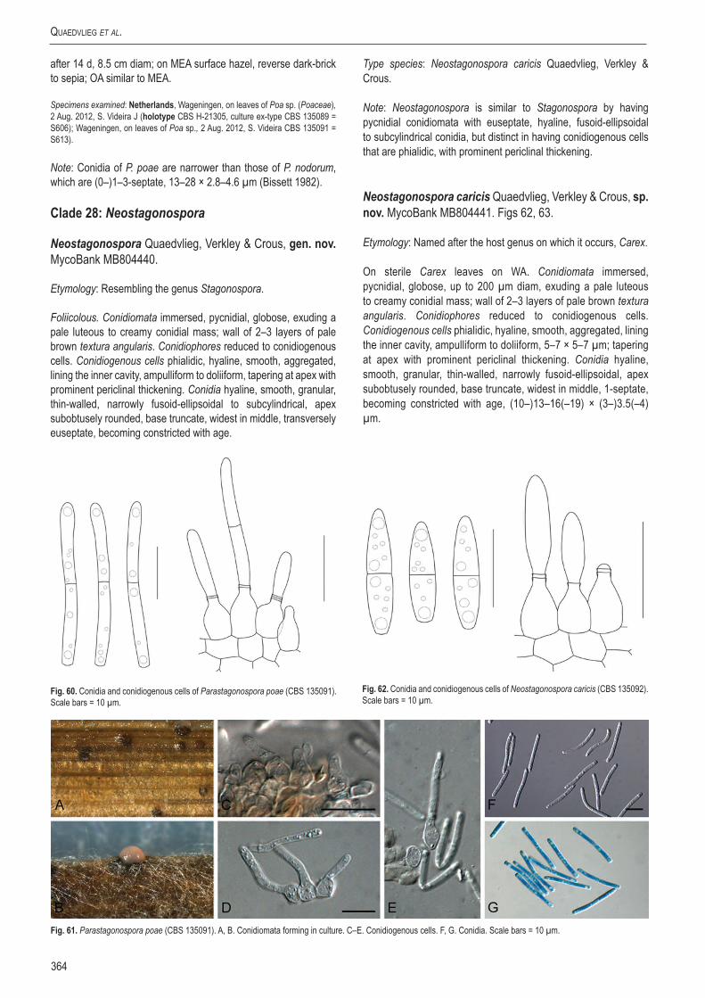
Quaedvlieg et al.
364
after 14 d, 8.5 cm diam; on MEA surface hazel, reverse dark-brick to sepia; OA similar to MEA.
Specimens examined: Netherlands, Wageningen, on leaves of Poa sp. (Poaceae), 2 Aug. 2012, S. Videira J (holotype CBS H-21305, culture ex-type CBS 135089 = S606); Wageningen, on leaves of Poa sp., 2 Aug. 2012, S. Videira CBS 135091 = S613).
Note: Conidia of P. poae are narrower than those of P. nodorum, which are (0–)1–3-septate, 13–28 × 2.8–4.6 µm (Bissett 1982).
Clade 28: Neostagonospora
Neostagonospora Quaedvlieg, Verkley & Crous, gen. nov. MycoBank MB804440.
Etymology: Resembling the genus Stagonospora.
Foliicolous. Conidiomata immersed, pycnidial, globose, exuding a pale luteous to creamy conidial mass; wall of 2–3 layers of pale brown textura angularis. Conidiophores reduced to conidiogenous cells. Conidiogenous cells phialidic, hyaline, smooth, aggregated, lining the inner cavity, ampulliform to doliiform, tapering at apex with prominent periclinal thickening. Conidia hyaline, smooth, granular, thin-walled, narrowly fusoid-ellipsoidal to subcylindrical, apex subobtusely rounded, base truncate, widest in middle, transversely euseptate, becoming constricted with age.
Type species: Neostagonospora caricis Quaedvlieg, Verkley & Crous.
Note: Neostagonospora is similar to Stagonospora by having pycnidial conidiomata with euseptate, hyaline, fusoid-ellipsoidal to subcylindrical conidia, but distinct in having conidiogenous cells that are phialidic, with prominent periclinal thickening.
Neostagonospora caricis Quaedvlieg, Verkley & Crous, sp. nov. MycoBank MB804441. Figs 62, 63.
Etymology: Named after the host genus on which it occurs, Carex.
On sterile Carex leaves on WA. Conidiomata immersed, pycnidial, globose, up to 200 µm diam, exuding a pale luteous to creamy conidial mass; wall of 2–3 layers of pale brown textura angularis. Conidiophores reduced to conidiogenous cells. Conidiogenous cells phialidic, hyaline, smooth, aggregated, lining the inner cavity, ampulliform to doliiform, 5–7 × 5–7 µm; tapering at apex with prominent periclinal thickening. Conidia hyaline, smooth, granular, thin-walled, narrowly fusoid-ellipsoidal, apex subobtusely rounded, base truncate, widest in middle, 1-septate, becoming constricted with age, (10–)13–16(–19) × (3–)3.5(–4) µm.
Fig. 60. Conidia and conidiogenous cells of Parastagonospora poae (CBS 135091). Scale bars = 10 µm.
Fig. 61. Parastagonospora poae (CBS 135091). A, B. Conidiomata forming in culture. C–E. Conidiogenous cells. F, G. Conidia. Scale bars = 10 µm.
Fig. 62. Conidia and conidiogenous cells of Neostagonospora caricis (CBS 135092). Scale bars = 10 µm.

www.studiesinmycology.org
Sizing up Septoria
365
Culture characteristics: Colonies on PDA flat, undulate, with sparse, powdery white aerial mycelium, surface greyish-sepia to isabelline, reverse olivaceous-grey to pale olivaceous-grey, after 14 d, 8.5 cm diam; on MEA erumpent, circular, with fine white aerial mycelium, surface honey, reverse cinnamon, after 14 d, 6 cm diam; on OA similar to PDA but surface honey, reverse cinnamon.
Specimen examined: Netherlands, Veenendaal, de Blauwe Hel, on leaves of Carex acutiformis (Cyperaceae), Aug. 2012, W. Quaedvlieg (holotype CBS H-21306, culture ex-type CBS 135092 = S616).
Note: Neostagonospora caricis is similar to Septoria caricis (conidia 1-septate, 20–35 × 2.5–3 µm; Ellis & Ellis 1997), although its conidia are shorter.
Neostagonospora elegiae Quaedvlieg, Verkley & Crous, sp. nov. MycoBank MB804442. Figs 64, 65.
Etymology: Named after the host genus from which it was collected, Elegia.
On Anthriscus stem. Conidiomata pycnidial, up to 150 µm diam, erumpent, globose, brown, opening by a central ostiole, exuding a crystalline conidial mass; wall consisting of 3–6 layers of pale brown textura angularis. Conidiophores reduced to conidiogenous cells. Conidiogenous cells phialidic, lining the inner cavity, hyaline, smooth, ampulliform, 4–7 × 4–6 µm; apex with prominent periclinal thickening. Conidia hyaline, smooth, guttulate to granular, scolecosporous, irregularly curved, subcylindrical, apex subobtuse, base truncate (slight taper from apical septum to apex and basal septum to hilum visible in some conidia), (0–)3-septate, (20–)50–65(–70) × (2.5–)3 µm.
Culture characteristics: Colonies spreading, erumpent with moderate aerial mycelium and smooth, even margins; reaching 35 mm diam after 2 wk. On OA pale luteous. On MEA dirty white on surface, luteous in reverse. On PDA dirty white on surface, pale luteous in reverse.
Specimen examined: South Africa, Western Cape Province, Harold Porter Botanical Garden, on leaves of Elegia cuspidata (Restionaceae), 30 Nov. 2001, S. Lee (holotype CBS H-21307, culture ex-type CBS 135101 = CPC 16977).
Notes: No septoria-like fungi are presently known from Elegia (Lee et al. 2004). Neostagonospora elegiae is distinguished from N. caricis based on its conidial morphology.
Clade 29: Phaeosphaeriopsis
Phaeosphaeriopsis M.P.S. Câmara, M.E. Palm & A.W. Ramaley, Mycol. Res. 107: 519. 2003.
Saprobic or plant pathogenic. Ascomata solitary or aggregated, immersed, subepidermal to erumpent, pushing up flaps of the epidermis, globose to pyriform, often papillate, solitary or gregarious in a stroma of scleroplectenchyma or dark brown textura angularis, often surrounded by septate, brown hyphae extending into the host tissues. Asci 8-spored, bitunicate, cylindrical to broadly fusoid, short stipitate, with visible apical chamber. Ascospores uni- to triseriate, cylindrical, broadly rounded at apex, tapering to narrowly rounded base, 4–5-septate, first septum submedian, often constricted, medium brown, echinulate, punctate or verrucose. Asexual morph coniothyrium-like or phaeostagonospora-like. Conidiomata pseudoparenchymatous, sometimes of scleroplectenchyma. Conidiogenous cells lining locule, ampulliform, hyaline, proliferating percurrently, resulting in inconspicuous annellations. Conidia cylindrical, with bluntly rounded ends, 0–3-septate, yellowish brown, punctate (Câmara et al. 2003, Zhang et al. 2012).
Fig. 63. Neostagonospora caricis (CBS 135092). A. Conidioma forming in culture. B, C. Conidiogenous cells. D. Conidia. Scale bars = 10 µm.
Fig. 64. Conidia and conidiogenous cells of Neostagonospora elegiae (CBS 135101). Scale bars = 10 µm.

Quaedvlieg et al.
366
Type species: Phaeosphaeriopsis glaucopunctata (Grev.) M.P.S. Câmara, M.E. Palm & A.W. Ramaley, Mycol. Res., 107: 519. 2003.
Phaeosphaeriopsis glaucopunctata (Grev.) M.P.S. Câmara, M.E. Palm & A.W. Ramaley, Mycol. Res. 107: 519. 2003. Figs 66, 67.Basionym: Cryptosphaeria glaucopunctata Grev., Fl. Edin.: 362. 1824.
≡ Paraphaeosphaeria glaucopunctata (Grev.) Shoemaker & C. E. Babc., Can. J. Bot. 63: 1286. 1985.
= Sphaeria rusci Wallr., Fl. Crypt. Germ. 2: 776. 1833.≡ Leptosphaeria rusci (Wallr.) Sacc., Syll. Fung. 2: 74. 1883.≡ Paraphaeosphaeria rusci (Wallr.) O. E. Erikss., Ark. Bot., Ser. 2 6: 406. 1967.
Ascomata scattered or aggregated, immersed, globose to subglobose, up to 250 µm diam; peridium up to 25 µm wide, of thick-walled textura angularis; hamathecium of dense, wide, cellular pseudoparaphyses, 3–5 μm diam. Asci 8-spored, bitunicate, cylindrical to broadly fusoid, with a short pedicel and small apical chamber, 50–110 × 10–16 μm. Ascospores uni- to triseriate, cylindrical, medium brown, 4(−5)-septate, without constriction or slightly constricted at the basal septum, the forth cell from the apex usually slightly inflated, the basal cell often longer, 14–28 × (3.5–)5–7.5 μm. Conidiomata pycnidial, immersed, scattered or aggregated, dark brown, subglobose, ostiolate, up to 200 μm diam. Conidiophores reduced to conidiogenous cells. Conidiogenous cells lining the inner cavity, ampulliform, hyaline, smooth, 5–10 × 3–6 µm; proliferating percurrently at apex. Conidia aseptate, smooth to finely verruculose, medium brown, subcylindrical, straight to reniform with obtuse ends, (5–)7–9(–10) × (2.5–)3(–5) μm.
Culture characteristics: On PDA colonies flat, spreading, with sparse aerial mycelium and smooth, lobate, even margins, surface primrose, reverse olivaceous-buff, On OA buff with patches of isabelline due to sporulating conidiomata. On MEA dirty white on surface, isabelline in reverse (centre), cinnamon in outer region.
Specimen examined: Switzerland, Kt. Basel-Stadt, Park Basel, on Ruscus aculeatus (Ruscaceae), 25 Sep. 1980, A. Leuchtmann (CBS H-21308, culture CBS 653.86).
Notes: The genus Phaeosphaeriopsis is characterised by having uni- or multiloculate stromata and 4–5-septate ascospores. It presently contains species with coniothyrium-like, and phaeostagonospora-like asexual morphs (e.g. P. musae; Arzanlou & Crous 2006). The type species, Phaeosphaeriopsis glaucopunctata, is associated with leaf spot and necrosis on Ruscus aculeatus (Câmara et al. 2003, Golzar & Wang 2012). The fact that an isolate identified as Chaetosphaeronema hispidulum (lectotype of Chaetosphaeronema) clusters in this clade is puzzling. The genus Chaetosphaeronema is characterised by setose, dark brown pycnidia with thick-walled outer cell layers, producing hyaline, 1-septate conidia (Sutton 1980). Isolate CBS 216.75 proved to be sterile, however, so this matter could unfortunately not be resolved.
Clade 30: Sclerostagonospora
Description: See above.
Type species: S. heraclei (Sacc.) Höhn., Hedwigia 59: 252. 1917.
Sclerostagonospora phragmiticola Quaedvlieg, Verkley & Crous, sp. nov. MycoBank MB804443. Fig. 68.
Fig. 65. Neostagonospora elegiae (CBS 135101). A. Conidioma forming in culture. B–D. Conidiogenous cells. E. Conidia. Scale bars: A = 150 µm, all others = 10 µm.
Fig. 66. Conidia and conidiogenous cells of Phaeosphaeriopsis glaucopunctata (CBS 653.86). Scale bar = 10 µm.
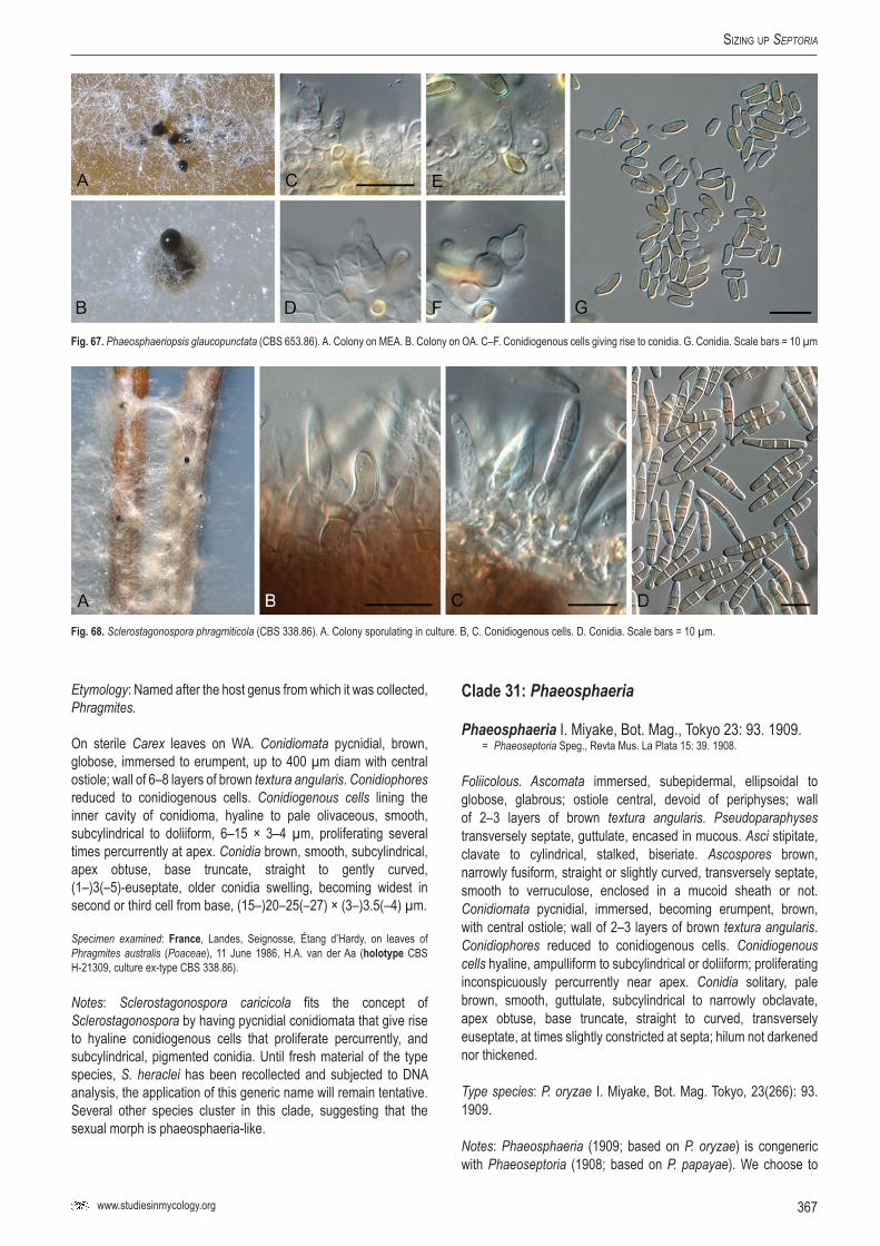
www.studiesinmycology.org
Sizing up Septoria
367
Etymology: Named after the host genus from which it was collected, Phragmites.
On sterile Carex leaves on WA. Conidiomata pycnidial, brown, globose, immersed to erumpent, up to 400 µm diam with central ostiole; wall of 6–8 layers of brown textura angularis. Conidiophores reduced to conidiogenous cells. Conidiogenous cells lining the inner cavity of conidioma, hyaline to pale olivaceous, smooth, subcylindrical to doliiform, 6–15 × 3–4 µm, proliferating several times percurrently at apex. Conidia brown, smooth, subcylindrical, apex obtuse, base truncate, straight to gently curved, (1–)3(–5)-euseptate, older conidia swelling, becoming widest in second or third cell from base, (15–)20–25(–27) × (3–)3.5(–4) µm.
Specimen examined: France, Landes, Seignosse, Étang d’Hardy, on leaves of Phragmites australis (Poaceae), 11 June 1986, H.A. van der Aa (holotype CBS H-21309, culture ex-type CBS 338.86).
Notes: Sclerostagonospora caricicola fits the concept of Sclerostagonospora by having pycnidial conidiomata that give rise to hyaline conidiogenous cells that proliferate percurrently, and subcylindrical, pigmented conidia. Until fresh material of the type species, S. heraclei has been recollected and subjected to DNA analysis, the application of this generic name will remain tentative. Several other species cluster in this clade, suggesting that the sexual morph is phaeosphaeria-like.
Clade 31: Phaeosphaeria
Phaeosphaeria I. Miyake, Bot. Mag., Tokyo 23: 93. 1909.= Phaeoseptoria Speg., Revta Mus. La Plata 15: 39. 1908.
Foliicolous. Ascomata immersed, subepidermal, ellipsoidal to globose, glabrous; ostiole central, devoid of periphyses; wall of 2–3 layers of brown textura angularis. Pseudoparaphyses transversely septate, guttulate, encased in mucous. Asci stipitate, clavate to cylindrical, stalked, biseriate. Ascospores brown, narrowly fusiform, straight or slightly curved, transversely septate, smooth to verruculose, enclosed in a mucoid sheath or not. Conidiomata pycnidial, immersed, becoming erumpent, brown, with central ostiole; wall of 2–3 layers of brown textura angularis. Conidiophores reduced to conidiogenous cells. Conidiogenous cells hyaline, ampulliform to subcylindrical or doliiform; proliferating inconspicuously percurrently near apex. Conidia solitary, pale brown, smooth, guttulate, subcylindrical to narrowly obclavate, apex obtuse, base truncate, straight to curved, transversely euseptate, at times slightly constricted at septa; hilum not darkened nor thickened.
Type species: P. oryzae I. Miyake, Bot. Mag. Tokyo, 23(266): 93. 1909.
Notes: Phaeosphaeria (1909; based on P. oryzae) is congeneric with Phaeoseptoria (1908; based on P. papayae). We choose to
Fig. 67. Phaeosphaeriopsis glaucopunctata (CBS 653.86). A. Colony on MEA. B. Colony on OA. C–F. Conidiogenous cells giving rise to conidia. G. Conidia. Scale bars = 10 µm
Fig. 68. Sclerostagonospora phragmiticola (CBS 338.86). A. Colony sporulating in culture. B, C. Conidiogenous cells. D. Conidia. Scale bars = 10 µm.

Quaedvlieg et al.
368
use the sexual name Phaeosphaeria, as it is well established, and less confused than Phaeoseptoria, which has become a confused concept applied to numerous septoria-like taxa with pigmented conidia (see Walker et al. 1992).
Phaeosphaeria oryzae I. Miyake, Bot. Mag. Tokyo, 23(266): 93. 1909. Figs 69, 70.
≡ Pleospora oryzae (I. Miyake) Hara, J. Agric. Soc. Japan 31(361): 17. 1927.≡ Trematosphaerella oryzae (I. Miyake) Padwick, A manual of rice diseases: 153. 1950.≡ Leptosphaerella oryzae (I. Miyake) Hara, A monograph of rice diseases: 53. 1959.
≡ Leptosphaerulina oryzae (I. Miyake) Karan, Mycopath. Mycol. Appl. 24: 88. 1964.
= Phaeoseptoria oryzae I. Miyake, J. Coll. Agric. Imp. Univ. Tokyo 2(4): 260. 1910.
Ascomata immersed, subepidermal, ellipsoidal to globose, glabrous, up to 150 µm diam, ostiole central, up to 20 µm diam, devoid of periphyses; wall of 2–3 layers of brown textura angularis. Pseudoparaphyses 2–3 µm diam, transversely septate, guttulate, encased in mucous. Asci stipitate, cylindrical, 30–55 × 7–9 µm, stalked, biseriate. Ascospores brown, narrowly fusiform, straight or slightly curved, (15–)17–20(–23) × 4(–5) µm, 3-septate, uniformly verruculose, enclosed in a mucoid sheath; after discharge, ascospores become prominently swollen, up to 33 µm long and 8 µm wide.
Specimens examined: Japan, No. 196178, on 2, Prov. Susuya Shizuoka, Sep. 1907, ex Herb. Sydow, ex S., as Leptosphaeria oryzae Hori = Phaeosphaeria oryzae I. Miyake, slides prepared by O. Eriksson, lectotype (UPS). Korea, on leaf of Oryza sativa (Poaceae), intercepted at Port San Francisco, CA, 29 Dec. 1997, coll. L. Hausch, det. M.E. Palm, epitype designated here as BPI 744438, culture ex-epitype CBS 110110 (MBT175330).
Notes: Several detailed accounts of this species are available (Eriksson 1967, Shoemaker & Babcock 1989, Fukuhara 2002). The epitype chosen here closely matches the lectotype in morphology.
Phaeosphaeria papayae (Speg.) Quaedvlieg, Verkley & Crous, comb. nov. MycoBank MB804444. Figs 71, 72.Basionym: Phaeoseptoria papayae Speg., Revta Mus. La Plata: 39. 1908.
Leaf spots associated with infections of Asperisporium caricae, amphigenous, pale brown to grey-white, subcircular to angular, 1–5 mm diam, with red-purple margin; conidiomata developing
Fig. 70. Phaeosphaeria oryzae (BPI 744438). A. Ascomata on host tissue. B–G. Asci. H. Ascospores. Scale bars = 10 µm.
Fig. 69. Asci and ascospores of Phaeosphaeria oryzae (BPI 744438). Scale bars = 10 µm.

www.studiesinmycology.org
Sizing up Septoria
369
and sporulating on leaves when incubated in moist chambers, with white, fluffy mycelium erumpting from lesions. Conidiomata amphigenous, pycnidial, brown, globose, up to 120 µm diam, with central ostiole, exuding a brown conidial cirrhus; wall of 3–4 layers of brown textura angularis. Conidiophores reduced to conidiogenous cells. Conidiogenous cells lining the inner cavity, hyaline, smooth, ampulliform to subcylindrical or doliiform, 5–12 × 4–6 µm; proliferating inconspicuously percurrently near
apex (conidiogenous cells disintegrating at maturity). Conidia solitary, pale brown, smooth, guttulate, subcylindrical to narrowly obclavate, apex obtuse, base truncate, (1–)3(–4)-septate, at times slightly constricted at septa, straight to slightly curved, (15–)26–32(–35) × (2.5–)3 µm; hilum not darkened nor thickened, 2 µm diam. Ascomata developed after 4 wk in culture on sterile nettle stems: aggregated in black clusters, globose, up to 150 µm diam, with central ostiole; wall of 2–3 layers of brown textura angularis. Asci bitunicate, curved to straight, fasciculate, short stipitate with ocular chamber, 40–60 × 8–11 µm. Pseudoparaphyses hyaline, smooth, 2–3 µm, septate, constricted at septa, not anastomosing, hypha-like with obtuse ends, distributed among asci. Ascospores tri to multiseriate, fusoid, curved to straight, brown, verruculose throughout, somewhat constricted at septa with age, second cell from apex swollen, (18–)24–26(–29) × (3–)4(–5) µm.
Culture characteristics: Colonies with abundant aerial mycelium, covering dish within 2 wk at 24 ºC, fast growing, olivaceous-grey on MEA (surface and reverse); margins smooth, even, sterile on MEA, PDA and OA, as well as on SNA with sterile barley leaves.
Specimens examined: Brazil, São Paulo, Botanical Garden, on leaves of Carica papaya (Caricaceae), Sep. 1908, IMI 246301, slide ex-holotype; Minas Gerais, Viçosa, UFV campus, on leaves of Carica papaya, Mar. 2013, A.C. Alfenas, epitype designated here as CBS H-21310, culture ex-epitype CBS 135416 (MBT175331).
Notes: It is interesting to note that Walker et al. (1992) also observed Phaeoseptoria papayae to co-occur with Asperisporium
Fig. 71. Conidia, ascospores and ascus of Phaeosphaeria papayae (CBS H-21310). Scale bars = 10 µm.
Fig. 72. Phaeosphaeria papayae (CBS H-21310). A. Leaf spot. B. Conidioma with ostiole (arrow). C–E. Conidiogenous cells. F. Conidia. G–K. Asci and ascospores. Scale bars = 10 µm.

Quaedvlieg et al.
370
caricae on the holotype specimen (noted by Spegazzini as Cercospora caricae), suggesting that the co-occurrence of these two pathogens is quite common. The fresh collection obtained in this study enabled us to elucide the conidiogenesis of the fungus (not observed by Walker et al. 1992), and also designate an epitype specimen. Phylogenetically it is closely related to Phaeosphaeria oryzae, which has Phaeoseptoria oryzae as asexual morph.
Clade 32: Neosetophoma
Neosetophoma Gruyter, Aveskamp & Verkley, Mycologia 102(5): 1075. 2010.
Foliicolous, plant pathogenic. Conidiomata pycnidial, solitary to confluent, on upper surface of agar, globose to irregular, with mycelial outgrowths, or confluent, with papillate ostioles, sometimes developing long necks, honey to olivaceous or olivaceous-black, with up to 10 layers of pseudoparenchymatal cells. Conidiogenous cells hyaline, monophyalidic. Conidia slightly yellowish, 0–1(–3)-septate, ellipsoidal to cylindrical, usally attenuate at one end, often guttulate.
Type species: N. samarorum Gruyter, Aveskamp & Verkley, Mycologia 102(5): 1075. 2010.
Note: The fact that several strains with a phaeosphaeria-like morphology cluster in this clade, suggests that sexual states do exist for species of Neosetophoma.
Clade 33: Paraphoma
Paraphoma Morgan-Jones & J.F. White, Mycotaxon 18: 58. 1983.
Mycelium consisting of branched, septate, subhyaline to pale brown, smooth hyphae. Conidiomata pycnidial, solitary to aggregated, superficial to immersed, dark brown, globose to subglobose, papillate, uniloculate, setose; ostiole circular, single; wall of 3–6 layers of brown textura angularis. Setae copious, straight to flexuous, smooth to verruculose, thick-walled, septate, pale brown to brown. Conidiogenous cells lageniform, monophalidic, formed from inner layer of conidiomatal wall, hyaline to subhyaline, discrete. Conidia ellipsoid, aseptate, hyaline, smooth, guttulate. Chlamydospores if present unicellular.
Type species: P. radicina (McAlpine) Morgan-Jones & J.F. White, Mycotaxon 18: 60. 1983.
Paraphoma dioscoreae Quaedvlieg, H.D. Shin, Verkley & Crous, sp. nov. MycoBank MB804445. Figs 73, 74.
Etymology: Named after the host genus from which it was collected, Dioscorea.
On Anthriscus stem. Conidiomata pycnidial, separate, immersed becoming erumpent, globose, with papillate neck and central ostiole exuding a crystalline conidial mass; conidiomata up to 350 µm diam, neck up to 150 µm diam, of darker brown cells than body, which is pale brown; wall of 3–6 layers of pale brown textura angularis. Conidiophores hyaline, smooth, subcylindrical, reduced to conidiogenous cells, 1–5-septate, irregularly branched, 5–20 × 3–5 µm. Conidiogenous cells phialidic, hyaline, smooth, ampulliform to subcylindrical (long, elongated neck on Anthriscus stem, but not on MEA), 5–15 × 2–3 µm; apex with prominent periclinal thickening, or with several percurrent prolferations (especially on conidiogenous cells with elongated necks). Conidia solitary, straight to slightly curved, hyaline, smooth, aseptate, cylindrical with obtuse ends and a guttule at each end, (5–)6(–7) × 2(–2.5) µm.
Fig. 74. Paraphoma dioscoreae (CBS 135100). A. Conidioma forming in culture. B–E. Conidiogenous cells. F. Conidia. Scale bars: B = 350 µm, all others = 10 µm.
Fig. 73. Conidia and conidiogenous cells of Paraphoma dioscoreae (CBS 135100). Scale bar = 10 µm.

www.studiesinmycology.org
Sizing up Septoria
371
Culture characteristics: Colonies flat, spreading with sparse aerial mycelium and even, smooth margins; after 2 wk reaching 30 mm diam on MEA, 40 mm on PDA and 50 mm on OA. On PDA dark brick, reverse fuscous-black. On OA dark brick with patches of sienna and ochreous. On MEA surface dirty white (due to aerial mycelium), also somewhat sectored, reverse umber.
Specimen examined: South Korea, on leaves of Dioscorea tokoro (Dioscoreaceae), 24 Oct. 2003, H.D. Shin (holotype CBS H-21311, culture ex-type CPC 11357 = CBS 135100).
Note: Paraphoma dioscoreae is phylogenetically distinct from the three other species presently known in the genus (de Gruyter et al. 2010).
Clade 34: Xenoseptoria
Xenoseptoria Quaedvlieg, H.D. Shin, Verkley & Crous, gen. nov. MycoBank MB804446.
Etymology: Similar to the genus Septoria s. str., but distinct.
Foliicolous, plant pathogenic. Conidiomata separate, pycnidial, immersed becoming erumpent, globose, brown, developing 1–3 papillate necks, exuding a pink to orange conidial mass; wall of 4–8 layers of brown textura angularis. Conidiophores hyaline, smooth, reduced to conidiogenous cells or septate, branched below. Conidiogenous cells lining the inner cavity, hyaline, smooth, ampulliform to doliiform or subcylindrical, mono- to polyphialidic, with prominent periclinal thickening, but also with percurrent proliferation. Conidia hyaline, smooth, guttulate, scolecosporous, straight to irregularly curved, cylindrical to obclavate, transversely euseptate, tapering to subobtuse apex, base obtuse.
Type species: Xenoseptoria neosaccardoi Quaedvlieg, Verkley & Crous.
Xenoseptoria neosaccardoi Quaedvlieg, H.D. Shin, Verkley & Crous, sp. nov. MycoBank MB804447. Figs 75, 76.
Etymology: Resembling Septoria saccardoi, but morphologically distinct.
Leaf spots on the upper leaf surface, scattered, distinct, circular, 2–4 mm diam, initially appearing as reddish brown discolouration, later turning brown to reddish brown without a distinct border line, finally central area becoming greyish brown to dull grey and surrounded by reddish to dark brown margin, reddish pigments may diffuse outward to form a halo; on the lower leaf surface initially showing reddish discolouration, later becoming brown with distinct border line, center greyish brown to grey with indistinct border (Shin & Sameva 2004). On sterile Carex leaves on WA. Conidiomata separate, pycnidial, immersed becoming erumpent, globose, up to 350 µm diam, brown, becoming ostiolate, developing 1–3 papillate necks, exuding a pink to orange conidial mass; wall of 4–8 layers of brown textura angularis. Conidiophores hyaline,
Fig. 75. Conidia and conidiogenous cells of Xenoseptoria neosaccardoi (CBS 128665). Scale bars = 10 µm.
Fig. 76. Xenoseptoria neosaccardoi (CBS 128665). A, B. Pycnidia forming in culture. C–E. Conidiogenous cells. F, G. Conidia. Scale bars: B = 170 µm, all others = 10 µm.

Quaedvlieg et al.
372
smooth, reduced to conidiogenous cells or 1–2-septate, branched below, 10–20 × 4–6 µm. Conidiogenous cells lining the inner cavity, hyaline, smooth, ampulliform to doliiform or subcylindrical, mono- to polyphialidic, with prominent periclinal thickening, but also with percurrent proliferation, 5–15 × 3–5 µm. Conidia hyaline, smooth, guttulate, scolecosporous, straight to irregularly curved, cylindrical to obclavate, (1–)3-septate, (23–)33–45(–48) × (2.5-)3(–4) µm, tapering to subobtuse apex, base obtuse, 2–2.5 µm diam.
Culture characteristics: Colonies flat, spreading, with sparse aerial mycelium and lobate, feathery mergins, reaching 30 mm after 2 wk. On PDA surface iron-grey, reverse olivaceous-grey; on OA surface olivaceous-grey; on MEA surface folded, bay, reverse umber.
Specimen examined: South Korea, Pyeongchang, on leaves of Lysimachia vulgaris var. davurica (Primulaceae), 30 May 2007, H.D. Shin (holotype CBS H-21312, culture ex-type CBS 128665 = KACC 43962 = SMKC 23666).
Notes: An isolate of Septoria saccardoi (CBS 128756) clusters in Septoria s. str., thus well apart from this taxon, which was collected in Korea. The Korean collection closely matches that of the original description of Septoria saccardoi (on Lysimachia vulgaris in Italy), having 3-septate, curved, cylindrical conidia, 38–40 × 3.5 µm, 3-septate (Saccardo & Saccardo 1906). Xenoseptoria is however distinct from Septoria s. str. in forming pycnidia with multiple papillate necks, and having conidiogenous cells that are mono- or polyphialidic.
Clade 35: Vrystaatia
Vrystaatia Quaedvlieg, W.J. Swart, Verkley & Crous, gen. nov. MycoBank MB804448.
Etymology: Named after the Free State Province in South Africa, “Vrystaat” in Afrikaans, where this fungus was collected.
Foliicolous. Conidiomata black, globose, pycnidial with central, dark brown ostiolar area, substomatal on host, erumpent in culture;
wall of 6–8 layers of pale brown textura angularis; exuding cirrhus of orange conidia. Conidiophores reduced to conidiogenous cells. Conidiogenous cells lining the inner cavity of conidioma, globose to ampulliform, rarely allantoid, hyaline, smooth; with prominent periclinal thickening, or proliferating several times percurrently near apex, giving rise to macro- and microconidia. Macroconidia solitary, hyaline, smooth, guttulate, subcylindrical to narrowly obclavate or acicular, apex obtuse to subobtuse, base truncate to long obconically truncate, conidia widest at or just above basal septum, transversely euseptate. Microconidia hyaline, smooth, aseptate, pear-shaped to globose or ellipsoid, apex obtuse, base truncate.
Type species: Vrystaatia aloeicola Quaedvlieg, Verkley, W.J. Swart & Crous.
Vrystaatia aloeicola Quaedvlieg, Verkley, W.J. Swart & Crous, sp. nov. MycoBank MB804449. Figs 77, 78.
Etymology: Named after the host genus from which it was collected, Aloe.
Fig. 78. Vrystaatia aloeicola (CBS 135107). A. Conidiomata sporulating on PDA, with characteristic orange conidial cirrhi. B–D. Conidiogenous cells. E, F. Conidia. Scale bars = 10 µm.
Fig. 77. Macro- and microconidia and conidiogenous cells of Vrystaatia aloeicola (CBS 135107). Scale bars = 10 µm.

www.studiesinmycology.org
Sizing up Septoria
373
On sterile Carex leaves on WA. Conidiomata black, globose, pycnidial with central, dark brown ostiolar area, substomatal on host, erumpent in culture; wall of 6–8 layers of pale brown textura angularis; exuding cirrhus of orange conidia. Conidiophores reduced to conidiogenous cells. Conidiogenous cells lining the inner cavity of conidioma, globose to ampulliform, rarely allantoid, hyaline, smooth, 5–12 × 4–6 µm; with prominent periclinal thickening, or proliferating several times percurrently near apex, 2–2.5 µm diam, giving rise to macro- and microconidia. Macroconidia solitary, hyaline, smooth, guttulate, subcylindrical to narrowly obclavate or acicular, apex obtuse to subobtuse, base truncate to long obconically truncate, conidia widest at or just above basal septum, (1–)3-septate, (30–)40–52(–65) × (2.5–) 3(–3.5) µm. Microconidia hyaline, smooth, aseptate, pear-shaped to globose or ellipsoid, apex obtuse, base truncate, 4–6 × 3–3.5 µm.
Culture characteristics: On MEA colonies spreading fast, with moderate aerial mycelium and smooth, even margin, reaching 30 mm diam after 2 wk; surface with concentric zones of umber and apricot; reverse umber, produces brown exudates; on PDA round lobate margins, lacking aerial mycelium, reaching 20 mm diam after 2 wk, surface fuscous-black to greyish-sepia for younger mycelium, reverse fuscous-black to greyish-sepia for younger mycelium; on OA round, lobate, lacking aerial mycelium, reaching 30 mm diam after 2 wk, surface vinaceous-grey, reverse greyish sepia.
Specimen examined: South Africa, Orange Free State, Bloemfontein, Free State National Botanical Garden, on dead leaf tips of Aloe maculata (Aloaceae), 7 May 2012, P.W. Crous & W.J. Swart (holotype CBS H-21313, culture ex-type CBS 135107 = CPC 20617).
Notes: Vrystaatia is distinct from Septoria s. str. in that it has phialidic conidiogenous cells that proliferate percurrently or with prominent periclinal thickening, and form macro- as well as microconidia in culture, which is not typical of Septoria. Rhabdospora aloetica was described from stems of Aloe sp. in Portugal, with aseptate conidia, 12–16 × 1.5 µm (Saccardo & Saccardo 1906); it is likely this is an asexual morph of Diaporthe. As far as we could establish, no septoria-like fungi have thus far been described from Aloe.
Clade 36: Setophoma
Setophoma Gruyter, Aveskamp & Verkley, Mycologia 102: 1077. 2010.
Conidiomata pycnidial, solitary to confluent, superficial or submerged in agar, globose to subglobose, setose, with papillate ostioles, honey to olivaceous to olivaceous-black, with 2–7(–11) layers of pseudoparenchymatal cells. Conidiogenous cells hyaline, monophyalidic. Conidia aseptate, ellipsoidal to subcylindrical to subfusoid, guttulate.
Type species: S. terrestris (H.N. Hansen) Gruyter, Aveskamp & Verkley, Mycologia 102: 1077. 2010.
Setophoma chromolaenae Quaedvlieg, Verkley, R.W. Barreto & Crous, sp. nov. MycoBank MB804450. Figs 79, 80.
Fig. 80. Setophoma chromolaenae (CBS 135105). A. Conidiomata forming on OA. B, C, F. Conidiomata with setae. D, E. Conidiogenous cells. G. Conidia. Scale bars: B = 22 µm, C, F = 45 µm, all others = 10 µm.
Fig. 79. Conidia and conidiogenous cells of Setophoma chromolaenae (CBS 135105). Scale bar = 10 µm.
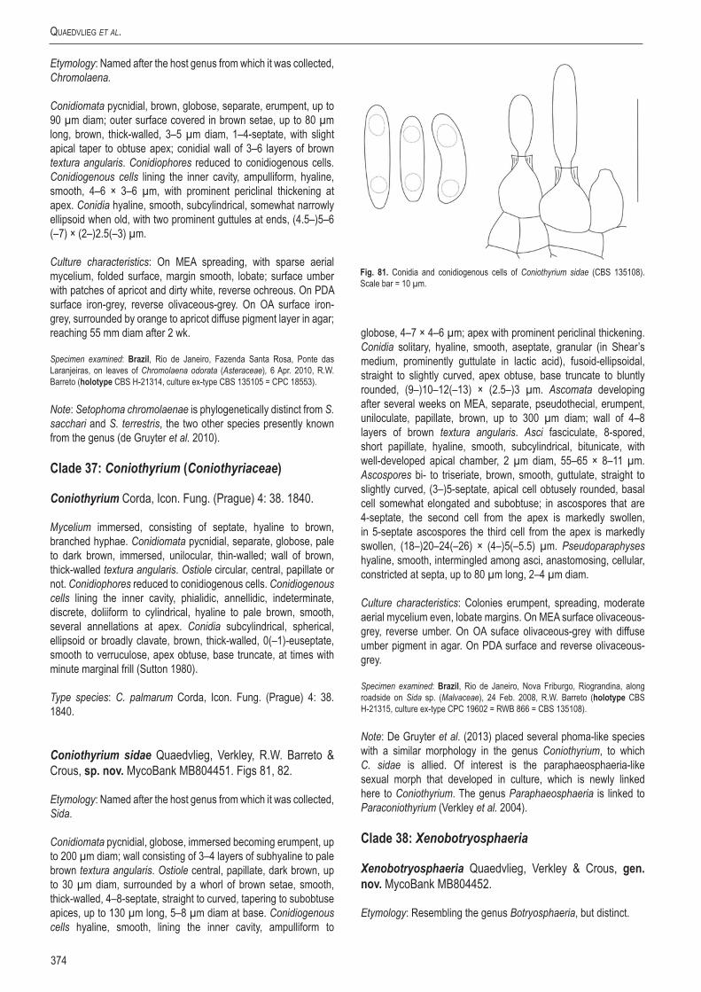
Quaedvlieg et al.
374
Etymology: Named after the host genus from which it was collected, Chromolaena.
Conidiomata pycnidial, brown, globose, separate, erumpent, up to 90 µm diam; outer surface covered in brown setae, up to 80 µm long, brown, thick-walled, 3–5 µm diam, 1–4-septate, with slight apical taper to obtuse apex; conidial wall of 3–6 layers of brown textura angularis. Conidiophores reduced to conidiogenous cells. Conidiogenous cells lining the inner cavity, ampulliform, hyaline, smooth, 4–6 × 3–6 µm, with prominent periclinal thickening at apex. Conidia hyaline, smooth, subcylindrical, somewhat narrowly ellipsoid when old, with two prominent guttules at ends, (4.5–)5–6 (–7) × (2–)2.5(–3) µm.
Culture characteristics: On MEA spreading, with sparse aerial mycelium, folded surface, margin smooth, lobate; surface umber with patches of apricot and dirty white, reverse ochreous. On PDA surface iron-grey, reverse olivaceous-grey. On OA surface iron-grey, surrounded by orange to apricot diffuse pigment layer in agar; reaching 55 mm diam after 2 wk.
Specimen examined: Brazil, Rio de Janeiro, Fazenda Santa Rosa, Ponte das Laranjeiras, on leaves of Chromolaena odorata (Asteraceae), 6 Apr. 2010, R.W. Barreto (holotype CBS H-21314, culture ex-type CBS 135105 = CPC 18553).
Note: Setophoma chromolaenae is phylogenetically distinct from S. sacchari and S. terrestris, the two other species presently known from the genus (de Gruyter et al. 2010).
Clade 37: Coniothyrium (Coniothyriaceae)
Coniothyrium Corda, Icon. Fung. (Prague) 4: 38. 1840.
Mycelium immersed, consisting of septate, hyaline to brown, branched hyphae. Conidiomata pycnidial, separate, globose, pale to dark brown, immersed, unilocular, thin-walled; wall of brown, thick-walled textura angularis. Ostiole circular, central, papillate or not. Conidiophores reduced to conidiogenous cells. Conidiogenous cells lining the inner cavity, phialidic, annellidic, indeterminate, discrete, doliiform to cylindrical, hyaline to pale brown, smooth, several annellations at apex. Conidia subcylindrical, spherical, ellipsoid or broadly clavate, brown, thick-walled, 0(–1)-euseptate, smooth to verruculose, apex obtuse, base truncate, at times with minute marginal frill (Sutton 1980).
Type species: C. palmarum Corda, Icon. Fung. (Prague) 4: 38. 1840.
Coniothyrium sidae Quaedvlieg, Verkley, R.W. Barreto & Crous, sp. nov. MycoBank MB804451. Figs 81, 82.
Etymology: Named after the host genus from which it was collected, Sida.
Conidiomata pycnidial, globose, immersed becoming erumpent, up to 200 µm diam; wall consisting of 3–4 layers of subhyaline to pale brown textura angularis. Ostiole central, papillate, dark brown, up to 30 µm diam, surrounded by a whorl of brown setae, smooth, thick-walled, 4–8-septate, straight to curved, tapering to subobtuse apices, up to 130 µm long, 5–8 µm diam at base. Conidiogenous cells hyaline, smooth, lining the inner cavity, ampulliform to
globose, 4–7 × 4–6 µm; apex with prominent periclinal thickening. Conidia solitary, hyaline, smooth, aseptate, granular (in Shear’s medium, prominently guttulate in lactic acid), fusoid-ellipsoidal, straight to slightly curved, apex obtuse, base truncate to bluntly rounded, (9–)10–12(–13) × (2.5–)3 µm. Ascomata developing after several weeks on MEA, separate, pseudothecial, erumpent, uniloculate, papillate, brown, up to 300 µm diam; wall of 4–8 layers of brown textura angularis. Asci fasciculate, 8-spored, short papillate, hyaline, smooth, subcylindrical, bitunicate, with well-developed apical chamber, 2 µm diam, 55–65 × 8–11 µm. Ascospores bi- to triseriate, brown, smooth, guttulate, straight to slightly curved, (3–)5-septate, apical cell obtusely rounded, basal cell somewhat elongated and subobtuse; in ascospores that are 4-septate, the second cell from the apex is markedly swollen, in 5-septate ascospores the third cell from the apex is markedly swollen, (18–)20–24(–26) × (4–)5(–5.5) µm. Pseudoparaphyses hyaline, smooth, intermingled among asci, anastomosing, cellular, constricted at septa, up to 80 µm long, 2–4 µm diam.
Culture characteristics: Colonies erumpent, spreading, moderate aerial mycelium even, lobate margins. On MEA surface olivaceous-grey, reverse umber. On OA suface olivaceous-grey with diffuse umber pigment in agar. On PDA surface and reverse olivaceous-grey.
Specimen examined: Brazil, Rio de Janeiro, Nova Friburgo, Riograndina, along roadside on Sida sp. (Malvaceae), 24 Feb. 2008, R.W. Barreto (holotype CBS H-21315, culture ex-type CPC 19602 = RWB 866 = CBS 135108).
Note: De Gruyter et al. (2013) placed several phoma-like species with a similar morphology in the genus Coniothyrium, to which C. sidae is allied. Of interest is the paraphaeosphaeria-like sexual morph that developed in culture, which is newly linked here to Coniothyrium. The genus Paraphaeosphaeria is linked to Paraconiothyrium (Verkley et al. 2004).
Clade 38: Xenobotryosphaeria
Xenobotryosphaeria Quaedvlieg, Verkley & Crous, gen. nov. MycoBank MB804452.
Etymology: Resembling the genus Botryosphaeria, but distinct.
Fig. 81. Conidia and conidiogenous cells of Coniothyrium sidae (CBS 135108). Scale bar = 10 µm.

www.studiesinmycology.org
Sizing up Septoria
375
Ascomata brown, globose, smooth, ostiolate, superficial on stems; wall of 3–4 layers of brown textura angularis. Asci clavate, hyaline, smooth, short stipitate, fasciculate, bitunicate, thin-walled, apical chamber not visible, 6–8-spored. Ascospores multiseriate, hyaline, smooth and thin-walled, granular, broadly ellipsoid, ends obtuse, aseptate. Pseudoparaphyses not seen.
Type species: Xenobotryosphaeria calamagrostidis Quaedvlieg, Verkley & Crous.
Xenobotryosphaeria calamagrostidis Quaedvlieg, Verkley & Crous, sp. nov. MycoBank MB804453. Figs 83, 84.
Etymology: Named after the host genus from which it was collected, Calamagrostis.
On Anthriscus stem. Ascomata brown, globose, smooth, superficial on stems, ostiolate, up to 180 µm diam; wall of 3–4 layers of brown textura angularis. Asci clavate, hyaline, smooth, short stipitate, fasciculate, bitunicate, thin-walled, apical chamber not visible, 6–8-spored, 60–80 × 30–40 µm. Ascospores multiseriate, hyaline, smooth and thin-walled, granular, broadly ellipsoid, ends obtuse, aseptate, (17–)18–20(–24) × (11–)12–13(–14) µm. Pseudoparaphyses not seen.
Culture characteristics: Colonies flat, spreading, with sparse to no aerial mycelium. On PDA surface and reverse dirty white; on MEA concolorous with agar; on OA pale pink on surface.
Specimen examined: Italy, Bergamo Vigolo, on Calamagrostis sp. (Poaceae), 20 Jun. 1967, G.A. Hedjaroude (holotype CBS H-21316, culture ex-type CBS 303.71).
Notes: Hedjaroude (1968) studied the specimen (ETH 7131; as Phaeosphaeria silvatica), but obviously the incorrect fungus was cultivated, as X. calamagrostidis is quite distinct from P. silvatica, which has cylindrical-fusoid, brown, 6–8-septate ascospores, 18–18 × 4–5 µm. Xenobotryosphaeria is reminiscent of genera in the Botryosphaeriales, but is phylogenetically distinct (Crous et al. 2006, Phillips et al. 2008, Liu et al. 2012). It also resembles species of Muyocopron (Muyocopronaceae), but the latter genus differs in that it has circular, flattened ascomata, as well as prominent pseudoparaphyses, which are absent in Xenobotryosphaeria.
Clade 39: Phoma
Note: See Aveskamp et al. (2010), de Gruyter et al. (2009, 2013).
Fig. 82. Coniothyrium sidae (CBS 135108). A–E. Conidiomata forming in culture, showing setae. F, G. Conidiogenous cells. H. Conidia. I–K. Asci and ascospores. Scale bars: B, D, E = 100 µm, all others = 10 µm.
Fig. 83. Ascospores and asci of Xenobotryosphaeria calamagrostidis (CBS 303.71). Scale bars = 10 µm.

Quaedvlieg et al.
376
Clade 40: Acicuseptoria
Acicuseptoria Quaedvlieg, Verkley & Crous, gen. nov. MycoBank MB804454.
Etymology: Acicu- from acicular (conidia), and Septoria = septoria-like.
Conidiomata pycnidial, erumpent, brown, globose, with central ostiole, exuding a cream conidial mass; wall consisting of 3–6 layers of thin, brown textura angularis. Conidiophores reduced
to conidiogenous cells. Conidiogenous cells hyaline, smooth, ampulliform; proliferating inconspicuously and percurrently at apex, or simply appearing holoblastic. Conidia solitary, hyaline, granular, acicular, straight to gently curved, tapering towards apex that is acutely rounded, base truncate, transversely euseptate.
Type species: Acicuseptoria rumicis Quaedvlieg, Verkley & Crous.
Acicuseptoria rumicis Quaedvlieg, Verkley & Crous, sp. nov. MycoBank MB804455. Fig. 85.
Fig. 84. Xenobotryosphaeria calamagrostidis (CBS 303.71). A, C. Ascomata forming in culture. E, G. broken wall with asci. B, D, F. Asci. H. Ascospores. Scale bars: C = 45 µm, all others = 10 µm.
Fig. 85. Acicuseptoria rumicis (CBS 522.78). A. Conidiomata sporulating in culture. B–E. Conidiogenous cells. F, G. Conidia. Scale bars = 10 µm.

www.studiesinmycology.org
Sizing up Septoria
377
Etymology: Named after the host genus from which it was collected, Rumex.
On sterile Carex leaves on WA. Conidiomata pycnidial, erumpent, brown, globose, up to 300 µm diam, with central ostiole, exuding a cream conidial mass; wall consisting of 3–6 layers of thin, brown textura angularis. Conidiophores reduced to conidiogenous cells. Conidiogenous cells hyaline, smooth, ampulliform, 7–15 × 5–7 µm; proliferating inconspicuously and percurrently at apex, or simply appearing holoblastic. Conidia solitary, hyaline, granular, acicular, straight to gently curved, tapering towards apex that is acutely rounded, base truncate, 1.5–2 µm diam, up to 8-septate, (32–)40–60(–70) × 2(–2.5) µm.
Culture characteristics: Colonies lobate, flat with little appressed, white aerial mycelium. On MEA surface olivaceous-grey, reverse umber. On OA suface olivaceous-grey. On PDA surface and reverse olivaceous-grey.
Specimen examined: France, Haute Savoie, Mt. Beaudin, 2000 m alt., stem of Rumex alpinus (Polygonaceae), Oct. 1978, H.A. van der Aa (holotype CBS H-18163, culture ex-type CBS 522.78).
Notes: Acicuseptoria rumicis was originally deposited as Septoria rumicum, but is distinct from the latter in having acicular, narrower conidia. Acicuseptoria is distinct from Septoria s. str. in having acicular conidia.
Clade 41: Stagonospora
Stagonospora (Sacc.) Sacc., Syll. Fung. (Abellini) 3: 445. 1884.
Description: See above.
Type species: S. paludosa (Sacc. & Speg.) Sacc., Syll. Fung. (Abellini) 3: 453. 1884.
Stagonospora duoseptata Quaedvlieg, Verkley & Crous, sp. nov. MycoBank MB804459. Figs 86, 87.
Etymology: Named after the fact that conidia are 2-septate.
On sterile Carex leaves on WA. Conidiomata dark brown, immersed, subepidermal, pycnidial, globose, up to 400 µm diam, exuding a short, hyaline cirrhus of conidia; wall of 3–4 layers of medium brown textura angularis. Conidiophores hyaline, smooth, lining inner cavity, 0–1-septate, subcylindrical, 10–20 × 4–5 µm. Conidiogenous cells phialidic, hyaline, smooth, aggregated, lining the inner cavity, subcylindrical to ampulliform or doliiform, 6–8 × 3–4 µm; phialidic with several apical percurrent proliferations. Conidia hyaline, smooth, thin-walled, granular, fusoid-ellipsoidal, 2-septate, with septa 4–6 µm inwards from both obtuse conidial ends; conidia widest in middle, (18–)20–23(–25) × (5–)6(–7) µm.
Culture characteristics: Colonies on PDA flattened, circular with lobate edges, and fine grey aerial mycelium, surface mouse-grey, reverse olivaceous-black, after 14 d, 4 cm diam; on MEA after 14 d, 4.5 cm diam; on OA similar to MEA.
Specimen examined: Netherlands, Nijmegen, de Duffelt, on leaves of a Carex acutiformis (Cyperaceae), 29 Jul. 2012, W. Quaedvlieg (holotype CBS H-21321, culture ex-type CBS 135093 = S618).
Notes: Stagonospora duoseptata is distinct from other species occurring on Carex in that it has fusoid-ellipsoidal, 2-septate conidia, (18–)20–23(–25) × (5–)6(–7) µm, with septa positioned 4–6 µm inwards from its obtuse conidial ends. Stagonospora biseptata (occurring on Carex lanuginosa, Wisconsin, USA) has conidia that are larger, (35–)40–50(–55) × (2–)10–11(–13) µm (Greene 1961).
Fig. 86. Conidia and conidiogenous cells of Stagonospora duoseptata (CBS 135093). Scale bars = 10 µm.
Fig. 87. Stagonospora duoseptata (CBS 135093). A. Conidiomata forming in culture. B, C. Conidiogenous cells. D. Conidia. Scale bars = 10 µm.

Quaedvlieg et al.
378
Stagonospora paludosa (Sacc. & Speg.) Sacc., Syll. Fung. (Abellini) 3: 453. 1884. Figs 88, 89.Basionym: Hendersonia paludosa Sacc. & Speg., Michelia 1(no. 3): 353. 1878.
On sterile Carex leaves on WA. Conidiomata black, immersed, subepidermal, pycnidial, globose, up to 400 µm diam, exuding a short, hyaline cirrhus of conidia; wall of 3–4 layers of medium brown textura angularis. Conidiophores reduced to conidiogenous cells. Conidiogenous cells phialidic, hyaline, smooth, aggregated, lining the inner cavity, ampulliform to doliiform, 5–10 × 5–10 µm; tapering at apex with prominent periclinal thickening or 1–2 inconspicuous percurrent proliferations visible at apex. Conidia hyaline, smooth, thin-walled, granular, or each cell with a large central guttule, subcylindrical to fusoid, apex subobtusely to obtusely rounded, base truncate (4–7 µm diam), (6–)7–8-septate (becoming constricted at septa with age), (45–)55–63(–65) × (9–)10–11 µm.
Culture characteristics: Colonies on PDA flat, circular, with grey aerial mycelium, reverse olivaceous-black to buff at the margins, after 14 d, 8.5 cm diam; on MEA umbonate, round, with appressed, grey aerial mycelium, with white patches; OA similar to PDA, but reverse buff with iron-grey patches at the outer region.
Specimens examined: Italy, on Carex riparia (Cyperaceae), Feb. 1878, holotype (presumably lost). Netherlands, Utrecht, Veenendal, de Blauwe Hel, Carex acutiformis (Cyperaceae), 23 Jul. 2012, W. Quaedvlieg (neotype designated here CBS H-21317, culture ex-type S601 = CBS 135088) (MBT175339).
Notes: For more than a century, Stagonospora was confused with Septoria. The introduction of molecular techniques around the turn of the century made it possible to definitively establish that Stagonospora was not linked to Septoria, and that it in fact clusters with other important plant pathogenic genera like Phoma and Leptosphaeria in the Pleosporales (Cunfer & Ueng 1999, Solomon et al. 2006). The type of Stagonospora (S. paludosa) was recollected from a Carex during this study and phylogenetic analyses showed that this species clustered separately from most other known “Stagonospora” spp. (mostly isolated from Poaceae), but together with several other Stagonospora species that were also collected from Carex. This led to the conclusion that Stagonospora s. str. was limited to Carex, and that other commercially important stagonospora-like species on Poaceae (e.g. S. avenae and S. nodorum) in fact belonged to different genera.
Stagonospora perfecta Quaedvlieg, Verkley & Crous, sp. nov. MycoBank MB804458. Figs 90, 91.
Etymology: Named after the fact that both sexual and asexual morphs of the fungus developed in culture.
On sterile Carex leaves on SNA. Ascomata developing on SNA, solitary, globose, brown, erumpent, up to 300 µm diam, with central ostiole; wall of 3–4 layers of brown textura angularis. Pseudoparaphyses intermingled among asci, hyaline, smooth, guttulate, multi-septate, constricted at septa, branched, hyphal-like, 4–6 µm diam, filling entire cavity. Asci stipitate, hyaline, smooth, clavate to fusoid-ellipsoidal, bitunicate, with prominent apiculus, 1.5–2.5 µm diam, 8-spored, 45–100 × 12–18 µm. Ascospores hyaline, smooth, 3- to multi-seriate in ascus,
Fig. 88. Conidia and conidiogenous cells of Stagonospora paludosa (CBS 135088). Scale bars = 10 µm.
Fig. 89. Stagonospora paludosa (CBS 135088). A, B. Conidiomata forming in culture. C, D. Conidiogenous cells. E, F. Conidia. Scale bars: B = 400 µm, all others = 10 µm.
Fig. 90. Conidia, conidiogenous cells and ascus with ascospores of Stagonospora perfecta (CBS 135099). Scale bars = 10 µm.

www.studiesinmycology.org
Sizing up Septoria
379
fusoid-ellipsoidal with median septum, prominently constricted at septum, tapering towards subobtuse apices, with 1–2 large guttules per cell, thin-walled, widest just above septum in upper cell, (20–)23–25(–27) × (5–)6–7(–8) µm. Conidiomata up to 300 µm diam, brown, immersed, subepidermal, pycnidial, subglobose with central ostiole, exuding crystalline to creamy conidial mass; wall of 2–3 layers of brown textura angularis. Conidiophores reduced to conidiogenous cells. Conidiogenous cells phialidic, hyaline, smooth, aggregated, lining the inner cavity, ampulliform to doliiform or subcylindrical, with several percurrent proliferations near apex, 5–12 × 4–6 µm. Conidia hyaline, smooth, thin-walled, subcylindrical to narrowly fusoid-ellipsoidal, with obtuse apex and bluntly rounded base, 2–3-septate, slightly constricted at septa, with 1–2 large guttules per cell, (19–)25–29(–32) × (6–)7(–8) µm.
Culture characteristics: Colonies on PDA flattened, convex, circular, with white aerial mycelium, surface fuscous-black, reverse iron-grey to black, after 14 d, 8.5 cm diam; on MEA surface fuscous-black, reverse olivaceous-black; on OA surface isabelline, reverse fuscous-black.
Specimen examined: Netherlands, Limburg, Weert, Moerselpeel, on leaves of Carex acutiformis (Cyperaceae), Sep. 2012, W. Quaedvlieg (holotype CBS H-21320, culture ex-type CBS 135099 = S656).
Notes: Stagonospora perfecta is the first species with a confirmed sexual state in the genus Stagonospora. Of interest is the fact that it is didymella-like, rather than phaeosphaeria-like in morphology, which also explains it clustering in the Didymellaceae. Morphologically S. perfecta resembles S. vitensis (18–32 × 4–6 µm, 2–3(–4)-septate; Ellis & Ellis 1997), but conidia are wider. Stagonospora perfecta is closely related to S. pseudovitensis, though in the latter conidia are
slightly longer, more fusoid-ellipsoidal in shape, and lack a sexual morph in culture.
Stagonospora pseudocaricis Quaedvlieg, Verkley, Gardiennet & Crous, sp. nov. MycoBank MB804456. Figs 92, 93.
Etymology: Named after the species that it resembles, Stagonospora caricis.
On sterile Carex leaves on WA. Conidiomata black, immersed, subepidermal, pycnidial, globose, up to 400 µm diam, exuding a short, hyaline cirrhus of conidia; wall of 3–4 layers of medium brown textura angularis. Conidiophores reduced to conidiogenous cells. Conidiogenous cells phialidic, hyaline, smooth, aggregated, lining the inner cavity, ampulliform to doliiform, 5–9 × 5–8 µm; tapering at apex with prominent periclinal thickening or 1–2 inconspicuous percurrent proliferations visible at apex. Conidia hyaline, smooth, thin-walled, granular, or each cell with a large central guttule, subcylindrical to fusoid, apex subobtusely to obtusely rounded, base truncate, (5–)6(–7)-septate, (35–)42–48(–50) × (6–)7–8 µm.
Culture characteristics: Colonies on PDA flat, circular, with appressed, grey aerial mycelium, surface sepia, reverse olivaceous-black to buff, after 14 d, 8.5 cm diam; on MEA umbonate, round, with appressed, grey aerial mycelium with white patches, surface greyish sepia, reverse fuscous-black to olivaceous-black; OA similar to PDA.
Specimens examined: France, Foncegrive, Rive de la Venelle, on Carex acutiformis (Cyperaceae), Oct. 2012, A. Gardiennet (holotype CBS H-21318, culture ex-type CBS 135132 = S610); ibed., S609 = CBS 135414).
Fig. 91. Stagonospora perfecta (CBS 135099). A. Conidiomata forming in culture. B. Ascomata forming in culture. C–F, I, J. Asci and pseudoparaphyses. G, H. Conidiogenous cells. K. Conidia. Scale bars: B = 300 µm, all others = 10 µm.

Quaedvlieg et al.
380
Note: Conidia of S. pseudocaricis closely resemble those of S. caricis (25–45 × 4–8 µm, 5–7-septate; Ellis & Ellis 1997), but are longer.
Stagonospora pseudovitensis Quaedvlieg, Verkley & Crous, sp. nov. MycoBank MB804457. Figs 94, 95.
Etymology: Named after the species that it resembles, Stagonospora vitensis.
On sterile Carex leaves on WA. Conidiomata black, immersed, subepidermal, pycnidial, globose with central ostiole, up to 180 µm diam; wall of 3–4 layers of pale brown textura angularis. Conidiophores reduced to conidiogenous cells. Conidiogenous cells phialidic, hyaline, smooth, aggregated, lining the inner cavity, ampulliform to doliiform, 5–7 × 4–5 µm; tapering at apex with inconspicuous periclinal thickening or percurrent proliferation. Conidia hyaline, smooth, thin-walled, granular, subcylindrical with obtuse apex and truncate to bluntly rounded base, 3–4 µm diam, 3-septate, with large central guttule in each cell, (25–)28–33(–36) × (6–)7(–8) µm.
Culture characteristics: Colonies on PDA flat, circular, aerial mycelium consisting of some grey tufts, surface pale mouse-grey, reverse olivaceous-black, after 14 d, 8.5 cm diam; on MEA similar to PDA, but with appressed, white aerial mycelium, and with some grey tufts; OA similar to MEA, but reverse olivaceous-grey.
Specimens examined: Netherlands, Veenendaal, de Blauwe Hel, on leaves of Carex acutiformis (Cyperaceae), 23 Jul. 2012, W. Quaedvlieg (holotype CBS H-21319, culture ex-type CBS 135094 = S620); ibed., S602.
Note: Conidia of S. pseudovitensis differ from that of S. vitensis (18–32 × 4–6 µm, 2–3(–4)-septate; Ellis & Ellis 1997), by having consistently 3-septate, wider conidia.
Stagonospora uniseptata Quaedvlieg, Verkley & Crous, sp. nov. MycoBank MB804460. Figs 96, 97.
Etymology: Named after the fact that conidia are 1-septate.
On sterile Carex leaves on WA. Conidiomata up to 150 µm diam, black, immersed, subepidermal, pycnidial, globose with central ostiole, exuding yellow conidial masses; wall of 3–4 layers of red-brown textura angularis. Conidiophores reduced to conidiogenous cells. Conidiogenous cells phialidic, hyaline, smooth, aggregated, lining the inner cavity, ampulliform to subcylindrical, 5–8 × 3–4 µm, with percurrent proliferation at apex. Conidia hyaline, smooth, thin-walled, fusoid-ellipsoidal, with obtuse apex and truncate to bluntly rounded base (2 µm diam), medianly 1-septate, prominently constricted at septum, straight to irregularly curved, widest in middle of either apical or basal cell, granular, including yellow-green reflective guttules, (13–)16–20(–22) × (5–)5.5(–6) µm.
Fig. 92. Conidia of Stagonospora pseudocaricis (CBS 135132). Scale bar = 10 µm.
Fig. 93. Stagonospora pseudocaricis (CBS 135132). A. Conidiomata forming in culture. B, C. Conidia. Scale bars = 10 µm.
Fig. 94. Conidia and conidiogenous cells of Stagonospora pseudovitensis (CBS 135094). Scale bars = 10 µm.

www.studiesinmycology.org
Sizing up Septoria
381
Culture characteristics: Colonies on PDA appressed, circular, with short, greyish-white aerial mycelium, surface fusous-black, reverse olivaceous-black to hazel, after 14 d, 8.5 cm diam; on MEA surface hazel, reverse cinnamon; on OA with patches of white aerial mycelium, surface isabelline, reverse olivaceous to fuscous-black.
Specimens examined: Netherlands, Nijmegen, de Duffelt, on leaves of a Carex acutiformis (Cyperaceae), 29 Jul. 2012, W. Quaedvlieg, (holotype CBS H-21322, culture ex-type CBS 135090 = S611); ibed., S607, S608 = CPC 22151 and CPC 22150.
Notes: Of the Stagonospora and Septoria species occurring on Carex, Stagonospora uniseptata is most similar to Septoria caricis (conidia 20–35 × 2.5–3 µm, 1-septate; Ellis & Ellis 1997), but distinct in that conidia are shorter and wider.
Clade 42: Corynespora
Corynespora Güssow, Z. PflKrankh. PflPath. PflSchutz 16: 10. 1906.
Mycelium immersed or superficial. Stroma present in some species. Setae and hyphopodia absent. Conidiophores macronematous, mononematous, straight or flexuous, unbranched, brown or olivaceous brown, smooth. Conidiogenous cells monotretic, integrated, terminal, percurrent, cylindrical or doliiform. Conidia solitary or catenate, dry, acrogenous, simple, obclavate, rarely cylindrical, subhyaline, pale to dark brown or olivaceous brown or straw-coloured, euseptate or distoseptate, smooth, rarely verruculose (Ellis 1971).
Fig. 95. Stagonospora pseudovitensis (CBS 135094). A. Conidiomata forming in culture. B, C. Conidiogenous cells. D. Conidia. Scale bars = 10 µm.
Fig. 96. Conidia and conidiogenous cells of Stagonospora uniseptata (CBS 135090). Scale bars = 10 µm.
Fig. 97. Stagonospora uniseptata (CBS 135090). A. Conidiomata sporulating in culture. B. Conidiogenous cells. C. Conidia. Scale bars = 10 µm.

Quaedvlieg et al.
382
Type species: C. mazei Güssow, Consp. Regni Veget. (Leipzig) 16: 13. 1906. [= C. cassiicola (Berk. & M.A. Curtis) C.T. Wei, Mycol. Pap. 34: 5. 1950.]
Corynespora leucadendri Quaedvlieg, Verkley & Crous, sp. nov. MycoBank MB804461. Figs 98, 99.
Etymology: Named after the host genus from which it was collected, Leucadendron.
On MEA and PDA after 2 wk. Mycelium consisting of creeping, branched, septate, hyaline, smooth, 3–4(–5) µm diam hyphae that become brown close to conidiophores; stroma lacking. Conidiophores subcylindrical, erect, medium brown, 100–300 µm tall, 4–6(–7) µm diam, thick-walled, transversely multiseptate, with several swollen nodes of conidiophore rejuvenation (up to 12 µm diam). Conidiogenous cells terminal, cylindrical, medium brown, smooth, ends swollen or not, central locus somewhat darkened or inconspicuous, 15–40 × 5–6(–7) µm. Conidia medium brown, obclavate to subcylindrical, straight to slightly curved, thick-walled, (3–)4–6(–10)-distoseptate, basal locus thickened, darkened, protruding, 2–3 µm diam, (35–)70–110(–170) × (6–)7–8(–11) µm.
Culture characteristics: Colonies erumpent, spreading with moderate aerial mycelium and smooth, even margin; reaching 25 mm diam after 2 wk. On MEA surface dirty white, reverse cinnamon. On PDA surface dirty white, reverse buff to rosy buff with diffuse rosy buff pigment. On OA surface dirty white with diffuse rosy buff pigment in agar.
Specimen examined: South Africa, Western Cape Province, Helderberg Nature Reserve, from the leaves of Leucadendron sp. (Proteaceae), 14 Aug. 2000, S. Lee (holotype CBS H-21323, culture ex-type CBS 135133 = CPC 19345).
Notes: This species was not treated by Marincowitz et al. (2008), and presently no species of Corynespora are known from Leucadendron. Furthermore, based on conidial morphology, none of the species treated by Ellis (1971, 1976) resemble C. leucadendri, nor is it similar to any Corynespora sequence presently deposited in GenBank. For these reasons we thus introduce C. leucadendri as a new taxon.
Clade 43: Setoseptoria
Setoseptoria Quaedvlieg, Verkley & Crous, gen. nov. MycoBank MB804462.
Etymology: Named after its conidiomata which are septoria-like, but setose.
Conidiomata pycnidial, brown, immersed, globose with central ostiole, somewhat papillate, apical erumpent part at times with brown, verruculose to warty setae; wall of 6–8 layers of brown textura angularis; inner layer of 6–10 layers of hyaline textura angularis. Conidiophores lining the inner cavity, reduced to conidiogenous cells, or with one supporting cell. Conidiogenous cells hyaline, smooth, subcylindrical to doliiform; apical region with several inconspicuous percurrent proliferations, or with periclinal thickening; collarette inconspicuous, or prominent, flared. Conidia hyaline, smooth, becoming somewhat olivaceous and verruculose in older cultures, subcylindrical, tapering in apical part to obtuse or subobtuse apex, base truncate, transversely euseptate, straight to somewhat curved, mostly with one large central guttule per cell, older conidia becoming constricted at septa, disarticulating into phragmospores.
Type species: Setoseptoria phragmites Quaedvlieg, Verkley & Crous.
Fig. 99. Corynespora leucadendri (CBS 135133). A–C. Conidiogenous cells giving rise to conidia. D. Conidia. Scale bars = 10 µm.
Fig. 98. Conidia and conidiogenous loci of Corynespora leucadendri (CBS 135133). Scale bar = 10 µm.

www.studiesinmycology.org
Sizing up Septoria
383
Setoseptoria phragmitis Quaedvlieg, Verkley & Crous, sp. nov. MycoBank MB804463. Fig. 100.
Etymology: Named after the host genus from which it was collected, Phragmites.
On sterile Carex leaves on WA. Conidiomata pycnidial, brown, immersed, globose with central ostiole, up to 30 µm diam, somewhat papillate, up to 200 µm diam, apical erumpent part at times with brown, verruculose to warty setae; wall of 6–8 layers of brown textura angularis; inner layer of 6–10 layers of hyaline textura angularis. Conidiophores lining the inner cavity, reduced to conidiogenous cells, or with one supporting cell. Conidiogenous cells hyaline, smooth, subcylindrical to doliiform, 7–12 × 3–4 µm; apical region with several inconspicuous percurrent proliferations, or with periclinal thickening; collarette inconspicuous, or prominent, flared. Conidia hyaline, smooth, becoming somewhat olivaceous and verruculose in older cultures, subcylindrical, (1–)3-septate, (19–)25–35(–38) × (3.5–)4 µm, tapering in apical part to obtuse or subobtuse apex, base truncate, 1.5–2.5 µm diam, straight to somewhat curved, mostly with one large central guttule per cell, older conidia becoming constricted at septa, disarticulating into phragmospores.
Culture characteristics: Colonies on PDA umbonate, round, fluffy grey white aerial mycelium on the younger parts with longer grey blackish tufts on the older parts, surface olivaceous-black to buff at the younger mycelium, reverse olivaceous-black at the older parts to buff at the younger mycelium, after 14 days 6 cm diam; on MEA similar toPDA but after 14 d, 7 cm diam; on OA similar to PDA.
Specimens examined: hong Kong, Mai Po Mangrove, from the leaves of Phragmites australis (Poaceae), 12 Mar. 1998, K.D. Hyde (holotype CBS H-21324, culture ex-type CBS 114802 = HKUCC 2689); ibid., 3 Feb. 2000, K.D. Hyde (CBS 114966 = HKUCC 6029).
Notes: Setoseptoria needs to be compared to Dearnessia and Trichoseptoria (see above). The genus Trichoseptoria is poorly known, and details about its conidiogenesis is lacking, and thus it cannot be compared until it has been recollected. Setoseptoria is distinct from Dearnessia in that it has conidiogenous cells with prominent percurrent proliferation, and conidia that tend to become olivaceous and verruculose in older cultures, and disarticulate into phragmospores. Several Septoria species have been described from Phragmites, including S. phragmitis (conidia 20–30 × 1.5–2
µm), S. arundinacea (conidia 6–7-septate, 60–70 × 5–6 µm), S. curva (conidia 14–20 × 3.5–4.5 µm), and S. graminum (conidia multiseptate, 55–75 × 1–1.3 µm), all of which appear to differ from Setoseptoria phragmitis based on its conidial morphology.
Clade 44: Septorioides
Septorioides Quaedvlieg, Verkley & Crous, gen. nov. MycoBank MB804464.
Etymology: Resembling the genus Septoria.
Foliicolous. Conidiomata black, unilocular, globose, flattened, opening by means of irregular rupture; wall consisting of 6–10 layers of dark brown textura irregularis to angularis, exuding a crystal conidial mass. Paraphyses intermingled among conidiophores, hyaline, cylindrical, branched at base, septate with obtuse ends. Microconidia hyaline, smooth, cylindrical, mostly straight, apex obtuse, base truncate. Conidiophores reduced to conidiogenous cells or with a supporting cell. Conidiogenous cells lining the inner cavity in basal layer, hyaline, smooth, subcylindrical to ampulliform, giving rise to macro- and microconidia. Spermatia formed in conidiomata, cylindrical, hyaline, smooth, straight to curved. Macroconidia hyaline, smooth, guttulate, subcylindrical, straight to irregularly curved, tapering in apical cell to subobtuse apex, base truncate, transversely euseptate.
Type species: Septorioides pini-thunbergii (S. Kaneko) Quaedvlieg, Verkley & Crous.
Septorioides pini-thunbergii (S. Kaneko) Quaedvlieg, Verkley & Crous, comb. nov. MycoBank MB804465. Fig. 101.Basionym: Septoria pini-thunbergii S. Kaneko, Trans. Mycol. Soc. Japan 30(4): 463. 1989.
Associated with needle blight, or isolated as endophyte. On PDA. Conidiomata black, unilocular, globose, flattened, up to 400 µm diam, opening by means of irregular rupture; wall consisting of 6–10 layers of dark brown textura irregularis to angularis, exuding a crystal conidial mass. Paraphyses intermingled among conidiophores, hyaline, cylindrical, branched at base, septate with obtuse ends, 2–2.5 µm diam, up to 80 µm long. Microconidia hyaline, smooth,
Fig. 100. Setoseptoria phragmitis (CBS 114802). Conidioma sporulating in culture. B. Setae. C, D. Conidiogenous cells. E. Conidia. Scale bars = 10 µm.

Quaedvlieg et al.
384
cylindrical, mostly straight, apex obtuse, base truncate, 5–15 × 2–2.5 µm. Conidiophores reduced to conidiogenous cells or with a supporting cell. Conidiogenous cells lining the inner cavity in basal layer, hyaline, smooth, subcylindrical to ampulliform, 10–15 × 4–6 µm, giving rise to macro- and microconidia. Spermatia formed in conidiomata, cylindrical, hyaline, smooth, straight to curved, 3–7 × 2 µm. Macroconidia hyaline, smooth, guttulate, subcylindrical, straight to irregularly curved, tapering in apical cell to subobtuse apex, base truncate, (60–)70–80(–110) × 3.5(–4) µm, (1–)3–6(–10)-septate.
Specimen examined: Japan, Akita Prefecture, Tenno-cho, on needles of Pinus thunbergii (Pinaceae), Aug. 1984, S. Kaneko & Y. Zinno, culture ex-type of Septoria pini-thunberghii (CBS 473.91).
Note: Septorioides is distinguished from Septoria by having conidiomata that open by means of an irregular split (acervular), and having paraphyses intermingled among its conidiophores. Septorioides pini-thunbergii was originally described from blighted needles of Pinus thunbergii in Japan (Kaneko et al. 1989). It was also recently isolated as endophyte from needles of P. densiflora in Korea (Yoo & Eom 2012).
Clade 45: Phlyctema
Phlyctema Desm., Ann. Sci. Nat., Sér. 3, 8: 16. 1847. = Allantozythia Höhn., Mykol. Unters. 3: 322. 1923.
Mycelium immersed, branched, septate, hyaline. Conidiomata eustromatic, immersed, erumpent, sporodochial, separate, yellowish brown, pulvinate, circular, unilocular but convoluted, thick-walled; wall of textura angularis, darker brown and thicker-walled at base than at the sides. Ostiole absent, dehiscence by irregular rupture. Conidiophores hyaline, septate, branched irregularly, cylindrical to filiform, formed from the wall lining the conidiomata. Conidiogenous cells enteroblastic, phialidic, integrated or discrete, determinate, hyaline, with minute collarette and periclinal thickening. Conidia hyaline, aseptate, fusiform, eguttulate, straight to slightly curved or irregular (Sutton 1980).
Type species: P. vagabunda Desm., Ann. Sci. Nat., Bot., Sér. 3, 8: 16. 1847.
Notes: Phlyctema is characterised by having eustromatic, convulated, pulvinate to sporodochial conidiomata, branched,
hyaline conidiophores, and phialidic conidiogenous cells that give rise to hyaline, aseptate, fusiform, straight to curved conidia. The genus has more than 80 names, and is in need of revision. . The type species is linked to a sexual morph known as Neofabraea alba (Verkley 1999).
Phlyctema vincetoxici Quaedvlieg, Verkley & Crous, sp. nov. MycoBank MB804466. Figs 102, 103.
Etymology: Named after the host genus from which it was collected, Vincetoxicum.
Conidiomata immersed, separate, eustromatic, unilocular, convulated, opening by irregular rupture, becoming acervular to sporodochial, up to 450 µm diam; wall of 3–6 layers of brown textura angularis; outer surface covered in brown, warty hyphae. Conidiophores hyaline, smooth, subcylindrical, lining the inner layer, branched, 1–4-septate, 15–50 × 4–5 µm. Conidiogenous cells phialidic, hyaline, smooth, subcylindrical to cymbiform or doliiform, with apical periclinal thickening and minute, non-flaring collarette, 7–18 × 3.5–5 µm. Conidia hyaline, smooth, guttulate, aseptate, fusiform, curved, tapering to subobtuse apex and truncate base, (27–)33–37(–40) × 3(–3.5) µm.
Fig. 101. Septorioides pini-thunbergii (CBS 473.91). A. Colony sporulating on PDA. B. Spermatia. C–E. Conidiogenous cells. F. Conidia. Scale bars = 10 µm.
Fig. 102. Conidia and conidiogenous cells of Phlyctema vincetoxici (CBS 123727). Scale bar = 10 µm.
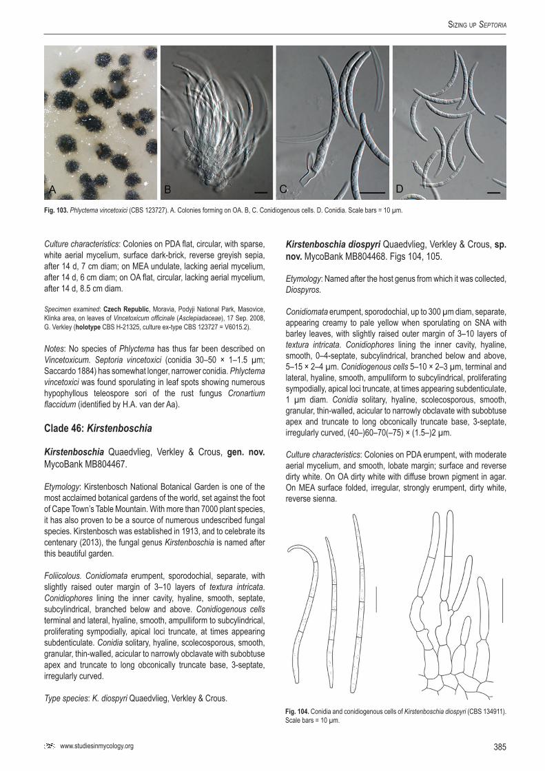
www.studiesinmycology.org
Sizing up Septoria
385
Culture characteristics: Colonies on PDA flat, circular, with sparse, white aerial mycelium, surface dark-brick, reverse greyish sepia, after 14 d, 7 cm diam; on MEA undulate, lacking aerial mycelium, after 14 d, 6 cm diam; on OA flat, circular, lacking aerial mycelium, after 14 d, 8.5 cm diam.
Specimen examined: czech republic, Moravia, Podyji National Park, Masovice, Klinka area, on leaves of Vincetoxicum officinale (Asclepiadaceae), 17 Sep. 2008, G. Verkley (holotype CBS H-21325, culture ex-type CBS 123727 = V6015.2).
Notes: No species of Phlyctema has thus far been described on Vincetoxicum. Septoria vincetoxici (conidia 30–50 × 1–1.5 µm; Saccardo 1884) has somewhat longer, narrower conidia. Phlyctema vincetoxici was found sporulating in leaf spots showing numerous hypophyllous teleospore sori of the rust fungus Cronartium flaccidum (identified by H.A. van der Aa).
Clade 46: Kirstenboschia
Kirstenboschia Quaedvlieg, Verkley & Crous, gen. nov. MycoBank MB804467.
Etymology: Kirstenbosch National Botanical Garden is one of the most acclaimed botanical gardens of the world, set against the foot of Cape Town’s Table Mountain. With more than 7000 plant species, it has also proven to be a source of numerous undescribed fungal species. Kirstenbosch was established in 1913, and to celebrate its centenary (2013), the fungal genus Kirstenboschia is named after this beautiful garden.
Foliicolous. Conidiomata erumpent, sporodochial, separate, with slightly raised outer margin of 3–10 layers of textura intricata. Conidiophores lining the inner cavity, hyaline, smooth, septate, subcylindrical, branched below and above. Conidiogenous cells terminal and lateral, hyaline, smooth, ampulliform to subcylindrical, proliferating sympodially, apical loci truncate, at times appearing subdenticulate. Conidia solitary, hyaline, scolecosporous, smooth, granular, thin-walled, acicular to narrowly obclavate with subobtuse apex and truncate to long obconically truncate base, 3-septate, irregularly curved.
Type species: K. diospyri Quaedvlieg, Verkley & Crous.
Kirstenboschia diospyri Quaedvlieg, Verkley & Crous, sp. nov. MycoBank MB804468. Figs 104, 105.
Etymology: Named after the host genus from which it was collected, Diospyros.
Conidiomata erumpent, sporodochial, up to 300 µm diam, separate, appearing creamy to pale yellow when sporulating on SNA with barley leaves, with slightly raised outer margin of 3–10 layers of textura intricata. Conidiophores lining the inner cavity, hyaline, smooth, 0–4-septate, subcylindrical, branched below and above, 5–15 × 2–4 µm. Conidiogenous cells 5–10 × 2–3 µm, terminal and lateral, hyaline, smooth, ampulliform to subcylindrical, proliferating sympodially, apical loci truncate, at times appearing subdenticulate, 1 µm diam. Conidia solitary, hyaline, scolecosporous, smooth, granular, thin-walled, acicular to narrowly obclavate with subobtuse apex and truncate to long obconically truncate base, 3-septate, irregularly curved, (40–)60–70(–75) × (1.5–)2 µm.
Culture characteristics: Colonies on PDA erumpent, with moderate aerial mycelium, and smooth, lobate margin; surface and reverse dirty white. On OA dirty white with diffuse brown pigment in agar. On MEA surface folded, irregular, strongly erumpent, dirty white, reverse sienna.
Fig. 103. Phlyctema vincetoxici (CBS 123727). A. Colonies forming on OA. B, C. Conidiogenous cells. D. Conidia. Scale bars = 10 µm.
Fig. 104. Conidia and conidiogenous cells of Kirstenboschia diospyri (CBS 134911). Scale bars = 10 µm.
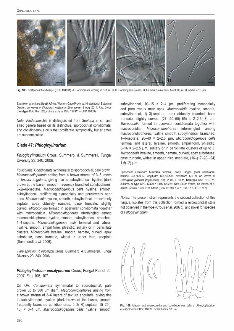
Quaedvlieg et al.
386
Specimen examined: South Africa, Western Cape Province, Kirstenbosch Botanical Garden, on leaves of Diospyros whyteana (Ebenaceae), 9 Aug. 2011, P.W. Crous (holotype CBS H-21326, culture ex-type CBS 134911 = CPC 19869).
Note: Kirstenboschia is distinguished from Septoria s. str. and allied genera based on its distinctive, sporodochial conidiomata, and conidiogenous cells that proliferate sympodially, but at times are subdenticulate.
Clade 47: Phlogicylindrium
Phlogicylindrium Crous, Summerb. & Summerell, Fungal Diversity 23: 340. 2006.
Foliicolous. Conidiomata synnematal to sporodochial, pale brown. Macroconidiophores arising from a brown stroma of 3–6 layers of textura angularis, giving rise to subcylindrical, hyaline (dark brown at the base), smooth, frequently branched conidiophores, 0–2(–6)-septate. Macroconidiogenous cells hyaline, smooth, subcylindrical, proliferating sympodially and percurrently near apex. Macroconidia hyaline, smooth, subcylindrical, transversely septate, apex obtusely rounded, base truncate, slightly curved. Microconidia formed in acervular conidiomata together with macroconidia. Microconidiophores intermingled among macroconidiophores, hyaline, smooth, subcylindrical, branched, 1–4-septate. Microconidiogenous cells terminal and lateral, hyaline, smooth, ampulliform, phialidic, solitary or in penicillate clusters. Microconidia hyaline, smooth, hamate, curved, apex subobtuse, base truncate, widest in upper third, aseptate (Summerell et al. 2006).
Type species: P. eucalypti Crous, Summerb. & Summerell, Fungal Diversity 23: 340. 2006.
Phlogicylindrium eucalyptorum Crous, Fungal Planet 20. 2007. Figs 106, 107.
On OA. Conidiomata synnematal to sporodochial, pale brown up to 300 μm diam. Macroconidiophores arising from a brown stroma of 3–6 layers of textura angularis, giving rise to subcylindrical, hyaline (dark brown at the base), smooth, frequently branched conidiophores, 0–2(–6)-septate, 15–25(–45) × 3–4 μm. Macroconidiogenous cells hyaline, smooth,
subcylindrical, 10–15 × 2–4 μm, proliferating sympodially and percurrently near apex. Macroconidia hyaline, smooth, subcylindrical, 1(–3)-septate, apex obtusely rounded, base truncate, slightly curved, (27–)40–50(–55) × 2–2.5(–3) μm. Microconidia formed in acervular conidiomata together with macroconidia. Microconidiophores intermingled among macroconidiophores, hyaline, smooth, subcylindrical, branched, 1–4-septate, 20–40 × 2–2.5 µm. Microconidiogenous cells terminal and lateral, hyaline, smooth, ampulliform, phialidic, 5–16 × 2–2.5 µm, solitary or in penicillate clusters of up to 3. Microconidia hyaline, smooth, hamate, curved, apex subobtuse, base truncate, widest in upper third, aseptate, (16–)17–20(–24) 1.5(–2) µm.
Specimens examined: Australia, Victoria, Otway Ranges, (near Gellibrand), latitude: -38.568412, longitude: 143.539586, elevation: 175 m, on leaves of Eucalyptus globulus (Myrtaceae), Sep. 2005, I. Smith, holotype CBS H-19771, cultures ex-type CPC 12429 = CBS 120221; New South Wales, on leaves of E. nitens, 22 Nov. 1996, P.W. Crous (CBS 111689 = CPC 1547 = STE-U 1547).
Notes: The present strain represents the second collection of this fungus. Isolates from this collection formed a microconidial state not observed in the type (Crous et al. 2007c), and novel for species of Phlogicylindrium.
Fig. 105. Kirstenboschia diospyri (CBS 134911). A. Conidiomata forming in culture. B, C. Conidiogenous cells. D. Conidia. Scale bars: A = 300 µm, all others = 10 µm.
Fig. 106. Macro- and microconidia and conidiogenous cells of Phlogicylindrium eucalyptorum (CBS 111689). Scale bars = 10 µm.

www.studiesinmycology.org
Sizing up Septoria
387
dIScuSSIoN
The main question considered in the present study was: what is Septoria? To address this we included 370 isolates representing 170 species, sampled from six continents. Furthermore, we also generated several phylogenetic datasets based on partial sequences of the ITS, LSU, Btub, RPB2 and EF-1α loci. In the final analysis, it was clear that Septoria is a well-defined genus and phylogenetic clade, with pycnidial, ostiolate conidiomata, conidiophores reduced to conidiogenous cells that proliferate sympodially, and hyaline, filiform conidia with transverse eusepta, fitting the original concept of Sutton (1980). However, when host material has been incubated for a while, several pycnidial species tend to form acervuli (also not clearly defined when studied in culture on normal agar media), and conidiogenous cells could have a combination of sympodial and percurrent proliferation (as observed in Pseudocercospora; Crous et al. 2013).
The present study, including that of Verkley et al. (2013) defined an additional 15 genera that were formerly treated as “septoria” in the widest sense. Although it has recently been shown that Phoma is a generic complex representing many morphologic and phylogenetic genera (Aveskamp et al. 2010, de Gruyter et al. 2010, 2013), this was not expected to also be the case for Septoria. Furthermore, many of the septoria-like genera discussed earlier in this paper are presently still not known from sequence, and thus their phylogeny remains to be resolved, meaning that they could add futher entities to the list of acknowledged septoria-like genera.
Although Septoria s. str. is a genus in the Mycosphaerellaceae (Capnodiales), several of the septoria-like genera clustered outside this family. Species of Septoria are morphologically conserved, and in the past many taxa were identified based on host, which has been shown to be unreliable (see Verkley et al. 2013), as several taxa have wide host ranges. Another complication revealed in the present study is that many septoria-like genera cluster in different phylogenetic clades, but have still retained the Septoria morphological characters, which means that as in Phoma, future
identifications in this complex will also have to rely on DNA sequence data to support morphological conclusions.
The genus Stagonospora has always been separated from Septoria on the basis that Septoria has conidiogenous cells with sympodial proliferation, whereas in Stagonospora they proliferate percurrently. As shown in the present study, however, conidiogenesis is far too broad a feature to define all genera that express these modes of proliferation in their conidiogenous cells. Stagonospora, which is based on S. paludosa, was epitypified in this study, and shown to cluster apart from Septoria s. str. Another major surprise lies in the fact that Septoria nodorum blotch, caused by “Stagonospora” nodorum, clusters in a distinct genus, unrelated to Stagonospora s. str., and also separate from Phaeosphaeria s. str. A repercussion from these findings is the fact that the common cereal pathogens, which are neither Stagonospora, Septoria, Phaeosphaeria or Leptosphaeria (see de Gruyter et al. 2013), now have to be accommodated in a new genus, Parastagonospora. Furthermore, it appears that Stagonospora s. str. occurs on Poaceae, but has thus far only been confirmed from Carex, though further sampling will undoubtedly extend the host range of this genus. Parastagonospora is thus a novel, distinct stagonospora-like genus, which has sexual morphs that are phaeosphaeria-like in morphology, thus quite unlike those of Stagonospora s. str., which are more didymella-like in morphology.
The genus Phaeosphaeria is based on P. oryzae (asexual morph Phaeoseptoria oryzae), for which we could designate an epitype in this study. Furthermore, we also recollected the type species of Phaeoseptoria, P. papayae, for which we also designated an epitype. As expected, Phaeoseptoria clusters with Phaeosphaeria, for which we choose the name of the sexual morph, Phaeosphaeria, on the basis that it is clearly resolved, and well established in literature. In contrast, Phaeoseptoria has in recent years become a muddled concept harbouring unrelated coelomycetes with pigmented conidia.
Obtaining a culture of Cytostagonospora martiniana clarified the phylogenetic position of the genus as distinct from Septoria, resolving the difference of opinion between von Arx (1983), who regarded it as synonym of Septoria, versus Sutton (1980), who
Fig. 107. Phlogicylindrium eucalyptorum (CBS 111689). A. Colony on OA. B, E. Microcyclic conidiation with macro- and macroconidia. C. Macroconidiogenous cells. D. Microconidia. F. Macroconidia. Scale bars = 10 µm.

Quaedvlieg et al.
388
retained it as separate genus. Of interest is the unique mode of conidiogenesis, ranging from holoblastic sympodial to polyphialides with periclinal thickening to percurrent proliferation. It should be noted, however, that although this is a distinct genus, C. mariniana is not the type of Cytostagonospora, and C. photiniicola (occurring on Photinia serrulata, Austria) will have to be recollected to confirm that these two fungi are congeneric.
The genus Phloeospora (based on P. ulmi) has for long been assumed to be a synonym of Septoria based on morphology. It is thus good to finally see it resolved as separate phylogenetic lineage, which is also supported morphologically based on its acervular conidiomata and conidiogenous cells with prominent percurrent proliferation. In spite of resolving 21 genera, several lineages remain unresolved, and are simply treated as “septoria-like” awaiting the recollection of additional material.
It is surprising that so many of the cereal pathogens actually have a confused taxonomy. Eyespot disease of wheat, formely treated as Tapesia (Ramulispora asexual states), was shown to represent a distinct genus Oculimacula (Helgardia asexual states) (Crous et al. 2003), while Quaedvlieg et al. (2011) determined that Septoria tritici blotch, caused by “Septoria” tritici, is in fact better accommodated in a new genus, Zymoseptoria, which appears to be restricted to members of Poaceae. The present study also resolved the phylogenetic position of Septoria nodorum blotch, which proved to not represent a member of Septoria, Stagonospora, or Phaeosphaeria, but to represent a distinct genus, described here as Parastagonospora. Clearly more attention should be directed towards resolving the taxonomy of the pathogens of agricultural crops of major economic importance in future, as these findings also have implications for genomic studies, where organisms from different genera, and even families get compared to one another, and new evolutionary hypotheses are proposed on the assumption that these taxa are congeneric. To clarify the taxonomy of well-known plant pathogens, however, many species will have to be recollected, and epitypified, so that authentic cultures and DNA barcodes will become available to fix the genetic application of these names.
General conclusions
The genus Septoria is defined by having pycnidial to acervular conidiomata, and hyaline conidiophores that give rise to conidiogenous cells that proliferate sympodially and percurrently, forming hyaline, filiform conidia with transverse eusepta. Many species have wide host ranges, and host occurrence should not be used as primary character for identification (see Verkley et al. 2013, this issue). Although species of Septoria and several of the novel genera introduced here have mycosphaerella-like sexual states, the name Mycosphaerella is restricted to the genus Ramularia, and is unavailable for species of Septoria and related genera.
aCKNoWlEdGEMENTs
We thank the technical staff, Arien van Iperen (cultures), and Marjan Vermaas (photographic plates), for their invaluable assistance. The research leading to these results has received funding from the European Community’s Seventh Framework Program (FP7/2007–2013)/grant agreement no. 226482 (Project: QBOL - Development of a new diagnostic tool using DNA barcoding to identify quarantine organisms in support of plant health) by the European Commission under the theme “Development of new diagnostic methods in support of Plant Health policy” (no. KBBE-2008-1-4-01). Special thanks also go to Dr Ellen van Agtmaal who prepared the line drawings included in this paper from photomicrographs and published materials (Sutton 1980) using Adobe Photoshop CS3.
rEFErENCEs
Aptroot A (2006). Mycosphaerella and its anamorphs. 2, Conspectus of Mycosphaerella. CBS Biodiversity Series 5. CBS-KNAW Fungal Biodiversity Centre, Utrecht, The Netherlands.
Arx JA von (1983). Mycosphaerella and its anamorphs. Proceedings of the Koninklijke Nederlandse Akademie van Wetenschappen Series C-Biological and Medical Sciences 86(1): 15–54.
Arzanlou M, Crous PW (2006). Phaeosphaeriopsis musae. Fungal Planet 9. CBS-KNAW Fungal Biodiversity Centre, Utrecht, The Netherlands.
Aveskamp M, Gruyter H de, Woudenberg J, Verkley G, Crous PW (2010). Highlights of the Didymellaceae: A polyphasic approach to characterise Phoma and related pleosporalean genera. Studies in Mycology 65: 1–60.
Barnes I, Crous PW, Wingfield BD, Wingfield MJ (2004). Multigene phylogenies reveal that red band needle blight of Pinus is caused by two distinct species of Dothistroma, D. septosporum and D. pini. Studies in Mycology 50: 551–565.
Beach WS (1919). Biologic specialization in the genus Septoria. American Journal of Botany 6: 1–32.
Bedlan G (2011). Septoria juliae sp. nov. – a new Septoria species on Nerium oleander. Journal für Kulturpflanzen 63: 430–431.
Bissett J (1982). Stagonospora avenae. Fungi Canadenses 239. National Mycological Herbarium, Biosystematics Research Institute, Agriculture Canada, Ottawa.
Braun U (1990). Taxonomic problems of the Ramularia / Cercosporella complex. Studies in Mycology 32: 65–75.
Braun U (1995). A monograph of Cercosporella, Ramularia and allied genera (Phytopathogenic Hyphomycetes). Vol. 1. IHW-Verlag, Eching.
Braun U (1998). A monograph of Cercosporella, Ramularia and allied genera (Phytopathogenic Hyphomycetes). Vol. 2. IHW-Verlag, Eching.
Câmara MPS, Ramaley AW, Castlebury LA, Palm ME (2003). Neophaeosphaeria and Phaeosphaeriopsis , segregates of Paraphaeosphaeria. Mycological Research 107: 516–522.
Carbone I, Kohn LM (1999). A method for designing primer sets for speciation studies in filamentous ascomycetes. Mycologia 91: 553–556.
Cheewangkoon R, Crous PW, Hyde KD, Groenewald JZ, To-anan C (2008). Species of Mycosphaerella and related anamorphs on Eucalyptus leaves from Thailand. Persoonia 21: 77–91.
Constantinescu O (1984). Taxonomic revision of Septoria-like fungi parasitic on Betulaceae. Transactions of the British Mycological Society 83: 383–398.
Crous PW (1998). Mycosphaerella spp. and their anamorphs associated with leaf spot diseases of Eucalyptus. Mycologia Memoir 21. APS Press, MN, USA.
Crous PW, Braun U (2003). Mycosphaerella and its anamorphs: 1. Names published in Cercospora and Passalora. CBS Biodiversity Series 1. CBS-KNAW Fungal Biodiversity Centre, Utrecht, The Netherlands.
Crous PW, Braun U, Groenewald JZ (2007a). Mycosphaerella is polyphyletic. Studies in Mycology 58: 1–32.
Crous PW, Braun U, Hunter GC, Wingfield MJ, Verkley GJM, et al. (2013). Phylogenetic lineages in Pseudocercospora. Studies in Mycology 75: 37–114.
Crous PW, Braun U, Schubert K, Groenewald JZ (2007b). Delimiting Cladosporium from morphologically similar genera. Studies in Mycology 58: 33–56.
Crous PW, Ferreira FA, Sutton BC (1997). A comparison of the fungal genera Phaeophleospora and Kirramyces (coelomycetes). South African Journal of Botany 63: 111–115.
Crous PW, Groenewald JZ, Gams W (2003). Eyespot of cereals revisited: ITS phylogeny reveals new species relationships. European Journal of Plant Pathology 109: 841–850.
Crous PW, Groenewald JZ, Smith IW (2007c). Phlogicylindrium eucalyptorum. Fungal Planet 20. CBS-KNAW Fungal Biodiversity Centre, Utrecht, The Netherlands.
Crous PW, Kang JC, Braun U (2001). A phylogenetic redefinition of anamorph genera in Mycosphaerella based on ITS rDNA sequence and morphology. Mycologia 93: 1081–1101.
Crous PW, Schoch CL, Hyde KD, Wood AR, Gueidan C, et al. (2009a). Phylogenetic lineages in the Capnodiales. Studies in Mycology 64: 17–47.
Crous PW, Shivas RG, Wingfield MJ, Summerell BA, Rossman AY, et al. (2012a). Fungal Planet description sheets 128–127. Persoonia 29: 138–153.
Crous PW, Slippers B, Wingfield MJ, Rheeder J, Marasas WFO, et al. (2006). Phylogenetic lineages in the Botryosphaeriaceae. Studies in Mycology 55: 235–253.
Crous PW, Summerell BA, Carnegie AJ, Wingfield MJ, Groenewald JZ (2009b). Novel species of Mycosphaerellaceae and Teratosphaeriaceae. Persoonia 23: 119–146.
Crous PW, Summerell BA, Carnegie AJ, Wingfield MJ, Hunter GC, et al. (2009c). Unravelling Mycosphaerella: do you believe in genera? Persoonia 23: 99–118.
Crous PW, Tanaka K, Summerell BA, Groenewald JZ (2011). Additions to the Mycosphaerella complex. IMA Fungus 2: 49–64.
www.studiesinmycology.org
Sizing up Septoria
389
Crous PW, Verkley GJM, Christensen M, Castañeda-Ruiz RF, Groenewald JZ (2012b). How important are conidial appendages? Persoonia 28: 126–137.
Crous PW, Verkley GJM, Groenewald JZ, Samson RA (2009d). CBS Laboratory Manual Series 1. CBS-KNAW Fungal Biodiversity Centre, Utrecht, The Netherlands.
Crous PW, Wingfield MJ, Park RF (1991). Mycosphaerella nubilosa a synonym of M. molleriana. Mycological Research 95: 628–632.
Cunfer BM (2000). Stagonospora and Septoria diseases of barley, oat, and rye. Canadian Journal of Plant Pathology 22: 332–348.
Cunfer BM, Ueng PP (1999). Taxonomy and identification of Septoria and Stagonospora species on small-grain cereals. Annual Review of Phytopathology 37: 267–284.
Deighton FC (1987). New species of Pseudocercospora and Mycovellosiella, and new combinations into Pseudocercospora and Phaeoramularia. Transactions of the British Mycological Society 88: 365–391.
Demaree JB, Wilcox MS (1943). The fungus causing the so-called “Septoria leaf-spot disease” of raspberry. Phytopathology 33: 986–1003.
Desmazières JBHJ (1847). Quatorzième notice sur les plantes cryptogames récemment découvertes en France. Annales des Sciences Naturelles Botanique, Série 3, 8: 9–37, 172–193.
Donk MA (1964). Nomina conervana proposita. Deuteromycetes. Regnum Vegetable 34: 7–15.
Dyko BJ, Sutton BC (1979). Two new and unusual deuteromycetes. Transactions of the British Mycological Society 72: 411–417.
Ellis MB (1971). Dematiaceous Hyphomycetes. Commonwealth Mycological Institute, Kew, Surrey, UK.
Ellis MB (1976). More Dematiaceous Hyphomycetes. Commonwealth Mycological Institute, Kew, Surrey, UK.
Ellis MB, Ellis JP (1997). Microfungi on Land Plants – An Identification Handbook. Richmond Publishing, England.
Eriksson OE (1967). On graminicolous pyrenomycetes from Fennoscandia I, II, Ill. Phragmosporous and scolecosporous species. Arkiv för Botanik Series 2, 6: 381–440.
Evans HC (1984). The genus Mycosphaerella and its anamorphs Cercoseptoria, Dothistroma and Lecanosticta on pines. Mycological Papers 153: 1–102.
Eyal Z, Sharen AL, Prescott JM, Ginkel M van (1987). The Septoria diseases of wheat: concepts and methods of disease management. Mexico, DF, CIMMYT.
Farr DF (1991). Septoria species on cornus. Mycologia 83: 611–623.Farr DF (1992). Species of Septoria on the Fabaceae, subfamily Faboidae, tribe
Genistae. Sydowia 44: 13–31.Farr DF, Rossman AY (2013). Fungal Databases, Systematic Mycology and
Microbiology Laboratory, ARS, USDA. Retrieved May 3, 2013, from http://nt.ars-grin.gov/fungaldatabases/
Feau N, Hamelin RC, Bernier L (2006). Attributes and congruence of three molecular data sets: Inferring phylogenies among Septoria related species from woody perennial plants. Molecular Phylogenetics and Evolution 40: 808–829.
Ferreira FA (1989). Patologia Florestal. Principais Doenças Florestais No Brasil. Sociedade de Investigaçoes Florestais, Viçosa, MG, Brasil.
Frank J, Crous PW, Groenewald JZ, Oertel B, Hyde KD, et al. (2010). Microcyclospora and Microcyclosporella: novel genera accommodating epiphytic fungi causing sooty blotch on apple. Persoonia 24: 93–105.
Fukuhara M (2002). Three Phaeosphaeria species and Paraphaeosphaeria michotii isolated from Phragmites leaves in Osaka, Japan. Mycoscience 43: 275–382.
Golzar H, Wang C (2012). First report of Phaeosphaeriopsis glaucopunctata as the cause of leaf spot and nectrosis on Ruscus aculeatus in Australia. Australasian Plant Disease Notes 7: 13–15.
Goodwin SB (2004). Minimum phylogenetic coverage: An additional criterion to guide the selection of microbial pathogens for initial genomic sequencing efforts. Phytopathology 94: 800–804.
Greene HC (1961). Notes on Wisconsin parasitic fungi. XXVII. Wisconsin Academy of Sciences, Arts and Letters 50: 141–161.
Groenewald JZ, Nakashima C, Nishikawa J, Shin H-D, Park J-H, et al. (2013). Species concepts in Cercospora: spotting the weeds among the roses. Studies in Mycology 75: 115–170.
Gruyter J de, Woudenberg JHC, Aveskamp MM, Verkley GJM, Groenewald JZ, Crous PW (2010). Systematic reappraisal of species in Phoma section Paraphoma, Pyrenochaeta and Pleurophoma. Mycologia 102: 1066–1081.
Gruyter J de, Woudenberg JHC, Aveskamp MM, Verkley GJM, Groenewald JZ, Crous PW (2013). Redisposition of Phoma-like anamorphs in Pleosporales. Studies in Mycology 75: 1–36.
Hall TA (1999). BioEdit: A user-friendly biological sequence alignment editor and analysis program for Windows 95/98/NT. Nucleic Acids Symposium Series 41: 95–98.
Hawksworth DL, Crous PW, Redhead SA, Reynolds DR, Samson RA, et al. (2011). The Amsterdam Declaration on Fungal Nomenclature. IMA Fungus 2: 105–112.
Huelsenbeck JP, Ronquist F (2001). MRBAYES: Bayesian inference of phylogenetic trees. Bioinformatics 17: 754–755.
Inácio CA, Dianese JC (1998). Some foliicolous fungi on Tabebuia species. Mycological Research 102: 695–708.
Jørstad I (1965). Septoria and septoroid fungi on dicotyleones in Norway. Oslo University Press, Oslo.
Jørstad I (1967). Septoria and Septoroid fungi on Gramineae in Norway. Oslo University Press, Oslo.
Kaneko S, Fujioka H, Zinno Y (1989). A new species of Septoria on Japanese black pine. Transactions of the Mycological Society of Japan 30: 463–466.
Katoh K, Misawa K, Kuma K, Miyata T (2002). MAFFT: a novel method for rapid multiple sequence alignment based on fast Fourier transform. Nucleic Acids Research 30: 3059–3066.
Lamprecht SC, Crous PW, Groenewald JZ, Tewoldemedhin YT, Marasas WFO (2011). Diaporthaceae associated with root rot of maize. IMA Fungus 2: 13–24.
Lee S, Mel’nik V, Taylor JE, Crous PW (2004). Diversity of saprobic hyphomycetes on Proteaceae and Restionaceae from South Africa. Fungal Diversity 17: 91–114.
Li HY, Sun GY, Zhai XR, Batzer JC, Mayfield DA, et al. (2012). Dissoconiaceae associated with sooty blotch and flyspeck on fruits in China and the United States. Persoonia 28: 113–125.
Liu JK, Phookamsak R, Mingkhuan M, Wikee S, Li YM, et al. (2012) Towards a natural classification of Botryosphaeriales. Fungal Diversity 57: 149–210.
Liu Y, Whelen S, Hall B (1999). Phylogenetic relationships among ascomycetes: evidence from an RNA polymerse II subunit. Molecular Biology and Evolution 16: 1799–1808.
Lombard L, Crous PW, Wingfield BD, Wingfield MJ (2010). Species concepts in Calonectria (Cylindrocladium). Studies in Mycology 66: 1–13.
Marincowitz S, Crous PW, Groenewald JZ, Wingfield MJ (2008). Microfungi occurring on Proteaceae in the fynbos. CBS Biodiversity Series 7. CBS-KNAW Fungal Biodiversity Centre, Utrecht, The Netherlands.
Mason-Gamer RJ, Kellogg EA (1996). Testing for phylogenetic conflict among molecular data sets in the tribe Triticeae (Gramineae). Systematic Biology 45: 524–545.
McDonald MC, Razavi M, Friesen TL, Brunner PC, McDonald BA (2012). Phylogenetic and population genetic analyses of Phaeosphaeria nodorum and its close relatives indicate cryptic species and an origin in the Fertile Crescent. Fungal Genetics and Biology 49: 882–895.
Michaelides T, Morgan DP, Doster MA (1995). Foliar and fruit fungal diseases. In: Pistachio Production (L Ferguson, ed). Center for Fruit and Nut Crop Research and Information, Pomology Department, University of California, Davis CA.: 148–159.
Monod M (1983). Monographie taxonomique des gnomoniacées ascomycètes de l’ordre des Diaporthales. Sydowia 9: 1–315.
Nag Raj TR (1993). Coelomycetous anamorphs with appendage-bearing conidia. Mycologue Publications, Waterloo, Ontario.
Niekerk JM van, Groenewald JZ, Verkley GJM, Fourie PH, Wingfield MJ, Crous PW (2004). Systematic reappraisal of Coniella and Pilidiella, with specific reference to species occurring on Eucalyptus and Vitis in South Africa. Mycological Research 108: 283–303.
Nylander JAA (2004). MrModeltest v2. Program distributed by the author. Evolutionary Biology Centre Uppsala University 2: 1–2.
O’Donnell K, Kistler HC, Cigelnik E, Ploetz RC (1998). Multiple evolutionary origins of the fungus causing Panama disease of banana: Concordant evidence from nuclear and mitochondrial gene genealogies. Proceedings of the National Academy of Sciences of the United States of America 95: 2044–2049.
Petrak F (1957). Über die auf Aconitum vorkommenden Arten der gattung Septoria. Sydowia 11: 375–379.
Petrak F, Sydow H (1927). Die Gattungen der Pyrenomyzeten, Sphaeropsideen und Melanconieen. 1. Teil. Die phaeosporen Sphaeropsideen und die Gattung Macrophoma. Repertorium specierum novarum regni vegetabilis, Beihefte Bd 42.
Phillips AJL, Alves A, Pennycook SR, Johnston PR, Ramaley A, et al. (2008). Resolving the phylogenetic and taxonomic status of dark-spored teleomorph genera in the Botryosphaeriaceae. Persoonia 21: 29–55.
Priest MJ (2006). Fungi of Australia: Septoria. ABRS, Canberra: CSIRO publishing, Melbourne, Australia.
Punithalingam E (1976). Septoria chrysanthemella. CMI Descriptions of Pathogenic Fungi and Bacteria 669. Commonwealth Mycological Institute, Kew, UK
Quaedvlieg W, Groenewald JZ, de Jesús Yáñez-Morales M, Crous PW (2012). DNA barcoding of Mycosphaerella species of quarantine importance to Europe. Persoonia 29: 101–115.
Quaedvlieg W, Kema GHJ, Groenewald JZ, Verkley GJM, Seifbarghi S, et al. (2011). Zymoseptoria gen. nov.: a new genus to accommodate Septoria-like species occurring on graminicolous hosts. Persoonia 26: 57–69.
Rayner RW (1970). A mycological colour chart. CMI and British Mycological Society. Kew, UK.
Rogers DP (1949). Nomina conservanda proposita and nomina confusa – Fungi: Nomina conservanda. Farlowia 3: 425–493.

Quaedvlieg et al.
390
Saccardo PA (1884). Sylloge Fungorum: Sylloge Sphaeropsidearum et Melanconiearum 3: 542. Padova, Italy.
Saccardo PA (1895). Sylloge Fungorum: Supplementum Universale, Pars III 11: 542. Padova, Italy.
Saccardo PA, Saccardo D (1906). Sylloge Fungorum: Supplementum Universale, Pars VII 18: 1–828. Padova, Italy.
Saccardo PA, Trotter A (1913). Sylloge Fungorum: Supplementum Universale, Pars IX 22: 1–1612. Padova, Italy.
Shin HD (1995). New fungal diseases of economic resource plants in Korea (II). Korean Journal of Plant Pathology 11: 120–131.
Shin HD, Sameva EF (2002). Taxonomic notes on the genus Septoria in Korea (II). Mycotaxon 83: 287–300.
Shin HD, Sameva EF (2004). Septoria in Korea. National Institute of Agricultural Science and Technology, Republic of Korea.
Shoemaker RA, Babcock CE (1989). Phaeosphaeria. Canadian Journal of Botany 67: 1500–1599.
Sivanesan A (1984). The Bitunicate Ascomycetes and their anamorphs. J. Cramer, Vaduz, Germany.
Solomon PS, Lowe RGT, Tan KC, Waters ODC, Oliver RP (2006). Stagonospora nodorum: cause of stagonospora nodorum blotch of wheat. Molecular Plant Pathology 7: 147–156.
Stukenbrock EH, Quaedvlieg W, Javan-Nikhah M, Zala M, Crous PW, McDonald BA (2012). Zymoseptoria ardabilia and Z. pseudotritici, two progenitor species of the septoria tritici leaf blotch fungus Z. tritici (synonym: Mycosphaerella graminicola). Mycologia 104: 1397–1407.
Summerell BA, Groenewald JZ, Carnegie AJ, Summerbell RC, Crous PW (2006). Eucalyptus microfungi known from culture. 2. Alysidiella, Fusculina and Phlogicylindrium genera nova, with notes on some other poorly known taxa. Fungal Diversity 23: 323–350.
Sutton B, Pollack F (1974). Microfungi on Cercocarpus. Mycopathologia 52: 331–351.
Sutton BC, Pascoe IG (1987). Septoria species on Acacia. Transactions of the British Mycological Society 89: 521–532.
Sutton BC, Pascoe IG (1989). Some Septoria species on native Australian plants. Studies in Mycology 31: 177–186.
Sutton BC, Swart HJ (1986). Australian leaf-inhabiting fungi XXIII. Colletogloeum species and similar fungi on Acacia. Transactions of the British Mycological Society 87: 93–102.
Sutton BC (1964). Coelomycetes III. Annellolacinia gen. nov., Aristastoma, Phaecytostroma, Seimatosporium, etc. Mycological Papers 64. Commonwealth Mycological Institute, Kew, UK.
Sutton BC (1977). Coelomycetes VI. Nomenclature of generic names proposed for Coelomycetes. Mycological Papers 141. Commonwealth Mycological Institute, Kew, UK.
Sutton BC (1980). The coelomycetes. Fungi imperfecti with pycnidia, acervuli and stromata. Commonwealth Mycological Institute, Kew, UK.
Teterevnikova-Babayan DN (1987). Fungi of the genus Septoria in the U.S.S.R. Akademiya Nauk Armyanskoi SSR, Yerevan.
Verkley GJM (1999). A monograph of the genus Pezicula and its anamorphs. Studies in Mycology 44: 1–180.
Verkley GJM, Priest MJ (2000). Septoria and similar coelomycetous anamorphs of Mycosphaerella. Studies in Mycology 45: 123–128.
Verkley GJM, Crous PW, Groenewald JZ, Braun U, Aptroot A (2004a). Mycosphaerella punctiformis revisited: morphology, phylogeny, and epitypification of the type species of the genus Mycosphaerella (Dothideales, Ascomycota). Mycological Research 108: 1271–1282.
Verkley GJM, Silva da M, Wicklow DT, Crous PW (2004b). Paraconiothyrium, a new genus to accommodate the mycoparasite Coniothyrium minitans, anamorphs of Paraphaeosphaeria, and four new species. Studies in Mycology 50: 323–335.
Verkley GJM, Starink-Willemse M, van Iperen A, Abeln ECA (2004c). Phylogenetic analyses of Septoria species based on the ITS and LSU-D2 regions of nuclear ribosomal DNA. Mycologia 96: 558–571.
Verkley GJM, Quaedvlieg W, Shin HD, Crous PW (2013). A new approach to species delimitation in Septoria. Studies in Mycology 75: 213–305.
Vilgalys R, Hester M. 1990. Rapid genetic identification and mapping of enzymatically amplified ribosomal DNA from several Cryptococcus species. Journal of Bacteriology 172: 4238–4246.
Wakefield EM (1940). Nomina generica conservanda. Transactions of the British Mycological Society 24: 282–293.
Walker J, Sutton BC, Pascoe IG (1992). Phaeoseptoria eucalypti and similar fungi on Eucalyptus, with description of Kirramyces gen. nov. (Coelomycetes). Mycological Research 96: 911–924.
White TJ, Bruns T, Lee S, Taylor J (1990). Amplification and direct sequencing of fungal ribosomal RNA genes for phylogenetics. In: PCR Protocols: a guide to methods and applications (Innis MA, Gelfand DH, Sninsky JJ, White TJ, eds). Academic Press, San Diego, California: 315–322.
Wingfield MJ, De Beer ZW, Slippers B, Wingfield BD, Groenewald JZ, et al. (2012). One fungus, one name promotes progressive plant pathology. Molecular Plant Pathology 13: 604–613.
Yoo J-J, Eom A-H (2012). Molecular identification of endophytic fungi isolated from needle leaves of conifers in Bohyeon Mountain, Korea. Mycobiology 40: 231–235.
Zhang Y, Crous PW, Schoch CL, Hyde KD (2012). Pleosporales. Fungal Diversity 53: 1–221.