Size and location relationships of stalked barnacles of the genus Octolasmis on the mangrove crab,...
12
JOURNAL OF CRUSTACEAN BIOLOGY, 20(3): 483494, 2000 SIZE AND LOCATION RELATIONSHIPS OF STALKED BARNACLES OF THE GENUS OCTOLASMZS ON THE MANGROVE CRAB SCYLLA SERRATA Harold K. Voris, William B. Jeffries, and Sombat Poovachiranon (HKV) Department of Zoology, Field Museum of Natural History, Chicago, Illinois 60605, U.S.A. (e-mail: hvoris @fmnh.org); (WBJ) Department of Biology, Dickinson College, Carlisle, Pennsylvania 17013, U.S.A. (e-mail: [email protected]); (SP) Phuket Marine Biological Center, Phuket, Thailand ABSTRACT Cyprid larvae of the lepadomorph Octolasmis colonize the gill chambers of the edible mangrove crab Scylla seratta (Forskbl, 1755). In a natural population of 856 mangrove crabs from southern Thailand, 260 individuals were infested by 3,670 Octolasmis cor and 1,758 0. angulata, including 1,014 subadults, 168 cyprids, and 38 peduncles of the two species. This population of gill cham- ber symbionts was examined to investigate the relationship between barnacle size and barnacle spatial distributions. The habitat of the branchial chamber was partitioned for study into gills one through eight, the inside (hypobranchial) and outside (hyperbranchial) gill surfaces, and the prox- imal, medial, and distal regions of each gill. The collective data from 260 crabs were pooled for an analysis that showed a nonrandom relationship between the size of octolasmids and thier loca- tion within the gill chamber. On the inside gill surfaces 0. angulata attained its largest average size on gills 3, 7, and 8, whereas on the outside surface the barnacles were largest on gills 4 and 5. Octolasmis cor attained its greatest average size on the inside surface of gill number 6. Com- parisons of barnacles from the three gill regions also revealed some significant differences in av- erage barnacle size. Positive correlations among barnacle size, barnacle number, and barnacle den- sity were present. Moreover, there was a significant correlation between the total numbers of bar- nacles and the average size of 0. cor on the inside surfaces of gill numbers 1 to 8, whereas it was not significant for 0. angulata. There was also a significant correlation between barnacle densities and barnacle size in 0. cor but not 0 angulata. Positive correlations were also observed among higher numbers of barnacles, larger barnacles, and higher numbers of advanced reproductive stages. Areas with higher densities also were areas of higher average fecundity. Pedunculate or Lepadomorph barnacles are in the Order Pedunculata Newman, 1987, within the Superorder Thoracica Darwin, 1854. Barnacles of the genus Octolasmis Gray, 1825 (Crustacea: Cirripedia: Poecilas- matidae Annandale, 1909) are pedunculates but their habitats differ from common goose- neck barnacles such as Lepas anatifera Lin- naeus, 1767, which live attached to floating objects (Anderson, 1994). The majority of Octolasmis species are symbiotic (sensu lato, de Bary, 1879) on the integument of deca- pod Crustacea. As cyprid larvae, the barna- cles cement themselves to these ephemeral substrates. They metamorphose on them into juveniles, grow, and live out their adult lives. At host ecdysis the life cycle ends because both the integument (exuviae) and attached barnacles are shed. Thus the life cycle of the symbionts must exist within the life cycle of their hosts, and it is likely that the two are integrated and may be regulated by some of the same hormones. Colonization of newly molted mangrove crabs Scylla serruta (Forskil, 1755) by Oc- tolusmis cyprid larvae is significantly pulsed because the cyprids aggregate on premolt crabs (but do not attach) and at the time of host ecdysis the cyprids move from the old exuviae to the integument of the newly molted crab (Jeffries et al., 1989). Thus, cyprid larvae exploring recently molted deca- pods may interact with each other, but there is no chance of them encountering attached adult Octolasmis. Knowledge of infestation levels, attach- ment sites, numbers and density of barnacles on the host integment-along with the sex, age, and physiological condition of the hosts-provides a basis for understanding the host symbiont relationship. For example, in a sample of 856 mangrove crabs S. serruta, 30 percent harbored Octolasmis in their gill chambers, and crabs with carapace widths of less than 34.3 mm bore no Octolasmis. The percentage of crabs hosting Octolasmis in- 483
-
Upload
fieldmuseum -
Category
Documents
-
view
0 -
download
0
Transcript of Size and location relationships of stalked barnacles of the genus Octolasmis on the mangrove crab,...

JOURNAL OF CRUSTACEAN BIOLOGY, 20(3): 483494, 2000
SIZE AND LOCATION RELATIONSHIPS OF STALKED BARNACLES OF THE GENUS OCTOLASMZS ON THE MANGROVE CRAB SCYLLA SERRATA
Harold K. Voris, William B. Jeffries, and Sombat Poovachiranon
(HKV) Department of Zoology, Field Museum of Natural History, Chicago, Illinois 60605, U.S.A. (e-mail: hvoris @fmnh.org); (WBJ) Department of Biology, Dickinson College,
Carlisle, Pennsylvania 17013, U.S.A. (e-mail: [email protected]); (SP) Phuket Marine Biological Center, Phuket, Thailand
A B S T R A C T
Cyprid larvae of the lepadomorph Octolasmis colonize the gill chambers of the edible mangrove crab Scylla seratta (Forskbl, 1755). In a natural population of 856 mangrove crabs from southern Thailand, 260 individuals were infested by 3,670 Octolasmis cor and 1,758 0. angulata, including 1,014 subadults, 168 cyprids, and 38 peduncles of the two species. This population of gill cham- ber symbionts was examined to investigate the relationship between barnacle size and barnacle spatial distributions. The habitat of the branchial chamber was partitioned for study into gills one through eight, the inside (hypobranchial) and outside (hyperbranchial) gill surfaces, and the prox- imal, medial, and distal regions of each gill. The collective data from 260 crabs were pooled for an analysis that showed a nonrandom relationship between the size of octolasmids and thier loca- tion within the gill chamber. On the inside gill surfaces 0. angulata attained its largest average size on gills 3, 7, and 8, whereas on the outside surface the barnacles were largest on gills 4 and 5. Octolasmis cor attained its greatest average size on the inside surface of gill number 6. Com- parisons of barnacles from the three gill regions also revealed some significant differences in av- erage barnacle size. Positive correlations among barnacle size, barnacle number, and barnacle den- sity were present. Moreover, there was a significant correlation between the total numbers of bar- nacles and the average size of 0. cor on the inside surfaces of gill numbers 1 to 8, whereas it was not significant for 0. angulata. There was also a significant correlation between barnacle densities and barnacle size in 0. cor but not 0 angulata. Positive correlations were also observed among higher numbers of barnacles, larger barnacles, and higher numbers of advanced reproductive stages. Areas with higher densities also were areas of higher average fecundity.
Pedunculate or Lepadomorph barnacles are in the Order Pedunculata Newman, 1987, within the Superorder Thoracica Darwin, 1854. Barnacles of the genus Octolasmis Gray, 1825 (Crustacea: Cirripedia: Poecilas- matidae Annandale, 1909) are pedunculates but their habitats differ from common goose- neck barnacles such as Lepas anatifera Lin- naeus, 1767, which live attached to floating objects (Anderson, 1994). The majority of Octolasmis species are symbiotic (sensu lato, de Bary, 1879) on the integument of deca- pod Crustacea. As cyprid larvae, the barna- cles cement themselves to these ephemeral substrates. They metamorphose on them into juveniles, grow, and live out their adult lives. At host ecdysis the life cycle ends because both the integument (exuviae) and attached barnacles are shed. Thus the life cycle of the symbionts must exist within the life cycle of their hosts, and it is likely that the two are integrated and may be regulated by some of the same hormones.
Colonization of newly molted mangrove crabs Scylla serruta (Forskil, 1755) by Oc- tolusmis cyprid larvae is significantly pulsed because the cyprids aggregate on premolt crabs (but do not attach) and at the time of host ecdysis the cyprids move from the old exuviae to the integument of the newly molted crab (Jeffries et al., 1989). Thus, cyprid larvae exploring recently molted deca- pods may interact with each other, but there is no chance of them encountering attached adult Octolasmis.
Knowledge of infestation levels, attach- ment sites, numbers and density of barnacles on the host integment-along with the sex, age, and physiological condition of the hosts-provides a basis for understanding the host symbiont relationship. For example, in a sample of 856 mangrove crabs S. serruta, 30 percent harbored Octolasmis in their gill chambers, and crabs with carapace widths of less than 34.3 mm bore no Octolasmis. The percentage of crabs hosting Octolasmis in-
483
484 JOURNAL OF CRUSTACEAN BIOLOGY, VOL. W, NO. 3, ZMO
Fig. I . Poitcrwr YICU of pll 5 o i rhc mangrove crab .SI.?//U .serroru Ocro/o.sniis ori,qii/uto can he iccn 10 dominate the outside (hyprrbranchial) surface whciear 0. L ' O ~ dominates the inside (hypobranchiall surface. The proximal, medial. and dihtill regions arc also shown.
creased as the crabs approached sexual ma- turity, and the magnitude of infestation in- creased with crab size. Among immature crabs all barnacles were on the inside (bypo- branchial) gill surfaces, whereas among ma- ture crabs l l percent were on the outside (by- perhranchial) gill surfaces. The number of barnacles on the hypo- and hyperbranchial gill surfaces of mature crabs was a function of the number of barnacles in the gill cham- ber (Jeffries ef al., 1992). Ocfolasmis angu- lafa (Aurivillius, 1894) and 0. cor (Aurivil- lius, 1892) were commonly observed living together in the branchial chambers of S. ser- ram. Among the 6,648 barnacles observed, there were about twice as many 0. cor as 0. angulafa. The spatial distributions of the two species on the gills of S. serrata were non- random, uneven, and did not reflect available surface area. The two barnacle species were distributed differently on the hypo- and hy- perbranchial gill surfaces and were distrib- uted differently on the gills of immature (< 70 mm carapace width) and mature (> 70 mm) crabs (Voris ef al., 1994).
In this study we investigated the relation- ship between barnacle size and barnacle spa- tial distribution for 0. angulata and 0. cor residing in the gill chambers of a natural pop- ulation of the mangrove crab S. serrata. The relationships between size and the numbers and densities of barnacles in various locations in the branchial chambers of the crabs were also examined. Abiotic and biotic factors that may influence the settlement patterns of the two barnacle species are presented.
MATERIALS AND METHODS Crab and Barnacle Somples.-The same mangrove crab specimens and symbiotic octolasmids used in two previ- ous studies form the foundation for the current repon (Jef- fries et a/., 1992; Voris ef al., 1994). The sample of 856 mangrove crabs, which included 403 males and 453 fe- males ranging in size from 10.9 to 132.3 mm carapace width (instars 5-18), were collected from a natural pop- ulation in southern T h a i h d . The carapace of each crab was removed and the gills examined for Octolasmis cyprids, juveniles, and adults. Adult barnacles werc iden- tified to species whereas cyprids, small juveniles, and some intermediate-sized juveniles could not be so iden- tified. The exact site of barnacle attachment (left or right gill chamber, gill number, inside (hypobranchiall or out-

VORIS ET AL. : BARNACLE SIZE AND LOCATION ON MANGROVE CRABS 485
Table 1. The number and mean capitular length (MCL) of Octolasmis angulata and 0. coy taken from the inside and outside surfaces of gills 1 through 8 of 260 infected Scylla serrata. In cases where the number is three or less the MCL is not applicable (na).
Octolasmrs angulutu ocro1nsmrs COT
Clll Inside Outside Inside Outside number Number MCL Number MCL Number MCL Number MCL
1 3 na 15 1.90 60 1.81 0 na 2 2 Wd 16 1.98 8 1.70 14 1.83 3 72 1.97 23 1.79 442 2.02 1 na 4 84 1.83 92 1.95 1,128 2.08 5 1.75 5 140 1.68 228 1.84 751 2.05 23 1.95 6 265 1.83 88 1.65 789 2.16 3 na 7 514 1.99 21 1.36 368 2.03 1 na 8 187 1.98 3 na 70 1.76 1 na
Totals = 1,267 486 3,616 48
side (hyperbranchial) gill surface, proximal, medial, or distal gill region, see Fig. 1) and the length (mm) of the capitulum of each barnacle were recorded using meth- ods employed by Jeffries and Voris (1983). A dissecting microscope was used to determine the reproductive sta- tus of the barnacles.
Of the 856 crabs examined, 260 hosted the 6,648 in- dividuals of Octolasmis. The barnacle population included 1,758 0. angulata with a mean capitular length (MCL) of 1.88 mm (range from 0.858 to 4.004 mmj, 3,670 0. cor with a 2.065 mm MCL (range from 0.572 to 4.719 mm), 1,014 unidentified juveniles with a 0.841 mm MCL (range from 0.572 to 1.287 mm), 38 peduncles only, and 168 cyprids.
Octolasmis angulata and 0. cor were differentiated on the basis of external features such as overall shape, ca- pitular shape, and capitular plate morphology (Jeffries et a/., 1991). In addition, 0. cor regularly attained a larger size than 0. angulata, e.g., there were very few 0. an- gulata at or above a 3.00 mm capitular length, whereas over 100 0. cor equaled or surpassed that capitular length. Juveniles were sub-adult barnacles of both species that could not be assigned to species with certainty.
Treatment of Unidentijied Juveniles.-To account for the size of all individuals in each area, an adjusted mean ca- pitular length (AMCL) for both 0. angulata and 0. cor was computed. The AMCL for each species in each re- gion was based on the measurements of both the identi- fied adults and unidentified juveniles in the same propor- tion as the adults in that region. Therefore, the AMCL pro- vides an estimate of the average size of all the individuals of each of the species in a given region. Several conclu- sions emerged from an analysis of these derived figures.
Gil/ Area Measurements.-Because the various gill re- gions have very different surface areas we computed bar- nacle densities for each region. The average percentage of the total inside surface area of the gills of S. serrata are distributed as follows: gill 1, 6.48%; gill 2, 5.07%; gill 3, 10.07%; gill 4, 11.64%; gill 5 , 18.82%; gill 6, 18.24%; gill 7, 15.02%; gill 8, 14.66%. Areas for the proximal, medial, and distal regions of each gill were also determined from direct measurements using a digitizer. Densities of barnacles were determined for each region using the measured areas.
Reproductive Stages.-During the data collection phase of this research each 0. angulata and 0. cor was assigned
to one of four reproductive categories that represent pro- gressively more advanced stages of reproductive condi- tion or readiness. All observations were made using a dis- secting microscope. The stages were defined as follows: Non-reproductive-there were no pre-ovary cells, no or- ganized islands of cells, and no recognizable strands of pre-ovary cells within the peduncle; Turgid-the ovary was clearly recognizable as a branched, lobulated, organ of tumescent cells within the peduncle. This stage was characterized by a distinct ovary; Gravid-ovigerous lamellae (Darwin, 185 l), i s . , bilateral concave, plate-like, aggregates of developmental stages were clearly recog- nizable within the capitular cavity; Eyed-the ovigerous lamellae were punctuated with many minute black spots, each representing the median eye of a larva and collec- tively indicating an imminent larval release. The black eye spots observed suggested that this ontogenetic stage is comparable to stage N in the development of Pollicipes polymerus (Sowerby, 1833) (see Lewis, 1975).
Levels of Analysis-The relationship between the size of barnacles and their location in the gill chambers of the host crabs was considered from several perspectives. In one approach the distribution of each species of bar- nacle was pooled over all 260 crabs that had one or more barnacles in their gill chambers. Thus, for each species we recorded the number and mean capitular length (MCL) of barnacles located in the left and right gill chambers, on gills 1 to 8, on the inside and outside of each gill snr- face, and on the proximal, medial, or distal gill regions. Table 1 provides the numbers and MCLs of the two bar- nacle species over the 8 gills on the inside and outside gill surfaces. For this table the left and right chambers of all crabs were pooled, and the proximal, medial and distal regions along the gills were pooled.
Because the analysis of these data required a large number of comparisons a conservative approach was war- ranted. For multiple comparisons of MCLs between gill regions, we used the Tukey Honest Significant Difference Test for unequal sample sizes (Tukey #N HSD Test, see Spjotvoll and Stoline, 1973). In some cases where a lim- ited number of comparisons were involved, we also pro- vided the probabilities based on the Least Significance Difference (LSD) test or individual t-tests, but these tests offer less protection from error resulting from multiple post hoc comparisons.
In a second approach we used the patterns of differ- ences observed in the analysis of the pooled samples to
486 JOURNAL OF CRUSTACEAN BIOLOGY, VOL. 20, NO. 3,2000
28
E Z 4 -
2 0 ~ c 4 ? 1 6 ~ ii
0
1 2 -
direct specific comparisons within the individual gill chambers of a subset of crabs. In this analysis the repli- cate was the individual crab chamber with its unique his- tory and assemblage. The within-chamber comparisons were limited by the relatively small number of crabs with large numbers of barnacles. Even among these crabs, sam- ple sizes in specific regions were often so small that they precluded meaningful comparisons. One subset of crabs consisted of 17 individuals (34 chambers) that had more than 100 barnacles (both chambers combined). Within this sample we scored each chamber for a given comparison (e.g., MCL on gill number 5 larger than on gill 6 ) and then compared the results for all 34 chambers to a bino- mial distribution using a Chi-square test. In addition, in some cases, in a subset of just 5 crabs that had the largest numbers of barnacles (160 to 326 barnacles, two cham- bers combined) we compared the MCLs of barnacles lo- cated in particular regions. For these selected tests we used both the Tukey Honest Significant Difference Test for unequal sample sizes (Tukey f N HSD Test) and the Least Significance Difference (LSD) test.
Octolasrne ;I+$i'l angulala
RESULTS
Gill by Gill Comparison on the Inside and Outside Surfaces
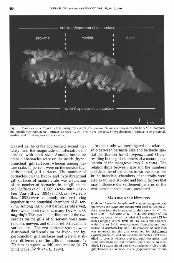
Octolasmis angulata.-The mean capitular lengths (MCL) of the 0. angulata located on the inside surfaces of gills 1 to 8 of all 260 S. serrata with one or more octolasmids are presented in Fig. 2A. Sample sizes varied among gills from 2 0. angulata on gill num- ber 2 to 514 on gill 7. Gills 1 and 2 were not considered further because of small sample sizes (Table 1). Gills 3, 7, and 8 had 0. an- gulata with similar MCLs (Fig. 2A). Gills 4, 5 , and 6 had populations with smaller MCLs. The MCLs of 0. angulata on gills 3, 7, and 8 were each significantly different from the MCL of the group on gill number 5 (Tukey #N HSD test, gill number 3 vs. gill 5, P = 0.013; gill 7 vs. gill 5 , P < 0.000; gill 8 vs. gill 5 , P < 0.000). The MCLs of 0. angulata on gills 4 and 6 were not significantly dif- ferent from the MCLs on gill 5 (Tukey #N HSD test, P > 0.05).
The pattern of size differences on the out- side surfaces of the gills was different from that on the inside surfaces (Fig. 2B). Gill number 8 was not included in this analysis because of a sample of only 3 (Table 1), and sample sizes were generally smaller on the outside surface with a maximum of 228 0. angulata on gill 5. In general the pattern was of gradually declining MCLs from gill 4 to gill 7. The MCLs of 0. angulata on gills 1 to 5 showed no significant differences (LSD test, P > 0.05; Tukey #N HSD test, P > 0.05),
1 2 -
, LLLPi?, L L
1 2 -
, LLLPi?, L L
2 6 1
Y L 1 2 -
1 2 3 4 5 6 7 6
GILL NUMBER (INSIDE SURFACE)
Fig. 2. The mean capitular lengths (MCL), one standard error, and one standard deviation are given for gills 1 to 8 for (A) the Octolasmis angulata located on the inside (hypobranchial) surfaces, (B) the 0. angulata located on the outside (hyperbranchial) surfaces, and (C) the 0. cor located on the inside surfaces. The barnacles were located in the gill chambers of 260 Scylla sermta, and the sam- ple sizes for each gill are given in Table 1.
but the MCL of 0. angulata on gill 4 dif- fered from both the MCLs on gills 6 and 7 (Tukey #N HSD test, P = 0.003 and P = 0.006 respectively).
Octolasmis cor.-The number of 0. coy on the inside surfaces of gills 1 through 8 far exceeded that on the outside surfaces (Table 1); thus, only data from the inside surfaces were analyzed. Because the sample size on gill number 2 is only 8 (Table 1), it was not included in the following comparisons (Fig. 2C). The MCLs of the 0. COY located on the inside surfaces of gills 3, 4, 5, and 7 did not
VORIS ET AL.: BARNACLE SIZE AND LOCATION ON MANGROVE CRABS 487 I
3.2
2.6
2.0
1.4
0.8 h
E v ' 3.2 I 6 2.6
-I
4 0.8 3
w' 2.0
(r 1.4
t
i 0 GILL 1
i 0 GILL 2
E I - - 1
I 0 GILL 4
i 0 GILL 5
T5 :f-g---Jpl 1 4
0 8 I 0 I 0
GILL 7 GILL 8
GILL 3
E GILL 6
1 &td. Dev. 0 4 t d . Err.
INSIDE (i) GILL SURFACES VERSUS OUTSIDE (0) GILL SURFACES FOR 0. angulata
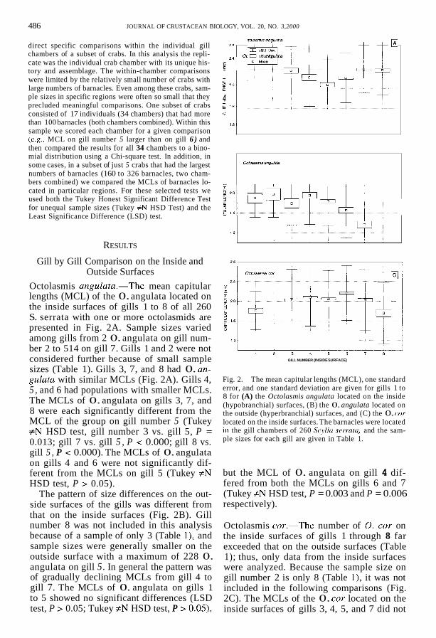
Fig. 3. The mean capitular lengths (MCL), one standard error, and one standard deviation are given for Octolus- mis angulutu located on the inside (hypobranchial) and outside (hyperbranchial) surfaces of gills 1 to 8 of Scylla serrafu. The sample sizes for each gill are provided in Table 1.
show significant differences (Tukey #N HSD test, P > 0.05). Both gills 1 and 8 had animals with smaller MCLs, and the 0. cor on gill 6 had the largest MCL (Table 1, Fig. 2C). In fact, the MCL of 0. coy on gill 6 was sig- nificantly larger than the MCLs on gills 1, 3, 5, 7, and 8 (Tukey #N HSD test, P < 0.05).
Comparison of the Inside and Outside Surfaces
Octolasmis angu1ata.-The 0. angulata on the inside gill surfaces outnumbered those on the outside gill surfaces 2.6 to 1 (Table 1). The MCL of 0. angulata located on the in- side surfaces of all gills (pooled) was larger (X = 1.905 mm, s = 0.511, n = 1,272) than the MCL of those located on the outside of the gills (X = 1.811 mm, s = 0.532, n = 486; t = 3.422, d.f. = 8, n = 1,756, P < 0.001).
A gill by gill comparison of MCLs of 0. angulata between the inside and outside sur- faces using an analysis of variance resulted in a significant F value ( F = 7.520, d.f. = 16, P < 0.001). Gills 3, 6, and 7 have 0. angu- lata with larger MCLs on the inside than the outside (Fig. 3). The differences between in- side and outside surfaces on gills 3 and 6 are
not significant (gill 3, inside X = 1.970 mm, s = 0.486, n = 72; outside X = 1.791 mm, s = 0.439, n = 23; Tukey #N HSD, P > 0.05; gill 6, inside X = 1.825 mm, s = 0.506, n = 265; outside X = 1.653 mm, s = 0.558, n = 88; Tukey #N HSD, P > 0.05). The MCL of 0. angulata on the inside of gill 7 was signifi- cantly larger than the MCL of 0. angulata on the outside (inside X = 1.995 mm, s = 0.480, n = 514; outside X = 1.362 mm, s = 0.315, n = 21; Tukey #N HSD, P = 0.005).
Octolasmis angulata located on the inside surfaces of gills 1, 2, and 8 were smaller in average size than those located on the out- side (Fig. 3), but these differences were not credible because the sample sizes were low (Table 1).
Octolasmis angulata located on the inside surface of gills 4 and 5 were smaller in av- erage size than those located on the outside surfaces, but these difference were not sig- nificant (gill 4, inside X = 1.83 mm, s = 0.583, n = 84; outside X = 1.951 mm, s = 0.583; Tukey #N HSD, P > 0.05; gill 5, inside X = 1.68 mm, s = 0.506, n = 140; outside X = 1.836 mm, s = 0.502, n = 228; Tukey #N HSD, P > 0.05).
488 JOURNAL OF CRUSTACEAN BIOLOGY, VOL. 20, NO. 3.2000
h
E E v
E
cc 3 4 k a a 0
2.6 -
2 4 -
2 2 -
2.0
1 .a
1.6
1.4
1.2 ' "1 486 684 1: 1 I 284 f 32 I 0.8
P m d P m d GILL SURFACE. OUTSIDE GILL SURFACE: INSIDE
PROXIMAL (p) - MEDIAL (m) - DISTAL (d)
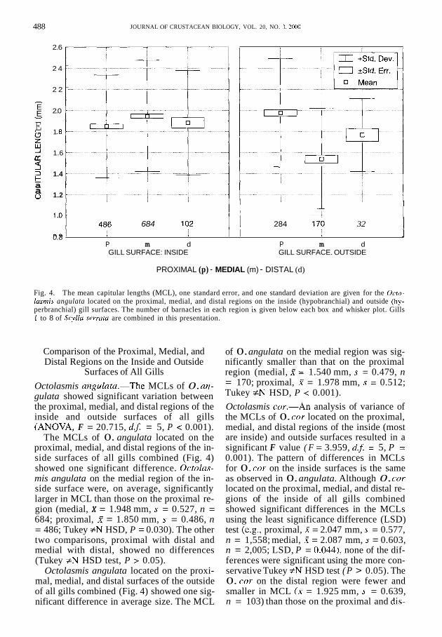
Fig. 4. The mean capitular lengths (MCL), one standard error, and one standard deviation are given for the Octo- lasrnis angulata located on the proximal, medial, and distal regions on the inside (hypobranchial) and outside (hy- perbranchial) gill surfaces. The number of barnacles in each region is given below each box and whisker plot. Gills 1 to 8 of Scyllu serrutu are combined in this presentation.
Comparison of the Proximal, Medial, and Distal Regions on the Inside and Outside
Surfaces of All Gills Octolasmis angu1ata.-The MCLs of 0. an- gulata showed significant variation between the proximal, medial, and distal regions of the inside and outside surfaces of all gills (ANOVA, F = 20.715, d. j = 5, P < 0.001).
The MCLs of 0. angulata located on the proximal, medial, and distal regions of the in- side surfaces of all gills combined (Fig. 4) showed one significant difference. Octolas- mis angulata on the medial region of the in- side surface were, on average, significantly larger in MCL than those on the proximal re- gion (medial, X = 1.948 mm, s = 0.527, n = 684; proximal, X = 1.850 mm, s = 0.486, n = 486; Tukey f N HSD, P = 0.030). The other two comparisons, proximal with distal and medial with distal, showed no differences (Tukey #N HSD test, P > 0.05).
Octolasmis angulata located on the proxi- mal, medial, and distal surfaces of the outside of all gills combined (Fig. 4) showed one sig- nificant difference in average size. The MCL
of 0. angulata on the medial region was sig- nificantly smaller than that on the proximal region (medial, X = 1.540 mm, s = 0.479, n = 170; proximal, X = 1.978 mm, s = 0.512; Tukey #N HSD, P < 0.001). Octolasmis cor.-An analysis of variance of the MCLs of 0. cor located on the proximal, medial, and distal regions of the inside (most are inside) and outside surfaces resulted in a significant F value ( F = 3.959, d.5 = 5, P = 0.001). The pattern of differences in MCLs for 0. cor on the inside surfaces is the same as observed in 0. angulata. Although 0. COT
located on the proximal, medial, and distal re- gions of the inside of all gills combined showed significant differences in the MCLs using the least significance difference (LSD) test (e.g., proximal, X = 2.047 mm, s = 0.577, n = 1,558; medial, X = 2.087 mm, s = 0.603, n = 2,005; LSD, P = 0.044), none of the dif- ferences were significant using the more con- servative Tukey #N HSD test ( P > 0.05). The 0. cor on the distal region were fewer and smaller in MCL (X = 1.925 mm, s = 0.639, n = 103) than those on the proximal and dis-
VORlS ET AL.: BARNACLE SIZE AND LOCATION ON MANGROVE CRABS 489
2.8 I 1
1 152 ~ 324 - 38 P rn d
GILL SURFACE. INSIDE
0 8
---i 151 - 68 -
0 &td. Err. I
17 Mean
1 7 - 1 3 P rn d GILL SURFACE: OUTSIDE
PROXIMAL (p) - MEDIAL (m) - DISTAL (d)
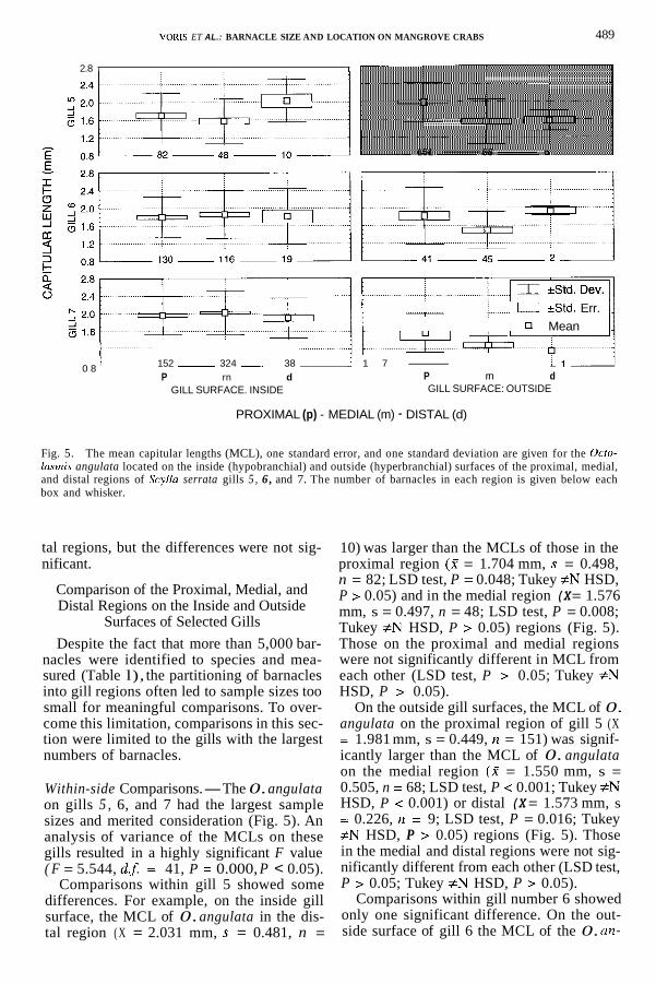
Fig. 5. The mean capitular lengths (MCL), one standard error, and one standard deviation are given for the Ocfo- lasrnis angulata located on the inside (hypobranchial) and outside (hyperbranchial) surfaces of the proximal, medial, and distal regions of Scylla serrata gills 5 , 6 , and 7. The number of barnacles in each region is given below each box and whisker.
tal regions, but the differences were not sig- nificant.
Comparison of the Proximal, Medial, and Distal Regions on the Inside and Outside
Surfaces of Selected Gills Despite the fact that more than 5,000 bar-
nacles were identified to species and mea- sured (Table l), the partitioning of barnacles into gill regions often led to sample sizes too small for meaningful comparisons. To over- come this limitation, comparisons in this sec- tion were limited to the gills with the largest numbers of barnacles.
Within-side Comparisons.-The 0. angulata on gills 5 , 6, and 7 had the largest sample sizes and merited consideration (Fig. 5). An analysis of variance of the MCLs on these gills resulted in a highly significant F value
Comparisons within gill 5 showed some differences. For example, on the inside gill surface, the MCL of 0. angulata in the dis- tal region ( X = 2.031 mm, s = 0.481, n =
( F = 5.544, d.5 = 41, P = 0.000, P < 0.05).
10) was larger than the MCLs of those in the proximal region (2 = 1.704 mm, s = 0.498, n = 82; LSD test, P = 0.048; Tukey #N HSD, P > 0.05) and in the medial region (X = 1.576 mm, s = 0.497, n = 48; LSD test, P = 0.008; Tukey #N HSD, P > 0.05) regions (Fig. 5). Those on the proximal and medial regions were not significantly different in MCL from each other (LSD test, P > 0.05; Tukey #N HSD, P > 0.05).
On the outside gill surfaces, the MCL of 0. angulata on the proximal region of gill 5 (X = 1.981 mm, s = 0.449, n = 151) was signif- icantly larger than the MCL of 0. angulata on the medial region (X = 1.550 mm, s = 0.505, n = 68; LSD test, P < 0.001; Tukey #N HSD, P < 0.001) or distal (X = 1.573 mm, s = 0.226, n = 9; LSD test, P = 0.016; Tukey #N HSD, P > 0.05) regions (Fig. 5). Those in the medial and distal regions were not sig- nificantly different from each other (LSD test, P > 0.05; Tukey #N HSD, P > 0.05).
Comparisons within gill number 6 showed only one significant difference. On the out- side surface of gill 6 the MCL of the 0. an-
490
375 393 21
JOURNAL OF CRUSTACEAN BIOLOGY, VOL. 20, NO. 3,2000
I 133 218 17
Octolasmis cor INSIDE GILL SURFACE
3.0
2.4
1.8 h
1.2 v
I 0.6 4
4 k a
W -I [r 3.0
2.4 a 0 1.8
1.2
0.6
f t
P rn d P rn d GILL 3 GILL 4
T I T 1 ‘“T. I;;
P rn d GILL 5
I tStd Dev
0 +Std Err
22 42 - 6 - 1 P rn d
GILL 8
PROXIMAL (p) - MEDIAL (m) - DISTAL (d)
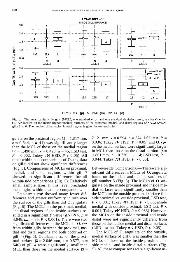
Fig. 6. The mean capitular lengths (MCL), one standard error, and one standard deviation are given for Octolas- mis cor located on the inside (hypobranchial) surfaces of the proximal, medial, and distal regions of Scyllu serrutu gills 3 to 6. The number of barnacles in each region is given below each plot.
gulata on the proximal region (X = 1.8 17 mm, s = 0.644, n = 41) was significantly larger than the MCL of those on the medial region (W = 1.490 mm, s = 0.428, n = 45; LSD test, P = 0.002; Tukey #N HSD, P > 0.05). All other within-side comparisons of 0. angulata on gill 6 did not show significant differences (Fig. 5). Comparisons of MCLs on proximal, medial, and distal regions within gill 7 showed no significant differences for all within-side comparisons (Fig. 5). Relatively small sample sizes at this level precluded meaningful within-chamber comparisons.
Octolasmis cor showed many fewer dif- ferences and greater uniformity in size over the surface of the gills than did 0. angulata (Fig. 6). The MCLs on the proximal, medial, and distal regions of the inside surfaces re- sulted in a significant F value (ANOVA, F = 3.048, d.5 = 35, P < 0.001). There were two significant differences in the MCLs of 0. cor from within gills, between the proximal, me- dial and distal regions and both occurred on gill 4 (Fig. 6). Octolasmis cor on the proxi- mal surface (X = 2.046 mm, s = 0.577, n = 540) of gill 4 were significantly smaller in MCL than those on the medial surface (X =
2.121 mm, s = 0.594, n = 574; LSD test, P = 0.036; Tukey #N HSD, P > 0.05) and 0. cor on the medial surface were significantly larger in MCL than those on the distal portion (X = 1.801 mm, s = 0.730, n = 14; LSD test, P = 0.044; Tukey #N HSD, P > 0.05).
Between-side Comparisons.-There were sig- nificant differences in MCLs of 0. angulata found on the inside and outside surfaces of gill number 5 (Fig. 5). The MCLs of 0. an- gulata on the inside proximal and inside me- dial surfaces were significantly smaller than the MCL on the outside proximal surface (in- side proximal vs. outside proximal, LSD test, P < 0.001; Tukey #N HSD, P > 0.05; inside medial with outside proximal, LSD test, P < 0.001; Tukey #N HSD, P = 0.033). However, the MCLs on the inside proximal and inside distal were not significantly different from those on the outside medial and distal surfaces (LSD test and Tukey #N HSD, P > 0.05).
The MCL of 0. angulata on the outside, medial surface of gill 6 was smaller than the MCLs of those on the inside proximal, in- side medial, and inside distal surfaces (Fig. 5). All these comparisons were significant us-
VORIS ET AL. : BARNACLE SIZE AND LOCATION ON MANGROVE CRABS 49 1
ing the LSD planned comparison test ( P < 0.05) but none were significant using the more conservative Tukey #N HSD test (P > 0.05). This same pattern of difference was also ap- parent on gill 7, but the sample size on the me- dial outside surface was only 13 and the MCLs were not significantly different ( P > 0.05).
Individual Gill Chamber Analysis To determine if the above findings based
on pooled samples could be demonstrated in individual gill chambers, we investigated a subset of 17 crabs (34 gill chambers) that had more than 100 barnacles in both chambers combined. To do this, we scored each of the 34 chambers with respect to each compari- son. For example, the largest between-gill dif- ference in Fig. 2C fell between gills 6 and 8. A scoring of the 34 chambers of the 17 crabs resulted in 6 chambers that had larger 0. coy on gill number 8 than on gill 6 and 4 cham- bers that had larger ones on gill number 6 than on gill 8. Twenty-six chambers could not be scored because one of the two gills (usu- ally gill 8) lacked any 0. cor. Thus, small samples within chambers on gill 8 prevented a conclusive comparison.
However, a chamber tally showed gills 4 and 7 differing significantly, with 15 cham- bers containing 0. cor with MCLs greater on gill 4 than on gill 7 and only 5 chambers with MCLs greater on gill 7 (chi-square, P < 0.025). A comparison of the MCLs on gills 4 and 5 in the 34 chambers also produced a significant deviation from 1: 1, with 24 cham- bers with MCLs larger on gill 4 than on gill 5 and 5 chambers with MCLs larger on gill 5 (chi-square, P < 0.01). Gill 4 was found to have larger MCLs than gill 3 in 26 cham- bers, whereas gill 3 had larger MCLs in only 7 of the chambers (chi-square, P < 0.001). Thus, in several comparisons where sample sizes were sufficiently large, within-chamber comparisons corroborated the results based on pooled samples. However, in the major- ity of comparisons between particular regions (e.g., gill 4, inside surface, distal region with gill 4, outside surface, distal region), sample sizes proved too small for any meaningful comparisons.
Relationships Between Barnacle Size, Numbers, and Density
The distribution of all barnacles (both species, all stages) over gills 1 to 8 deviated
sharply from the expected distribution based on gill area alone (chi-square = 1,614.7; P < 0.001). Thus, barnacles were not distributed according to available surface area alone.
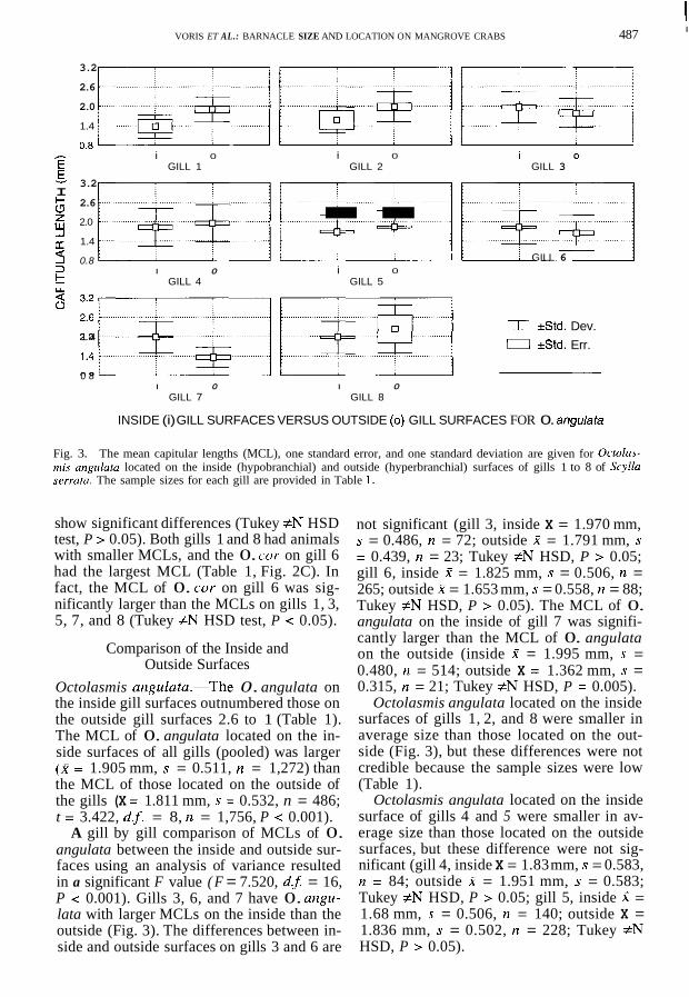
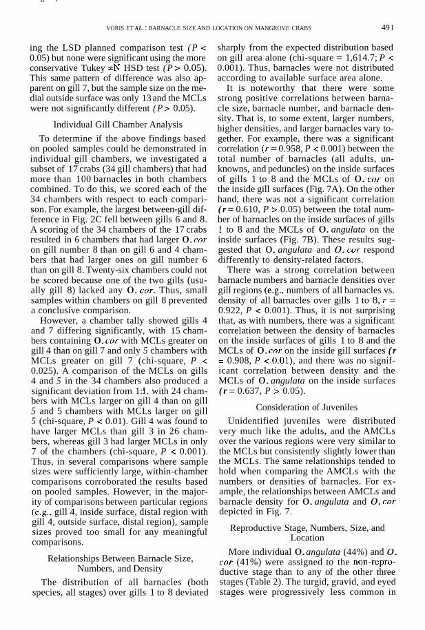
It is noteworthy that there were some strong positive correlations between barna- cle size, barnacle number, and barnacle den- sity. That is, to some extent, larger numbers, higher densities, and larger barnacles vary to- gether. For example, there was a significant correlation ( r = 0.958, P < 0.001) between the total number of barnacles (all adults, un- knowns, and peduncles) on the inside surfaces of gills 1 to 8 and the MCLs of 0. cor on the inside gill surfaces (Fig. 7A). On the other hand, there was not a significant correlation ( r = 0.610, P > 0.05) between the total num- ber of barnacles on the inside surfaces of gills 1 to 8 and the MCLs of 0. angulata on the inside surfaces (Fig. 7B). These results sug- gested that 0. angulata and 0. cor respond differently to density-related factors.
There was a strong correlation between barnacle numbers and barnacle densities over gill regions (e.g., numbers of all barnacles vs. density of all barnacles over gills 1 to 8, r = 0.922, P < 0.001). Thus, it is not surprising that, as with numbers, there was a significant correlation between the density of barnacles on the inside surfaces of gills 1 to 8 and the MCLs of 0. cor on the inside gill surfaces ( r = 0.908, P < 0.01), and there was no signif- icant correlation between density and the MCLs of 0. angulata on the inside surfaces ( r = 0.637, P > 0.05).
Consideration of Juveniles Unidentified juveniles were distributed
very much like the adults, and the AMCLs over the various regions were very similar to the MCLs but consistently slightly lower than the MCLs. The same relationships tended to hold when comparing the AMCLs with the numbers or densities of barnacles. For ex- ample, the relationships between AMCLs and barnacle density for 0. angulata and 0. coy depicted in Fig. 7.
Reproductive Stage, Numbers, Size, and Location
More individual 0. angulata (44%) and 0. cor (41%) were assigned to the non-repro- ductive stage than to any of the other three stages (Table 2). The turgid, gravid, and eyed stages were progressively less common in
492
GILL NUMBER (INSIDE SURFACE)
~ -
JOURNAL OF CRUSTACEAN BIOLOGY, VOL. 20, NO. 3,2000
2 2 2 1 8 400 800 1200 1600
MEAN CAPITULAR LENGTH (mm)
;ILL NUMBER (INSIDE SURFACE)
NUMBER OF ALL BARNACLES
' 2 1 8 1 6 1 4 400 800 1200 1600
MEAN CAPITULAR LENGTH (mm) NUMBER OF ALL BARNACLES
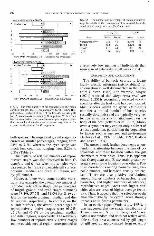
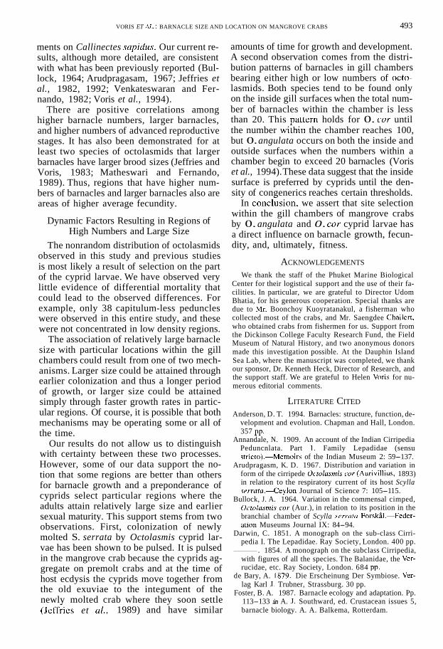
Fig. 7. The total number of all barnacles and the mean capitular lengths (MCL) are compared for the inside (hy- pobranchial) surfaces of each of the 8 Scylla serrata gills for (A) Ocfolasmis cor and (B) 0. angulata. Within each bar the rank order from smallest to largest is given. Note that the ranks of numbers and size are very similar for 0. cor but dissimilar for 0. angulata.
both species. The turgid and gravid stages oc- curred as similar percentages, ranging from 24% to 31%, whereas the eyed stage was much less common, ranging from 5.2% to 6.5% (Table 2).
This pattern of relative numbers of repro- ductive stages was also observed in both 0. angulata and 0. cor when the samples were categorized by inside and outside gill surface, proximal, medial, and distal gill region, and by gill number.
However, there were some notable varia- tions. For example, the overall percentages of reproductively active stages (the percentages of turgid, gravid, and eyed stages summed) were 58.2%, 57.5%, and 51.0% for 0. angu- lata on the inside proximal, medial, and dis- tal regions, respectively. In contrast, on the outside surfaces, the overall percentages of reproductively active stages were 68.7%, 27.6%, and 46.9% on the proximal, medial, and distal regions, respectively. The relatively low numbers of reproductively active stages on the outside medial region corresponded to
Table 2. The number and percentage of each reproductive stage for adults of the two species of octolasmid barnacle found on 260 mangrove crabs (Scylla serratu).
0 angu/nra 0. oor
Stage Number Percent Number Percent
Nonreproductive 773 44 1,490 41 Turgid 454 26 1,129 31
417 24 861 24 Gravid Eyed 114 6.5 189 5.2
a relatively low number of individuals that were also of relatively small size (Fig. 4).
DISCUSSION AND CONCLUSIONS
The ability of barnacle cyprids to locate highly specific substrates (microhabitats) for colonization is well documented in the liter- ature (Foster, 1987). For example, Moyse (197 1) reported that Megatrema anglicum (Gray, 1825) is secondarily attracted to con- specifics after the host coral has been located. Most species within the genus Octolasmis colonize a limited number of host species (usually decapods) and are typically very se- lective as to the site of attachment on the body of the host (Jeffries et al., 1982). More- over, a symbiont may exploit only a subset of a host population, partitioning the population by factors such as age, sex, and environment (Jeffries et al., 1992; Shields, 1992; Hudson and Lester, 1994).
The present work further documents a non- random relationship between the size of oc- tolasmids and their location within the gill chambers of their hosts. Thus, it is apparent that 0. angulata and 0. cor attain greater av- erage size in some locations over others. Pos- itive correlations among barnacle size, bar- nacle number, and barnacle density are pre- sent. There are also positive correlations among higher numbers of barnacles, larger barnacles, and higher numbers of advanced reproductive stages. Areas with higher den- sities also are areas of higher average fecun- dity. Thus, it appears that the selection of the attachment site by the cyprid larvae strongly impacts adult fitness parameters.
In an earlier paper (Voris et al., 1994), it was suggested that the spatial distribution of 0. angulata and 0. cor on the gills of S. ser- rata is nonrandom and does not reflect avail- able surface area as measured by gill length or gill area as approximated from measure-
ments on Callinectes sapidus. Our current re- sults, although more detailed, are consistent with what has been previously reported (Bul- lock, 1964; Arudpragasam, 1967; Jeffries et al., 1982, 1992; Venkateswaran and Fer- nando, 1982; Voris et al., 1994).
There are positive correlations among higher barnacle numbers, larger barnacles, and higher numbers of advanced reproductive stages. It has also been demonstrated for at least two species of octolasmids that larger barnacles have larger brood sizes (Jeffries and Voris, 1983; Matheswari and Fernando, 1989). Thus, regions that have higher num- bers of barnacles and larger barnacles also are areas of higher average fecundity.
Dynamic Factors Resulting in Regions of High Numbers and Large Size
The nonrandom distribution of octolasmids observed in this study and previous studies is most likely a result of selection on the part of the cyprid larvae. We have observed very little evidence of differential mortality that could lead to the observed differences. For example, only 38 capitulum-less peduncles were observed in this entire study, and these were not concentrated in low density regions.
The association of relatively large barnacle size with particular locations within the gill chambers could result from one of two mech- anisms. Larger size could be attained through earlier colonization and thus a longer period of growth, or larger size could be attained simply through faster growth rates in partic- ular regions. Of course, it is possible that both mechanisms may be operating some or all of the time.
Our results do not allow us to distinguish with certainty between these two processes. However, some of our data support the no- tion that some regions are better than others for barnacle growth and a preponderance of cyprids select particular regions where the adults attain relatively large size and earlier sexual maturity. This support stems from two observations. First, colonization of newly molted S. serrata by Octolasmis cyprid lar- vae has been shown to be pulsed. It is pulsed in the mangrove crab because the cyprids ag- gregate on premolt crabs and at the time of host ecdysis the cyprids move together from the old exuviae to the integument of the newly molted crab where they soon settle
amounts of time for growth and development. A second observation comes from the distri- bution patterns of barnacles in gill chambers bearing either high or low numbers of octo- lasmids. Both species tend to be found only on the inside gill surfaces when the total num- ber of barnacles within the chamber is less than 20. This Dattern holds for 0. cor until the number wiihin the chamber reaches 100, but 0. angulata occurs on both the inside and outside surfaces when the numbers within a chamber begin to exceed 20 barnacles (Voris et al., 1994). These data suggest that the inside surface is preferred by cyprids until the den- sity of congenerics reaches certain thresholds.
In conclkion. we assert that site selection within the gill chambers of mangrove crabs by 0. angulata and 0. coy cyprid larvae has a direct influence on barnacle growth, fecun- dity, and, ultimately, fitness.
ACKNOWLEDGEMENTS
We thank the staff of the Phuket Marine Biological Center for their logistical support and the use of their fa- cilities. In particular, we are grateful to Director Udom Bhatia, for his generous cooperation. Special thanks are due to Mi-. Boonchoy Kuoyratanakul, a fisherman who collected most of the crabs, and Mr. Saengdee Chailert, who obtained crabs from fishermen for us. Support from the Dickinson College Faculty Research Fund, the Field Museum of Natural History, and two anonymous donors made this investigation possible. At the Dauphin Island Sea Lab, where the manuscript was completed, we thank our sponsor, Dr. Kenneth Heck, Director of Research, and the support staff. We are grateful to Helen Voris for nu- merous editorial comments.
LITERATURE CITED
Anderson, D. T. 1994. Barnacles: structure, function, de- velopment and evolution. Chapman and Hall, London.
Annandale, N. 1909. An account of the Indian Cirripedia Peduncnlata. Part 1. Family Lepadidae (sensu stricto).-Memoirs of the Indian Museum 2: 59-137.
Arudpragasm, K. D. 1967. Distribution and variation in form of the cirripede Ocfolasmis cor (Aurivillius, 1893) in relation to the respiratory current of its host Scylla serrata.-Ceylon Journal of Science 7: 105-1 15.
Bullock, J. A. 1964. Variation in the commensal cimped, Octolasmis cor (Aur.), in relation to its position in the branchial chamber of Scylla serrata ForskBI.-Feder- ation Museums Journal IX: 84-94.
Darwin, C. 1851. A monograph on the sub-class Cirri- pedia I. The Lepadidae. Ray Society, London. 400 pp.
. 1854. A monograph on the subclass Cirripedia, with figures of all the species. The Balanidae, the Ver- rucidae, etc. Ray Society, London. 684 pp.
de Bary, A. 1879. Die Erscheinung Der Symbiose. Ver- lag Karl J. Trubner, Strassburg. 30 pp.
Foster, B. A. 1987. Barnacle ecology and adaptation. Pp. 113-133 in A. J. Southward, ed. Crustacean issues 5,
357 pp.
VORIS ET RL.: BARNACLE SIZE AND LOCATION ON MANGROVE CRABS 493
(Jeffiies et al., 1989) and have similar barnacle biology. A. A. Balkema, Rotterdam.
494 JOURNAL OF CRUSTACEAN BIOLOGY, VOL. 20, NO. 3,2000
Gray, J. E. 1825. A synopsis of the genera of cirripedes arranged in natural families, with a description of some new species.-Annals of Philosophy 10: 97-107.
Hudson, D. A,, and R. J. G. Lester. 1994. Parasites and symbionts of wild mud crabs Scylla serrutu (Forski]) of potential significance in aquaculture.-Aquaculture
Jeffries, W. B., and H. K. Voris. 1983. The distribution, size, and reproduction of the pedunculate barnacle, Oc- tolasrnis miilleri (Coker, 1902), on the blue crab, Cal- linectes supidus (Rathbun, 1896).-Fieldiana (new se- ries) 16: 1-10.
~~ , and S. Poovachiranon. 1992. Age of the mangrove crab, Scylla serrutu, at colonization by stalked barnacles of the genus 0ctolnsmis.-Biologi- cal Bulletin 182: 188-194.
~~ , and C. M. Yang. 1982. Diversity and distribution of the pedunculate barnacle Octolasmis in the seas adjacent to Singapore.-Journal of Crustacean Biology 2: 562-569.
, and ~ . 1989. A new mechanism of host colonization: pedunculate barnacles of the genus Octolasmis on the mangrove crab Scylla serrutu.- Ophelia 31: 51-58.
_______ , and - . 1991. Species recognition among the pedunculate barnacles (Cirripedia: Tho- racica) on the mangrove crab, Scylla serrata.-Raf- fles Bulletin of Zoology 40: 83-92.
Lewis, C. A. 1975. Development of the gooseneck bar- nacle Pollicipes polymerus (Cirripedia: Lepadomor- pha): fertilization through settlement.-Marine Biol- ogy, Berlin 32: 141-154.
120: 183-199.
~~
Matheswari, R., and S. A. Fernando. 1989. Fecundity of epizoic barnacles.-Indian Journal of Invertebrate Zoology and Aquatic Biology 1: 31-35.
Moyse, J. 1971. Settlement and growth pattern of the parasitic barnacle, Pyrgorna anglicum. Pp. 125-141 in D. J. Crisp, ed. Fourth European Marine Biology Sym- posium. Cambridge University Press, Cambridge.
Newman, W. A. 1987. Evolution of cirripedes and their major groups. Pp. 3 4 2 in A. J. Southward, ed. Crus- tacean issues 5, barnacle biology. A. A. Balkema, Rot- terdam.
Shields, J. D. 1992. Parasites and symbionts of the crab Portunus pelagicus from Moreton bay, eastern Aus- tralia.-Journal of Crustacean Biology 12: 94-100.
Spjotvoll, E., and M. R. Stoline. 1973. An extension of the T-method of multiple comparison to include the cases with unequal sample sizes.-Journal of the Amer- ican Statistical Association 68: 976-978.
Venkateswaran, K., and S. A. Fernando. 1982. Distri- bution and variation in form of the epizoic cirriped Oc- tolasrnis cor (Aurivillius, 1893).-Indian Journal of Marine Science 11 : 243-246.
Voris, H. K., W. B. Jeffries, and S. Poovachiranon. 1994. Patterns of distributions of two barnacle species on the mangrove crab Scylla serrata.-Biological Bulletin 187: 346-354.
RECEIVED: 5 February 1999. ACCEPTED: 10 February 2000.