Historical Antisemitism, Ethnic Specialization, and Financial ...

Journal of Experimental Marine Biology and Ecology 392 (2010) 115–124
Contents lists available at ScienceDirect
Journal of Experimental Marine Biology and Ecology
j ourna l homepage: www.e lsev ie r.com/ locate / jembe
Antennular specialization in cyprids of coral-associated barnacles
I. Brickner a, J.T. Høeg b,⁎a Department of Zoology, Tel Aviv University, Tel Aviv 69978, Israelb Comparative Zoology, Department of Biology, University of Copenhagen, Universitetsparken 15, DK-2100, Denmark
⁎ Corresponding author. Tel.: +45 35321247; fax: +E-mail address: [email protected] (J.T. Høeg).
0022-0981/$ – see front matter © 2010 Elsevier B.V. Aldoi:10.1016/j.jembe.2010.04.015
a b s t r a c t
a r t i c l e i n f oKeywords:EvolutionLarval biologyMetamorphosisSEMSensory organSettlement
We used video, light and scanning electron microscopy to study the cypris larvae in species of coralinhabiting barnacles (Pyrgomatidae) in search of adaptations to settlement on their highly unusualsubstratum. Species studied were Savignium crenatum, Trevathana jensi, Trevathana margaretae, Trevathanamizrachae and Trevathana sarae. In all five species the third antennular segment was shaped like a spearheadwith only an extremely narrow attachment disc. This morphology represents the most extreme antennularspecialization known from cirripedes, and it is not even matched by those parasitic barnacles that use theantennule for active penetration of cuticles. Compared to the third segment, the first, second and fourthsegments exhibit no obvious specializations, and the armament of sensory setae is also as very comparable tothat seen in balanomorphan cirripedes. The coral barnacles also have the nauplius eye, compound eyes,frontal filaments, lattice organs and cement glands known from other barnacles. Only T. sarae differed byhaving two unusually shaped setae terminally on the fourth segment. Video observations showed that thecoral barnacle cyprids display the exploratory walking behaviour known from cyprids settling on hardbottom substrata. We interpret the pointed structure of the third antennular segment as an adaptation topenetrate through live coral tissue. Projecting this character onto a recent phylogenetic tree ofbalanomorphan species indicates that it is an apomorphy for a large clade of coral inhabiting barnacles.
45 35321200.
l rights reserved.
© 2010 Elsevier B.V. All rights reserved.
1. Introduction
Many cirripedes are epibiotic and inhabit a wide range of othermarine animals including both vertebrates and invertebrates. A wholesuperorder, the Rhizocephala, are even parasitic, and they represent themost specialized of all barnacles. But within the superorder Thoracicaalmost all of the epibiotic forms are suspension feeders, where the hostanimal serves principally as a mere substratum, but sometimes alsooffering transport, protection, or both to the sessile barnacle.Within theThoracicaBalanomorpha there arenumerous species of coral-associatedbarnacles, most of which are placed in the family Pyrgomatidae(Anderson, 1994; Simon-Blecher et al., 2007). The coral barnacles aresuspension feederswith the body enclosed in a house of shell plates, butthey differ from almost all other epibiotic thoracicans in not just beingattached on an inert body armour such as a cuticle or a shell, but livingalmost fully embedded within the coral tissue. This begs the questionhow the cypris larvae of coral barnacles manage to settle on the surfaceof a live coral and how coral tissue is penetrated.
In all cirripedes larval development terminates with a cyprid that ishighly specialized for locating andattaching to a specific substratumandmetamorphosing into a sessile juvenile. The vast majority of cirripedesattach on a more or less solid surface, and the settlement process is
basically similar whether the substratum is a rock, a piece of wood, aship's hull, or the body armour of marine animals such as crustaceans,bivalves, sea turtles or thecate hydroids. Even in the highly specializedRhizocephala the cyprid first cements itself onto the solid cuticle of thepotential host crab as would any other barnacle, and it is only theensuing kentrogon stage that completes the invasion of the parasite intothe host (Glenner, 2001). Dovetailing with this it seems that cirripedes,despite pronounced differences in the habitat and biology of the adultanimal, have remarkably similar cypris larvae (Høeg et al., 2004; Pérez-Losadaet al., 2009. This similarity even extends to the antennules,whichare used both in the exploratory walking over the substratum prior tosettlement and in the final cementation. They seem to have the samegeneral segmentation and musculature and the same position andmorphology of the so-called attachment organ used in both temporaryand permanent attachments (Fig. 1). The only remarkable differencebetween cirripede cypris larvae concerns the number and morphologyof sensory organs including those on the antennules (Glenner et al.,1989; Kolbasov and Høeg, 2007; Bielecki et al., 2009).
In contrast to other cirripedes, the cyprid of a coral barnacle mustfirst negotiate the live and potentially lethal epidermis of the hostbefore it can reach and attach to the underlying exoskeleton. Butnothing is known about the actual settlement process, and the cypridhas only been superficially studied in a single species, the AtlanticMegatrema anglica. Here Moyse et al. (1995) found a remarkablemorphology of the third antennular segment, which is pointed as aspearhead and virtually without any attachment disc, a feature
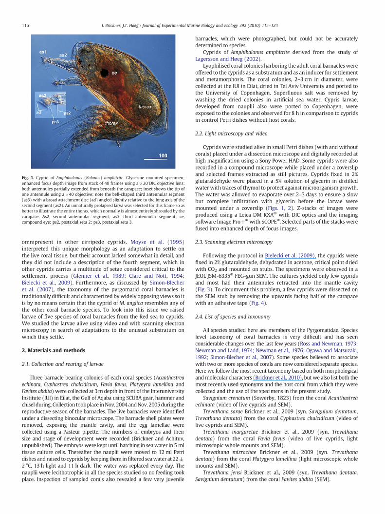
Fig. 1. Cyprid of Amphibalanus (Balanus) amphitrite. Glycerine mounted specimen;enhanced focus depth image from stack of 40 frames using a ×20 DIC objective lens;both antennules partially extended from beneath the carapace; inset shows the tip ofone antennule using a ×40 objective; note the bell-shaped third antennular segment(as3) with a broad attachment disc (ad) angled slightly relative to the long axis of thesecond segment (as2). An unnaturally prolapsed larva was selected for this frame so asbetter to illustrate the entire thorax, which normally is almost entirely shrouded by thecarapace. As2, second antennular segment; as3, third antennular segment; ce,compound eye; ps2, postaxial seta 2; ps3, postaxial seta 3.
116 I. Brickner, J.T. Høeg / Journal of Experimental Marine Biology and Ecology 392 (2010) 115–124
omnipresent in other cirripede cyprids. Moyse et al. (1995)interpreted this unique morphology as an adaptation to settle onthe live coral tissue, but their account lacked somewhat in detail, andthey did not include a description of the fourth segment, which inother cyprids carries a multitude of setae considered critical to thesettlement process (Glenner et al., 1989; Clare and Nott, 1994;Bielecki et al., 2009). Furthermore, as discussed by Simon-Blecheret al. (2007), the taxonomy of the pyrgomatid coral barnacles istraditionally difficult and characterized bywidely opposing views so itis by no means certain that the cyprid of M. anglica resembles any ofthe other coral barnacle species. To look into this issue we raisedlarvae of five species of coral barnacles from the Red sea to cyprids.We studied the larvae alive using video and with scanning electronmicroscopy in search of adaptations to the unusual substratum onwhich they settle.
2. Materials and methods
2.1. Collection and rearing of larvae
Three barnacle bearing colonies of each coral species (Acanthastreaechinata, Cyphastrea chalcidicum, Favia favus, Platygyra lamellina andFavites abdita) were collected at 3m depth in front of the InteruniversityInstitute (IUI) in Eilat, the Gulf of Aqaba using SCUBA gear, hammer andchisel during. Collection tookplace inNov. 2004andNov. 2005during thereproductive season of the barnacles. The live barnacles were identifiedunder a dissecting binocular microscope. The barnacle shell plates wereremoved, exposing the mantle cavity, and the egg lamellae werecollected using a Pasteur pipette. The numbers of embryos and theirsize and stage of development were recorded (Brickner and Achituv,unpublished). The embryoswere kept until hatching in seawater in 5 mltissue culture cells. Thereafter the nauplii were moved to 12 ml Petridishes and raised to cyprids by keeping them infiltered seawater at 22±2 °C, 13 h light and 11 h dark. The water was replaced every day. Thenauplii were lecithotrophic in all the species studied so no feeding tookplace. Inspection of sampled corals also revealed a few very juvenile
barnacles, which were photographed, but could not be accuratelydetermined to species.
Cyprids of Amphibalanus amphitrite derived from the study ofLagersson and Høeg (2002).
Lyophilised coral colonies harboring the adult coral barnacles wereoffered to the cyprids as a substratum and as an inducer for settlementand metamorphosis. The coral colonies, 2–3 cm in diameter, werecollected at the IUI in Eilat, dried in Tel Aviv University and ported tothe University of Copenhagen. Superfluous salt was removed bywashing the dried colonies in artificial sea water. Cypris larvae,developed from nauplii also were ported to Copenhagen, wereexposed to the colonies and observed for 8 h in comparison to cypridsin control Petri dishes without host corals.
2.2. Light microscopy and video
Cyprids were studied alive in small Petri dishes (with and withoutcorals) placed under a dissection microscope and digitally recorded athigh magnification using a Sony Power HAD. Some cyprids were alsorecorded in a compound microscope while placed under a coverslipand selected frames extracted as still pictures. Cyprids fixed in 2%glutaraldehyde were placed in a 5% solution of glycerin in distilledwater with traces of thymol to protect against microorganism growth.The water was allowed to evaporate over 2–3 days to ensure a slowbut complete infiltration with glycerin before the larvae weremounted under a coverslip (Figs. 1, 2). Z-stacks of images wereproduced using a Leica DM RXA® with DIC optics and the imagingsoftware Image Pro+® with SCOPE®. Selected parts of the stacks werefused into enhanced depth of focus images.
2.3. Scanning electron microscopy
Following the protocol in Bielecki et al. (2009), the cyprids werefixed in 2% glutaraldehyde, dehydrated in acetone, critical point driedwith CO2 and mounted on stubs. The specimens were observed in aJEOL JSM-6335® FEG-gun SEM. The cultures yielded only few cypridsand most had their antennules retracted into the mantle cavity(Fig. 3). To circumvent this problem, a few cyprids were dissected onthe SEM stub by removing the upwards facing half of the carapacewith an adhesive tape (Fig. 4).
2.4. List of species and taxonomy
All species studied here are members of the Pyrgomatidae. Specieslevel taxonomy of coral barnacles is very difficult and has seenconsiderable changes over the last few years (Ross and Newman, 1973;Newman and Ladd, 1974; Newman et al., 1976; Ogawa and Matsuzaki,1992; Simon-Blecher et al., 2007). Some species believed to associatewith two or more species of corals are now considered separate species.Here we follow the most recent taxonomy based on both morphologicalandmolecular characters (Brickner et al., 2010), but we also list both themost recently used synonyms and the host coral from which they werecollected and the use of the specimens in the present study.
Savignium crenatum (Sowerby, 1823) from the coral Acanthastreaechinata (video of live cyprids and SEM).
Trevathana sarae Brickner et al., 2009 (syn. Savignium dentatum,Trevathana dentata) from the coral Cyphastrea chalcidicum (video oflive cyprids and SEM).
Trevathana margaretae Brickner et al., 2009 (syn. Trevathanadentata) from the coral Favia favus (video of live cyprids, lightmicroscopic whole mounts and SEM).
Trevathana mizrachae Brickner et al., 2009 (syn. Trevathanadentata) from the coral Platygyra lamellina (light microscopic wholemounts and SEM).
Trevathana jensi Brickner et al., 2009 (syn. Trevathana dentata,Savignium dentatum) from the coral Favites abdita (SEM).

Fig. 2. A. Trevathana sarae. Live cyprid, mounted under a coverslip; photo extractedfrom a video sequence. B. Trevathana mizrachae. Anterior end of cypris showing cementgland, antennules, the nauplius eye and both compound eyes. Both antennulesretracted, but the spear-shaped third segments are visible. As2, second antennularsegment; as3; third antennular segment; ce; compound eye; ne, nauplius eye.
Fig. 3. General cyprid features. A. Trevathana sarae. SEM. Ventral view of anterior end ofcyprid; frontal horn pores (fhp) near the carapace rim; the ventral rim of the carapacevalves (ca) donotmeet completely, but thin pliable cuticle inside the rim effects the actualclosure, obscuring the retracted antennules. B. Savigniumdentatum. SEM.Ventral view; thetwo compound eyes (ce) and one of the frontal filaments (ff) visible between the partiallyclosed rims of the carapace; one lens of the eyes is outlined. C. Trevathanamargaretae. SEM.Lattice organ of pore-field shape with large terminal pore as typical for thoracican andrhizocephalan cirripedes (Høeg et al., 2004); from dorsal part of cyprid.
117I. Brickner, J.T. Høeg / Journal of Experimental Marine Biology and Ecology 392 (2010) 115–124
3. Results
3.1. Observations on live cyprids
The cyprids explored the surface of the glass vessel by walking onthe antennules in the manner typical for cirripede cyprids as firstdescribed by Visscher (1928) and accurately modelled by Lagersonand Høeg (2002). We observed no difference in the exploratorybehaviour between cyprids exposed to dead, treated corals and thosein the control dishes, and no cyprids attached or metamorphosed.Video sequence 1 of T. margaretae (supplementary material) showshow the cyprid alternately walks step wise ahead and stops toexamine a particular substratum area. It is always attached with oneantennule, while the other is extended, probing the surroundingsubstratum until deciding whether to complete the new step.Occasional beating of the thoracopods seen while probing the bottommay serve to pull the body away from the substratum and thus assessthe adhesive force at this particular spot. Scrutiny of live cypridstrapped under a coverslip (Fig. 2A, video sequence 2) showed that theantennular segments, including the third and fourth, had a degree infreedom of movement more or less comparable to that described forAmphibalanus (Balanus) amphitrite by Lagerson and Høeg (2002).
3.2. General cypris morphology
Aside from the specializations in the antennules described below,we found no features where the coral barnacle cyprids differed fromother thoracicans (compare Figs. 1 and 2). We found all the sensoryand glandular structures normally seen in cyprids including lattice

Fig. 4. Savignium crenatum. Cyprid. SEM of antennule. A. Medial view of right antennule,freely exposed. B, C. Medial view of right antennule from specimen opened bydissection to reveal an antennule retracted into the mantle cavity; antennule partlytucked into the thin mantle cuticle; in B, note how the subterminal and terminal setaeare situated almost at the same level and the pores in the subterminal setae; In C rowsof minute fringes line the inside of the mantle wall. Am, arthrodial membrane; as1-4,first— fourth antennular segments; dsas1, distal sclerite of first antennular segment, po,pore; psas1, proximal sclerite of first antennular segment; ps2, postaxial seta 2; ps3,postaxial seta 3; sts 1–4, subterminal setae 1–4; ts, terminal setae; ts-A to D, terminalsetae A–D.
118 I. Brickner, J.T. Høeg / Journal of Experimental Marine Biology and Ecology 392 (2010) 115–124
organs of a the pore-field type (T. sarae, T. margaretae), pores for thefrontal horn glands (all five species), frontal filaments (S. crenatum,T. margaretae, T.mizrachae, T. jensi) associated with paired compoundeyes (S. crenatum, T. sarae, T. margaretae, T. mizrachae), a nauplius eye(T. sarae, T. margaretae, T. mizrachae) and paired cement glands(T. sarae, T. margaretae). Failure to observe any of these organs in aparticular species does not indicate that we believe they are absent,but just that the particular structure was not exposed to view in ourpreparations.
The cement glands were visible in light microscopy of the glycerinmounted cyprids (Fig. 2). Situated in the cyprid body, they consist ofseveral gland cells standing in a semicircular row just as depicted byWalley (1969) and Walker (1971). The light microscopy also showed
the nauplius eyes, not visible with SEM, and the spearhead shape ofthe third segment described below.
3.3. Morphology of cypris antennules
For Savignium crenatum we had several specimens available,where the antennules were exposed to SEM observation. Hence weprovide the most comprehensive description for that species. For theother species, we emphasize the differences from S. crenatum. Weemploy the same terminology for antennular structures and setae asrecently set down in Bielecki et al. (2009).
All investigated species have four segmented antennules, con-structed as described by Glenner and Høeg (1995), Lagerson and Høeg(2002) and Bielecki et al. (2009). The first, second and fourthsegments are rather similar, both between the species studied hereand compared to Amphibalanus amphitrite (Figs. 1, 2). In contrast, allthe coral barnacles had a small and pointed third segment, with no oronly a very narrow attachment disc visible (Figs. 2, 4–8). As a resultthe third segment resembles a spearhead mounted on the elongatedsegment. Due to this morphology, the fourth segment, retaining theshape and size seen in other barnacles, looks unusually largecompared to the third.
3.3.1. Savignium crenatum (Figs. 4, 5)
3.3.1.1. The first segment. Consists of two articulating sclerites (Fig. 4).They are articulated so they form a posteriorly directed elbow, whenthe antennules are fully or partially retracted into the mantle cavity(Fig. 4B).
3.3.1.2. The second segment. Has an elongated and laterally com-pressed shape, looking broad (high) in lateral view but narrow inventral and dorsal views. In lateral view, it is highest, where itarticulates proximally to the first segment (distal sclerite), but abouthalf way from the base the ventral edge forms an abrupt angle(Fig. 4A, arrow), whence the height decreases gradually towards thetip. In dorsal or ventral views the segment tapers only slightly towardsthe tip. The only seta is a rather short postaxial seta 2 situatedventrally near the distal end (Fig. 4A).
3.3.1.3. The third segment. Has, at the joint, the same width and heightas the distal end of the second segment, but it narrows gradually toterminate in a very pointed tip (Fig. 4). As a result, the third segmentlooks exactly like a spear point at the tip of the elongated secondsegment. The attachment disc extends along the narrow ventral sideof the segment (Fig. 5A). The whole disc is obscured by a series ofoverlapping cuticular flaps, which line both the lateral and medialsides of the segment and have a rectangular shape, being longer thanwide (Fig. 5C). The flaps consist of thin, pliable cuticle bendinginwards towards the ventral midline, whence they obscure any villipresent on the disc surface.
A long and simple seta, the postaxial seta 3 (ps3), sits in the ventralmidline, proximally to the cuticular flaps, and level with the insertionof the fourth segment (Fig. 5A). Also in the midline, about in themiddle of the segment, sits the axial disc seta (ads). It consists of abroad basal hump on top of which a small seta protrudes from a smalldepression (Fig. 5B,C). More distally and slightly laterally sits a spine-like projection (sp). It has a broad base, but we could not detect anyapical pores that could indicate whether it is a modified seta. Wefound two pairs of radial disc setae (rds). The distal-most pair, nearthe tip, looks like two small spines and did not have any visible pores.The proximal pair sits between the axial disc seta and the spinousprocess (Fig. 5B,C). They resemble small spines but sported distinct,subterminally situated pores indicating that they are sensory setae. Aseta like process sits in the disc, proximally to the axial disc seta(arrow in Fig. 5C). By position it could correspond to the postaxial disc

Fig. 5. Savignium crenatum. Cyprid. SEM of the third and fourth antennular segments. A. Oblique medial view; the attachment disc on the third segment faces up. B. Detail of A; thedistal part of the third segment; the attachment disc proper is obscured by rows of overlapping and inwardly flexing flaps of cuticle. C. Distal end of the third segment. Inset in A showsthe ornaments on terminal seta D. Insets in B shows the axial disc seta. Insets in C shows the axial disc seta and a radial disc seta, the latter with a pore (arrow). Ads, axial disc seta; as2–4, second–fourth antennular segments; ps2, postaxial seta 2; ps3, postaxial seta 3; rds, radial disc seta; se, seta; sp; spinous process; ts-C, terminal seta C; ts-D, terminal seta D.
119I. Brickner, J.T. Høeg / Journal of Experimental Marine Biology and Ecology 392 (2010) 115–124
seta (pds) found in other barnacle cyprids, but it might also be a moremedially situated radial disc seta.
3.3.1.4. The fourth segment. Is articulated to the lateral side of the thirdsegment near the base (Fig. 4). It has a circular cross section, and froma fairly narrow base it widens somewhat towards the flat apex so italtogether has an inverted cone-shape. It carries a subterminal groupof four setae and a terminal group of five setae, but the two platformscarrying these setae are located almost at the same level (Fig. 4C). Theterminology is therefore used mainly for comparison with othercirripede cyprids, where these platforms are more clearly separated.The four subterminal setae (sts 1–4) are simple, more or less straightand tapering towards a distinct terminal pore. Terminal seta A and Bappear to be similar to each other, and they carry long and thinsetules, although these were hard see on most SEMmicrographs. SetaC is minute and with a terminal pore. Seta D is very conspicuous,broad, almost isodiametrical to the tip and with a distinctly wrinkledsurface indicating that its cuticle consists of thin areas interspacedwith thicker reinforcing parts (inset in Fig. 5A). Seta E is simple andterminates in a pore.
3.3.2. Trevathana jensi (Fig. 6, 7A)The second segment is relatively shorter and stouter than in
S. crenatum, whence the pointed third segment seems to sit as a finepoint at the tip of a blunt spar (Fig. 6A). The second segment carries arather short ps2 seta near the distal end. In ventral and dorsal view thethird segment tapers gradually towards the pointed tip. UnlikeS. crenatum, the ventral surface of the third segment has a distinct,but very narrow attachment disc covered with long and thin cuticularvilli (Figs. 6B, 7A). The villus-covered area extends for about the distaltwo thirds of the ventral surface and continues to the very tip of thesegment. In the proximal half of the disc the villi are distinctly longer
and they also seem to stand denser than in the more distal part(Fig. 7A). The entire disc is surrounded by a series of cuticular flapsthat seem not to overlap but form a single row. Along the sides of thesegment these flaps have a rectangular shape, wider than high andlower than the villi so the latter are clearly visible in lateral views(Fig. 7A). The row of flaps continues around the proximal end of thedisc, where they are still rectangular, but distinctly higher than wide(Fig. 6B). A postaxial seta 3 inserts in the ventral midline, being placedproximally to the attachment disc and at level with the insertion ofthe fourth segment (Fig. 6B). The disc sports an axial disc seta(Fig. 6C), a more distally placed spinous process, and close to the tip atleast two spine-shaped radial disc seta (rds).
The fourth segment inserts as in S. crenatum. It carries the samenumber and type of subterminal and terminal setae as in S. crenatum,but is relatively longer and narrower than in that species (Fig. 6B). Thesubterminal setae sit close to, but distinctly lower than the terminalones. Setae A and B are long and clearly armed with fine setules(Fig. 6A), but we could not study details of the other setae in thematerial.
3.3.3. Trevathana margaretae (Fig. 7B,C)In SEM, the third segment is very long and narrow. It appears not
to taper but retains almost the same narrow width (seen in ventralview) until the distal end. A narrow attachment disc extends alongmost of the ventral surface and bears cuticular villi that seem to standin rather regular rows, but with naked areas of cuticle exposed in themidline (Fig. 7B).
A single row of rectangular cuticular flaps surround the disc, atleast laterally and distally (Fig. 7B). The flaps are few and veryelongated laterally but shorter around the distal end. They are so lowthat the villi of the disc extend above them in lateral views. Due toobscuration by a contorted fourth segment we could not observe the

Fig. 6. Trevathana jensi. Cyprid. SEM of antennule. A. Oblique ventral view of extended antennule, only distal part of first segment visible. B. Detailed view of second and thirdsegments; inset shows the spinous process. C. Detail of B, showing the axial disc seta and cuticular villi on the attachment disc. D. Detail from the attachment disc showing two radialdisc setae (arrows), both with distinct sub-apically situated pores. Ads, axial disc seta; as1-4, second–fourth antennular segments; ob, oban; ps3, postaxial seta 3; rds, radial disc seta;sp; spinous process; sts, subterminal setae; ts-A to E, terminal seta A–E.
120 I. Brickner, J.T. Høeg / Journal of Experimental Marine Biology and Ecology 392 (2010) 115–124
proximal part of the disc. Near the tip the disc carries two smallprojections and slightly more proximally, a pair of similar, but muchlonger projections that seem to have an apical pore (Fig. 7C). As inS. crenatum, we interpret these four projections as radial disc setae(rds). As the cuticular villi, they seem to have a rather soft cuticle. Aprominent spine-like structure sits slightly more proximally thanradial setae with its tip pointing towards the base of the segment. Thisstructure, seen on both right and left antennule in the single cypridwecould observe in detail, sits not in the midline but displaced laterally.Details of the fourth segment could not be seen clearly, but it seems tocarry the same setae as in S. crenatum and Trevathana jensi.
3.3.4. Trevathana sarae (Figs. 2A, 8)Light microscopy revealed a very pointed third segment (Fig. 2A).
The material prepared for SEM was insufficient for a detaileddescription of the third segment, but did confirm that it has astructure resembling that in T. jensi and T. margaretae. The narrowattachment disc carries some cuticular villi and is surrounded byrectangular cuticular flaps. The third segment also carries two distallyplaced pairs of radial disc setae and a slightly more proximally placedspinous process, but we could not establish whether an axial disc setais present.
For the fourth segment, both SEM and still images extracted fromdigital video revealed the same number of setae as in S. dentatum andT. jensi (Figs. 2A, 8). The subterminal setae are ornamented with smallspine-like setules and apical pores. Both the light microscopy and SEM
showed that terminal setae A and B are very long and beset with thinsetules along the shaft (Fig. 8A). Terminal seta C is small and slender.What we identify as terminal seta D and E have unusual shapescomparedwith the other species studied here (Fig. 8B). Terminal sera Dhas a narrow base carrying an almost spear-shaped distal part,ornamented with longitudinal ridges and terminating in a small pore.Terminal seta E is alsounusual in consistingof awide basal part towhicharticulates a much narrower distal part with an apical pore (inset inFig. 8B).
3.3.5. Trevathana mizrachae (Fig. 2B)Light microscopy of whole mounted cyprids revealed a pair of
cement glands just as in Trevathana margaretae (Fig. 2B). Thesepreparations, and SEM (not shown) also showed that the antennulehas a spear-shaped and very pointed third segment.
3.4. Juvenile specimens (Fig. 9)
Two very early juveniles of unidentified species of coral barnacleswere found on the coral exoskeletons. The shells of the juveniles werearched, not flat as in the adults. It is clear that the juveniles werefirmly attached to the septae of the coral exoskeleton. Live coral tissuewith zooxanthellae surrounded the juveniles. Due to the scarcity ofthe material, we could not establish whether the wall of the shell wasfused or consisted of more or less individual plates at this earlyontogenetic stage.

Fig. 8. Trevathana sarae. Cyprid. SEM, of third and fourth antennular segments. A. Thirdsegment distinctly pointed; fourth segment with long and clearly setulated terminalsetae A and B. B. Detail of A; fourth segment; terminal seta E consists of a wide basal partarticulating (curved arrow and inset) with a simple shaped distal part terminating in apore; terminal setae D consists of a narrow basal part carrying a short, spear-shapedand longitudinally ridged distal part; the subterminal setae are ornamented with smallspines and terminate in pores. As3, third antennular segment; as4, fourth antennularsegment; po, pore; set; setule; sts, subterminal setae; ts-A to D, terminal setae A-D.
Fig. 7. A. Trevathana jensi. Cyprid. SEM, lateral view of third antennular segment, ventralside with attachment disc faces up. Note the somewhat concave shape of the discsurface, which in this species carries long and thin cuticular villi. Insets show a radialdisc seta and the spinous process. B, C. Trevathana margaretae. Cyprid. SEM of thirdantennular segment; lateral view (B) shows the elongated flaps bordering theattachment disc, two pairs of radial disc setae and the spinous process; ventral view(C) shows distal part of the segment, with the same structures as in B and the longcuticular villi almost regularly arranged in longitudinal rows; arrow indicates wheretwo flaps are abutting or slightly overlapping. Ps3, postaxial seta 3; rds, radial disc seta;sp, spinous process.
121I. Brickner, J.T. Høeg / Journal of Experimental Marine Biology and Ecology 392 (2010) 115–124
4. Discussion
We interpret the spearhead-shaped structure of the third anten-nular segment, found in cyprids of all five coral barnacles studied here,as an extreme adaptation to settle on the live tissue of a coral colony.This morphology represents the most extreme modification of theantennular attachment organ anywhere in the Cirripedia. In otherbarnacles the form of the third antennular segment can vary from abell-shape to an elongated hand-shape, but it always features a largeattachment disc, oval or circular in outline, and with a variously densecover of cuticular villi (Fig. 1). Such an attachment disc characterizesnot only the rocky shore inhabiting cirripedes such as Pollicipes andmany balanomorphans but also the epibiotic forms including even theparasitic Rhizocephala (Moyse et al., 1995; Glenner et al., 1989;Kolbasov and Høeg, 2007).
4.1. The attachment disc
The cuticular flaps found around the attachment disc in coralbarnacle cyprids resemble both the large velum found in manythoracicans and the much lower and more or less continuous skirtenveloping the disc in rhizocephalans (Moyse et al., 1995).We use theneutral term “flaps” to avoid any premature statement of homologybetween these structures. The sensory setae and other structures onthe third segment have only been mapped in a handful of thoracicanbarnacles (Nott and Foster, 1969; Moyse et al.; 1995; Blomsterberget al., 2004; Bielecki et al., 2009). We found fewer structures on thethird segment in the coral barnacle cyprids than in other thoracicans,but nearly all of them could be homologized by both position and

Fig. 9. Very juvenile coral barnacles. Species unknown. In both A and B, the base of thejuvenile is firmly attached to the coral exoskeleton. Note the zooxanthellae in the coraltissue surrounding the barnacles. At settlement, the cyprid must penetrate this tissue toreach down to cement the antennule to the skeleton.
122 I. Brickner, J.T. Høeg / Journal of Experimental Marine Biology and Ecology 392 (2010) 115–124
morphology with structures in other species. Cyprids of balanomor-phan barnacles seem to have a total of 6 or 8 radial disc setae situatedat the rim of the attachment disc but always inside the velum (Nottand Foster, 1969; Bielecki et al., 2009). By their relative size andposition we suggest that the two pairs of radial disc setae found in thecoral barnacles corresponds to radial disc setae 1–4 in otherbalanomorphans, although they are more spine-shaped than inthese forms. Both the axial disc seta and the postaxial seta 3corresponds almost perfectly to the similarly named setae in otherbalanomorphans (Bielecki et al., 2009). Our observation of only 4radial disc setae and our failure in finding a postaxial disc seta couldsignify either that these structures are truly absent or that they areobscured beneath the folds of cuticular flaps. Therefore, in coralbarnacles the third segment may possibly carry fewer setae, but thesensory armament is not otherwise specialized compared to otherthoracican species. Only the spinous process seems to lack ahomologue in other thoracicans, but, as explained below, aninteresting parallel may exist in some rhizocephalans.
4.2. First and second segments
In coral barnacle cyprids, the first and second segments of theantennule are identical to those found in all other cirripede cyprids. Itis the shape and musculature of these segments that, jointly with theattachment organ, enables the complex exploratorywalking displayedby the cyprid prior to the final cementation. Our video observationsconfirm that such surface exploration to test the substratum is indeedcarried out also in coral barnacles. But unless the cyprids have some
means of neutralizing the cnidocyte defenses in the epidermis, thelarvae would expectedly try to minimize time spent exploring thecoral surface. This is certainly the case in rhizocephalan cyprids thatare highly susceptible to the grooming defenses of their host animals(Ritchie and Høeg, 1981; Høeg et al., 2005).
4.3. The fourth segment
Within species of the Balanomorpha the sensory armament of thefourth antennular segment seems to comprise four identical, rathershort and subterminally situated setae and five variously shapedterminal setae (Clare and Nott, 1994; Glenner and Høeg, 1995; Bieleckiet al., 2009). Among the terminal setae, ts-A and ts-B are armed withsetules and believed to be pure mechanoreceptors, sensing hydrody-namic forces (Lagersson et al., 2003). The remaining, three terminalsetae are either chemoreceptors or combined (bimodal) mechano andchemoreceptors. The laterally extending fourth segment and itsvariously sized setae enables the cyprid to cover a much wider swathof surface during exploratory walking than the tiny area beneath theattachment discs (Bielecki et al., 2009, video sequence 2). Thisdisposition of setae is therefore crucial for effective location of a site tosettle. Our observation of exactly the same setae in coral barnaclecyprids bears evidence that the process of surface exploration proceedssimilarly in all balanomorphan cirripedes. The fourth segment inTrevathana sarae represents the only deviation, since, in that species,the complex base of terminal seta E and the short terminal seta D differfromwhat is found in any other thoracican species (Blomsterberg et al.,2004; Lagersson et al., 2003; Bielecki et al., 2009). The explanation forthis might be found in the host coral. Cyprids of the epibioticAcrothoracica and notably of the parasitic Rhizocephala have a setationof the fourth antennular segment that deviates rather extensively fromthe pattern seen in thoracican cirripedes (Glenner et al., 1989; Kolbasovand Høeg, 2007). This, and themorphology found in T. sarae, leads us tosuggest that modifications in the setation of the fourth antennularsegment, considered crucial to the process of substrate location (Clareand Nott, 1994), occur principally in species that are highly adapted toan epibiotic mode of life and varies with the type of host animal.
4.4. Variation between species
We found little variation in antennular morphology between thespecies investigated here, but especially the number and shape ofcuticular flaps surrounding the disc seem to vary. In Savigniumcrenatum these flaps are so elaborate that they entirely obscure thewhole disc and makes it impossiible to assess the number of villi. Inthe four species of Trevathana some villi could be spotted on the disc,but far fewer than in other balanomorphan barnacles (Moyse et al.,1995). In Trevathana sarae the morphology of terminal setae D and Edeviated from those seen in the other species. These differences,although visible by SEM only, provide additional support for thespecies level taxonomy in Brickner et al. (2010) and show that cypridstructure can provide systematically important information betweenotherwise very similar species of coral barnacles.
The coral barnacle cyprids possessed all sensory and glandularstructures normally found in cirripede cyprids. The presence of latticeorgans, a nauplius eye, compound eyes and frontal filaments bearevidence that the specialized substratum used in coral barnacles hasnot resulted in either loss or significant modification of sensoryorgans.
4.5. Settlement
Our observations on live larvae show that the coral barnaclecyprids possess the same behavioral repertoire during surfaceexploration as recorded by Lagerson and Høeg (2002) in Amphibala-nus amphitrite, but it is quite possible that theywould show additional
123I. Brickner, J.T. Høeg / Journal of Experimental Marine Biology and Ecology 392 (2010) 115–124
behavioral patterns when exploring live coral tissue. Our failure toinduce attachment and metamorphosis is probably due to the use ofdead colonies, since a juvenile barnacle can expectedly only surviveon a live coral.
The actual attachment process has never been observed in coralbarnacles, but our observation of typical cement glands in the cypridsand of very small juveniles firmly attached to the coral exoskeletonshows that the permanent fixation occurs as in other cirripedes(Anderson, 1994; Aldred and Clare, 2008). Prior to this, the cypridmust pierce down through the coral tissue covering the skeleton, andwe suggest that the spearhead shape of the third segment literallyfacilitates this process. Unless the cyprid can move its entire bodydown through the live coral, it must find an area, where the tissuecovering the skeleton does not exceed the length of the extendedantennule. It is likely that the cover of villi found on cypris attachmentorgans serves an important adhesive function during either surfaceexploration, permanent cementation or both (Phang et al., 2008,2009) and that their density is correlated with forces, such as watercurrents, that could dislodge the settling cyprid in its natural habitat.The presence of cuticular villi on the attachment disc in coral-associated barnacles testifies to the primary importance of this featureduring the settlement process for cirripedes in general (Moyse et al.,1995). But we also suggest that there is a link between the unusualscarcity of these villi in coral barnacle cyprids and the highly peculiarsubstratum of these species, where host defenses, rather thanhydrodynamic forces, pose the superior risk. The extensive rim ofcuticular flaps around the disc could perhaps shield either the villi orthe sensory setae against hostile coral tissue. If so, the setae on thefourth segment must have completed their function prior to actualpenetration of the coral tissue, because they remain fully exposedduring settlement.
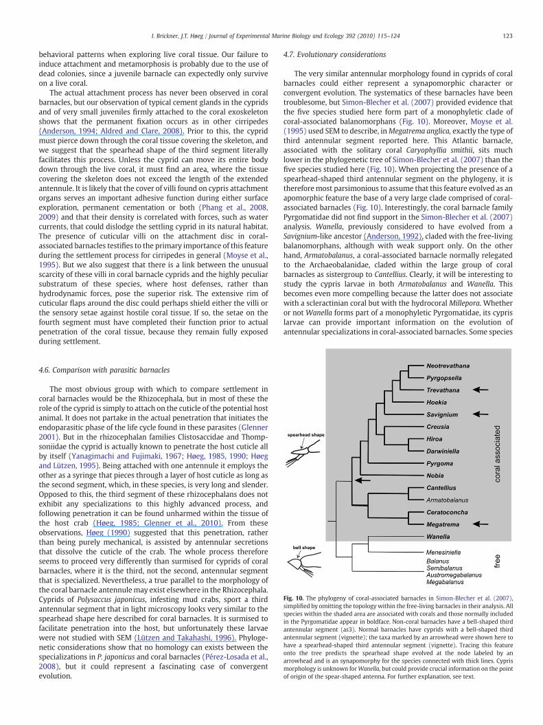
Fig. 10. The phylogeny of coral-associated barnacles in Simon-Blecher et al. (2007),simplified by omitting the topology within the free-living barnacles in their analysis. Allspecies within the shaded area are associated with corals and those normally includedin the Pyrgomatidae appear in boldface. Non-coral barnacles have a bell-shaped thirdantennular segment (as3). Normal barnacles have cyprids with a bell-shaped thirdantennular segment (vignette); the taxa marked by an arrowhead were shown here tohave a spearhead-shaped third antennular segment (vignette). Tracing this featureonto the tree predicts the spearhead shape evolved at the node labeled by anarrowhead and is an synapomorphy for the species connected with thick lines. Cyprismorphology is unknown forWanella, but could provide crucial information on the pointof origin of the spear-shaped antenna. For further explanation, see text.
4.6. Comparison with parasitic barnacles
The most obvious group with which to compare settlement incoral barnacles would be the Rhizocephala, but in most of these therole of the cyprid is simply to attach on the cuticle of the potential hostanimal. It does not partake in the actual penetration that initiates theendoparasitic phase of the life cycle found in these parasites (Glenner2001). But in the rhizocephalan families Clistosaccidae and Thomp-soniidae the cyprid is actually known to penetrate the host cuticle allby itself (Yanagimachi and Fujimaki, 1967; Høeg, 1985, 1990; Høegand Lützen, 1995). Being attached with one antennule it employs theother as a syringe that pieces through a layer of host cuticle as long asthe second segment, which, in these species, is very long and slender.Opposed to this, the third segment of these rhizocephalans does notexhibit any specializations to this highly advanced process, andfollowing penetration it can be found unharmed within the tissue ofthe host crab (Høeg, 1985; Glenner et al., 2010). From theseobservations, Høeg (1990) suggested that this penetration, ratherthan being purely mechanical, is assisted by antennular secretionsthat dissolve the cuticle of the crab. The whole process thereforeseems to proceed very differently than surmised for cyprids of coralbarnacles, where it is the third, not the second, antennular segmentthat is specialized. Nevertheless, a true parallel to the morphology ofthe coral barnacle antennulemay exist elsewhere in the Rhizocephala.Cyprids of Polysaccus japonicus, infesting mud crabs, sport a thirdantennular segment that in light microscopy looks very similar to thespearhead shape here described for coral barnacles. It is surmised tofacilitate penetration into the host, but unfortunately these larvaewere not studied with SEM (Lützen and Takahashi, 1996). Phyloge-netic considerations show that no homology can exists between thespecializations in P. japonicus and coral barnacles (Pérez-Losada et al.,2008), but it could represent a fascinating case of convergentevolution.
4.7. Evolutionary considerations
The very similar antennular morphology found in cyprids of coralbarnacles could either represent a synapomorphic character orconvergent evolution. The systematics of these barnacles have beentroublesome, but Simon-Blecher et al. (2007) provided evidence thatthe five species studied here form part of a monophyletic clade ofcoral-associated balanomorphans (Fig. 10). Moreover, Moyse et al.(1995) used SEM to describe, inMegatrema anglica, exactly the type ofthird antennular segment reported here. This Atlantic barnacle,associated with the solitary coral Caryophyllia smithii, sits muchlower in the phylogenetic tree of Simon-Blecher et al. (2007) than thefive species studied here (Fig. 10). When projecting the presence of aspearhead-shaped third antennular segment on the phylogeny, it isthereforemost parsimonious to assume that this feature evolved as anapomorphic feature the base of a very large clade comprised of coral-associated barnacles (Fig. 10). Interestingly, the coral barnacle familyPyrgomatidae did not find support in the Simon-Blecher et al. (2007)analysis. Wanella, previously considered to have evolved from aSavignium-like ancestor (Anderson, 1992), claded with the free-livingbalanomorphans, although with weak support only. On the otherhand, Armatobalanus, a coral-associated barnacle normally relegatedto the Archaeobalanidae, claded within the large group of coralbarnacles as sistergroup to Cantellius. Clearly, it will be interesting tostudy the cypris larvae in both Armatobalanus and Wanella. Thisbecomes even more compelling because the latter does not associatewith a scleractinian coral but with the hydrocoral Millepora. Whetheror not Wanella forms part of a monophyletic Pyrgomatidae, its cyprislarvae can provide important information on the evolution ofantennular specializations in coral-associated barnacles. Some species

124 I. Brickner, J.T. Høeg / Journal of Experimental Marine Biology and Ecology 392 (2010) 115–124
of Megabalanus also live on corals and provide another avenue fortesting the association between this mode of life and antennularshape. There are also other thoracican cirripedes such as spongebarnacles (Moyse, 1971), whale barnacles and the turtle barnaclePlatylepas that settle on and partially penetrate into live tissues. In awider perspective, it would be fascinating to investigate theseepibiotic cirripedes in search of possible adaptations in the morphol-ogy and metamorphosis of their cypris larvae. Nogata and Matsumura(2006) claimed that settlement and metamorphosis in whalebarnacles proceed as in other cirripedes but did not offer any detailedmicroscopical observations. Accuratemorphological descriptions as inNott and Foster (1969) and Bielecki et al. (2009) are needed to assessthe extent to which variations in the type of substratum are correlatedwith adaptations in cyprid sensory and attachment organs. Suchcomparative information will be valuable when generalizing frommeticulous experimental studies on settlement in model species.
Acknowledgements
I. Brickner was financed by two grants from the European UnionSYNTHESYS program. We take this opportunity to extend our sincerethanks to Professor Henrik Enghoff, The Danish Natural HistoryMuseum, for his tireless efforts to set up the COBICE and SYNTHESYSprograms. I Brickner also thank the Interuniversity Institute forMarine Studies in Eilat (IUI) for assistance, use of facilities and mostlyfor their friendship. Jens Høeg acknowledges the support from theCarlsberg Foundation and the Danish Research Academy (FNU). Healso wishes to honour the late Dr. Margaret Barnes, long time editor ofJEMBE, who edited four of his first eight papers in a both friendly andhighly professional manner. Few have like her furthered barnaclebiology by helping newcomers to the field. [SS]
Appendix A. Supplementary data
Supplementary data associated with this article can be found, inthe online version, at doi:10.1016/j.jembe.2010.04.015.
References
Aldred, N., Clare, A., 2008. The adhesive strategies of cyprids and development ofbarnacle-resistant marine coatings. Biofouling 24, 351–363.
Anderson, D.T., 1992. Structure, function and phylogeny of coral inhabiting barnacles(Cirripedia, Balanoidea). Zool. J. Linn. Soc. 106, 277–339.
Anderson, D.T., 1994. Barnacles — Structure, Function, Development and Evolution.Chapmann and Hall, London, pp. 1–3571. [available 1993].
Bielecki, J., Chan, B.K.K., Hoeg, J.T., Sari, A., 2009. Antennular sensory organs in cyprids ofbalanomorphan cirripedes: standardizing terminology using Megabalanus rosa.Biofouling 25 (3), 203–214.
Blomsterberg, M., Høeg, J.T., Jeffries, W.B., Lagersson, N.C., 2004. Antennulary sensoryorgans in cyprids of Octolasmis angulata and three Species of Lepas (Crustacea:Thecostraca: Cirripedia: Thoracica): a Scanning Electron Microscopy Study.J. Morphol. 200, 141–153.
Brickner, I., Simon-blecher, N., Achituv, Y., 2010. Darwin's Pyrgoma (Cirripedia)revisited: Revision of the Savignium group, molecular analysis and description ofnew species. J. Crust. Biol. 30, 266–291.
Clare, A.S., Nott, J.A., 1994. Scanning electron microscopy of the fourth antennularsegment of Balanus amphitrite amphitrite. J. mar. biol. Ass. U.K. 74, 967–970.
Glenner, H., 2001. Cypris metamorphosis, injection and earliest internal developmentof the kentrogonid rhizocephalan Loxothylacus panopaei (Gissler). Crustacea:Cirripedia: Rhizocephala: Sacculinidae. J. morphol. 249, 43–75.
Glenner, H., Høeg, J.T., 1995. Scanning electron microscopy of cyprid larvae in Balanusamphitrite amphitrite (Crustacea: Cirripedia: Thoracica: Balanomorpha). J. crustaceanBiol. 15 (3), 523–536.
Glenner, H., Høeg, J.T., Klysner, A., Brodin Larsen, B., 1989. Cypris ultrastructure,metamorphosis and sex in seven families of parasitic barnacles (Crustacea:Cirripedia: Rhizocephala). Acta zool. 70, 229–242.
Glenner, H., Høeg, J.T., Stenderup, J., Rybakov, A.V., 2010. The monophyletic origin of aremarkable sexual system in akentrogonid rhizocephalan parasites is confirmed bymolecular and larval structural data. Exp. Parasitol. 125, 3–12.
Høeg, J.T., 1985. Male cypris settlement in Clistosaccus paguri Lilljeborg (Crustacea:Cirripedia: Rhizocephala). J. exp. mar. Biol. Ecol. 89, 221–235.
Høeg, J.T., 1990. “Akentrogonid” host invasion and an entirely new type of life cycle inthe rhizocephalan parasite Clistosaccus paguri (Thecostraca: Cirripedia). J. crusta-cean Biol. 10, 37–52.
Høeg, J.T., Lützen, J., 1995. Life cycle and reproduction in the Cirripedia Rhizocephala.Oceanogr. Mar. Biol. Annu. Rev. 33, 427–485.
Høeg, J.T., Lagersson, N.C., Glenner, H., 2004. The complete cypris larva and itssignificance in thecostracan phylogeny. In: Scholtz, G. (Ed.), Evolutionary andDevelopmental Biology of Crustacea. Crustacean Issues 15. A.A. Balkema/Lisse,Abingdon Exton (PA) & Tokyo, pp. 197–215.
Høeg, J.T., Glenner, H., Shields, J., 2005. Cirripedia Thoracica and Rhizocephala (barnacles).In: Rohde, K. (Ed.), Marine Parasites. CABI Publishing Wallingford U.K. & CSIROPublishing Collingwood Victoria Australia, pp. 154–165.
Kolbasov, G.A., Høeg, J.T., 2007. Cypris larvae of acrothoracican barnacles (Thecostraca:Cirripedia: Acrothoracica). Zool. Anz. 246 (2), 127–151.
Lagerson, N.C., Høeg, J.T., 2002. Settlement behavior and antennulary biomechanics andin cypris larvae of Balanus amphitrite (Crustacea: Thecostraca: Cirripedia). Mar.Biol. 141, 513–526.
Lagersson, N.C., Garm, A.L., Høeg, J.T., 2003. Notes on the ultrastructure of the setae onthe fourth antennular segment of the Balanus amphitrite cyprid (Crustacea:Cirripedia: Thoracica). J. mar. biol. Ass. U.K. 83, 361–365.
Lützen, J., Takahashi, T., 1996. Morphology and biology of Polysaccus japonicus(Crustacea, Rhizocephala, Akentrogonida, Polysaccidae, fam. n.), a parasite of theghost–shrimp Callianassa japonica. Zool. Scr. 25 (2), 171–181.
Moyse, J., 1971. Settlement and growth pattern of the parasitic barnacle Pyrgomaanglicum. In: Crisp, D.J. (Ed.), Fourth European Marine Biology Symposium.Cambridge University Press, Cambridge, New York, pp. 125–142.
Moyse, J., Jensen, P.G., Høeg, J.T., Al-Yahya, H., 1995. Attachment organs in cypris larvae:using scanning electronmicroscopy. In: Schram, F.R., Høeg, J.T. (Eds.), NewFrontiers inBarnacle Evolution. Crustacean Issues 10. A.A. Balkema, Rotterdam, pp. 153–178.
Newman, W.A., Ladd, H.S., 1974. Origin of coral-inhabiting balanids (Cirripedia,Thoracica). Verh. Naturf. Ges. Basel 84, 381–396.
Newman, W.A., Jumars, P.A., Ross, A., 1976. Diversity trends in coral-inhabitingbarnacles (Cirripedia, Pyrgomatinae). Micronesica 12, 69–82.
Nogata, Y., Matsumura, K., 2006. Larval development and settlement of a whalebarnacle. Biol. Lett. 2, 92–93.
Nott, J., Foster, B., 1969. On the structure of the antennular attachment organ of thecypris larva of Balanus balanoides (L.). Phil. Trans. R. Soc. Lond. B. 256, 115–134.
Ogawa, K., Matsuzaki, K., 1992. An essay on host specificity, systematic taxonomy, andevolution of coral-barnacles. Bull. Biogeograph. Soc. Japan 47 (10), 87–101.
Pérez-Losada, M., Harp, M., Høeg, J.T., Achituv, Y., Jones, D., Watanabe, H., Crandall, K.A.,2008. The tempo andmode of barnacle evolution. Mol. Phylogen. Evol. 46, 328–346.
Pérez-Losada, M., Høeg, J.T., Crandall, K.A., 2009. Remarkable convergent evolution inspecialized parasitic Thecostraca (Crustacea). BMC Biol. 7, 15.
Phang, I.Y., Aldred, N., Clare, A.S., Vancso, G.J., 2008. Towards a nanomechanical basis fortemporary adhesion in barnacle cyprids (Semibalanus balanoides). J. R. Soc.interface 5 (21), 397–402.
Phang, I.Y., Chaw, K.C., Choo, S.S.H., Kang, R.K.C., Lee, S.S.C., Birch,W., Teo, S.L.M., Vancso,G.J., 2009. Marine biofouling field tests, settlement assay and footprint micromor-phology of cyprid larvae of Balanus amphitrite on model surfaces. Biofouling 25 (2),139–147.
Ritchie, L.E., Høeg, J.T., 1981. The life history of Lernaeodiscus porcellanae (Cirripedia:Rhizocephala) and co-evolution with its porcellanid host. J. crustacean Biol. 1,334–347.
Ross, A., Newman, W.A., 1973. Revision of the coral-inhabiting barnacles (Cirripedia:Balanidae). Trans. S. Diego Soc. nat. Hist. 17 (12), 137–174.
Simon-Blecher, N., Huchon, D., Achituv, Y., 2007. Phylogeny of coral inhabitingbarnacles (Cirripedia; Thoracica; Pyrgomatidae) based on 12S, 16S and 18S rDNAanalysis. Mol. Phylogen. Evol. 44, 1333–1341.
Visscher, J.P., 1928. Reactions of the cyprid larvae of barnacles at the time ofattachment. Biol. Bull. (Woods Hole) 54, 327–335.
Walker, G., 1971. A study of the cement apparatus of the cypris larva of the barnacleBalanus balanoides. Mar. Biol 9, 205–212.
Walley, L.J., 1969. Studies on the larval structure and metamorphosis of Balanusbalanoides (L.). Phil. Trans. R. Soc. Lond. B. 256, 237–280.
Yanagimachi, R., Fujimaki, N., 1967. Studies on the sexual organization of theRhizocephala. IV. On the nature of the “testis” of Thompsonia. Annotnes zool. jap40, 98–104.
Copyright © 2022 FDOKUMEN