Quantitative observations of a major coral bleaching event in Barbados, Southeastern Caribbean
Upload
khangminh22Category
view
0download
0
MARINA TONETTI BOTANA
The role of Symbiodinium membrane lipids in
response to heat shock: implications for coral
bleaching
Thesis presented to the Instituto Oceanográfico
of the Universidade de São Paulo, in partial
fulfillment of the requirements for obtaining the
degree of Master in Sciences, Oceanography
Program, concentration area of Biological
Oceanography.
Supervisor: Prof. Dr. Paulo Y. G. Sumida
São Paulo
2019

Universidade de São Paulo
Instituto Oceanográfico
The role of Symbiodinium membrane lipids in
response to heat shock: implications for coral
bleaching
Marina Tonetti Botana
Thesis presented to the Instituto Oceanográfico of the
Universidade de São Paulo, in partial fulfillment of the
requirements for obtaining the degree of Master in Sciences,
Oceanography Program, concentration area of Biological
Oceanography.
Evaluated in:
___________________________________ ____________________
Prof(a). Dr.(a) Grade
___________________________________ ____________________
Prof(a). Dr.(a) Grade
___________________________________ ____________________
Prof(a). Dr.(a) Grade

“Look up at the stars and not down at your feet. Try to make sense of what you see and
wonder about what makes the universe exist. Be curious. Science is not only a
discipline of reason, but also, one of romance and passion”
Steve Hawking
“Commit yourself to the process. Not the goal. The final score take care of itself”
James Clear

ACKNOWLEDGEMENTS
I would like to thank my advisor Paulo Sumida for giving me the opportunity to pursue my
masters in his lab. He allowed me to work on something I am very passionate for and
even supported my parallel career as a triathlete. I feel privileged for having him in my life
as a tutor and friend. My gratitude also goes to Dr. Marcos Yoshinaga who first presented
to me this fascinating world of lipids and without whom this work would never have been
possible. I thank all his tutoring and friendship, both pivotal for guiding this moment of my
career. Additionally, immense thanks to Prof. Sayuri Miyamoto who opened the doors of
her lab to my project and from whom I also could learn a lot about lipids metabolism and
oxidative stress. My deep gratitude extends to Dra. Flavia Saldanha-Correa who helped
me with the experiments and showed me other cool applications of studying lipids in other
phytoplankton phylotypes. Thanks to Dr. Marius Müller for the skype talks adding ideas
on the ecological implications of my results.
Regarding my learning process I also thanks Dr. Matthias Kellerman and Prof. Ray
Valentine for kindly teaching me about the universal principle of bioenergetics and how
the existence of life is utterly dependent on lipid membranes. My great appreciation also
to Dr. Florence Schubotz for sending valuable lipid standards and enabling better results.
For all their gentle help in the lab I thank Alex Inague, Adriano Brito and Debora Kutner.
Also, Thomas Banha and Arthur Güth for the many talks about my experiment design and
application of lipids in coral reefs ecology. Thanks to everyone from Sumida’s and
Sayuri’s laboratories for our daily routines full of scientific learnings and lots of funny
stories in the last two years.
Special thanks to my training buddies and triathlon coaches who also were part of my
routine from Sunday to Sunday in the last years and showed me that everything is
possible. Thanks for teaching me that interest is different from commitment and that we
must commit every day to the process of doing things that we really believe and are
passionate for, because that is what make us successful in the long term. That is what
make us grow as better athletes, scientists and human beings.

Last, but not least, I would mainly thank my parents for being supportive along the way
and believing in my career as a scientist. Since I was little, I was a very curious for ocean
things and they gave me an environment in which my curiosity could grow into passion
and then I could turn it into my something useful for nature and society. I would never be
able to put into words how grateful I feel for being your daughter.
This study was financed in part by the Coordenação de Aperfeiçoamento de Nível
Superior (CAPES) – Finance Code 001.

RESUMO
Recifes de coral do mundo inteiro vêm sendo devastados pelo fenômeno de
branqueamento, o qual as evidências indicam que seja causado pelo stress oxidativo
promovido pelo aquecimento global e eventos catastróficos de El Niño. A grande
variabilidade genética da Família Symbiodiniacea também é sugerida como
determinante da susceptibilidade do coral hospedeiro porque cada espécie possui limites
fisiológicos específicos, tanto no modo de vida livre como em simbiose. Neste estudo
apresentamos pela primeira vez o sucesso da utilização da de técnicas de lipidômica (i.e,
caracterização dos lipídeos globais em um determinado organismo) oferecendo suporte
para as investigações moleculares de investigação dos mecanismos relacionados ao
stress térmico em espécies de endosimbiontes de coral. Symbiodinium minutum foi
sensível às temperaturas elevadas, enquanto S. microadriaticum e S. goreaui
apresentaram distintos níveis de termo tolerância. Os fenótipos lipídicos das espécies
após o stress, incluindo o transportador de elétrons do fotossistema II – plastoquinona –
sugerem que cada um apresentou uma estratégia diferente para sobreviver. Além disso,
os lipídeos específicos do cloroplasto com ácidos graxos poliisaturados (PUFA) formado,
principalmente, por espécies com ômega 3 (n-3) foram essenciais para manter a
bioenergética celular à longo prazo (10 dias após stress) em todos os Symbiodinium spp.
A capacidade de manter altas concentrações de n-3 na membrana dos cloroplastos
determinou a sobrevivência dos S. microadriaticum e S. goreaui. Os dados apresentados
nesta dissertação revelam, pela primeira vez, o aumento de ácidos graxos oxidados na
membrana do cloroplasto e também na forma livre (FFA) em resposta aos dados de
stress oxidativo causados pelo calor. O estudo das membranas lipídicas é fundamental
para melhor compreensão da bioenergética dos simbiontes e para determinar a
vulnerabilidade da relação de simbiose com o coral aos estressores climáticos em um
futuro com temperaturas mais elevadas.
Palavras-chave: Symbiodinium, recifes de coral, lipidômica, stress térmico, stress
oxidativo.

ABSTRACT
Coral reefs around the world have been largely devastated by the phenomenon of “coral
bleaching”, which causes have been reported to be strongly related to oxidative stress
promoted by climate change drivers, including mainly global warming and catastrophic El
Niño events. Genetic variability in coral endosymbionts from the Family Symbiodiniacea
was also suggested as determinant of host susceptibility to stress because they present
distinct physiological boundaries when in free living or in symbiosis. Here we present for
the first time the successful use of lipidomics (i.e., the global characterization of lipids in
a given organism) supporting molecular investigation in the oxidative mechanisms related
to thermal stress in coral endosymbionts phylotypes. Symbiodinium minutum was thermal
sensitive, whereas S. microadriaticum and S. goreaui presented different levels of thermal
tolerance. Their lipid phenotypes after stress, including the photosystem electron
transporter - plastoquinone - suggested they had different survival strategies. In addition,
chloroplast specific lipids with polyunsaturated fatty acids (PUFAs) mainly formed by
omega 3 (n-3) seemed to be essential to sustain Symbiodinium cells bioenergetics in the
long term (10 days after stress). S. microadriaticum and S. goreaui capability of keeping
high n-3 concentrations in the chloroplast membranes determined their survival. The
present thesis reports, for the first-time, upregulation of oxidized lipids derived from
precursor chloroplast membranes and free fatty acids (FFA) in response to oxidative
stress damage caused by heat. The study of lipid membranes is of paramount importance
to better understand the bioenergetics of symbionts and to determine the
host/endosymbiont vulnerability to climate change stressors in a warmer future.
Key words: Symbiodinium, coral reefs, lipidomics, thermal stress, oxidative stress

LIST OF ACRONYMS AND ABBREVIATIONS
ARA - arachidonic acid
AC - Symbiodinium phylotype A1 control sample
AT – Symbiodinium phylotype A1 temperature stressed sample
ATP – adenosine triphosphate
BC - Symbiodinium phylotype B1 control sample
BT - Symbiodinium phylotype B1 temperature stressed sample
CC - Symbiodinium phylotype C1 control sample
CE – cholesterol ester
Cer - ceramide
Chl-a -chlorophyl-a
CL - cardiolipin
CT - Symbiodinium phylotype C1 temperature stressed sample
DAG – diacylglycerol
DGCC - 1,2-diacylglyceryl-3-(O-carboxyhydroxymethylcholine)
DGDG – digalactosyldiacylglycerol
DGTS - diacylglyceroltrimethylhomoserine
DHA - docosahexaenoic acid
EPA - eicosapentaenoic acid
ER - endoplasmic reticulum
ESI-TOFMS – electron spray ionization time of flight mass spectrometer
FFA - free fatty acids

Gluc Acid – glucoronic acid
HUFA – High unsaturated fatty acids
LC - liquid chromatography
MGDG - monogalactosyldiacylglycerol
MUFA - monounsaturated fatty acid
MS – mass spectrometer
PC – phosphatidylcholine
PE - phosphatidylethanolamine
PG – phosphatidylglycerol
PI - phosphatidylinositol
PL – polar lipids
PUFA - polyunsaturated fatty acid
RPLC – reverse phase liquid chromatography
ROS - reactive oxygen species
SFA - saturated fatty acid
SQDG – sulfoquinovosyldiacylglycerol
SM - sphingomyelin
TAG - triacylglycerol
UHPLC - ultra-high-performance liquid chromatography

LIST OF FIGURES
List is presented in the order they appeared in the text.
Figure 1.1: Bleached corals of the genus Mussismilia hispida, endemic from the tropical waters of
Brazil, observed in Ubatuba (São Paulo State north shore) in the summer of 2019……………………..2
Figure 1.2: Exemplification of how membrane lipids are affected by fatty acid composition and
temperature. Figure shows how polar lipids with unsaturated fatty acids attribute high motion and low
viscosity to the membrane. Plus, elevated temperatures also promote higher membrane’s motion and
fluidity. Combination of elevated temperatures and high polyunsaturated fatty acids can compromise
membrane’s electron transport chain viability……………………………………………………………………..4
Figure 1.3: Electron transport chain resulting in ATP synthesis in the thylakoid membranes of
chloroplasts. Protein complexes photosystem II (PSII), cytochrome b6-f (cyt b6-f) and photosystem I (PSI)
are highlighted. Plastoquinone pool responsible for electron transport from PSII to cyt b6-f is circled in
red……………………………………………………………………………………………………………………..5
Figure 2.1: Diversity of lipid species identified in Symbiodinium phylotypes A1, B1 and C1 sorted
into their respective lipid classes. Abbreviations: AMINO = aminolipids, GLYCO = glycolipids, SPHINGO
= sphingolipids, PHOSPHO = phospholipids. Detailed description of lipid subclasses abbreviations is shown
in Table S2.1………………………………………………………………………………………………………...17
Figure 2.2: Lipid molecular species profiles of Symbiodinium phylotypes A1, B1 and C1 control
samples. Cultures grew at 22 ºC; 80 µmols photons m-² s-1; 12L:12D cycle. Bars show the mean abundance
of each lipid class per cells and their respective standard errors. Storage lipids encompass cholesterol
esters and triacylglycerol; membrane lipids group sphingolipids, phospholipids, glycolipids and aminolipids;
Diacylglycerol (DAG) and free fatty acids (FFA) are presented alone. * represents statistically significantly
different (p < 0.05) mean values inside their respective lipid groups. Fig.S1.1 exhibits separately details
about storage lipids and membrane lipids. AC, BC and CC stand for control samples of A1, B1 and C1,
respectively………………………………………………………………………………………………………….18
Figure 2.3: Plastoquinone concentration in Symbiodinium phylotypes A1, B1 and C1; and its
respective molecular structure. * indicates that only BC is significantly different (p < 0.05) from other
mean values in AC and CC. AC, BC and CC stand for control samples of A1, B1 and C1, respectively……22
Figure 2.4: Heatmap of the most significantly different lipid molecular species between
Symbiodinium phylotypes A1, B1 and C1. In total, 40 lipid molecular species are shown in rows and

samples in column. Statistical significance was evaluated by one-way ANOVA followed by Turkey’s post-
hoc test (p < 0.05) using Metaboanalyst. Distance was measured in Pearson, and Ward´s clustering
algorithm. Each colored cell on heatmap corresponds to normalized concentrations. Log transformation
was used for data normalization value. Red color indicates upregulation, whereas blue means
downregulation. AC, BC and CC stand for control samples of A1, B1 and C1, respectively…………………24
Figure 2.5: Distribution of main omega 3 (n-3) fatty acids found in Symbiodinium phylotypes A1, B1
and C1 on their respective lipid polar heads. a) octadecatetraenoic acid (18:4) distribution; b)
octadecapentaenoic acid (18:5) distribution; c) docosahexaenoic acid distribution; d) relative percentage of
main omega 3 (n-3) fatty acids to total lipids in the 3 studied phylotypes. Means showing * are significantly
different (p < 0.05) from other mean values inside their respective lipid groups. Means showing # are
exclusively significantly different from each other (same p < 0.05). Vertical lines represent the standard
error. AC, BC and CC stand for control samples of A1, B1 and C1, respectively……………………………..26
Figure S2.1: Membrane and storage lipid subclasses in Symbiodinium phylotypes A1, B1 and C1.
a) main membrane lipids subclasses. Sphingolipids are not represented here because their concentrations
were lower than 10-4; b) storage lipid subclasses. Means showing an * are significantly different (p < 0.05)
from other mean values inside their respective lipid subclasses. Vertical lines represent the standard error.
AC, BC and CC stand for control samples of A1, B1 and C1, respectively…………………………………….32
Figure S2.2: Pigments in Symbiodinium phylotypes A1, B1 and C1. a) most abundant pigments; b)
minor representative pigments. Means were not significantly different. Vertical lines represent the standard
error. AC, BC and CC stand for control samples of A1, B1 and C1, respectively……………………………33
Figure S2.3: Most abundant lipid subclasses in Symbiodinium phylotypes A1, B1 and C1. a)
monogalactosyldiacylglycerol (MGDG); b) digalactosyldiacylglycerol (DGDG); c)1,2-diacylglyceryl-3-(O-
carboxyhydroxymethylcholine) – DGCC; concentrations of oxo-DHA were significantly lower in phylotype
C1, plus concentrations of DHA-OH were significantly higher in B1; d) phosphatidylcholine (PC); e)
triacylglycerol (TAG); f) free fatty acids (FFA). Means showing an * are significantly different (p < 0.05) from
other mean values inside their respective lipid subclasses. Means showing # are exclusively significantly
different from each other (same p < 0.05). Vertical lines represent the standard error. AC, BC and CC stand
for control samples of A1, B1 and C1, respectively………………………………………………………………34
Figure S2.4: Minor abundant phospholipids in Symbiodinium phylotypes A1, B1 and C1. a)
phosphatidylglycerol (PG); b) phosphatidyletanolamina; c) phosphatidylinositol. Means were not
significantly different. Vertical lines represent the standard error. AC, BC and CC stand for control samples
of A1, B1 and C1, respectively……………………………………………………………………………………..35
Figure S2.5: Minor compounds significantly different in Symbiodinium phylotypes A1, B1 and C1.
a) diacylglyceroltrimethylhomoserine (DGTS); b) Sphingolipids; c) Cholesterol ester (CE). Means showing
an * are significantly different (p < 0.05) from other mean values inside their respective lipid subclasses.

Means showing # are exclusively significantly different from each other (same p < 0.05). Vertical lines
represent the standard error. AC, BC and CC stand for control samples of A1, B1 and C1, respectively…..36
Figure S2.6: Other minor compounds not significantly different in Symbiodinium phylotypes A1, B1
and C1. a) diacylglycerol (DAG); b) Cholesterol; c) sulfoquinovosyldiacylglycerol (SQDG); d) Glucoronic
acid (Gluc Acid). Vertical lines represent the standard error. AC, BC and CC stand for control samples of
A1, B1 and C1, respectively………………………………………………………………………………………..37
Figure S2.7: Chlorophyll-a molecule fragments break spectrum. Molecule draw and exact mass
calculations were performed with ChemDraw………………………………………………………………38
Figure S2.8: Monogalactosyldiacylglycerol molecule ionized with ammonium fragments break
spectrum. High peak of 333 indicates double 18:4 fatty acids and 161 represents sugar head.
Molecule draw and exact mass calculations were performed with ChemDraw…………………………38
Figure S2.9: 1,2-diacylglyceryl-3-(O-carboxyhydroxymethylcholine) molecule fragments break
spectrum. 562 represents loss of DHA fatty acids and 544 represents loss of DHA plus water.
Molecule draw and exact mass calculations were performed with ChemDraw…………………………39
Figure S2.10: Diacylglyceroltrimethylhomoserine molecule fragments break spectrum. 236
represents polar head and 500 represents loss of 18:1 fatty acids. Molecule draw and exact mass
calculations were performed with ChemDraw………………………………………………………………..39
Figure S2.11: Phosphatidylcholine molecule ionized with acetate fragments break spectrum. 168
represents its polar head and each indicated arrow on figure indicate fatty acid losses. Molecule
draw and exact mass calculations were performed with ChemDraw……………………………………40
Figure S2.11: Phytoceramide molecule fragments ionized with acetate break spectrum. Molecule
draw and exact mass calculations were performed with ChemDraw……………………………………..40
Figure S2.12: Triacylglycerol molecule ionized with ammonium fragments break spectrum. Fatty
acids were determined based on exact mass loss. Higher peak of 577 indicates double loss of 16:0
and 551 peak indicates loss of 18:1. Molecule draw and exact mass calculations were performed
with ChemDraw……………………………………………………………………………………………………41
Figure S2.13: Cholesterol ester molecule ionized with ammonium fragments break spectrum.
Molecule draw and exact mass calculations were performed with ChemDraw…………………………41

Figure 3.1: Growth rate curves of control (AC, BC, CC) and temperature stressed Symbiodinium
phylotypes cultures (AT, BT, CT) with its respective µ values and standard deviation. Regression
curves were obtained with n=3 (triplicates). a) Growth rate curves of phylotype A1; b) Growth rate curves
of phylotype B1; c) Growth rate curves of phylotype C1. Images below graphs show final culture
appearance. Note that lighter coloration of cultures denotes lower cell
densities……………………………………………………………………………………………………………..50
Figure 3.2: Lipid remodeling data indicating distinct phylotype response time that are
correspondent to growth rate curves behavior. a) number of significant modified lipid species overtime
after heat shock event comparing samples from stressed cultures against samples from control (C x T). X
axis presents time in hours after heat shock event. Data points were based on the cultures sampling made
4 h, 24 h (1 day) and 240 hours (10 days) after stress; b) same significant modified compounds sorted into
the main lipid groups and the same sampling times. Bars above zero show species that were “upregulated”
compared to control; whereas bars below zero show lipids that were “downregulated” compared to control.
The * represents no upregulation of glycolipids in phylotype B1 after 10 days. Differences were evaluated
through Volcano-Plot analysis (FC = 1.5 and p < 0.05; FDR adjusted). Detailed data and molecular lipid
compounds are presented on Table S3.1………………………………………………………………………52
Figure 3.3: Total variation of omega 3 polyunsaturated fatty acids over time. Relative percentage of
all omega 3 (n-3) fatty acids (DHA, EPA, DPA, 18:3, 18:4 & 18:5) was considered for each Symbiodinium
phylotype considering 4- and 24-hours variations as short term and 10 days as long-term effect. Variations
were calculated based on values from stressed minus control lipid data in each sampling time. Bars show
standard error values obtained from triplicates…………………………………………………………………..53
Figure 3.4: Correlation of polyunsaturated fatty acids (PUFA) in the most abundant membrane lipids
and plastoquinone. Respective R² and p values are represented together in each graph. Glycolipids were
calculated considering monogalactosyldiacylglycerol (MGDG), digalactosyldiacylglycerol (DGDG),
sulfoquinovosyldiacylglycerol (SQDG) and glucoronic acid (GlucA). 1,2-Diacylglyceryl-3-(O-
carboxyhydroxymethylcholine) was the only aminolipid associated with PUFAs, as well as
phosphatidylcholine (PC) for phospholipids. All lipid data is represented in ng per cell………………………55
Figure 3.5: Pigments ratios that changed significantly after heat shock. Ratios are represented in
logarithmic scale to evidence its up or downregulation comparing temperature stressed cultures (T) and
control cultures (C) in three distinct sampling times: a) 4 hours; b) 24 hours; c) 10 days after heat shock
event. * represent samples statistically significant altered based on Volcano Plot analysis (FC = 1.5 and p
< 0.05) and details are on Table S3.1……………………………………………………………………………..56
Figure 3.6: Ratios of total concentrations of oxidized fatty acids and their precursors from polar
lipids (PLs) and free fatty acids (FFAs). 15 molecules represented by free fatty acids (6) and polar lipids
(8) were identified and monitored during our experiment. Ratios considered total concentration of each
identified fatty acid in either free or membrane associated forms. a) short term (4h and 24h after heat

shock); b) long term (10 days after heat shock). Bars represent average concentrations with respective
standard errors. Temperature stressed samples values are represented by (T), while control samples are
represented by (C)………………………………………………………………………………………………….58
Figure 3.7: Heatmap of oxylipins ratios that changed significantly after heat shock. In total, 12
oxylipins ratios are shown in rows and samples in columns. Statistical significance was evaluated by one-
way ANOVA followed by Turkey’s post-hoc test (p < 0.05) using Metaboanalyst. Distance was measured
in Pearson, and Ward´s clustering algorithm. Each colored cell on heatmap corresponds to normalized
concentrations. Log transformation was used for data normalization values. Red color indicates
upregulation, whereas blue means downregulation of oxylipins ratios. AC, BC and CC samples represent
controls. AT, BT and CT represent temperature stressed samples. 4h, 24h and 10 days represent sampling
times after heat shock for both stressed (T) and control (C) samples. Middle black rectangle groups
phylotype B1 samples after heat shock and green rectangle highlights single higher concentrations of FFA
oxylipins……………………………………………………………………………………………………………...60
Figure F1: Summary of Symbiodinium phylotypes cell’s proposed mechanisms caused by
upregulation of ROS. Temperature alters membrane viscosity and enable scape of high energy electrons
from electron transport chain. This might be responsible for upregulation of ROS that will concomitantly
lead to the peroxidation of most vulnerable omega-3 polyunsaturated fatty acids and activate quenching
mechanisms. We propose that phylotype B1 did not survive because it could not avoid lipid peroxidation of
essential ω-3 in the membranes, neither synthesize de novo epoxidized lipids. On the other hand, it was
not true for phylotypes A1 and C1. Both kept efficient energy output, although we suggest that they followed
distinct strategies already discussed in chapter 3. Protein complexes photosystem II (PSII), cytochrome b6-
f (cyt b6-f) and photosystem I (PSI) were highlighted. Plastoquinone pool responsible for electron transport
from PSII to cyt b6-f was also highlighted in red. Scape of high energy electron leading to ROS was
highlighted in dark red………………………………………………………………………………………………70

LIST OF TABLES
Table S2.1: Internal standards used for quantification of sphingolipids, phospholipids, storage
lipids and plastoquinone described in the material and methods section……………………………42
Table S2.2: Additional standards used for quantification of glycolipids, aminolipids and pigments
described in the material and methods section………………………………………………………………42
Table S2.3: All identified lipid compounds including their exact mass and retention time in both
positive and negative modes. File is in excel spreadsheet available in Research Gate profile
website…………………………………………………………………………………………………………INDEX
.
Table S3.1: Description of lipids and pigments compounds either up or downregulated after heat
shock for Symbiodinium phylotypes A1, B1 and C1 after 4h, 24h and 240h. File is in excel
spreadsheet available in Research Gate profile website………………………………………………INDEX

SUMMARY
CHAPTER 1: GENERAL INTRODUCTION
1.1 Coral reefs and their symbionts in the context of global warming……………………………………………1
1.2 Coral bleaching and the oxidative stress theory………………………………………………………...…….2
1.3 The effect of high temperature in membrane’s viscosity leading to free radical and ROS formation in the
chloroplast…………………………………………………………………………………………………………….3
1.4 Lipid membrane profiles and symbionts fate after thermal stress……………………………………….…6
1.4.1 Lipidomics as tool for better explanation of thermal sensibility………………………………………........7
2. Aimed goals and scope of this thesis……………………….……………………………………………….…..8
CHAPTER 2: Lipidomics as a throughput method for profiling lipids and pigments in Symbiodinium
phylotypes (A1, B1 & C1): physiological and ecological applications
1. INTRODUCTION………………………………………………………………………………………………...10
2. MATERIALS AND METHODS…………………………………………………………………………….……12
2.1 Experiment design…………………………………………………………………………………………..….12
2.2 Internal standards…………………………………………………………………………..…………………..12
2.3 Other standards……………………………………………………………………………………………..….12
2.4 Lipid extraction………………………………………………………………………………………………….13
2.5 Lipidomics analysis………………………………………………………………….………………………....13
2.6 Data processing………………………………………………………………………….……………………..14
2.7 Statistical analysis…………………………………………………………………………………………..….15
3. RESULTS………………………………………………………………………………………………………...16
3.1 Description of total lipids and pigments from Symbiodinium phylotypes A1, B1 and C1……………….18
3.1.1 Pigments………………………………………………………………………………………………….…..19
3.1.2 Membrane lipids……………………………………………………………………………………………...19

3.1.2.1 Glycolipids…………………………………………………………………………………………………..19
3.1.2.2 Aminolipids……………………………………………………………………………………………….…20
3.1.2.3 Phospholipids…………………………………………………………………………………………...….20
3.1.2.4 Sphingolipids…………………………………………………………………………………………….…20
3.1.2.5 Cholesterol………………………………………………………………………………………………....21
3.1.3 Storage lipids……………………………………………………………………………………………...….21
3.1.4 Other compounds…………………………………………………………………………………………….21
3.2 Main distinctions in Symbiodinium phylotypes are revealed in membrane lipids composition………..…22
4. DISCUSSION…………………………………………………………………………………………………….27
Supporting information……………………………………………………………………………………………..32
CHAPTER 3: Concentration of omega 3 fatty acids in membrane lipids determine Symbiodinium
phylotypes (A1, B1 & C1) fate after temperature stress
1. INTRODUCTION…………………………………………………………………………………………….…..43
2. MATERIALS AND METHODS………………………………………………………………………………….47
2.1 Experiment design…………………………………….…………………………………………………….….47
2.1 Population growth rates………………………………………………………………………………………..47
2.3 Standards, lipid analysis and data processing……………………………………………………………….48
2.4 Statistical analysis…………………………………………………………………………………………...…48
3. RESULTS…………………………………………………………………………………………………….…..49
3.1. Heat shock experiment revealed two resistant Symbiodinium spp. and drastic changes in lipids and
pigments profiles……………………………………………………………………………………………….…...49
3.2 Changes in lipid composition after heat shock……………………………………………………..………..50
3.3 Polyunsaturated fatty acids in membrane lipids were pivotal for Symbiodinium spp. long term
survival……………………………………………………………………………………………………………….53
3.4 Changes in pigments and oxylipins as evidences of oxidative stress caused by heat shock…………....55

4. DISCUSSION…………………………………………………………………………………………………….61
4.1. Chloroplast bioenergetics and oxidative stress: a benefit – risk story…………………………………...61
4.2 Ecological implications…………………………………………………………………………………………66
4.2.1 Lipid ecology of zooxanthellae in coral reefs symbiosis and global warming perspectives…………...66
Supporting Information………………………………………...…………………………………………………..68
FINAL REMARKS…………………………………………………………………………………...……………..69
Importance of phytoplankton lipids in the global carbon budget and bottom-up effects in the marine food
chains…………………………………………………………………………………………………………….….72
Additional considerations……………………………………………………………………………………….….74
FUTURE PERSPECTIVES………………………………………………………………………………………..76
REFERENCES………………………………………………………………………………………...……………77

CHAPTER 1: GENERAL INTRODUCTION
1.1 Coral reefs and their symbionts in the context of global warming
Coral reefs are one of the most productive ecosystems on Earth, representing one of the
most diverse marine environments (Grigg et al., 1984). They hold high biodiversity and
ecosystem services important for sustaining higher trophic level organisms and providing
goods for large numbers of people (Martinez et al., 2007; Alves de Lima et al., 2010;
Knowlton et al., 2010). Corals are defined as ecosystem engineering organisms
responsible for controlling the availability of resources to other organisms through
physical changes in biotic and abiotic materials (Jones et al., 1994). Therefore, studying
them is pivotal to understand marine community structure and production of resources to
humankind.
Tropical calcium carbonate reefs are built by stony corals (Scleractinia) associated with
dinoflagellates of the family Symbiodinidaceae perceived as a mutualistic symbiosis.
Photosynthetically produced endosymbiont metabolites are exchanged with coral hosts
guaranteeing their survival and growth even in nutrient-poor waters (Muscatine & Porter,
1977; Leggat et al., 2003). In addition, the family Symbiodinidaceae is divided into nine
clades (A-I) (Pochon & Gates, 2010) and multiple phylotypes within each clade (Thornhill
et al., 2014), which shows distinct degrees of host-specificity and different tolerances to
environmental conditions (Toller et al., 2001; Baker, 2003; Chen et al., 2003; Coffroth &
Santos, 2005; LaJeunesse, 2005; Berkelmans & van Oppen, 2006; Goulet, 2006). Recent
research has focused on the strength of this mutualistic relationship in response to
predicted climate alteration (Davy & Cook, 2001; Cervino et al., 2004; Barneah et
al.,2007; Hoegh-Guldberg et al.,2007), which is the main culprit causing extensive coral
reef degradation (Hoegh-Guldberg & Smith, 1989; Graham et al. 2008; Wilkinson, 2008).

1.2 Coral bleaching and the oxidative stress theory
Coral bleaching is defined by visible whitening of the coral as a result of decreasing
densities of Symbiodinium spp. cells and/or declines in their photosynthetic pigments
(Fig.1.1). Since corals are extremely dependent on endosymbiont metabolites, their lack
leads to coral degradation, eventually followed by coral death (Glynn, 1996; Brown,
1997). A recognized biochemical explanation for the phenomenon was first proposed by
Lesser (1997) and coined by Downs et al. (2002) as “the oxidative stress theory of coral
bleaching”. They proposed that excessive light and temperature cause physiological
stress in the symbiont in combination with increased production of reactive oxygen
species (ROS). ROS (i.e., hydrogen peroxide, superoxide, hydroxyl and singlet oxygen -
Valko et al. 2007) can trigger the oxidation of essential biomolecules such as proteins
and lipids in both coral and Symbiodinium spp. cells, thereby leading to disruption of
symbiotic association.
Figure 1.1: Bleached corals of the genus Mussismilia hispida, endemic from the tropical waters of
Brazil, observed in Ubatuba (São Paulo State north shore) in the summer of 2019.
The concept of ROS playing a key role in bleaching events has been an important subject
of research in the last decades. The increased ROS production in symbiont cells may be
caused by combination of temperature, light and even other stressors (e.g., alterations in
carbonate chemistry) (Tchernov et al., 2004; Smith et al., 2005; Ragni et al., 2010;
Roberty et al., 2015; Goyen, 2017). Today, despite the evidences, the specific causes
triggering excessive ROS production leading to bleaching and harmful impacts on host
physiology and impairment of symbiosis are still a matter of debate and further
investigation.
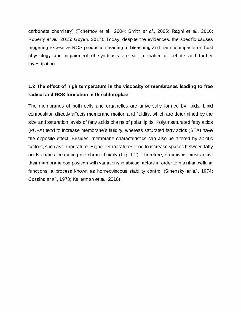
1.3 The effect of high temperature in the viscosity of membranes leading to free
radical and ROS formation in the chloroplast
The membranes of both cells and organelles are universally formed by lipids. Lipid
composition directly affects membrane motion and fluidity, which are determined by the
size and saturation levels of fatty acids chains of polar lipids. Polyunsaturated fatty acids
(PUFA) tend to increase membrane’s fluidity, whereas saturated fatty acids (SFA) have
the opposite effect. Besides, membrane characteristics can also be altered by abiotic
factors, such as temperature. Higher temperatures tend to increase spaces between fatty
acids chains increasing membrane fluidity (Fig. 1.2). Therefore, organisms must adjust
their membrane composition with variations in abiotic factors in order to maintain cellular
functions, a process known as homeoviscous stability control (Sinensky et al., 1974;
Cossins et al., 1978; Kellerman et al., 2016).

Figure 1.2: Exemplification of how membrane lipids are affected by fatty acid composition and
temperature. Figure shows how polar lipids with unsaturated fatty acids attribute high motion and low
viscosity to the membrane. Plus, elevated temperatures also promote higher membrane’s motion and
fluidity. Combination of elevated temperatures and high polyunsaturated fatty acids can compromise
membrane’s electron transport chain viability. Source: adapted from bio.libretexts.org.
Controlling membrane composition is pivotal for survival of all life forms, from bacteria to
plants to mammals. The lipid composition of energy transducing membranes (i.e.,
cytoplasmic membranes of bacteria, thylakoid membranes of chloroplasts and
mitochondrial inner membranes) is extremely specialized and must perform two essential
functions: 1) control permeability of ions such as protons and sodium; and 2) tighten the
electron transport at the membrane level. In chloroplasts (Fig. 1.3), the high-energy
electrons coming from the water split at photosystem II (PS II) and must be safely
transported within membranes by a plastoquinone to energize protein complexes or
proton pumps (cytochrome b6f and PSI), a process known as electron transport chain.
These pumps generate a proton gradient (high in the lumen and low in the cytoplasm)
needed to promote adenosine triphosphate (ATP) synthesis (Fig.1.3). In retrospect, this
ion gradient can only be established by controlling permeability of energy transducing
membranes. All living forms must always adapt and keep membrane motion and fluidity
stable in order to make ATP or energy, thus, lipids and bioenergetics are inseparable.

Figure 1.3: Electron transport chain resulting in ATP synthesis in the thylakoid membranes of
chloroplasts. Protein complexes photosystem II (PSII), cytochrome b6-f (cyt b6-f) and photosystem I (PSI)
are highlighted. Plastoquinone pool responsible for electron transport from PSII to cyt b6-f is circled in red.
Source: Adapted from Wada & Murata, 2009.
Thylakoid membranes of chloroplasts are very unique in that they are composed of
glycolipids and specially at protein complexes such as the PS II. They are also decorated
with several pigments including carotenoids and chlorophylls (Wada & Murata et al.,
2009). The arrangement or conformation of lipids and pigments has been honed by
Darwinian evolution to prevent the leakage of both protons and electrons at the
membrane level. Any leakage of protons through the membrane may prevent the
generation of proton gradients needed for ATP synthesis (e.g., Kellerman et al., 2016;
Yoshinaga et al., 2016). On the other hand, leakage of high-energy electrons may lead
to free radical generation, which combined with oxygen leads to the formation of ROS
(Polle, 1996). Both free radicals and ROS, if not contained by the cell’s antioxidant
machinery (including carotenoids and antioxidant enzymes such as catalases), can cause
oxidation of biomolecules: proteins, pigments and lipids (Augusto & Miyamoto, 2011; Yin
et al. 2011). Lipid peroxidation is the substitution of a bis-allylic hydrogen in the lipid
structure by a free radical, yielding the formation of a lipid radical (L•). This lipid radical
can easily react with oxygen, generating a lipid peroxyl radical (LOO•) and undergo
complex cyclic reactions that might propagate through the membrane through a process
best known as chain reaction (Niki, 2009; Yin et al., 2011). In this context, therefore, fatty
acids containing high unsaturation levels, such as polyunsaturated fatty acids (PUFAs),

are the main targets of radical and ROS attack in their double bounds and propagate lipid
oxidation even further unless stopped by an antioxidant agent.
The combination of high temperatures and high concentration of PUFAs in the
chloroplasts of Symbiodinium is likely a strong trigger for coral bleaching, and the
associated “oxidative theory of coral bleaching” (Lesser, 1997; Downs et al, 2002). Drastic
changes in membrane permeability and fluidity are expected to occur as a consequence
of high temperatures. Altering the thylakoid membrane conformation most likely promotes
leakage of protons and electrons, thereby leading to decreased energy production and
increased oxidative stress, respectively. It is, however, unknown whether the disruption
of symbiosis occurs by a decreased supply of metabolites to the coral host, death of
symbionts or simply symbionts themselves representing a potential threat to the host due
to high ROS production.
1.4 Lipid membrane profiles of symbionts and their fate after thermal stress
Strong evidence suggests that Symbiodinium spp. can modulate the lipid composition of
cells and organelles membranes in order to keep homeoviscous stability and adapt to
environmental alterations (D’amico et al., 2006). Higher abundance of SFAs relative to
PUFAs has been reported to enhance physical stability of thylakoid membranes of
Symbiodinium sp. in response to thermal stress (Tchernov et al., 2004; Bachok et al.,
2006; Tolosa et al., 2011). The rationale is that Symbiodinium spp. thylakoid membranes
are enriched in PUFA, which are highly susceptible to oxidative damage by free radicals
and ROS (Wada, 1994; Lesser, 2006; Catalá, 2009). However, Symbiodinium spp. may
protect the photosynthetic membranes against ROS and thus acquire thermal tolerance
altering the ratio saturated/unsaturated fatty acids. Tchernov et al. (2004) have even
suggested that thermal tolerance is not associated with a single monophyletic phylotype,
but rather with the level of saturation of their membrane lipids.
The above-mentioned studies marked the initial investigations of the role of lipids in coral
bleaching. They were essential to establish that bulk fatty acid composition is crucial for
survival and supports the “oxidative theory of coral bleaching” (Lesser, 1997; Downs et

al, 2002). In this dissertation, we generated data based on the global lipidome of some
Symbiodinium phylotypes (e.g., glycolipids, phospholipids, aminolipids and storage
lipids), including their pigments, in response to thermal stress. Thus, we not only report
data on fatty acids, but also the lipid molecular species containing these fatty acids. That
is, we are able to pinpoint whether thermal stress affects thylakoid membranes by
characterizing their specific glycolipids rather than bulk fatty acids derived from other pool
of lipids such as the triglycerides or phospholipids. Such detailed and comprehensive lipid
analysis could only be achieved by recent analytical developments in mass spectrometry
and the advance of lipidomics (Jones et al., 2012; Yao et al., 2015; Nygren et al., 2017).
1.4.1 Lipidomics as tool to better characterize microalgae lipids
Lipidomics is a fairly recent technique that evolved from metabolomics in its own research
field (Tomita & Nishioka, 2006; German et al., 2007). Previous lipid analytical techniques
allowed qualitative information about polar lipids such as acquired by thin-layer
chromatography or quantitative analysis of bulk fatty acids by gas chromatography.
Contrasting with past lipid analytical techniques, lipid characterization by liquid
chromatography coupled to mass spectrometry (LC-MS) enabled precise
characterization and quantification of every lipid molecular species present in a given
sample (German et al., 2007; Oresic et al., 2008), including molecules that are specific
markers of chloroplast, such as glycolipids and plastoquinones. For example, a great
diversity of glyco and amino membrane lipids in Symbiodinium spp. and other
dinoflagellates has been described by LC-MS analysis, including few alterations when
growing in distinct temperatures (Leblond et al., 2000, 2006, 2010, 2015; Gray et al.,
2009; Dahmen et al., 2013; Flaim et al., 2014; Anesi et al., 2015, 2016). These studies
were mostly focused on a specific class of polar lipid such as glycolipids or aminolipids,
and not aimed at characterizing the global lipidome together with specialized lipids, such
as plastoquinone and pigments using LC-MS. Besides the culture-based investigations,
important data have been generated in environmental studies reported by Van Mooy et
al. (2006, 2009 and 2010), Moutin et al. (2007), Schubotz et al. (2009), Xie et al. (2014)
and Becker et al. (2018). These include not only data from phospho, glyco and

aminolipids, but also storage lipids and quinone molecules (e.g., ubiquinone in Becker et
al., 2018). These studies describe the lipidome of the water column of the oceans, where
a diverse variety of phytoplankton occurs, in response to diel temperature oscillations and
distinct nutrient conditions. The present study is, to the best of our knowledge, the first
attempt to characterize the global lipidome of a microalga, describing not only membrane
lipids, but also storage lipids, pigments and plastoquinone from Symbiodinium
phylotypes. This information will be linked to cell physiology data to further knowledge on
their thermal sensitivity.
2. GOALS AND SCOPE OF THIS THESIS
The overall goal of this thesis is to investigate how the lipidomes of S. microadriaticum
(phylotype A1), S. minutum (phylotype B1) and S. goreaui (phylotype C1) most
prevalently associated with scleractinian corals are related to their thermal sensitivity.
Description of Symbiodinium spp. lipids and pigments is very scarcely found in the
literature, let alone their lipid alterations with stress events. For best comprehension all
studied Symbiodinium spp. will be referred as their phylotypes A1, B1 and C1. Therefore,
this thesis is divided into two further chapters:
Chapter 2:
Description and quantification of global lipidome and pigment profiles of Symbiodinium
phylotypes A1, B1 and C1 growing under optimum conditions of temperature, light and
nutrients. Here, the goals are to examine whether differences in lipidome and pigments
profiles between Symbiodinium phylotypes can predict their thermal tolerance to heat
stress events based on previous studies (e.g., Tchernov et al., 2004).

Chapter 3:
Monitor Symbiodinium phylotypes (A1, B1 and C1) growth rates and lipidome/pigment
alterations after a heat shock event (4h under 34˚C) describing short-term (4 and 24 h)
and long-term phenotypical alterations (10 days).
Main assumption: “High temperatures known to change thylakoid membrane stability and
enabling scape of high energy electrons generate higher concentration of ROS leading
to lipid peroxidation accompanied by reduction of ATP production. Both facts affect
Symbiodinium spp. growth rates and reflect in drastic changes in their lipidomes and
pigments profiles”.
Specific hypotheses:
1) Symbiodinium spp. growth rates are negatively affected by heat shock;
2) Changes in lipids and pigments are different between Symbiodinium phylotypes after
heat shock;
3) Lipidome and pigments profiles of Symbiodinium phylotypes after stress are good
indicators of oxidative stress caused by heat shock experiment;
4) Lipid peroxidation preferably occurs in polyunsaturated fatty acids (PUFA) from
chloroplast membranes.

FINAL REMARKS
In Chapter 2 we verified the use of lipidomics as a precise tool for description,
quantification and monitoring of total lipids and pigments of Symbiodinium phylotypes A1,
B1 and C1 growing under optimum conditions of temperature, light and nutrients. We also
showed that statistically significant differences among phylotypes were mainly
determined by their membrane lipids. Although A1 and B1 were more similar based in the
heatmap analysis, A1 was more alike to C1 considering their higher concentrations of
omega-3 polyunsaturated fatty acids and plastoquinone.
In Chapter 3 we analyzed variations in Symbiodinium growth rates and both short and
long terms lipidomes after a heat shock event summarized in Figure F1. Phylotype B1
was not heat shock resistant and its high decrease in biomass and cell death occurred
after a downregulation of essential membrane omega-3 fatty acids and all identified
pigments from chloroplasts. Thus, lipidome and pigments changes of B1 could not
guarantee its cell’s energetic requirements. However, phylotypes A1 and C1 both resisted
mainly because they were capable of maintaining higher concentrations of essential
omega-3 fatty acids in the thylakoid membranes and supply cells energetic requirements
in the long term.
Combined information from both chapters demonstrated lipidomics as a functional tool to
comprehend cell physiological alterations caused by thermal stress by a unified concept.
We noticed that functional thylakoid membrane lipid structure cannot vary much in order
to feasibly supply the cell bioenergetic demands. Therefore, the fate of organisms is likely
to be determined by the cell antioxidant machinery, which were not analyzed in the
present study, but that might protect membranes against ROS and keep efficient energy
output even under stress conditions.

Figure F1: Summary of Symbiodinium phylotypes cell’s proposed mechanisms caused by
upregulation of ROS. Temperature alters membrane viscosity and enable scape of high energy electrons
from electron transport chain. This might be responsible for upregulation of ROS that will concomitantly
lead to the peroxidation of most vulnerable omega-3 polyunsaturated fatty acids and activate quenching
mechanisms. We propose that phylotype B1 did not survive because it could not avoid lipid peroxidation of
essential ω-3 in the membranes, neither synthesize de novo epoxidized lipids. On the other hand, it was
not true for phylotypes A1 and C1. Both kept efficient energy output, although we suggest that they followed
distinct strategies already discussed in chapter 3. Protein complexes photosystem II (PSII), cytochrome b6-
f (cyt b6-f) and photosystem I (PSI) were highlighted. Plastoquinone pool responsible for electron transport
from PSII to cyt b6-f was also highlighted in red. Scape of high energy electron leading to ROS was
highlighted in dark red. Source: Adapted from Wada & Murata, 2009.
Finally, we attempted to elucidate specific hypothesis delineated in this
dissertation/thesis:
1) “Symbiodinium spp. growth rates are negatively affected by heat shock”
Growth rates were all reduced after heat shock. Besides, response was different between
phylotypes. S. minutum (B1) did not survive stress, whereas S. microadriaticum (A1) and
S. goreau (C1) were resistant to heat shock but decreased their growth rates.

2) “Changes in lipids and pigments are different between Symbiodinium phylotypes after
heat shock”
Symbiodinium spp. lipidomes and pigments profiles were differently altered by heat
shock. However, downregulation of n-3 fatty acids 4 hours after stress was common in all
phylotypes. After 24 hours and in the long term considered as 10 days, each phylotype
had a specific survival strategy and fate summarized in figure 4. This demonstrates the
essential role of omega-3 fatty acids for cellular energy as suggested by Valentine and
Valentine (2004).
3) “Lipidome and pigments profiles of Symbiodinium spp. after stress are good indicators
of oxidative stress caused by heat shock experiment”
We considered variations in the pigments ratios as indicators of chloroplast “health status”
and chlorophyll decrease strongly indicated damage in the chloroplasts caused by heat
shock. This data together with variations in epoxy/de-epoxy xanthophylls suggest that
damage was likely caused by oxidative stress but could not prove it. However, oxidized
fatty acids in the free form and connected with polar lipids analytically proved damage
provoked by oxidative stress.
4) “Lipid peroxidation preferably occurs in polyunsaturated fatty acids (PUFAs) from
chloroplast membranes”
Lipid peroxidation was evidenced by the presence of oxidized fatty acids mostly derived
from omega 3 polyunsaturated fatty acids in free (FFA) and membrane associated (PL)
forms of MGDG, DGDG, DGCC and PC. Glycolipids (MGDG and DGDG) are well known
structural thylakoid membrane lipids, plus, evidences discussed in chapter 3 strongly
indicated DGCC presence also in the chloroplast. Therefore, chloroplast membranes
were main targets of lipid peroxidation. Plus, significant changes in DGCC and DGDG
compounds suggest that they might be specifically located closer to chloroplast ROS

formation sources when compared to MGDG because they did not change significantly
after heat shock.
Importance of phytoplankton lipids in the global carbon budget and bottom-up
effects in the marine food chains
Symbiodinium sp. takes part of the Dinophyceae class and many lipid compounds
characterized and monitored in our work have been previously reported in the phenotypes
of relatives from the same class and other microalgal classes that inhabit oligotrophic
ocean gyres (e.g., Crytophyceae, Haptophyceae) (Van Mooy et al., 2009, 2010; Shemi
et al., 2016). We suggest our temperature-related alterations in the lipidome of
Symbiodinium spp. are similar to those that may occur in phytoplankton communities
considering a universal biochemical principle of lipid composition in bioactive membranes.
Becker et al. (2018) showed that in the North Pacific subtropical gyre (NPSG) nearly half
of the relative abundance of organisms was composed of Dinophyceae and
Haptophyceae (ca. 25.3% each). This region comprises 40% of Earth’s total surface area,
representing the world’s largest biome (Emmerson et al., 1997; Sarmiento et al., 2004).
Local and global alterations in phytoplankton communities were noticed with elevated
temperatures and other climate change stressors in the past years leading to alterations
in global primary production and carbon sinking (Sarmiento et al., 2004; Behrenfeld et al.,
2006; Schmittner et al., 2008; Nagelkerken & Connell, 2010), but the physiological
mechanisms responsible for such alterations were poorly discussed. We suggest that
changes are likely to happen because of phytoplankton photosynthetic structure
vulnerability to oxidative stress considering that they are the major DHA producers in the
biosphere (Valentine, 2009). High concentrations of DHA were reported by Becker et al.
(2018) in the NPSG community associated with DGCC betaine polar head. Their most
abundant DGCC had C38:6 – 800.6035 m/z - acyl chains of palmitic acid (16:0) and
docosahexaenoic acid (DHA), which was also the most abundant feature in our samples.
Photosynthesis and phytoplankton growth in phenotypes with high DHA concentration
can be impaired through mechanisms already discussed in our work.

Furthermore, the high abundance of DHA in Symbiodinium phylotypes and in other
flagellate microalgae (Leblond et al., 2000, 2006, 2015; Gray et al., 2009; Awai et al.,
2012; Armada et al., 2013; Dahmen et al., 2013; Anesi & Guella, 2015; Anesi et al., 2016)
sustains the idea proposed by Valentine (2009): phytoplankton represents the global
stock of DHA production in the marine ecosystem. Life cycle of zooplankton species
depend on the nutritional quality of phytoplankton, which is defined by lipid content
(Søreide et al., 2010). Ingestion of DHA and other omega 3 enables development of their
sensorial mechanisms essential for survival, growth and reproduction (Müller-Navarra et
al., 2000; Falkowski & Oliver, 2007). Higher in the food chains, other organisms also
evolved with the same dependence in n-3 and their populations may either decrease or
present individuals with neurological and sensorial deficiencies if n-3 consumption is low
(Davis et al., 1992; Budge et al., 2001; Jonasdottir et al., 2002). Consequently, we
highlight the importance of DHA ingestion in the marine food chains and how they are
also likely to be impacted by global warming. Behrenfeld et al. (2006) showed that from
1999 to 2004 the average global primary productivity dropped by about 200 tons a year.
Local changes had a decrease as high as 50% (see also Bopp et al., 2013). If ocean
temperatures keep increasing progressively as predicted by climate models (Hansen et
al., 2010; Rogelj et al., 2012; Cabré et al., 2015), omega 3 producing phytoplankton might
follow an opposite way and promote a bottom-up effect in all marine food chains that
could potentially lead to ecological collapse of the whole ecosystem
Omega-3 presence in microalgae membranes mediate cell death cascades with
temperature changes in the environment (Valentine, 2009). It was highly evidenced in our
experiments with Symbiodinium spp. and generated valuable data for coral reef symbiosis
and bleaching research. Besides, the concomitant omega-3 presence in phytoplankton
populations with large distribution patterns enhances our insights of large detrimental
effects in marine food chains following ocean warming. Therefore, we highlight the
importance of the present study in mechanistically explaining a universal biochemical
principle for all living creatures, from algae cells to more complex organisms. A principle
useful to understanding cell physiology and also how slight modifications can impact the
whole environment.

REFERENCES
Alves de Lima, L., Migliolo, L., Barreiro e Castro, C., de Oliveira Pires, D., López-
Abarrategui, C., Ferreira Goncalves, E., ... & Campos Dias, S. (2013). Identification of a
novel antimicrobial peptide from Brazilian coast coral Phyllogorgia dilatata. Protein and
peptide letters, 20(10), 1153-1158.
Andras, J. P., Kirk, N. L., & Drew Harvell, C. (2011). Range‐wide population genetic
structure of Symbiodinium associated with the Caribbean Sea fan coral, Gorgonia
ventalina. Molecular Ecology, 20(12), 2525-2542.
Anesi, A., & Guella, G. (2015). A fast liquid chromatography-mass spectrometry
methodology for membrane lipid profiling through hydrophilic interaction liquid
chromatography. Journal of chromatography A, 1384, 44-52.
Anesi, A., Obertegger, U., Hansen, G., Sukenik, A., Flaim, G., & Guella, G. (2016).
Comparative analysis of membrane lipids in psychrophilic and mesophilic freshwater
dinoflagellates. Frontiers in plant science, 7, 524.
Anthony KRN, Hoogenboom MO, Maynard JA, Grottoli AG, Middlebrook R (2009)
Energetics approach to predicting mortality risk from environmental stress: a case study
of coral bleaching. Funct Ecol 23:539-550.
Armada, I., Hachero-Cruzado, I., Mazuelos, N., Ríos, J. L., Manchado, M., & Cañavate,
J. P. (2013). Differences in betaine lipids and fatty acids between Pseudoisochrysis
paradoxa VLP and Diacronema vlkianum VLP isolates (Haptophyta). Phytochemistry, 95,
224-233.
Augusto, O., & Miyamoto, S. (2011). Oxygen radicals and related species. Principles of
free radical biomedicine, 1, 19-42.
Awai, K., Matsuoka, R., & Shioi, Y. (2012). Lipid and fatty acid compositions of
Symbiodinium phylotypes. In Proceedings of the 12th International Coral Reef
Symposium.
Bachok, Z., Mfilinge, P., & Tsuchiya, M. (2006). Characterization of fatty acid composition
in healthy and bleached corals from Okinawa, Japan. Coral Reefs, 25(4), 545-554.
Barneah O. Brickner, M. Hooge (2007) Three party symbiosis: acoelomorph worms,
corals and unicellular algal symbiont in Eilat (Red Sea). Marine Biology 151: 1215

Baker A. (2003) Flexibility and specificity in coral-algal symbiosis: diversity, ecology, and
biogeography of Symbiodinium. Annual Review Ecology Evolution Systems, 34: 661689
Bastien, O., Botella, C., Chevalier, F., Block, M. A., Jouhet, J., Breton, C., ... & Maréchal,
E. (2016). New insights on thylakoid biogenesis in plant cells. In International review of
cell and molecular biology (Vol. 323, pp. 1-30). Academic Press.
Bates, P. D., Fatihi, A., Snapp, A. R., Carlsson, A. S., & Lu, C. (2012). Acyl editing and
headgroup exchange are the major mechanisms that direct polyunsaturated fatty acid flux
into triacylglycerols. Plant physiology, pp-112.
Babiychuk E, Müller F, Eubel H, Braun HP, Frentzen M AND Kushnir S (2003)
Arabidopsis phosphatidylglycerophosphate synthase 1 is essential for chloroplast
differentiation but is dispensable for mitochondrial function. Plant J 33: 899–909
Becker, K. W., Collins, J. R., Durham, B. P., Groussman, R. D., White, A. E., Fredricks,
H. F., ... & Van Mooy, B. A. (2018). Daily changes in phytoplankton lipidomes reveal
mechanisms of energy storage in the open ocean. Nature communications, 9(1), 5179.
Behrenfeld, M. J., O’Malley, R. T., Siegel, D. A., McClain, C. R., Sarmiento, J. L.,
Feldman, G. C., ... & Boss, E. S. (2006). Climate-driven trends in contemporary ocean
productivity. Nature, 444(7120), 752.
Berkelmanns, R. & Van Oppen, M. J. H. (2006) Flexible partners in coral symbiosis: a
‘nugget of hope’ for coral reefs in an era of climate change. Proc. R. Soc. B 273, 2305–
2312
Bligh, E. G., & Dyer, W. J. (1959). Canadian journal of biochemistry and physiology. A
rapid method of lipid extraction and purification, 37, 911-917.
Block, M. A., Dorne, A. J., Joyard, J., & Douce, R. (1983). Preparation and
characterization of membrane fractions enriched in outer and inner envelope membranes
from spinach chloroplasts. II. Biochemical characterization. Journal of Biological
Chemistry, 258(21), 13281-13286.
Bopp, L., Resplandy, L., Orr, J. C., Doney, S. C., Dunne, J. P., Gehlen, M., ... & Tjiputra,
J. (2013). Multiple stressors of ocean ecosystems in the 21st century: projections with
CMIP5 models. Biogeosciences, 10, 6225-6245.
Boudière, L., Botté, C. Y., Saidani, N., Lajoie, M., Marion, J., Brehelin, L., ... & Bastien,
O. (2012). Galvestine-1, a novel chemical probe for the study of the glycerolipid
homeostasis system in plant cells. Molecular BioSystems, 8(8), 2023-2035.
Boudière, L., Michaud, M., Petroutsos, D., Rébeillé, F., Falconet, D., Bastien, O., ... &
Block, M. A. (2014). Glycerolipids in photosynthesis: composition, synthesis and
trafficking. Biochimica et Biophysica Acta (BBA)-Bioenergetics, 1837(4), 470-480.g

Brown BE (1997) Coral bleaching: causes and consequences. Coral Reefs 16: S129–
S138
Budge, S. M., Parrish, C. C., & McKenzie, C. H. (2001). Fatty acid composition of
phytoplankton, settling particulate matter and sediments at a sheltered bivalve
aquaculture site. Marine Chemistry, 76(4), 285-303.
Budin, I., de Rond, T., Chen, Y., Chan, L. J. G., Petzold, C. J., & Keasling, J. D. (2018).
Viscous control of cellular respiration by membrane lipid
composition. Science, 362(6419), 1186-1189.
Cabré, A., Marinov, I., & Leung, S. (2015). Consistent global responses of marine
ecosystems to future climate change across the IPCC AR5 earth system models. Climate
Dynamics, 45(5-6), 1253-1280.
Cañavate, J. P., Armada, I., Ríos, J. L., & Hachero-Cruzado, I. (2016). Exploring
occurrence and molecular diversity of betaine lipids across taxonomy of marine
microalgae. Phytochemistry, 124, 68-78.
Cardol, P., Forti, G., & Finazzi G. (2011). Regulation of electron transport in
microalgae. Biochimica et Biophysica Acta (BBA)-Bioenergetics, 1807(8), 912-918.
Catalá A (2009) Lipid peroxidation of membrane phospholipids generates hydroxy-
alkenals and oxidized phospholipids active in physiological and/or pathological
conditions. Chemical Physical Lipids 157:1–11
Cervino, J.M., Hayes, R., Goreau, T.J., Smith, G.W. (2004) Zooxanthellae regulation in
yellow blotch/band and other coral diseases contrasted with temperature related
bleaching: in situ destruction vs expulsion. Symbiosis 37: 63–85
Chen C.A., Lam K.K., Nakano Y., Tsai W.S. (2003) Stable association of stresstolerant
zooxanthellae, Symbiodinium clade D, with the low-temperature tolerant coral Oulastrea
crispata, (Scleractinia: Faviidae) in subtropical non-reefal coral communities. Zoology
Studies 42: 540-550
Coffroth M.A., Lewis C., Santos S., Weaver J. (2006) Environmental populations of
symbiotic dinoflagellates in the genus Symbiodinium can initiate symbioses with reef
cnidarians. Current Biology 16: 985–987
Cooper, T. F., Lai, M., Ulstrup, K. E., Saunders, S. M., Flematti, G. R., Radford, B., & van
Oppen, M. J. (2011). Symbiodinium genotypic and environmental controls on lipids in reef
building corals. PLoS One, 6(5), e20434.
Cossins, A. R., & Prosser, C. L. (1978). Evolutionary adaptation of membranes to
temperature. Proceedings of the National Academy of Sciences, 75(4), 2040-2043.

D'Amico, S., Collins, T., Marx, J. C., Feller, G., & Gerday, C. (2006). Psychrophilic
microorganisms: challenges for life. EMBO reports, 7(4), 385-389.
D Lambreva, M., Russo, D., Polticelli, F., Scognamiglio, V., Antonacci, A., Zobnina, V., ...
& Rea, G. (2014). Structure/function/dynamics of photosystem II plastoquinone binding
sites. Current Protein and Peptide Science, 15(4), 285-295.
Da Costa, E., Silva, J., Mendonça, S., Abreu, M., & Domingues, M. (2016). Lipidomic
approaches towards deciphering glycolipids from microalgae as a reservoir of bioactive
lipids. Marine drugs, 14(5), 101.
Dahmen, J. L., Khadka, M., Dodson, V. J., & Leblond, J. D. (2013). Mono-and
digalactosyldiacylglycerol composition of dinoflagellates. VI. Biochemical and genomic
comparison of galactolipid biosynthesis between Chromera velia (Chromerida), a
photosynthetic alveolate with red algal plastid ancestry, and the dinoflagellate,
Lingulodinium polyedrum. European journal of phycology, 48(3), 268-277.
Davis, M. W., & Olla, B. L. (1992). Comparison of growth, behavior and lipid
concentrations of walleye pollock Theragra chalcogramma larvae fed lipid-enriched, lipid-
deficient and field-collected prey. Marine ecology progress series. Oldendorf, 90(1), 23-
30.
Daum G & Vance JE (1997) Import of lipids into mitochondria. Prog Lipid Res 36: 103–
130
Davy S.V.K., Cook C.V. (2001) The influence of ‘host release factor’ on carbon release
by zooxanthellae isolated from fed and starved Aiptasia pallida. Comparative
Biochemistry and Physiology Part A 129: 487-494.
Demmig-Adams, B., & Adams III, W. W. (1996). The role of xan-thophyll cycle
carotenoids in the protection of photosyn(Doctoral dissertation, thesis. Trends in Plant
Science 1, 21-6).
Díaz-Almeyda, E., Thomé, P. E., El Hafidi, M., & Iglesias-Prieto, R. (2011). Differential
stability of photosynthetic membranes and fatty acid composition at elevated temperature
in Symbiodinium. Coral Reefs, 30(1), 217-225.
Díaz-Almeyda, E. M., Prada, C., Ohdera, A. H., Moran, H., Civitello, D. J., Iglesias-Prieto,
R., & Medina, M. (2017). Intraspecific and interspecific variation in thermotolerance and
photoacclimation in Symbiodinium dinoflagellates. Proceedings of the Royal Society B:
Biological Sciences, 284(1868), 20171767.
Dietz, K. J., Turkan, I., & Krieger-Liszkay, A. (2016). Redox-and reactive oxygen species-
dependent signaling into and out of the photosynthesizing chloroplast. Plant
Physiology, 171(3), 1541-1550.

Downs CA, Fauth JE, Halas JC, Dustan P, Bemiss J, et al. (2002) Oxidative stress and
seasonal coral bleaching. Free Radical Biological Medicine 33: 533–543.
Eichenberger, W., & Gribi, C. (1997). Lipids of Pavlova lutheri: cellular site and metabolic
role of DGCC. Phytochemistry, 45(8), 1561-1567.
Emerson, S., Quay, P., Karl, D., Winn, C., Tupas, L., & Landry, M. (1997). Experimental
determination of the organic carbon flux from open-ocean surface
waters. Nature, 389(6654), 951.
Evans, T. W., Wörmer, L., Lever, M. A., Lipp, J. S., Lagostina, L., Lin, Y. S., ... & Hinrichs,
K. U. (2017). Size and composition of subseafloor microbial community in the Benguela
upwelling area examined from intact membrane lipid and DNA analysis. Organic
Geochemistry, 111, 86-100.
Falkowski, P. G., & Oliver, M. J. (2007). Mix and match: how climate selects
phytoplankton. Nature reviews microbiology, 5(10), 813.
Flaim, G., Obertegger, U., Anesi, A., & Guella, G. (2014). Temperature‐induced changes
in lipid biomarkers and mycosporine‐like amino acids in the psychrophilic dinoflagellate P
eridinium aciculiferum. Freshwater biology, 59(5), 985-997.
Fitt, W. K., Brown, B. E., Warner, M. E., & Dunne, R. P. (2001). Coral bleaching:
interpretation of thermal tolerance limits and thermal thresholds in tropical corals. Coral
reefs, 20(1), 51-65.
Gates RD, Baghdasarian G, Muscatine L (1992) Temperature stress causes host cell
detachment in symbiotic cnidarians: implications for coral bleaching. Biological Bull 182:
324–332
German, J. B., Gillies, L. A., Smilowitz, J. T., Zivkovic, A. M., & Watkins, S. M. (2007).
Lipidomics and lipid profiling in metabolomics. Current opinion in lipidology, 18(1), 66-71.
Glynn, P. W. 1991. Coral reef bleaching in the 1980s and possible connections with global
warming. Trends Ecol. Evol. 6: 175-179
Glynn, P. W. 1996. Coral reef bleaching: facts, hypotheses and implications. Glob
Change Biol 2:495-509. Glynn, P. W. 1996. Coral reef bleaching: facts, hypotheses and
implications. Glob Change Biol 2:495-509.
Goss R, Lohr M, LatowskI D, Grzyb J, VIeler A, Wilhelm C & Strzalka K (2005) Role of
hexagonal structure-forming lipids in diadinoxanthin and violaxanthin solubilization and
de-epoxidation. Biochemistry 44: 4028–4036
Goulet, T. L., Simmons, C., & Goulet, D. (2008). Worldwide biogeography of
Symbiodinium in tropical octocorals. Marine Ecology Progress Series, 355, 45-58.

Gordon, B. R., Martin, D. E., Bambery, K. R., & Motti, C. A. (2018). Chemical imaging of
a Symbiodinium sp. cell using synchrotron infrared microspectroscopy: a feasibility
study. Journal of microscopy, 270(1), 83-91.
Goyen, S., Pernice, M., Szabó, M., Warner, M. E., Ralph, P. J., & Suggett, D. J. (2017).
A molecular physiology basis for functional diversity of hydrogen peroxide production
amongst Symbiodinium spp. (Dinophyceae). Marine Biology, 164(3), 46.
Graham, N.A.J., Mcclanahan, T.R., Macneil, M.A., Wilson, S.K., Polunin, N.V.C.,
Jennings, S. et al. (2008) Climate warming, marine protected areas and the ocean-scale
integrity of coral reef ecosystems. PLoS ONE 3: e3039.
Gray, C. G., Lasiter, A. D., LI, C., & Leblond, J. D. (2009). Mono-and
digalactosyldiacylglycerol composition of dinoflagellates. I. Peridinin-containing
taxa. European Journal of Phycology, 44(2), 191-197.
Grottoli, A. G., Rodrigues, L. J., & Juarez, C. (2004). Lipids and stable carbon isotopes in
two species of Hawaiian corals, Porites compressa and Montipora verrucosa, following a
bleaching event. Marine Biology, 145(3), 621-631.
Grottoli AG, Rodrigues LJ, Palardy JE (2006) Heterotrophic plasticity and resilience in
bleached corals. Nature 440: 1186-1189.
Grigg, R. W., Polovina, J. J., & Atkinson, M. J. (1984) Model of a coral reef
ecosystem. Coral Reefs, 3(1), 23-27.
Guillard, R. (1975) Culture of phytoplankton for feeding marine invertebrates. Culture
of Marine Invertebrate Animals, Springer, New York, p. 29–60.
Gunderson, A. R., Armstrong, E. J., & Stillman, J. H. (2016). Multiple stressors in a
changing world: the need for an improved perspective on physiological responses to
the dynamic marine environment. Annual Review of Marine Science, 8, 357-378.
Gurr, M.I., Harwood, J.L., and Frayn, K.N. (2002) Lipid Biochemistry. An Introduction,
5th ed. Blackwell, Oxford, 320 pp.
Guschina, I. A., & Harwood, J. L. (2006) Lipids and lipid metabolism in eukaryotic
algae. Progress in lipid research, 45(2), 160-186.
Guschina, I. A., & Harwood, J. L. (2009) Algal lipids and effect of the environment on their
biochemistry. In Lipids in aquatic ecosystems (pp. 1-24). Springer, New York, NY.
Hansen, J., Ruedy, R., Sato, M., & Lo, K. (2010) Global surface temperature
change. Reviews of Geophysics, 48(4).
Harland, A. D., Fixter, L. M., Davies, P. S., & Anderson, R. A. (1991) Distribution of lipids
between the zooxanthellae and animal compartment in the symbiotic sea

anemoneAnemonia viridis: Wax esters, triglycerides and fatty acids. Marine
Biology, 110(1), 13-19.
Harland, A. D., Navarro, J. C., Davies, P. S., & Fixter, L. M. (1993). Lipids of some
Caribbean and Red Sea corals: total lipid, wax esters, triglycerides and fatty acids. Marine
Biology, 117(1), 113-117.
Havaux, M., & Tardy, F. (1996). Temperature-dependent adjustment of the thermal
stability of photosystem II in vivo: possible involvement of xanthophyll-cycle
pigments. Planta, 198(3), 324-333.
Havaux, M. (2014). Carotenoid oxidation products as stress signals in plants. The Plant
Journal, 79(4), 597-606.
Hazel, J. R. (1995). Thermal adaptation in biological membranes: is homeoviscous
adaptation the explanation? Annual review of physiology, 57(1), 19-42.
Hendry, George AF, Jennifer D. Houghton, and Stanley B. Brown. "The degradation of
chlorophyll—a biological enigma." New Phytologist 107.2 (1987): 255-302.
Hoegh-Guldberg O, Smith GJ (1989) The effect of sudden changes in temperature, light
and salinity on the population density and export of zooxanthellae from the reef corals
Stylophora pistillata Esper and Seriatopora hystrix Dana. J Exp Marine Biology Ecology
129: 279–303.
Hoegh-Guldberg, O. AND Bruno, J.F. (2010) The impact of climate change on the world’s
marine ecosystems. Science 328: 1523–1528
Hu Q, Sommerfeld M, Jarvis E, GhirardI M, Posewitz M, Seibert M & Darzins A (2008)
Microalgal triacylglycerols as feedstocks for biofuel production: perspectives and
advances. Plant J 54: 621–639
Holzwarth AR, Miloslavina Y, Nilkens M, Jahns P (2009) Identifi cation of two quenching
sites active in the regulation of photosynthetic light-harvesting studied by time-resolved fl
uorescence. Chem Phys Lett 483:262–267
Hughes, T. P., Kerry, J. T., Álvarez-Noriega, M., Álvarez-Romero, J. G., Anderson, K. D.,
Baird, A. H., ... & Bridge, T. C. (2017). Global warming and recurrent mass bleaching of
corals. Nature, 543(7645), 373-377.
Imbs, A. B., Demidkova, D. A., Latypov, Y. Y., & Pham, L. Q. (2007). Application of fatty
acids for chemotaxonomy of reef-building corals. Lipids, 42(11), 1035-1046.
Imbs, A. B., & Yakovleva, I. M. (2012). Dynamics of lipid and fatty acid composition of
shallow-water corals under thermal stress: an experimental approach. Coral
Reefs, 31(1), 41-53.

Jahns, P., & Holzwarth, A. R. (2012). The role of the xanthophyll cycle and of lutein in
photoprotection of photosystem II. Biochimica et Biophysica Acta (BBA)-
Bioenergetics, 1817(1), 182-193
Jonasdottir, S., Gudfinnsson, H., Gislason, A., & Astthorsson, O. (2002). Diet composition
and quality for Calanus finmarchicus egg production and hatching success off south-west
Iceland. Marine Biology, 140(6), 1195-1206.
Jones, C. G., Lawton, J. H. & Shachak, M. 1994. Organisms as ecosystem engineers.
Oikos 69:373-86.
Jones M. R. (2007) Lipids in photosynthetic reaction centres: structural roles and
functional holes. Progress in Lipid Research 46: 56–87
Jones, J., Manning, S., Montoya, M., Keller, K., & Poenie, M. (2012). Extraction of algal
lipids and their analysis by HPLC and mass spectrometry. Journal of the American Oil
Chemists' Society, 89(8), 1371-1381.
Kato, M., Sakai, M., Adachi, K., Ikemoto, H., & Sano, H. (1996). Distribution of betaine
lipids in marine algae. Phytochemistry, 42(5), 1341-1345.
Kellermann, M. Y., Yoshinaga, M. Y., Valentine, R. C., Wörmer, L., & Valentine, D. L.
(2016). Important roles for membrane lipids in haloarchaeal bioenergetics. Biochimica et
Biophysica Acta (BBA)-Biomembranes, 1858(11), 2940-2956.
Khozin-Goldberg, I., & Cohen, Z. (2006). The effect of phosphate starvation on the lipid
and fatty acid composition of the fresh water eustigmatophyte Monodus
subterraneus. Phytochemistry, 67(7), 696-701.
Klueter, A., Crandall, J. B., Frederick, I., Archer, F. I., Teece, M. A., & Coffroth, M.A.
(2015). Taxonomicandenvironmentalvariationofmetaboliteprofilesin marine
dinoflagellates of the genus Symbiodinium. Metabolites 5, 74–99. doi:
10.3390/metabo5010074
Kneeland, J., Hughen, K., Cervino, J., Hauff, B., & Eglinton, T. (2013). Lipid biomarkers
in Symbiodinium dinoflagellates: new indicators of thermal stress. Coral Reefs, 32(4),
923-934.
Knowlton, N., Brainard, R. E., Fisher, R., Moews, M., Plaisance, L., & Caley, M. J. (2010).
Coral reef biodiversity. Life in the world’s oceans: diversity distribution and abundance,
65-74.
Krinsky, N. I. (1989). Antioxidant functions of carotenoids. Free Radical Biology and
Medicine, 7(6), 617-635

Krueger, T., Becker, S., Pontasch, S., Dove, S., Hoegh‐Guldberg, O., Leggat, W., ... &
Davy, S. K. (2014). Antioxidant plasticity and thermal sensitivity in four types of S
ymbiodinium sp. Journal of phycology, 50(6), 1035-1047.
Krueger, T., Fisher, P. L., Becker, S., Pontasch, S., Dove, S., Hoegh-Guldberg, O., ... &
Davy, S. K. (2015a). Transcriptomic characterization of the enzymatic antioxidants
FeSOD, MnSOD, APX and KatG in the dinoflagellate genus Symbiodinium. BMC
evolutionary biology, 15(1), 48.
Krueger, T., Hawkins, T. D., Becker, S., Pontasch, S., Dove, S., Hoegh-Guldberg, O., ...
& Davy, S. K. (2015b). Differential coral bleaching—Contrasting the activity and response
of enzymatic antioxidants in symbiotic partners under thermal stress. Comparative
Biochemistry and Physiology Part A: Molecular & Integrative Physiology, 190, 15-25.
Künzler, K., & Eichenberger, W. (1997). Betaine lipids and zwitterionic phospholipids in
plants and fungi. Phytochemistry, 46(5), 883-892.
LaJeunesse, T. (2002). Diversity and community structure of symbiotic dinoflagellates
from Caribbean coral reefs. Marine Biology, 141(2), 387-400.
LaJeunesse, T.C. (2005) ‘Species’ radiations of symbiotic dinoflagellates in the Atlantic
and Indo-Pacific since the Miocene-Pliocene transition. Molecular Biology Evolution 22:
570-581.
LaJeunesse, T. C., Parkinson, J. E., Gabrielson, P. W., Jeong, H. J., Reimer, J. D.,
Voolstra, C. R., & Santos, S. R. (2018). Systematic revision of Symbiodiniaceae highlights
the antiquity and diversity of coral endosymbionts. Current Biology, 28(16), 2570-2580.
Leblond, J. D., & Chapman, P. J. (2000). Lipid class distribution of highly unsaturated
long chain fatty acids in marine dinoflagellates. Journal of Phycology, 36(6), 1103-1108.
Leblond, J. D., Anderson, B., Kofink, D., Logares, R., Rengefors, K., & Kremp, A. (2006).
Fatty acid and sterol composition of two evolutionarily closely related dinoflagellate
morphospecies from cold Scandinavian brackish and freshwaters. European Journal of
Phycology, 41(3), 303-311.
Leblond, J. D., Dahmen, J. L., & Evens, T. J. (2010). Mono-and digalactosyldiacylglycerol
composition of dinoflagellates. IV. Temperature-induced modulation of fatty acid
regiochemistry as observed by electrospray ionization/mass spectrometry. European
Journal of Phycology, 45(1), 13-18.
Leblond, J. D., Khadka, M., Duong, L., & Dahmen, J. L. (2015). Squishy lipids:
Temperature effects on the betaine and galactolipid profiles of a C 18/C 18 peridinin‐
containing dinoflagellate, Symbiodinium microadriaticum (Dinophyceae), isolated from
the mangrove jellyfish, Cassiopea xamachana. Phycological research, 63(3), 219-230.

Leggat W, Buck BH, Grice AM, Yellowlees D (2003) The impact of bleaching on the
metabolic contribution of dinoflagellate symbionts to their giant clam host. Plant Cell
Environ, 26:1951-1961.
Leggat, W., Hoegh‐Guldberg, O., Dove, S., & Yellowlees, D. (2007). Analysis of an EST
library from the dinoflagellate (Symbiodinium sp.) symbiont of reef‐building corals
1. Journal of Phycology, 43(5), 1010-1021.
Lesser M P (1996) Elevated temperatures and ultraviolet radiation cause oxidative stress
and inhibit photosynthesis in symbiotic dinoflagellates. Limnology Oceanography 41:
271–283.
Lesser M. P. (1997) Oxidative stress causes coral bleaching during exposure to elevated
temperatures. Coral reefs, 16(3), 187-192.
Lesser M. P. (2006) Oxidative stress in marine environments: Biochemistry and
physiological ecology. Annual Review Physiology 68:253–278.
Lesser, M. P. (2011). Coral bleaching: causes and mechanisms. In Coral reefs: an
ecosystem in transition (pp. 405-419). Springer, Dordrecht.
Lesser, M. P. (2019). Phylogenetic signature of light and thermal stress for the
endosymbiotic dinoflagellates of corals (Family Symbiodiniaceae). Limnology and
Oceanography.
Manning, M. M., & Gates, R. D. (2008). Diversity in populations of free‐living
Symbiodinium from a Caribbean and Pacific reef. Limnology and Oceanography, 53(5),
1853-1861.
Mansour, M. P., Volkman, J. K., Jackson, A. E., & Blackburn, S. I. (1999). The fatty acid
and sterol composition of five marine dinoflagellates. Journal of Phycology, 35(4), 710-
720.
Mansour, J. S., Pollock, F. J., Díaz-Almeyda, E., Iglesias-Prieto, R., & Medina, M. (2018).
Intra-and interspecific variation and phenotypic plasticity in thylakoid membrane
properties across two Symbiodinium clades. Coral Reefs, 37(3), 841-850.
Martínez, M., Intralawan, A., Vázquez, G., Pérez-Maqueo, O., Sutton, P. & Landgrave,
R. 2007. The coasts of our world: Ecological, economic and social importance. Ecol Econ
63:254-72
Matthews, J. L., Oakley, C. A., Lutz, A., Hillyer, K. E., Roessner, U., Grossman, A. R., ...
& Davy, S. K. (2018). Partner switching and metabolic flux in a model cnidarian–
dinoflagellate symbiosis. Proceedings of the Royal Society B, 285(1892), 20182336.

Mcclanahan, T. R., Ateweberhan, M., Graham, N. A. J., Wilson, S. K., Sebastian, C. R.,
Guillaume, M. M., & Bruggemann, J. H. (2007). Western Indian Ocean coral communities:
bleaching responses and susceptibility to extinction. Marine Ecology Progress
Series, 337, 1-13.
McGinty, E. S., Pieczonka, J., & Mydlarz, L. D. (2012). Variations in reactive oxygen
release and antioxidant activity in multiple Symbiodinium types in response to elevated
temperature. Microbial Ecology, 64(4), 1000-1007.
Mizusawa, N., & Wada, H. (2012). The role of lipids in photosystem II. Biochimica et
Biophysica Acta (BBA)-Bioenergetics, 1817(1), 194-208
Moberg, F., & Folke, C. (1999). Ecological goods and services of coral reef
ecosystems. Ecological economics, 29(2), 215-233
Mongrand, S., Bessoule, J. J., Cabantous, F., & Cassagne, C. (1998). The C16: 3\C18:
3 fatty acid balance in photosynthetic tissues from 468 plant
species. Phytochemistry, 49(4), 1049-1064.
Moutin, T., Karl, D. M., Duhamel, S., Rimmelin, P., Raimbault, P., Van Mooy, B. A., &
Claustre, H. (2007). Phosphate availability and the ultimate control of new nitrogen input
by nitrogen fixation in the tropical Pacific Ocean. Biogeosciences Discussions, 4(4),
2407-2440.
Müller-Navarra, D. C., Brett, M. T., Liston, A. M., & Goldman, C. R. (2000). A highly
unsaturated fatty acid predicts carbon transfer between primary producers and
consumers. Nature, 403(6765), 74.
Muscatine L., Porter J.W. (1977) Reef corals: mutualistic symbioses adapted to nutrient-
poor environments. BioSciences 27: 454–460.
Murphy D. J. (1986) The molecular organisation of the photosynthetic membranes of
higher plants. Biochim Biophys Acta 864: 33–94
Nagelkerken, I., & Connell, S. D. (2015). Global alteration of ocean ecosystem functioning
due to increasing human CO2 emissions. Proceedings of the National Academy of
Sciences, 112(43), 13272-13277.
Nygren, H., Seppänen-Laakso, T., & Rischer, H. (2017). Liquid Chromatography-Mass
Spectrometry (LC-MS)-Based Analysis of Molecular Lipids in Algae Samples.
Nanjo, Y., Mizusawa, N., Wada, H., Slabas, A. R., Hayashi, H., & Nishiyama, Y. (2010).
Synthesis of fatty acids de novo is required for photosynthetic acclimation of
Synechocystis sp. PCC 6803 to high temperature. Biochimica et Biophysica Acta (BBA)-
Bioenergetics, 1797(8), 1483-1490.

Niedzwiedzki, D. M., Jiang, J., LO, C. S., & Blankenship, R. E. (2014). Spectroscopic
properties of the Chlorophyll a–Chlorophyll c2–Peridinin-Protein-Complex (acpPC) from
the coral symbiotic dinoflagellate Symbiodinium. Photosynthesis research, 120(1-2), 125-
139.
Niki E (2009) Lipid peroxidation: Physiological levels and dual biological effects. Free
Radic Biol Med 47:469–484. doi: 10.1016/j.freeradbiomed.2009.05.032
Nisar, N., Li, L., Lu, S., Khin, N. C., & Pogson, B. J. (2015). Carotenoid metabolism in
plants. Molecular plant, 8(1), 68-82.
Ohlrogge JB & Browse J (1995) Lipid biosynthesis. Plant Cell 7: 957–97
Olivotto I, Planas M, Simões N, et al (2011) Advances in breeding and rearing marine
ornamentals. Journal of the World Aquaculture Society 42:135–166.
Orešič, M., Hänninen, V. A., & Vidal-Puig, A. (2008). Lipidomics: a new window to
biomedical frontiers. Trends in biotechnology, 26(12), 647-652.
Parkinson, J. E., Baumgarten, S., Michell, C. T., Baums, I. B., LaJeunesse, T. C., &
Voolstra, C. R. (2016). Gene expression variation resolves species and individual
phylotypes among coral-associated dinoflagellates within the genus
Symbiodinium. Genome biology and evolution, 8(3), 665-680.
Picciani, N., e Seiblitz, I. G. D. L., de Paiva, P. C., e Castro, C. B., & Zilberberg, C. (2016).
Geographic patterns of Symbiodinium diversity associated with the coral Mussismilia
hispida (Cnidaria, Scleractinia) correlate with major reef regions in the Southwestern
Atlantic Ocean. Marine biology, 163(11), 236.
Parkinson, J. E., Coffroth, M. A., & LaJeunesse, T. C. (2015). New species of Clade B
Symbiodinium (Dinophyceae) from the greater Caribbean belong to different functional
guilds: S. aenigmaticum sp. nov., S. antillogorgium sp. nov., S. endomadracis sp. nov.,
and S. pseudominutum sp. nov. Journal of phycology, 51(5), 850-858.
Pochon X., Gates R.D. (2010) A new Symbiodinium clade (Dinophyceae) from soritid
foraminifera in Hawaii. Molecular Phylogenetics and Evolution 56: 492–497.
Polle, A. (1996). Mehler reaction: friend or foe in photosynthesis? Botanica Acta, 109(2),
84-89.
Ragni, M et al. PSII photoinhibition and photorepair in Symbiodinium (Pyrrhophyta) differs
between thermally tolerant and sensitive phylotypes. Marine Ecology Progress Series, v.
406, p. 57-70, 2010.

Ramel, F., Birtic, S., Ginies, C., Soubigou-Taconnat, L., Triantaphylidès, C., & Havaux,
M. (2012). Carotenoid oxidation products are stress signals that mediate gene responses
to singlet oxygen in plants. Proceedings of the National Academy of Sciences, 109(14),
5535-5540.
Raven, J. A., & Allen, J. F. (2003). Genomics and chloroplast evolution: what did
cyanobacteria do for plants? Genome biology, 4(3), 209.
Rees, S. A., Opdyke, B. N., Wilson, P. A., & Fifield, L. K. (2005). Coral reef sedimentation
on Rodrigues and the Western Indian Ocean and its impact on the carbon
cycle. Philosophical Transactions of the Royal Society of London A: Mathematical,
Physical and Engineering Sciences, 363(1826), 101-120.
Roberty, S., Fransolet, D., Cardol, P., Plumier, J. C., & Franck, F. (2015). Imbalance
between oxygen photoreduction and antioxidant capacities in Symbiodinium cells
exposed to combined heat and high light stress. Coral Reefs, 34(4), 1063-1073.
Rodrigues, L. J., GrottolI, A. G., & Pease, T. K. (2008). Lipid class composition of
bleached and recovering Porites compressa Dana, 1846 and Montipora capitata Dana,
1846 corals from Hawaii. Journal of Experimental Marine Biology and Ecology, 358(2),
136-143.
Rogelj, J., Meinshausen, M., & Knutti, R. (2012). Global warming under old and new
scenarios using IPCC climate sensitivity range estimates. Nature climate change, 2(4),
248.
Rowan, R., Knowlton, N., Baker, A., & Jara, J. (1997). Landscape ecology of algal
symbionts creates variation in episodes of coral bleaching. Nature, 388(6639), 265.
Salvat, B. (1992). Coral reefs: a challenging ecosystem for human societies. Global
Environmental Change, 2(1), 12-18.
Saragosti, E., Tchernov, D., Katsir, A., & Shaked, Y. (2010). Extracellular production and
degradation of superoxide in the coral Stylophora pistillata and cultured
Symbiodinium. PLoS One, 5(9), e12508.
Sarmiento, J. L., Slater, R., Barber, R., Bopp, L., Doney, S. C., Hirst, A. C., ... & Soldatov,
V. (2004). Response of ocean ecosystems to climate warming. Global Biogeochemical
Cycles, 18(3).
Sato, N., Hagio, M., Wada, H., & Tsuzuki, M. (2000). Environmental effects on acidic
lipids of thylakoid membranes.

Sato, N., Suda, K., & Tsuzuki, M. (2004). Responsibility of phosphatidylglycerol for
biogenesis of the PSI complex. Biochimica et Biophysica Acta (BBA)-
Bioenergetics, 1658(3), 235-243.
Schubotz, F., Wakeham, S. G., Lipp, J. S., Fredricks, H. F., & Hinrichs, K. U. (2009).
Detection of microbial biomass by intact polar membrane lipid analysis in the water
column and surface sediments of the Black Sea. Environmental Microbiology, 11(10),
2720-2734.
Schmittner, A., Oschlies, A., Matthews, H. D., & Galbraith, E. D. (2008). Future changes
in climate, ocean circulation, ecosystems, and biogeochemical cycling simulated for a
business‐as‐usual CO2 emission scenario until year 4000 AD. Global biogeochemical
cycles, 22(1).
Seemann, J., Sawall, Y., Auel, H., & Richter, C. (2013). The use of lipids and fatty acids
to measure the trophic plasticity of the coral Stylophora subseriata. Lipids, 48(3), 275-
286.
Shemi, A., Schatz, D., Fredricks, H. F., Van Mooy, B. A., Porat, Z., & Vardi, A. (2016).
Phosphorus starvation induces membrane remodeling and recycling in Emiliania
huxleyi. New Phytologist, 211(3), 886-898.
Sheppard, C. R. (1998). Biodiversity patterns in Indian Ocean corals, and effects of
taxonomic error in data. Biodiversity and Conservation, 7(7), 847-868.
Shi, X. M., & Chen, F. (2002). High‐Yield Production of Lutein by the Green Microalga
Chlorella protothecoidesin Heterotrophic Fed‐Batch Culture. Biotechnology
progress, 18(4), 723-727.
Shimojima, M., Ohta, H., & Nakamura, Y. (2009). Biosynthesis and function of chloroplast
lipids. In Lipids in Photosynthesis (pp. 35-55). Springer, Dordrecht.
Silverstein, R. N., Cunning, R., & Baker, A. C. (2017). Tenacious D: Symbiodinium in
clade D remain in reef corals at both high and low temperature extremes despite
impairment. Journal of Experimental Biology, 220(7), 1192-1196.
Sinensky, M. (1974). Homeoviscous adaptation—a homeostatic process that regulates
the viscosity of membrane lipids in Escherichia coli. Proceedings of the National Academy
of Sciences, 71(2), 522-525.
Smith D. J., Suggett DJ, Baker NR (2005) Is photoinhibition of zooxanthellae
photosynthesis the primary cause of thermal bleaching in corals? Global Change Biology
11: 1–11.

Søreide, J. E., Leu, E. V. A., Berge, J., Graeve, M., & Falk‐Petersen, S. T. I. G. (2010).
Timing of blooms, algal food quality and Calanus glacialis reproduction and growth in a
changing Arctic. Global change biology, 16(11), 3154-3163.
Spencer, T., Teleki, K. A., Bradshaw, C., & Spalding, M. D. (2000). Coral bleaching in the
southern Seychelles during the 1997–1998 Indian Ocean warm event. Marine Pollution
Bulletin, 40(7), 569-586.
Suzuki, N., & Mittler, R. (2006). Reactive oxygen species and temperature stresses: a
delicate balance between signaling and destruction. Physiologia plantarum, 126(1), 45-
51.
Takahashi, S., Whitney, S., Itoh, S., Maruyama, T., & Badger, M. (2008). Heat stress
causes inhibition of the de novo synthesis of antenna proteins and photobleaching in
cultured Symbiodinium. Proceedings of the National Academy of Sciences, 105(11),
4203-4208.
Tchernov, D., Gorbunov, M. Y., DE Vargas, C., Yadav, S. N., Milligan, A. J., Häggblom,
M., & Falkowski, P. G. (2004). Membrane lipids of symbiotic algae are diagnostic of
sensitivity to thermal bleaching in corals. Proceedings of the National Academy of
Sciences of the United States of America, 101(37), 13531-13535
Teece, M. A., Estes, B., Gelsleichter, E., & Lirman, D. (2011). Heterotrophic and
autotrophic assimilation of fatty acids by two scleractinian corals, Montastraea faveolata
and Porites astreoides. Limnology and Oceanography, 56(4), 1285-1296.
Terao, J., Minami, Y., & Bando, N. (2010). Singlet molecular oxygen-quenching activity
of carotenoids: relevance to protection of the skin from photoaging. Journal of clinical
biochemistry and nutrition, 48(1), 57-62.
Thornhill, D. J., Lewis, A. M., Wham, D. C., & LaJeunesse, T. C. (2014). Host‐specialist
lineages dominate the adaptive radiation of reef coral endosymbionts. Evolution, 68(2),
352-367.
Toller W.W., Rowan R., Knowlton N. (2001) Zooxanthellae of the Montastrea annularis
species complex: patterns of distribution of four taxa of Symbiodinium on different reefs
and across depths. Biological Bulletin 201: 348-359.
Tomita, M., & Nishioka, T. (Eds.). (2006). Metabolomics: the frontier of systems biology.
Springer Science & Business Media.
Tolosa I, Treignier C, Grover R, Ferrier-Page S C (2011) Impact of feeding and short-
term temperature stress on the content and isotopic signature of fatty acids, sterols, and
alcohols in the scleractinian coral Turbinaria reniformis. Coral Reefs 30:763–774

Tong, H., Cai, L., Zhou, G., Yuan, T., Zhang, W., Tian, R., ... & Qian, P. Y. (2017).
Temperature shapes coral-algal symbiosis in the South China Sea. Scientific reports, 7,
40118.
Towle, E. K., Palacio-Castro, A. M., Baker, A. C., & Langdon, C. (2017). Source location
and food availability determine the growth response of Orbicella faveolata to climate
change stressors. Regional Studies in Marine Science, 10, 107-115.
Treignier, C., Grover, R., Ferrier-Pages, C., & Tolosa, I. (2008). Effect of light and feeding
on the fatty acid and sterol composition of zooxanthellae and host tissue isolated from the
scleractinian coral Turbinaria reniformis. Limnology and Oceanography, 53(6), 2702-
2710.
Valentine, R. C., & Valentine, D. L. (2004). Omega-3 fatty acids in cellular membranes: a
unified concept. Progress in lipid research, 43(5), 383-402.
Valentine, R. C., & Valentine, D. L. (2009). Omega-3 fatty acids and the DHA principle.
CRC press.
Van Mooy, B. A., Rocap, G., Fredricks, H. F., Evans, C. T., & Devol, A. H. (2006).
Sulfolipids dramatically decrease phosphorus demand by picocyanobacteria in
oligotrophic marine environments. Proceedings of the National Academy of
Sciences, 103(23), 8607-8612.
Van Mooy, B. A., Fredricks, H. F., Pedler, B. E., Dyhrman, S. T., Karl, D. M., Koblížek,
M., ... & Rappé, M. S. (2009). Phytoplankton in the ocean use non-phosphorus lipids in
response to phosphorus scarcity. Nature, 458(7234), 69.
Van Mooy, B. A., & Fredricks, H. F. (2010). Bacterial and eukaryotic intact polar lipids in
the eastern subtropical South Pacific: water-column distribution, planktonic sources, and
fatty acid composition. Geochimica et Cosmochimica Acta, 74(22), 6499-6516.
Van Oppen, M. J., Palstra, F. P., Piquet, A. M. T., & Miller, D. J. (2001). Patterns of coral–
dinoflagellate associations in Acropora: significance of local availability and physiology of
Symbiodinium phylotypes and host–symbiont selectivity. Proceedings of the Royal
Society of London. Series B: Biological Sciences, 268(1478), 1759-1767.
Vigh, L., Escribá, P. V., Sonnleitner, A., Sonnleitner, M., Piotto, S., Maresca, B., ... &
Harwood, J. L. (2005). The significance of lipid composition for membrane activity: new
concepts and ways of assessing function. Progress in lipid research, 44(5), 303-344.
Vigh, L., Nakamoto, H., Landry, J., GOMEZ‐MUNOZ, A. N. T. O. N. I. O., Harwood, J. L.,
& Horvath, I. (2007). Membrane regulation of the stress response from prokaryotic models
to mammalian cells. Annals of the New York Academy of Sciences, 1113(1), 40-51.

Vogel, G., & Eichenberger, W. (1992). Betaine lipids in lower plants. Biosynthesis of
DGTS and DGTA in Ochromonas danica (Chrysophyceae) and the possible role of DGTS
in lipid metabolism. Plant and cell physiology, 33(4), 427-436.
Wada H (1994) Contribution of membrane lipids to the ability of the photosynthetic
machinery to tolerate temperature stress. Proc Natl Acad Sci USA 91:4273–4277
Wada, H., & Murata, N. (Eds.). (2009). Lipids in Photosynthesis. Springer Netherlands.
Wang, L., Shen, W., Kazachkov, M., Chen, G., Chen, Q., Carlsson, A. S., ... & Zou, J.
(2012). Metabolic interactions between the Lands cycle and the Kennedy pathway of
glycerolipid synthesis in Arabidopsis developing seeds. The Plant Cell, tpc-112.
Warner, M. E., Fitt, W. K., & Schmidt, G. W. (1999). Damage to photosystem II in
symbiotic dinoflagellates: a determinant of coral bleaching. Proceedings of the National
Academy of Sciences, 96(14), 8007-8012.
Weis, V. M. (2008). Cellular mechanisms of Cnidarian bleaching: stress causes the
collapse of symbiosis. Journal of Experimental Biology, 211(19), 3059-3066.
Wenk, M. R. (2005). The emerging field of lipidomics. Nature reviews Drug
discovery, 4(7), 594-610.
Widomska, J., Welc, R., & Gruszecki, W. I. (2019). The effect of carotenoids on the
concentration of singlet oxygen in lipid membranes. Biochimica et Biophysica Acta (BBA)-
Biomembranes.
Wilkinson C (2008) Status of coral reefs of the world: 2008. Global Coral Reef Monitoring
Network and Reef and Rainforest Research Centre, Townsville
Wohlers, J., Engel, A., Zöllner, E., Breithaupt, P., Jürgens, K., Hoppe, H. G., ... &
Riebesell, U. (2009). Changes in biogenic carbon flow in response to sea surface
warming. Proceedings of the national academy of sciences, 106(17), 7067-7072
Wu W. G. & Chi L. M. (1991) Conformational change of cholesterol side chain in lipid
bilayers. J Chem Soc 113: 4683–4685
Yamamoto H. Y. (2006) Functional roles of the major chloroplast lipids in the violaxanthin
cycle. Planta 224: 719–724
Yamashiro, H., Oku, H., & Onaga, K. (2005). Effect of bleaching on lipid content and
composition of Okinawan corals. Fisheries Science, 71(2), 448-453.
Yang, S. Y., Keshavmurthy, S., Obura, D., Sheppard, C. R., Visram, S., & Chen, C. A.
(2012). Diversity and distribution of Symbiodinium associated with seven common coral
species in the Chagos Archipelago, Central Indian Ocean. PLoS One, 7(5), e35836.

Yao, L., Gerde, J. A., Lee, S. L., Wang, T., & Harrata, K. A. (2015). Microalgae lipid
characterization. Journal of agricultural and food chemistry, 63(6), 1773-1787.
Yin, H.; Xu, L.; Porter, N. A. (2011) Free Radical Lipid Proxidation: Mechanisms and
Analysis. Chem. Rev., 111 (10), 5944–5972.
Yoshinaga, M. Y., Kellermann, M. Y., Valentine, D. L., & Valentine, R. C. (2016).
Phospholipids and glycolipids mediate proton containment and circulation along the
surface of energy-transducing membranes. Progress in lipid research, 64, 1-15.
Zhukova, N. V., & Titlyanov, E. A. (2003). Fatty acid variations in symbiotic dinoflagellates
from Okinawan corals. Phytochemistry, 62(2), 191-195.
Copyright © 2022 FDOKUMEN