Antioxidant defenses and oxidative stress parameters in tissues of mud crab ( Scylla serrata) with...
10
Author's personal copy Antioxidant defenses and oxidative stress parameters in tissues of mud crab (Scylla serrata) with reference to changing salinity Biswaranjan Paital a , G.B.N. Chainy a,b, ⁎ a Department of Zoology, Utkal University, Bhubaneswar-751 004, India b Department of Biotechnology, Utkal University, Bhubaneswar-751 004, India abstract article info Article history: Received 24 August 2009 Received in revised form 22 September 2009 Accepted 23 September 2009 Available online 29 September 2009 Keywords: Ascorbic acids Catalase DNA unwinding test Glutathione peroxidase Hydrogen peroxide Lipid peroxidation Glutathione reductase Protein carbonylation Salinity Scylla serrata Superoxide dismutase The effects of salinity (10, 17 and 35 ppt) on O 2 consumption, CO 2 release and NH 3 excretion by crabs and oxidative stress parameters and antioxidant defenses of its tissues were reported. An increase in salinity caused a decrease in O 2 consumption and CO 2 release and an increase in ammonia excretion by crabs. Lipid peroxidation, protein carbonyl, H 2 O 2 levels and total antioxidant capacity of the tissues elevated significantly at 35 ppt salinity except in abdominal muscle where H 2 O 2 content was low. Ascorbic acid content of tissues was higher at 17 ppt salinity than at 10 and 35 ppt salinities. With increasing salinity, a gradual decrease in SOD, an increase in catalase, no change in GPx and a decrease followed by an increase in GR activities were recorded for abdominal muscle. While for hepatopancreas, an increase followed by a decrease in SOD and catalase, decrease in GPx and GR activities were noticed with increasing salinity. In the case of gills, a decrease followed by an increase in SOD, a decrease in catalase and GPx and an increase in GR activities were noted when the salinity increased from 10 ppt to 35 ppt. These results suggest that salinity modulation of oxidative stress and antioxidant defenses in Scylla serrata is tissue specific. © 2009 Elsevier Inc. All rights reserved. 1. Introduction Salinity is one of the abiotic variables of estuarine ecosystem that fluctuates widely during the year and thereby, plays a significant role in the physiology of inhabiting invertebrate species (Schmidt-Nielsen, 1997). Consequently, various adaptations at biochemical, physiolog- ical and behavioral levels are observed in the estuarine inhabiting organisms to cope with the changing environmental salinity. Mud crab, an euryhaline species, generally inhabits intertidal zones and estuaries throughout the Indo-Pacific region (Chen and Chia, 1996a) including the Chilika lagoon of India (19° 28′ and 19° 54′ N and 85° 05′ and 85° 38′ E). It is an important commercial invertebrate species of the estuarine ecosystem. Several aspects of Scylla serrata are reported to be modulated by environmental salinity and temperature (Hill, 1974; Chen and Chia, 1996a,b; Hai et al., 1998; Ruscoe et al., 2004). It is also reported that larval survival, growth and development (Hill, 1974; Hamasaki 2003), oxygen uptake and ammonia excretion (Chen and Chia, 1996b) of S. serrata are considerably influenced by salinity. A seasonal variation in fat and protein content of abdominal muscles of the species was attributed to the changing salinity of the water (Zafar et al., 2004). Utilization of oxygen by a variety of biochemical reactions in aerobic organisms is the origin of partially reduced oxygen species that are commonly known as reactive oxygen species (ROS; such as superoxide radical, hydroxyl radical and hydrogen peroxide). The ROS being highly cytotoxic in nature produce deleterious effects on biomolecules. Cellular antioxidant defense system is one of the important biochemical strategies that give protection to cells against deleterious effects of endogenous ROS by keeping their level relatively low. Cellular antioxidant defense system comprises of both non- enzymatic small antioxidant molecules (such as reduced glutathione (GSH), ascorbic acid (AA), carotenoids etc) and a cascade of enzymes namely superoxide dismutase (SOD, EC1.15.1.1), catalase (CAT, EC 1.11.1.6) and glutathione peroxidase (GPx, EC1.11.1.9) (Halliwell and Gutteridge 2001). Antioxidant enzymes are interdependent in nature and subject to variations due to intrinsic biological cycles, ambient physico-chemical environment and anthropogenic pollutants (Sheehan and Power, 1999). There has been much interest in recent years in enzyme activities involved in metabolism of ROS in crustaceans such as prawns (Dandapat et al., 2000) and crabs (Gamble et al., 1995; Orbea et al., 2002; Kong et al., 2004, 2005, 2007, 2008; Vijayavel et al., 2004, 2005). There are reports that describe about the manifestation of Comparative Biochemistry and Physiology, Part C 151 (2010) 142–151 ⁎ Corresponding author. Department of Zoology, Utkal University, Bhubaneswar-751 004, India. Fax: +91 674 2587389. E-mail address: [email protected] (G.B.N. Chainy). 1532-0456/$ – see front matter © 2009 Elsevier Inc. All rights reserved. doi:10.1016/j.cbpc.2009.09.007 Contents lists available at ScienceDirect Comparative Biochemistry and Physiology, Part C journal homepage: www.elsevier.com/locate/cbpc
Transcript of Antioxidant defenses and oxidative stress parameters in tissues of mud crab ( Scylla serrata) with...

Author's personal copy
Antioxidant defenses and oxidative stress parameters in tissues of mud crab(Scylla serrata) with reference to changing salinity
Biswaranjan Paital a, G.B.N. Chainy a,b,⁎a Department of Zoology, Utkal University, Bhubaneswar-751 004, Indiab Department of Biotechnology, Utkal University, Bhubaneswar-751 004, India
a b s t r a c ta r t i c l e i n f o
Article history:Received 24 August 2009Received in revised form 22 September 2009Accepted 23 September 2009Available online 29 September 2009
Keywords:Ascorbic acidsCatalaseDNA unwinding testGlutathione peroxidaseHydrogen peroxideLipid peroxidationGlutathione reductaseProtein carbonylationSalinityScylla serrataSuperoxide dismutase
The effects of salinity (10, 17 and 35 ppt) on O2 consumption, CO2 release and NH3 excretion by crabs andoxidative stress parameters and antioxidant defenses of its tissues were reported. An increase in salinitycaused a decrease in O2 consumption and CO2 release and an increase in ammonia excretion by crabs. Lipidperoxidation, protein carbonyl, H2O2 levels and total antioxidant capacity of the tissues elevated significantlyat 35 ppt salinity except in abdominal muscle where H2O2 content was low. Ascorbic acid content of tissueswas higher at 17 ppt salinity than at 10 and 35 ppt salinities. With increasing salinity, a gradual decrease inSOD, an increase in catalase, no change in GPx and a decrease followed by an increase in GR activities wererecorded for abdominal muscle. While for hepatopancreas, an increase followed by a decrease in SOD andcatalase, decrease in GPx and GR activities were noticed with increasing salinity. In the case of gills, adecrease followed by an increase in SOD, a decrease in catalase and GPx and an increase in GR activities werenoted when the salinity increased from 10 ppt to 35 ppt. These results suggest that salinity modulation ofoxidative stress and antioxidant defenses in Scylla serrata is tissue specific.
© 2009 Elsevier Inc. All rights reserved.
1. Introduction
Salinity is one of the abiotic variables of estuarine ecosystem thatfluctuates widely during the year and thereby, plays a significant rolein the physiology of inhabiting invertebrate species (Schmidt-Nielsen,1997). Consequently, various adaptations at biochemical, physiolog-ical and behavioral levels are observed in the estuarine inhabitingorganisms to cope with the changing environmental salinity.
Mud crab, an euryhaline species, generally inhabits intertidal zonesand estuaries throughout the Indo-Pacific region (Chen and Chia,1996a) including the Chilika lagoon of India (19° 28′ and 19° 54′N and85° 05′ and 85° 38′ E). It is an important commercial invertebratespecies of the estuarine ecosystem. Several aspects of Scylla serrata arereported to be modulated by environmental salinity and temperature(Hill, 1974; Chen and Chia, 1996a,b; Hai et al., 1998; Ruscoe et al.,2004). It is also reported that larval survival, growth and development(Hill, 1974; Hamasaki 2003), oxygen uptake and ammonia excretion(Chen and Chia, 1996b) of S. serrata are considerably influenced bysalinity. A seasonal variation in fat and protein content of abdominal
muscles of the species was attributed to the changing salinity of thewater (Zafar et al., 2004).
Utilization of oxygen by a variety of biochemical reactions inaerobic organisms is the origin of partially reduced oxygen speciesthat are commonly known as reactive oxygen species (ROS; such assuperoxide radical, hydroxyl radical and hydrogen peroxide). The ROSbeing highly cytotoxic in nature produce deleterious effects onbiomolecules. Cellular antioxidant defense system is one of theimportant biochemical strategies that give protection to cells againstdeleterious effects of endogenous ROS by keeping their level relativelylow. Cellular antioxidant defense system comprises of both non-enzymatic small antioxidant molecules (such as reduced glutathione(GSH), ascorbic acid (AA), carotenoids etc) and a cascade of enzymesnamely superoxide dismutase (SOD, EC1.15.1.1), catalase (CAT, EC1.11.1.6) and glutathione peroxidase (GPx, EC1.11.1.9) (Halliwell andGutteridge 2001).
Antioxidant enzymes are interdependent in nature and subject tovariations due to intrinsic biological cycles, ambient physico-chemicalenvironment and anthropogenic pollutants (Sheehan and Power,1999). There has been much interest in recent years in enzymeactivities involved in metabolism of ROS in crustaceans such asprawns (Dandapat et al., 2000) and crabs (Gamble et al., 1995; Orbeaet al., 2002; Kong et al., 2004, 2005, 2007, 2008; Vijayavel et al., 2004,2005). There are reports that describe about the manifestation of
Comparative Biochemistry and Physiology, Part C 151 (2010) 142–151
⁎ Corresponding author. Department of Zoology, Utkal University, Bhubaneswar-751004, India. Fax: +91 674 2587389.
E-mail address: [email protected] (G.B.N. Chainy).
1532-0456/$ – see front matter © 2009 Elsevier Inc. All rights reserved.doi:10.1016/j.cbpc.2009.09.007
Contents lists available at ScienceDirect
Comparative Biochemistry and Physiology, Part C
j ourna l homepage: www.e lsev ie r.com/ locate /cbpc
Author's personal copy
physiological and biochemical variations especially that of the O2
consumption of euryhaline crabs at altered salinity conditions(Findley et al., 1978; Chen and Chia, 1996b; Robles et al., 2002).Since O2 uptake is directly related with the oxidative metabolism ofanimals, therefore, we hypothesize that changing salinity maychannelize with altered oxidative stress and antioxidant defense ofeuryhaline crab S. serrata. In this context, it is noteworthy to mentionthat information on responses of antioxidant defenses of S. serratato changing salinity is lacking. Therefore, the present study wasundertaken to investigate about the effect of salinity on (i) levelsof activities of enzymes of antioxidant defense system (such assuperoxide dismutase, catalase, glutathione peroxidase and glutathi-one reductase: GR); (ii) levels of non-enzymatic small antioxidantmolecule (such as ascorbic acid and non-protein sulphydriles: -SH)and total antioxidant capacity; and (iii) levels of oxidative stressparameters (such as lipid peroxidation: LPx, protein carbonylation: PCand hydrogen peroxide: H2O2) in tissues of mud crabs under labo-ratory conditions. Also rate of oxygen consumption and rate of releaseof carbon dioxide and ammonia by mud crabs were determined atdifferent salinity points. Besides, alkaline DNA unwinding level ofthe tissues was also checked to detect the effect of salinity on DNAdamage in the tissues of crabs. Results of the present study were notonly expected to provide additional contribution to the emerging fieldof invertebrate oxidative stress but also will help in understandingthe physiological and biochemical basis of adaptation of S. serrata tochanging salinity. In addition, the possible use of the results in thefuture to act as biomarkers in ecotoxicological investigations andaquaculture of mud crabs cannot be ruled out. Moreover, this is thefirst report regarding oxidative metabolism of an estuarine inverte-brate species with respect to changing salinity.
2. Materials and methods
2.1. Chemicals
Thiobarbituric acid, cumene-hydroperoxide, Sephadex G-25, BSA,glutathione reductase, horse radish peroxidase, homovanilic acid,dithiobis nitrobenzoic acid, triton-X 100, Hoechst dye number 33258,tert-butyl hydroperoxide, 2, 2-Diphenyl-1-picryl hydrazyl and Tris–Clwere purchased from Sigma-Aldrich Chemical Company, USA. Protein-ase-K and RNAse were obtained from Fermentas Life Sciences, USA.Dithiothretol was supplied by Biogene, USA. Sucrose, phenolnaphtaline,phenol, EDTA, riboflavin, phenylmethane sulphonyl fluoride (PMSF),hydroxylamine hydrochloride, L-methionine, oxidized and reducedglutathione, sodium azide, NADPH, chloroform and sodium molybdatewere purchased from SISCO Research Laboratory, Mumbai, India.Sodium dodecyl sulphate, H2O2 and isoamyl alcohol were obtainedfromSDFine chemicals,Mumbai, India. All other chemicals usedwere ofanalytical grade.
2.2. Animals
Adult male mud crab (S. serrata, Forskal) at intermoult stageweighing 65.95±8.49 g of body mass were collected from theArakhakuda region of the Chilika lagoon (19° 28′ and 19° 54′ N and85° 05′ and 85° 38′ E) of Orissa, India during May 2007 and weretransported to the laboratory in gunny bags containing see weeds.The transport time from the site of collection to the laboratory wasaround 3 h. The salinity of Chilika lagoon varies throughout the yearranging from nearly 10 ppt in the rainy season (July to September),17 ppt during winter (December and January) and 35 ppt duringsummer (April to June) with a temperature variation from 18 °C(winter) to 32 °C (summer) (Mohapatra et al., 2007; Panigrahi et al.,2007).
2.3. Experimental protocol
The crabs were disinfected by dipping them into 17 ppt salinewater with 500 ppm (parts per million) KMnO4 for 5–7 min and thenthey were acclimated in 17 ppt saline water without KMnO4 for 24 hin the laboratory. During this acclimatization period about 13%mortality rate was observed. Only active crabs were selected, dividedinto three groups containing 12 crabs each and released into threeglass aquaria (75 cm×30 cm×30 cm) with low (10 ppt), medium(17 ppt) and high (35 ppt) salinities of artificially made sea water.Artificial sea water of 35 ppt was made according to Robinson (1954)and diluted to 17 ppt and 10 ppt with tap water supplied byMunicipality Corporation, Bhubaneswar, when necessary. The aquariahad 5 cm high bed of sand that were pre-treated with KMnO4
followed by extensive washing with tap water and water level washigh enough to keep the crabs in submerged condition. All aquariawere continuously aerated and covered with opaque plastic covers tomake the least disturbance to the animals. Saline water was changeddaily at 18.00 h and the crabs were exposed to 12 h:12 h natural lightand day periods throughout the experimentation. The crabs werefed once daily at night (at 19.00 h) with fresh chick liver pieces.Temperature and salinity of water of aquaria were recorded once daily(at 8.00 h) with specific electrodes (μP based soil and water analysiskit, Esico. Co., NewDelhi, India). The mean salinity and temperature ofaquaria water of low, medium and high salinity during experimen-tation period of 21 days were 10.07±0.60 ppt and 29.2±2.0 °C,17.51±0.73 ppt and 29.31±1.99 °C, and 35.39±0.66 ppt and 29.7±1.6 °C, respectively. The mortality rate of the crabs in the aquaria wasaround 25%, 17% and 25% at 10 ppt, 17 ppt and 35 ppt, respectively.
2.4. Measurement of O2 consumption, CO2 release and NH3 excretion
One group of 10 male crabs was maintained at 10 ppt salinity forthree weeks after acclimatization to 17 ppt salinity in laboratoryconditions as described above in order to study O2 consumption, CO2
release and NH3 excretion. The same group of crabs was then shiftedto 17 ppt salinity for three weeks, followed by 35 ppt salinity foranother three weeks. O2 consumption, CO2 release and NH3 excretionby the crabs were measured at each salinity point prior to shiftingfrom one salinity to another.
A respiratory chamber (RC) was designed with a cylindrical glassjar (volume 3.121 L) fitted with a hard plastic net at 7 cm height at itsbottom. The RC was placed on a magnetic stirrer and a magnetic bidwas kept under the plastic net to circulate the water within the jar.The upper side of the RC was equipped with an O2 electrodeconnected with a monitor (µP based soil and water analysis kit,Esico. Co., New Delhi, India). One crab was introduced into thechamber filled with freshly O2 saturated artificial saline water ofrequired salinity. Prior to releasing the crabs into the RC, each wasacclimated in a similar chamber for 10 min with the water of the samesalinity. During experiments, the RC was sealed to prevent thediffusion of atmospheric O2 into the chamber. Fall of O2 concentrationin the chamber was recorded at 15 min interval up to 60 min and theresult was expressed as mg O2 consumed/100 g body mass/h. At theend of 1 h, a 500 mL water sample was drawn into an air tight darkbottle for CO2 measurement and 4 mL water into a microfuge tube forNH3 measurement. Before adding water into the RC, water sampleswere collected to assess initial O2, CO2 and NH3 present before theexperiment and the values were deduced from the respective resultedvalues after the end of the experiment. Free CO2 was measured bytitrating 50 mL of water sample with 0.1 mL of phenolphthaleinindicator against N/10 NaOH according to APHA (1985). The endpoint of titration was achieved when a pink color was developed inthe solution. The result was expressed as mg of CO2 released/100 gbody mass/h. NH3 in the water sample was quantified according toRussell (1944). To 1.5 mL of water sample 0.1 mL of manganous salt
143B. Paital, G.B.N. Chainy / Comparative Biochemistry and Physiology, Part C 151 (2010) 142–151
Author's personal copy
was added followed by 1 mL of 25% alkaline phenol and 0.5 mL ofhypochlorite solution. The mixture was gently rotated and boiledfor 5 min, cooled and was centrifuged at 200g for 1 min at 25 °C.Absorbance of the color of solution was measured at 625 nm. NH3
concentration was calculated from its standard curve and the resultswere expressed as mg of NH3 excreted/100 g body mass of crab/h.
2.5. Tissue collection and sub cellular fractionation
Animals were sacrificed by removing their carapace from theirabdomen with a jerk and the hepatopancrea, gills and abdominalmuscle tissues were dissected out quickly. Tissues were washed inice-cold normal saline (0.67%, w/v), blotted, flash frozen in liquidnitrogen and stored at −80 °C. A 10% or 20% (w/v) homogenateof tissues were prepared at 4 °C in the homogenizing buffer (50 mMTris–Cl, 1 mM EDTA, I mM DTT, 0.5 mM sucrose, 150 mM KCl and1 mM PMSF, pH 7.8) with the help of a Potter–Elvejhem type, motordriven glass Teflon homogenizer at 250 rpm speed with 7–8 up anddown strokes whereas due to rigidity, gill tissues were homogenizedin pre-cooled mortar and pestle. The crude homogenates werecentrifuged at 1,000g for 10 min at 4 °C in a cooling centrifuge (REMI,Model C-24) to sediment nuclei and tissue debris. The supernatantfractions (post-nuclear fraction: PNF) were centrifuged at 10,000gfor 10 min at 4 °C to obtain the clear supernatant which was referredas post-mitochondrial fraction (PMF).
2.6. Biochemical determinations
Protein estimation in PNF and PMF samples were made accordingto the method of Bradford (1976) using bovine serum albumin (BSA)as a standard.
2.6.1. Measurement of lipid peroxidationFor LPx estimation, post-nuclear fractions were diluted with a
cold KCl (1.15%; w/v) solution and the level of thiobarbituric acidreactive substances (TBARS) was measured according to the methodof Ohkawa et al. (1979). Results were calculated from the molarextinction coefficient of TBARS as 1.56×10⁵M−1cm−1 and wereexpressed as nmoles of TBARS formed per mg protein. To reduce theautoperoxidation of lipids during incubation, butylated hydroxyto-luene (0.02%, w/v) was added to samples prior to assay.
2.6.2. Measurement of protein carbonylationProtein carbonylation content was measured in post-nuclear
fractions of tissue samples according to the method of Levine et al.(1994). Carbonyl content was calculated from its molar absorptioncoefficient as 22,000 M−1cm−1 and results were expressed as nmolprotein carbonyl per mg protein.
2.6.3. Measurement of hydrogen peroxideHydrogen peroxide (H2O2) content in tissue samples were
measured spectrofluorimetrically according to Anguelov and Chi-chovska (2004). In brief, tissue samples were homogenized in 50 mMphosphate buffer containing 2 mM EDTA, pH 7.6 (PBE) at 4 °C. Thecrude homogenate (10%, w/v) was centrifuged at 1,000g for 10 min at4 °C to obtain PNF. The samples were pre-incubated for 1 h at 25 °C inthe assay mixture (0.6 mL) containing 2 U/mL horseradish peroxidase(HRP) and, 200 µM homovanillic acid (HVA) in PBE. Then the mixturewas added with 0.9 mL of PBE and 0.375 mL glycine-NaOH (0.1 M)buffer, pH 12. The HRP mediated dimer product of HVA by H2O2 wasmeasured with a fluorescence spectrophotometer (Hitachi, Japan,Model F 2500). Instrument specifications during measurement were5 nm of both extinction and emission slit widths, 400 PMT voltagesand 312 nm and 420 nm of extinction and emission wavelengths,respectively. Pure H2O2 was taken as standard and the results wereexpressed as µg of H2O2 per g of wet tissue. To calculate the internal
loss of H2O2 during sample preparation, muscle, gill and hepatopan-creas of a group of five crabs were processed as described above andprecipitated immediatelywith 5% trichloro acetic acid and centrifugedat 1, 000 rpm for 5 min to collect the supernatant. The samples werethen neutralized with tris base and H2O2 was measured as describedabove. A loss of 2.12±0.11, 1.51±0.07 and 1.56±0.12 fold of H2O2
was observed in TCA precipitated sample of muscle, hepatopancreasand gills, respectively. Therefore, the observed data obtained for non-precipitated samples were multiplied with the corresponding factorsfor calculation of actual H2O2 content in the tissues.
2.7. Determination of antioxidant enzyme activities
SOD, GPx and GR activities were measured in sephadex G-25column elutes of PMF of tissue samples. In brief, 200 µl of PMF samplewas loaded to a pre-cooled Sephadex G-25 column prepared in a 1 mLsyringe and equilibrated with the homogenizing buffer. The columnwas centrifuged at 2000rpm for 2 min at 4 °C to remove the bufferprior to sample loading. After loading of the sample, the syringe wasput in a cleaned cool centrifuge tube and centrifuged at 2000rpm for2 min at 4 °C and enzyme activities were assayed in the elute.Activities of all enzymes were measured at 25 °C.
2.7.1. Superoxide dismutaseSOD activity in samples was measured according to the method of
Das et al. (2000). The principle of the assay is based on the estimationof superoxide scavenging capacity of samples. Superoxide radicalswere generated in the assay system by photoreduction of riboflavin. Inbrief, the assay mixture (1.58 mL) contained hydroxylamine hydro-chloride (0.47 mM), L-methionine (0.9 mM), Triton-X 100 (0.026%),and riboflavin (2.5 µM) in 100 mM Tris buffer, pH 8. The reaction wasstarted by exposing themixture to two 20 W fluorescent lamps (fittedparallel to each other inside an aluminum coated wooden chamber)for 10 min at 25 °C. The nitrite produced by superoxide radicals wasfixed by adding 1 mL of Griess reagent and the intensity of the colorformed was measured at 540 nm against an appropriate blank.Enzyme activity was expressed in U (unit) per mg protein whereone unit is defined as the amount of protein that causes 50% inhibitionof nitrite production. Final SOD activity was calculated by subtractingthe value obtained for boiled (95 °C for 30 min) samples from thecorresponding unboiled samples.
2.7.2. CatalaseCAT activity wasmeasured in the samples according to themethod
of Aebi (1974) by monitoring the decrease in absorbance of H2O2 at240 nm. Prior to the assay, triton-X 100 (1%, v/v) and ethanol (1%)were added to the samples to increase the observable CAT activity byreleasing it from peroxisomes and to prevent formation of the inactivecomplex (complex-II) of CAT with H2O2, respectively (Cohen et al.,1970). In brief, fall of absorbance of 25 mM H2O2 was recorded by anUV-VIS spectrophotometer at 240 nm (Varian, CARY 100) in the assayvolume of 3 mL phosphate buffer (50 mM, pH 7). Enzyme activity wascalculated from the extinction coefficient of H2O2 as 43.6 M−1cm−1
and was expressed as nKat per mg protein. (One Kat is defined as onemole of H2O2 consumed per second per mg protein).
2.7.3. Glutathione peroxidaseGPx activity was measured according to the method of Paglia and
Valentine (1967) with cumene-hydroperoxide as the substrate. Inbrief, 1 mL of the assay system contained 50 mM phosphate buffer(pH 7.6), 30 mM glutathione, 30 mM sodium azide, 4.5 mM NADPH,10 U/mL glutathione reductase and 7.5 mM cumene-hydroperoxide.All the reagents were prepared in phosphate buffer. Utilization ofNADPH by glutathione system was recorded at 340 nm and resultswere calculated from the extinction coefficient of NADPH as
144 B. Paital, G.B.N. Chainy / Comparative Biochemistry and Physiology, Part C 151 (2010) 142–151
Author's personal copy
6.22 mM−1cm−1 and expressed as nmol of NADPH oxidized per minper mg protein.
2.7.4. Glutathione reductaseGR activity was measured according to the method of Massey
and Williams (1965) in which the rate of conversion of GSSG to GSHwas estimated by monitoring oxidation of NADPH in the assay systemat 340 nm. In brief, 1 mL of the assay system contained phosphatebuffer (50 mM, pH 7.6), 120 mM oxidized glutathione and 4.5 mMNADPH. The enzyme activity was calculated from the extinctioncoefficient of NADPH. The results were expressed in nmol of NADPHoxidized per min per mg protein.
2.8. Alkaline DNA unwinding test
Alkaline DNA unwinding assay was performed fluorimetricallyaccording to Shugart (1988a,b). In brief, DNA was isolated from tissuesamples by preparing a 10% (w/v) homogenate (50 mg tissue) in lysisbuffer (50 mM tris,100 mM EDTA, 0.5% SDS, pH 8) with the help of ahand homogenizer in 1.5 mL microfuge tubes at room temperature.RNAse (10 µg/mL) was added to it and was incubated for 1 h followedby proteinase-K (100 µg/mL) treatment for another 1 h. The sam-ples were then mixed with equal volumes of phenol and centrifugedat 12,000g for 10 min at 4 °C. The aqueous phase was transferred to anew tube and an equal volume of phenol:chloroform:isoamyl alcohol(25:24:1) was added to it and again centrifuged at 12,000×g at 4 °Cfor 10 min. The aqueous phase was extracted with an equal volumeof chloroform followed by centrifugation at 12,000g for 10 min at 4 °C.The aqueous phase was again transferred to another new tube andadded with a double volume of absolute alcohol and sodium acetate(0.3 M) and kept at −40 °C for 30 min. The sample was then cen-trifuged at 12,000g for 10 min at 4 °C to obtain DNA pellet. The pelletwas washed with 75% ethanol at 12,000g at 4 °C and dissolved in20 µL of Tris (10 mM) EDTA (1 mM) buffer, pH 8. DNA was quantifiedby taking absorbance of the sample at 260 nm at proper dilutionwith double distilled water. Around 5 µg of DNA was mixed with 1 µgof Hoechst dye (no. 33258) for DNA unwinding assay. Briefly, for eachsample, three sets of incubation were performed at 4 °C, 38 °C and80 °C with 50 µL of 0.05 N NaOH solution for 15 min. The mixturewas then neutralized with 50 µl of 0.05 N HCl solution. Then 5 µLof SDS (0.2%,w/v) containing 2 mM EDTA was added, mixed andfollowed by 100 μL of Hoechst dye (1 µg) and kept for 15 min at roomtemperature. The volume of reaction mixture was adjusted to 3 mLwith 0.2 MPB, pH 6.9. Fluorescence of the sample was measured at360 nm excitation and 450 nm emission wavelengths and 5 nm forboth the slit widths in a fluorescence spectrophotometer (Hitachi,Japan, Model F-2500). The results were calculated from the fluores-cence value of three incubations of each sample and expressed asF-value, where F-value=(F 38 °C–F 80 °C) / (F 4 °C–F 80 °C) andF indicated fluorescence of sample at the respective temperatures.The F-value closer to 1.0 or less than 1.0 indicates more intact DNAor more unwound DNA, respectively.
2.9. Measurement of small antioxidant molecules and total antioxidantcapacity
2.9.1. Ascorbic acid1 mL of the post-mitochondrial fraction samples was precipitated
with ice-cold TCA (5%, w/v) immediately after fractionation. TCAprecipitated samples were centrifuged at 10,000g for 10 min at 4 °C toobtain the supernatant. Ascorbic acid was estimated according to themethod of Mitsui and Ohta (1961) by monitoring the reduction ofphosphomolybdate by ascorbic acid. Briefly, 1 mL of assay mixturecontained 0.5 mL sample, 0.2 mL Na-molybdate (0.66%), 0.2 mL H2SO4
(0.05 N) and 0.1 Na-phosphate (0.025 mM) and was kept at 60 °Cwater bath for 40 min. It was then cooled under running water and
centrifuged. Absorbance of the clear supernatant was taken at 660 nm.AA was taken as standard and the results were expressed in ng ofascorbic acid per g wet tissue mass.
2.9.2. Non-protein -SH groupNon-protein -SH content was determined according to the method
of Sedlak and Lindsay (1968). In brief, 1.0 mL of the post-mitochondrialfractions of samples were precipitated with ice-cold TCA (5%, w/v)immediately after fractionation and centrifugedat 10,000g for 10 min at4 °C. To0.5 mLof sample, 80 µLof saturatedTrisbasewasadded tomakethe pH of the solution between 8 and 9. To it, 50 µL of 0.01 M dithiobisnitrobenzoic acid (DTNB) was added and the color product wasmeasured at 412 nm against a suitable blank. Results were calculatedfrom the standard curve of GSH and expressed in nmol of -SH contentper g of wet tissue mass.
2.9.3. Determination of total antioxidant capacityTissue samples were homogenized in 50 mM phosphate buffer
containing 2 mM EDTA, pH 7.6 (PBE) for measurement of totalantioxidant capacity of tissues in the form of DPPH (2, 2-diphenyl-1-picryl hydrazyl) scavenging activity. The crude homogenate (10%,w/v) was centrifuged at 1000g for 10 min to obtain PNF. DPPHscavenging assay was performed according to Marxen et al. (2007)in the aqueous phase of PNF. The results were expressed as % ofdecrease in absorbance by 100 µL of PNF from a control set (withoutsample) at 515 nm. The calculation was performed as % ofinhibition=(Abs control−Abs of sample)/Abs control×100 (Singet al., 2002).
2.10. Statistical analysis
Results were expressed as mean±standard deviation. Means of O2,CO2 and NH3 measurements were compared and analyzed fordifferences using repeated ANOVA (StatPages.org.). Means of biochem-ical estimations were compared and analyzed by one way ANOVAanalysis followed by Duncan's new multiple range test. Differenceamong the means was considered significant at P≤0.05 levels.
3. Results
3.1. Oxygen consumption, carbon dioxide release and ammonia excretion
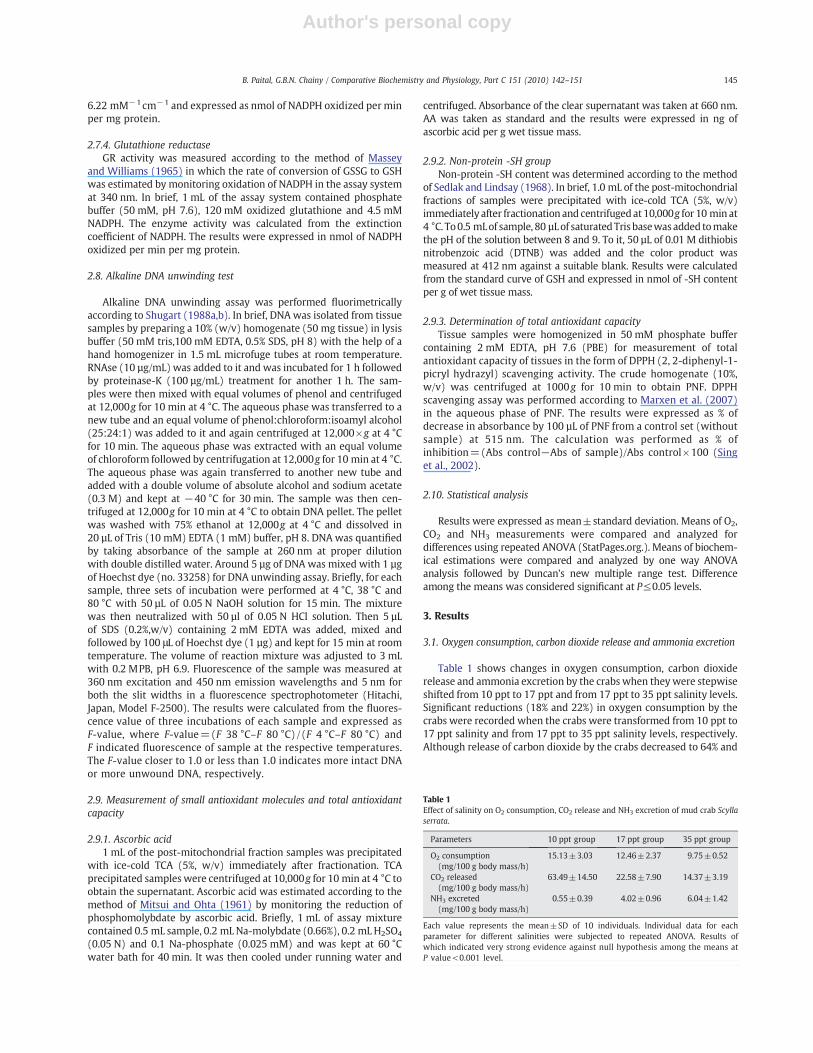
Table 1 shows changes in oxygen consumption, carbon dioxiderelease and ammonia excretion by the crabs when they were stepwiseshifted from 10 ppt to 17 ppt and from 17 ppt to 35 ppt salinity levels.Significant reductions (18% and 22%) in oxygen consumption by thecrabs were recorded when the crabs were transformed from 10 ppt to17 ppt salinity and from 17 ppt to 35 ppt salinity levels, respectively.Although release of carbon dioxide by the crabs decreased to 64% and
Table 1Effect of salinity on O2 consumption, CO2 release and NH3 excretion of mud crab Scyllaserrata.
Parameters 10 ppt group 17 ppt group 35 ppt group
O2 consumption(mg/100 g body mass/h)
15.13±3.03 12.46±2.37 9.75±0.52
CO2 released(mg/100 g body mass/h)
63.49±14.50 22.58±7.90 14.37±3.19
NH3 excreted(mg/100 g body mass/h)
0.55±0.39 4.02±0.96 6.04±1.42
Each value represents the mean±SD of 10 individuals. Individual data for eachparameter for different salinities were subjected to repeated ANOVA. Results ofwhich indicated very strong evidence against null hypothesis among the means atP value<0.001 level.
145B. Paital, G.B.N. Chainy / Comparative Biochemistry and Physiology, Part C 151 (2010) 142–151

Author's personal copy
37% when they were stepwise transformed from 10 ppt to 17 ppt andfrom 17 ppt to 35 ppt salinity levels, respectively; the differencebetween values of 17 ppt and 35 ppt was not statistically significant.On the other hand, it was noticed that total ammonia excretion by thecrabs elevated to around 630% and 50% when they were shifted from10 ppt to 17 ppt and from 17 ppt to 35 ppt salinity levels, respectively.
3.2. Oxidative stress parameters
Table 2 depicts changes in various oxidative stress parameters inabdominal muscle, hepatopancreas and gills of adult male crabsmaintained at different salinities.
3.2.1. Lipid peroxidationA gradual elevation in LPx value of abdominal muscle of the crabs
was recorded in response to increasing salinity. Lipid peroxidationlevel in abdominal muscle of the crabs was 150% and 500% higher at17 ppt and 35 ppt salinity in comparison to 10 ppt salinity, respec-tively. Although the LPx value of hepatopancreas of the crabs did notvary significantly between 10 ppt and 17 ppt salinity levels, its valuewas 45% higher at 35 ppt salinity level than that of 10 ppt and 17 ppt.In case of gills of the crabs, LPx value was 45% and 110% higher at17 ppt and 35 ppt in comparison with 10 ppt salinity level,respectively.
3.2.2. Protein carbonyl contentIn all the three tissues viz. abdominal muscle, hepatopancreas and
gills of the crabs, protein carbonyl content was significantly higher at35 ppt salinity in comparison with 10 ppt salinity. However, the
magnitude of change in protein carbonyl content was different fromone tissue to the other. Protein carbonyl content of abdominal muscleof the crabs at 35 ppt was 234% and 182% higher than 10 ppt and17 ppt salinity groups, respectively. It was further noticed that theprotein carbonyl content of hepatopancreas of crabs of the 35 pptgroup was 53% and 23% higher than that of 10 ppt and 17 ppt groups,respectively. Protein carbonyl content of gills of the crabs was 29% and53% higher at 17 ppt and 35 ppt groups in comparison with 10 pptsalinity group, respectively.
3.2.3. Hydrogen peroxide contentA small but a significant decrease (12%) in hydrogen peroxide
content of abdominal muscle of the crabs of 35 ppt salinity group wasrecorded in comparisonwith 10 ppt salinity group. On the other hand,a significant 22% and 41% increase in hydrogen peroxide contentof hepatopancreas of the crabs of 35 ppt group were recorded incomparison with 10 ppt and 17 ppt groups, respectively. Similarly,29% and 23% elevations in hydrogen peroxide content of gills of thecrabs of 35 ppt groupwas noted in comparisonwith 10 ppt and 17 pptgroups, respectively.
3.2.3. DNA unwinding testThe DNA unwinding value of the three tissues of crabs studied did
not change in response to changing salinity.
3.3. Antioxidant enzymes
Table 3 shows changes in levels of various antioxidant enzymes ofabdominal muscle, hepatopancreas and gills of adult male crabs inresponse to different salinities.
3.3.1. Superoxide dismutaseIt was noticed that the level of SOD in abdominal muscle of the
crabs decreased gradually as the level of salinity increased. The activ-ity of the enzyme in abdominal muscle tissue of the crabs diminished55% and 85% in 17 ppt and 35 ppt groups in comparison with 10 pptgroup, respectively. In case of hepatopancreas, the enzyme activity ofthe crabs at 17 ppt salinity was observed to be higher (56%) than thatof 10 ppt group. But a significant 22% and 51% decrease in its levelwere recorded in the tissues of crabs of 35 ppt groups in comparisonwith 10 ppt and 17 ppt groups, respectively. On the other hand, thelevel of the enzyme was 22% lower in gills of the crabs of 17 pptgroups in comparison with 10 ppt and 35 ppt groups, respectively.
3.3.2. CatalaseThe response of catalase to different salinities varied from one
tissue to another. Significant 119% and 155% increments in the level ofcatalase of abdominal muscle of the crabs of 35 ppt group were noted
Table 2Effect of salinity on lipid peroxidation (LPx), protein carbonyl (PC) and hydrogenperoxide (H2O2) and DNA unwinding value of gills and abdominal muscle (M),hepatopancreas (HP) and gills (G) of mud crab Scylla serrata.
Parameters 10 ppt group 17 ppt group 35 ppt group
LPx (nmol TBARS/mg protein) M 0.04±0.01a 0.1±0.02b 0.24±0.02c
HP 0.90±0.23a 0.89±0.22a 1.32±0.16b
G 0.13±0.04a 0.18±0.06b 0.28±0.05c
PC (nmol PC/mg protein) M 1.82±0.72a 2.15±0.89a 6.08±1.21b
HP 2.46±0.13a 2.67±0.31a 3.29±0.60b
G 1.69±0.26a 2.19±0.71b 2.60±0.71b
H2O2 (μg/g wet tissue) M 0.91±0.04a 0.97±0.04b 0.80±0.04c
HP 8.19±1.51a 7.11±1.36a 10.05±1.27b
G 1.22±0.48a 1.28±0.02a 1.58±0.13b
DNA UW (F-value) M 0.76±0.14 0.88±0.10 0.85±0.12HP 0.81±0.12 0.85±0.08 0.84±0.06G 0.80±0.89 0.81±0.09 0.80±0.08
Each value represents the mean±SD of 9 individuals. Superscripts (a, b and c) indicatestatistical significance within the groups at P≤0.05 level. Groups with differentsuperscripts are statistically different from each other.
Table 3Effect of salinity on superoxide dismutase (SOD), catalase (CAT), glutathione peroxidase (GPx) and glutathione reductase (GR) activities of abdominal muscle (M), hepatopancreas(HP) and gills (G) of mud crab Scylla serrata.
Parameters 10 ppt group 17 ppt group 35 ppt group
SOD (U/mg protein) M 2.64±0.35a 1.20±0.18b 0.38±0.14c
HP 4.31±0.63a 6.74±0.73b 3.33±0.79c
G 1.42±0.50a 0.83±0.10b 1.43±0.26a
CAT (nKAT/mg protein) M 62.58±20.20a 53.54±19.98a 136.75±23.09b
HP 513.75±89.59a 721.0±93.85b 535.43±54.18a
G 489.02±61.19a 268.74±38.17b 69.66±21.06c
GPx(nmol of NADPH oxidized/min/mg protein)
M 18.29±4.80 13.98±5.15 13.55±6.59HP 140.0±13.02a 205.57±86.73b 97.16±14.41c
G 65.69±18.04a 45.84±20.42b 45.58±5.47b
GR(nmol of NADPH oxidized/min/mg protein)
M 0.32±0.11ab 0.19±0.06a 0.46±0.23b
HP 1.41±0.42a 1.17±0.62a 0.71±0.26b
G 0.43±0.12a 0.50±0.33a 0.70±0.13b
Each value represents the mean±SD of 9 individuals. Superscripts (a, b and c) indicate statistical significance within the groups at P≤0.05 level. Groups with different superscriptsare statistically different from each other.
146 B. Paital, G.B.N. Chainy / Comparative Biochemistry and Physiology, Part C 151 (2010) 142–151

Author's personal copy
in comparison with 10 ppt and 17 ppt groups, respectively. In case ofhepatopancreas, a 40% increase in enzyme activity was recorded forthe crabs of 17 ppt than that of 10 ppt group. But the activity of theenzyme was observed to be low (25%) in the tissue of the crabs of35 ppt group in comparisonwith 17 ppt group. The enzyme activity ingills of the crabs gradually decreased as the level of salinity increased.It was 85% and 74% lower in gills of the crabs of 35 ppt group incomparison with 10 ppt and 17 ppt groups, respectively.
3.3.3. Glutathione peroxidaseGlutathione peroxidase activity of the abdominal muscle of the
crabs did not change significantly in response to changing salinity. Asignificant increase (46%) in the enzyme activity of hepatopancreaswas recorded in 17 ppt group than that of crabs of 10 ppt group.However, the enzyme level was 53% lower in the tissue of the crabs of35 ppt group than that of 17 ppt group. It was observed that theenzyme level in gills of the crabs decreased to 30% in the case of 17 pptgroup in comparison with 10 ppt group and did not change further in35 ppt group.
3.3.4. Glutathione reductaseThe response of glutathione reductase to changing salinity varies
from one tissue to another. Although the activity of the enzyme inabdominal muscle of the crabs did not vary significantly between10 ppt and 17 ppt groups, it was significantly high (142%) in the crabsof 35 ppt groups than that of 17 ppt group. It was noticed that theenzyme activity was 49% and 39% lower in hepatopancreas of thecrabs of 35 ppt in comparison with 10 ppt and 17 ppt groups,respectively. On the other hand, the enzyme activity was 62% and40% higher in gills of the crabs of 35 ppt group than that of 10 ppt and17 ppt groups, respectively.
3.4. Small antioxidants and total antioxidant capacity (Table 4)
3.4.1. Free -SH groupThe non-protein free -SH group content of abdominal muscle of
the crabs of 35 ppt groupwas 20% higher than that of 10 ppt group. Onthe other hand, a small but significant decrease in -SH group contentof hepatopancreas of 17 ppt group of the crabs was recorded incomparison wtih 10 and 35 ppt groups. A 67% decrease in -SH contentof gills of the crabs of 35 ppt group was recorded in comparison with10 ppt and 17 ppt groups.
3.4.2. Ascorbic acidIt was observed that the ascorbic acid content of abdominal muscle
of the crabs of 17 ppt group was 764% higher than that of 10 pptgroup. The value decreased to 53% in the tissue of 35 ppt group thanthat of 17 ppt group. Although a similar pattern was also observed inthe case of hepatopancreas, the increase in the case of 17 ppt group incomparison with 10 ppt was only 45%. A significant 12% and 16%decrease in ascorbic acid content of gills of the crabs of 35 ppt groupwere recorded in comparisonwith 10 and 17 ppt groups, respectively.
3.4.3. Total antioxidant capacityResult of the present experiment depicts that all the three tissues
exhibited a similar pattern. DPPH scavenging activity was recorded tobe the lowest at 10 ppt which reached to a maximum value at 35 pptsalinity. A positive correlation was observed between salinity andtotal antioxidant capacity of tissues of crabs (figure not given).
4. Discussion
A distinct tissue specific pattern was noticed for antioxidantenzymes in S. serrata that were acclimatized in the laboratory at17 ppt of salinity. Similar tissue variation regarding activities of anti-oxidant enzymes was reported for several other species belonging to
phylum Arthropoda: M. rosenbergi (Dandapat et al., 2000), Charybdisjaponica (Pan and Zhang, 2006), M. malcolmsoni (Arun and Subrama-nian, 1998), Chasmagnathus granulata (now Neohelice granulata)(Maciel et al., 2004), and Orconectus limosus (Borkovic et al., 2008).Our results regarding tissue distribution of antioxidant enzymes in thecase of S. serrata are different from that reported for another estuarinecrab species C. granulata. It is reported that gills of C. granulata havehigher SOD (de Oliveira et al., 2005, 2006) and catalase (Maciel et al.,2004) activities than hepatopancreas. On the other hand, GR activity ingills of C. granulata was found to be low in comparison to hepatopan-creas (Maciel et al., 2004). It is noticed that catalase and GPx activitiesin hepatopancreas of crayfish (Orconectus limosus) were high in com-parison to gills and muscle (Borkovic et al., 2008). The same authorsnoted lower SOD and GR activities in hepatopancreas of the samespecies than gills and muscle tissues (Borkovic et al., 2008). This sug-gests that antioxidant defense strategies employed by tissues ofcrustaceans not only vary among themselves but also vary from onespecies to other.
In the present study, crabs were acclimatized at 17 ppt of salinityin the laboratory before they were shifted to low i.e. 10 ppt or high i.e.35 ppt salinity. It was observed that oxygen consumption by the crabsincreased (around 30%) at 10 ppt than that of 17 ppt. On the otherhand, oxygen consumption by the crabs decreased (around 30%)at 35 ppt in comparison with 17 ppt. Therefore, the possibility ofexperiencing a transition state of internal organs of crabs fromnormoxia to hypoxia (at 35 ppt) or to hyperoxia (at 10 ppt) couldnot be excluded as a consequence of decrease or increase in oxygenconsumption rate, respectively. Our results are in good agreementwith earlier results for other euryhaline crab species which exhibitedelevated (Engel and Eggert, 1974; Engel et al., 1975; Sabourin, 1983)or reduced (Findley et al., 1978; Chen and Chia, 1996b; Robles et al.,2002) oxygen consumption rate when they were acclimated to lowor high salinities. Increased consumption of oxygen by adult S. serrataat low salinity suggests that a higher energy demand by the organismmay be due to active pumping of ions from environment tohemolymph. It is now a well established fact that most euryhalinecrab species are hyperosmoregulators at low salinities, activelypumping ions from sea water into hemolymph (Mantel and Farmer,1983; Lucu, 1990). Reduced rate of oxygen consumption and CO2
production by crabs at high salinity indicate low oxidativemetabolismof the organism to regulate its hyperionic state.
Ammonia, amino acids and urea are the three principal end prod-ucts of protein metabolism that are released to the environmentthrough gills in decapods (Regnault, 1987). Chen and Chia (1996a)reported that a shift in nitrogen excretion pattern from ammoniote-lism to ureotelism takes place in S. serrata when crabs were movedfrom lower salinity to higher salinity. Chen and Chia (1996a) observeda decrease in ammonia-N release at high salinity. On the contrary, wehave noted an increase in ammonia release by crabs at higher salinity.The discrepancies between our results and that of Chen and Chia(1996a) may be due to different experimental designing. Chen andChia (1996a) have kept crabs in different salinities for 7 days only intheir experiments. The duration of exposure to higher salinity maynot be sufficient enough for crabs to acclimatize to salinities and maybe one of the reasons for change in their nitrogen excretion patternfrom ammonotelic to ureotelic. The increase release of ammoniaat higher salinity may be attributed to high protein catabolism.
The hepatopancreas in crustaceans is a major organ which is main-ly associated with diverse metabolic activities ranging from supply ofnutrition to ovary, to digestion and absorption (Sureshkumar andKurup, 1999; Sang and Fotedar, 2004; Comoglio et al., 2008). It canbe considered metabolically more active than gills and muscle tissue. Itis difficult to explain the physiological basis for the observed decreasein activities of antioxidant enzymes in hepatopancreas of mud crabsat 10 ppt without any change in oxidative stress markers. It is possiblethat production of ROS in hepatopancreas is reduced at 10 ppt salinity
147B. Paital, G.B.N. Chainy / Comparative Biochemistry and Physiology, Part C 151 (2010) 142–151
Author's personal copy
whichmight have caused decrease in synthesis of antioxidant enzymes.It is interesting tonote that a significantdecrease in lipid peroxidation inabdominal muscle (60%) and gills (28%) and in protein carbonylation ingills (33%) was recorded at 10 ppt salinity. Observed increase in SODactivity in abdominal muscle and SOD, catalase and GPx in gills may beresponsible for lowering LPx values. It contributes to the mechanism tounderstand that due to higher oxygen consumption by the organism atlow salinity, these two tissues might have experienced higher oxygentension and consumption which might have resulted in enhancedproduction of ROS. Consequently, levels of antioxidant enzyme wereenhanced to counteract the ROS. In crabs, gills are primarily responsiblefor respiration, acid–base balance and osmotic and ionic regulation(Henry and Cameron, 1983; Henry, 1984; Lucu, 1990). Furthermore ithas been noted that a battery of biochemical and physiological changestakes place in gills of euryhaline crabs in response to low salinity stressin order to maintain osmotic and ionic homeostasis (Piller et al., 1995).It has been reported that exposure of cells to hyperoxia resulted inincrease generation of ROS by enzymatic and non-enzymatic mechan-isms (Freeman and Crapo, 1981). The increased consumption of oxygenby gills in crabs at low salinity would have enhanced production ofsuperoxide radicals. In order to neutralize the increase of superoxideradicals, tissue level SOD activity might have induced. The consequenceof increased SOD activity would have resulted in more production ofhydrogen peroxide. Hydrogen peroxide is highly toxic to cells andsubsequently generates highly toxic and reactive hydroxyl radicals byclassical Fenton reactions. Consequently, activities of both the enzymeselevated in gills of crabs to keep the hydrogen peroxide level at basallevel. Generally, in almost all animals hydrogen peroxide is neutralizedby catalase and GPx enzymes. Nevertheless, neutralization of hydrogenperoxide by muscles and gills of mud crabs was quite distinct. In theformer tissue, where only GPx plays an active role in neutralizinghydrogen peroxide, both the enzymes are required for the later tissue.It is evident from the present study that enzymatic neutralizationmechanism of ROS at low salinity is different among the tissues of mudcrabs.
It is well known that the rate of production of ROS at themitochondrial level in many biological systems is proportional tooxygen consumption and to mitochondrial metabolic rate. But in ourexperiment the decrease in oxygen consumption in crabs at 35 pptsalinity have resulted in higher production of ROS. Several studieshave clearly established that deprivation of oxygen to cells or tissueshas resulted in generation of ROS (Vanden Hoek et al., 1997; Arroyoet al., 1987; Bolli et al., 1988; Ward, 2006). Several reasons have beendescribed as the cause for increased generation of ROS duringhypoxia such as residual oxygen level in tissues (de Oliveira et al.,2006) and switch over of biochemical reaction (Storey and Storey,1990; Hochachka and Lutz, 2001). Limiting oxygen supply to crabsduring high salinity will slow down the electron transport in thelower part of the ETC propagating ROS formation from downwardelectron carriers such as complex III ubiquinone (Brand 2000; Staniekand Nohl, 2000). There are several reports that confirmed increasedproduction of ROS during hypoxia condition in marine invertebrates(Abele and Puntarulo, 2004). Among the three organs, hepatopan-creas is the one most subjected to oxidative stress during transitionof crabs from 17 ppt to 35 ppt salinity as evident by augmentation ofLPx, PC and hydrogen peroxide levels. Lipid peroxidation and proteincarbonylation are considered as consequences of oxidation of lipidsand proteins by ROS (Halliwell and Gutteridge, 2001). Therefore,both parameters i.e. lipid peroxidation and protein carbonylation areconsidered as indices of tissue oxidative stress in almost all animalsincluding that of estuarine invertebrates (Monserrat et al., 2007). Inthe present study, elevated levels of TBARS and PC contents in tissuesof S. serrata at high salinity suggest induction of oxidative stress.
Not much information is available in the literature on biochemicalchanges taking place in hepatopancreas of S. serrata in response tochanging environmental factors in general and salinity in particular. A
significant decrease in SOD activity was observed in hepatopancreasof crabs at high salinity which is accompanied with reduced catalaseand GPx activities may explain for the observed high hydrogenperoxide level in the tissue. GPx is also considered to remove lipidperoxides besides hydrogen peroxide (Fernandez-Diaz et al., 2006).The physiological significance of reduced activities of antioxidantenzymes at high salinity is difficult to explain at present. However,it may be attributed to low oxidative metabolism of the crabs asconsumption of oxygen by crabs decreased during its exposure to highsalinity.
SOD activity of gills of crabs elevated when the crabs weresubjected to high salinity condition. High salinity stress might haveproduced a large amount of ROS in gills which could have inducedSOD to scavenge superoxide radicals. However, decrease in catalaseactivity along with GPx at high salinity might not be able to removeexcessive hydrogen peroxide causing enhancement of lipid peroxidelevel in the tissue. Gills, the principal organ for respiration in crabs, aredirectly exposed to the surrounding environment. It also maintainsthe osmotic balance of body fluid. Several biochemical adaptations ingills of crabs to changing salinity are reported. For example, Na+–K+
ATPase (Cryptograpus angulatus: Lopez-Mananes et al., 2002) andcarbonic anhydrase (Carcinus maenus: Henry et al., 2003) are reportedto be influenced by environmental salinity.
In comparison with hepatopancreas and gills, abdominal muscle ismetabolically less active. SOD activity of the abdominal muscledecreased with high salinity. The present results provide an idea tosupport the fact that at high salinity, the metabolic activity of theabdominal muscle may be very low. Consequently, less amount ofsuperoxide radicals are generated. Hence, the tissue needs low SODactivity. Decreased level of SOD activity along with enhanced level ofcatalase in abdominal muscle at high salinity may be the reason forreduced hydrogen peroxide level in the tissue. It is here to note thatGR activity of the muscle increased in response to high salinity. GRregenerates GSH from GSSG which is formed by GPx. The biochemicaladaptations of muscles of S. serrata to varying environmental salinitiesare relatively unknown except for a few studies which have shownchange in alkaline phosphates activity (Pinoni and Lopez-Mananes,2004) andmobilization of lipids frommuscles (Luvizotto-Santos et al.,2003) of the euryhaline crab Cyrtograpus angulatus; enhancedarginine flux from muscle tissues of another crab species Callinectessapidus (Holt and Kinsey, 2002) in response to low salinity andincreased myosin ATPase activity of the flexor muscle of S. serrata inresponse to high salinity (Krishnamoorthy and Venkatramiah, 1969).
Results of the present study give more insight to think about themolecular mechanism by which salinity modulates antioxidantenzyme levels in tissues of mud crabs. It is to note here that post-translational regulation of SOD, catalase and GPx does not occur(Hermes-Lima, 2004). Brouwer et al. (2004) using microarraysconstructed from cDNA and subtractive hybridization studies onhepatopancreas of Callinectes sapidus concluded that hypoxia resultedin down regulation of several genes. Therefore, alteration in activitiesof antioxidant enzymes in tissues of crabs in response to salinity mustreflect alteration in their respective rate of expression and/ordegradation is to be a matter of future investigation.
In this study, the effect of salinity on two non-enzymatic anti-oxidant molecules in different tissues of mud crab is investigated.They are non-protein SH and ascorbic acid. GSH is also reported to actas an effective antioxidant inmarine animal system besides acting as asubstrate for GPx to neutralize hydrogen peroxide produced by SODenzyme. Also it acts as a reductant in conjugation with electrophilicsubstances. Therefore, GSH level may reflect the detoxification abilityof an organism (Kovacevic et al., 2008). It is here to mention that wehave measured non-protein free -SH group in the samples which maynot represent the true value for GSH. Nevertheless, in several studiesmeasurement of -SH group in this fraction is used to represent GSH(Monteiro et al., 2009; Sk and Bhattacharya, 2006). A significant
148 B. Paital, G.B.N. Chainy / Comparative Biochemistry and Physiology, Part C 151 (2010) 142–151

Author's personal copy
decrease (60%) in non-protein -SH level in gills but not in other tissueswas recorded at high salinity when oxygen consumption is less andLPx level of tissue is high. It indicates that non-protein -SHmight havebeen utilized by the tissue to remove hydrogen peroxides and otherlipoperoxides during exposure of crabs to high salinity by GSTenzymes. de Oliveira et al. (2005, 2006) have observed that GSTactivity of gills but not of hepatopancreas of C. granulata elevated inresponse to anoxia. It has been observed that the concentration of GSHwas maximum during nocturnal periods in gills of C. granulata and isparalleled by high oxygen consumption and high LPx level (Macielet al., 2004). Same authors have also noticed unaltered non-protein-SH group in hepatopancreaswith changing oxygen consumption. Thedifferences in response of non-protein -SH of tissues of crabs to highsalinity may be due to their specific metabolic characteristics accord-ing to their functions.
Ascorbic acid is a potent antioxidant which scavenges reactiveradicals such as hydroxyl, perhydroxyl, peroxyl and nitric oxide(Halliwell and Gutteridge, 2001), therefore, it serves as an importantprotectivemeasure against free radicals (Bendich et al., 1986; Karakoeet al., 1997). AA is believed to regenerate vitamin E from its oxidizedform (Wells et al., 1992) thereby raises the antioxidant status of cells.The significant decrease in AA content in muscle and hepatopancreasof mud crabs at high as well as low salinity signifies its importantrole in combating stress compared with the non-protein -SH group(Table 4). A small decrease in ascorbic acid content in gills in responseto high salinity suggests its obscure role in combating oxidative stressin the tissue. To the best of our knowledge there is no informationavailable in the literature on the effect of salinity on tissue AA contentof crabs.
DPPH is an established reagent in evaluating antioxidant proper-ties of several biological compounds such as amines, phenols, vita-mins, plant extracts etc. (Ionita, 2005). Results of our present workrevealed that DPPH radical scavenging capacity of crab tissues wasmaximum at high salinity in comparison with low salinity at thesame temperature. Therefore, it is possible that crabs that inhabitintertidal regions may develop some physiological mechanism tocounteract salinity induced stress. Interestingly, a positive correlationwas observed between salinity and DPPH radical scavenging capacityfor all three tissues of crabs studied (figure not given). To the best ofour knowledge no report is available in the literature which showseffect of salinity on DPPH radical scavenging activity inmud crabs. Theexact mechanism by which total antioxidant capacity of tissues ofcrabs elevated is not clear and needs further investigation.
Cells, made hypertonic with elevated levels of salt like NaCl,experience double stranded DNA breakage (Kultz and Chakravarty,2001). It is reported that exposure of marine animals to variousstressful conditions causes double stranded DNA damage (Rao et al.,2001; Verlecar et al., 2008) which is contributed by increasedproduction of ROS (Imlay and Linn, 1988; Almeida et al., 2007).Alkaline DNA unwinding test indicated that changing salinity did not
cause any damage to DNA of the tissues of crabs. It may be inferredthat the magnitude of generation of ROS in S. serrata at high salinitywas not able to surpass the threshold value to cause DNA damage atmolecular level.
In conclusion, results of the present study suggest that themagnitude of oxidative stress (measured in terms of lipid peroxida-tion and protein carbonylation) and status of antioxidant defensecomplexes (both enzymatic and non-enzymatic) of S. serrata aretissue specific and their responses to changing salinity vary from onetissue to the other. However, high salinity (35 ppt) induces moreoxidative stress in the tissues of S. serrata in comparison with lowsalinities (10 ppt and 17 ppt).
Acknowledgements
The work was supported by a financial grant of the UniversityGrants Commission, scheme no. F. No.31-284/2005 (SR) to GBNC. BRPis extremely grateful to the University Grants Commission, New Delhifor providing a fellowship under the RFSMS scheme to theDepartment of Zoology, Utkal University. The authors are highlythankful to the Head of the Departments of Zoology and Biotechnol-ogy, Utkal University, India, for providing the laboratory facilities.
References
Abele, D., Puntarulo, S., 2004. Formation of reactive species and induction of antioxidantdefence systems in polar and temperate marine invertebrates and fish. Comp.Biochem. Physiol. A 138, 405–415.
Aebi, H., 1974. Catalase. In: Bergeyer, H.U. (Ed.), Methods of Enzymatic Analysis, Vol. 2.Academic Press, New York, pp. 673–678.
Almeida, E.A., Bainy, A.C.D., de Melo, L.A.P., Martinez, G.R., Miyamoto, S., Onuki, J.,Barbosa, L.F., Garcia, C.C.M., Prado, F.M., Ronsein, G.E., Sigolo, C.A., Brochini, C.B.,Martins, A.M.G., de Medeiros, M.H.G., Di Mascio, P., 2007. Oxidative stress in Pernaperna and other bivalves as indicators of environmental stress in the Brazilianmarine environment: antioxidants, lipid peroxidation and DNA damage. Comp.Biochem. Physiol. A 146, 588–600.
Anguelov, A., Chichovska, M., 2004. Effect of paraquat intoxication and ambroxoltreatment on hydrogen peroxide production and lipid peroxidation in selectedorgans of rat. Verterinarski Archiv. 74, 141–155.
APHA, 1985. Standard Methods for the Examination of Water and Wastewater, 16thedition. Districts of Columbia, Washington.
Arroyo, C.M., Kramer, J.H., Leiboff, R.H., Mergner, G.W., Dickens, B.F., Weglicki, W.B.,1987. Spin trapping of oxygen and carbon-centered free radicals in ischemic caninemyocardium. Free Radic. Biol. Med. 3, 313–316.
Arun, S., Subramanian, P., 1998. Antioxidant enzymes activity in subcellular fraction offreshwater prawns M. malcolmsonii and M. lamarrei lamarrei. App. Biochem. Biotech.75, 187–192.
Bendich, A., Machlin, L.J., Scandurra, O., Burton, G.W., Wayner, D.D.M., 1986. Theantioxidant role of vitamin C. Adv. Free Radic. Biol. Med. 2, 419–444.
Bolli, R., Patel, B.S., Jeroudi, M.O., Lat, E.K., McCay, P.B., 1988. Demonstration of freeradical generation in “stunned”myocardium of intact dogs with the use of spin trapα-phenyl N-tert-butyl nitrone. J. Clin. Invest. 82, 476–485.
Borkovic, S.S., Pavlovic, S.Z., Kovacevic, T.B., Stajn, A.S., Petrovic, V.M., Saicic, Z.S., 2008.Antioxidant defence enzyme activities in hepatopancreas, gills and muscle of spinycheek crayfish (Orconectes limosus) from the River Danube. Comp. Biochem. Physiol. C147, 122–128.
Table 4Effect of salinity on free sulphydril group (-SH), ascorbic acid (AA) and total antioxidant capacity of abdominal muscle (M), hepatopancreas (HP) and gills (G) of mud crab Scyllaserrata.
Parameters 10 ppt group 17 ppt group 35 ppt group
-SH(nmol/g wet tissue)
M 21.36±1.21a 22.27±0.88ab 25.74±3.40b
HP 29.71±3.83a 23.86±3.95b 29.37±1.93a
G 27.46±2.76a 27.81±1.45a 09.06±2.12b
AA(ng/g wet tissue)
M 17.1±3.45a 147.07±22.76b 69.54±18.74c
HP 105.69±28.11a 154.57±17.25b 71.12±9.03c
G 201.0±20.17a 211.0±27.04a 177.0±24.97b
Total antioxidant capacity⁎(% of inhibition)
M 11.02±2.68a 13.33±2.56a 16.06±1.96b
HP 16.06±2.62a 20.27±1.95b 23.76±3.02c
G 15.12±1.25a 16.90±0.99b 21.74±0.86c
⁎Data are expressed as % of inhibition of absorbance of DPPH of sample tubes from absorbance of DPPH from control tubes having no sample.Each value represents the mean±SD of 9 individuals. Superscript (a, b and c) indicate statistical significance within the groups at P≤0.05 level. Groups having different superscriptsare statistically different from each other.
149B. Paital, G.B.N. Chainy / Comparative Biochemistry and Physiology, Part C 151 (2010) 142–151
Author's personal copy
Bradford, M.M., 1976. A rapid and sensitive method for the quantification of microgramquantities of protein utilizing the principle of protein-dye binding. Anal. Biochem.72, 248–254.
Brand, M.D., 2000. Uncoupling to survive? The role of mitochondrial inefficiency inageing. Exp. Gerentol. 35, 811–820.
Brouwer, M., Larkin, P., Brown-Peterson, N., King, C., Manning, S., Denslow, N., 2004.Effects of hypoxia on gene and protein expression in the blue crab, Callinectessapidus. Mar. Environ. Res. 58, 787–792.
Chen, J., Chia, P., 1996a. Haemolymph ammonia and urea and nitrogenous excretion ofScylla serrata at different temperature and salinity levels. Mar. Ecol. Prog. Ser. 139,119–125.
Chen, J., Chia, P., 1996b. Oxygen uptake and nitrogen excretion of juvenile Scylla serrataat different temperature and salinity levels. J. Crustacean Biol. 16, 437–442.
Cohen, G., Dembiec, D., Marcus, J., 1970. Measurement of catalase activity in tissueextract. Anal. Biochem. 34, 30–38.
Comoglio, L., Goldsmit, J., Amin, O., 2008. Starvation effects on physiological parametersand biochemical composition of thehepatopancreas of the southernking crab Lithodessantolla (Molina, 1782). Revista de Biología Marina y Oceanografía. 43, 345–353.
Dandapat, J., Chainy, G.B.N., Rao, K.J., 2000. Dietary vitamin-E modulates antioxidantdefence system in giant freshwater prawn, Macrobrachium resenbergii. Comp.Biochem. Physiol. C 127, 101–115.
Das, K., Samanta, L., Chainy, G.B.N., 2000. A modified spectrophotometric assay ofsuperoxide dismutase using nitrite formation by super oxide radicals. IndianJ. Biochem. Biophys. 37, 201–204.
de Oliveira, U.O., Araujo, A.S.R., Bello-Klein, A., Da Silva, R.S.M., Kucharski, L.C., 2005.Effects of environmental anoxia and different periods of reoxygenation on theoxidative balance in the anterior and posterior gills of the estuarine crab Chas-magnathus granulata. Comp. Biochem. Physiol. B 140, 51–57.
de Oliveira, U.O., Bello-Klein, A., Kucharski, L.C., 2006. Oxidative balance and immunode-tection of antioxidant enzymes in the hepatopancreas of the crab Chasmagnathusgranulata subjected to anoxia and reoxygenation. Can. J. Zool. 84, 677–684.
Engel, D.W., Eggert, L.D., 1974. The effect of salinity and sex on the respiration rates ofexcised gills of the blue crab. Callinectes sapidus. Comp. Biochem. Physiol. A 47,1005–1011.
Engel, D.W., Ferguson, R.L., Eggert, L.D., 1975. Respiration rates and ATP concentrationsin the excised gills of the blue crab as a function of salinity. Comp. Biochem. Physiol.A 52, 669–673.
Fernandez-Diaz, C., Kopecka, J., Canavate, J.P., Sarasquete, C., Sole, M., 2006. Variationson development and stress defences in Solea senegalensis larvae fed on live andmicroencapsulated diets. Aquaculture 251, 573–584.
Findley, A.M., Belisle, B.W., Stickle, W.B., 1978. Effect of salinity fluctuations on therespiration rate of the southern oyster drill Thais haemastoma and the blue crabCallinectes sapidus. Mar. Biol. 49, 59–67.
Freeman, B.A., Crapo, J.D., 1981. Hyperoxia increases oxygen radical production in ratlungs and lung mitochondria. J. Biol. Chem. 256, 10986–10992.
Gamble, S.C., Goldfarb, P.S., Porte, C., 1995. Glutathione peroxidase and otherantioxidant enzyme function in marine invertebrates (Mytilus edulis, Pectenmaximus, Carcinus maenas and Asterias rubens). Mar. Environ. Res. 39, 191–195.
Hai, T.N., Hassan, A.B., Law, A.T., Shazili, N.A.M., 1998. Effect of reduced water salinity onjuveniles of the mud crab Scylla serrata. International Forum on the Culture ofPortunid Crabs.1–4 December, 1998. Boracay, Philippines. Asian Fisheries Society,Quezon City, Philippines, p. 57.
Halliwell, B., Gutteridge, J.M.C., 2001. Free Radicals in Biology and Medicine, 3rd ed.Oxford University Press, New York.
Hamasaki, K., 2003. Effects of temperature on the egg incubation period, survival anddevelopment of larvae of the mud crab Scylla serrata (Forskal) (Brachyura:Portunidae) reared in the laboratory. Aquaculture 219, 561–572.
Henry, R.P., 1984. The role of carbonic anhydrase in blood ion and acid–base regulation.Am. Zool. 24, 241–251.
Henry, R.P., Cameron, J.N., 1983. The role of carbonic anhydrase in respiration, ionregulation and acid base balance in the aquatic crab Callinectes sapidus and theterrestrial crab Gecarcinus lateralis. J. Exp. Biol. 103, 205–223.
Henry, R.P., Gehnrich, S., Weihrauch, D., Towle, W.D., 2003. Salinity-mediated carbonicanhydrase induction in the gills of the euryhaline green crab, Carcinus maenas.Comp. Biochem. Physiol. A 136, 243–258.
Hermes-Lima, M., 2004. Oxygen in biology and biochemistry: role of free radicals. In:Storey, K.B. (Ed.), Functional metabolism: regulation and adaptation. John Wileyand Sons, pp. 319–368.
Hill, B.J., 1974. Salinity and temperature tolerance of the zoea of the Portunidae crabScylla serrata. Mar. Biol. 25, 21–24.
Hochachka, P.W., Lutz, P.L., 2001. Mechanism, origin, and evolution of anoxia tolerancein animals. Comp. Biochem. Physiol. B 130, 435–459.
Holt, S.M., Kinsey, S.T., 2002. Osmotic effects on arginine kinase function in livingmuscle of the blue crab Callinectes sapidus. J. Exp. Biol. 205, 1775–1785.
Imlay, J.A., Linn, S., 1988.DNAdamageandoxygen radical toxicity. Science. 240, 1302–1309.Ionita, P., 2005. Is DPPH stable free radical a good scavenger for oxygen active species?
Chem. Pap. 59, 11–16.Karakoe, F.T., Hewer, A., Philips, D.H., Gaines, A.F., Yuregir, G., 1997. Biomarkers of
marine pollution observed in species of mullet living in two eastern Mediterraneanharbors. Biomarkers. 2, 303–309.
Kong, X., Wang, G., Ai, C., Li, S., 2004. Comparative study on content of reactive oxygenspecies and activity of antisuperoxide anion free radicals in different organs andtissues of mud crab, Scylla serrata (in Chinese). Mar. Sci. 28, 1–4.
Kong, X., Wang, G., Li, S., Ai, C., 2005. Antioxidant effects and ATPase activity changesin hepatopancreas of mud crab Scylla serrata under low temperature acclimation(in Chinese). J. Fish Sci. China. 12, 708–713.
Kong, X., Wang, G., Li, S., 2007. Antioxidation and ATPase activity in the gill of mud crabScylla serrata under cold stress. Chin. J. Oceanol. Limnol. 25, 221–226.
Kong, X., Wang, G., Li, S., 2008. Seasonal variations of ATPase activity and antioxidantdefenses in gills of the mud crab Scylla serrata (Crustacea, Decapoda). Mar. Biol.154, 269–276.
Kovacevic, T.B., Borokovic, S.S., Pavlovic, S.Z., Despotovic, S.G., Saicic, Z.S., 2008.Glutathione as a suitable biomarker in hepatopancreas, gills and muscles of threefresh water cryfish species. Arch. Biol. Sci. 60, 59–66.
Krishnamoorthy, R.V., Venkatramiah, A., 1969. Myosin ATPase activity in an estuarinedecapod crustacean, Scylla serrata, as a function of salinity adaptation. Mar. Biol. 4,345–348.
Kultz, D., Chakravarty, D., 2001. Hyperosmolarity in the form of elevated NaCl but noturea causes DNA damage in murine kidney cells. Proc. Natl. Acad. Sci. USA 98,1999–2004.
Levine, R.L., Williams, J.A., Stadtman, E.R., Shacter, E., 1994. Carbonyl assays fordetermination of oxidatively modified proteins. In: Packer, L. (Ed.), Meths Enzymol,vol. 233, pp. 346–357.
Lopez-Mananes, A.A., Meligeni, C.D., Goldemberg, A.L., 2002. Response to environmen-tal salinity of Na+–K+ ATPase activity in individual gills of the euryhaline crabCyrtograpsus angulatus. J. Exp. Mar. Biol. Ecol. 274, 75–85.
Lucu, C., 1990. Ionic regulatorymechanisms in crustacean gill epithelia. Comp. Biochem.Physiol. A 97, 297–306.
Luvizotto-Santos, R., Lee, J., Branco, Z., Bianchini, A., Nery, L., 2003. Lipids as energysource during salinity acclimation in the euryhaline crab Chasmagnathus granulataDana, 1851 (Crustacea–Grapsidae). J. Exp. Zool. Comp. Exp. Biol. A 295, 200–205.
Maciel, F.E., Rosa, C.E., Santos, E.A., Monserrat, J.M., Nery, L.E.M., 2004. Daily variationsin oxygen consumption, antioxidant defenses, and lipid peroxidation in the gillsand hepatopancreas of an estuarine crab. Can. J. Zool. 82, 1871–1877.
Mantel, L.H., Farmer, L.L., 1983. Osmotic and ionic regulation. In: Mantel, L.H. (Ed.), TheBiology of Crustacea, vol. 5. Academic Press, London, pp. 53–161.
Marxen, K., Vanselow, K.H., Lippemeier, S., Hintze, R., Ruser, A., Hansen, U.P., 2007.Determination of DPPH radical oxidation caused by methanolic extracts of somemicroalgal species by linear regression analysis of spectrophotometric measure-ments. Sensors 7, 2080–2095.
Massey, V., Williams, C.H., 1965. On the reaction mechanism of yeast glutathionereductase. J. Biol. Chem. 240, 4470–4481.
Mitsui, A., Ohta, T., 1961. Photooxidative consumption and photoreductive formation ofascorbic acid in green leaves. Plant Cell Physiol. 2, 31–44.
Mohapatra, A., Mohanty, R.K., Mohanty, S.K., Bhatta, K.S., Das, N.R., 2007. Fisheriesenhancement and biodiversity assessment of fish, prawn and mud crab inChilika lagoon through hydrological intervention. Wetlands Ecol. Manage. 15,229–251.
Monserrat, J.M., Martínez, P.E., Geracitano, L.A., Amado, L.L., Martins, C.M.G., Pinho, G.L.L.,Chaves, I.S., Ferreira-Cravo, M., Ventura-Lima, J., Bianchini, A., 2007. Pollutionbiomarkers inestuarine animals: critical reviewandnewperspectives. Comp.Biochem.Physiol. C 146, 221–234.
Monteiro, D.A., Rantin, F.T., Kalinin, A.L., 2009. The effects of selenium on oxidative stressbiomarkers in the freshwater characidfishmatrinxa,Brycon cephalus (Gunther, 1869)exposed to organophosphate insecticide Folisuper 600 BR® (methyl parathion).Comp. Biochem. Physiol. C 149, 40–49.
Ohkawa, H., Ohishi, N., Yagi, K., 1979. Assay for lipid peroxides in animal tissue bythiobarbituric acid reaction. Anal. Biochem. 95, 351–358.
Orbea, A., Ortiz-Zarragoitia, M., Sole, M., Porte, C., Cajaraville, M.P., 2002. Antioxidantenzymes and peroxisome proliferation in relation to contaminant body burdens ofPAHs and PCBs in bivalve molluscs, crabs and fish from the Urdaibai and Plentziaestuaries (Bay of Biscay). Aquat. Toxicol. 58, 75–98.
Paglia, D.E., Valentine, W.N., 1967. Studies on the quantitative and qualitativecharacterization of erythrocyte glutathione peroxidase. J. Lab. Clin. Med. 70, 158–169.
Pan, L., Zhang, H., 2006. Metallothionein, antioxidant enzymes and DNA strand breaksas biomarkers of Cd exposure in amarine crab. Charybdis japonica. Comp. Biochem.Physiol. C 144, 67–75.
Panigrahi, S., Acharya, B.C., Panigrahy, R.C., Nayak, B.C., Banarjee, K., Sarkar, S.K., 2007.Anthropologic impact on water quality of Chilika lagoon RAMSAR site: a statisticalapproach. Wetlands Ecol. Manage. 15, 113–126.
Piller, S.C., Henry, R.P., Doeller, J.E., Kraus, D.W., 1995. A comparisonof thegill physiologyoftwo euryhaline crab species, Callinectes sapidus and Callinectes similis: energyproduction, transportrelated enzymes and osmoregulation as a function of acclima-tion salinity. J. Exp. Biol. 198, 349–358.
Pinoni, S.A., Lopez-Mananes, A.A., 2004. Alkaline phosphatase activity sensitive toenvironmental salinity and dopamine in muscle of the euryhaline crab Cyrtograpsusangulatus. J. Exp. Mar. Biol. Ecol. 307, 35–46.
Rao, M.V., Chinoy, N.J., Suthar, M.B., Rajvanshi, M.I., 2001. Role of ascorbic acid onmercury chloride induced genotoxicity in human blood cultures. Toxicol. In. vitro.15, 649–654.
Regnault, M., 1987. Nitrogen excretion in marine and fresh-water crustacea. Biol. Revs.62, 1–24.
Robinson, R.A., 1954. The vapour pressure and osmotic equivalence of sea water. F. Mar.Biol. Assoc., UK 33, 449–455.
Robles, R., Alvarez, F., Alcaraz, G., 2002. Oxygen consumption of the crab Callinectesrathbunae parasited by the rhizocephalan barnacle Loxothylacus texanus as afunction of salinity. Mar. Ecol. Prog. Ser. 235, 189–194.
Ruscoe, I.M., Shelley, C.C., Williams, G.R., 2004. The combined effects of temperatureand salinity on growth and survival of juvenile mud crabs (Scylla serrata, Forskal).Aquaculture 238, 239–247.
Russell, J.A., 1944. The colorimetric estimation of small amounts of ammonia by thephenol-hypochlorite reaction. J. Biol. Chem. 156, 457–461.
150 B. Paital, G.B.N. Chainy / Comparative Biochemistry and Physiology, Part C 151 (2010) 142–151

Author's personal copy
Sabourin, T.D., 1983. The relationship between fluctuating salinity and oxygen deliveryin adult blue crabs. Comp. Biochem. Physiol. A 78, 109–118.
Sang, H.M., Fotedar, R., 2004. Growth, survival, haemolymph osmolality and organoso-matic indices of the western king prawn (Penaeus latisulcatus Kishinouye, 1896)reared at different salinities. Aquaculture 234, 601–614.
Schmidt-Nielsen, K., 1997. Animal Physiology (Adaptation and Environment), 5th ed.Cambridge University Press, Cambridge, England.
Sedlak, J., Lindsay, R.H., 1968. Estimation of total, protein-bound and nonproteinsulfhydryl groups in tissue with Ellman's reagent. Anal. Biochem. 25, 192–205.
Sheehan, D., Power, A., 1999. Effects of seasonality on xenobiotic and antioxidantdefence mechanisms of bivalve molluscs. Comp. Biochem. Physiol. C 123, 193–199.
Shugart, L.R., 1988a. An alkaline unwinding assay for the detection of DNA damage inaquatic organisms. Mar. Environ. Res. 24, 321–325.
Shugart, L.R., 1988b. Quantitation of chemically induced damage to DNA of aquaticorganisms by alkaline unwinding assay. Aquat. Toxicol. 13, 43–52.
Sing, R.P., Chidambara, M., Jayaprakash, G.K., 2002. Studies on the antioxidant activity ofpomegranate (Punica granatum) peel and seed extracts using in vitromodels. J. Agr.Food Chem. 50, 81–86.
Sk, U.H., Bhattacharya, S., 2006. Prevention of cadmium induced lipid peroxidation,depletion of some antioxidative enzymes and glutathione by a series of novelorganoselenocyanates. Environ. Toxicol. Pharmacol. 22, 298–308.
Staniek, K., Nohl, H., 2000. Are mitochondria a permanent source of reactive oxygenspecies? Biochim. Biophys. Acta 1460, 268–275.
Storey, K.B., Storey, J.M., 1990. Facultative metabolic rate depression: molecularregulation and biochemical adaptation in anaerobiosis, hibernation and estivation.Q. Rev. Biol. 65, 145–174.
Sureshkumar, S., Kurup, B., 1999. Variations in hepatosomatic index and biochemicalprofiles among the male morphotypes of Macrobrachium rosenbergii. Aquaculture176, 285–293.
Vanden Hoek, T.L., Li, C., Shao, Z., Schumacker, P.T., Becker, L.B., 1997. Significant levelsof oxidants are generated by isolated cardiomyocytes during ischemia prior toreperfusion. J. Mol. Cell. Cardiol. 29, 2571–2583.
Verlecar, X.N., Jena, K.B., Chainy, G.B.N., 2008. Modulation of antioxidant defences indigestive gland of Perna viridis (L.), on mercury exposures. Chemosphere 71,1977–1985.
Vijayavel, K., Gomathi, R.D., Durgabhavani, K., Balsubramanian, M.P., 2004. Sub lethaleffect of naphthalene on lipid peroxidation and antioxidant status in the ediblemarine crab Scylla serrata. Mar. Poll. Bull. 48, 429–433.
Vijayavel, K., Anbuselvam, C., Balasubramanian, M.P., 2005. Napthalene-inducedhematological disturbances and oxidative stress in an estuarine edible crab,Scylla serrata. Environ. Toxicol. 20, 464–466.
Ward, J.P.T., 2006. Point: counterpoint: hypoxic pulmonary vasoconstriction is/is notmediated by increased production of reactive oxygen species. J. Appl. Physiol. 101,993–999.
Wells, W.W., Xu, D.P., Yang, Y., Rocque, P.A., 1992. Mammalian thioltransferase(glutredoxin) and protein disulphide isomerase have dehydroascorbate reductaseactivity. J. Biol. Chem. 265, 15361–15364.
Zafar, M., Siddiqui, M.J.H., Hoque, M.A., 2004. Biochemical composition in Scylla serrata(Forskal) of Chakaria Sundarban area. Bangladesh. Pak. J. Biol. Sci. 7, 2182–2186.
151B. Paital, G.B.N. Chainy / Comparative Biochemistry and Physiology, Part C 151 (2010) 142–151