
Sinus floor augmentation using large (1-2 mm) or small (0.25-1 mm) bovine bone mineral particles: a...
8
Sinus floor augmentation using large (1–2 mm) or small (0.25–1 mm) bovine bone mineral particles: a prospective, intra-individual controlled clinical, micro-computerized tomography and histomorphometric study Tali Chackartchi n Giovana Iezzi n Moshe Goldstein n Avigdor Klinger Aubrey Soskolne Adriano Piattelli Lior Shapira Authors’ affiliations: Tali Chackartchi, Moshe Goldstein, Avigdor Klinger, Aubrey Soskolne, Lior Shapira, Department of Periodontology, Hadassah and Hebrew University Medical Center, Faculty of Dental Medicine, Jerusalem, Israel Giovana Iezzi, Adriano Piattelli, Faculty of Dental Medicine, University of Chieti-Pescara, Chiety, Italy Corresponding author: Prof. Lior Shapira Department of Periodontology Hadassah and Hebrew University Medical Center Faculty of Dental Medicine PO Box 12272 Jerusalem 91120 Israel Tel.: þ 972 2 677 7826 Fax: þ 972 2 643 8705 e-mail: [email protected] Key words: bone augmentation, bovine bone mineral, human study, implant surgery, maxillary sinus, sinus floor augmentation Abstract Objectives: To compare the amount of newly formed bone after sinus floor augmentation with two different particle sizes of bovine bone mineral (BBM) using clinical, micro-computerized tomography (CT) and histological techniques. Methods: Bilateral sinus floor augmentations were performed in 10 patients. Six to 9 months later, bone samples were retrieved and analyzed. Results: Results: Both groups were not different in vertical bone height achieved after augmentation, post-operative complications and maximal torque for the insertion of implants. Micro-CT measurements could not detect a statistically significant difference in bone volume between the groups (with a tendency for new more bone in the small granules group). Histomorphometric analysis revealed that both granule sizes produced the same pattern of bone formation, surrounding the graft granules, and producing a shape of a network, ‘‘bridging’’ between the BBM particles. Multi- nucleated giant cells, probably osteoclasts, were observed directly on the BBM particle surface in both groups. The osteoclast-like cells preferred the small-size BBM particles and not the large particles both in the small-size and the large-size granules group. Conclusion: Both sizes of BBM granules preformed equally and achieved the aim of the sinus floor augmentation procedure clinically and histologically. Crestal bone resorption and pneumatization of the maxillary sinus are often evident after loss of maxillary posterior teeth (Tallgren 1972; Brane- mark et al. 1984; Blomqvist et al. 1996; Sharan & Madjar 2008). The edentulous posterior maxilla in these cases, often presents with an insufficient bone quantity for the restoration of posterior miss- ing teeth with dental implants. One of the com- mon methods for achieving sufficient bone volume is sinus floor augmentation. Clinical success has been obtained by grafting the maxillary sinus with different bone replacement materials (Hallman & Thor 2008; Nkenke & Stelzle 2009) before or simultaneously with implant placement. Various bone grafting materials were described for sinus floor augmentation, such as autogenous bone (Boyne & James 1980; Tatum 1986; Moy et al. 1993), human demineralized bone matrix (Groen- veld et al. 1999), bovine deproteinized bone matrix (Artzi et al. 2005), resorbable hydroxyapatite (Wagner 1991), porous hydroxyapatite (Smiler & Holmes 1987), tricalcium phosphate (Artzi et al. 2005) or bioactive glass particles (Turunen et al. 2004). Deproteinized bovine bone mineral (BBM) is a well-documented grafting material and very pop- ular for the augmentation of the maxillary sinus floor. It was proven as a scaffold for new bone formation in both animal models (Klinge et al. 1992; Spector 1994; Wetzel et al. 1995; Hurzeler et al. 1997) and human clinical trials (Valentini & Abensur 1997; Froum et al. 1998; Valentini et al. 1998; Piattelli et al. 1999; Tadjoedin et al. 2003). BBM is a calcium-deficient carbon apatite with a crystal size of approximately 10 nm (Smiler et al. 1992; Hurzeler et al. 1997). This material was found to be osteoconductive and the bovine bone granules provided a scaffold for new bone forma- tion (Klinge et al. 1992; Spector 1994; Wetzel et al. 1995; Hammerle et al. 1998). When the material was used for the augmentation of the n These authors had equal contribution to the study. T. C. for the micro-CT, G. I. for the histology and M. G. for the clinical part. Date: Accepted 24 June 2010 To cite this article: Chackartchi T, Iezzi G, Goldstein M, Klinger A, Soskolne A, Piattelli A, Shapira L. Sinus floor augmentation using large (1–2 mm) or small (0.25–1 mm) bovine bone mineral particles: a prospective, intra-individual controlled clinical, micro-computerized tomography and histomorphometric study. Clin. Oral Impl. Res 22, 2011; 473–480 doi: 10.1111/j.1600-0501.2010.02032.x c 2010 John Wiley & Sons A/S 473
-
Upload
independent -
Category
Documents
-
view
0 -
download
0
Transcript of Sinus floor augmentation using large (1-2 mm) or small (0.25-1 mm) bovine bone mineral particles: a...

Sinus floor augmentation using large(1–2 mm) or small (0.25–1 mm) bovinebone mineral particles: a prospective,intra-individual controlled clinical,micro-computerized tomography andhistomorphometric study
Tali Chackartchin
Giovana Iezzin
Moshe Goldsteinn
Avigdor KlingerAubrey SoskolneAdriano PiattelliLior Shapira
Authors’ affiliations:Tali Chackartchi, Moshe Goldstein, Avigdor Klinger,Aubrey Soskolne, Lior Shapira, Department ofPeriodontology, Hadassah and Hebrew UniversityMedical Center, Faculty of Dental Medicine, Jerusalem,IsraelGiovana Iezzi, Adriano Piattelli, Faculty of DentalMedicine, University of Chieti-Pescara, Chiety, Italy
Corresponding author:Prof. Lior ShapiraDepartment of PeriodontologyHadassah and Hebrew University Medical CenterFaculty of Dental MedicinePO Box 12272Jerusalem 91120IsraelTel.: þ 972 2 677 7826Fax: þ 972 2 643 8705e-mail: [email protected]
Key words: bone augmentation, bovine bone mineral, human study, implant surgery, maxillary
sinus, sinus floor augmentation
Abstract
Objectives: To compare the amount of newly formed bone after sinus floor augmentation with two
different particle sizes of bovine bone mineral (BBM) using clinical, micro-computerized tomography
(CT) and histological techniques.
Methods: Bilateral sinus floor augmentations were performed in 10 patients. Six to 9 months later,
bone samples were retrieved and analyzed.
Results: Results: Both groups were not different in vertical bone height achieved after augmentation,
post-operative complications and maximal torque for the insertion of implants. Micro-CT
measurements could not detect a statistically significant difference in bone volume between the
groups (with a tendency for new more bone in the small granules group). Histomorphometric analysis
revealed that both granule sizes produced the same pattern of bone formation, surrounding the graft
granules, and producing a shape of a network, ‘‘bridging’’ between the BBM particles. Multi-
nucleated giant cells, probably osteoclasts, were observed directly on the BBM particle surface in both
groups. The osteoclast-like cells preferred the small-size BBM particles and not the large particles both
in the small-size and the large-size granules group.
Conclusion: Both sizes of BBM granules preformed equally and achieved the aim of the sinus floor
augmentation procedure clinically and histologically.
Crestal bone resorption and pneumatization of the
maxillary sinus are often evident after loss of
maxillary posterior teeth (Tallgren 1972; Brane-
mark et al. 1984; Blomqvist et al. 1996; Sharan
& Madjar 2008). The edentulous posterior maxilla
in these cases, often presents with an insufficient
bone quantity for the restoration of posterior miss-
ing teeth with dental implants. One of the com-
mon methods for achieving sufficient bone volume
is sinus floor augmentation. Clinical success has
been obtained by grafting the maxillary sinus with
different bone replacement materials (Hallman &
Thor 2008; Nkenke & Stelzle 2009) before or
simultaneously with implant placement.
Various bone grafting materials were described for
sinus floor augmentation, such as autogenous bone
(Boyne & James 1980; Tatum 1986; Moy et al.
1993), human demineralized bone matrix (Groen-
veld et al. 1999), bovine deproteinized bone matrix
(Artzi et al. 2005), resorbable hydroxyapatite (Wagner
1991), porous hydroxyapatite (Smiler & Holmes
1987), tricalcium phosphate (Artzi et al. 2005) or
bioactive glass particles (Turunen et al. 2004).
Deproteinized bovine bone mineral (BBM) is a
well-documented grafting material and very pop-
ular for the augmentation of the maxillary sinus
floor. It was proven as a scaffold for new bone
formation in both animal models (Klinge et al.
1992; Spector 1994; Wetzel et al. 1995; Hurzeler
et al. 1997) and human clinical trials (Valentini &
Abensur 1997; Froum et al. 1998; Valentini et al.
1998; Piattelli et al. 1999; Tadjoedin et al. 2003).
BBM is a calcium-deficient carbon apatite with
a crystal size of approximately 10 nm (Smiler et
al. 1992; Hurzeler et al. 1997). This material was
found to be osteoconductive and the bovine bone
granules provided a scaffold for new bone forma-
tion (Klinge et al. 1992; Spector 1994; Wetzel et
al. 1995; Hammerle et al. 1998). When the
material was used for the augmentation of the
nThese authors had equal contribution to the study. T. C. forthe micro-CT, G. I. for the histology and M. G. for theclinical part.
Date:Accepted 24 June 2010
To cite this article:Chackartchi T, Iezzi G, Goldstein M, Klinger A, SoskolneA, Piattelli A, Shapira L. Sinus floor augmentation usinglarge (1–2 mm) or small (0.25–1 mm) bovine bone mineralparticles: a prospective, intra-individual controlled clinical,micro-computerized tomography and histomorphometricstudy.Clin. Oral Impl. Res 22, 2011; 473–480doi: 10.1111/j.1600-0501.2010.02032.x
c� 2010 John Wiley & Sons A/S 473

sinus floor, it had led to the formation of lamellar
bone (Wetzel et al. 1995; Hurzeler et al. 1997)
and to an increase in bone density (McAllister et
al. 1998). In humans, deproteinized bovine bone
granules alone or mixed with other materials
(autogenous bone or demineralized freeze dried
bone allograft) was found to be highly osteocon-
ductive and allowed the creation of bone bridges
between and around the graft granules (Wallace et
al. 1996; Valentini & Abensur 1997; Froum et al.
1998; Piattelli et al. 1999).
Geistlich BioOsss
is a commercially popular
BBM preparation, and it is available in two
particle sizes, 0.25–1 or 1–2 mm. Despite the
wide-scale research using this grafting material,
there are no available data comparing these two
particle sizes. Therefore, there is no scientific
basis to the clinician’s choice between these two
commercially available products.
The aim of this prospective human study was
to compare the amount of newly formed bone
after bilateral sinus floor augmentation with
BBM 0.25–1 or 1–2-mm-size particles using his-
tomorphometry (primary outcome measures),
micro-computerized tomography (CT) analysis,
radiographic and clinical measurements (second-
ary outcome measures). Our working hypothesis
was that due to the expected spaces between
the granules, using large granules will induce
the formation of more bone between the BBM
particles.
Material and methods
Patient selection
Ten patients, four females and six males, all non-
smokers, age range from 46 to 65 years with an
average age of 54.25, were included in this study.
All patients were candidates for bilateral max-
illary sinus augmentation, in a two-stage ap-
proach. The patients were included in the study
if they were able to comply with the study-
related procedures such as exercising good oral
hygiene and attending all follow-up procedures.
The exclusion criteria included pregnant and
nursing women, people who smoke more than
10 cigarettes a day, alcohol and drug abusers,
people suffering from uncontrolled diabetes
(HbA1c47.5), severe osteoporosis, rheumatic
arthritis, precancerous or neoplastic lesions of
oral cavity and people suffering from any diag-
nosed pathology in the maxillary sinus. All
patients were fully informed of the study protocol
and implications, and consent forms were signed
before treatment. The study and the consent
forms were approved by the Ethics Committee
of the Hadassah Medical Center, and the guide-
lines for Good Clinical Practice were respected.
In all 10 patients participating in the study, a
CT scan of the maxilla was taken. From the
tomography of each patient, three sagittal cuts
were selected from the mid portion of each sinus
and the residual bone height was recorded. These
three measurements were then used to obtain the
average residual alveolar height.
Surgical procedures
In each patient, one side was randomly assigned
(by a random table generated before the study by
T.C.) to be grafted with BBM 0.25–1 mm and the
contra-lateral side with BBM 1–2 mm (Geistlich
Biomaterials Inc., Wolhusen, Switzerland).
Thirty to 60 min before surgery, patients were
given 875 mg of amoxicillin/clavulanate potas-
sium (Augmentin, GSK, Worthing, West Sussex,
UK) plus 1.5 g amoxicillin, and analgesics,
according to the decision and choice of the treat-
ing surgeon. The maxillary sinus floor augmen-
tation was then preformed according to the lateral
window technique described by Boyne & James
(1980) and Tatum (1986) (Fig. 1). Under local
anasthesia, a muco-periosteal flap was elevated
and the lateral wall of the maxillary sinus was
exposed. An oval window was cut in the lateral
wall to enable the gentle elevation of the Schnei-
derian membrane. BBM (0.25–1 or 1–2 mm) and
blood clot were mixed and applied into the sinus
cavity by avoiding extreme packing. Before soft-
tissue closure, the entire obturated lateral win-
dow was covered with a resorbable collagen
membrane (Bio-Gide, Geistlich, Switzerland).
After surgery, the patients were prescribed Aug-
mentin 875 mg twice a day for a week, and
advised to rinse their mouth daily with chlorhex-
idine (0.2%) for 2 weeks. The patients were
examined 1 week post-surgery when the sutures
were removed. All patients were checked regu-
larly to verify healing. Any adverse reactions,
signs of infection, hamatomas or swelling were
recorded.
Six to 9 months after the augmentation proce-
dure, a second CT scan was performed (Fig. 2)
Fig. 1. Sinus floor augmentation technique used in this study. (a) Window preparation. (b) Membrane elevation. (c) Packing
using a syringe. (d) The packed granules.
Fig. 2. Example of a second-stage computerized tomography scan (6–9 months after augmentation procedure).
Chackartchi et al � Sinus floor augmentation with BioOsss
in different particle size
474 | Clin. Oral Impl. Res. 22, 2011 / 473–480 c� 2010 John Wiley & Sons A/S

and the bone height was measured as described
above. The patient was scheduled for implant
placement surgery. Before placing an implant, a
biopsy of the augmented tissue was retrieved
using a 3.5 mm internal diameter trephine bur
(Fig. 3). The insertion torque of each implant was
also recorded.
Initially, the samples were analyzed by three-
dimensional (3D) micro-CT. Subsequently, his-
tological sections were prepared for standard
histomorphometric measurements.
Micro-CT processing
The samples were fixed in 10% buffered forma-
lin, rinsed in saline and alcohol, and then
scanned using a high-resolution micro-CT sys-
tem (Scanco Medical, Bassersdorf, Switzerland).
The resolution of the scanning was about 16mm
in a multi-slice mode. Each 3D image data set
consisted of approximately 500 micro-CT slice
images. Scanning time for each specimen was
approximately 3 h. Micro-CT measurements of
the bone/graft were obtained by working on the
thresholds of the gray levels (gray for bone, white
for graft and black for marrow spaces, Fig. 4). The
micro-CT scan produces serial two-dimensional
(2D) sagittal slides that could be reproduced to a
3D image (Fig. 5) using the scanner software.
With the arbitrary threshold, it was possible to
visualize and measure separately the graft parti-
cles alone and the bone segments alone. It was
not possible to distinguish the residual and new
bone segments. Fig. 6 was presenting the CT scan
taken before second-stage surgery and the corre-
sponding micro-CT 3D reproduction of the core
biopsy. Using the micro-CT software, the 3D
image reproduction was measured for the follow-
ing: TV, total volume of the whole core (mm3);
BV, bone volume (mm3); BBM-BV, graft volume
(mm3); BV/TV, % bone volume from the whole
core; BS, linear calculation of the surface of the
bone/graft trabecules (mm2); BS/TV, total surface
of bone/graft trabecules out of total core volume
(mm2/mm3); Th, trabecular thickness (mm); Sp,
spaces between bone/graft trabecules (mm); and
TbN, trabecular number (1 mm� 1).
Histological processing
Following micro-CT scanning, the specimens
were processed to obtain thin ground sections
with the Precise 1 Automated System (Assing,
Rome, Italy). The specimens were dehydrated in
an ascending series of alcohol rinses and em-
bedded in a glycolmethacrylate resin (Techonovit
7200 VLC; Kulzer, Wehrheim, Germany). After
polymerization, the specimens were sectioned
along their longitudinal axis with a high-preci-
sion diamond disk at about 150mm and ground
down to about 30mm with a specially designed
grinding machine. The slides were stained with
acid fuchsin and toluidine blue. The slides were
observed in normal transmitted light under a
Leitz-Laborlux microscope (Laborlux S, Leitz,
Wetzlar, Germany). The histomorphometry was
performed using a light microscope (Laborlux S,
Leitz) connected to a high-resolution video cam-
era (3CCD JVC KYF55B), and interfaced to a
monitor and personal computer. This optical
system was associated with a digitizing pad
(Matrix Vision GmbH, Brescia, Italy) and a histo-
metry software package with image-capturing
capabilities (Image-Pro Plus 4.5; Media Cyber-
netics Inc., Immagin, Milano, Italy).
Statistical analysis
The results of the CT analysis and clinical mea-
surements were calculated as the means � SD
and ranges (95% confidence interval) for each
variable. The Wilcoxon signed-rank test was used
to assess the statistical significance of the micro-
CT and histological data, and the differences
between the large and small BBM granules. P-
values �0.05 were considered significant.
Results
Patient demographic and clinical data
Ten patients participated in the study (four fe-
males and six males). The pre-operative alveolar
ridge residual height was 2.45 � 1.46 mm
(90% confidence interval: 1–3.49 mm) in the
side augmented with large BBM particles, and
1.95 � 1.06 mm (95% confidence interval:
1.49–2.5 mm) in the side augmented with small
BBM particles (Table 1). No statistically signifi-
cant difference was measured between both sides
Fig. 3. Harvesting bone sample during second-stage surgery.
Fig. 4. An example of a micro-computerized tomography scan showing three gray-scale areas to be examined: white, gray and
black.
Chackartchi et al � Sinus floor augmentation with BioOsss
in different particle size
c� 2010 John Wiley & Sons A/S 475 | Clin. Oral Impl. Res. 22, 2011 / 473–480

using the Wilcoxon signed-rank test. Six to 9
months after sinus floor augmentation, the in-
crease in ridge height was measured from the
second CT scan. The post-operative alveolar ridge
residual height was 18� 2.915 mm (95% con-
fidence interval: 14.99–20.5 mm) in the side
augmented with the large BBM particles, and
18.3� 1.12 mm (95% confidence interval:
14.99–20.49 mm) in the side augmented with
the small BBM particles (Table 1). Again, no
statistically significant difference was measured
between both sides using the Wilcoxon signed-
rank test. At the second surgery, after taking the
biopsy, implants were inserted into the recon-
structed bone of the augmented sinus floor. Max-
imal implant insertion torque was 450 N for all
the inserted implants (Table 1), with no statisti-
cally significant difference between both sides.
The post-operative complications included
hematoma and swelling, regardless of the oper-
ated side or the granule size that was in use. In
one of the patients, there was a post-operative
infection, after the first surgery, on both sides
operated, but he recovered using antibiotic ther-
apy with no need for surgical intervention. In
another patient, during the second-stage surgery,
the augmented bone was found to be with a very
low density and was very porous in both sides of
the maxilla. No biopsies could be recovered from
this subject, and the patient was excluded from
the study.
Micro-CT analysis
The 2D slices produced from the CT scan al-
lowed us to distinguish the BBM granules from
the bone trabecules. Using the micro-CT soft-
ware, we defined the radio-opacity threshold for
bone and grafted material (white for grafted ma-
terial and gray for bone). Then we were able to
calculate from the 3D reconstruction the relative
volume and percentage of these elements out of
the whole core volume (TV) (Tables 2–5).
The morphometric results from the micro-CT
quantitative analysis are summarized in Tables 2
and 3. The relative bone volume in the speci-
mens retrieved from the large granules group was
7.99 � 4.23% (95% confidence interval: 3.99–
12.63%) while it was 14.64� 12.03% (95%
confidence interval: 4.73–26.91%) for the small
granules group. The differences between the two
groups were not significant (P¼0.15). For large
particles, the mean BV was 3.41 � 2.38 mm3
(95% confidence interval: 1.28–5.73 mm3) and
for the small particles, the mean BV was
5.39 � 4.72 mm3 (95% confidence interval:
1.79–10.34 mm3). The difference between the
volumes of the BBM between the two groups
was also non-significant. For large particles, the
mean BBM-BV was 9.41 � 3.08 mm3 (95%
Fig. 5. (a) Example of a micro-computerized tomography (CT) three-dimensional (3D) reconstruction. (b) An example of
micro-CT 3D slice reproduction from the same sample as in (a). The slice is made from 40 original CT slides, with different
staining for thresholds – white for BioOsss
and pink for bone.
Fig. 6. Sections from of a computerized tomography (CT) scan taken before implant placement. With a reproduction of the
micro-CT cores of the biopsies. (a) Right side, small particles, (b) left side, large particles.
Table 1. Clinical information
Large particles Small particles
Mean SD CI Mean SD CI
Alveolar ridge height pre operative (mm) 2.45 1.46 1–3.49n 1.95 1.06 1.49–2.5nn
Alveolar ridge height post operative (mm) 18 2.915 14.99–20.5nn 18.3 1.12 14.99–20.49nn
Max insertion torque 450 450
None of the differences between the groups were statistically significant (Wilcoxon’s signed-rank test).nCI 90%.nnCI 95%.
SD, standard deviation; CI, confidence interval.
Chackartchi et al � Sinus floor augmentation with BioOsss
in different particle size
476 | Clin. Oral Impl. Res. 22, 2011 / 473–480 c� 2010 John Wiley & Sons A/S

confidence interval: 6.1–12.52 mm3) and for the
small particles, the mean BBM-BV was 8.77 �3.91 mm3 (95% confidence interval: 4.5–
12.71 mm3). When subtracting the bone volume
(BV) and the graft volume (BBM-BV) from the
whole core (TV), we could calculate the volume
of the soft-tissue in-between this calcified mate-
rial (Tables 2 and 3). The percentage of soft-tissue
volume in the cores containing large granules
was 68.89 � 4.14% (95% confidence interval:
65.22–73.04%) and the percentage of soft-tissue
volume in the cores containing small granules
was 62.47 � 14.95% (95% confidence interval:
47.18–73.37%).
Table 4 presents the micro-CT data calculated
for bone. The residual and new bone cannot
be distinguished, and therefore were calcula-
ted together. The mean structural values of con-
nectivity were as follows: TbN¼1 (1 mm� 1),
Sp¼0.97 mm, Th¼0.15 mm for large size gran-
ules and TbN¼1.45 (1 mm� 1), Sp¼0.8 mm,
Th¼0.15 mm for small size granules. For
any of the tested parameters, there was no statis-
tical difference between the groups using the
Wilcoxon signed rank test. Table 5 presents the
same parameters calculated for the BBM gran-
ules. There was no statistical difference between
the groups in all measured parameters using the
Wilcoxon signed rank test.
Histology and histomorphometric analysis
At low magnification, the differences in the
histological sections between the large and the
small particles were clear (Fig. 7). In both experi-
mental groups, after 6–8 month of healing, most
of the particles were surrounded by newly formed
bone with well-organized osteons. The new bone
produced a shape of a network, ‘‘bridging’’ be-
tween the BBM particles. The BBM particles
presented marked staining differences from the
host bone and had a lower affinity for the stains.
The spaces between the mineralized tissues were
occupied by small and large blood vessels, with
the small blood vessels in close proximity to the
new bone and the BBM particles. In many fields,
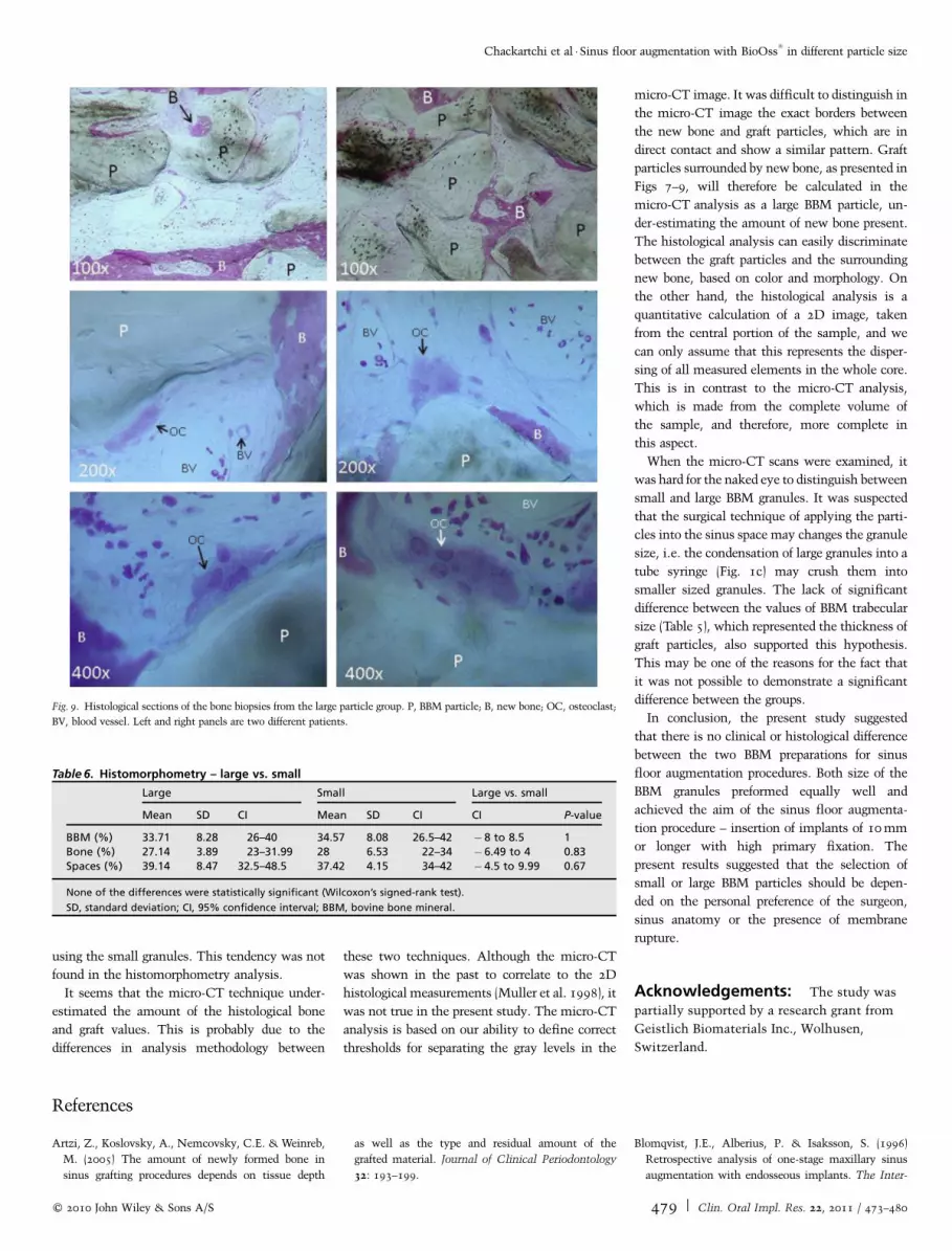
multi-nucleated giant cells, probably osteoclasts,
were observed directly on the BBM particles sur-
face (Figs 8 and 9). It was interesting to see that in
both groups (large vs. small particles), the osteo-
clast-like cells preferred the small-size BBM par-
ticles and not the large particles (Fig. 9). No gaps
were present at the bone–particles interface, and
the bone was always in close contact with the
particles. No inflammatory cell infiltrate was
present around the particles or at the interface
with bone.
The morphometric results from the standard
histological quantative analysis were summar-
ized in Table 6. From the samples representing
the large granules, the mean percentage of
BioOsss
granules out of the whole slide was
33.71 � 8.28% (95% confidence interval: 26–
40%). The percentage of bone volume was
27.14þ3.89% (95% confidence interval: 23–
31.99%) and the percentage of spaces was
39.14þ8.47% (95% confidence interval: 32.5–
48.5%). From the samples representing the small
granules, the mean percentage of BBM granules
out of the whole slide was 34.57þ8.08% (95%
confidence interval: 26.5–42%). The percentage
of bone volume was 28þ6% (22–34%), and the
percentage of spaces was 37.42þ4.15% (95%
confidence interval: 34–42%). There was no
statistical difference between the groups for all
measurements (Table 6).
Discussion
In the present study, the clinical performances of
the two different particle size of BBM were
compared in sinus floor augmentation proce-
dures. The results imply that there were no
differences between these two BBM preparations
Table 2. Micro-CT-large vs. small particles
Large particles Small particles Large vs. small
Mean SD CI Mean SD CI CI P-value
BBM (%) 23.12 6.02 15.82–28.22 22.89 8.32 13.86–9.87 � 3.18 to 2.96 0.68Bone (%) 7.99 4.23 3.99–12.63 14.64 12.03 4.73–26.91 � 18.93 to 1.78 0.15Soft tissue (%) 68.89 4.14 65.22–73.04 62.47 14.95 47.18–3.37 � 2.47 to 21.71 0.46
None of the differences were statistically significant (Wilcoxon’s signed-rank test).
SD, standard deviation; CI, 95% confidence interval; CT, computerized tomography.
Table 3. BBM, bone and soft-tissue volumes
Large particles Small particles
Mean SD CI Mean SD CI
Total volume (mm3) 41.22 10.31 31.38–51.76 38.09 6.94 31.85–44.73BBM volume (mm3) 9.41 3.08 6.1–12.52 8.77 3.91 4.5–12.71Bone volume (mm3) 3.41 2.38 1.28–5.73 5.39 4.72 1.79–10.37Soft tissue (mm3) 28.4 23.9
None of the difference were statistically significant (Wilcoxon’s signed-rank test).
SD, standard deviation; CI, 95% confidence interval; BBM, bovine bone mineral.
Table 4. Bone values
Large particles Small particles
Mean SD CI Mean SD CI
Bone/total volume (%) 7.99 4.23 3.99–12.63 14.64 12.03 4.73–26.91Bone surface (mm2) 71.58 41.56 29.83–114.99 112 103.13 36.91–219.12BS/TV (mm2/mm3) 1.72 0.78 0.93–2.5 3.01 2.59 0.96–5.55Th (mm) 0.15 0.03 0.11–0.17 0.15 0.02 0.13–0.15Sp (mm) 0.97 0.19 0.77–1.1 0.8 0.43 0.43–1.19TbN (1/mm) 1 0.22 0.87–1.21 1.45 0.66 1–2.16
None of the difference were statistically significant (Wilcoxon’s signed-rank test).
SD, standard deviation; CI, 95% confidence interval; BS/TV, total surface of bone/graft trabecules out of total core
volume; Th, trabecular thickness; Sp, spaces between bone/graft trabecules; TbN, trabecular number.
Table 5. BBM values
Large particles Small particles
Mean SD CI Mean SD CI
BBM/total volume (%) 23.12 6.02 15.82–28.22 22.89 8.32 13.86–29.87BBM surface (mm2) 189.84 57.25 115.98–237.85 195.89 93.07 95.3–288.67BBM BS/TV (mm2/mm3) 4.64 1.06 3.56–5.72 5.1 2.03 2.45–6.7Th (mm) 0.18 0.02 0.16–0.19 0.17 0.01 0.15–0.18Sp (mm) 0.51 0.23 0.3–0.72 0.67 0.37 0.39–1.11TbN (1/mm) 1.98 0.55 1.51–2.45 1.69 0.59 0.92–2.18
None of the difference were statistically significant (Wilcoxon’s signed-rank test).
SD, standard deviation; CI, 95% confidence interval; BS/TV, total surface of bone/graft trabecules out of total core
volume; Th, trabecular thickness; Sp, spaces between bone/graft trabecules; TbN, trabecular number; BBM, bovine
bone mineral.
Chackartchi et al � Sinus floor augmentation with BioOsss
in different particle size
c� 2010 John Wiley & Sons A/S 477 | Clin. Oral Impl. Res. 22, 2011 / 473–480

in sinus floor augmentation procedures in hu-
mans, using clinical, histological and micro-CT
measurements. Both groups performed equally
well, allowing stable implant placement and
high insertion torque (450 N). As expected,
there was a large variability between the indivi-
dual data from patient to patient, but the intra-
individual design minimizes problems of varia-
bility between individuals.
BBM is a well-documented material for the
augmentation of the maxillary sinus (Wallace et
al. 1996; Valentini & Abensur 1997; Froum et al.
1998; Piattelli et al. 1999). The most popular and
documented preparation of BBM, BioOsss
, is
commercially available for clinical use in two
particle sizes – 0.25–1 and 1–2 mm, with no
available data regarding the histological, clinical
or other aspects related to the different particle
sizes. There are studies investigating the effect of
different particle sizes of harvested autogenous
bone with no conclusive results (Zaner & Yukna
1984; Fucini et al. 1993; Xu et al. 2003; Springer
et al. 2004; Murai et al. 2006; Coradazzi et al.
2007; Walsh et al. 2008; Kon et al. 2009). In
another type of study, demineralized freeze-dried
bone allografts with 850–1000 mm particles
yielded more favorable results than the 250–
500 mm particles in human periodontal defects
(Fucini et al. 1993). However, no clinical or
histological study was made so far relating the
particle size of BBM.
In the present study, two quantitative methods
for the evaluation of the mineralized material
occupying the sinus orifice were used. The mi-
cro-CT system was first introduced by Feldkamp
et al. (1989). This system was further validated as
a 3D analysis method by Muller et al. (1998)
when demonstrating the good correlation be-
tween the 3D analysis results with the well-
accepted 2D histology. This method of analysis
allows 3D measurements of bone quantity
and a 3D assessment of the architecture and
microstructure of the bone, which are important
for the estimation of bone durability and strength.
One of the great advantages of the micro-CT
system is that this is a not destructive system.
Therefore, all of these parameters can still be
validated in the same biopsy sample with the 2D
conventional histology. The histomorphometric
analysis has the ability to more accurately eval-
uate the inter-phase between the graft particles
and the newly formed bone, the cellular charac-
terization, and will allow validation of the volu-
metric results. Using micro-CT volumetric
analysis, there were no significant differences
between the two groups for any of the tested
parameters, as defined using radio-opacity
thresholds to discriminate between bone, BBM
and non-calcified tissue. There was a tendency
toward the creation of more new bone when
Fig. 7. A sample of the histological sections of the biopsies from one patient, � 30 magnification. (a) Small particles; (b) large
particle. The difference in size of the BioOsss
particles is evident.
Fig. 8. Histological sections of the bone biopsies from the small particle group. P, BBM particle; B, new bone; OC, osteoclast;
BV, blood vessel. Left and right panels are two different patients.
Chackartchi et al � Sinus floor augmentation with BioOsss
in different particle size
478 | Clin. Oral Impl. Res. 22, 2011 / 473–480 c� 2010 John Wiley & Sons A/S
using the small granules. This tendency was not
found in the histomorphometry analysis.
It seems that the micro-CT technique under-
estimated the amount of the histological bone
and graft values. This is probably due to the
differences in analysis methodology between
these two techniques. Although the micro-CT
was shown in the past to correlate to the 2D
histological measurements (Muller et al. 1998), it
was not true in the present study. The micro-CT
analysis is based on our ability to define correct
thresholds for separating the gray levels in the
micro-CT image. It was difficult to distinguish in
the micro-CT image the exact borders between
the new bone and graft particles, which are in
direct contact and show a similar pattern. Graft
particles surrounded by new bone, as presented in
Figs 7–9, will therefore be calculated in the
micro-CT analysis as a large BBM particle, un-
der-estimating the amount of new bone present.
The histological analysis can easily discriminate
between the graft particles and the surrounding
new bone, based on color and morphology. On
the other hand, the histological analysis is a
quantitative calculation of a 2D image, taken
from the central portion of the sample, and we
can only assume that this represents the disper-
sing of all measured elements in the whole core.
This is in contrast to the micro-CT analysis,
which is made from the complete volume of
the sample, and therefore, more complete in
this aspect.
When the micro-CT scans were examined, it
was hard for the naked eye to distinguish between
small and large BBM granules. It was suspected
that the surgical technique of applying the parti-
cles into the sinus space may changes the granule
size, i.e. the condensation of large granules into a
tube syringe (Fig. 1c) may crush them into
smaller sized granules. The lack of significant
difference between the values of BBM trabecular
size (Table 5), which represented the thickness of
graft particles, also supported this hypothesis.
This may be one of the reasons for the fact that
it was not possible to demonstrate a significant
difference between the groups.
In conclusion, the present study suggested
that there is no clinical or histological difference
between the two BBM preparations for sinus
floor augmentation procedures. Both size of the
BBM granules preformed equally well and
achieved the aim of the sinus floor augmenta-
tion procedure – insertion of implants of 10 mm
or longer with high primary fixation. The
present results suggested that the selection of
small or large BBM particles should be depen-
ded on the personal preference of the surgeon,
sinus anatomy or the presence of membrane
rupture.
Acknowledgements: The study was
partially supported by a research grant from
Geistlich Biomaterials Inc., Wolhusen,
Switzerland.
References
Artzi, Z., Koslovsky, A., Nemcovsky, C.E. & Weinreb,
M. (2005) The amount of newly formed bone in
sinus grafting procedures depends on tissue depth
as well as the type and residual amount of the
grafted material. Journal of Clinical Periodontology
32: 193–199.
Blomqvist, J.E., Alberius, P. & Isaksson, S. (1996)
Retrospective analysis of one-stage maxillary sinus
augmentation with endosseous implants. The Inter-
Fig. 9. Histological sections of the bone biopsies from the large particle group. P, BBM particle; B, new bone; OC, osteoclast;
BV, blood vessel. Left and right panels are two different patients.
Table 6. Histomorphometry – large vs. small
Large Small Large vs. small
Mean SD CI Mean SD CI CI P-value
BBM (%) 33.71 8.28 26–40 34.57 8.08 26.5–42 � 8 to 8.5 1Bone (%) 27.14 3.89 23–31.99 28 6.53 22–34 � 6.49 to 4 0.83Spaces (%) 39.14 8.47 32.5–48.5 37.42 4.15 34–42 � 4.5 to 9.99 0.67
None of the differences were statistically significant (Wilcoxon’s signed-rank test).
SD, standard deviation; CI, 95% confidence interval; BBM, bovine bone mineral.
Chackartchi et al � Sinus floor augmentation with BioOsss
in different particle size
c� 2010 John Wiley & Sons A/S 479 | Clin. Oral Impl. Res. 22, 2011 / 473–480

national Journal of Oral & Maxillofacial Implants
11: 512–521.
Boyne, P.J. & James, R.A. (1980) Grafting of the
maxillary sinus floor with autogenous marrow and
bone. Journal of Oral Surgery 38: 613–616.
Branemark, P.I., Adell, R., Albrektsson, T., Lekholm, U.,
Lindstrom, J. & Rockler, B. (1984) An experimental
and clinical study of osseointegrated implants penetrat-
ing the nasal cavity and maxillary sinus. Journal of
Oral and Maxillofacial Surgery 42: 497–505.
Coradazzi, L.F., Garcia, I.R Jr. & Manfrin, T.M. (2007)
Evaluation of autogenous bone grafts, particulate or
collected during osteotomy with implant burs: histo-
logic and histomorphometric analysis in rabbits. The
International Journal of Oral & Maxillofacial Im-
plants 22: 201–207.
Feldkamp, L.A., Goldstein, S.A., Parfitt, A.M., Jesion,
G. & Kleerekoper, M. (1989) The direct examination
of three-dimensional bone architecture in vitro by
computed tomography. Journal of Bone and Mineral
Research 4: 3–11.
Froum, S.J., Tarnow, D.P., Wallace, S.S., Rohrer, M.D. &
Cho, S.C. (1998) Sinus floor elevation using anorganic
bovine bone matrix (osteograf/n) with and without
autogenous bone: a clinical, histologic, radiographic,
and histomorphometric analysis – part 2 of an ongoing
prospective study. The International Journal of Perio-
dontics and Restorative Dentistry 18: 528–543.
Fucini, S.E., Quintero, G., Gher, M.E., Black, B.S. &
Richardson, A.C. (1993) Small versus large particles
of demineralized freeze-dried bone allografts in hu-
man intrabony periodontal defects. Journal of Perio-
dontology 64: 844–847.
Groenveld, H.H., van den Bergh, J.P., Holzmann, P.,
ten Bruggenkate, C.M., Tuinzing, D.B. & Burger,
E.H. (1999) Histological observations of a bilateral
maxillary sinus floor elevation 6 and 12 months after
grafting with osteogenic protein-1 device. Journal of
Clinical Periodontology 26: 841–846.
Hallman, M. & Thor, A. (2008) Bone substitutes and
growth factors as an alternative/complement to auto-
genous bone for grafting in implant dentistry. Perio-
dontology 2000 47: 172–192.
Hammerle, C.H., Chiantella, G.C., Karring, T. & Lang,
N.P. (1998) The effect of a deproteinized bovine bone
mineral on bone regeneration around titanium dental
implants. Clinical Oral Implants Research 9: 151–162.
Hurzeler, M.B., Quinones, C.R., Kirsch, A., Gloker, C.,
Schupbach, P., Strub, J.R. & Caffesse, R.G. (1997)
Maxillary sinus augmentation using different grafting
materials and dental implants in monkeys. Part i.
Evaluation of anorganic bovine-derived bone matrix.
Clinical Oral Implants Research 8: 476–486.
Klinge, B., Alberius, P., Isaksson, S. & Jonsson, J. (1992)
Osseous response to implanted natural bone mineral
and synthetic hydroxylapatite ceramic in the repair of
experimental skull bone defects. Journal of Oral and
Maxillofacial Surgery 50: 241–249.
Kon, K., Shiota, M., Ozeki, M., Yamashita, Y. &
Kasugai, S. (2009) Bone augmentation ability of
autogenous bone graft particles with different sizes:
a histological and micro-computed tomography study.
Clinical Oral Implants Research 20: 1240–1246.
McAllister, B.S., Margolin, M.D., Cogan, A.G., Taylor,
M. & Wollins, J. (1998) Residual lateral wall defects
following sinus grafting with recombinant human
osteogenic protein-1 or bio-oss in the chimpanzee.
The International Journal of Periodontics and Re-
storative Dentistry 18: 227–239.
Moy, P.K., Lundgren, S. & Holmes, R.E. (1993) Max-
illary sinus augmentation: histomorphometric analy-
sis of graft materials for maxillary sinus floor
augmentation. Journal of Oral and Maxillofacial
Surgery 51: 857–862.
Muller, R., Van Campenhout, H., Van Damme, B., Van
Der Perre, G., Dequeker, J., Hildebrand, T. & Rueg-
segger, P. (1998) Morphometric analysis of human
bone biopsies: a quantitative structural comparison of
histological sections and micro-computed tomogra-
phy. Bone 23: 59–66.
Murai, M., Sato, S., Fukase, Y., Yamada, Y., Ko-
miyama, K. & Ito, K. (2006) Effects of different sizes
of beta-tricalcium phosphate particles on bone aug-
mentation within a titanium cap in rabbit calvarium.
Dental Materials Journal 25: 87–96.
Nkenke, E. & Stelzle, F. (2009) Clinical outcomes of
sinus floor augmentation for implant placement using
autogenous bone or bone substitutes: a systematic
review. Clinical Oral Implants Research 20 (Suppl.
4): 124–133.
Piattelli, M., Favero, G.A., Scarano, A., Orsini, G. &
Piattelli, A. (1999) Bone reactions to anorganic bovine
bone (bio-oss) used in sinus augmentation procedures:
a histologic long-term report of 20 cases in humans.
The International Journal of Oral & Maxillofacial
Implants 14: 835–840.
Sharan, A. & Madjar, D. (2008) Maxillary sinus pneu-
matization following extractions: a radiographic
study. The International Journal of Oral & Maxillo-
facial Implants 23: 48–56.
Smiler, D.G. & Holmes, R.E. (1987) Sinus lift proce-
dure using porous hydroxyapatite: a preliminary
clinical report. Journal of Oral Implantology 13:
239–253.
Smiler, D.G., Johnson, P.W., Lozada, J.L., Misch, C.,
Rosenlicht, J.L., Tatum, O.H Jr. & Wagner, J.R.
(1992) Sinus lift grafts and endosseous implants.
Treatment of the atrophic posterior maxilla. Dental
Clinics of North America 36: 151–187.
Spector, M. (1994) Anorganic bovine bone and ceramic
analogs of bone mineral as implants to facilitate bone
regeneration. Clinical Plastic Surgery 21: 437–444.
Springer, I.N., Terheyden, H., Geiss, S., Harle, F.,
Hedderich, J. & Acil, Y. (2004) Particulated bone
grafts – effectiveness of bone cell supply. Clinical
Oral Implants Research 15: 205–212.
Tadjoedin, E.S., de Lange, G.L., Bronckers, A.L.,
Lyaruu, D.M. & Burger, E.H. (2003) Deproteinized
cancellous bovine bone (bio-oss) as bone substitute for
sinus floor elevation. A retrospective, histomorpho-
metrical study of five cases. Journal of Clinical
Periodontology 30: 261–270.
Tallgren, A. (1972) The continuing reduction of the
residual alveolar ridges in complete denture wearers:
a mixed-longitudinal study covering 25 years. Journal
of Prosthetic Dentistry 27: 120–132.
Tatum, H. Jr. (1986) Maxillary and sinus implant
reconstructions. Dental Clinics of North America
30: 207–229.
Turunen, T., Peltola, J., Yli-Urpo, A. & Happonen, R.P.
(2004) Bioactive glass granules as a bone adjunctive
material in maxillary sinus floor augmentation. Clin-
ical Oral Implant Research 15: 135–141.
Valentini, P. & Abensur, D. (1997) Maxillary sinus floor
elevation for implant placement with demineralized
freeze-dried bone and bovine bone (bio-oss): a clinical
study of 20 patients. The International Journal of
Periodontics and Restorative Dentistry 17: 232–241.
Valentini, P., Abensur, D., Densari, D., Graziani, J.N.
& Hammerle, C. (1998) Histological evaluation of
bio-oss in a 2-stage sinus floor elevation and implan-
tation procedure. A human case report. Clinical Oral
Implants Research 9: 59–64.
Wagner, J.R. (1991) A 3 1/2-year clinical evaluation of
resorbable hydroxylapatite osteogen (ha resorb) used
for sinus lift augmentations in conjunction with the
insertion of endosseous implants. Journal of Oral
Implantology 17: 152–164.
Wallace, S.S., Froum, S.J. & Tarnow, D.P. (1996) His-
tologic evaluation of a sinus elevation procedure: a
clinical report. The International Journal of Perio-
dontics and Restorative Dentistry 16: 46–51.
Walsh, W.R., Vizesi, F., Michael, D., Auld, J., Lang-
down, A., Oliver, R., Yu, Y., Irie, H. & Bruce, W.
(2008) Beta-tcp bone graft substitutes in a bilateral
rabbit tibial defect model. Biomaterials 29: 266–271.
Wetzel, A.C., Stich, H. & Caffesse, R.G. (1995) Bone
apposition onto oral implants in the sinus area filled
with different grafting materials. A histological study
in beagle dogs. Clinical Oral Implants Research 6:
155–163.
Xu, H., Shimizu, Y., Asai, S. & Ooya, K. (2003)
Experimental sinus grafting with the use of deprotei-
nized bone particles of different sizes. Clinical Oral
Implants Research 14: 548–555.
Zaner, D.J. & Yukna, R.A. (1984) Particle size of
periodontal bone grafting materials. Journal of Perio-
dontology 55: 406–409.
Chackartchi et al � Sinus floor augmentation with BioOsss
in different particle size
480 | Clin. Oral Impl. Res. 22, 2011 / 473–480 c� 2010 John Wiley & Sons A/S